Review

Molecular and Regulatory Mechanisms of Desensitization and Resensitization of GABAA Receptors with a Special Reference to Propofol/Barbiturate

1
Department of Behavioral Physiology, Graduate School of Human Sciences, Osaka University, Osaka 565-0871, Japan
2
Department of Neurobiology and Physiology, School of Dentistry, Seoul National University, Seoul 110-749, Korea
3
Department of Oral Physiology, Graduate School of Medical and Dental Sciences, Kagoshima University, Sakuragaoka 8-35-1, Kagoshima 890-8544, Japan
4
Department of Neuroscience and Oral Physiology, Osaka University Graduate School of Dentistry, Osaka 565-0871, Japan
*
Authors to whom correspondence should be addressed.
Int. J. Mol. Sci. 2020, 21(2), 563; https://doi.org/10.3390/ijms21020563
Submission received: 25 November 2019 / Revised: 11 January 2020 / Accepted: 14 January 2020 / Published: 15 January 2020
(This article belongs to the Special Issue Pharmacology and Neurobiology of GABA Receptors)

Abstract

:
It is known that desensitization of GABAA receptor (GABAAR)-mediated currents is paradoxically correlated with the slowdown of their deactivation, i.e., resensitization. It has been shown that an upregulation of calcineurin enhances the desensitization of GABAAR-mediated currents but paradoxically prolongs the decay phase of inhibitory postsynaptic currents/potentials without appreciable diminution of their amplitudes. The paradoxical correlation between desensitization and resensitization of GABAAR-mediated currents can be more clearly seen in response to a prolonged application of GABA to allow more desensitization, instead of brief pulse used in previous studies. Indeed, hump-like GABAAR currents were produced after a strong desensitization at the offset of a prolonged puff application of GABA in pyramidal cells of the barrel cortex, in which calcineurin activity was enhanced by deleting phospholipase C-related catalytically inactive proteins to enhance the desensitization/resensitization of GABAAR-mediated currents. Hump-like GABAAR currents were also evoked at the offset of propofol or barbiturate applications in hippocampal or sensory neurons, but not GABA applications. Propofol and barbiturate are useful to treat benzodiazepine/alcohol withdrawal syndrome, suggesting that regulatory mechanisms of desensitization/resensitization of GABAAR-mediated currents are important in understanding benzodiazepine/alcohol withdrawal syndrome. In this review, we will discuss the molecular and regulatory mechanisms underlying the desensitization and resensitization of GABAAR-mediated currents and their functional significances.

1. Introduction

Ligand-gated channels open in response to the neurotransmitter binding but also close (desensitize) for long periods with the agonist still bound [1,2]. It is demonstrated that desensitization of GABAA receptor (GABAAR)-mediated currents is paradoxically correlated with the slowdown of their deactivation, i.e., resensitization [3]. Desensitization tends to prolong inhibitory currents and keeps the transmitter in the bound state of GABAARs. The rate at which the receptors enter the desensitization state will affect the shape of inhibitory currents [4,5,6].
The desensitization of GABAAR-mediated currents is modulated by various signal transductions. The PKA-mediated phosphorylation modulates the desensitization of GABAAR-mediated currents in chick cortical neurons [7], rat sympathetic ganglion neurons [8], rat cerebellar granule neurons [9], and recombinant GABAARs [10]. The PKC- and PKG-mediated phosphorylation decreases the fast component of desensitization in recombinant α1β1 GABAARs [11] and rat cerebellar granule cells [9], respectively. CaMKII (Ca2+/calmodulin-dependent protein kinase II) decreased the desensitization of GABAAR-mediated currents in rat spinal dorsal horn neurons [12], while calcineurin enhanced the desensitization of GABAAR-mediated currents in rat hippocampal neurons [13]. Calcineurin directly binds to the intracellular loop of the GABAAR γ2 subunit, thereby dephosphorylating the receptor [14]. Interestingly, it is reported that the desensitization of GABAAR-mediated currents, which is caused by the enhanced calcineurin activity, paradoxically prolongs the decay phase of inhibitory postsynaptic currents/potentials without appreciable diminution of their amplitudes [4].
The paradoxical correlation between desensitization and resensitization of GABAAR-mediated currents can be seen in response to a brief pulse in previous studies [3,4]. However, this relationship can be more clearly seen in response to a prolonged application of GABA for enough time to allow full desensitization. Indeed, hump-like GABAAR currents were produced after a strong desensitization at the offset of puff applications of GABA for 2 s in pyramidal cells of the barrel cortex in the phospholipase C-related catalytically inactive proteins (PRIP-1/2) double-knockout (PRIP-DKO) mice [15]. In these neurons, the increased calcineurin activity due to the potentiated Ca2+-induced Ca2+ release (CICR) and store-operated Ca2+ entry (SOCE) enhances the desensitization of GABAAR-mediated currents and subsequently causes resensitization of GABAAR-mediated currents [15]. GABARAP (GABAAR-associated protein) plays an important role in intracellular trafficking/clustering of GABAARs [16,17] and the clustered GABAARs display lower apparent affinity for GABA, faster deactivation, and slower desensitization [18]. The kinases and molecules involved in desensitization and resensitization (slowdown of deactivation) of GABAAR-mediated currents are summarized in Table 1.
Hump-like GABAAR currents after a strong desensitization were also seen at the offset of propofol applications at a high concentration (600 μM) in hippocampal pyramidal neurons [19], etomidate applications at a high concentration (1 mM) in rat spinal dorsal horn neurons [20], pentobarbital applications at high concentrations (1–3 mM) in frog sensory neurons [21,22], rat hippocampal neurons [23], and recombinant GABAARs [24,25,26,27,28,29] or phenobarbital applications at a high concentration (10 mM) in rat hippocampal neurons [23], although these were not seen at the offset of GABA applications. Drugs that cause desensitization and resensitization of GABAAR-mediated currents are summarized in Table 2. It is believed that the generation of hump-like currents may be caused by the removal of the blockade by anesthetic agents as partial antagonists [24], although their mechanisms remain unclear and the involvement of desensitization is not necessarily denied. Propofol and barbiturate are clinically used for treatment of benzodiazepine/alcohol withdrawal syndrome [30,31,32]. Considering that hump-like GABAAR currents that are seen after a strong desensitization or blockade were evoked at the offset of propofol or barbiturate applications, the regulatory mechanisms of desensitization/resensitization of GABAAR-mediated currents might be important for understanding benzodiazepine/alcohol withdrawal syndrome. Here, we discuss the molecular and regulatory mechanisms underlying the desensitization and resensitization of GABAAR-mediated currents in neurons of PRIP-DKO mice and their functional significances.

2. PRIP-1/2 are Involved in Desensitization and Resensitization of GABAAR-Mediated Currents

PRIP-1/2 are involved in the membrane trafficking of GABAARs and the regulation of intracellular Ca2+ stores [16,17]. Thus, it was investigated whether and how the deletion of PRIP-1/2 affects GABAAR-mediated currents evoked by puff applications of GABA in layer III pyramidal cells of the barrel cortex. It was found that the deletion of PRIP-1/2 enhanced the desensitization of GABAAR-mediated currents but paradoxically induced a hump-like tail-current at the offset of the GABA puff (Figure 1) [15]. Thus, it is likely that PRIP-1/2 are involved in the desensitization and resensitization of GABAAR-mediated currents. Although similar tail-currents were observed following the removal of propofol [19], etomidate [20], pentobarbital [21,22,23,24,25,26,27,28,29], and phenobarbital [23], it was the first report on such hump-like tail-currents that were induced by GABA itself.

3. [Ca2+]i Dependence of Desensitization and Resensitization of GABAAR-Mediated Currents and Their Abolishment by a Calcineurin Inhibitor

It is well known that the desensitization of GABAAR-mediated currents is accelerated by increases in [Ca2+]i [33,34]. As expected, it was clearly demonstrated that both the acceleration of desensitization of GABAAR-mediated currents and the generation of the hump-like tail-currents were caused by increases in [Ca2+]i [15]. Consistent with the idea that desensitization is mechanistically related to the deactivation of GABAAR-mediated currents [3], the progress of desensitization of GABAAR-mediated currents was invariably accompanied by the enhancement of the hump-like tail-currents [15]. These results suggested that the deletion of PRIP-1/2 results in an enhancement of the desensitization and resensitization of GABAAR-mediated currents through increases in [Ca2+]i. The involvement of CICR and the following SOCE in both the desensitization of GABAAR-mediated currents and the generation of the hump-like tail-currents in PRIP-DKO pyramidal cells was also demonstrated by an intracellular application of ruthenium red [15].
It has been demonstrated that a calcineurin inhibitor, cyclosporin A-cyclophilin A complex, suppressed the desensitization of GABAAR-mediated currents in acutely dissociated hippocampal neurons [13]. It has also been reported that the inhibition of calcineurin increased the rate of GABA unbinding from GABAARs [4]. Consistent with these previous studies, the bath application of a calcineurin inhibitor, fenvalerate, alleviated the desensitization of GABAAR-mediated currents and markedly decreased the hump-like tail-currents [15]. Thus, it is likely that the hump-like tail-currents in PRIP-DKO pyramidal cells were generated as a result of an acceleration of desensitization of GABAAR-mediated currents coupled with a slowdown of the GABA unbinding, which was mediated by Ca2+-dependent activation of calcineurin. Furthermore, Ca2+ imaging revealed that CICR and the following SOCE were more potent in PRIP-DKO pyramidal cells than in wild-type pyramidal cells [15]. Taken together, these results strongly suggest that the enhancement of desensitization and resensitization of GABAAR-mediated currents in PRIP-DKO pyramidal cells was largely mediated by the upregulation of Ca2+-dependent activity of calcineurin due to the potentiation of CICR followed by SOCE.

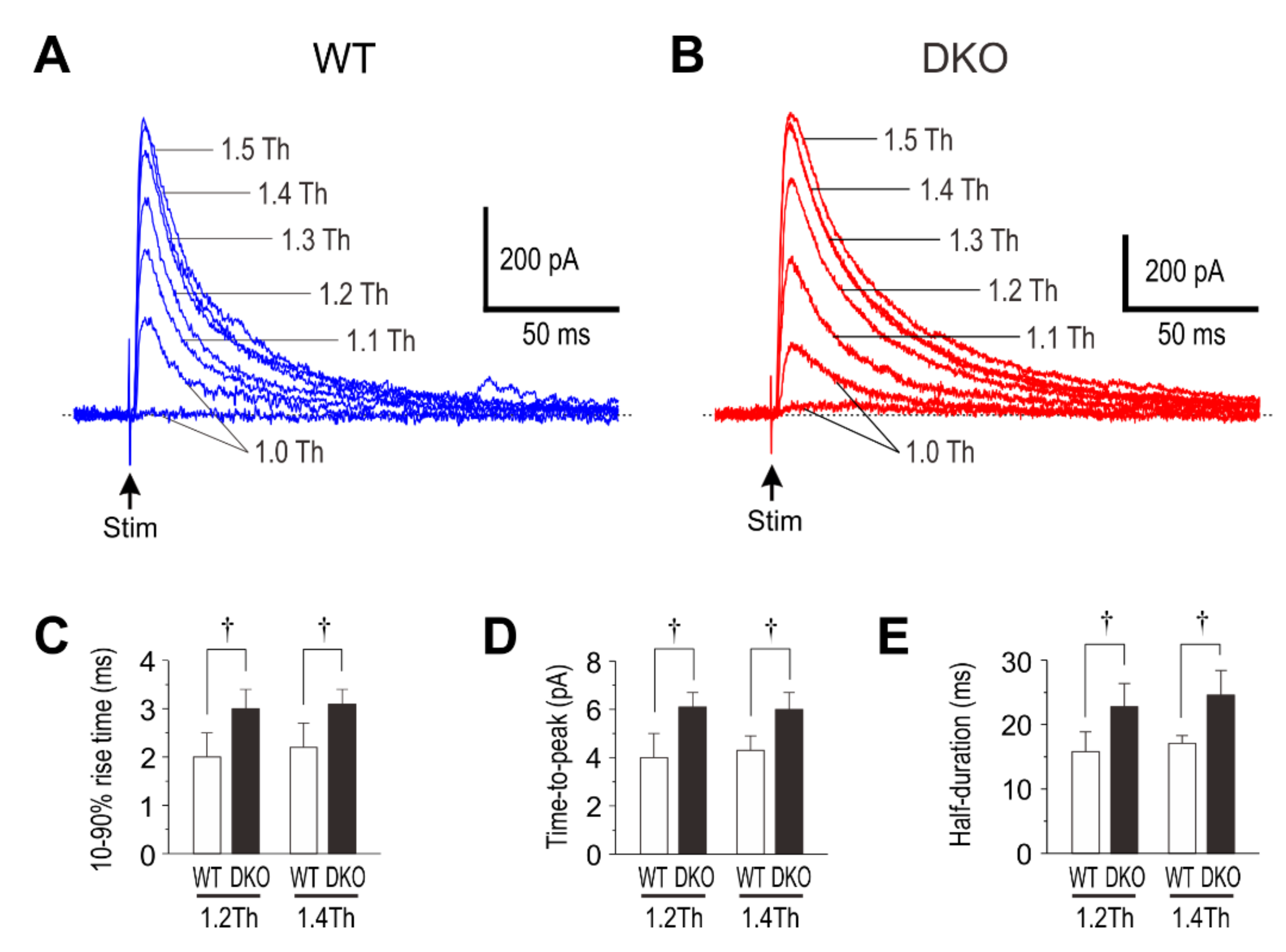
4. Deletion of PRIP-1/2 Prolongs eIPSCs in Layer II/III Pyramidal Cells

The differences in the kinetic properties of GABAAR-mediated currents between pyramidal cells of wild-type and PRIP-DKO mice should be reflected in the difference in inhibitory postsynaptic responses. Then, it was investigated how inhibitory postsynaptic responses reflect the changes in the kinetic properties of the GABAAR-mediated currents in layer III pyramidal cells of the PRIP-DKO barrel cortex.
It was found that the deletion of PRIP-1/2 resulted in the prolongation of the decay phase of inhibitory postsynaptic currents/potentials (IPSCs/IPSPs) in layer II/III pyramidal cells evoked by stimulation of layer III (Figure 2), leaving the overall features of miniature IPSCs unchanged [35]. These observations suggest that the prolongation of inhibitory synaptic actions is likely to result from an enhancement of desensitization followed by an enhanced resensitization of GABAAR-mediated currents. It has been reported that the PRIP-DKO mice exhibited a reduced expression of synaptic GABAARs containing γ2 subunits by 40% in hippocampal neurons [36] and by 18% in cerebellar granule cells [37] as a consequence of the lack of binding between PRIP-1/2 and GABAAR-associated protein [38]. The mean peak amplitudes of the IPSCs and IPSPs in the PRIP-DKO pyramidal cells were not significantly different from those in the wild-type pyramidal cells. In any case, the amplitude of eIPSPs would not be increased by deletion of PRIP-1/2 [35]. Then, an increase in duration instead of amplitude of eIPSPs is likely to be caused in PRIP-DKO mice.

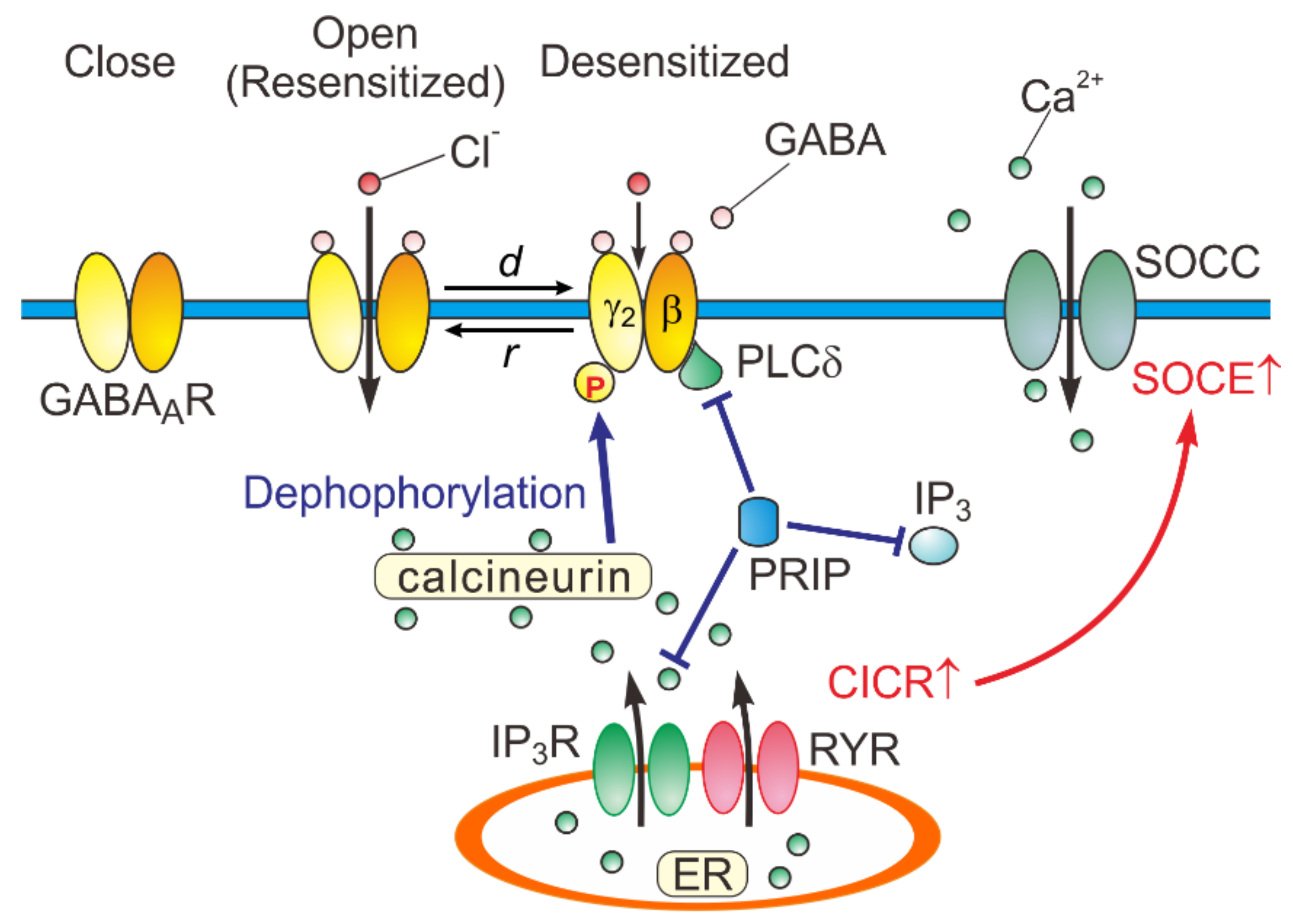
5. A Possible Kinetic Mechanism Underlying the Generation of the Hump-Like Tail-Currents and the Prolongation of eIPSCs

To understand the kinetic mechanisms underlying the generation of the hump-like tail-currents and the prolongation of eIPSCs, these currents were simulated using a previously proposed model [3] (Figure 3). It was examined whether the possible increase in the fast desensitization rate (d2) and the possible decrease in the unbinding rate (koff) can lead to a generation of the hump-like tail-current at the offset of the GABA puff.
It is known that GABA binding affinity was much larger in the desensitized GABAARs compared to the non-desensitized GABAARs and the binding affinity of the desensitized GABAARs increased depending on the concentration of the pre-applied GABA as was the case with the degree of desensitization of GABAAR-mediated currents [39]. Then, when the probability of being in the desensitized state (Dfast) for GABAARs was increased by increasing GABA concentration ([GABA]) or during the 2 s puff application of GABA, Dfast would be further recruited, leaving Open2 unchanged. Thus, it is reasonable to assume that the d2, but not β2, increase in a manner dependent on [GABA] [15,39]. Because Bound2, which is bifurcated into Open2 and Dfast, increases in a manner dependent on [GABA], the idea was incorporated in this model by defining d2 as follows;
d 2 =   d m a x 1 +   K h GABA n
where dmax is the maximum desensitization rate, Kh is the [GABA] that yields the half maximum desensitization rate, and n is the Hill coefficient [15]. It was assumed that calcineurin increased d2 by increasing its [GABA] dependency through a reduction of kh, and the d2 and koff were changed between the simulated wild-type and PRIP-DKO pyramidal cells. These changes were comparable to those caused by the activation of calcineurin reported previously [4,13].
In this simulation, the onset and offset of the 2 s puff application of GABA were assumed to be attenuated with a time constant raging between 0.1 and 0.3 s. In the simulated wild-type pyramidal cell, GABAAR-mediated currents were induced without a hump-like tail-current in response to 2 s GABA puff at 50 µM [15]. In contrast, in the simulated PRIP-DKO pyramidal cell, GABAAR-mediated currents displayed a prominent desensitization and were followed by a prominent hump-like tail-current [15]. Thus, a slowdown of koff and an acceleration of d2 resulted in a generation of a hump-like tail-current. Following a sharp decrease in [GABA] at the offset of GABA puff, a sharp decrease in d2 to a level smaller than the fast de-desensitization (i.e., resensitization) rate constant (r2) occurred to subsequently induce a hump-like tail-current. Indeed, decreases in the decay time constant at the offset of GABA puff pulse from 0.3 to 0.1 sec decreased the half-duration of the hump-like tail-current, leaving its amplitude almost unchanged [15]. Only PRIP-DKO pyramidal cells, but not wild-type pyramidal cells, displayed hump-like tail-currents in response to the same GABA puff that may have decayed slowly. These observations clearly indicate that the generation of the hump-like tail-current reflects kinetic differences between GABAAR-mediated currents in wild-type and PRIP-DKO pyramidal cells. Taken together, it can be concluded that a higher calcineurin activity in PRIP-DKO layer III pyramidal cells might have caused a slowdown of koff and an acceleration of d2 through the modulation of its GABA concentration dependency, leading to a generation of hump-like tail-currents in PRIP-DKO pyramidal cells.
Because there were no significant differences in the single-channel current and the number of GABAARs between eIPSCs in PRIP-DKO and wild-type pyramidal cells [35], it can be investigated whether the increase in d2 and the decrease in koff can also lead to the prolongation of eIPSCs. Simulated IPSCs in PRIP-DKO and the wild-type pyramidal cells that have half-durations similar to those obtained in the real experiments [35] revealed that a prolongation of eIPSCs/eIPSPs in PRIP-DKO pyramidal cells results from resensitization of GABAAR-mediated currents, which is brought about by an acceleration of d2 through the modulation of its [GABA] dependency together with a slowdown of koff. The finding of a negative skewness coefficient in PRIP-DKO eIPSCs obtained by the nonstationary variance analysis [35] is consistent with the occurrence of de-desensitization (resensitization) of GABAAR-mediated currents during the decay phase of PRIP-DKO eIPSCs.
Based on the experimental and simulation studies, the regulatory mechanisms of GABAARs are schematically depicted (Figure 4).

6. Physiological Significance of Desensitization and Resensitization of GABAAR-Mediated Currents

A single whisker deflection elicits an excitation in a subset of layer IV neurons within a single barrel-related column [41], which subsequently causes an excitation in layer II/III in the same column and then spreads horizontally into neighboring columns [42,43]. The spatio-temporal profile of the excitation spread in layer II/III evoked by stimulation of layer IV was narrower and faster in the barrel cortex of the PRIP-DKO mice compared to the wild-type mice [35].
Such a horizontal excitation spread in layer II/III seems to be strictly controlled by GABAAR-mediated lateral inhibition [42,44,45]. Indeed, bicuculline application abolished such a difference in the spatio-temporal profile of the excitation spread in layer II/III between the two genotypes [35]. It is reported that the PRIP-DKO mice exhibited a greater decrease in performance in the rotarod test [36], which is commonly used to assess the sensorimotor integration [46]. Then, the enhanced phasic inhibition caused by the PRIP-1/2 deletion would suppress the inter-columnar integration in the barrel cortex, consequently decreasing spatial recognition. Further studies are required to clarify the roles of PRIP-1/2 in sensorimotor processing in the barrel cortex.

7. Clinical Significance of Desensitization and Resensitization of GABAAR-Mediated Currents

Central nervous system depressants slow brain activity, making them useful for treating anxiety, panic, and sleep disorders. Alcohol and benzodiazepine are useful to mitigate anxiety through enhancing GABAAR-mediated inhibition. However, alcohol and benzodiazepine are known as abused drugs. Alcohol or benzodiazepine withdrawal syndrome appears following a reduction in alcohol or benzodiazepine use after a period of excessive use [47,48,49,50]. The alcohol or benzodiazepine withdrawal symptoms typically include anxiety, sweating, hand tremor, and sleep disturbance. The underlying mechanisms involve neuronal adaptations, which are revealed as decreased GABAergic responses [51] and enhancement of NMDA responses [52,53,54,55]. Although the exact mechanism for the reduced responsiveness of GABAARs remains uncertain, changes in surface GABAAR protein level and subunit composition, changes in turnover, recycling, and production rates, degree of phosphorylation, and decreased coupling mechanisms between GABA and alcohol/benzodiazepine sites are thought to be involved in the reduced responsiveness [56,57,58,59]. It has recently been demonstrated that the benzodiazepine diazepam caused downregulation of GABAergic inhibition through the phospholipase C (PLCδ)/Ca2+/calcineurin signaling pathway [40]. The study showed that overexpression of PRIP-1 suppressed diazepam-dependent activation of PLCδ and diazepam-dependent downregulation of GABAARs in HEK293 cells [40], indicating that PRIP-1 acts as an inhibitor by outcompeting the PLCδ binding to GABAARs. Because intracellular Ca2+ and calcineurin activity are increased in PRIP-DKO mice [15], these findings suggest that the diazepam-induced long-term downregulation of GABAergic inhibition is mediated by the PLCδ/Ca2+/calcineurin signaling pathway. Nevertheless, it is also true that calcineurin causes resensitization of GABAAR-mediated currents by facilitating their desensitization [4,15]. Given the apparently contradictory behaviors of GABAAR-mediated currents by calcineurin activation, the two different behaviors of GABAAR-mediated currents may depend on whether calcineurin activation occurs before or after activation of GABAARs.
As for the treatment of benzodiazepine/alcohol withdrawal syndrome, propofol and barbiturate which enhance GABAAR-mediated inhibition are useful. Indeed, it was demonstrated that propofol and barbiturates (pentobarbital and phenobarbital) were effective for the treatment of alcohol withdrawal syndrome [30,32] and barbiturate (pentobarbital) was effective for the treatment of benzodiazepine withdrawal syndrome [60]. However, it remains unclear how propofol and barbiturate ameliorate reduced GABA responsiveness in patients with benzodiazepine/alcohol withdrawal syndrome. Although the concentrations of propofol and barbiturates that generated the hump-like current are very high [19,21,22] compared to the dose used for treatment of the withdrawal syndrome [30,32], the generation of hump-like GABAAR currents itself may suggest the occurrence of resensitization of GABAAR-mediated currents. Indeed, the desensitization and deactivation of GABAAR-mediated currents are facilitated and slowed, respectively, by propofol/barbiturate at much lower concentrations [19,22]. Then, propofol and barbiturate may improve the reduced GABA responsiveness through the resensitization of GABAAR-mediated currents. Therefore, the regulatory mechanisms of desensitization/resensitization of GABAAR-mediated currents are important to better understand benzodiazepine/alcohol withdrawal syndrome and to develop the treatment method.

Author Contributions

Y.K. and H.T. conceptualized, drafted, reviewed, and revised the manuscript. M.S. performed a simulation study. All authors have read and agreed to the published version of the manuscript

Funding

This research was funded by Japan Society for the Promotion of Science (17K07055 to Y.K. and 17K08538 to H.T.).

Conflicts of Interest

The authors declare no conflict of interest.

Abbreviations

CaMKIICa2+/calmodulin-dependent protein kinase II
CICRCa2+-induced Ca2+ release
DKOdouble-knockout
GABAARGABAA receptor
GABARAPGABAAR-associated protein
IPSCinhibitory postsynaptic current
IPSPinhibitory postsynaptic potential
NMDAN-methyl-D-aspartate
PLCphospholipase C
PRIPphospholipase C-related catalytically inactive protein
RYRryanodine receptor
SOCCstore-operated Ca2+ channel
SOCEstore-operated Ca2+ entry

References

  1. Jones, M.V.; Westbrook, G.L. The impact of receptor desensitization on fast synaptic transmission. Trends Neurosci. 1996, 19, 96–101. [Google Scholar] [CrossRef]
  2. Keramidas, A.; Lynch, J.W. An outline of desensitization in pentameric ligand-gated ion channel receptors. Cell Mol. Life Sci. 2013, 70, 1241–1253. [Google Scholar] [CrossRef] [PubMed]
  3. Jones, M.V.; Westbrook, G.L. Desensitized states prolong GABAA channel responses to brief agonist pulses. Neuron 1995, 15, 181–191. [Google Scholar] [CrossRef] [Green Version]
  4. Jones, M.V.; Westbrook, G.L. Shaping of IPSCs by endogenous calcineurin activity. J. Neurosci. 1997, 17, 7626–7633. [Google Scholar] [CrossRef] [PubMed] [Green Version]
  5. Overstreet, L.S.; Jones, M.V.; Westbrook, G.L. Slow desensitization regulates the availability of synaptic GABAA receptors. J. Neurosci. 2000, 20, 7914–7921. [Google Scholar] [CrossRef] [PubMed] [Green Version]
  6. Qazi, S.; Caberlin, M.; Nigam, N. Mechanism of psychoactive drug action in the brain: Simulation modeling of GABAA receptor interactions at non-equilibrium conditions. Curr. Pharm. Des. 2007, 13, 1437–1455. [Google Scholar] [CrossRef] [PubMed] [Green Version]
  7. Tehrani, M.H.; Hablitz, J.J.; Barnes, E.M., Jr. cAMP increases the rate of GABAA receptor desensitization in chick cortical neurons. Synapse 1989, 4, 126–131. [Google Scholar] [CrossRef]
  8. Moss, S.J.; Smart, T.G.; Blackstone, C.D.; Huganir, R.L. Functional modulation of GABAA receptors by cAMP-dependent protein phosphorylation. Science 1992, 257, 661–665. [Google Scholar] [CrossRef]
  9. Robello, M.; Amico, C.; Cupello, A. Evidence of two populations of GABAA receptors in cerebellar granule cells in culture: Different desensitization kinetics, pharmacology, serine/threonine kinase sensitivity, and localization. Biochem. Biophys. Res. Commun. 1999, 266, 603–608. [Google Scholar] [CrossRef]
  10. Hinkle, D.J.; Macdonald, R.L. Beta subunit phosphorylation selectively increases fast desensitization and prolongs deactivation of α1β1γ2L and α1β3γ2L GABAA receptor currents. J. Neurosci. 2003, 23, 11698–11710. [Google Scholar] [CrossRef] [Green Version]
  11. Krishek, B.J.; Xie, X.; Blackstone, C.; Huganir, R.L.; Moss, S.J.; Smart, T.G. Regulation of GABAA receptor function by protein kinase C phosphorylation. Neuron 1994, 12, 1081–1095. [Google Scholar] [CrossRef]
  12. Wang, R.A.; Cheng, G.; Kolaj, M.; Randic, M. Alpha-subunit of calcium/calmodulin-dependent protein kinase II enhances gamma-aminobutyric acid and inhibitory synaptic responses of rat neurons in vitro. J. Neurophysiol. 1995, 73, 2099–2106. [Google Scholar] [CrossRef] [PubMed]
  13. Martina, M.; Mozrzymas, J.W.; Boddeke, H.W.; Cherubini, E. The calcineurin inhibitor cyclosporin A-cyclophilin A complex reduces desensitization of GABAA-mediated responses in acutely dissociated rat hippocampal neurons. Neurosci. Lett. 1996, 215, 95–98. [Google Scholar] [CrossRef]
  14. Muir, J.; Arancibia-Carcamo, I.L.; MacAskill, A.F.; Smith, K.R.; Griffin, L.D.; Kittler, J.T. NMDA receptors regulate GABAA receptor lateral mobility and clustering at inhibitory synapses through serine 327 on the 2 subunit. Proc. Natl. Acad. Sci. USA 2010, 107, 16679–16684. [Google Scholar] [CrossRef] [PubMed] [Green Version]
  15. Toyoda, H.; Saito, M.; Sato, H.; Tanaka, T.; Ogawa, T.; Yatani, H.; Kanematsu, T.; Hirata, M.; Kang, Y. Deletion of phospholipase C-related inactive protein-1/2 enhances desensitization and resensitization of GABAA receptors in pyramidal cells of the barrel cortex. Pflugers Arch. 2015, 467, 267–284. [Google Scholar] [CrossRef]
  16. Jacob, T.C.; Moss, S.J.; Jurd, R. GABAA receptor trafficking and its role in the dynamic modulation of neuronal inhibition. Nat. Rev. Neurosci. 2008, 9, 331–343. [Google Scholar] [CrossRef] [Green Version]
  17. Luscher, B.; Fuchs, T.; Kilpatrick, C.L. GABAA receptor trafficking-mediated plasticity of inhibitory synapses. Neuron 2011, 70, 385–409. [Google Scholar] [CrossRef] [Green Version]
  18. Chen, L.; Wang, H.; Vicini, S.; Olsen, R.W. The γ-aminobutyric acid type A (GABAA) receptor-associated protein (GABARAP) promotes GABAA receptor clustering and modulates the channel kinetics. Proc. Natl. Acad. Sci. USA 2000, 97, 11557–11562. [Google Scholar] [CrossRef] [Green Version]
  19. Orser, B.A.; Wang, L.Y.; Pennefather, P.S.; MacDonald, J.F. Propofol modulates activation and desensitization of GABAA receptors in cultured murine hippocampal neurons. J. Neurosci. 1994, 14, 7747–7760. [Google Scholar] [CrossRef] [Green Version]
  20. Zhang, Z.X.; Lu, H.; Dong, X.P.; Liu, J.; Xu, T.L. Kinetics of etomidate actions on GABAA receptors in the rat spinal dorsal horn neurons. Brain Res. 2002, 953, 93–100. [Google Scholar] [CrossRef]
  21. Akaike, N.; Hattori, K.; Inomata, N.; Oomura, Y. γ-Aminobutyric-acid- and pentobarbitone-gated chloride currents in internally perfused frog sensory neurones. J. Physiol. 1985, 360, 367–386. [Google Scholar] [CrossRef] [PubMed]
  22. Akaike, N.; Maruyama, T.; Tokutomi, N. Kinetic properties of the pentobarbitone-gated chloride current in frog sensory neurones. J. Physiol. 1987, 394, 85–98. [Google Scholar] [CrossRef] [PubMed]
  23. Rho, J.M.; Donevan, S.D.; Rogawski, M.A. Direct activation of GABAA receptors by barbiturates in cultured rat hippocampal neurons. J. Physiol. 1996, 497, 509–522. [Google Scholar] [CrossRef] [PubMed]
  24. Gingrich, K.J.; Burkat, P.M.; Roberts, W.A. Pentobarbital produces activation and block of α1β2γ2S GABAA receptors in rapidly perfused whole cells and membrane patches: Divergent results can be explained by pharmacokinetics. J. Gen. Physiol. 2009, 133, 171–188. [Google Scholar] [CrossRef] [PubMed] [Green Version]
  25. Feng, H.J.; Bianchi, M.T.; Macdonald, R.L. Pentobarbital differentially modulates α1β3δ and α1β3γ2L GABAA receptor currents. Mol. Pharmacol. 2004, 66, 988–1003. [Google Scholar] [CrossRef]
  26. Krampfl, K.; Wolfes, H.; Dengler, R.; Bufler, J. Kinetic analysis of the agonistic and blocking properties of pentobarbital on recombinant rat α1β2γ2S GABAA receptor channels. Eur. J. Pharmacol. 2002, 435, 1–8. [Google Scholar] [CrossRef]
  27. Thompson, S.A.; Whiting, P.J.; Wafford, K.A. Barbiturate interactions at the human GABAA receptor: Dependence on receptor subunit combination. Br. J. Pharmacol. 1996, 117, 521–527. [Google Scholar] [CrossRef] [Green Version]
  28. Wooltorton, J.R.; Moss, S.J.; Smart, T.G. Pharmacological and physiological characterization of murine homomeric β3 GABAA receptors. Eur. J. Neurosci. 1997, 9, 2225–2235. [Google Scholar] [CrossRef]
  29. Ziemba, A.M.; Forman, S.A. Correction for inhibition leads to an allosteric co-agonist model for pentobarbital modulation and activation of α1β3γ2L GABAA receptors. PLoS ONE 2016, 11, e0154031. [Google Scholar] [CrossRef] [Green Version]
  30. Brotherton, A.L.; Hamilton, E.P.; Kloss, H.G.; Hammond, D.A. Propofol for treatment of refractory alcohol withdrawal syndrome: A review of the literature. Pharmacotherapy 2016, 36, 433–442. [Google Scholar] [CrossRef]
  31. MacKinnon, G.L.; Parker, W.A. Benzodiazepine withdrawal syndrome: A literature review and evaluation. Am. J. Drug Alcohol Abuse 1982, 9, 19–33. [Google Scholar] [CrossRef] [PubMed]
  32. Martin, K.; Katz, A. The role of barbiturates for alcohol withdrawal syndrome. Psychosomatics 2016, 57, 341–347. [Google Scholar] [CrossRef] [PubMed]
  33. Inoue, M.; Oomura, Y.; Yakushiji, T.; Akaike, N. Intracellular calcium ions decrease the affinity of the GABA receptor. Nature 1986, 324, 156–158. [Google Scholar] [CrossRef] [PubMed]
  34. Mozrzymas, J.W.; Cherubini, E. Changes in intracellular calcium concentration affect desensitization of GABAA receptors in acutely dissociated P2–P6 rat hippocampal neurons. J. Neurophysiol. 1998, 79, 1321–1328. [Google Scholar] [CrossRef]
  35. Toyoda, H.; Saito, M.; Sato, H.; Kawano, T.; Kawakami, S.; Yatani, H.; Kanematsu, T.; Hirata, M.; Kang, Y. Enhanced lateral inhibition in the barrel cortex by deletion of phospholipase C-related catalytically inactive protein-1/2 in mice. Pflugers Arch. 2015, 467, 1445–1456. [Google Scholar] [CrossRef]
  36. Mizokami, A.; Tanaka, H.; Ishibashi, H.; Umebayashi, H.; Fukami, K.; Takenawa, T.; Nakayama, K.I.; Yokoyama, T.; Nabekura, J.; Kanematsu, T.; et al. GABAA receptor subunit alteration-dependent diazepam insensitivity in the cerebellum of phospholipase C-related inactive protein knockout mice. J. Neurochem. 2010, 114, 302–310. [Google Scholar] [CrossRef]
  37. Mizokami, A.; Kanematsu, T.; Ishibashi, H.; Yamaguchi, T.; Tanida, I.; Takenaka, K.; Nakayama, K.I.; Fukami, K.; Takenawa, T.; Kominami, E.; et al. Phospholipase C-related inactive protein is involved in trafficking of γ2 subunit-containing GABAA receptors to the cell surface. J. Neurosci. 2007, 27, 1692–1701. [Google Scholar] [CrossRef] [Green Version]
  38. Kanematsu, T.; Jang, I.S.; Yamaguchi, T.; Nagahama, H.; Yoshimura, K.; Hidaka, K.; Matsuda, M.; Takeuchi, H.; Misumi, Y.; Nakayama, K.; et al. Role of the PLC-related, catalytically inactive protein p130 in GABAA receptor function. EMBO J. 2002, 21, 1004–1011. [Google Scholar] [CrossRef] [Green Version]
  39. Chang, Y.; Ghansah, E.; Chen, Y.; Ye, J.; Weiss, D.S. Desensitization mechanism of GABA receptors revealed by single oocyte binding and receptor function. J. Neurosci. 2002, 22, 7982–7990. [Google Scholar] [CrossRef]
  40. Nicholson, M.W.; Sweeney, A.; Pekle, E.; Alam, S.; Ali, A.B.; Duchen, M.; Jovanovic, J.N. Diazepam-induced loss of inhibitory synapses mediated by PLCδ/Ca2+/calcineurin signalling downstream of GABAA receptors. Mol. Psychiatry 2018, 23, 1851–1867. [Google Scholar] [CrossRef]
  41. Armstrong-James, M.; Fox, K.; Das-Gupta, A. Flow of excitation within rat barrel cortex on striking a single vibrissa. J. Neurophysiol. 1992, 68, 1345–1358. [Google Scholar] [CrossRef] [PubMed]
  42. Petersen, C.C.; Sakmann, B. Functionally independent columns of rat somatosensory barrel cortex revealed with voltage-sensitive dye imaging. J. Neurosci. 2001, 21, 8435–8446. [Google Scholar] [CrossRef] [PubMed] [Green Version]
  43. Sato, H.; Shimanuki, Y.; Saito, M.; Toyoda, H.; Nokubi, T.; Maeda, Y.; Yamamoto, T.; Kang, Y. Differential columnar processing in local circuits of barrel and insular cortices. J. Neurosci. 2008, 28, 3076–3089. [Google Scholar] [CrossRef] [PubMed]
  44. Laaris, N.; Carlson, G.C.; Keller, A. Thalamic-evoked synaptic interactions in barrel cortex revealed by optical imaging. J. Neurosci. 2000, 20, 1529–1537. [Google Scholar] [CrossRef] [Green Version]
  45. Laaris, N.; Keller, A. Functional independence of layer IV barrels. J. Neurophysiol. 2002, 87, 1028–1034. [Google Scholar] [CrossRef] [Green Version]
  46. Anstrom, K.K.; Schallert, T.; Woodlee, M.T.; Shattuck, A.; Roberts, D.C. Repetitive vibrissae-elicited forelimb placing before and immediately after unilateral 6-hydroxydopamine improves outcome in a model of Parkinson’s disease. Behav. Brain Res. 2007, 179, 183–191. [Google Scholar] [CrossRef]
  47. Bayard, M.; McIntyre, J.; Hill, K.R.; Woodside, J., Jr. Alcohol withdrawal syndrome. Am. Fam. Physician 2004, 69, 1443–1450. [Google Scholar]
  48. McKeon, A.; Frye, M.A.; Delanty, N. The alcohol withdrawal syndrome. J. Neurol. Neurosurg. Psychiatry 2008, 79, 854–862. [Google Scholar] [CrossRef] [Green Version]
  49. Onyett, S.R. The benzodiazepine withdrawal syndrome and its management. J. R. Coll. Gen. Pract. 1989, 39, 160–163. [Google Scholar]
  50. Petursson, H. The benzodiazepine withdrawal syndrome. Addiction 1994, 89, 1455–1459. [Google Scholar] [CrossRef]
  51. Allison, C.; Pratt, J.A. Neuroadaptive processes in GABAergic and glutamatergic systems in benzodiazepine dependence. Pharmacol. Ther. 2003, 98, 171–195. [Google Scholar] [CrossRef]
  52. Chandler, L.J.; Newsom, H.; Sumners, C.; Crews, F. Chronic ethanol exposure potentiates NMDA excitotoxicity in cerebral cortical neurons. J. Neurochem. 1993, 60, 1578–1581. [Google Scholar] [CrossRef] [PubMed]
  53. Grant, K.A.; Valverius, P.; Hudspith, M.; Tabakoff, B. Ethanol withdrawal seizures and the NMDA receptor complex. Eur. J. Pharmacol. 1990, 176, 289–296. [Google Scholar] [CrossRef]
  54. Koff, J.M.; Pritchard, G.A.; Greenblatt, D.J.; Miller, L.G. The NMDA receptor competitive antagonist CPP modulates benzodiazepine tolerance and discontinuation. Pharmacology 1997, 55, 217–227. [Google Scholar] [CrossRef]
  55. Tsuda, M.; Shimizu, N.; Yajima, Y.; Suzuki, T.; Misawa, M. Hypersusceptibility to DMCM-induced seizures during diazepam withdrawal in mice: Evidence for upregulation of NMDA receptors. Naunyn Schmiedebergs Arch. Pharmacol. 1998, 357, 309–315. [Google Scholar] [CrossRef]
  56. Olsen, R.W.; Liang, J.; Cagetti, E.; Spigelman, I. Plasticity of GABAA receptors in brains of rats treated with chronic intermittent ethanol. Neurochem. Res. 2005, 30, 1579–1588. [Google Scholar] [CrossRef]
  57. Olsen, R.W.; Spigelman, I. GABAA Receptor Plasticity in Alcohol Withdrawal. In Jasper’s Basic Mechanisms of the Epilepsies, 4th ed.; Noebels, J.L., Avoli, M., Rogawski, M.A., Olsen, R.W., Delgado-Escueta, A.V., Eds.; National Center for Biotechnology Information: Bethesda, MD, USA, 2012. [Google Scholar]
  58. Tan, K.R.; Rudolph, U.; Luscher, C. Hooked on benzodiazepines: GABAA receptor subtypes and addiction. Trends Neurosci. 2011, 34, 188–197. [Google Scholar] [CrossRef] [Green Version]
  59. Wafford, K.A. GABAA receptor subtypes: Any clues to the mechanism of benzodiazepine dependence? Curr. Opin. Pharmacol. 2005, 5, 47–52. [Google Scholar] [CrossRef]
  60. Preskorn, S.H.; Denner, L.J. Benzodiazepines and withdrawal psychosis. Report of three cases. JAMA 1977, 237, 36–38. [Google Scholar] [CrossRef]
Figure 1. GABAAR-mediated currents evoked by GABA puff applications in wild-type and PRIP-DKO pyramidal cells. (A and B) Sample traces of GABAAR-mediated currents evoked at 0 mV in wild-type and PRIP-DKO pyramidal cells dialyzed with 5 mM EGTA, respectively, by puff application (4 and 6 psi) of GABA for 2 s. a, b, and c are the peak amplitude, the amplitude at the offset of the puff application, and the peak amplitude after the offset of the puff application, respectively. # and § are the durations at half amplitudes of desensitized component ([(a + b)/2]) and of tail-currents, respectively. (C) The relationship between the desensitization degree [Ds = (a – b)/a] of the GABAAR-mediated currents and half-duration of the tail-current (§) induced by a puff with 4 psi. †: p <0.01. (D) The relationship between the half-desensitization time of the GABAAR-mediated currents (#) and half-duration of the tail-current (§) induced by a puff with 4 psi. †: p <0.01. Adopted from [15].
Ijms 21 00563 g001
Figure 2. Evoked IPSCs (eIPSCs) in wild-type and PRIP-DKO pyramidal cells. (A and B) Superimposed sample traces of IPSCs evoked by stimulation with 1.0–1.5 times threshold (1.0–1.5 Th) in wild-type (A) and PRIP-DKO pyramidal cells (B). (C) The mean 10%–90% rise times of IPSCs evoked by stimulation with 1.2 Th in wild-type (n = 8) and PRIP-DKO pyramidal cells (n = 7) and those evoked by stimulation with 1.4 Th in wild-type (n = 8) and PRIP-DKO pyramidal cells (n = 7). †: p <0.01. (D) The mean times-to-peak of IPSCs evoked by stimulation with 1.2 Th in wild-type (n = 8) and PRIP-DKO pyramidal cells (n = 7) and those evoked by stimulation with 1.4 Th in wild-type (n = 8) and PRIP-DKO pyramidal cells (n = 7). †: p <0.01. (E) The mean half-durations of IPSCs evoked by stimulation with 1.2 Th in wild-type (n = 8) and PRIP-DKO pyramidal cells (n = 7) and those evoked by stimulation with 1.4 Th in wild-type (n = 8) and PRIP-DKO pyramidal cells (n = 7). †: p <0.01. Adopted from [35].
Ijms 21 00563 g002
Figure 3. A kinetic model for a hump-like tail-current. (A) A kinetic model of GABAARs representing mono- and double-liganded states, each providing access to open and desensitized states. (B and C) Top; Presumed [GABA] changes created by puff application of GABA with a rectangular pressure pulse through a puff pipette containing 200 μM GABA in the extracellular medium was assumed to be diluted 4 times and the onset and offset of the puff application were assumed to be attenuated with a time constant ranging between 0.1 and 0.3 s. Bottom; superimposed traces of the simulated GABAAR-mediated currents under the condition that the attenuation time constant is 0.3 and 0.1 s (solid and interrupted traces, respectively) in simulated wild-type (B) and PRIP-DKO (C) pyramidal cells. The rate constants were as follows (in s−1): kon = 15 μM−1, β2 = 2500, α2 = 142, r2 = 50, β1 = 200, α1 = 1100, r1 = 0.35, d1 = 6, q = 1 × 10⁻⁸ μM−1, and p = 1. The values of koff in WT and PRIP-DKO GABAARs were 90 and 30 s−1, respectively. The value of dmax in WT and PRIP-DKO GABAARs was 3600. The values of kh in WT and PRIP-DKO GABAARs were 2000 and 200, respectively. (D) Superimposed traces of a simulated wild-type and PRIP-DKO eIPSC induced by a GABA transient shown on an expanded time scale (inset) with a small maximum conductance. The rate constants were as follows (in s−1): kon = 20 μM−1, β2 = 2500, α2 = 195, r2 = 55, β1 = 100, α1 = 600, r1 = 0.35, d1 = 11, q = 1 × 10⁻⁸ μM−1, p = 0, and dmax = 3100. The values of koff in WT and PRIP-DKO GABAARs were 550 and 410 s−1, respectively. The value of dmax in WT and PRIP-DKO GABAARs was 310. The values of kh in WT and PRIP-DKO GABAARs were 2000 and 150, respectively. Adopted from [15] and [35].
Ijms 21 00563 g003
Figure 4. Close, open (resensitized), and desensitized states of GABAARs. When GABA binds to GABAARs, the receptors open the pore and consequently increase the permeability of the ion pore to Cl-. In response to a prolonged application of GABA, GABAARs are desensitized (d) by increased calcineurin activity due to potentiated Ca2+-induced Ca2+ release (CICR) followed by store-operated Ca2+ entry (SOCE) [15]. GABAARs are resensitized through de-desensitization (r) at the offset of the GABA puff. PRIP outcompetes the PLCδ in binding to GABAAR β subunits [40]. d: desensitization, r: resensitization, RYR: ryanodine receptor, SOCC: store-operated Ca2+ channel, IP3R: inositol trisphosphate receptor.
Ijms 21 00563 g004
Table 1. Kinases and molecules involved in desensitization and slowdown of deactivation of GABAAR-mediated currents.
Kinases/
Molecules
Neuron/Recombinant GABAARsEffectsReferences
PKAChick cortical neuronsincreases desensitization[7]
Rat sympathetic ganglion neuronsdecreases peak amplitude and increases fast desensitization[8]
Rat cerebellar granule cellsdecreases fast desensitization[9]
α1β1γ2S, α1β3γ2LSincreases desensitization and slows deactivation[10]
PKCα1β1decreases fast desensitization[11]
PKGRat cerebellar granule cellsdecreases fast desensitization[9]
CaMKIIRat spinal dorsal horn neuronsdecreases desensitization[12]
CalcineurinRat hippocampal neuronsincreases desensitization and slows deactivation[4]
PRIPMouse cortical pyramidal neuronsPRIP deletion increases desensitization and generates hump-like currents through increased calcineurin activity[15]
GABARAPα1β2γ2Lpromotes clustering of GABAARs, facilitates deactivation, and slows desensitization[18]
Table 2. Drugs that modulate GABA responses and directly activate GABAARs at higher concentrations.
DrugsNeurons/
Recombinant GABAARs
EffectsRefs.
Anesthetics
PropofolMouse hippocampal neuronsslows deactivation and increases apparent desensitization of GABA responses at low concentrations and directly elicits after-responses upon washout at high concentrations[19]
EtomidateRat spinal dorsal horn neuronsslows deactivation of GABA responses at low concentrations while directly eliciting tail currents upon washout at high concentrations[20]
Barbiturate
Pentobarbital Frog sensory neurons slows deactivation and increases apparent desensitization of GABA responses at low concentrations and directly elicits hump currents upon washout at high concentrations[21,22]
Rat hippocampal neuronsslows deactivation and increases apparent desensitization of GABA responses at low concentrations and directly elicits rebound currents upon washout at high concentrations[23]
α1β2γ2Ldirectly elicits tail currents upon washout at high concentrations[24,26]
α1β3γ2L slows deactivation and increases apparent desensitization of GABA responses at low concentrations and directly elicits rebound currents upon washout at high concentrations[25]
α1β2γ2S, α6β2γ2Sdirectly elicits hump currents upon washout at high concentrations[27]
β3increases apparent desensitization of GABA responses and directly elicits rebound currents upon washout at high concentrations[28]
α1β3γ2Ldirectly elicits tail currents upon washout at high concentrations[29]
PhenobarbitalRat hippocampal neuronsslows deactivation and increases apparent desensitization of GABA responses at low concentrations and directly elicits rebound currents upon washout at high concentrations[23]

