Article

Ovule Development and in Planta Transformation of Paphiopedilum Maudiae by Agrobacterium-Mediated Ovary-Injection

1
Key Laboratory of South China Agricultural Plant Molecular Analysis and Gene Improvement, South China Botanical Garden, Chinese Academy of Sciences, Guangzhou 510650, China
2
University of Chinese Academy of Sciences, Beijing 100049, China
3
Independent Researcher, P. O. Box 7, Miki-cho Post Office, Ikenobe 3011-2, Kagawa-ken 761-0799, Japan
4
Guangdong Provincial Key Laboratory of Applied Botany, South China Botanical Garden, Chinese Academy of Sciences, Guangzhou 510650, China
*
Authors to whom correspondence should be addressed.
The first three authors contributed equally to this work.
Int. J. Mol. Sci. 2021, 22(1), 84; https://doi.org/10.3390/ijms22010084
Submission received: 6 December 2020 / Revised: 20 December 2020 / Accepted: 21 December 2020 / Published: 23 December 2020
(This article belongs to the Collection Genetics and Molecular Breeding in Plants)

Abstract

:
In this paper, the development of the Paphiopedilum Maudiae embryo sac at different developmental stages after pollination was assessed by confocal laser scanning microscopy. The mature seeds of P. Maudiae consisted of an exopleura and a spherical embryo, but without an endosperm, while the inner integument cells were absorbed by the developing embryo. The P. Maudiae embryo sac exhibited an Allium type of development. The time taken for the embryo to develop to a mature sac was 45-50 days after pollination (DAP) and most mature embryo sacs had completed fertilization and formed zygotes by about 50–54 DAP. In planta transformation was achieved by injection of the ovaries by Agrobacterium, resulting in 38 protocorms or seedlings after several rounds of hygromycin selection, corresponding to 2, 7, 5, 1, 3, 4, 9, and 7 plantlets from Agrobacterium-mediated ovary-injection at 30, 35, 42, 43, 45, 48, 50, and 53 DAP, respectively. Transformation efficiency was highest at 50 DAP (2.54%), followed by 2.48% at 53 DAP and 2.45% at 48 DAP. Four randomly selected hygromycin-resistant plants were GUS-positive after PCR analysis. Semi-quantitative PCR and quantitative real-time PCR analysis revealed the expression of the hpt gene in the leaves of eight hygromycin-resistant seedlings following Agrobacterium-mediated ovary-injection at 30, 35, 42, 43, 45, 48, 50, and 53 DAP, while hpt expression was not detected in the control. The best time to inject P. Maudiae ovaries in planta with Agrobacterium is 48-53 DAP, which corresponds to the period of fertilization. This protocol represents the first genetic transformation protocol for any Paphiopedilum species and will allow for expanded molecular breeding programs to introduce useful and interesting genes that can expand its ornamental and horticulturally important characteristics.

1. Introduction

Paphiopedilum is one of the most exquisite orchid genera with a peculiar flower shape, including pocket-shaped lips, leading these orchids to be commonly referred to as the slipper orchids [1,2]. There are about 107 species in the Paphiopedilum genus that are distributed in tropical Asia to the Pacific islands, including 31 species that are found in China, all of which have ornamental value as cut flowers or pot plants [3,4,5]. Due to human-induced destruction of natural habitats and over-harvesting, wild orchid species are under a constant threat of depletion and extinction. The trade of all wild Paphiopedilum orchid plants, which are listed in the Convention on International Trade in Endangered Species of Wild Fauna and Flora (CITES) Appendix I, is prohibited [6,7]. Therefore, there is tremendous urgency and need to find solutions for the protection and sustainable utilization of Paphiopedilum resources. P. Maudiae, which is a cross-fertilized product of P. callosum (♀) and P. lawrenceanum (♂), has flowers whose color can range from dark red, red, green, to white green, and is a popular ornamental variety in Chinese and international markets due to its high ornamental value and strong adaptability to multiple environments [8]. However, due to limited success in tissue culture protocols from ex vitro-derived explants because of the rarity of materials, difficulties caused by bacterial and fungal decontamination, and the poor development of explants that survive under in vitro conditions [7,9,10], commercial Paphiopedilum propagation by growers has, to date, still been entirely through asymbiotic seed germination [2,7,11].
Generative propagation is an important period of growth and development in angiosperms. Ovules form and develop in fruits, and the normal development of ovules is a prerequisite for embryo and seed development. Seed quality is determined by the development of the embryo sac before fertilization, and the development of the embryo and endosperm after fertilization [12]. Since no studies exist on the embryonic development of P. Maudiae, this study aimed to research the reproductive biology of this orchid.
There are many methods to assess embryo sac and embryonic development in angiosperms, such as by paraffin and semi-thin sections that are then observed by a light or electron microscope [12,13,14,15,16,17,18]. Confocal laser scanning microscopy (CLSM) is a simple and convenient method [19]. CLSM allows for optical sectioning of live or fixed cells and tissues to obtain information in each layer, like a “microscopic CT image”. In addition, samples can be continuously scanned by CLSM to obtain a three-dimensional structure by computer processing that allows for a clear understanding of the spatial distribution of cells in samples [20]. Even though some reports exist on the use of CLSM to examine embryo sac formation and development in cotton (Gossypium) [20], rice (Oryza sativa) [21,22], cherry (Prunus avium) [23], and some other crops, the technique has not yet been applied to any orchid, including Paphiopedilum. In this paper, using CLSM, the reproductive biology of P. Maudiae was examined to better understand embryo sac and embryonic development.
Cross breeding in Paphiopedilum results in high sterility and a low seed germination rate while the vegetative growth phase of seedlings is long [7,11,24]. Since the success of a breeding program depends on the fine balance between desired and undesirable traits, it is difficult to obtain ideal hybrids. In the last two decades, significant developments in plant transformation technologies aimed at inserting foreign DNA into orchid genomes have been made, and the most popular techniques for genetic transformation are mediated by Agrobacterium tumefaciens or microparticle bombardment (biolistics) [25]. In most orchids, protocorm-like bodies (PLBs) were used as target tissue for gene transformation since the origin of these PLBs are single somatic cells, and since PLBs are also easy to root and develop into plantlets [26]. However, they are not suitable for Paphiopedilum because it is very difficult to establish a callus or PLB-based regeneration system for Paphiopedilum. To date, there are no reports on the genetic transformation of Paphiopedilum. Nevertheless, the pollen-tube pathway method [27], which bypasses conventional regeneration, is a relatively simple procedure for genetic transformation [28], and the procedure is very cost effective [16]. The success of Agrobacterium-mediated ovary-injection (AMOI) with target genes or plasmids in other orchids (Dendrobium nobile and Doritis pulcherrima) has been reported by this paper’s corresponding author’s research group who found that transformation efficiency was significantly affected by the period of ovary-injection [29,30].
Pahiopedilum orchids usually have a long juvenile phase [31]. FT (Flowering Locus T) functional genes are key genes that determine flowering time in the photoperiodic pathway and play a very important role in the regulatory processes in flowering plants that underlie the transition from vegetative to reproductive growth [32,33]. Cloning FT genes and transforming them into Pahiopedilum could elucidate their role during floral bud development and shorten the juvenile phase. In this paper, we established a transgenic protocol for Pahiopedilum using the exogenous CeFT (Cymbidium ensifolium Flowering Locus T) functional gene by AMOI. The ability to obtain transgenic Paphiopedilum plants would allow for the creation of new varieties through the introduction of horticulturally useful and ornamentally attractive genes and lay the foundation for genetic breeding research programs for this orchid.

2. Results

2.1. Embryo Sac Development, Megasporogenesis, and Female Gametophyte Formation

When P. Maudiae flowered (Figure S1a), just prior to pollination, the placenta was fully developed. At about 6 DAP, the ovaries began to swell and the placenta formed many protuberances, then lengthened and developed into many branches. At about 10 DAP, ovular primordia in the terminus of the branches of the placental protuberances enlarged and differentiated into archesporial cells, which were distinguished by their large nucleus and rich cytoplasmic content. Each was composed of an axial row of cells covered by an epidermal cell layer (Figure 1a). The funicle, which consisted of 4 to 5 cells at the base of the archesporial cell, began to bend and form anatropous ovules until fertilization (Figure 1b). Ovules with a single layer of integument consisted of two layers of cells: Epidermal cells at the bottom of the archesporial cell that then grew to cover the nucellus, leaving the micropyle on top. Nucellus cells developed and formed a single layer of epidermal cells on the outside of the archesporial cells, which differentiated into megasporocytes by elongating and enlarging. Their cytoplasm of megasporocytes was very thick, their nucleus was obvious, and most deviated from the center, very close to the micropyle on top (Figure 1c). As cell volume increased, nucellus cells became gradually compressed. When megasporocytes divided, nucellus cells gradually disintegrated, leaving only the nucellus cells of the chalaza, and thus providing nutrition for the growth of the embryo sac. At about 25 DAP, megasporocytes divided and formed two uniform dyads (Figure 1d). At 25 to 30 DAP, one of the dyads near the micropyle degenerated, while the other near the chalaza continued to divide to form two megaspores of unequal size. The smaller microspore was near the degraded micropyle while the larger microspore, which was near the chalaza, developed into a functional megaspore (Figure 1e). At 35 to 40 DAP, the functional megaspore formed two haploid megaspore nuclei by mitotic division, and since a cell wall did not form between the nuclei, this was referred to as the two-nucleate embryo sac stage (Figure 1f). At 42 to 44 DAP, a four-nucleate embryo sac formed (Figure 1g). At 45 to 50 DAP, eight nuclei formed, and this period was referred to as the eight-nucleate embryo sac, which had an egg cell near the micropyle (Figure 1h), and an antipodal cell near the chalaza (Figure 1i). The two nuclei from the micropyle and chalaza moved to the center of the embryo sac, forming the polar nucleus of the central cell (Figure 1h). The embryo sac matured at about 45–50 DAP while three antipodal cells formed by 54 DAP (Figure 1i).

2.2. Fertilization

During the period of fertilization, generative cells divided into two sperm cells, pollen tubes passed a degenerated synergid along the micropyle, entered into the embryo sac and released the two sperm cells which fused with the egg and nucleus, thereby completing double fertilization. The time interval was 50–54 days from pollination to fertilization, and fusion of the pole nuclei formed secondary nuclei before fertilization that formed two endosperm nuclei after fertilization (Figure 2a–c), moving to opposite poles of the embryo sac. One endosperm nucleus was close to the zygote while the other was close to the embryo sac’s chalaza (Figure 2d). In most cases, during embryonic development, the endosperm nuclei no longer divided and gradually degenerated during a later developmental period.

2.3. Embryonic Development

The embryo sac of P. Maudiae developed to maturity by about 54 DAP when most ovules had become fertilized, becoming zygotes (Figure 3a). As zygote volume increased, polarity strengthened, the cell nucleus was located in the chalaza, which had a nucleolus, and the cytoplasm became very thick. As the embryo developed, the endosperm cell nucleus disintegrated and finally disappeared, leaving mature seeds with no endosperm. A small number of unfertilized ovules did not produce zygotes, and their embryo sac chambers had no embryos. Zygotes underwent uneven horizontal mitosis forming two cells, namely, the basal cell near the micropyle and the apical cell near the chalaza. The volume of the basal cell was larger, it had less cytoplasm and a large vacuole while the volume of the apical cell was small, and the cytoplasm was thicker than that of the basal cell, although the development of basal and apical cells was asynchronous. At 60 DAP, a two-cell pre-embryo stage emerged (Figure 3b). Thereafter, the apical cell formed the embryo after many cell divisions while the basal cell grew into the suspensor in subsequent development. At 70 DAP, the apical cell divided into a linear three-cell pre-embryo (Figure 3c). The basal cell did not divide at this time, and remained vacuolized. At 76 DAP, the cell at the top of the three-cell pre-embryo formed a four-cell T-shaped pre-embryo (Figure 3d). A six-cell embryo formed by periclinal division at 85 DAP, at which time the cytoplasm of the basal cell was thicker, and the cell began to divide and gradually formed the suspensor (Figure 3e). At 90 DAP, the six-cell pre-embryo, following periclinal divisions, formed an eight-cell embryo with a two-cell suspensor with highly vacuolized suspensor cells (Figure 3f). At 100 DAP, the pre-embryo developed into an early globular 16-cell embryo whose suspensor had 2-4 closely arranged cells (Figure 3g). At 105 DAP, the pre-embryo developed into a globular embryo whose suspensor had two cells that showed a high degree of vacuolization (Figure 3h). At 109 DAP, the globular embryo occupied the entire embryo sac chamber, and starch and lipid globules continued to accumulate. The suspensor started to degenerate (Figure 3i). At 114 DAP, the globular embryo remained the same, while the suspensor, which had three cells that were highly vacuolized, gradually degenerated (Figure 3j). At 120 DAP, there was a large number of starch and lipid globules (possible nutrition for seed germination in this stage), but the suspensor had not completely degenerated, and some residue was observed (Figure 3k). At 125 DAP, starch and lipid globules accumulated further (Figure 3l). At 130 DAP, the embryo was globular with a residual suspensor (Figure 3m). At 140 DAP, the testa had wrapped around the globular embryo (Figure 3n). At 150 DAP, the ellipsoid embryo was mature with a large number of starch and lipid globules (Figure 3o). At 160 DAP, the suspensor had degraded completely (Figure 3p). At 170 DAP, the shape of the embryonic body remained unchanged (Figure 3q). At 180 DAP, there were still starch and lipid globules in the embryo (Figure 3r). A similar structure was observed in two other orchids, Paphiopedilum armeniacum18 and Cypripedium foemosanum [34]. In addition, in the process of embryo development, an inner layer of integument cells was absorbed, leaving a layer of cells that developed into the testa (Figure 3b–g). The ovules became overgrown with placenta in three carpels and the ovary had a free cavity at an early developmental stage (from 10 to 90 DAP) (Figure S1c) that filled as development progressed (this was observed when capsules were cut for aseptic sowing). The developmental process of the P. Maudiae megagametophyte and zygotic embryo is summarized in Table 1.

2.4. Morphological Characteristics and Seed Viability Tests

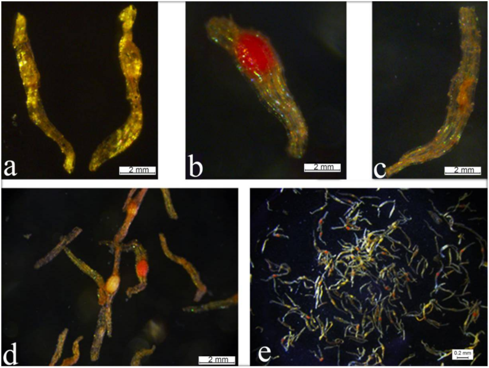
Mature P. Maudiae capsules were green (Figure S1b,c) and the seeds were yellow-brown, with an average length of 0.33 mm, fusiform of subacute at both ends, one end with a tip while the other end is blunt. There is a small ellipsoid embryo without an endosperm, and it is only composed of a one-layer testa and an ellipsoidal embryo. The testa is rectangular with a thin cytoderm, brown, transparent, and without any content (Figure 4a). The embryo of P. Maudiae seeds did not differentiate completely and consisted only of a mass of undifferentiated embryonic cells, which was the pre-embryo stage. Seeds have no endosperm and do not store nutrients, so they are difficult to germinate in natural conditions without the help of a symbiotic fungus [25]. The embryos of P. Maudiae seeds could be stained by triphenyl tetrazolium chloride (TTC) (Figure 4b): If the seeds had an incomplete embryo, seeds were only slightly stained (Figure 4c) or could not be stained in seeds without an embryo (Figure 4d), the embryos of viable seeds were red or light red (Figure 4b,d), while the embryos of aborted seeds did not stain, or stained slightly (Figure 4d,e). However, some seeds with an embryo were not stained (Figure 4d), as was also reported by Fu et al. [11].

2.5. In Planta Transformation by Agrobacterium-Mediated Ovary-Injection

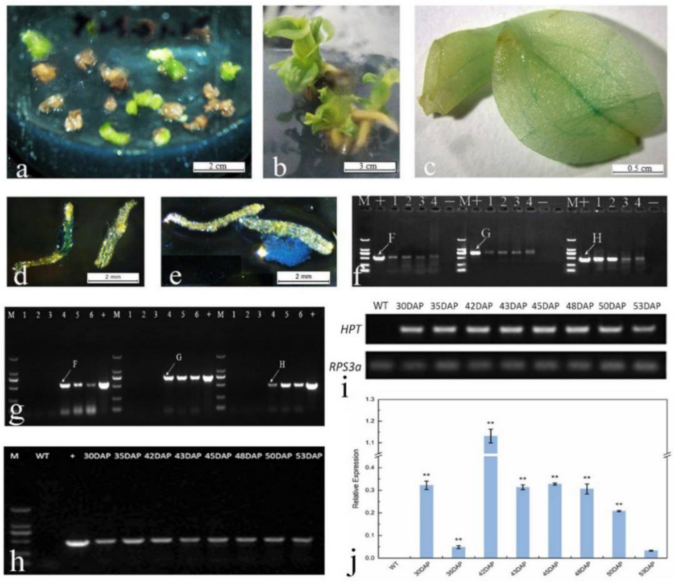
The percentage of seeds that stained positive for TTC from the non-injected fruits was 60.61%, which was significantly higher than that of injected fruits at all stages (DAPs), while the percentage of stained seeds in injected fruits at 50 DAP (43.01%, Figure 5d,e) was significantly higher than that at other injected stages, except at 53 DAP (32.98%, Table 2).

2.6. TTC and GUS Staining and Preliminary Detection of Transgenic Seeds

The seeds of non-injected fruits by AMOI did not stain GUS-positive. However, some seeds and placentas of injected fruits were stained by GUS (Figure 5d,e), but the level of GUS staining was extremely low (<0.1%, data not shown). Simultaneously, the seeds of injected fruits were used to detect the GUS gene by PCR analysis, which showed a positive band for this gene (Figure 5f), suggesting that this exogenous gene was transferred to P. Maudiae seeds.

2.7. In Vitro Germination

In the pericarp of mature capsules used in AMOI and the control (no injection), we only sterilized by dipping in 75% (v/v) ethanol for 1 min, followed by agitation for 15 min in 0.1% (w/v) mercuric chloride (HgCl2) solution, and seeds were then sown onto H26 medium [35]. In this case, seed contamination was 100% for fruits that were injected by Agrobacterium, or by other fungi or bacteria, but fruits of the control group were not contaminated. Consequently, we first sterilized the pericarp of mature capsules (treatment and control) using the steps indicated above, then disinfected the seeds after splitting the capsules with a scalpel using NaOCl (0.5% available chlorine) for 40 min. The level of contamination of injected seeds decreased to 20% while no contamination was observed in the control group. Uncontaminated seeds in media were used in follow-up experiments.
Seed germination rate differed at different injection stages in the AMOI treatment and control. Seed germination of the control was highest (36.30%) and significantly higher than seeds at all injection stages, while the highest seed germination of injected fruits was 15.21% at 50 DAP (Table 3).

2.8. Selection Pressure and Hygromycin Screening

Uniform sized uninjected protocorms were subcultured on H26 medium containing different concentrations of hygromycin (Hyg). Highest survival (95.33%) was observed on medium without Hyg. As the concentration of Hyg increased, protocorm survival declined, but the rate of decline differed. Highest rates of decline were observed between 5 and 25 mg L−1, and between 60 and 75 mg L−1, while all protocorms died at 150 mg L−1 (Figure 6). A selection pressure gradient was suitable for screening Hyg resistance by successively transferring protocorms to different concentrations of Hyg (25, 40, 40, 25, then 0 mg L−1), with each sub-culture period being about 45 d.
Thirty-eight seedlings (with 3–4 leaves and 2–3 roots) were obtained after four rounds of Hyg selection and a single round of Hyg-free culture, resulting in 2, 7, 5, 1, 3, 4, 9, and 7 Hyg-resistant plantlets with a corresponding transformation frequency of 0.45%, 1.89%, 2.16%, 1.07%, 0.90%, 2.45%, 2.54%, and 2.48% from protocorms which germinated from AMOI seeds at 30, 35, 42, 43, 45, 48, 50, and 53 DAP, respectively. No Hyg-resistance plantlets formed from seeds of uninjected capsules or from injected capsules older than 55 DAP. The highest transformation frequency was obtained at 48, 50, and 53 DAP, which were significantly higher than other AMOI times. The highest transformation frequency of all sown seeds was obtained at 50 DAP, which was significantly higher than other AMOI times, except at 35 and 53 DAP (Table 4). Hyg-resistant plantlets transferred to Hyg-free H26 media could form complete plants.

2.9. GUS Staining and Molecular Detection of Hyg-Resistant Plants

The leaves of all putatively Hyg-resistant plants stained blue after GUS staining (Figure 5c). Veins were stained most intensely, indicating that the GUS stain may be easier to permeate into vein tissue. Four randomly selected Hyg-resistant plants were subjected to PCR analysis, and positive bands for the GUS and FT genes were observed (Figure 5g). PCR analysis and semi-quantitative PCR were used to determine the presence of the hpt gene and its expression in leaves of Hyg-resistant seedlings from AMOI at 30, 35, 42, 43, 45, 48, 50, and 53 DAP (Figure 5h,i). hpt expression was detected by quantitative real-time PCR analysis in these tested seedlings as previously described, but at different levels, indicating that exogenous genes were integrated into the plant genome (Figure 5j). Transformation efficiency at 50 DAP was highest (2.54%), followed by 2.48% at 53 DAP and 2.45% at 48 DAP (Table 4). The best time to inject P. Maudiae fruits using the in planta AMOI method for genetic transformation was 48–53 DAP.

3. Discussion

3.1. Ovules and Embryo Development

Seed germination rates and genetic transformation efficiency by ovary-injection in orchids are significantly affected by the embryo developmental stage [2,18,30]. The period of fertilization, and the time it takes for pollen to fall on the stigma and induce the fusion of its sperm with the eggs and polar nucleus differs in most plant species, and the process tends to be complete in 10-48 h [36,37]. However, most orchids need more time, usually 40 to 90 d [12,14]. In this paper, we found that the time required from pollination to fertilization in P. Maudiae is about 50 to 54 d. In P. Maudiae flowers, the ovaries are small, but by about 21 DAP, the capsules expand rapidly, which may be due to the growth of pollen tubes in the ovaries. Pollen inclusions may have some physiological regulatory substances, including auxin and ethylene, which can promote ovule development, seed coat formation and the maturation of female gametophytes [12,38]. The exact reason is unclear, however, and the main active substances and the mechanism of action need to be further researched.
During the development of the female P. Maudiae gametophytes, only the megasporocytes were surrounded by a layer of nucellus cells, thus P. Maudiae is a thin nucellus-type plant. Based on a reference standard of angiosperm embryo sac development types, the P. Maudiae embryo sac development is classified as Allium type [39]. The process of zygote division and differentiation into embryos proceeded according to standard patterns, and the difference in embryonic development between angiosperms are the first few cell divisions of the zygote [40]. Embryonic development in P. Maudiae first shows a transverse division in which the topical cell divides transversally and the basal cell is involved in the formation of the embryonic body and division to form suspensor cells. Then, germ cells develop into zygotes, which continue to develop and do not undergo dormancy, unlike in Paphiopedilum godefroy [41] in which zygotes begin to divide after 40–45 d of dormancy. Based on the reference standard of angiosperm embryonic development types, P. godefroy embryonic development is a Solanum type [39,40].
Embryonic development of mature P. Maudiae seeds generated some seeds without embryos, which may be due to two reasons: (1) Development of the female gametophyte is not normal during embryo sac development, and a mature embryo sac does not form; (2) only a part of the embryo sac can fertilize normally and form a zygote. However, development of the male gametophyte and the processes of pollination and fertilization require further research.
Development of the embryo sac is the process in which the female generative organ of a plant forms, and is an important aspect of reproductive biology. Normal paraffin sections and semi-thin sections are currently the most common methods to observe embryo sacs and embryo development in orchids [13,14,15,16,17,18,41]. The procedural preparation for these two methods is long and tedious, and it is very difficult to isolate several nuclei in embryo sac development within the same time section. CLSM is a combination of common optical microscopy and computer-aided laser technology whose software allows for continuous optical sectioning, allowing for the observation of dynamic changes to the cytoskeleton and other structures at the subcellular level [19]. CLSM significantly shortens the operation time, making it more convenient, and also allowing different levels of organizational structure to be accurately observed. A series of images allows for 3D reconstruction to observe the internal structure and space relationships from different angles [20].
In this paper, treated P. Maudiae embryo sacs and embryo development were observed by CLSM, and clear pictures of both were obtained. This allowed us to determine the developmental stages of embryo sacs and embryos at different periods after pollination, laying down a foundation for further research on developmental studies in this orchid.

3.2. Agrobacterium-Mediated Ovary-Injection

Currently, the most common ways to transform orchids are by Agrobacterium [25,42,43,44,45,46] and particle bombardment [47,48,49,50,51,52], as has been reviewed [53]. The pollen tube pathway was successfully used for transgenic breeding in corn, wheat, melons, cucumbers and other crops such as cotton [28,54,55,56,57]. However, there are few reports for orchids because most orchids contain an abundance of seeds in each capsule that cannot germinate under natural conditions. Orchid seeds thus need an artificial regeneration system to be established, either symbiotically [58], or asymbiotically [59], as has been shown for another orchid genus, Dendrobium. Moreover, the plasmid or DNA may degrade easily which could be problematic because most orchids generally require 40-90 days from pollination to fertilization [12]. Nevertheless, the fruits of most orchids are large, and the ovary has large cavities that can hold in excess of 10,000 seeds, allowing more injection solution to be loaded for transformation.
Thus, the injection of Agrobacterium carrying a plasmid with DNA of interest into the ovary may provide an effective way to introduce novel genes into orchids. Two preliminary studies recorded success in other orchids by some of the authors of this paper, namely in Dendrobium nobile [29] and Doritis pulcherrima [30].
An appropriate selective marker is important for an effective resistance screening test. In this experiment, the Ti plasmid contained the Hyg resistance gene. Orchid protocorms and seedlings are not particularly sensitive to kanamycin, but effective screening can be achieved when using Hyg, as in our study, and which was also shown to be suitable for other orchids, including Dendrobium nobile [29,59] and Doritis pulcherima [30]. The appropriate selection pressure (i.e., Hyg concentration) is also important in resistance screening, and if the Hyg concentration is too low, untransformed protocorms will continue to grow and divide, and putative transformants may be lost. If, on the other hand, the Hyg concentration is too high, protocorm growth will be suppressed or they may even die, even killing off transformed cells. The selection gradient employed for screening transgenic protocorms in this experiment was suitable. A total of 38 Hyg-resistant plants with 3 to 4 leaves and 2 to 3 roots were obtained after four cycles of Hyg resistance screening and a single round of Hyg-free culture, the leaves of all resistant plants stained GUS-positive, and four randomly selected Hyg-resistant plants tested positive for the GUS and FT genes by PCR analysis. These results show that these two exogenous genes could be integrated into the P. Maudiae genome. Semi-quantitative PCR and quantitative real-time PCR analysis of total RNA isolated from the leaves of Hyg-resistant seedlings demonstrates the same result, i.e., confirms the integration of these genes. However, the stable inheritance of these exogenous genes in subsequent generations, and whether the FT gene will shorten the juvenile stage when overexpressed still need to be researched. In general, P. Maudiae plants need about three years to flower from in vitro seedlings, so there is hope that plants transformed with the FT gene may flower precociously. This is because the biological function of FT is a putative phosphatidylethanolamine-binding protein gene, as was shown in Cymbidium, that may regulate the vegetative to reproductive transition in flowers and act similarly to AtFT by regulating the transition from vegetative state to flowering by activating AtAP1 [60,61,62].
The period of exogenous DNA transformation by the pollen tube pathway using transgenic technology is limited to the time between fusion of the sperm and egg and division of the zygote [28,63,64,65,66]. The exogenous gene enters the embryo sac via the pollen, placental surface, micropyle and nucellus channel, and can directly transform the generative cells without a cell wall in the fusion period [67,68,69]. The distance traveled by exogenous DNA before entering the embryo is an important factor that influences transformation frequency [28,29,30]. In our experiments, AMOI provided a shorter entrance pathway for exogenous DNA and thus a shorter entrance period, a greater quantity of DNA entering the embryo sacs, and less degradation of exogenous DNA. A shorter DNA entrance pathway would therefore greatly improve the capacity of exogenous DNA to enter the embryo sacs and enhance the probability of its integration into the host genome. In this study, 38 Hyg-resistant plantlets were obtained from capsules that had been injected between 30 and 53 DAP, with the highest transformation frequency (2.45% to 2.48%) from 48 to 53 DAP. No Hyg-resistant plantlets were obtained when capsules older than 55 DAP were injected. When considering that fertilization in P. Maudiae takes place between 50 and 54 DAP, the best time to employ AMOI would then be when double fertilization of the eggs takes place. However, the lower transformation frequency of capsules injected at 43 and 45 DAP than that of an earlier injection period is difficult to explain because less degradation of exogenous DNA occurred in a shorter time frame from injection of exogenous DNA to fertilization. A possible reason for the lower transformation frequency of all sown seeds form ovules injected 43-48 DAP is that the embryo is easily damaged by AMOI when the embryo sac is in the four- to eight-nucleate stage of development. However, we could not detect CeFT gene mRNA from AMOI at 30, 35, 42, 43, 45, 48, 50, and 53 DAP, possibly due to the loss or silencing of heterologous CeFT gene during seed germination and growth, or because the Ubi promoter was unable to activate the expression of CeFT in P. Maudiae.
In conclusion, the P. Maudiae embryo sac exhibited an Allium type of development. AMOI is an attractive and effective method for the genetic transformation of Paphiopedilum. The fertilization time of P. Maudiae was 50 to 54 DAP, and the best injection time was coincidentally 48 to 53 DAP, which corresponds to the period of fertilization. Thirty-eight protocorms or seedlings were obtained after several rounds of Hyg selection and four randomly selected Hyg-resistant plants were GUS-positive. Semi-quantitative PCR and quantitative real-time PCR analyses revealed the expression of the hpt gene in the leaves of eight Hyg-resistant seedlings. This protocol represents the first effective genetic transformation protocol for any Paphiopedilum species and will allow for expanded molecular breeding programs to introduce useful and interesting genes that can expand its ornamental and horticulturally important characteristics.

4. Materials and Methods

4.1. Plant Material and Agrobacterium Strain

Paphiopedilum Maudiae plants were maintained in a glass greenhouse with a water curtain and blowers for cooling and ventilation in the South China Botanical Garden, Guangzhou, China. The average temperature and humidity range were 15–35 °C and 65–95%, respectively. The flowers from three-year-old adult plants were labeled and artificially self-pollinated by transferring pollen onto the stigma of the same flower as they became fully opened in March to April, in 2014. The seed setting percentage exceeded 90%.
The A. tumefaciens EHA105 strain carried the Flowering Locus T gene (CsFT) from Cymbidium sinense, the β-glucose acid gene (GusA), the Hyg resistance gene (hpt) with a 35S CaMV promoter and a NOS terminator [60].

4.2. Histological and Histochemical Studies

Fruit capsules (Figure S1a) of different developmental stages from 5 to 180 DAP were collected at 5- or 10-d intervals. Capsules were removed and both ends were sliced horizontally, then fixed in a solution of FAA (50% ethanol:acetic acid:formalin; 18:1:1, v/v) at 4 °C overnight. Ovules, which were excised from fixed capsules, were stained according to a previous protocol [22], as follows: (1) A rinse with double distilled water six times, each time for 20 min; (2) rehydration in an ethanol gradient with 50%, 70%, 90% ethanol, each time for 20 min; (3) staining with solution (95% ethanol, 5% eosin, and 5% golden orange; 96:3:1, v/v) for 21 h; (4) rinse in double distilled water six times, each time for 20 min; (5) dehydration in an ethanol gradient with 50%, 70%, 90% ethanol, each time for 20 min, then dehydrated twice with 100% ethanol, each time for 30 min; (6) treatment in a mixed solution (100% ethanol, methyl salicylate; 1:1, v/v) for 3 h, then added to methyl salicylate three times (first time = 3 h; second and third times = 15 h each). Transparent ovules were continually preserved in methyl salicylate; (7) whole transparent ovules were mounted on microslides, covered with coverslips (microslides and coverslips: Leica Biosystems Inc., Buffalo Grove, IL, USA), and observed and photographed at 543 nm laser excitation using a laser scanning confocal microscope (Lsm 510 Meta, Zeiss, Bonn, Germany) for CLSM studies.

4.3. Injection of Ovaries with Agrobacterium

In the glass greenhouse, 20 µL of Agrobacterium suspension was injected laterally into the middle of capsules (ovaries) using a 25 µL microinjector (Shanghai Eastsen Analytical Instrument Co. Ltd., Shanghai, China) on 30, 35, 40, 42, 43, 44, 45, 46, 47, 48, 50, 53, 55, 57, 60, 65, and 70 DAP, i.e., a total of 17 time treatments, with nine different capsules per time treatment. The control consisted of capsules that were not injected with Agrobacterium after artificial pollination. Seed from 150 DAP fruits were sown aseptically.

4.4. Aseptic Sowing

Capsules that had (or had not) been injected with Agrobacterium were aseptically sown, as follows. At first, capsules were rinsed under running tap water to remove remains of the epidermis and disinfected by dipping into 75% (v/v) ethanol for 1 min, then immersed for 15 min in 0.1% (w/v) mercuric chloride (HgCl2) solution. Capsules were then rinsed four times with sterilized (by autoclave) deionized water and their surfaces were dabbed dry on sterilized filter paper to remove surface moisture. Capsules were split longitudinally along the suture with a sterilized scalpel, seeds were gently placed into sterilized 100-mL conical flasks for sterilizing with NaOCl containing 0.5% available chlorine for 40 min. Seeds were then rinsed three times with sterile deionized water (filtered through sterilized filter paper; Hangzhou WoHua Filter Paper Co. Ltd., Hangzhou, China) and placed into sterile deionized water to create a suspension liquid with approximately 100 seeds/mL for aseptic sowing.
Three ml of a suspension with about 300 seeds from all injection stages were dispersed uniformly on Hyponex N026 medium [2], which contained 1.5 g L−1 Hyponex I (Taihe Horticultural Co. Ltd., Taiwan, China), 15 g L−1 sucrose, 2 g L−1 peptone, 5.5 g L−1 agar (Huankai Microbial Sci. & Tech, Co., Ltd., Guangzhou, China), 1.5 g L−1 activated charcoal (AC), 10 mg L−1 vitamin B1, 1 mg L−1 vitamin B6, 1 mg L−1 niacin, 2 mg L−1 glycine, 1 mg L−1 NAA, 1 mg L−1 myo-inositol, and 50 mL L−1 coconut milk (CM). All chemicals and reagents were purchased from Sigma-Aldrich (St. Louis, MI, USA), unless specified otherwise. The CM used in these experiments was obtained from 6- to 7-month-old green coconuts from Hainan Province, China, after filtering through a single sheet of filter paper [35]. Medium was added to 500-mL conical flasks (Xuzhou Hualian Glass Products Co. Ltd., Xuzhou, China) closed with perforated rubber stoppers and plugged with cotton. Each flask contained about 100 mL of medium. Medium pH was adjusted to 5.6 with 1 mol L−1 KOH or HCl before autoclaving at 121 °C for 18 min at 1.06 kg cm−2. Cultures were incubated at 26 ± 1 °C and a 16-h photoperiod under 40 W cool white fluorescent lamps (Shanghai Xianyi Lighting and Electrical Appliance Co. Ltd., Shanghai, China) delivering a photosynthetic photon flux density of ca. 45 μmol m−2 s−1. The germination rate of seeds was calculated using a previously reported method [2].

4.5. Seed Viability Test

Mature P. Maudiae seeds at all tested DAP were added into labelled 1.5-mL Eppendorf tubes. Approximately 100 seeds were submerged in 1.0 mL of 0.5% triphenyl tetrazolium chloride (TTC) solution, incubated at 37 °C, stained for 24 h, then rinsed with deionized water. TTC solution was made by first dissolving 0.1 g of TTC in 200 mL phosphate buffer (pH 7.17), then placing the TTC solution into an amber laboratory bottle at 4 °C. Stained seeds were viewed with a stereomicroscope (Leica DFC 450, Wetzlar, Germany) to count the number of seeds with an embryo and the number of stained seeds. Measurements were repeated three times for each treatment (i.e., DAP) at all injection stages.

4.6. GUS Assay for Seeds and Transgenic Seedlings

Mature P. Maudiae seeds (150 DAP) at each injection stage were placed into separate Eppendorf tubes. Seeds were imbibed (no vacuum infiltration) in GUS solution (100 mM L−1 sodium phosphate buffer (Na3PO4), 0.1% Triton X–100, 10 mM l−1 EDTA, 0.5 mM l−1 potassium ferricyanide, 0.5 mM L−1 potassium ferrocyanide, 1 mg ml−1 X-Gluc), placed in an incubator and stained for 12 h at 37 °C, destained in 75% ethanol for 1-2 d, then placed on a microslide and viewed by a Leica DFC 450 stereomicroscope. The leaves of putatively transgenic seedlings with 3–4 leaves resistant to Hyg were detected by GUS staining using the same method employed for the GUS staining of seeds.

4.7. Selection Pressure and Resistance Screening of Protocorms

Well-developed protocorms of the control group with uniform growth and size (approx. 0.4 cm in diameter) were inoculated onto Hyponex N026 medium containing different concentrations of Hyg (0, 5, 10, 15, 20, 25, 30, 40, 50, 60, 70, 75, 100, 150, and 200 mg L−1). Each culture dish (90 mm × 16 mm) was inoculated with 50 protocorms, with five dishes for each treatment. Trials indicated that 25–40 mg L−1 was the most effective concentration range for screening the resistance of protocorms to Hyg. Protocorms that survived were cultured successively in media with different concentrations of Hyg (25, 40, 40, 25, and 0 mg L−1), each culture cycle being 45 d. The gradually decrease in the concentration of Hyg in the last two rounds was to encourage the growth of transgenic material to develop robust plants.

4.8. PCR Detection of Seeds and Resistant Seedlings

CeFT, GUS and hpt genes were detected in seeds at 150 DAP from ovary-injection of Agrobacterium at 48 DAP, and the hpt gene was detected in Hyg-resistant seedlings from AMOI at 30, 35, 42, 43, 45, 48, 50, and 53 DAP. The DNA of Hyg-resistant seedlings was extracted by the cetyl trimethyl ammonium bromide (CTAB) method [58]. The PCU1301 plasmid, carrying exogenous genes, served as the positive control and untransformed seedlings served as the negative control. A map of the T-DNA region is indicated in Figure S2. The following primers were used for PCR of the hpt and GUS genes [58], and the FT gene [60]: HPTL (forward) 5′-GATGTTGGCGACCTCGTATT-3′; HPTR (reverse) 5′-GTGCTTGACATTGGGGAGT-3′; GUSL (forward) 5′-GTGAATCCGCACCTCTGG-3′; GUSR (reverse) 5′-ATCGCCGCTTTGGACATA-3′; FTL (forward) 5′-CGCGGATCCATGAATAGAGAGAGAGACT-3′; FTR (reverse) 5′-CGCGGATCCTCAATCCTGCATCCTTCTTCCG-3′. PCR was run over 35 cycles of 95 °C for 30 s, 58 °C for 30 s, and 72 °C for 1 min. PCR products were detected by 0.8% agarose gel (Biowest Agarose G-10, Baygene Biotech. Co., Ltd., Shanghai, China) electrophoresis, stained with 0.1 µg ml−1 ethidium bromide (Leagene Biotech. Co., Ltd., Beijing, China), observed and images were collected by a UVP GelDoc-It 310 imaging system (Shanghai Yuansheng Instrument Equipment Co., Ltd., Shanghai, China).

4.9. Semi-Quantitative PCR and Quantitative Real-Time PCR Analysis

The hpt gene was detected in Hyg-resistant seedlings from AMOI at 30, 35, 42, 43, 45, 48, 50, and 53 DAP. Total RNA from these leaves was extracted using a Column Plant RNAOUT 2.0 kit (TIANDZ Biotech. Co., Ltd., Beijing, China), and 1 μg of each RNA sample were reverse transcribed using the GoScript™ Reverse Transcription System (Promega Biotech Co., Ltd., Beijing, China) according to the manufacturers’ instructions. The cDNA products were diluted to 200 ng µL−1 for further studies.
The success of reverse transcription was verified by PCR using the RPS3a reference gene [70]. The gene-specific primer pairs were designed by the online PrimerQuest tool [71] (RPS3aL (forward) 5′-TGATGTGAAGACCACGGATAAC-3′, RPS3aR (reverse) 5′-CAGGTTCGCTTGACTTGATTTG-3′). Semi-quantitative PCR was performed using the Golden Star T6 Super PCR Mix (TsingKe Biotech Co., Ltd., Beijing, China) with first-strand cDNA as the template in a 25 μL standard PCR reaction. The PCR reaction was performed by denaturation at 95 °C for 2 min, followed by 30 cycles of 10 s denaturation at 98 °C, 10 s annealing at 65 °C, 10 s extension at 72 °C, and a final extension at 72 °C for 1 min. The amplified fragments were separated on a 1% agarose gel containing 0.1 µg mL−1 ethidium bromide in TAE buffer.
The cDNA protocol described above was also used for quantitative real-time PCR analysis, which was performed using the iTaq Universal SYBR Green Supermix (Bio-Rad Laboratories, Inc., Beijing, China) in an Applied Biosystems® ABI 7500 Real-time system (Thermo Fisher Scientific Inc., Waltham, MA, USA). Amplification conditions were 95 °C for 30 s, followed by 40 cycles of amplification (95 °C for 15 s, 60 °C for 35 s).

4.10. Data Analysis

The experiments of TTC staining and seed germination were conducted in a completely randomized design. All data were analyzed with SPSS 17.0 for Windows (Microsoft Corp., Washington, USA) and expressed as means ± standard error (SE) using one-way analysis of variance (ANOVA) followed by Duncan’s multiple range test at p ≤ 0.05.

Supplementary Materials

The following are available online at https://www.mdpi.com/1422-0067/22/1/84/s1.

Author Contributions

B.-X.L., L.Z., F.Z., K.-L.W., J.A.T.d.S., L.F., G.-H.M., and S.-J.Z., conceived, designed, and assessed the experiments; B.-X.L., L.Z., F.Z., S.-J.Z., and L.L. performed the experiments; B.-X.L., L.Z., S.-J.Z., X.-H.Z., and L.F. analyzed the data; K.-L.W. and L.L. contributed reagents/materials/analysis tools; B.-X.L., L.Z., L.F., S.-J.Z., and J.A.T.d.S. wrote the manuscript. All authors have read and agreed to the published version of the manuscript.

Funding

This work was funded by the National Key Research and Development Program of China (2016YFC0503104; 2018YFD1000403), the National Natural Science Foundation of China (31902062), the Natural Science Foundation of Guangdong Province (2018A030313686), the Guangdong Key Technology Research and Development Program (2018B020202001-3), the Guangdong Modern Agricultural Industry Technology System Program (2018LM2176, 2020KJ121), the Biological Resources Program, the Chinese Academy of Sciences (KFJ-BRP-005), and the Key Laboratory of South China Agricultural Plant Molecular Analysis and Genetic Improvement, South China Botanical Garden, Chinese Academy of Sciences (KF202006).

Institutional Review Board Statement

The study was conducted according to the guidelines of the Declaration of Helsinki, and approved by the Institutional Review Board of South China Botanical Garden, Chinese Academy of Sciences.

Informed Consent Statement

There were no human or animal research subjects in this study. Consequently, informed consent was not required.

Data Availability Statement

Data may be found within the article or supplementary material. Raw data available upon reasonable request.

Conflicts of Interest

The authors declare no conflict of interest.

References

  1. Liu, Z.J.; Chen, S.C.; Chen, L.J.; Lei, S.P. The Genus Paphiopedilum in China; Science Press: Beijing, China, 2009; pp. 13–349. [Google Scholar]
  2. Zeng, S.J.; Wu, K.L.; Teixeira da Silva, J.A.; Zhang, J.X.; Chen, Z.L.; Xia, N.H.; Duan, J. Asymboiotic seed germination, seedling development and reintroduction of Paphiopedilum wardii Sumerh., an endangered terrestrial orchid. Sci. Hortic. 2012, 138, 198–209. [Google Scholar] [CrossRef]
  3. Cribb, P. The Genus Paphiopedilum, 2nd ed.; National History Publications: Borneo, Malaysia, 1998; pp. 48–396. [Google Scholar]
  4. Wu, Z.Y.; Raven, P.H.; Hong, D.Y. Flora of China; Science Press: Beijing, China; Missouri Botanical Garden Press: St., Louis, MI, USA, 2009; Volume 25, pp. 33–44. [Google Scholar]
  5. World Checklist of Selected Plant Families. Available online: http://apps.kew.org/wcsp/qsearch.do (accessed on 22 December 2020).
  6. The IUCN Red List of Threatened Species. Version 2020-2. Available online: http://www.iucnredlist.org/search (accessed on 22 December 2020).
  7. Zeng, S.J.; Huang, W.C.; Wu, K.L.; Zhang, J.X.; Teixeira da Silva, J.A.; Duan, J. In vitro propagation of Paphiopedilum orchids. Crit. Rev. Biotechnol. 2016, 36, 521–534. [Google Scholar]
  8. Zeng, S.J.; Tian, R.X.; Chen, Z.L.; Wu, K.L.; Duan, J. Research progress on cross breeding of Paphiopedilum. J. Trop. Subtrop. Bot. 2010, 18, 459–468. [Google Scholar]
  9. Huang, L.C. A procedure for asexual multiplication of Paphiopedilum in vitro. Am. Orchid Soc. Bull. 1988, 57, 274–278. [Google Scholar]
  10. Liao, Y.J.; Tsai, Y.C.; Sun, Y.W.; Lin, R.S.; Wu, F.S. In vitro shoot induction and plant regeneration from flower buds in Paphiopedilum orchids. In Vitro Cell. Dev. Biol. Plant 2011, 47, 702–709. [Google Scholar]
  11. Fu, Y.Y.; Jiang, N.; Wu, K.L.; Zhang, J.X.; Teixeira da Silva, J.A.; Duan, J.; Liu, H.T.; Zeng, S.J. Stimulatory effects of sodium hypochlorite and ultrasonic treatments on tetrazolium staining and seed germination in vitro of Paphiopedilum SCBG Red Jewel. Seed Sci. Technol. 2016, 44, 77–90. [Google Scholar]
  12. Zhang, X.S.; O’Neill, S.D. Ovary and gametophyte development are coordinately regulated by auxin and ethylene following pollination. Plant Cell 1993, 5, 403–418. [Google Scholar] [CrossRef] [PubMed]
  13. Ye, X.L.; Guo, J.Y. Female gametophyte development and embryogenesis in Cymbidium sinense (Andr.) Willd. J. Trop. Subtrop. Bot. 1995, 3, 54–58. [Google Scholar]
  14. Tang, Y.J.; Ye, X.L.; Chen, Z.L. The studies of the female gametophyte development and the embryogenesis in Doritis pulcherrima Lindl. J. Trop. Subtrop. Bot. 1998, 6, 289–292. [Google Scholar]
  15. Li, D.M.; Wu, C.H.; Ye, X.L.; Liang, C.Y. Ultrastructural observation on embryo sac development in Phaius tankervilliae (Aiton) Bl. Plant Sci. J. 2012, 30, 188–198. [Google Scholar] [CrossRef]
  16. Liu, F.; Tian, M.; Wang, C.X.; Gong, M.J.; Li, Q.J. Observation on fruit growth dynamics and embryo development process of Cymbidium japonicum. J. Plant Res. Dev. 2012, 21, 28–35. [Google Scholar]
  17. Zhang, Y.; Zhang, Q.X.; Zhao, S.W.; Ling, C.Y. Embryo and integument development of the endangered species Cypripedium macrathos Sw. Acta Hortic. Sin. 2010, 37, 72–76. [Google Scholar] [CrossRef]
  18. Zhang, Y.Y.; Wu, K.L.; Zhang, J.X.; Deng, R.F.; Duan, J.; Teixeira da Silva, J.A.; Huang, W.C.; Zeng, S.J. Embryo development in association with asymbiotic seed germination in vitro of Paphiopedilum armeniacum S. C. Chen et F. Y. Liu. Sci. Rep. 2015, 5, 16356. [Google Scholar] [CrossRef] [PubMed] [Green Version]
  19. Zhou, L.H.; Huang, G.W. Application of laser scanning confocal microscope in botany. Acta Laser Biol. Sin. 2005, 14, 76–79. [Google Scholar]
  20. Li, J.X.; Dai, X.M.; Zhang, J.J.; Zhang, Y.S. Using a whole stain-clearing technique to observe embryo sac of cotton. Exp. Technol. Manag. 2014, 31, 60–62. [Google Scholar]
  21. Dai, X.M.; Huang, Q.C.; Li, G.P.; Hu, X.M.; Qin, G.Y. Observation on the process of double fertilization in rice using laser scanning confocal microscopy. J. Henan Agric. Sci. 2003, 6, 25–30. [Google Scholar]
  22. Zhang, H.H.; Feng, J.H.; Lu, Y.G.; Yang, B.Y.; Liu, X.D. Observation on formation and development of autotetraploid rice embryo sac using laser scanning confocal microscope. J. Chin. Electron Microsc. Soc. 2003, 22, 380–384. [Google Scholar]
  23. Zhu, D.Z.; Wang, J.W.; Zong, X.J.; Wei, H.R.; Liu, Q.Z. Observation of female gametophyte development of sweet cheery with confocal scanning microscope. Int. J. Fruit Sci. 2014, 31, 36–40. [Google Scholar]
  24. Johnson, T.R.; Kane, M.E. Asymbiotic germination of ornamental Vanda: In vitro germination and development of three hybrids. Plant Cell Tissue Organ Cult. 2007, 91, 251–261. [Google Scholar] [CrossRef]
  25. Hossain, M.M.; Kant, R.; Van, P.T.; Winarto, B.; Liu, J.J.; Zeng, S.J.; Teixeira da Silva, J.A. The application of biotechnology to orchids. Crit. Rev. Plant Sci. 2013, 32, 69–139. [Google Scholar] [CrossRef]
  26. Teixeira da Silva, J.A. Orchids: Advances in tissue culture, genetics, phytochemistry and transgenic biotechnology. Floricult. Ornam. Biotechnol. 2013, 7, 1–52. [Google Scholar]
  27. Zhou, G.Y.; Weng, J.; Zeng, Y.; Huang, J.; Qian, S.; Liu, G. Introduction of exogenous DNA into cotton embryos. Meth. Enzymol. 1983, 101, 433–481. [Google Scholar]
  28. Yang, A.F.; Su, L.J.; An, L.J. Ovary-drip transformation: A simple method for directly generating vector- and marker-free transgenic maize (Zea mays L.) with a linear GFP cassette transformation. Planta 2009, 229, 793–801. [Google Scholar] [CrossRef] [PubMed]
  29. Liu, Q.F.; Dong, H.; Zeng, S.J.; Chen, Z.L.; Wu, K.L.; Zhang, J.X.; Duan, J. In planta transformation of Dendrobium nobile by ovary-injection of Agrobacterium. J. South China Agric. Univ. 2013, 34, 378–382. [Google Scholar]
  30. Wang, J.; Zeng, S.J.; Chen, Z.L.; Wu, K.L.; Zhang, J.X.; Duan, J. Genetic transformation of Doritis pulcherima (Orchidaceae) via ovary-injection. Chin. J. Trop. Crops 2013, 34, 1498–1501. [Google Scholar]
  31. Teixeira da Silva, J.A.; Kerbauy, G.B.; Zeng, S.J.; Chen, Z.L.; Duan, J. In vitro flowering of orchids. Crit. Rev. Biotechnol. 2014, 34, 56–76. [Google Scholar] [CrossRef]
  32. Kardailsky, I.; Shukla, V.K.; Ahn, J.H.; Dagenais, N.; Christensen, S.K.; Nguyen, J.T.; Chory, J.; Harrison, M.J.; Weigel, D. Activation tagging of the floral inducer FT. Science 1999, 286, 1962–1965. [Google Scholar] [CrossRef] [Green Version]
  33. Jia, Z.; Zhao, F.Y.; Wu, C.X. Pleiotropic effects of FT and its orthologues on plant development. Acta Bot. Boreal. Occid. Sin. 2011, 31, 2558–2564. [Google Scholar]
  34. Lee, Y.I.; Lee, N.; Yeung, E.C.; Chung, M.C. Embryo development of Cypripedium formosanum in relation to seed germination in vitro. J. Am. Soc. Hortic. Sci. 2005, 130, 747–753. [Google Scholar] [CrossRef] [Green Version]
  35. Zeng, S.J.; Chen, Z.L.; Wu, K.L.; Zhang, J.X.; Bai, C.K.; Teixeira da Silva, J.A.; Duan, J. Asymbiotic seed germination, induction of calli and protocorm-like bodies, and in vitro seedling development of the rare and endangered Nothodoritis zhejiangensis Chinese orchid. HortScience 2011, 46, 460–465. [Google Scholar] [CrossRef] [Green Version]
  36. Sun, H.F. The time interval from pollination to fertilization. Biol. Bull. 1990, 10, 8. [Google Scholar]
  37. Peng, Z.Z.; Zhao, Y.; Zeng, Q.; Huang, L.H.; Chen, H.D.; Li, Y.G.; Zhang, X.W. Inplanta transformation of Gossypium hirsutum by ovary-injection of Agrobacterium. J. Cotton Sci. 2011, 23, 311–316. [Google Scholar]
  38. Yam, T.W.; Yeung, E.C.; Ye, X.L.; Zee, S.Y.; Arditti, J. Orchid embryos. In Orchid Biology: Reviews and Perspectives, 8th ed.; Kull, T., Arditti, J., Eds.; Kluwer Academic Publishers: Dordrecht, The Netherlands, 2002; pp. 287–385. [Google Scholar]
  39. Liu, J.P. The embryonic development of angiosperms. Biol. Bull. 1984, 5, 4–6. [Google Scholar]
  40. Ren, L.; Wang, F.H. Embryological studies of Paphiopedilum godefroy Srein. Acta Bot. Sin. 1987, 29, 14–21. [Google Scholar]
  41. Chin, D.P.; Mishiba, K.; Mii, M. Agrobacterium-mediated transformation of protocorm-like bodies in Cymbidium. Plant Cell Rep. 2007, 26, 735–743. [Google Scholar] [CrossRef] [PubMed]
  42. Chan, Y.L.; Lin, K.H.; Sanjaya; Liao, L.J.; Chen, W.H.; Chan, M.T. Gene stacking in Phalaenopsis orchid enhances dual tolerance to pathogen attack. Transgenic Res. 2005, 14, 279–288. [Google Scholar] [CrossRef]
  43. Liau, C.H.; You, S.J.; Prasad, V.; Hsiao, H.H.; Lu, J.C.; Yang, N.S.; Chan, M.T. Agrobacterium tumefaciens-mediated transformation of an Oncidium orchid. Plant Cell Rep. 2003, 21, 993–998. [Google Scholar] [CrossRef]
  44. Mishiba, K.; Chin, D.P.; Mii, M. Agrobacterium-mediated transformation of Phalaenopsis by targeting protocorms at an early stage after germination. Plant Cell Rep. 2005, 24, 297–303. [Google Scholar] [CrossRef]
  45. Sjahril, R.; Mii, M. High-efficiency Agrobacterium-mediated transformation of Phalaenopsis using meropenem, a novel antibiotic to eliminate Agrobacterium. J. Hortic. Sci. Biotech. 2006, 81, 458–464. [Google Scholar] [CrossRef]
  46. Nan, G.L.; Kuehnle, A.R. Factors affecting gene delivery by particle bombardment of Dendrobium orchids. Biol. Plant. 1995, 31, 131–136. [Google Scholar]
  47. Yang, J.; Lee, H.J.; Shin, D.H.; Oh, S.K.; Seon, J.H.; Paek, K.Y.; Han, K.H. Genetic transformation of Cymbidium orchid by particle bombardment. Plant Cell Rep. 1999, 18, 978–984. [Google Scholar] [CrossRef]
  48. Knapp, J.E.; Kausch, A.P.; Chandlee, J.M. Transformation of three genera of orchid using the bar gene as a selectable marker. Plant Cell Rep. 2000, 19, 893–898. [Google Scholar] [CrossRef] [PubMed]
  49. Men, S.; Ming, X.; Wang, Y.; Liu, R.; Wei, C.; Li, Y. Genetic transformation of two species of orchid by biolistic bombardment. Plant Cell Rep. 2003, 21, 592–598. [Google Scholar] [CrossRef] [PubMed]
  50. Tee, C.S.; Marziah, M.; Tan, C.S.; Abdullah, M.P. Evaluation of different promoters driving the GFP report gene and selected target tissues for particle bombardment of Dendrobium Sonia 17. Plant Cell Rep. 2003, 21, 452–458. [Google Scholar] [CrossRef]
  51. Li, S.H.; Kuoh, C.S.; Chen, Y.H.; Chen, H.H.; Chen, W.H. Osmotic sucrose enhancement of single-cell embryogenesis and transformation efficiency in Oncidium. Plant Cell Tissue Organ Cult. 2005, 81, 183–192. [Google Scholar] [CrossRef]
  52. Teixeira da Silva, J.A.; Chin, D.P.; Van, P.T.; Mii, M. Transgenic orchids. Sci. Hortic. 2011, 130, 673–680. [Google Scholar] [CrossRef]
  53. Zhou, G.Y.; Weng, J.; Gong, Z.Z.; Zhen, Y.S.; Yang, W.X.; Shen, W.F.; Wang, Z.F.; Tao, Q.Z.; Huang, J.G.; Qian, S.Y.; et al. Molecular breeding of agriculture: A technique for introducing exogenous DNA into plants after self-pollination. Sci. Agric. Sin. 1988, 21, 1–6. [Google Scholar]
  54. Clough, S.J.; Bent, A.F. Floral dip: A simplified method for Agrobacterium-mediated transformation of Arabidopsis thaliana. Plant J. 1998, 16, 735–743. [Google Scholar] [CrossRef] [Green Version]
  55. Bent, A.F. Arabidopsisin planta transformation. uses, mechanisms, and prospects for transformation of other species. Plant Physiol. 2000, 124, 1540–1547. [Google Scholar] [CrossRef] [Green Version]
  56. Yang, S.; Li, G.; Li, M.G.; Wang, J.H. Transgenic soybean with low phytate content constructed by Agrobacterium transformation and pollen-tube pathway. Euphytica 2010, 177, 375–382. [Google Scholar] [CrossRef]
  57. Teixeira da Silva, J.A.; Tsavkelova, E.; Zeng, S.J.; Ng, T.B.; Dobránszki, J.; Parthibhan, S.; Cardoso, J.C.; Rao, M.V. Symbiotic in vitro seed propagation of Dendrobium: Fungal and bacterial partners and their influence on plant growth and development. Planta 2015, 242, 1–22. [Google Scholar] [CrossRef] [PubMed]
  58. Teixeira da Silva, J.A.; Tsavkelova, E.; Ng, T.B.; Dobránszki, J.; Parthibhan, S.; Cardoso, J.C.; Rao, M.V.; Zeng, S.J. Asymbiotic in vitro seed propagation of Dendrobium. Plant Cell Rep. 2015, 34, 1685–1706. [Google Scholar] [CrossRef] [PubMed]
  59. Chen, Z.L.; Duan, J.; Zeng, S.J.; Liang, C.Y.; Ye, X.L. Agrobacterium-mediated transformation of Dendrobium orchid by targeting protocorms. Acta Sci. Nat. Univ. Sunyatseni 2007, 46, 86–90. [Google Scholar]
  60. Huang, W.T.; Fang, Z.M.; Zeng, S.J.; Zhang, J.X.; Wu, K.L.; Chen, Z.L.; Teixeira da Silva, J.A.; Duan, J. Molecular cloning and functional analysis of three FLOWERING LOCUS T (FT) homologous genes from Chinese Cymbidium. Int. J. Mol. Sci. 2012, 13, 11385–11398. [Google Scholar] [CrossRef] [PubMed] [Green Version]
  61. Song, G.Q.; Walworth, A.; Zhao, D.Y.; Jiang, N.; Hancock, J.F. The Vaccinium corymbosum Flowering Locus T-like gene (VcFT): A flowering activator reverses photoperiodic and chilling requirements in blueberry. Plant Cell Rep. 2013, 32, 1759–1769. [Google Scholar] [CrossRef] [PubMed]
  62. Smart, M.; Roden, L.C. Initiation of flowering in Protea compacta × Protea neriifolia hybrid ‘Carnival’ coincides with expression of the Flowering Locus T homologue. Plant Mol. Biol. Rep. 2014, 32, 372–381. [Google Scholar] [CrossRef]
  63. Yu, G.R.; Yin, J.; Ren, J.P.; Guo, X.Y. Transferring Anti-TrxS gene into wheat by methods of pollen tube pathway and ovary injection. J. Henan Agric. Sci. 2004, 6, 6–10. [Google Scholar]
  64. Gao, X.R.; Wang, G.K.; Su, Q.; Wang, Y.; An, L.J. Phytase expression in transgenic soybeans: Stable transformation with a vector-less construct. Biotechnol. Lett. 2007, 29, 1781–1787. [Google Scholar] [CrossRef]
  65. Chen, Y. Study on techniques of introduction of exogenous DNA into plants via pollen tube pathway. Northern Hortic. 2010, 13, 226–228. [Google Scholar]
  66. Wang, Y.; Wu, Y.; Wang, Y.; Li, Z.B.; Wang, G.X. The five kinds of DNA transformation techniques. Rainfed Crops 2010, 30, 186–189. [Google Scholar]
  67. Wang, G.L.; Fang, H.J.; Zhu, Y.M. Plant Genetic Engineering; Science Press: Beijing, China, 2009; pp. 287–288. [Google Scholar]
  68. Li, W.J.; Zhang, J.P.; Chen, F.; Sun, W.J. Transformation of herbicide resistant gene (bar) into flax by pollen tube pathway. Chin. Agric. Sci. Bull. 2013, 29, 96–100. [Google Scholar]
  69. Wang, M.; Zhang, B.H.; Wang, Q.L. Transgenic Cotton Methods and Protocols; Humana Press: Totowa, NJ, USA, 2013; pp. 71–77. [Google Scholar]
  70. Luo, H.L.; Luo, K.C.; Luo, L.P.; Li, E.X.; Guan, B.C.; Xiong, D.J.; Sun, B.T.; Peng, K.; Yang, B.Y. Evaluation of candidate reference genes for gene expression studies in Cymbidium kanran. Sci. Hortic. 2014, 167, 43–48. [Google Scholar] [CrossRef]
  71. NCBI Primer-BLAST. Available online: https://www.ncbi.nlm.nih.gov/tools/primer-blast/index.cgi?LINK_LOC=BlastHome (accessed on 22 December 2020).
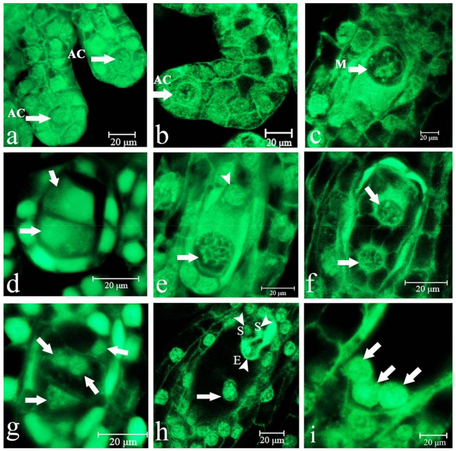
Figure 1. Development of the Paphiopedilum Maudiae embryo sac at several stages before pollination. (a): Archesporial cell (AC) at 10 days after pollination (DAP) (arrow); (b): AC at 20 DAP (arrow) in which the ovule primordium began to curve back to eventually face the placenta; (c): Megasporocyte (M) at 25 DAP (arrow); (d): First meiotic cell division with the formation of two dyads (arrow) at 25 DAP; the chalazal dyad enlarged prior to the second meiotic cell division; (e): Second meiotic cell division formed two megaspores of unequal size at 30 DAP; the smaller megaspore gradually degenerated (arrow); (f): First mitotic nuclear division with the formation of a two-nucleate embryo sac (arrow) at 35 DAP; (g): Second mitotic nuclear division with the formation of a four-nucleate embryo sac (arrow) at 42 DAP; (h): Polar nucleus (arrow), egg cell (E), two synergids (S) at 45 DAP; (i): Three antipodal cells (arrow) at 54 DAP.
Ijms 22 00084 g001
Figure 2. Polar nucleus of Paphiopedilum Maudiae embryo sac development after pollination. (ac): Fertilization of secondary nucleus; (d): Chalazal endosperm nucleus (arrow). SC = seed coat.
Ijms 22 00084 g002
Figure 3. Development of the Paphiopedilum Maudiae embryo at different stages after pollination. (a): Zygote (Z) just fertilized at 54 days after pollination (DAP) (arrow); (b): Two-cell pre-embryo at 60 DAP (arrow); (c): Three-cell pre-embryo at 70 DAP (arrow); (d): T-shaped pre-embryo with four cells at 76 DAP; (e): Six-cell embryo at 85 DAP; (f): Eight-cell embryo at 90 DAP; (g): Sixteen-cell embryo (E) at 100 DAP, suspensor (S); (h): Spherical embryo at 105 DAP; (i): Spherical embryo at 109 DAP; (j): Suspensor (S) with three highly vacuolated cells at 114 DAP (arrow); (k): Suspensor (S) with three highly vacuolated cells had not completely degenerated at 120 DAP (arrow); (l): Abundant starch and lipid globule accumulation at 125 DAP; (m): The suspensor degenerated, but remained residual at 130 DAP; (n): Spheric embryo was very close to the testa at 140 DAP (arrow); SC = seed coat; (o): Mature spherical embryo at 150 DAP; (p): The suspensor disappeared at 160 DAP; (q): Shape of the spherical embryo remained unchanged at 170 DAP; (r): Mature spherical embryo with large starch and lipid globules (LG) at 180 DAP.
Ijms 22 00084 g003
Figure 4. Viability test of Paphiopedilum Maudiae seeds. (a): Seeds of P. Maudiae; (b): Triphenyl tetrazolium chloride (TTC) staining of viable seed; (c): Slight TTC staining of seeds with an aborted embryo; (d): Seeds with an unstained embryo; (e): TTC staining of seeds from a capsule.
Ijms 22 00084 g004
Figure 5. In planta transformation of Paphiopedilum Maudiae by Agrobacterium-mediated ovary-injection (AMOI). (a): Protocorms selected on H26 medium [35] with 40 mg L−1 hygromycin (Hyg) in second screening; (b): Hyg-resistant plants after Hyg selection on H26 medium without Hyg; (c): Leaves of Hyg-resistant plants were stained blue after GUS staining; (d): GUS expression in the placenta; (e): GUS expression in seeds; (f): Detection of CeFT, GUS and hpt genes; lanes 1-4, AMOI at 35, 48, 50 and 53 DAP; + lane, positive control;—lane, negative control; M, DL2000 DNA marker ladder (TaKaRa, Dalian, China); (g): Detection of CeFT, GUS and hpt genes in 150 DAP seeds after AMOI at 48 DAP; lanes 1, 2 and 3: seeds from no AMOI fruits; lanes 4, 5 and 6: seeds from AMOI fruits; + lane, positive control; M, DL2000 DNA marker ladder; (h): PCR analysis of hpt in Hyg-resistant seedlings from AMOI at 30, 35, 42, 43, 45, 48, 50, and 53 DAP. M: DL2000 DNA marker ladder, WT: wild-type plant, +: positive plasmid control. (i): Semi-quantitative PCR analysis of hpt in Hyg-resistant seedlings from AMOI at 30, 35, 42, 43, 45, 48, 50, and 53 DAP. (j): hpt expression in WT and Hyg-resistant seedlings as revealed by quantitative real-time PCR. Data were calculated using the 2−ΔΔCt formula. Error bars represent standard error of three replicates. Double asterisks (**) indicate a significant difference relative to WT according to Duncan’s multiple range test (p < 0.01).
Ijms 22 00084 g005
Figure 6. Effect of different Hyg concentrations on Paphiopedilum Maudiae protocorms. For each treatment, 50 protocorms were inoculated per culture dish (90 mm × 16 mm). All experiments consisted of five independent replicates. Values followed by different letters indicate significant differences according to Duncan’s multiple range test (p < 0.05).
Ijms 22 00084 g006
Table 1. Major microscopic structural events taking place in the developing megagametophyte and embryo of Paphiopedilum Maudiae.
Days after Pollination (DAP)Developmental Stage
6, 10, 20Formation of an archesporial cell
20, 25Formation of megasporocyte
25, 30Formation of functional megaspore
35, 38, 40Two-nucleate embryo sac
42, 44Four-nucleate embryo sac
45, 50Eight-nucleate embryo sac
50, 54Mature embryo sac, fertilization
54, 60First division of the zygote
70, 76, 80, 85, 90Pre-embryo with 2-8 cells, suspensor with 1–2 cells and two-nucleate endosperm
95, 100, 105, 109, 114Pre-embryo with 10-40 cells, suspensor with 2–4 cells and nucleate endosperm
120, 125, 130, 140Embryo with more than 100 cells, suspensor and nucleate endosperm degenerated, massive starch and lipid globule found in the embryonal cells
150Seed mature
160Seed fully mature
180Capsules begin to open
Table 2. TTC staining rates of seeds derived from Agrobacterium-mediated ovary-injection at different days after pollination (DAP).
Injection Time (DAP)TTC Staining (%) *
Control (no injection)60.61 ± 0.05 a
3512.11 ± 4.41 ef
4014.63 ± 5.33 def
4418.63 ± 2.61 cde
4624.85 ± 3.70 cde
5043.01 ± 3.60 b
5332.98 ± 2.47 bc
5522.31 ± 0.84 cdef
5719.96 ± 6.79 cd
6014.04 ± 1.24 def
6511.91 ± 7.51 def
7010.43 ± 4.06 f
* For each treatment, approximately 300 seeds were stained by TTC. All experiments consisted of three independent replicates. Values followed by different lower-case letters within the column are significantly different at p < 0.05 according to Duncan’s multiple range test.
Table 3. Germination rates of seeds, 150 days after pollination (DAP), derived from Agrobacterium-mediated ovary-injection at different injection times on H26 medium [35] at 120 days after culture.
Injection Time (DAP)Seed Germination (%) *
Control (no injection)36.30 ± 2.13 a
357.26 ± 2.50 bc
407.53 ± 1.65 bc
448.78 ± 1.15 bc
4610.32 ± 1.26 bc
5015.21 ± 2.91 bc
539.31 ± 0.43 bc
555.46 ± 1.23 c
574.71 ± 0.10 c
604.61 ± 0.91 c
653.66 ± 0.93 c
702.84 ± 1.08 c
* For each treatment, approximately 300 seeds were cultured in a 500-mL culture flask containing 90 mL of medium. All experiments consisted of three independent replicates with 10 culture flasks per replicate. Values followed by different lower-case letters within the column are significantly different at p < 0.05 according to Duncan’s multiple range test.
Table 4. Effect on transformation rates of Paphiopedilum Maudiae derived from Agrobacterium-mediated ovary-injection at different days after pollination (DAP).
Injection Time (DAP)Number of Hygromycin-Resistant SeedlingsNumber of Germinated SeedsPercentage of Resistant Seedlings (Transformation Frequency (%))Percentage of Hygromycin-Resistant Seedlings from all Seeds Sown *
(Transformation Frequency (%))
No injection05300 g0 e
3024430.45 f0.13 cd
3573711.89 c0.47 ab
4252312.16 b0.33 bc
4312351.07 d0.07 de
4533340.90 e0.20 cd
4841632.45 a0.27 c
5093542.54 a0.60 a
5372822.48 a0.47 ab
5502420 g0 e
5702300 g0 e
* For each injection time, approximately 1500 seeds were sown. Values followed by different lower-case letters within the column are significantly different at p < 0.05 according to Duncan’s multiple range test.
Publisher’s Note: MDPI stays neutral with regard to jurisdictional claims in published maps and institutional affiliations.