Article

Hybrid Cyclobutane/Proline-Containing Peptidomimetics: The Conformational Constraint Influences Their Cell-Penetration Ability

1
Departament de Química, Universitat Autònoma de Barcelona, 08193 Cerdanyola del Vallès, Spain
2
Institut de Recerca Biomèdica, c/Baldiri Reixac 10, 08028 Barcelona, Spain
3
Centro de Investigación Biomédica en Red Bioingeniería, Biomateriales y Nanomedicina (CIBER-BBN), c/ Jordi Girona 18–26, 08034 Barcelona, Spain
4
Departamento de Química, Universidad del Tolima, Santa Helena Parte Alta, Ibagué 730006299, Tolima, Colombia
5
Centro de Investigaciones Biológicas Margarita Salas, c/ Ramiro de Maeztu 9, CSIC, 28040 Madrid, Spain
6
Departament de Biologia Cellular, Fisiologia i Immunologia, Universitat Autònoma de Barcelona, 08193 Cerdanyola del Vallès, Spain
7
Institut de Química Avançada de Catalunya (IQAC-CSIC), c/ Jordi Girona, 18-26, 08034 Barcelona, Spain
*
Authors to whom correspondence should be addressed.
Int. J. Mol. Sci. 2021, 22(10), 5092; https://doi.org/10.3390/ijms22105092
Submission received: 19 March 2021 / Revised: 6 May 2021 / Accepted: 7 May 2021 / Published: 11 May 2021
(This article belongs to the Section Molecular Pharmacology)

Abstract

:
A new family of hybrid β,γ-peptidomimetics consisting of a repetitive unit formed by a chiral cyclobutane-containing trans-β-amino acid plus a Nα-functionalized trans-γ-amino-l-proline joined in alternation were synthesized and evaluated as cell penetrating peptides (CPP). They lack toxicity on the human tumoral cell line HeLa, with an almost negligible cell uptake. The dodecapeptide showed a substantial microbicidal activity on Leishmania parasites at 50 µM but with a modest intracellular accumulation. Their previously published γ,γ-homologues, with a cyclobutane γ-amino acid, showed a well-defined secondary structure with an average inter-guanidinium distance of 8–10 Å, a higher leishmanicidal activity as well as a significant intracellular accumulation. The presence of a very rigid cyclobutane β-amino acid in the peptide backbone precludes the acquisition of a defined conformation suitable for their cell uptake ability. Our results unveiled the preorganized charge-display as a relevant parameter, additional to the separation among the charged groups as previously described. The data herein reinforce the relevance of these descriptors in the design of CPPs with improved properties.

1. Introduction

Cell penetrating peptides (CPPs) [1,2,3] are potential carriers for drug delivery systems (DDS). They usually consist of peptide oligomers that convey the molecular payload into the intracellular space in the absence of a cognate transporter/receptor. One of their major drawbacks is their poor cellular specificity. This can be partially overcome by their local activation through the degradation of an inhibitory fragment carried out by specific proteinases, under environmental conditions (pH, hypoxia, etc.) or by the inclusion into their sequence of recognition motifs for a specific receptor located in the targeted cell [4]. A step ahead is the delivery of a cargo molecule into a certain organelle by inclusion of a particular import sequence or by physicochemical characteristics of a given peptide [5]. The pharmacological implementation of CPPs as DDS [6,7,8] is supported by their versatility to transport cargo molecules through covalent and non-covalent interactions, high bioavailability, fine tuning of their solubility, and by the feasibility to design multifunctional DDS [9]. As such, CPPs were implemented as a new tool in the therapeutics of a variety of diseases, with a special relevance in cancer [10].
CPP engineering aims to achieve an optimal biological performance by ensuring an adequate cell uptake and endosomal escape, with minimal toxicity, as well as to expand the half-life of the peptide under biological environments. This last property mostly relies on the improvement of its proteolytic resistance. To this end, the inclusion in their sequence of non-natural or stereochemically modified amino acids, as well as of peptide bond surrogates have been described [11].
The biological activity and selectivity of proteins and peptides is highly influenced by the conformational restriction of their polypeptide backbone. A case in point is the neuropeptide Y (NPY), which controls central and peripheral processes by the activation of the G protein coupled receptors YxR. Meanwhile, the parental peptide shows promiscuous binding among different receptors of this family, analogues of its C-terminal fragment containing cyclobutane or cyclopentane β-amino acids displayed exclusively binding to Y4R [12]. A brand-new example is the incorporation of six proline residues in the spike glycoprotein of coronavirus virions with improved recombinant expression, and tolerance heat stress, thus allowing their storage at room temperature [13]. Otherwise, local constriction in biologically active peptides is another useful tool to improve their activity. For instance, the modification of the hydrazide bridge in biphalin, an opioid octapeptide, significantly enhances its analgesic properties [14]. This is the case, for instance, of biphalin analog AM94, where the native hydrazine linker has been replaced by a more constrained and less planar piperazine ring. AM94 displayed a higher potency than biphalin and a comparable effect with morphine [15]. Another example is provided by the incorporation of side-chain constrained α- or β-substituted amino acids into bioactive peptide ligands as key pharmacophore residues. It has proven to be a powerful method for understanding ligand–receptors binding interaction and in peptidomimetics design [16].
Conformational constraints were also introduced to stabilize the secondary structure of CPPs, sometimes associated with a better cell uptake [11]. For cationic CPPs, uptake and toxicity are highly dependent on the number and spatial distribution of positive charges throughout their sequence. With these two goals in mind, diverse CPPs were designed by combining in a single unit two cyclic amino acids, mainly proline and γ-aminoproline [17,18], helical peptide foldamers [19,20,21], and cyclic peptide backbones [21,22,23,24].
In previous works, we reported the biological performance as CPP of hybrid γ,γ-peptides formed by repetition of a dimeric unit constituted by a protected derivative of either cyclobutane γ-amino acids (γ-CBAA), 1 or 2 combined with cis-γ-amino-l-proline, 3 (Figure 1). High-resolution NMR spectra of these compounds showed very rigid and compact structures due to the intra- and inter-residue hydrogen-bonded ring formation [26].
For these peptides, the uptake by tumoral human cell line HeLa increased with its length, while the stereochemistry of the γ-CBAA was scarcely relevant. A good polar-hydrophobicity balance was achieved by the alternation of the guanidinium groups and the hydrophobicity of the (gem-dimethyl) cyclobutane ring [26,27]. These cyclobutane-containing CPPs showed a lower toxicity than those made exclusively of guanidinylated γ-amino proline residues though preserving a similar cell uptake [28], likely due to their halved number of guanidinium groups compared with the γ-aminoproline peptides of the same length.
Later, we synthesized the oligomers (from 8 to 14 residues) formed by 1 and cis- or trans-γ-amino-l-proline, 3 and 4, as the bases for γ-CC and γ-CT, respectively (Figure 1). These peptides bear guanidinium groups attached to the backbone through a flexible spacer. They were conjugated to carboxyfluorescein (CF) used as a fluorophore and were evaluated as CPPs and as selective vectors for anti-Leishmania DDS. These peptides and their respective CF-conjugates lacked cytotoxicity on HeLa cells with a moderate cell-uptake on them. In contrast, both γ-CC and γ-CT tetradecamers were microbicidal on Leishmania beyond 25 µM, with a significant intracellular accumulation in this protozoan parasite [29].
These peptides adopted a defined conformational preference in solution, as assessed by circular dichroism (CD) spectroscopy. In addition, well-defined conformations for both tetradecamers were inferred by molecular dynamics (MD) simulations, driven by the formation of intra- and long-range inter-residue hydrogen bonds. As an example, representative conformations for γ-CT and for CF-γ-CT are displayed in Figure 2. To note, the conformational restrictions for γ,γ-peptides are much higher than for peptides made of α-amino acids. Moreover, an important fact is the location of polar guanidinium groups at the periphery of the peptide facing the solvent. This will improve their solubility and the interaction with the negatively charged peptidoglycans of the extracellular matrix of the targeted cell [29].
To pursue a better definition on the role of conformational restriction of cyclobutane-containing peptidomimetics with respect to their cell penetrating activity, we synthetized new hybrid β,γ-oligomers that incorporate the highly constrained (1S,2S)-2-aminocyclobutane-1-carboxylic acid, 5, (Figure 3). In previous studies, it was observed that the inclusion of 5 in short oligomers (2–12 residues) led to well defined helical folding in solution prompted by the formation of eight-membered hydrogen-bonded rings involving i and i + 1 residues [30,31,32]. This type of hydrogen bonding was also found in preliminary studies on hybrid β,γ-oligomers (2–4 residues) containing Nα-Boc aminoproline derivatives (Boc = tert-butoxycarbonyl) [33] (Figure 3).
In order to get a better insight into the relevance of conformational constraints in the CPP scaffolds and their biological performance, peptides 611 (Figure 4) were compared with the homonymous oligomers containing the γ-amino acid 1 (Figure 1). Their cell penetrating activity was assayed in two different biological models, HeLa cells and the two major forms of Leishmania, a human protozoan parasite. In addition, MD simulations were carried out to predict the representative conformations. Altogether, the importance of a specific topology of the guanidium group on CPPs incorporating β,γ- or γ,γ- amino acids was verified.

2. Results and Discussion

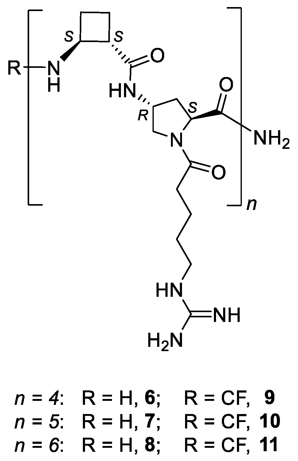
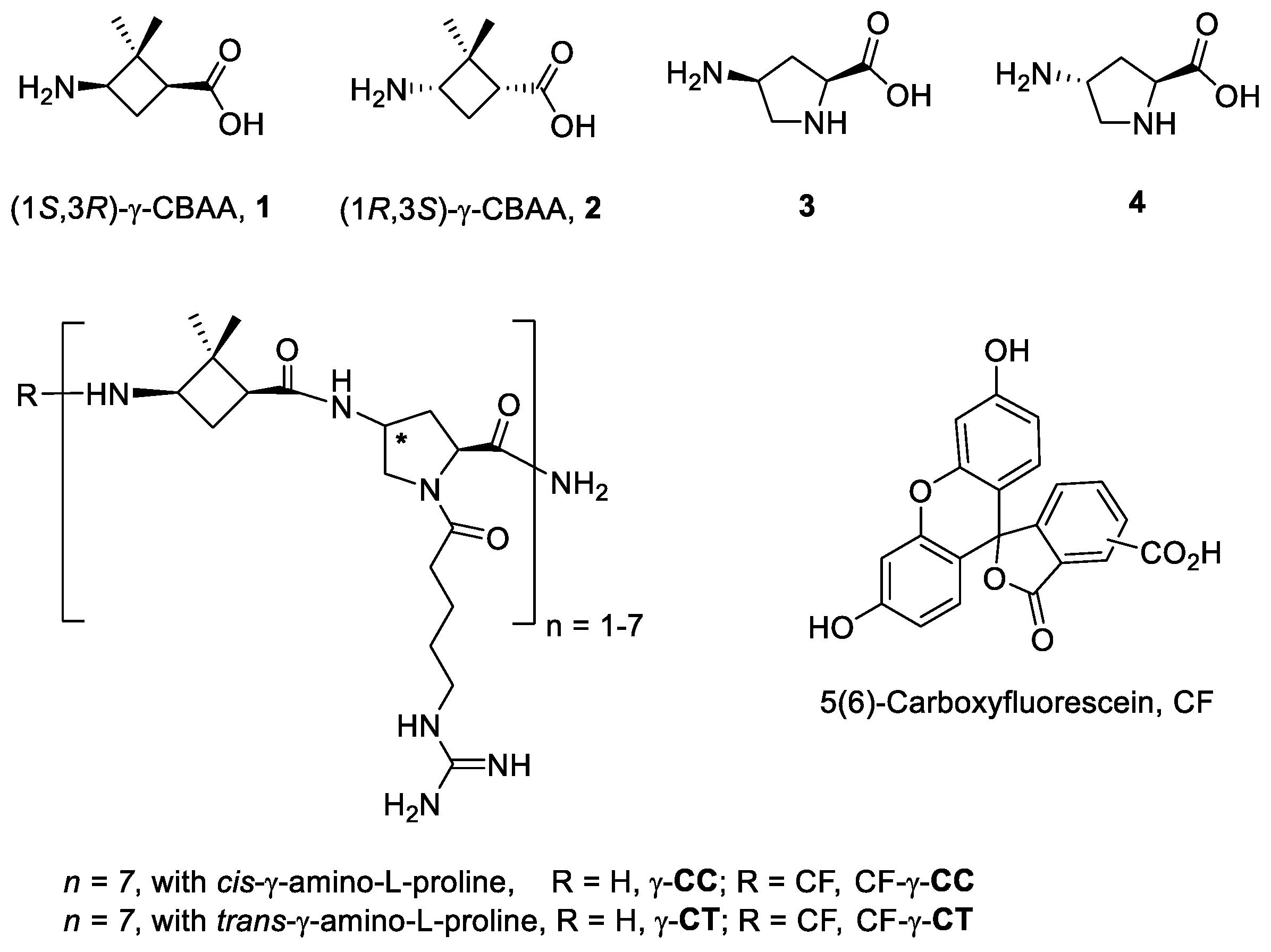
2.1. Synthesis of the Peptides

To analyze the role of peptide length on the cell uptake, a series of hybrid β,γ-peptides 6–11 (Figure 4, n = 4–6) was synthesized. They were formed by the repetition of a dipeptide motif made up by (1S,2S)-β-CBAA, 5 [34] and trans-γ-amino-l-proline, 4. All of them were prepared using standard protocols of solid phase synthesis methods (SPPS), either in their free N-terminus form (68) or with CF (911), as detailed in Section 4 and in the Supplementary Materials (Supplementary Tables S1–S3).
TAT48–57 (transactivator of transcription, TAT) [35,36,37] was also synthesized as a reference CPP, either with a free or carboxyfluoresceinated N-terminus as previously described [29].

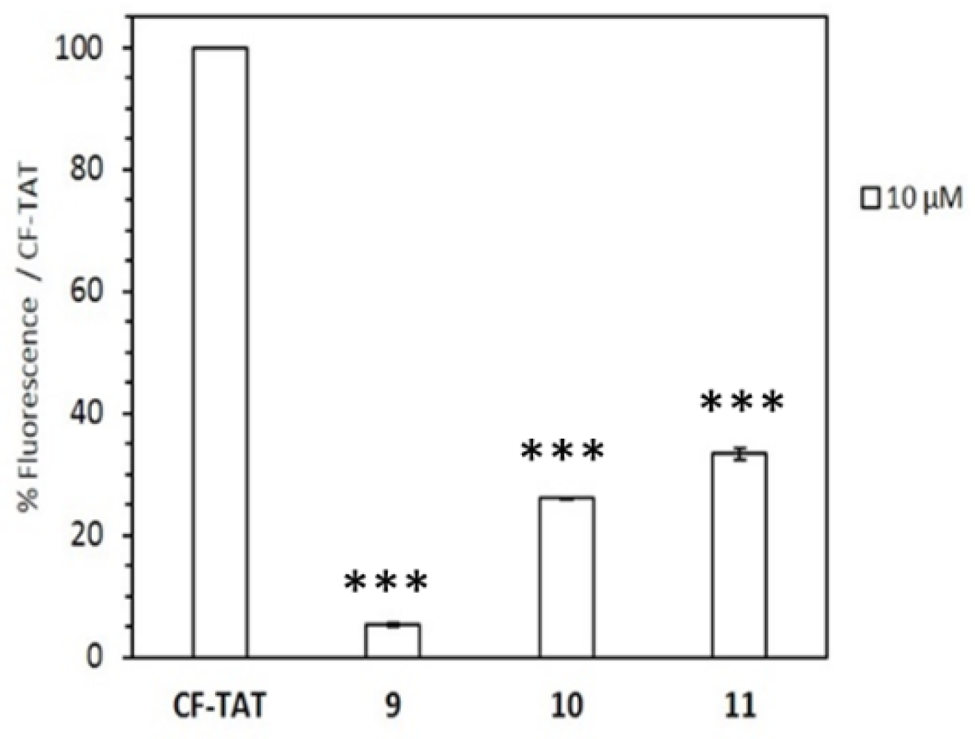
2.2. Cytotoxicity and Cellular Uptake in HeLa Cells

The cellular viability of peptides 611 was determined by the reduction of MTT (3-(4,5-dimethylthiazol-2-yl)-2,5-diphenyltetrazolium bromide) assay [38] after 24 h of cell incubation with the respective oligomer. The measure of the formazan formed from the reduction of MTT by mitochondrial dehydrogenases is directly related to the cellular viability (Figure 5). Even at the highest concentration assayed (50 µM), viability was over 90% for this set of peptides. Peptide toxicity was not dependent either on the number of guanidinium groups in the sequence or on the carboxyfluoresceination of the terminal amino group, in agreement with the previous data on their γ,γ-counterparts, γ-CC and γ-CT (Figure 1) [29].
Their cell internalization ability in HeLa cells was assessed by flow cytometry. To this end, the cell-associated fluorescence was quantified after incubation with peptides 911 at 10 and 25 µM and compared with that obtained with CF-TAT as a standard reference (Figure 6). Results showed a modest cell-uptake for all peptides, being the dodecamer the peptide with the highest value.
The intracellular uptake of the peptides was assessed by confocal microscopy. Figure 7 shows HeLa cells after 24 h incubation with the dodecamer 11 at 10 µM. CellMask deep red and Hoechst were used to stain the plasma membrane (red), and DNA (blue), respectively. The CF-peptide accounts for the green fluorescence.

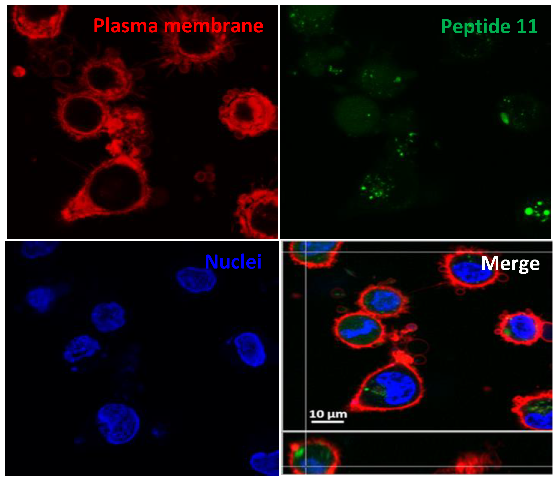
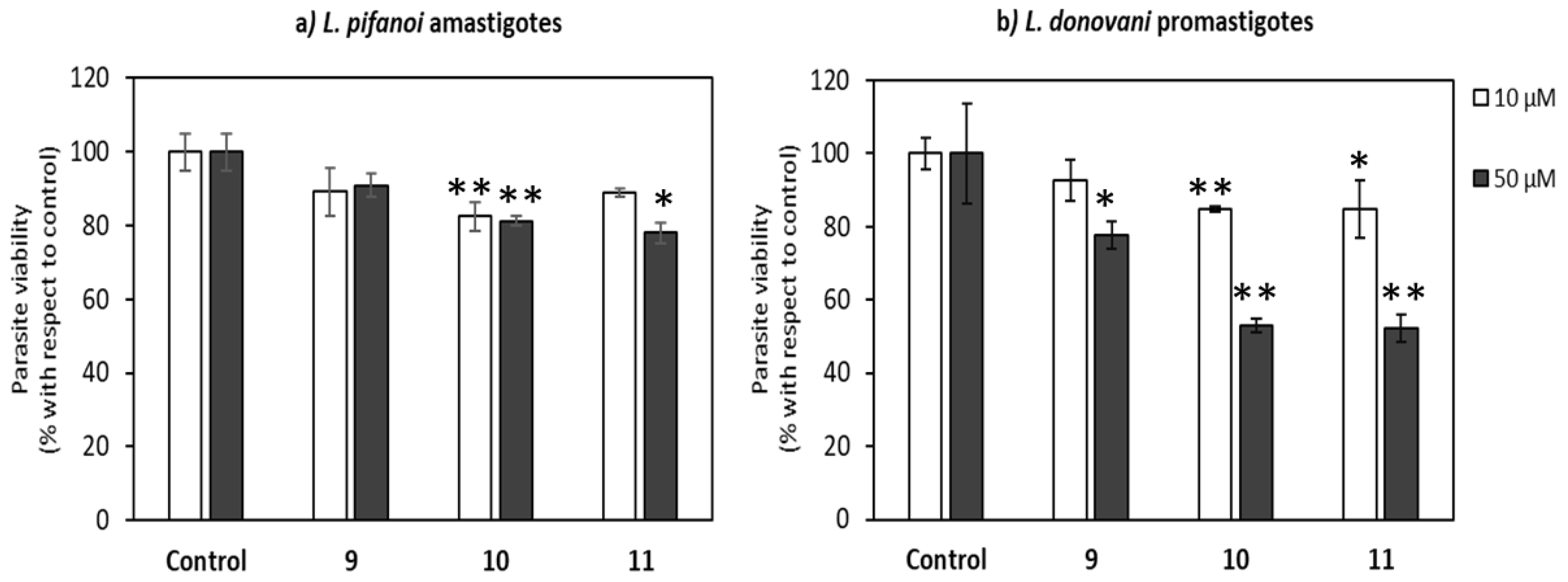
2.3. Uptake, Microbicidal Activity, and Intracellular Location of Peptides on Leishmania Parasites

Once the lack of cytotoxicity and poor uptake of the peptides respect to TAT on HeLa cells was evidenced, their performance on Leishmania parasites was approached. This is a challenging human protozoan parasite in reference to peptide membrane interaction. The cell membrane of Leishmania is reinforced by a longitudinal subpellicular layer of microtubules [39]. This confines all the membrane traffic to a small membrane area devoid of microtubules, the flagellar pocket [40]. In addition, it is endowed with a strong proteolytic armamentarium not only in the surface, but also in lysosomal enzymes, especially in the amastigote form of those species, as L. pifanoi, belonging to the mexicana complex [41,42]. To this end, peptides were assayed for viability, uptake and location on the protozoan Leishmania parasites.
The decrease on parasite viability was dependent both on the length of the peptide and on its concentration. The toxicity increases with the length of the peptide for both forms of the parasite, the dodecamer (11) and the decamer (10) being substantially more toxic than the octamer (9), likely due to the increase of the overall positive charge of the peptide [43]. This inhibitory effect was lower for L. pifanoi amastigotes, despite of their high content in cysteine proteinases with a broad substrate specificity, accounting for 5% of their total protein content. A similar trend was also observed for their γ, γ-counterparts, although with a higher quantitative effect [29].
Because of the nil inclusion of α-amino acids in the peptide composition, the divergence among Leishmania stages is not due to differences in their proteinase content, but likely mirrored differences in their membrane architecture. Almost 40% of the promastigote surface is covered with lipophosphoglycan formed by repetition of a phosphorylated disaccharide unit, whereas the amastigote hardly expresses a few hundred copies [44]. This oligosaccharide forms a strong anionic coating that likely acts as an initial anchor point for the peptides prior to their internalization.
To ascertain the role of length of the peptide in its uptake on L. donovani promastigotes, the set of β,γ-peptides were incubated for 4 h at a fixed concentration of 10 μM. The choice of these conditions relies both on the higher differences in cytotoxicity observed for promastigotes over amastigotes, as well as to the scarce toxicity of the peptides at this concentration, always less than <20% (Figure 8). According to Figure 9, the peptide uptake increased with its length although for all the β,γ-peptides, the uptake was consistently lower than that obtained for CF-TAT at identical concentration, used as a reference for CPP.
The internalization of these peptides in Leishmania donovani promastigotes was assessed by confocal microscopy. Thus, promastigotes were incubated with peptides 9–11 as well as TAT at a final concentration of 10 μM for 4 h at 26 °C. DAPI was used as a nucleic acid dye to stain the nucleus and the kinetoplast (blue fluorescence) (Figure 10).

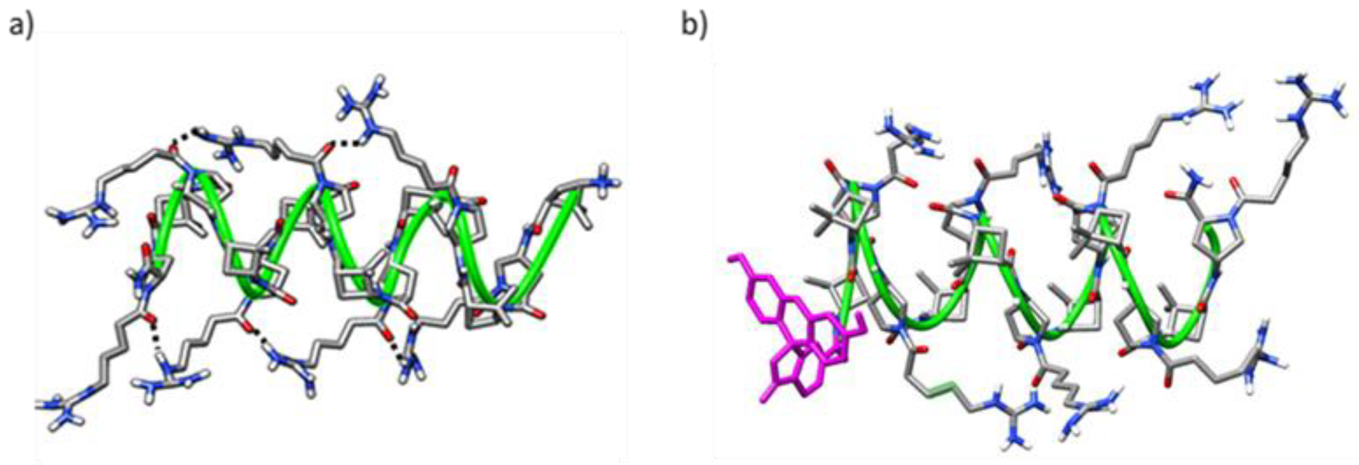
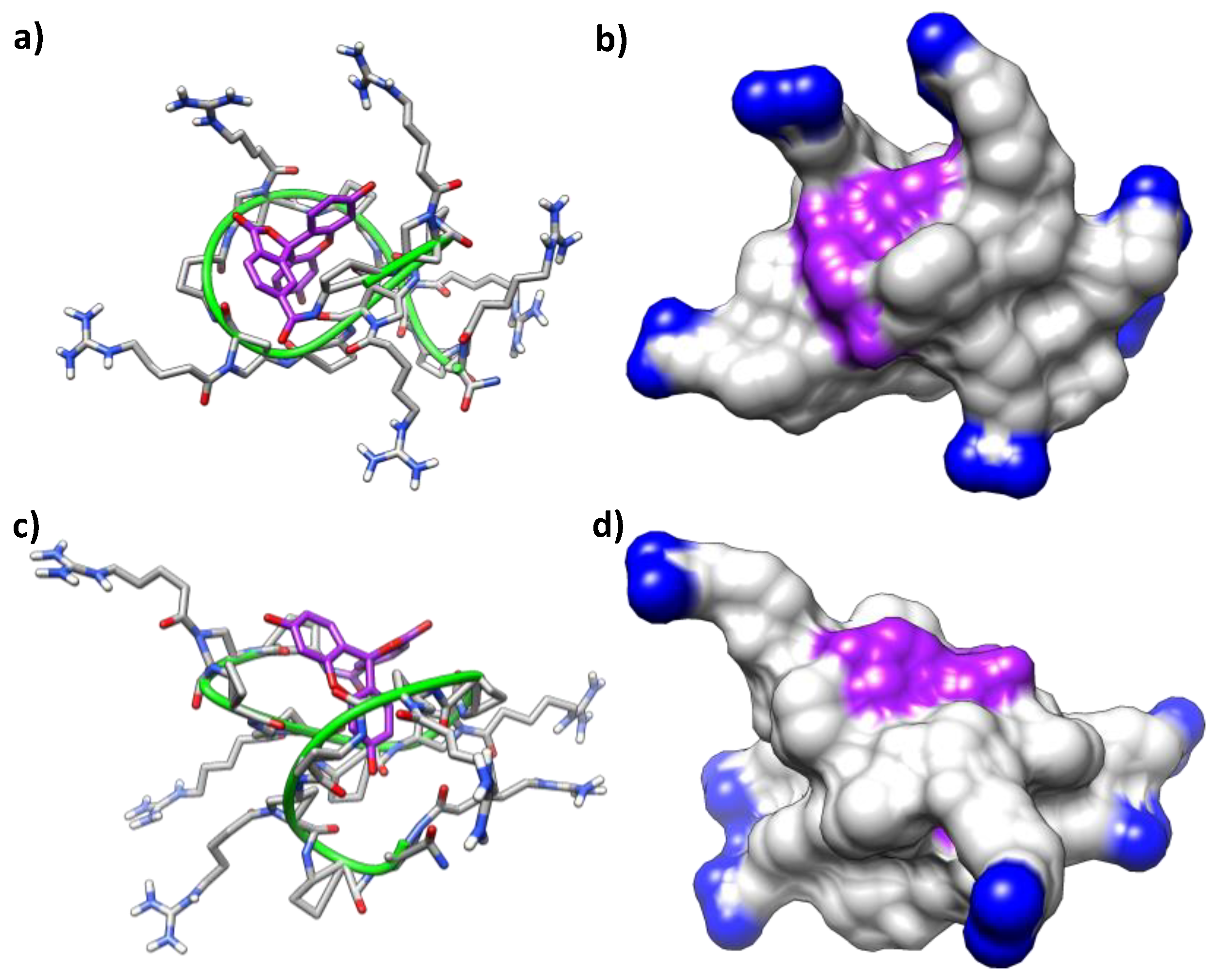
2.4. Molecular Modeling

To ascertain the folding of the peptides in aqueous solution as well as the influence of the fluorophore moiety (CF), molecular dynamics (MD) simulations under an explicit solvent scenario were carried out on the hybrid β,γ-peptide 8 and its CF-conjugate 11 (MD length of 900 ns and 600 ns, respectively). Both oligomers showed highly disorganized structures along the MD simulations, without any ordered secondary structure motif such as helical or β-sheet-like structures. Considering the backbone carbons of the peptides and a RMSD threshold of 2.0 Å, no cluster accounting for at least 1% of the simulation was found for the peptide 8, whereas the simulation of the CF-conjugate 11 led to twenty clusters containing, at least, 1% of the frames. Consequently, the incorporation of the CF induced a slight structural stabilization of the peptide. An inspection of the representative structures of the two most populated clusters (21% and 8% of the frames) supported the acquisition by the peptide of a globular shape where the peptide wrapped the CF moiety (Figure 11). This trend is observed in a series of MD clusters, but its high dynamics precluded a better identification of any stable interactions between the CF and the peptide.
As mentioned in the introduction, previous studies reported well-defined secondary structures for short oligomers made up exclusively with (1S,2S)-2-aminocyclobutane-1-carboxylic acid, 5, driven by the formation of eight-membered hydrogen-bonded rings (Figure 3) [30,31,32,33]. Strikingly, no stable hydrogen bonding patterns were found along the MD simulations, with a total of 721 and 456 different hydrogen bonds observed that appeared/disappeared throughout the trajectories of the peptide 8 and the CF-conjugate 11, respectively. None of them stands more than 15% of the time of simulation, suggesting that eight-membered stable rings are either absent, or their formation is quite transitory, precluding their observation. Another relevant factor involved in the cellular uptake of CPPs is the cationic character of the peptide, as it endorses a high affinity binding into the anionic proteoglycans exposed at the cell-surface, mandatory for their further internalization and endosomal escape [45,46,47]. A proper orientation of the cationic groups facing the solvent will speed up and strengthen this interaction. In this regard, most of the time the terminal nitrogen atoms of the guanidinium groups are exposed to the external aqueous medium according to MD simulations for peptide 8 and its CF-conjugate 11.
Apart from visual inspection of the representative structures (Figure 11), a solvent accessible surface area (SASA) analysis confirmed that the guanidinium groups of oligomers 8 and 11 have a good orientation toward the solvent, with average SASA values of 1.145 ± 0.084 nm2 and 1.160 ± 0.078 nm2, respectively (Table 1).
Otherwise, the influence of the spatial arrangement of positively charged amino acids of CPPs on their translocation profile is still controversial. First, a study on a series of arginine-rich CPPs [23] inferred that large inter-guanidinium distances, up to 15 Å, enhance uptake kinetics. However, a subsequent study suggested that shorter distances from 8 to 10 Å were necessary to match the topology of anionic groups in cell-surface glycans [20]. Indeed, despite the conformational flexibility of the lipophosphoglycan of Leishmania donovani, as a putative anchor for CPPs, a length of 160 Å was described for a structure formed by the extended conformation of the 16mer of the phosphorylated disaccharide, its repetitive forming unit. This accounts for an average separation of 10 Å [48].
In a recent study of ours on γ,γ-CPPs, MD simulations suggested inter-guanidinium distances in the range 8–10 Å both for the most active peptides (Figure 2) or their CF-conjugates [29]. Nevertheless, a similar analysis on 8 and 11 shows average inter-guanidium distances of 14.84 ± 4.46 Å and 16.42 ± 4.17 Å, respectively, not matching the reported as optimal distance among close guanidium groups. These observations offer a possible reason for the poorer activity of the current β,γ-CPPs since they are not prone to form the suitable charge distribution for efficient cell membrane translocation.

3. Conclusions

A new family of octa-, deca-, and dodecameric hybrid β,γ-peptides and their CF-conjugates were synthesized and evaluated as CPPs for toxicity and cell uptake ability. These peptides were made by a repetitive unit made of a chiral cyclobutane-containing trans-β-amino acid linked to a Nα-functionalized trans-γ-amino-l-proline. Their uptake and cytotoxicity on HeLa cells were not significant. In contrast, toxicity on Leishmania pifanoi amastigotes and especially on Leishmania donovani promastigotes was remarkable at 50 µM, with a toxicity ranking: 12-mer ≅ 10-mer >8-mer. Peptide internalization by Leishmania was practically nil on L. pifanoi amastigotes and modest on L. donovani promastigotes with a substantial increase with the length of the peptide. By MD simulations, dodecamer 8 and its CF-conjugate 11 are devoid of any conformational bias. These results differ from those reported for their γ,γ-homologues, with a cyclobutane-containing cis-γ-amino acid and a cis- or trans-γ-amino-l-proline as their structural units [29]. This γ,γ-CPP family adopted a well-defined conformation in solution, with modest uptake, and a toxicity on Leishmania parasites higher than the hybrid β, γ-peptides described herein.
The SASA analysis of the β,γ-dodecamers (8, and its CF-version, 11) confirmed that the guanidinium groups are exposed to the external aqueous medium for most of the time with an average inter-guanidinium distance of about 15 and 16 Å, respectively. This value did not fall into the interval 8–10 Å described as the optimal distance to match with the anionic groups of cell-surface glycans [20,48]. On the contrary, the γ,γ-counterparts, with a less constrained 1,3-disubstituted cyclobutane moiety, presented more stable conformations with inter-guanidium distances of 8–10 Å for the most active CPPs. Consequently, our results extended the notion of conformational restraint, previously described for α-peptides [49], to lineal CPPs by the inclusion of non-natural amino acids endowed with limited conformational freedom into their sequence. As a conclusion, the use of peptides with tuned conformational restriction, achieved by a rational incorporation of cyclobutane β- or γ-amino acids in their sequences, allows the exploration of the topology of guanidium groups and their intramolecular dynamics with a mobility limited by the stiffness of the peptide skeleton. Consequently, both stable peptide folding and convenient guanidinium structural patterns are reinforced as CPP key descriptors for an efficient cell-membrane translocation. As such, considering dynamical simulations as a part of future CPPs-engineering protocols appears promising.

4. Materials and Methods

The reagents and solvents were purchased from commercial sources and used without further purification. Unless otherwise stated, the reagents were purchased from Sigma Aldrich, Barcelona, Spain.

4.1. Synthesis of the β, γ-Peptides 6–11 and TAT48–57 Peptide

β,γ-Peptides were synthesized manually on solid-phase using Aminomethyl-ChemMatrix® resin (f = 0.74 mmol NH2/g resin). The resin was conditioned with successive washes with DCM (dichloromethane), TFA (trifluoroacetic acid)/DCM (4:5, v/v), DIPEA (N-diisopropylethylamine)/DCM (4:5, v/v), DMF (dimethylformamide) and DCM. The linker was coupled using 3:3:3 Fmoc-Rink amide-Linker/DIC/OxymaPure® (Fmoc = fluorenylmethoxycarbonyl, DIC = N,N′-Diisopropylcarbodiimide) in DMF for 1 h. β,γ-Peptides 6-11 backbone was elongated by alternate addition of (2S,4S)-4-Fmoc-amino-1-Alloc-pyrrolidin carboxylic acid (Alloc = Allyloxycarbonyl) and (1S,2S)-2-Fmoc-aminocyclobutane-1-carboxylic acid using DIC/OxymaPure® (3 equiv/3 equiv/3 equiv) as coupling reagents in DMF for 2 h. Fmoc group was released by treatment with piperidine-DMF (2:8, v/v). After each Fmoc elimination step, the peptidyl–resins were washed with DMF and DCM. The couplings were monitored by the Kaiser (ninhydrin) test. Once the peptide backbone syntheses were finished, Alloc groups were eliminated by catalytic reduction using 12/0.1 PhSiH3/Pd(PPh3)4 in DCM, and the guanidinium-containing side-chain introduced to the α-amine function of the Amp residues (Amp = 4-aminoproline). Then, the Fmoc group in N-terminal position was eliminated. At this point, the peptidyl-resin was split in two parts. One of these two fractions was conjugated to 5(6)-Carboxyfluorescein using CF/OxymaPure®/PyBOP/DIPEA (4:6:4:6) in DMF (PyBOP = benzotriazol-1-yloxytripyrrolidinophosphonium hexafluorophosphate). Finally, β, γ-peptides 911 were cleaved from the peptidyl-resins by treatment with TFA/H2O/TIS (95:2.5:2.5, v/v/v) (TIS = triisopropylsilane) for 3 h, precipitated in cold diethyl ether, dissolved in CH3CN/H2O (1:1, v/v) and lyophilized. The synthesis of TAT48–57 was performed by using standard Fmoc/tert-Bu SPPS methods as previously described [29]. See the Supplementary Materials for more details and Supplementary Tables S1–S3.

4.2. Peptide Purification

Crude peptides were purified by semipreparative RP-HPLC-UV-MS using a system composed by a binary gradient Waters 2545, a Waters Alliance 2767 sample manager module and an automatic fraction collector coupled to Waters 2487 dual UV-vis absorbance detector and an electrospray ion source (ESI-MS) Micromass ZQ mass spectrometer detector. The chromatographic separation of the peptides was achieved using a semipreparative column XBridge® Prep BEH C18 (19 × 100 mm, 5 µm) (Waters, Cerdanyola del Vallès, Spain) with dual solvent system formed by A: 0.1% TFA in H2O and B: 0.1% TFA in CH3CN at a flow rate of 25 mL/min. λ = 220 nm. Elution gradient was specific for each peptide.

4.3. Peptide Characterization

Peptide analysis and determination of the respective retention time (tR), analytical column XBridge™ BEH 130 C18 (4.6 × 100 mm, 3.5 μM) (Waters, Cerdanyola del Vallès, Spain) was used. Peptide elution was monitored at 220 nm.

4.3.1. Octameric Peptide (6)

Purification: RP-HPLC-UV-MS gradient: 5 to 40% B in 10 min; 40 to 60% B in 1 min, and to 100% B in 2 min. Characterization: RP-HPLC. Purity >95%, tR 3.4 min. m/z (ESI): Ms Calcd for C64H107N25O12 [M + 3H/3]+: 474.6; Experimental: 473.2. Ms Calcd para C64H107N25O12 [(M + 4H)/4]+: 355.6; Experimental: 355.7. Ms Calcd for C64H107N25O12 [(M + 5H)/5]+: 284.7; Experimental: 284.7. MALDI-TOF: Ms Calcd for C64H107N25O12 [(M + H)]+: 1419.72; Experimental: 1419.02. Ms Calcd para C64H107N25O12 [M + Na]+: 1457.82; Experimental: 1457.00.

4.3.2. Decameric Peptide (7)

Purification: RP-HPLC-UV-MS gradient: 5 to 40% B in 10 min; 40 to 60% B in 1 min, and to 100% B in 2 min. Characterization: RP-HPLC. Purity >98%, tR 3.4 min. m/z (ESI): Ms Calcd for C80H133N31O15 [(M + 3H)/3]+: 590.7; Experimental: 590.7. Ms Calcd for C80H133N31O15 [(M + 4H)/4]+: 445.2; Experimental: 445.3. Ms Calcd for C80H133N31O15 [(M + 5H)/5]+: 354.8; Experimental: 354.7. MALDI-TOF: Ms Calcd for C80H133N31O15 [M + H]+: 1769.06; Experimental: 1769.22.

4.3.3. Dodecameric Peptide (8)

Purification: RP-HPLC-UV-MS gradient: 10 to 60% B in 11 min, and to 100% B in 2 min. Characterization: RP-HPLC. Purity >99%, tR 3.5 min. m/z (ESI): Ms Calcd for C96H159N37O18 [(M + 4H)/4]+: 530.8; Experimental: 530.9. Ms Calcd for C96H159N37O18 [(M + 5H)/5] +: 424.9; Experimental: 424.8. MALDI-TOF: Ms calcd for C96H159N37O18 [M + H]+: 2119.27; Experimental: 2119.47. Ms Calcd for C96H159N37O18 [M + Na]+: 2141.26; Experimental: 2141.40.

4.3.4. Octameric CF-Peptide (9)

Purification: RP-HPLC-UV-MS gradient: 5 to 40% B in 10 min; 40 to 60% B in 1 min, and to 100% B in 2 min. Characterization: RP-HPLC. Purity >99%, tR 5.9 min. m/z (ESI): Ms Calcd for C85H117N25O18 [(M + 2H)/2]+: 889.5; Experimental: 889.3. Ms Calcd for C85H117N25O18 [(M + 3H)/3]+: 593.3; Experimental: 593.7. Ms Calcd para C85H117N25O18 [(M + 4H)/4]+: 445.2; Experimental: 445.1. Ms Calcd for C85H117N25O18 [(M + 5H)/5]+: 356.4; Experimental: 356.3. MALDI-TOF: Ms calcd for C85H117N25O18 [(M + 3H)]+: 1778.03; Experimental: 1778.20. Ms Calcd for C85H117N25O18 [M + Na]+: 1800.02; Experimental: 1799.22.

4.3.5. Decameric CF-Peptide (10)

Purification: RP-HPLC-UV-MS gradient: 5 to 50% B in 10 min; 50 to 60% B in 1 min, and to 100% B in 2 min. Characterization: RP-HPLC. Purity >99%, tR 5.5 min. m/z (ESI): Ms Calcd for C101H143N31O21 [(M + 3H)/3]+: 710.1; Experimental: 710.4. Ms Calcd for C101H143N31O21 [(M + 4H)/4]+: 532.8; Experimental: 532.8. Ms Calcd for C101H143N31O21 [(M + 5H)/5]+: 426.4; Experimental: 426.4. MALDI-TOF: Ms Calcd for C101H143N31O21 [M + H]+: 2127.11; Experimental: 2127.32.

4.3.6. Dodecameric CF-Peptide (11)

Purification: RP-HPLC-UV-MS gradient: 10 to 60% B in 11 min, and to 100% B in 2 min. Characterization: RP-HPLC. Purity >99%, tR 4.8 min. m/z (ESI): Ms Calcd for C117H169N37O24 [(M + 3H)/3]+: 826.9; Experimental: 826.5. Ms Calcd for C117H169N37O24 [(M + 4H)/4]+: 620.4; Experimental: 620.3. Ms Calcd for C117H169N37O24 [(M + 5H)/5]+: 496.5; Experimental: 496.5. MALDI-TOF: Ms Calcd for C117H169N37O24 [M + H]+: 2477.31; Experimental: 2477.46.

4.4. Cellular Viability, Internalization, and Localization Experiments with HeLa Cells

The HeLa cell line, derived from a human cervical cancer, was used to perform the biological assays. CPP dissolved in non-supplemented Minimum Essential Medium (MEM) (Gibco, Thermo Fisher Scientific, Cornellà de Llobregat, Spain) at their final concentration were sterilized by filtration through a Whatman® Puradisc 0.2 µm polycarbonate filter.

4.4.1. HeLa Cells Culture

The cells were cultured in 25 cm2 flasks in MEM culture media supplemented with 10% of heat inactivated fetal bovine serum (FBS, Gibco) plus 2 mM L-glutamine (Biowest, Labclinics, Barcelona, Spain) at 37 °C, saturated humidity and 5% of CO2 (standard conditions).

4.4.2. Cellular Viability

The MTT assay was used to assess the cytotoxicity of the peptides. This method is based on the ability of living cells to reduce the MTT to formazan salts by mitochondrial reductases. HeLa cells were seeded into 24 microwell plates at 6 × 10⁴ cells/mL (30,000 cells/well). After 24 h of incubation, the medium was replaced with fresh medium with the respective peptide concentration and incubated for 24 h. Then, the cells were washed three times with Hanks buffered saline solution (HBSS) (Biowest, Labclinics, Barcelona, Spain), and 500 µL of 0.1 mg/mL MTT in HBSS were added to the cell suspension and incubated for 3 h at 37 °C in darkness. The cell layer was dried in darkness, and the resulting formazan solubilized in pure DMSO. The absorbance was measured at 540 nm in an X3 Multilabel Plate Reader coupled to Perkin Elmer 2030 Manager control software. At least three independent experiments with four different replicates of each peptide and concentration were performed. Controls, non-treated cells, or cells incubated with CF were included at identical concentrations. The absorbance of non-treated cells was taken as 100% cellular viability.

4.4.3. Peptide Internalization

For flow cytometry experiments, HeLa cells were seeded into 35 mm culture dishes (2 × 10⁵ cells/dish). After 24 h incubation under standard conditions, culture medium was removed, and cells were incubated for 2 h with the corresponding peptides at 10 and 25 μM. Next, the culture medium was removed, the cells were washed twice with HBSS and then trypsinized with 0.5 mL of 0.25% trypsin-EDTA (Gibco, Thermo Fisher Scientific, Cornellà de Llobregat, Spain). After 5 min incubation at 37 °C, 2 mL of MEM + 10% FCS was added to the cells to stop trypsinization and the mixture was centrifuged (5 min, 300× g). The cells were then additionally washed with 2 mL of HBSS under the same conditions. Finally, the cell pellet was resuspended in 200 μL of PBS at pH = 6.0 to detach any peptide adhering to the plasma membrane. To exclude dead cells from gating, 5 µg/mL propidium iodide (PI, Sigma-Aldrich, Barcelona, Spain) was added to the cells immediately before the flow cytometric analysis, carried out in a BD FACSCanto cytometer (Bio-Rad, Alcobendas, Spain) coupled to FACSDiva v.7.0 software using 488 nm and 635 nm lasers to excite the peptides and PI, respectively.
A total of 10,000 single cells were analyzed per sample, and at least three independent experiments were performed with each peptide and concentration. Untreated cells (autofluorescence control) and cell cultures incubated with TAT as positive reference CPP, and CF as negative reference, were included in each experiment. The fluorescence intensity of cells treated with CF was taken as the arbitrary unit for normalization. For confocal microscopy, HeLa cells were seeded into glass bottom culture dishes (MatTek, Bratislava, Slovakia) at a density of 2 × 10⁵ cell/dish. After 24 h of incubation, the culture medium was removed, and the cells were incubated for 2 h in the presence of peptides at 25 µM. Then, cells were rinsed three times with PBS, and nuclei and plasma membrane were counterstained with 1 µL/mL of Hoechst 33,342 (10 mg/mL, Thermo Fisher, Scientific, Cornellà de Llobregat, Spain) and 1 µL/mL CellMask™ deep red plasma membrane stain (5 mg/mL, Thermo Fisher Scientific, Cornellà de Llobregat, Spain), respectively. Finally, the cells were washed with PBS prior to be resuspended in PBS pH = 6.0. The experiments were performed using an Olympus Fluoview FV1000 confocal laser scanning microscope (Olympus Iberia, Hospitalet de Llobregat, Spain) equipped with Olympus Fluoview as control software. The excitation wavelengths used were 405, 488, and 658 nm to visualize the nuclei, the peptides and the plasma membrane, respectively; the wavelength of emission was 460, 510 and 690 nm, respectively. A 3D reconstruction was generated to obtain orthogonal projections using the ImageJ/fiji software.

4.5. Cellular Viability, Internalization, and Localization Experiments with Leishmania Parasites

The experiments were carried out with Leishmania donovani promastigotes (MHOM/SI/00/1S-2D strain) and Leishmania pifanoi amastigotes (MHOM/VE/60/Ltrod strain), respectively. Promastigotes were grown at 26 °C in RPMI medium supplemented with 10% of FCS, 2 mM l-glutamine, 20 U/mL unicilin 20 plus 48 µg/mL gentamicin.

4.5.1. Cellular Viability

The viability assays were carried out using the reduction of MTT as described. The parasites were aliquoted into 96-microwell plates at a final concentration of 20 × 106 cells/mL in HBSS supplemented with 10 mM d-glucose (Glc). The peptides were incubated for 4 h at the corresponding peptide concentration at 26 °C or 32 °C for promastigotes and axenic amastigotes, respectively. Afterwards, 0.5 mg/mL MTT in HBSS + 10 mM Glc was added and the cells were incubated for two additional hours. The resulting formazan was solubilized with DMSO (1% final concentration) and read at 595 nm in a Bio-Rad 640 microplate reader.

4.5.2. Peptide Uptake for Leishmania donovani Promastigotes

The parasites were resuspended in HBSS + Glc and dispensed in 24-microwell plates (2 mL/well) at a final concentration of 20 × 106 cells/mL. After incubation with the peptides for 4 h at 26 °C, the parasites were washed twice with 2 mL of HBSS + Glc plus 1% fatty-acid free bovine seroalbumin in order to remove the non-internalized peptides and resuspended in the same medium at 1 × 10⁶ cells/mL. Propidium iodide (PI) at a final concentration of 5 µg/mL was added immediately to the flow cytometric analysis to gate the viable cells exclusively. Flow cytometry was carried out in a FC500 flow cytometer, using λEXC = 488 nm and λEM = 525 nm for fluoresceinated peptides and λEXC = 488 nm.

4.5.3. Intracellular Localization of the Internalized Peptides in Leishmania donovani Promastigotes

The parasites (2 × 10⁶ cells/mL in HBSS + Glc, 200 μL) were incubated with the corresponding peptides (2 h, 26 °C). Afterwards, they were collected from the well and washed twice with 2 mL of HBSS + Glc plus 1% bovine seroalbumin fatty-acid free. Prior to carrying out confocal microscopy, the parasites were incubated with 10 µL/mL of DAPI (4′,6-diamidine-2′-phenylindole dihydrochloride) for 10 min at 26 °C and washed twice with 2 mL of HBSS + Glc. Living parasites were observed using a Leica SP2 confocal microscope (Leica Microsistemas, S.A., Barcelona, Spain) at λEXC = 358 nm/λEM = 461 nm.

4.5.4. Statistical Treatment

Statistical significance was inferred with t-test, using the statistics platform of the SigmaPlot v.11.0 software: ((p ≤ 0.05, (*); p ≤ 0.01, (**); p ≤ 0.001, (***)).

4.6. Computational Details

Geometries without steric clashes of the hybrid β, γ-peptides 8 and 11 were obtained through RDKit ETKDGv3 conformer generation [50], taking the conformer with lowest energy as the initial structure for the MD simulations. Atomic charges were computed with the restrained electrostatic potential (RESP) protocol [51]. The atom types and force field parameters were assigned through antechamber and parmchk2 tools of the AmberTools20 package [52]. The peptides were solvated with a cubic box of TIP3P water molecules and the positive charges of the guanidinium groups were neutralized with six chloride ions in each case, employing the ions94 library. The GAFF force field [53] was used for all atoms of the peptides. MD simulations were carried out under periodic boundary conditions with the OpenMM engine [54] using the OMMProtocol application [55].
The convergence of the trajectories was assessed by RMSD, all-to-all RMSD, PCA and cluster counting analyses [56], taking the exploration of the conformational space defined by the twelve backbone carbons of the peptides (see Supplementary Materials, Table S4 and Figures S1 and S2 for further details). In particular, the simulation of the β, γ-peptide 8 was extended up to 900 ns, whereas the β, γ-peptide 11 simulation was of 600 ns length. UCSF Chimera [57] was used to visually analyze the trajectories. Pytraj from the AmberTools20 package [53] was used for the hydrogen bond detection and guanidinium distance calculation. Quality threshold clustering [58], with a threshold of 2.0 Å and a selection of the alpha carbons, was used to generate the clusters and the representative structures. The SASA values of the guanidinium groups (carbon, two terminal nitrogen atoms, and their corresponding hydrogen atoms) were calculated with the MDTraj [59] implementation of the Shrake and Rupley’s algorithm.

Supplementary Materials

The following are available online at https://www.mdpi.com/article/10.3390/ijms22105092/s1, Synthesis of the monomers for solid phase peptide synthesis (SPPS); NMR spectra of the new monomers; SPPS procedures; HPLC chromatograms and MS spectra of the purified peptides 611; convergence of Molecular Dynamics simulations; Table S1. General protocol for the synthesis of the peptides. Table S2. General protocol for the derivatization of the α-amine function using the solid phase synthesis. Table S3. General protocol for the incorporation of the 5(6)-carboxyfluorescein. Table S4: Specifications of the system for the MD simulations; Figure S1: Convergence studies for the MD simulation of peptide 8. (a) RMSD against the first frame of the trajectory. (b) Principal component analysis. Plot of the first principal component against the second principal component along the MD. (c) Cluster counting along the MD with a cluster cutoff of 5.0 Å. (d) All-to-all frames RMSD along the MD; Figure S2: Convergence studies for the MD simulation of CF-conjugate 11. (a) RMSD against the first frame of the trajectory. (b) Principal component analysis. Plot of the first principal component against the second principal component along the MD. (c) Cluster counting along the MD with a cluster cutoff of 5.0 Å. (d) All-to-all frames RMSD along the MD.

Author Contributions

Conceptualization, O.I., J.-D.M., M.R. and R.M.O.; Funding acquisition, O.I., C.N., L.R., J.-D.M., M.R. and R.M.O.; Investigation, J.O., J.-E.S.-A., X.P., D.C., M.Á.A., N.G., L.R. and J.-D.M.; Methodology, C.N., L.R., J.-D.M. and M.R.; Supervision, O.I., J.-D.M., M.R. and R.M.O.; Writing—Original draft, R.M.O.; Writing—Review & editing, O.I., J.-E.S.-A., C.N., L.R., J.-D.M., M.R. and R.M.O. All authors have given approval to the final version of the manuscript. All authors have read and agreed to the published version of the manuscript.

Funding

This research was funded by Spanish Ministry of Economy, Industry and Competitivity (Grants CTQ2016-77978-R, MAT2017-86357-C3-3-R, SAF2014-60138-R, and RTI2018-093831-B-I00), Generalitat de Catalunya (2017-SGR-503 and 2017-SGR-1439), Subdirección General de Redes y Centros de Investigación Cooperativa-FEDER (RICET RD16/0027/0010) and CSIC (PIE 201620E038) and CIBER BBN (CB/06/01/0074).

Institutional Review Board Statement

Not applicable.

Informed Consent Statement

Not applicable.

Data Availability Statement

The data presented in this study are available on request from the corresponding author. The data are not publicly available due to their use in further studies.

Acknowledgments

The authors would like to thank the staff at the Servei de Microscòpia de la Universitat Autònoma de Barcelona and the Peptide Synthesis Unit of the NANBIOSIS ICTS (U3, CIBER BBN-IQAC-CSIC) for technical assistance.

Conflicts of Interest

The authors declare no conflict of interest. The funders had no role in the design of the study; in the collection, analyses, or interpretation of data; in the writing of the manuscript, or in the decision to publish the results.

Abbreviations

ACNAcetonitrile
AllocAllyloxycarbonyl
Amp4-Aminoproline
CBAACyclobutane Amino Acid
CF5(6)-Carboxyfluorescein
CPPCell Penetrating Peptide
DAPI4′,6-Diamidino-2-phenylindole
DCMDichloromethane
DDSDrug Delivery Systems
DFTDensity Functional Theory
DICN,N′-Diisopropylcarbodiimide
DIPEAN-Diisopropylethylamine
DMFDimethylformamide
DMSODimethyl Sulfoxide
EDTAEthylenediaminetetraacetic Acid
ESIElectrospray Ionization
FBSFetal Bovine Serum
FmocFluorenylmethyloxycarbonyl
GlcGlucose
HBSSHanks Buffered Saline Solution
HPLCHigh-Performance Liquid Chromatography
LPGLypophosphoglycan
MALDIMatrix Assisted Laser Desorption Ionization
MSMass Spectroscopy
MDMolecular Dynamics
MEMMinimum Essential Medium
MTT3-(4,5-Dimethylthiazol-2-yl)-2,5-diphenyltetrazolium Bromide
NMRNuclear Magnetic Resonance
PBSPhosphate-Buffered Saline
PCAPrincipal Component Analysis
PESPotential Energy Surface
PIPropidium Iodide
PyBOP(7-Azabenzotriazol-1-yloxy)-tripyrrolidinophosphonium Hexafluorophosphate
RESPRestrained Electrostatic Potential
RMSDRoot-Mean-Square Deviation
RMSFRoot-Mean-Square Fluctuation
RPReverse Phase
SASASolvent Accessible Surface Area
SDStandard Deviation
SMSupplementary Materials
SPPSSolid Phase Peptide Synthesis
TISTriisopropylsilane
TOFTime of Flight
UVUltraviolet Spectroscopy

References

  1. Lindgren, M.; Hällbrink, M.; Prochiantz, A.; Langel, Ü. Cell-penetrating Peptides. Trends Pharmacol. Sci. 2000, 21, 99–103. [Google Scholar] [CrossRef]
  2. Langel, Ü. Cell-Penetrating Peptides in Processes and Applications; CRC Press Pharmacology and Toxicology Series; CRC Press: Boca Raton, FL, USA, 2002. [Google Scholar] [CrossRef]
  3. Lundberg, P.; Langel, Ü. A brief introduction to cell-penetrating peptides. J. Mol. Recognit. 2003, 16, 227–233. [Google Scholar] [CrossRef]
  4. Kim, G.C.; Cheon, D.H.; Lee, Y. Challenge to overcome current limitations of cell-penetrating peptides. Biochim. Biophys. Acta Proteins Proteom. 2021, 1869, 140604. [Google Scholar] [CrossRef]
  5. Wu, J.; Li, J.; Wang, H.; Liu, C.B. Mitochondrial-targeted penetrating peptide delivery for cancer therapy. Expert Opin. Drug Deliv. 2021, 15, 951–964. [Google Scholar] [CrossRef] [PubMed]
  6. Vivès, E.; Schmidt, J.; Pèlegrin, A. Cell-penetrating and cell-targeting peptides in drug delivery. Biochim. Biophys. Acta 2008, 1786, 126–138. [Google Scholar] [CrossRef] [PubMed] [Green Version]
  7. Koren, E.; Torchillin, V.P. Cell-penetrating peptides: Breaking through to the other side. Trends Mol. Med. 2012, 18, 385–393. [Google Scholar] [CrossRef]
  8. Copolovici, D.M.; Langel, K.; Eriste, E.; Langel, Ü. Cell-Penetrating Peptides: Design, Synthesis, and Applications. ACS Nano 2014, 8, 1972–1994. [Google Scholar] [CrossRef] [PubMed]
  9. Zhang, D.; Wang, J.; Xu, D. Cell penetrating peptides as noninvasive transmembrane vectors for the development of new functional drug delivery systems. J. Control. Release 2016, 229, 130–139. [Google Scholar] [CrossRef] [PubMed] [Green Version]
  10. Dissanayake, S.; Denny, W.A.; Gamage, S.; Sarojini, V. Recent developments in anticancer drug delivery using cell penetrating and tumor targeting peptides. J. Control. Release 2017, 250, 62–76. [Google Scholar] [CrossRef] [PubMed]
  11. Fominaya, J.; Bravo, J.; Rebollo, A. Strategies to stabilize cell penetrating peptides for in vivo applications. Ther. Deliv. 2015, 6, 1171–1194. [Google Scholar] [CrossRef]
  12. Berlicki, Ł.; Kaske, M.; Gutiérrez-Abad, R.; Bernhardt, G.; Illa, O.; Ortuño, R.M.; Cabrele, C.; Buschauer, A.; Reiser, O. Replacement of Th32 and Gln34 in the C-terminal neuropeptide Y fragment 25-36 by cis-cyclobutane- and cis-cyclopentane-amino acids shifts selectivity toward the Y4 receptor. J. Med. Chem. 2013, 56, 8422–8431. [Google Scholar] [CrossRef]
  13. Hsieh, C.L.; Maynard, J.A.; Schaub, J.M.; DiVenere, A.M.; Kuo, H.C.; Javanmardi, K.; Le, K.C.; Wrapp, D.; Lee, A.G.; Liu, Y.; et al. Structure-based design of prefusion-stabilized SARS-CoV-2 spikes. Science 2020, 369, 1501–1505. [Google Scholar] [CrossRef] [PubMed]
  14. Feliciani, F.; Pinnen, F.; Stefanuccia, A.; Costante, R.; Cacciatore, I.; Lucente, G.; Mollica, A. Structure-Activity Relationships of Biphalin Analogs and their Biological Evaluation on Opioid Receptors. Mini-Rev. Med. Chem. 2013, 13, 11–33. [Google Scholar] [CrossRef]
  15. Mollica, A.; Costante, R.; Stefanucci, A.; Pinnen, F.; Lucente, G.; Fidanzad, S.; Pieretti, S. Antinociceptive profile of potent opioid peptide AM94, a fluorinated analogue of biphalin with non-hydrazine linker. J. Pept. Sci. 2013, 19, 233–239. [Google Scholar] [CrossRef] [PubMed]
  16. Stefanucci, A.; Pinnen, F.; Feliciani, F.; Cacciatore, I.; Lucente, G.; Mollica, A. Conformationally Constrained Histidines in the Design of Peptidomimetics: Strategies for the χ-Space Control. Int. J. Mol. Sci. 2011, 12, 2853–2890. [Google Scholar] [CrossRef] [PubMed] [Green Version]
  17. Pujals, S.; Giralt, E. Proline-rich, amphipathic cell-penetrating peptides. Adv. Drug Deliv. Rev. 2008, 60, 473–484. [Google Scholar] [CrossRef]
  18. Dobitz, S.; Aronoff, M.R.; Wennemers, H. Oligoprolines as molecular entities for controlling distance in biological and material sciences. Acc. Chem. Res. 2017, 50, 2420–2428. [Google Scholar] [CrossRef] [PubMed]
  19. Potocky, T.B.; Menon, A.K.; Gellman, S.H. Effects of conformational stability and geometry of guanidinium display on cell entry by β-peptides. J. Am. Chem. Soc. 2005, 127, 3686–3687. [Google Scholar] [CrossRef]
  20. Nagel, Y.A.; Raschle, P.S.; Wennemers, H. Effect of preorganized charge-display on the cell-penetrating properties of cationic peptides. Angew. Chem. Int. Ed. 2017, 56, 122–126. [Google Scholar] [CrossRef] [PubMed]
  21. Tian, Y.; Zeng, X.; Li, J.; Jiang, Y.; Zhao, H.; Wang, D.; Huang, X.; Li, Z. Achieving enhanced cell penetration of short conformationally constrained peptides through amphiphilicity tuning. Chem. Sci. 2017, 8, 7576–7581. [Google Scholar] [CrossRef] [Green Version]
  22. Nischan, N.; Herce, H.D.; Natale, F.; Bohlke, N.; Budisa, N.; Cardoso, M.C.; Hackenberger, C.P.R. Covalent attachment of cyclic TAT peptides to GFP results in protein delivery into live cells with immediate bioavailability. Angew. Chem. Int. Ed. 2015, 54, 1950–1953. [Google Scholar] [CrossRef]
  23. Lättig-Tünnemann, G.; Prinz, M.; Hoffmann, D.; Behlke, J.; Palm-Apergi, C.; Morano, I.; Herce, H.D.; Cardoso, M.C. Backbone rigidity and static presentation of guanidinium groups increases cellular uptake of arginine-rich cell-penetrating peptides. Nat. Commun. 2011, 2, 453. [Google Scholar] [CrossRef] [PubMed]
  24. Qian, Z.; Martyna, A.; Hard, R.L.; Wang, J.; Appiah-Kubi, G.; Coss, C.; Phelps, M.A.; Rossman, J.S.; Pei, D. Discovery and mechanism of highly efficient cyclic cell-penetrating peptides. Biochemistry 2016, 55, 2601–2612. [Google Scholar] [CrossRef]
  25. Dougherty, P.G.; Sahni, A.; Pei, D. Understanding Cell Penetration of Cyclic Peptides. Chem. Rev. 2019, 119, 10241–10287. [Google Scholar] [CrossRef] [PubMed]
  26. Gutiérrez-Abad, R.; Carbajo, D.; Nolis, P.; Acosta-Silva, C.; Cobos, J.A.; Illa, O.; Royo, M.; Ortuño, R.M. Synthesis and structural study of highly constrained hybrid cyclobutane-proline γ,γ-peptides. Amino Acids 2011, 41, 673–686. [Google Scholar] [CrossRef] [PubMed]
  27. Gorrea, E.; Carbajo, D.; Gutiérrez-Abad, R.; Illa, O.; Branchadell, V.; Royo, M.; Ortuño, R.M. Searching for new cell-penetrating agents: Hybrid cyclobutane–proline γ,γ-peptides. Org. Biomol. Chem. 2012, 10, 4050–4057. [Google Scholar] [CrossRef]
  28. Farrera-Sinfreu, J.; Giralt, E.; Castel, S.; Albericio, F.; Royo, M. Cell-penetrating cis-γ-amino-L-proline-derived peptides. J. Am. Chem. Soc. 2005, 127, 9459–9468. [Google Scholar] [CrossRef] [PubMed]
  29. Illa, O.; Olivares, J.A.; Gaztelumendi, N.; Martínez-Castro, L.; Ospina, J.; Abengozar, M.Á.; Sciortino, G.; Maréchal, J.D.; Nogués, C.; Royo, M.; et al. Chiral cyclobutane-containing cell-penetrating peptides as selective vectors for anti-Leishmania drug delivery systems. Int. J. Mol. Sci. 2020, 21, 7502. [Google Scholar] [CrossRef] [PubMed]
  30. Torres, E.; Gorrea, E.; Da Silva, E.; Nolis, P.; Branchadell, V.; Ortuño, R.M. Prevalence of eight-membered hydrogen-bonded rings in some bis(cyclobutane)-dipeptides including residues with trans stereochemistry. Org. Lett. 2009, 11, 2301–2304. [Google Scholar] [CrossRef]
  31. Fernandes, C.; Faure, S.; Pereira, E.; Théry, V.; Declerck, V.; Guillot, R.; Aitken, D.J. 12-Helix folding of cyclobutane β-amino acid oligomers. Org. Lett. 2010, 12, 3606–3609. [Google Scholar] [CrossRef]
  32. Gorrea, E.; Pohl, G.; Nolis, P.; Celis, S.; Burusco, K.; Branchadell, V.; Perczel, A.; Ortuño, R.M. Secondary structure of short β-peptides as the chiral expression of monomeric building units: A rational and predictive model. J. Org. Chem. 2012, 77, 9795–9806. [Google Scholar] [CrossRef] [PubMed]
  33. Illa, O.; Olivares, J.A.; Nolis, P.; Ortuño, R.M. The relevance of the relative configuration in the folding of hybrid peptides containing β-cyclobutane amino acids and γ-amino-L-proline residues. Tetrahedron 2017, 73, 6286–6295. [Google Scholar] [CrossRef]
  34. Fernandes, C.; Gauzy, C.; Yang, Y.; Roy, O.; Pereira, E.; Faure, S.; Aitken, D.J. [2+2] Photocycloadditions with chiral uracyl derivatives: Access to all four stereoisomers of 2-aminocyclobutanecarboxylic acid. Synthesis 2007, 2222–2232. [Google Scholar] [CrossRef]
  35. Green, M.; Loewenstein, P.M. Autonomous functional domains of chemically synthesized human immunodeficiency virus TAT trans-activator protein. Cell 1988, 55, 1179–1188. [Google Scholar] [CrossRef]
  36. Frankel, A.D.; Pabo, C.O. Cellular uptake of the TAT protein from human immunodeficiency virus. Cell 1988, 55, 1189–1193. [Google Scholar] [CrossRef]
  37. Vivès, E.; Brodin, P.; Lebleu, B. A truncated HIV-1 TAT protein basic domain rapidly translocates through the plasma membrane and accumulates in the cell nucleus. J. Biol. Chem. 1997, 272, 16010–16017. [Google Scholar] [CrossRef] [Green Version]
  38. Mosnann, T. Rapid Colorimetric Assay for Cellular Growth and Survival: Application to Proliferation and Cytotoxicity Assays. J. Immunol. Methods 1983, 65, 55–63. [Google Scholar] [CrossRef]
  39. Sinclair, A.N.; de Graffenried, C.L. More than Microtubules: The Structure and Function of the Subpellicular Array in Trypanosomatids. Trends Parasitol. 2019, 35, 760–777. [Google Scholar] [CrossRef]
  40. Halliday, C.; de Castro-Neto, A.; Alcantara, C.L.; Cunha-e-Silva, N.L.; Vaughan, S.; Sunter, J.D. Trypanosomatid Flagellar Pocket from Structure to Function. Trends Parasitol. 2021, 37, 317–329. [Google Scholar] [CrossRef] [PubMed]
  41. Mottram, J.C.; Coombs, G.H.; Alexander, J. Cysteine peptidases as virulence factors of Leishmania. Curr. Opin. Microbiol. 2004, 7, 375–381. [Google Scholar] [CrossRef] [PubMed]
  42. Pupkis, M.F.; Tetley, L.; Coombs, G.H. Leishmania mexicana: Amastigote hydrolases in unusual lysosomes. Exp. Parasitol. 1986, 62, 29–39. [Google Scholar] [CrossRef]
  43. Aguilera, T.A.; Timmers, M.M.; Olson, E.S.; Jiang, T.; Tsien, R.Y. Systemic in vivo distribution of activatable cell penetrating peptides is superior to cell penetrating peptides. Integr. Biol. 2009, 1, 371–381. [Google Scholar] [CrossRef] [Green Version]
  44. Descoteaux, A.; Turco, S.J. Glycoconjugates in Leishmania infectivity. Biochim. Biophys. Acta Mol. Basis Dis. 1999, 1455, 341–355. [Google Scholar] [CrossRef] [Green Version]
  45. Pae, J.; Liivamägi, L.; Lubenets, D.; Arukuusk, P.; Langel, Ü.; Pooga, M. Glycosaminoglycans are required for translocation of amphipathic cell-penetrating peptides across membranes. Biochim. Biophys. Acta Biomembr. 2016, 1858, 1860–1867. [Google Scholar] [CrossRef]
  46. Nakase, I.; Takeuchi, T.; Tanaka, G.; Futaki, S. Methodological and cellular aspects that govern the internalization mechanisms of arginine-rich cell-penetrating peptides. Adv. Drug Deliv. Rev. 2008, 60, 598–607. [Google Scholar] [CrossRef] [PubMed]
  47. Vazdar, M.; Heyda, J.; Mason, P.E.; Tesei, G.; Allolio, C.; Lund, M.; Jungwirth, P. Arginine “Magic”: Guanidinium Like-Charge Ion Pairing from Aqueous Salts to Cell Penetrating Peptides. Acc. Chem. Res. 2018, 51, 1455–1464. [Google Scholar] [CrossRef] [PubMed] [Green Version]
  48. Homans, S.W.; Mehlert, A.; Turco, S.J. Solution Structure of the Lipophosphoglycan of Leishmania donovani. Biochemistry 1992, 31, 654–661. [Google Scholar] [CrossRef]
  49. Ablan, F.D.O.; Spaller, B.L.; Abdo, K.I.; Almeida, P.F. Charge Distribution Fine-Tunes the Translocation of a-Helical Amphipatic Peptides across Membranes. Biophys. J. 2016, 111, 1738–1749. [Google Scholar] [CrossRef] [Green Version]
  50. Wang, S.; Witek, J.; Landrum, G.A.; Riniker, S. Improving Conformer Generation for Small Rings and Macrocycles Based on Distance Geometry and Experimental Torsional-Angle Preferences. J. Chem. Inf. Model. 2020, 60, 2044–2058. [Google Scholar] [CrossRef]
  51. Bayly, C.I.; Cieplak, P.; Cornell, W.D.; Kollman, P.A. A well-behaved electrostatic potential based method using charge restraints for deriving atomic charges: The RESP model. J. Phys. Chem. 1993, 97, 10269–10280. [Google Scholar] [CrossRef]
  52. Case, D.A.; Belfon, K.; Ben-Shalom, I.Y.; Brozell, S.R.; Cerutti, D.S.; Cheatham, T.E.; Cruzeiro, V.W.D.; Darden, T.A.; Duke, R.E.; Giambasu, G.; et al. Amber 2020; University of California: San Francisco, CA, USA, 2020. [Google Scholar]
  53. Wang, J.; Wolf, R.M.; Caldwell, J.W.; Kollman, P.A.; Case, D.A. Development and testing of a general Amber force field. J. Comput. Chem. 2004, 25, 1157–1174. [Google Scholar] [CrossRef] [PubMed]
  54. Eastman, P.; Swails, J.; Chodera, J.D.; McGibbon, R.T.; Zhao, Y.; Beauchamp, K.A.; Wang, L.P.; Simmonett, A.C.; Harrigan, M.P.; Stern, C.D.; et al. OpenMM 7: Rapid development of high-performance algorithms for molecular dynamics. PLoS Comput. Biol. 2017, 13, e1005659. [Google Scholar] [CrossRef]
  55. Rodríguez-Guerra Pedregal, J.; Alonso-Cotchico, L.; Velasco-Carneros, L.; Maréchal, J.-D. OMMProtocol: A command line application to launch molecular dynamics simulations with OpenMM. ChemRxiv 2018. [Google Scholar] [CrossRef]
  56. Sciortino, G.; Sánchez-Aparicio, J.-E.; Rodríguez-Guerra Pedregal, J.; Garribba, E.; Maréchal, J.-D. Computational insight into the interaction of oxaliplatin with insulin. Metallomics 2019, 11, 765–773. [Google Scholar] [CrossRef] [PubMed]
  57. Pettersen, E.F.; Goddard, T.D.; Huang, C.C.; Couch, G.S.; Greenblatt, D.M.; Meng, E.C.; Ferrin, T.E. UCSF Chimera—A visualization system for exploratory research and analysis. J. Comput. Chem. 2004, 25, 1605–1612. [Google Scholar] [CrossRef] [PubMed] [Green Version]
  58. González-Alemán, R.; Hernández-Castillo, D.; Caballero, J.; Montero-Cabrera, L.A. Quality Threshold Clustering of Molecular Dynamics: A Word of Caution. J. Chem. Inf. Model. 2020, 60, 467–472. [Google Scholar] [CrossRef] [PubMed]
  59. McGibbon, R.T.; Beauchamp, K.A.; Harrigan, M.P.; Klein, C.; Swails, J.M.; Hernández, C.X.; Schwantes, C.R.; Wang, L.-P.; Lane, T.J.; Pande, V.S. MDTraj: A Modern Open Library for the Analysis of Molecular Dynamics Trajectories. Biophys. J. 2015, 109, 1528–1532. [Google Scholar] [CrossRef] [PubMed] [Green Version]
Figure 1. Monomers, type of peptides, and carboxyfluorescein (CF) conjugates investigated in our earlier studies [26,27,29].
Ijms 22 05092 g001
Figure 2. Representative MD conformations for (a) peptide γ-CT, with inter-residue interactions are marked; and (b) CF-γ-CT. The green ribbon stands out for the peptide scaffold. CF moiety is represented in magenta.
Ijms 22 05092 g002
Figure 3. Eight-membered hydrogen-bonded rings in oligomers containing the cyclobutane amino acid 5.
Ijms 22 05092 g003
Figure 4. Hybrid β,γ-peptides and their CF-conjugates involved in this work.
Ijms 22 05092 g004
Figure 5. Cytotoxicity of peptides 611 on HeLa cells. Peptides with (a) Free terminal amino group; (b) terminal amino group conjugated with CF. Cells were incubated with the respective peptide for 24 h and their viability assayed by MTT reduction. Cell viability was expressed as the percentage of MTT reduction with respect to control cells ± SD. Experiments were repeated thrice independently. Statistical significance respect to untreated control (p ≤ 0.05, (*); p ≤ 0.001, (***)).
Ijms 22 05092 g005
Figure 6. Cellular internalization of carboxyfluoresceinated peptides 911 normalized with respect to CF-TAT (100%). HeLa cells were incubated with the respective peptide at 10 μM (empty column), or 25 μM (black column) for 2 h at 37 °C, and the level of fluorescence associated with the cells assessed by flow cytometry (λEXC = 488 nm and λEM = 530 nm). Results were expressed as mean ± SD. Three independent experiments were carried out. Statistical significance respect to CF-TAT (p ≤ 0.001, (***)).
Ijms 22 05092 g006
Figure 7. Intracellular location of the fluorescent peptide 11. Confocal microscopy of HeLa cells incubated with the carboxyfluoresceinated dodecamer 11 (incubation conditions: 2 h, 37 °C, 10 µM) (green fluorescence λEXC = 488 nm; λEM = 510 nm). Plasma membrane was additionally stained with CellMask, deep red (red fluorescence, λEXC = 658 nm; λEM = 690 nm), and nucleic acids with Hoechst (blue fluorescence, λEXC = 405 nm; λEM = 460 nm). Magnification bar: 10 µm.
Ijms 22 05092 g007
Figure 8. Leishmanicidal effect of the peptides. Panel (a) Viability of L. pifanoi amastigotes. Panel (b) L. donovani promastigotes. Viability was measured after incubation with peptides 911 for 4 h. Leishmania parasites (20 × 106 cells/mL) were incubated with the peptides, and MTT reduction measured immediately after 4 h incubation at 26 °C at 10 (white column) or 50 µM (black column) final peptide concentration. Viability was represented as the percentage of MTT reduction (mean ± SD). Samples were made in triplicate. Statistical significance respect to untreated control (p ≤ 0.05, (*); p ≤ 0.01, (**)).
Ijms 22 05092 g008
Figure 9. Uptake of CF-β,γ-peptides 9–11 by Leishmania donovani promastigotes. Parasites (20 × 106 cells/mL) were incubated for 4 h with the respective peptides at 10 μM. Afterwards, peptide incorporation into parasites was assessed by flow cytometry (λEXC = 488 nm, λEM = 519 nm) and represented as the percentage with respect to CF-TAT ± SD. Error bars represent standard deviation (SD) from the mean value of three independent experiments with each peptide. Samples were made in triplicate. Statistical significance respect to untreated control. (p ≤ 0.001, (***)).
Ijms 22 05092 g009
Figure 10. Confocal microscopy of Leishmania donovani promastigotes incubated with dodecamer 11 (10 μM final concentration, 4 h, 26 °C). Afterwards, they were observed without further fixation. CF-peptides (green fluorescence: λEXC = 488 nm/λEM = 519 nm). DAPI (DNA probe, blue fluorescence: λEXC = 358 nm/λEM = 461 nm). Magnification bar = 15 μm.
Ijms 22 05092 g010
Figure 11. MD simulation of the CF-conjugate 11. Representative snapshots of the two most populated clusters for the peptide are included. Ribbon and stick representation (a,c), with the CF motif highlighted in purple. Surface representation (b,d), with the CF motif highlighted in purple and the terminal nitrogen atoms of the guanidinium groups highlighted in blue. (a,b) Representative structure of cluster one. (c,d) Representative structure of cluster two.
Ijms 22 05092 g011
Table 1. Solvent accessible surface area (SASA) for the guanidinium groups of peptide 8 and CF-conjugate 11 along the MD simulations.
Guanidinium GroupPeptide 8CF-Conjugate 11
SASA (nm2)Percentage 1SASA (nm2)Percentage 1
11.119 ± 0.24085.1 ± 18.3%1.194 ± 0.18690.8 ± 14.1%
21.105 ± 0.25084.0 ± 19.0%1.143 ± 0.19486.9 ± 14.8%
31.083 ± 0.25782.4 ± 19.5%1.212 ± 0.16492.2 ± 12.5%
41.147 ± 0.21387.2 ± 16.2%1.159 ± 0.20188.1 ± 15.3%
51.185 ± 0.17190.1 ± 13.0%1.154 ± 0.19687.8 ± 14.9%
61.232 ± 0.15193.7 ± 11.5%1.099 ± 0.22983.6 ± 17.4%
Average1.145 ± 0.08487.1 ± 6.4%1.160 ± 0.07888.2 ± 5.9%
1 Percentage of SASA value of the peptide guanidinium group over a fully solvated guanidinium group (1.315 nm2).
Publisher’s Note: MDPI stays neutral with regard to jurisdictional claims in published maps and institutional affiliations.

