Article

Choline-Sigma-1R as an Additional Mechanism for Potentiation of Orexin by Cocaine

1
Center for Substance Abuse Research, Lewis Katz School of Medicine at Temple University, Philadelphia, PA 19140, USA
2
Department of Pharmaceutical Sciences, Jefferson College of Pharmacy, Thomas Jefferson University, Philadelphia, PA 19107, USA
*
Author to whom correspondence should be addressed.
Current address: Cancer and Immunotherapies Group, Sanford Research, Sioux Falls, SD 57104, USA.
Int. J. Mol. Sci. 2021, 22(10), 5160; https://doi.org/10.3390/ijms22105160
Submission received: 5 April 2021 / Revised: 9 May 2021 / Accepted: 11 May 2021 / Published: 13 May 2021

Abstract

:
Orexin A, an endogenous peptide involved in several functions including reward, acts via activation of orexin receptors OX1 and OX2, Gq-coupled GPCRs. We examined the effect of a selective OX1 agonist, OXA (17-33) on cytosolic calcium concentration, [Ca2+]i, in neurons of nucleus accumbens, an important area in the reward circuit. OXA (17-33) increased [Ca2+]i in a dose-dependent manner; the effect was prevented by SB-334867, a selective OX1 receptors antagonist. In Ca2+-free saline, the OXA (17-33)-induced increase in [Ca2+]i was not affected by pretreatment with bafilomycin A1, an endo-lysosomal calcium disrupter, but was blocked by 2-APB and xestospongin C, antagonists of inositol-1,4,5-trisphosphate (IP3) receptors. Pretreatment with VU0155056, PLD inhibitor, or BD-1047 and NE-100, Sigma-1R antagonists, reduced the [Ca2+]i response elicited by OXA (17-33). Cocaine potentiated the increase in [Ca2+]i by OXA (17-33); the potentiation was abolished by Sigma-1R antagonists. Our results support an additional signaling mechanism for orexin A-OX1 via choline-Sigma-1R and a critical role for Sigma-1R in the cocaine–orexin A interaction in nucleus accumbens neurons.

1. Introduction

Orexin A and B (also known as hypocretin-1 and -2) are endogenous neuropeptides synthesized in hypothalamic neurons that control appetite, sleep/wakefulness, hormone release, stress, and drug-seeking behavior [1,2,3]. Hypothalamic neurons expressing orexins project to several brain areas such as ventral tegmental area, nucleus accumbens, dorsal raphe nucleus, and locus coeruleus [2,4].
Orexins act via OX1 and OX2 receptors, Gq-coupled GPCRs that may signal also via Gs or Gi proteins [5,6]. OX1 receptors have a preferential role in addiction, reward, and motivation, while OX2 receptors are involved in arousal [3,7]. OX1 receptor activation leads to an increase in cytosolic Ca2+ concentration, [Ca2+]i, subsequent to activation of phospholipase C (PLC) and generation of inositol-1,4,5-trisphosphate (IP3) [6,8]. In addition to the PLC coupling, activation of OX1 receptor leads to phospholipase D (PLD) activation [9,10]. PLD-mediated hydrolysis of phosphatidylcholine produces choline and phosphatidic acid [11]. Choline activates Sigma-1R [12], a chaperone protein residing at the endoplasmic reticulum that potentiates IP3-induced Ca2+ release [13].
OX1 receptors were identified in brain nuclei from the reward circuit, including nucleus accumbens [14,15], and OX1-selective antagonists have been evaluated as potential therapeutic agents for addiction treatment [16,17,18]. Previous studies indicate that orexins via OX1 receptor activation are involved in the response to cocaine and play multiple roles in cocaine addiction-related behaviors [7,19,20,21]. Orexin-OX1 signaling is required for stimulant locomotor sensitization and cocaine seeking when it is driven by highly motivated states [3]. OX1 receptors in the nucleus accumbens mediate chronic cocaine-induced locomotor sensitization [22]. Other studies indicate that SB-334867, a selective OX1 receptor antagonist, prevents cocaine seeking and is a potential treatment target for cocaine relapse prevention [23].
Cocaine acts primarily by blocking the dopamine transporter, thus increasing dopamine transmission in the nucleus accumbens [24], an important area in the reward circuit [25]. In addition, cocaine binds to and activates Sigma-1R [26]. Since nucleus accumbens neurons express Sigma-1R [27,28,29] and OX1 receptors [14,15] and behavioral studies support the cocaine–orexin interaction at this level [3,22], in this work, we examined the underlying mechanisms and role of Sigma-1R in the cocaine–orexin interaction in nucleus accumbens neurons.

2. Results

2.1. OXA (17-33) Increases Cytosolic Ca2+, [Ca2+]i, in Nucleus Accumbens Neurons via OX1 Receptor Activation

OXA (17-33) (0.1–100 nM), i.e., truncated orexin A, a selective OX1 agonist [1,30], increased [Ca2+]i in nucleus accumbens neurons in a dose-dependent manner (Figure 1). OXA (17-33) (10 nM) increased the fluorescence F340/380 ratio of Fura-2AM-loaded nucleus accumbens neurons; the effect was prevented by pretreatment with SB-334867 (1 µM), a selective OX1 antagonist [31] (Figure 1A). OXA (17-33) (10 nM) produced a transient increase in [Ca2+]i that was abolished by SB-334867 (Figure 1B). Comparison of the amplitude of the increase in [Ca2+]i produced by different concentrations of OXA (17-33) (0.1, 1, 10, 100 nM) is illustrated in Figure 1C (n = 6 neurons/each concentration). Of note, 20–30 neurons were tested for each condition, and an increase in [Ca2+]i was identified in about 25% of neurons tested; the amplitude of [Ca2+]i from the response of six neurons was used for analysis.

2.2. OXA (17-33) Increases [Ca2+]i via IP3-Dependent Mechanism

In Ca2+-free saline, OXA (17-33) (10 nM) elicited an increase in [Ca2+]i of lower amplitude (Figure 2) than in Ca2+-containing saline (Figure 1). The Ca2+ response to OXA (17-33) (10 nM) in Ca2+-free saline was abolished by pretreatment with IP3 receptors antagonists 2-aminoethoxydiphenyl borate (2-APB, 100 µM, 15 min) and xestospongin C (10 µM, 15 min) [32], indicating a PLC-dependent mechanism. Disruption of lysosomal Ca2+ stores with bafilomycin A1 (1 µM, 1 h preincubation), a V-type ATPase inhibitor that prevents lysosomal acidification [33], did not affect the Ca2+ response to orexin (10 nM) (Figure 2). OXA (17-33) (10 nM)-induced Ca2+ responses (average ± SD) in Ca2+-free saline in nucleus accumbens neurons in the absence and presence of 2-APB and xestospongin C or bafilomycin A1 are illustrated in Figure 2A, and a comparison of the amplitude of the [Ca2+]i increase in each condition is illustrated in Figure 2B (n = 6 neurons/condition).

2.3. OXA (17-33) Increases [Ca2+]i via Choline-Sigma-1R-Dependent Mechanism

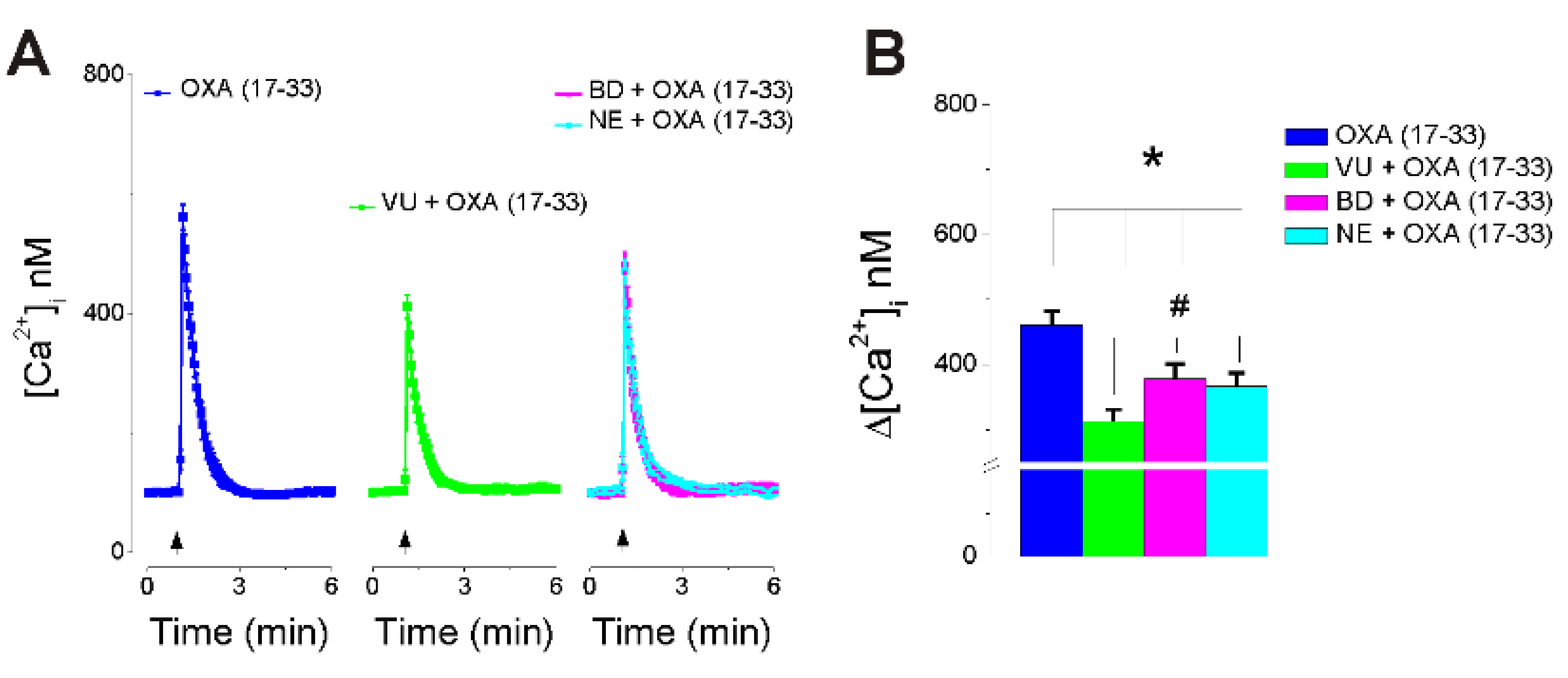
Pretreatment with VU0155056 (1 μM, 30 min), a PLD inhibitor [34], reduced the amplitude of OXA (17-33) (10 nM)-induced increase in [Ca2+]i by 33% (Figure 3). Pretreatment with BD1047 (50 µM, 30 min) or NE-100 (5 µM, 30 min) (Sigma-1R antagonists) [35,36] reduced the Ca2+ response to OXA (17-33) (10 nM) by 18.1% and 20.4%, respectively. Average Ca2+ responses induced by OXA (17-33) alone and in the presence of PLD inhibitor and Sigma-1R antagonists are illustrated in Figure 3A, and a comparison of the amplitude of the [Ca2+]i increase in each condition is illustrated in Figure 3B (n = 6 neurons/condition).

2.4. Cocaine Potentiates OXA (17-33)-Induced Increase in [Ca2+]i via Sigma-1R Activation

Cocaine (10 µM), while it did not elicit a Ca2+ response by itself, potentiated the increase in [Ca2+]i produced by OXA (17-33) (10 nM), when added at the same time as OXA (17-33) (Figure 4). Pretreatment with BD1047 (50 µM, 30 min) or NE-100 (5 µM, 30 min), Sigma-1R antagonists, reduced the increase in [Ca2+]i produced by cocaine + OXA (17-33) (10 nM), by 30.7% and 33.1%, respectively (Figure 4). This indicates that antagonism of Sigma-1R abolished the potentiation produced by cocaine and further reduced the Ca2+ response to OXA (17-33) (10 nM) to the same level as in neurons treated with Sigma-1R antagonists before OXA (17-33) alone (Figure 4 vs. Figure 3). A comparison of the amplitude of the [Ca2+]i increase in each condition is illustrated in Figure 4B (n = 6 neurons/condition).
A diagram summarizing the proposed mechanism of potentiation of orexin by cocaine via Sigma-1R activation in nucleus accumbens neurons is illustrated in Figure 5.

3. Discussion

Orexin A, via activation of OX1 receptor, can activate both phospholipase C (PLC) and phospholipase D (PLD) in various cell models [9,10] including neurons [37]. PLC activation leads to hydrolysis of phosphoinositides and formation of inositol-1,4,5- trisphosphate (IP3), the Ca2+-releasing second messenger that releases Ca2+ from endoplasmic reticulum (ER) through IP3 receptors [38]. PLD activation promotes the hydrolysis of phosphatidylcholine to choline and phosphatidic acid [11]. Whereas phosphatidic acid was considered the main effector downstream to PLD activation, we recently identified choline as a second messenger that activates Sigma-1R [12].
Sigma-1 receptor is a chaperone protein expressed in the endoplasmic reticulum (ER), mainly at the mitochondria-associated ER membrane domains (MAMs) [13]. Sigma-1Rs interact with many different signaling proteins. At the ER, Sigma-1Rs potentiate the Ca2+ release via IP3 receptors [13]; they also interact with STIM1, the Ca2+ sensor for store-operated Ca2+ entry [39]. Sigma-1R ligands include antidepressants, antipsychotics, and drugs of abuse [40]. Cocaine, in addition to its canonical target that elevates synaptic dopamine levels, binds to and activates Sigma-1Rs [41,42]. Neurons in the nucleus accumbens, a key area involved in the reward circuit [25], express Sigma-1R [27,28,29] and OX1 receptors [14,15]. Behavioral studies supported the cocaine–orexin interaction in nucleus accumbens [3,22], but the underlying mechanisms remained unclear; this prompted us to investigate the mechanisms of cocaine–orexin interaction at this level.
Orexin A has been reported to increase cytosolic Ca2+ concentration, [Ca2+]i, in various cells expressing orexin receptors [1], including neurons [43]. We first tested the effect of truncated orexin A peptide, OXA (17-33), a selective OX1 agonist [30], on [Ca2+]i in cultured nucleus accumbens neurons. OXA (17-33) increased [Ca2+]i in a dose-dependent manner; the effect was abolished by SB-334867 (1 µM), an OX1 antagonist [10,31] indicating that it was mediated by OX1 receptors.
We next demonstrated that the OXA (17-33)-induced increase in [Ca2+]i was mediated by IP3-dependent Ca2+ release from ER, as previously reported [6]; the effect was abolished by IP3 receptor antagonists, but not affected by disruption of lysosomal Ca2+ stores.
In other series of experiments, pretreatment with PLD inhibitor reduced the Ca2+ response elicited by OXA (17-33), supporting the involvement of PLD activation in addition to PLC/IP3-dependent mechanisms in nucleus ambiguus neurons. This is in agreement with previous studies reporting PLD-dependent mechanisms downstream to OX1 activation [9,10,37].
In addition, antagonism of Sigma-1R reduced the Ca2+ response produced by OXA (17-33), indicating for the first time the role of Sigma-1R in the response to OX1 activation in the nucleus accumbens. The reduction in the response to OXA (17-33) produced by PLD inhibition and Sigma-1R antagonism indicates that choline produced by PLD hydrolysis of phosphatidylcholine, acting on Sigma-1R, as recently reported [12], potentiates the IP3-mediated increase in [Ca2+]i.
Our results also indicate that cocaine, while it did not elicit a response by itself, potentiated the increase in [Ca2+]i induced by OXA(17-33). This is similar to the potentiation of orexin A-induced increase in [Ca2+]i by cocaine reported in VTA neurons [43]. In VTA neurons, the effect of orexin and the potentiation by cocaine were abolished by suvorexant (MK-4305), a dual orexin receptor OX1/OX2 antagonist [43,44]. Moreover, in nucleus accumbens neurons, the potentiation of orexin response by cocaine was abolished by Sigma-1R antagonists. Cocaine is a Sigma-1R agonist [26], and we previously reported that, in nucleus accumbens neurons, cocaine via Sigma-1R potentiates the IP3-mediated increase in [Ca2+]i [29]. Here, we identify an additional signaling mechanism for orexin A–OX1 via choline-Sigma-1R and a critical role for Sigma-1R in the cocaine–orexin A interaction in nucleus accumbens neurons.

4. Materials and Methods

4.1. Chemicals

OXA (17-33), i.e., truncated orexin A, a selective OX1 agonist [30], SB-334867, a selective nonpeptide OX1 antagonist [31], and BD-1047 and NE-100 (Sigma-1 antagonists) were obtained from Tocris (Bio-Techne Corporation, Minneapolis, MN, USA). VU0155056, a PLD inhibitor [34], was purchased from Avanti Polar Lipids (Alabaster, AL, USA). Cocaine was supplied by the National Institute on Drug Abuse’s Drug Supply Program. All other chemicals were from Sigma Aldrich (St. Louis, MO, USA), unless otherwise mentioned.

4.2. Neuronal Cell Culture

Nucleus accumbens neurons were dissociated from neonatal Sprague Dawley rats (Ace Animal Inc., Boyertown, PA, USA) of both sexes as previously described [29,45]. Newborn rats were decapitated, and the brains quickly removed surgically and immersed in ice-cold Hanks balanced salt solution (HBSS). The nucleus accumbens was identified, removed, minced, and subjected to enzymatic (papain, 37 °C) and mechanical dissociation. Cells were cultured in Neurobasal A medium (Life Technologies, ThermoFisher Scientific, Carlsbad, CA, USA) containing 10% fetal bovine serum, 1% GlutaMax, and 1% penicillin–streptomycin–amphotericin B solution at 37 °C in a humidified atmosphere with 5% CO2.The mitotic inhibitor cytosine β-arabinofuranoside (1 µM) was added to the culture to inhibit glial cell proliferation. For calcium imaging, neurons were cultured on round 25 mm diameter glass coverslips coated with poly-l-lysine, in six-well plates.

4.3. Measurement of Cytosolic Ca2+ Concentration

Cytosolic Ca2+ concentration, [Ca2+]i, was measured by calcium imaging methods in nucleus accumbens neurons loaded with Fura-2AM, as previously described [29,45]. Cells were incubated with 5 µM Fura-2AM (Invitrogen) in HBSS at room temperature for 45 min, in the dark, and then incubated for another 45 min in HBSS to allow for complete de-esterification of the dye. Coverslips (25 mm diameter) were subsequently mounted in an open bath chamber (Warner Instruments, Hamden, CT, USA) on the stage of an inverted microscope Nikon Eclipse TiE (Nikon Inc., Melville, NY, USA), equipped with a Perfect Focus System and a Photometrics CoolSnap HQ2 CCD camera (Photometrics, Tucson, AZ, USA). During the experiments, the Perfect Focus System was activated. Fura-2AM fluorescence (emission = 510 nm), following alternate excitation at 340 and 380 nm, was acquired at a frequency of 0.25 Hz. Images were acquired and analyzed using NIS-Elements AR software (Nikon Inc.). After appropriate calibration with ionomycin and CaCl2 and with Ca2+ free and EGTA, respectively, the ratio of the fluorescence signals (340/380 nm) was converted to Ca2+ concentrations [46].

4.4. Data Analysis

Data were expressed as the mean ± standard deviation (SD). Datasets were compared for statistically significant differences using one-way ANOVA followed by post hoc Bonferroni test. A p-value <0.05 was considered statistically significant.

Author Contributions

Conceptualization, E.B.; methodology, J.L.B., P.Z., G.C.B., and E.B.; validation, J.L.B., G.C.B., and E.B.; formal analysis, J.L.B., G.C.B., and E.B.; investigation, J.L.B., P.Z., G.C.B., and E.B.; writing—original draft preparation, G.C.B. and E.B.; writing—review and editing, J.L.B., P.Z., G.C.B., and E.B.; visualization, J.L.B., P.Z., G.C.B., and E.B.; supervision, E.B. All authors have read and agreed to the published version of the manuscript.

Funding

This research was funded by the National Institutes of Health grant number P30DA013429 and research funds from the Jefferson College of Pharmacy.

Institutional Review Board Statement

Animal protocols were approved by the Institutional Animal Care and Use Committee (protocol 01460 approved 20 January 2019).

Informed Consent Statement

Not applicable.

Data Availability Statement

The data generated and analyzed during this study are available in the manuscript.

Conflicts of Interest

The authors declare no conflict of interest.

References

  1. Sakurai, T.; Amemiya, A.; Ishii, M.; Matsuzaki, I.; Chemelli, R.M.; Tanaka, H.; Williams, S.C.; Richardson, J.A.; Kozlowski, G.P.; Wilson, S.; et al. Orexins and orexin receptors: A family of hypothalamic neuropeptides and G protein-coupled receptors that regulate feeding behavior. Cell 1998, 92, 573–585. [Google Scholar] [CrossRef] [Green Version]
  2. de Lecea, L.; Kilduff, T.S.; Peyron, C.; Gao, X.; Foye, P.E.; Danielson, P.E.; Fukuhara, C.; Battenberg, E.L.; Gautvik, V.T.; Bartlett, F.S.; et al. The hypocretins: Hypothalamus-specific peptides with neuroexcitatory activity. Proc. Natl. Acad. Sci. USA 1998, 95, 322–327. [Google Scholar] [CrossRef] [PubMed] [Green Version]
  3. Mahler, S.V.; Smith, R.J.; Moorman, D.E.; Sartor, G.C.; Aston-Jones, G. Multiple roles for orexin/hypocretin in addiction. Prog. Brain Res. 2012, 198, 79–121. [Google Scholar]
  4. Haghparast, A.; Fatahi, Z.; Arezoomandan, R.; Karimi, S.; Taslimi, Z.; Zarrabian, S. Functional roles of orexin/hypocretin receptors in reward circuit. Prog. Brain Res. 2017, 235, 139–154. [Google Scholar]
  5. Foord, S.M.; Bonner, T.I.; Neubig, R.R.; Rosser, E.M.; Pin, J.-P.; Davenport, A.P.; Spedding, M.; Harmar, A.J. International Union of Pharmacology. XLVI. G protein-coupled receptor list. Pharmacol. Rev. 2005, 57, 279–288. [Google Scholar] [CrossRef] [PubMed] [Green Version]
  6. Kukkonen, J.P. G-protein-dependency of orexin/hypocretin receptor signalling in recombinant Chinese hamster ovary cells. Biochem. Biophys. Res. Commun. 2016, 476, 379–385. [Google Scholar] [CrossRef]
  7. Hopf, F.W. Recent perspectives on orexin/hypocretin promotion of addiction-related behaviors. Neuropharmacology 2020, 168, 108013. [Google Scholar] [CrossRef] [PubMed]
  8. Kukkonen, J.P. Lipid signaling cascades of orexin/hypocretin receptors. Biochimie 2014, 96, 158–165. [Google Scholar] [CrossRef]
  9. Johansson, L.; Ekholm, M.E.; Kukkonen, J.P. Multiple phospholipase activation by OX(1) orexin/hypocretin receptors. Cell. Mol. Life Sci. 2008, 65, 1948–1956. [Google Scholar] [CrossRef]
  10. Jantti, M.H.; Putula, J.; Somerharju, P.; Frohman, M.A.; Kukkonen, J.P. OX1 orexin/hypocretin receptor activation of phospholipase D. Br. J. Pharmacol. 2012, 165, 1109–1123. [Google Scholar] [CrossRef] [PubMed] [Green Version]
  11. Exton, J.H. Phospholipase D. Ann. N. Y. Acad. Sci. 2000, 905, 61–68. [Google Scholar] [CrossRef]
  12. Brailoiu, E.; Chakraborty, S.; Brailoiu, G.C.; Zhao, P.; Barr, J.L.; Ilies, M.A.; Unterwald, E.M.; Abood, M.E.; Taylor, C.W. Choline is an intracellular messenger linking extracellular stimuli to IP3-evoked Ca(2+) signals through Sigma-1 receptors. Cell Rep. 2019, 26, 330–337. [Google Scholar] [CrossRef] [PubMed] [Green Version]
  13. Hayashi, T.; Su, T.P. Sigma-1 receptor chaperones at the ER-mitochondrion interface regulate Ca(2+) signaling and cell survival. Cell 2007, 131, 596–610. [Google Scholar] [CrossRef] [PubMed] [Green Version]
  14. D’Almeida, V.; Hipolide, D.C.; Raymond, R.; Barlow, K.B.L.; Parkes, J.H.; Pedrazzoli, M.; Tufik, S.; Nobrega, J.N. Opposite effects of sleep rebound on orexin OX1 and OX2 receptor expression in rat brain. Brain Res. Mol. Brain Res. 2005, 136, 148–157. [Google Scholar] [CrossRef]
  15. Lei, K.; Kwok, C.; Darevsky, D.; Wegner, S.A.; Yu, J.; Nakayama, L.; Pedrozo, V.; Anderson, L.; Ghotra, S.; Fouad, M.; et al. Nucleus accumbens shell Orexin-1 receptors are critical mediators of binge intake in excessive-drinking individuals. Front. Neurosci. 2019, 13, 88. [Google Scholar] [CrossRef] [Green Version]
  16. Perrey, D.A.; Zhang, Y. Therapeutics development for addiction: Orexin-1 receptor antagonists. Brain Res. 2020, 1731, 145922. [Google Scholar] [CrossRef]
  17. Han, Y.; Yuan, K.; Zheng, Y.; Lu, L. Orexin receptor antagonists as emerging treatments for psychiatric disorders. Neurosci. Bull. 2020, 36, 432–448. [Google Scholar] [CrossRef]
  18. Khoo, S.Y.; Brown, R.M. Orexin/hypocretin based pharmacotherapies for the treatment of addiction: DORA or SORA? CNS Drugs 2014, 28, 713–730. [Google Scholar] [CrossRef]
  19. Bentzley, B.S.; Aston-Jones, G. Orexin-1 receptor signaling increases motivation for cocaine-associated cues. Eur. J. Neurosci. 2015, 41, 1149–1156. [Google Scholar] [CrossRef]
  20. Zhou, L.; Ghee, S.M.; Chan, C.; Lin, L.; Cameron, M.D.; Kenny, P.J.; See, R.E. Orexin-1 receptor mediation of cocaine seeking in male and female rats. J. Pharmacol. Exp. Ther. 2012, 340, 801–809. [Google Scholar] [CrossRef]
  21. Prince, C.D.; Rau, A.R.; Yorgason, J.T.; Espana, R.A. Hypocretin/Orexin regulation of dopamine signaling and cocaine self-administration is mediated predominantly by hypocretin receptor 1. ACS Chem. Neurosci. 2015, 6, 138–146. [Google Scholar] [CrossRef] [PubMed] [Green Version]
  22. Yang, M.; Ma, H.; Jia, M.; Li, Y.; Miao, D.; Cui, C.; Wu, L. The role of the nucleus accumbens OXR1 in cocaine-induced locomotor sensitization. Behav. Brain Res. 2020, 379, 112365. [Google Scholar] [CrossRef] [PubMed]
  23. Martin-Fardon, R.; Weiss, F. Blockade of hypocretin receptor-1 preferentially prevents cocaine seeking: Comparison with natural reward seeking. Neuroreport 2014, 25, 485–488. [Google Scholar] [CrossRef] [PubMed]
  24. Giros, B.; Jaber, M.; Jones, S.R.; Wightman, R.M.; Garon, M.G. Hyperlocomotion and indifference to cocaine and amphetamine in mice lacking the dopamine transporter. Nature 1996, 379, 606–612. [Google Scholar] [CrossRef]
  25. Koob, G.F.; Volkow, N.D. Neurocircuitry of addiction. Neuropsychopharmacology 2010, 35, 217–238. [Google Scholar] [CrossRef] [Green Version]
  26. Sharkey, J.; Glen, K.A.; Wolfe, S.; Kuhar, M.J. Cocaine binding at sigma receptors. Eur. J. Pharmacol. 1988, 149, 171–174. [Google Scholar] [CrossRef]
  27. Gundlach, A.L.; Largent, B.L.; Snyder, S.H. Autoradiographic localization of sigma receptor binding sites in guinea pig and rat central nervous system with (+)3H-3-(3-hydroxyphenyl)-N-(1-propyl)piperidine. J. Neurosci. 1986, 6, 1757–1770. [Google Scholar] [CrossRef]
  28. Alonso, G.; Phan, V.; Guillemain, I.; Saunier, M.; Legrand, A.; Anoal, M.; Maurice, T. Immunocytochemical localization of the sigma(1) receptor in the adult rat central nervous system. Neuroscience 2000, 97, 155–170. [Google Scholar] [CrossRef]
  29. Barr, J.L.; Deliu, E.; Brailoiu, C.G.; Zhao, P.; Yan, G.; Abood, M.E.; Unterwald, E.M.; Brailoiu, E. Mechanisms of activation of nucleus accumbens neurons by cocaine via sigma-1 receptor-inositol 1,4,5-trisphosphate-transient receptor potential canonical channel pathways. Cell Calcium 2015, 58, 196–207. [Google Scholar] [CrossRef] [Green Version]
  30. Feng, X.M.; Mi, W.L.; Xia, F.; Mao-Ying, W.L.; Jiang, J.W.; Xiao, S.; Wang, Z.F.; Wang, Y.Q.; Wu, G.C. Involvement of spinal orexin A in the electroacupuncture analgesia in a rat model of post-laparotomy pain. BMC Complement. Altern. Med. 2012, 12, 225. [Google Scholar] [CrossRef] [Green Version]
  31. Smart, D.; Sabido-David, C.; Brough, S.J.; Jewitt, F.; Johns, A.; Porter, R.A.; Jerman, J.C. SB-334867-A: The first selective orexin-1 receptor antagonist. Br. J. Pharmacol. 2001, 132, 1179–1182. [Google Scholar] [CrossRef] [Green Version]
  32. Maruyama, T.; Kanaji, T.; Nakade, S.; Kanno, T.; Mikoshiba, K. 2APB, 2-aminoethoxydiphenyl borate, a membrane-penetrable modulator of Ins(1,4,5)P3-induced Ca2+ release. J. Biochem. 1997, 122, 498–505. [Google Scholar] [CrossRef] [PubMed]
  33. Bowman, E.J.; Siebers, A.; Altendorf, K. Bafilomycins: A class of inhibitors of membrane ATPases from microorganisms, animal cells, and plant cells. Proc. Natl. Acad. Sci. USA 1988, 85, 7972–7976. [Google Scholar] [CrossRef] [Green Version]
  34. Scott, S.A.; Selvy, P.E.; Buck, J.R.; Cho, H.P.; Criswell, T.L.; Thomas, A.L.; Armstrong, M.D.; Arteaga, C.L.; Lindsley, C.W.; Brown, H.A. Design of isoform-selective phospholipase D inhibitors that modulate cancer cell invasiveness. Nat. Chem. Biol. 2009, 5, 108–117. [Google Scholar] [CrossRef] [Green Version]
  35. Matsumoto, R.R.; Bowen, W.D.; Tom, M.A.; Vo, V.N.; Truong, D.D.; de Costa, B.R. Characterization of two novel sigma receptor ligands: Antidystonic effects in rats suggest sigma receptor antagonism. Eur. J. Pharmacol. 1995, 280, 301–310. [Google Scholar] [CrossRef]
  36. Chaki, S.; Okuyama, S.; Ogawa, S.; Tanaka, M.; Muramatsu, M.; Nakazato, A.; Tomisawa, K. Solubilization and characterization of binding sites for [3H]NE-100, a novel and potent sigma 1 ligand, from guinea pig brain. Life Sci. 1996, 59, 1331–1340. [Google Scholar] [CrossRef]
  37. Bjornstrom, K.; Turina, D.; Strid, T.; Sundqvist, T.; Eintrei, C. Orexin A inhibits propofol-induced neurite retraction by a phospholipase D/protein kinase Cepsilon-dependent mechanism in neurons. PLoS ONE 2014, 9, e97129. [Google Scholar] [CrossRef] [Green Version]
  38. Berridge, M.J. The Inositol Trisphosphate/Calcium signaling pathway in health and disease. Physiol. Rev. 2016, 96, 1261–1296. [Google Scholar] [CrossRef] [Green Version]
  39. Srivats, S.; Balasuriya, D.; Pasche, M.; Vistal, G.; Edwardson, J.M.; Taylor, C.W.; Murell-Lagnado, R.D. Sigma1 receptors inhibit store-operated Ca2+ entry by attenuating coupling of STIM1 to Orai1. J. Cell Biol. 2016, 213, 65–79. [Google Scholar] [CrossRef]
  40. Maurice, T.; Su, T.P. The pharmacology of sigma-1 receptors. Pharmacol. Ther. 2009, 124, 195–206. [Google Scholar] [CrossRef] [Green Version]
  41. Katz, J.L.; Hong, W.C.; Hiranita, T.; Su, T.P. A role for sigma receptors in stimulant self-administration and addiction. Pharmaceuticals 2011, 4, 880–914. [Google Scholar] [CrossRef] [Green Version]
  42. Hayashi, T.; Tsai, S.Y.; Mori, T.; Fujimoto, M.; Su, T.P. Targeting ligand-operated chaperone sigma-1 receptors in the treatment of neuropsychiatric disorders. Expert Opin. Ther. Targets 2011, 15, 557–577. [Google Scholar] [CrossRef] [PubMed] [Green Version]
  43. Gentile, T.A.; Simmons, S.J.; Watson, M.N.; Connelly, K.L.; Brailoiu, E.; Zhang, Y.; Muschamp, J.W. Effects of suvorexant, a dual Orexin/Hypocretin receptor antagonist, on impulsive behavior associated with cocaine. Neuropsychopharmacology 2018, 43, 1001–1009. [Google Scholar] [CrossRef] [Green Version]
  44. Cox, C.D.; Breslin, M.J.; Whitman, D.B.; Schreier, J.D.; McGaughey, G.B.; Bogusky, M.J.; Roecker, A.J.; Mercer, S.P.; Bednar, R.A.; Lemaire, W.; et al. Discovery of the dual orexin receptor antagonist [(7R)-4-(5-chloro-1,3-benzoxazol-2-yl)-7-methyl-1,4-diazepan-1-yl][5-methyl-2-(2H-1,2,3-triazol-2-yl)phenyl]methanone (MK-4305) for the treatment of insomnia. J. Med. Chem. 2010, 53, 5320–5332. [Google Scholar] [CrossRef]
  45. Brailoiu, G.C.; Deliu, E.; Barr, J.L.; Console-Bram, L.M.; Ciuciu, A.M.; Abood, M.E.; Unterwald, E.M.; Brailoiu, E. HIV Tat excites D1 receptor-like expressing neurons from rat nucleus accumbens. Drug Alcohol Depend. 2017, 178, 7–14. [Google Scholar] [CrossRef] [PubMed]
  46. Grynkiewicz, G.; Poenie, M.; Tsien, R.Y. A new generation of Ca2+ indicators with greatly improved fluorescence properties. J. Biol. Chem. 1985, 260, 3440–3450. [Google Scholar] [CrossRef]
Figure 1. OXA (17-33) increases cytosolic Ca2+ concentration, [Ca2+]i, in nucleus accumbens neurons via OX1 receptor activation. (A) Representative examples of fluorescence F340/380 ratio of Fura-2AM-loaded nucleus accumbens neurons in basal conditions (left) and after treatment with OXA (17-33) (10 nM), a selective OX1 agonist, alone (top right) or in the presence of OX1 antagonist, SB-334867 (1 µM) (bottom right). (B) OXA (17-33) (10 nM) produced a transient increase in [Ca2+]i; the effect was abolished by SB-334867. (C) Comparison of the amplitude of [Ca2+]i increase (mean + SD) produced by OXA (17–33) (0.1, 1, 10, and 100 nM); p < 0.05 as compared with the amplitude of [Ca2+]i increase produced by each concentration (*) or by OXA (17-33) (10 nM) (**); n = 6 neurons/each concentration tested.
Ijms 22 05160 g001
Figure 2. OXA (17-33) increases [Ca2+]i via IP3-dependent mechanism. (A) Illustration of average Ca2+ transients (± SD) induced in Ca2+-free saline by OXA (17-33) (10 nM) alone (left) and OXA (17-33) (10 nM) after pretreatment with 2-aminoethoxydiphenyl borate (2-APB, 100 µM) and xestospongin C (XeC, 10 µM, 15 min), IP3 receptor antagonists (middle), or with bafilomycin A1 (Baf, 1 µM) (right). (B) Comparison of the amplitude of the increase in [Ca2+]i (average + SD) in each condition. Pretreatment with 2-APB and xestospongin C abolished the Ca2+ response induced by OXA (17-33). * p < 0.05; n = 6 neurons/condition.
Ijms 22 05160 g002
Figure 3. OXA (17-33) A increases [Ca2+]i via choline-Sigma-1R-dependent mechanism. (A) Illustration of average Ca2+ transients (± SD) induced by OXA (17-33) (10 nM) alone (left) and in the presence of VU0155056 (1 μM), PLD inhibitor (middle), and BD1047 (50 µM) or NE-100 (5 µM), Sigma-1R antagonists (right). (B) Comparison of the amplitude of the increase in [Ca2+]i (average + SD) in each condition. Inhibition of PLD or antagonism of Sig-1R reduces the Ca2+ response elicited by OXA (17-33) (10 nM); p < 0.05 as compared to amplitude of [Ca2+]i increase produced by OXA (17-33) (*) or produced in the presence of the inhibitors (#) (n = 6 neurons/condition).
Ijms 22 05160 g003
Figure 4. Cocaine potentiates OXA (17-33)-induced increase in [Ca2+]i via Sigma-1R activation. (A) Illustration of average Ca2+ responses (± SD) produced by application of cocaine (10 µM) alone (left, no response), cocaine (10 µM) and OXA (17-33) (10 nM) (middle), and cocaine and OXA (17-33) in the presence of Sigma-1R antagonists BD1047 (50 µM) or NE-100 (5 µM) (right). (B) Comparison of the amplitude of the increase in [Ca2+]i (average + SD) in each condition. Cocaine potentiates the Ca2+ response induced by OXA (17-33), while antagonism of Sigma-1R abolished the potentiation produced by cocaine on the Ca2+ response elicited by OXA (17-33) (10 nM). * p < 0.05 (n = 6 neurons/condition).
Ijms 22 05160 g004
Figure 5. Diagram illustrating the proposed model of potentiation of orexin by cocaine via Sigma-1R in nucleus accumbens neurons. (A) Orexin A acting on OX1 receptor activates PLC and PLD. PLC increases IP3 level and promotes the Ca2+ release from endoplasmic reticulum via IP3 receptors (IP3R). PLD produces choline (from hydrolysis of phosphatidylcholine) that acts on Sigma-1R to potentiate Ca2+ increase via IP3 R (higher increase in [Ca2+]i). (B) Inhibition of PLD or antagonism of Sigma-1R limits the orexin A-OX1 receptor signaling to PLC-mediated IP3-dependent increase in Ca2+ (smaller increase in [Ca2+]i). (C) Cocaine, via Sigma-1R activation, potentiates the PLC- and PLD-mediated increase in [Ca2+]i produced by orexin A acting on OX1 (highest increase in [Ca2+]i). The diagram was created using the Motifolio Illustration Toolkit Neuroscience (https://www.motifolio.com accessed on 7 March 2021).
Ijms 22 05160 g005
Publisher’s Note: MDPI stays neutral with regard to jurisdictional claims in published maps and institutional affiliations.