
Current Practice in Bicistronic IRES Reporter Use: A Systematic Review


 , ,
, ,  and
and
Abstract
:1. Introduction
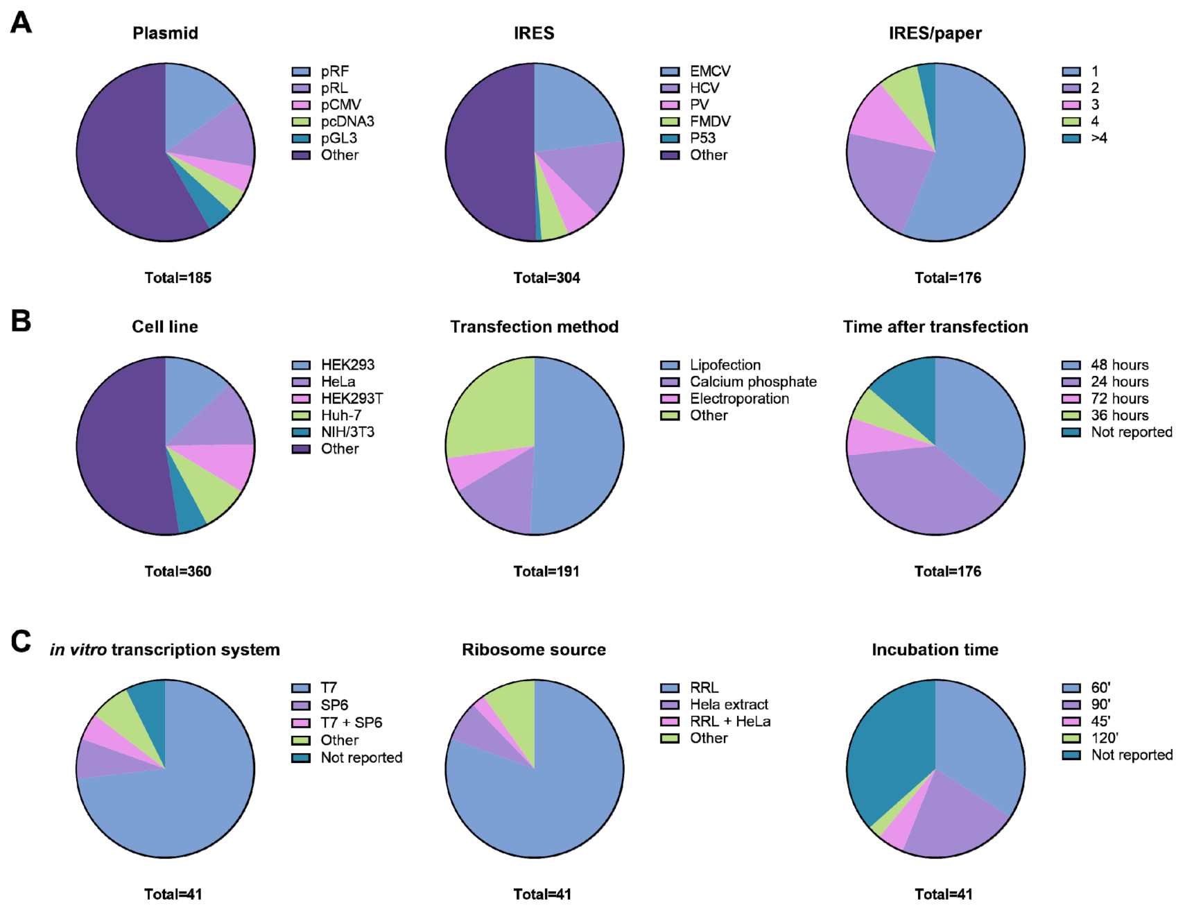
2. Results
2.1. Critical Determinants of Bicistronic Reporter Plasmid Design and IRES Activity
2.2. The Most Commonly Used Cell Lines and Cell Type-Specific IRES Activity
2.3. Methods for Transgene Expression and mRNA Translation In Vivo
2.4. Temporal Effects of Bicistronic Gene Transcription and mRNA Translation In Vivo
2.5. The Use of the Cell-Free Translation System
2.6. Visualization of Bicistronic Reporter Data: Ratio vs. Separate Cistrons
2.7. Normalization and Validation of Bicistronic Reporter Data
3. Discussion
- (1)
- Make plasmid backbones and IRES sequences available upon publication.
- (2)
- Carefully describe transfection procedures and the timeline of the experiment.
- (3)
- Report both the ratio and individual cistron data.
- (4)
- Make raw data available, including background signals.
- (5)
- Verify whether treatments affect cell number, total protein content, or cell viability.
- (6)
- For cellular IRES translational studies, an empty control plasmid to define background signals; a promoterless control plasmid to assess cryptic promoter activity of the IRES; and verification of equal and intact mRNA levels by RT-qPCR and RT-PCR, respectively, are feasible and represent important controls.
4. Methods
Search Strategy and Inclusion Criteria
Supplementary Materials
Author Contributions
Funding
Institutional Review Board Statement
Informed Consent Statement
Data Availability Statement
Conflicts of Interest
References
- Schenborn, E.; Groskreutz, D. Reporter Gene Vectors and Assays. Mol. Biotechnol. 1999, 13, 29–44. [Google Scholar] [CrossRef]
- Robison, L.R.; Seligsohn, R.; Lerner, S.A. Simplified Radioenzymatic Assay for Chloramphenicol. Antimicrob. Agents Chemother. 1978, 13, 25–29. [Google Scholar] [CrossRef] [PubMed] [Green Version]
- Ow, D.W.; de Wet, J.R.; Helinski, D.R.; Howell, S.H.; Wood, K.V.; Deluca, M. Transient and stable expression of the firefly luciferase gene in plant cells and transgenic plants. Science 1986, 234, 856–859. [Google Scholar] [CrossRef] [PubMed]
- Inouye, S.; Tsuji, F.I. Aequorea green fluorescent protein. Expression of the gene and fluorescence characteristics of the recom-binant protein. FEBS Lett. 1994, 341, 277–280. [Google Scholar] [CrossRef] [Green Version]
- Jing, Y.; Simmer, P.; Feng, J.Q. Cell Lineage Tracing: Colocalization of Cell Lineage Markers with a Fluorescent Reporter. Methods Mol. Biol. 2021, 2230, 325–335. [Google Scholar]
- Terenin, I.M.; Smirnova, V.V.; Andreev, D.E.; Dmitriev, S.E.; Shatsky, I.N. A researcher’s guide to the galaxy of IRESs. Cell. Mol. Life Sci. CMLS. 2017, 74, 1431–1455. [Google Scholar] [CrossRef]
- Blay, V.; Tolani, B.; Ho, S.P.; Arkin, M.R. High-Throughput Screening: Today’s biochemical and cell-based approaches. Drug Discov. Today 2020, 25, 1807–1821. [Google Scholar] [CrossRef]
- Kim, J.H.; Lee, S.R.; Li, L.H.; Park, H.J.; Park, J.H.; Lee, K.Y.; Choi, S.Y. High cleavage efficiency of a 2A peptide derived from porcine teschovirus-1 in human cell lines, zebrafish and mice. PLoS ONE 2011, 6, e18556. [Google Scholar] [CrossRef] [Green Version]
- Jang, S.K.; Davies, M.V.; Kaufman, R.J.; Wimmer, E. Initiation of protein synthesis by internal entry of ribosomes into the 5’ non-translated region of encephalomyocarditis virus RNA in vivo. J. Virol. 1989, 63, 1651–1660. [Google Scholar] [CrossRef] [Green Version]
- Jang, S.K.; Kräusslich, H.G.; Nicklin, M.J.; Duke, G.M.; Palmenberg, A.C.; Wimmer, E. A segment of the 5’ nontranslated region of encephalomyocarditis virus RNA directs internal entry of ribosomes during in vitro translation. J. Virol. 1988, 62, 2636–2643. [Google Scholar] [CrossRef] [Green Version]
- Kwan, T.; Thompson, S.R. Noncanonical Translation Initiation in Eukaryotes. In Cold Spring Harbor Perspectives in Biology; Cold Srping Harbor Laboratory Press: New York, NY, USA, 2019; Volume 11. [Google Scholar]
- Mokrejš, M.; Mašek, T.; Vopálenský, V.; Hlubuček, P.; Delbos, P.; Pospíšek, M. IRESite—A tool for the examination of viral and cellular internal ribosome entry sites. Nucleic Acids Res. 2009, 38, D131–D136. [Google Scholar] [CrossRef] [Green Version]
- Godet, A.-C.; David, F.; Hantelys, F.; Tatin, F.; Lacazette, E.; Garmy-Susini, B.; Prats, A.-C. IRES Trans-Acting Factors, Key Actors of the Stress Response. Int. J. Mol. Sci. 2019, 20, 924. [Google Scholar] [CrossRef] [Green Version]
- Jackson, R.J. The Current Status of Vertebrate Cellular mRNA IRESs. Cold Spring Harb. Perspect. Biol. 2013, 5, a011569. [Google Scholar] [CrossRef]
- Van Eden, M.E.; Byrd, M.P.; Sherrill, K.W.; Lloyd, R.E. Demonstrating internal ribosome entry sites in eukaryotic mRNAs using stringent RNA test procedures. RNA 2004, 10, 720–730. [Google Scholar] [CrossRef] [Green Version]
- Yang, Y.; Wang, Z. IRES-mediated cap-independent translation, a path leading to hidden proteome. J. Mol. Cell Biol. 2019, 11, 911–919. [Google Scholar] [CrossRef] [Green Version]
- Loughran, G.; Howard, M.T.; Firth, A.E.; Atkins, J.F. Avoidance of reporter assay distortions from fused dual reporters. RNA 2017, 23, 1285–1289. [Google Scholar] [CrossRef]
- Penzo, M.; Carnicelli, D.; Montanaro, L.; Brigotti, M. A reconstituted cell-free assay for the evaluation of the intrinsic activity of purified human ribosomes. Nat. Protoc. 2016, 11, 1309–1325. [Google Scholar] [CrossRef]
- Atkins, J.F.; Loughran, G.; Bhatt, P.R.; Firth, A.E.; Baranov, P.V. Ribosomal frameshifting and transcriptional slippage: From genetic steganography and cryptography to adventitious use. Nucleic Acids Res. 2016, 44, 7007–7078. [Google Scholar] [CrossRef] [Green Version]
- Jacobs, J.L.; Dinman, J.D. Systematic analysis of bicistronic reporter assay data. Nucleic Acids Res. 2004, 32, e160. [Google Scholar] [CrossRef]
- Ray, P.S.; Grover, R.; Das, S. Two internal ribosome entry sites mediate the translation of p53 isoforms. EMBO Rep. 2006, 7, 404–410. [Google Scholar] [CrossRef] [Green Version]
- Weingarten-Gabbay, S.; Segal, E. Toward a systematic understanding of translational regulatory elements in human and viruses. RNA Biol. 2016, 13, 927–933. [Google Scholar] [CrossRef] [PubMed] [Green Version]
- Weingarten-Gabbay, S.; Elias-Kirma, S.; Nir, R.; Gritsenko, A.A.; Stern-Ginossar, N.; Yakhini, Z.; Segal, E. Comparative genetics. Systematic discovery of cap-independent translation sequences in human and viral genomes. Science 2016, 351, aad4939. [Google Scholar] [CrossRef] [PubMed]
- Weingarten-Gabbay, S.; Nir, R.; Lubliner, S.; Sharon, E.; Kalma, Y.; Weinberger, A.; Segal, E. Systematic interrogation of human promoters. Genome Res. 2019, 29, 171–183. [Google Scholar] [CrossRef] [PubMed] [Green Version]
- Calamita, P.; Gatti, G.; Miluzio, A.; Scagliola, A.; Biffo, S. Translating the Game: Ribosomes as Active Players. Front. Genet. 2018, 9, 533. [Google Scholar] [CrossRef] [Green Version]
- Horos, R.; IJspeert, H.; Pospisilova, D.; Sendtner, R.; Andrieu-Soler, C.; Taskesen, E.; von Lindern, M. Ribosomal deficiencies in Dia-mond-Blackfan anemia impair translation of transcripts essential for differentiation of murine and human erythroblasts. Blood 2012, 119, 262–272. [Google Scholar] [CrossRef]
- Penzo, M.; Rocchi, L.; Brugiere, S.; Carnicelli, D.; Onofrillo, C.; Couté, Y.; Brigotti, M.; Montanaro, L. Human ribosomes from cells with reduced dyskerin levels are intrinsically altered in translation. FASEB J. 2015, 29, 3472–3482. [Google Scholar] [CrossRef] [Green Version]
- Marchand, V.; Pichot, F.; Neybecker, P.; Ayadi, L.; Bourguignon-Igel, V.; Wacheul, L.; Lafontaine, D.L.J.; Pinzano, A.; Helm, M.; Motorin, Y. HydraPsiSeq: A method for systematic and quantitative mapping of pseudouridines in RNA. Nucleic Acids Res. 2020, 48, e110. [Google Scholar] [CrossRef]
- Galvanin, A.; Ayadi, L.; Helm, M.; Motorin, Y.; Marchand, V. Mapping and Quantification of tRNA 2’-O-Methylation by Ribo-MethSeq. Methods Mol. Biol. 2019, 1870, 273–295. [Google Scholar]
- Wiener, D.; Schwartz, S. The epitranscriptome beyond m(6)A. In Nature Reviews Genetics; Nature Research: London, UK, 2020. [Google Scholar]
- Jonkhout, N.; Tran, J.; Smith, M.A.; Schonrock, N.; Mattick, J.S.; Novoa, E.M. The RNA modification landscape in human disease. RNA 2017, 23, 1754–1769. [Google Scholar] [CrossRef] [Green Version]
- Li, D.; Wang, J. Ribosome heterogeneity in stem cells and development. J. Cell Biol. 2020, 219, e202001108. [Google Scholar] [CrossRef]
- Venturi, G.; Montanaro, L. How Altered Ribosome Production Can Cause or Contribute to Human Disease: The Spectrum of Ribosomopathies. Cells 2020, 9, 2300. [Google Scholar] [CrossRef]
- Piskol, R.; Melo, F.D.S.E. Colon Cancer Heterogeneity: Welcome to the RiboZone. Cell Stem Cell 2020, 26, 797–799. [Google Scholar] [CrossRef]
- Catez, F.; Venezia, N.D.; Marcel, V.; Zorbas, C.; Lafontaine, D.L.J.; Diaz, J.-J. Ribosome biogenesis: An emerging druggable pathway for cancer therapeutics. Biochem. Pharmacol. 2019, 159, 74–81. [Google Scholar] [CrossRef] [Green Version]
- Ford, D. Ribosomal heterogeneity—A new inroad for pharmacological innovation. Biochem. Pharmacol. 2020, 175, 113874. [Google Scholar] [CrossRef]
- Whetter, L.E.; Day, S.P.; Elroy-Stein, O.; Brown, E.A.; Lemon, S.M. Low efficiency of the 5’ nontranslated region of hepatitis A virus RNA in directing cap-independent translation in permissive monkey kidney cells. J. Virol. 1994, 68, 5253–5263. [Google Scholar] [CrossRef] [Green Version]
- Schultz, D.E.; Honda, M.; Whetter, L.E.; McKnight, K.L.; Lemon, S.M. Mutations within the 5’ nontranslated RNA of cell cul-ture-adapted hepatitis A virus which enhance cap-independent translation in cultured African green monkey kidney cells. J. Virol. 1996, 70, 1041–1049. [Google Scholar] [CrossRef] [Green Version]
- Buratti, E.; Gerotto, M.; Pontisso, P.; Alberti, A.; Tisminetzky, S.; Baralle, F. In vivo translational efficiency of different hepatitis C virus 5′-UTRs. FEBS Lett. 1997, 411, 275–280. [Google Scholar] [CrossRef] [Green Version]
- Chon, S.K.; Perez, D.R.; Donis, R.O. Genetic Analysis of the Internal Ribosome Entry Segment of Bovine Viral Diarrhea Virus. Virology 1998, 251, 370–382. [Google Scholar] [CrossRef]
- Collier, A.J.; Tang, S.; Elliott, R.M. Translation efficiencies of the 5’ untranslated region from representatives of the six major genotypes of hepatitis C virus using a novel bicistronic reporter assay system. J. Gen. Virol. 1998, 79, 2359–2366. [Google Scholar] [CrossRef]
- Gan, W.; La Celle, M.; Rhoads, R.E. Functional Characterization of the Internal Ribosome Entry Site of eIF4G mRNA. J. Biol. Chem. 1998, 273, 5006–5012. [Google Scholar] [CrossRef] [Green Version]
- Huez, I.; Créancier, L.; Audigier, S.; Gensac, M.-C.; Prats, A.-C.; Prats, H. Two Independent Internal Ribosome Entry Sites Are Involved in Translation Initiation of Vascular Endothelial Growth Factor mRNA. Mol. Cell. Biol. 1998, 18, 6178–6190. [Google Scholar] [CrossRef] [Green Version]
- Hwang, L.-H.; Hsieh, C.-L.; Yen, A.; Chung, Y.-L.; Chen, D.S. Involvement of the 5′ Proximal Coding Sequences of Hepatitis C Virus with Internal Initiation of Viral Translation. Biochem. Biophys. Res. Commun. 1998, 252, 455–460. [Google Scholar] [CrossRef]
- Lopez de Quinto, S.; Martinez-Salas, E. Parameters influencing translational efficiency in aphthovirus IRES-based bicistronic expression vectors. Gene 1998, 217, 51–56. [Google Scholar] [CrossRef]
- Miller, D.L.; Dibbens, J.A.; Damert, A.; Risau, W.; Vadas, M.A.; Goodall, G.J. The vascular endothelial growth factor mRNA contains an internal ribosome entry site. FEBS Lett. 1998, 434, 417–420. [Google Scholar] [CrossRef] [Green Version]
- Roberts, L.O.; Seamons, R.A.; Belsham, G.J. Recognition of picornavirus internal ribosome entry sites within cells; influence of cel-lular and viral proteins. RNA 1998, 4, 520–529. [Google Scholar] [CrossRef] [Green Version]
- Stein, I.; Itin, A.; Einat, P.; Skaliter, R.; Grossman, Z.; Keshet, E. Translation of Vascular Endothelial Growth Factor mRNA by Internal Ribosome Entry: Implications for Translation under Hypoxia. Mol. Cell. Biol. 1998, 18, 3112–3119. [Google Scholar] [CrossRef] [Green Version]
- Stoneley, M.; Paulin, F.E.; Le Quesne, J.P.; Chappell, S.A.; Willis, A.E. C-Myc 5′ untranslated region contains an internal ribosome entry segment. Oncogene 1998, 16, 423–428. [Google Scholar] [CrossRef] [Green Version]
- Attal, J.; Théron, M.-C.; Houdebine, L.M. The optimal use of IRES (internal ribosome entry site) in expression vectors. Genet. Anal. Biomol. Eng. 1999, 15, 161–165. [Google Scholar] [CrossRef]
- De Gregorio, E.; Preiss, T.; Hentze, M.W. Translation driven by an eIF4G core domain in vivo. EMBO J. 1999, 18, 4865–4874. [Google Scholar] [CrossRef] [PubMed] [Green Version]
- Finkelstein, Y.; Faktor, O.; Elroy-Stein, O.; Levi, B.-Z. The use of bi-cistronic transfer vectors for the baculovirus expression system. J. Biotechnol. 1999, 75, 33–44. [Google Scholar] [CrossRef]
- Grace, K.; Gartland, M.; Karayiannis, P.; McGarvey, M.J.; Clarke, B. The 5′ untranslated region of GB virus B shows functional similarity to the internal ribosome entry site of hepatitis C virus. J. Gen. Virol. 1999, 80, 2337–2341. [Google Scholar] [CrossRef] [PubMed]
- Schiavi, A.; Hudder, A.; Werner, R. Connexin43 mRNA contains a functional internal ribosome entry site. FEBS Lett. 1999, 464, 118–122. [Google Scholar] [CrossRef] [Green Version]
- Tang, S.; Collier, A.J.; Elliott, R.M. Alterations to both the primary and predicted secondary structure of stem-loop IIIc of the hep-atitis C virus 1b 5’ untranslated region (5’UTR) lead to mutants severely defective in translation which cannot be complemented in trans by the wild-type 5’UTR sequence. J. Virol. 1999, 73, 2359–2364. [Google Scholar]
- Zhu, J.; Musco, M.L.; Grace, M.J. Three-color flow cytometry analysis of tricistronic expression of eBFP, eGFP, and eYFP using EMCV-IRES linkages. Cytometry. 1999, 37, 51–59. [Google Scholar] [CrossRef]
- Chappell, S.A.; Edelman, G.M.; Mauro, V.P. A 9-nt segment of a cellular mRNA can function as an internal ribosome entry site (IRES) and when present in linked multiple copies greatly enhances IRES activity. Proc. Natl. Acad. Sci. USA 2000, 97, 1536–1541. [Google Scholar] [CrossRef] [Green Version]
- Créancier, L.; Morello, D.; Mercier, P.; Prats, A.-C. Fibroblast Growth Factor 2 Internal Ribosome Entry Site (Ires) Activity Ex Vivo and in Transgenic Mice Reveals a Stringent Tissue-Specific Regulation. J. Cell Biol. 2000, 150, 275–281. [Google Scholar] [CrossRef]
- Henis-Korenblit, S.; Strumpf, N.L.; Goldstaub, D.; Kimchi, A. A Novel Form of DAP5 Protein Accumulates in Apoptotic Cells as a Result of Caspase Cleavage and Internal Ribosome Entry Site-Mediated Translation. Mol. Cell. Biol. 2000, 20, 496–506. [Google Scholar] [CrossRef] [Green Version]
- Hinton, T.M.; Li, F.; Crabb, B.S. Internal Ribosomal Entry Site-Mediated Translation Initiation in Equine Rhinitis A Virus: Similarities to and Differences from That of Foot-and-Mouth Disease Virus. J. Virol. 2000, 74, 11708–11716. [Google Scholar] [CrossRef] [Green Version]
- Honda, M.; Kaneko, S.; Matsushita, E.; Kobayashi, K.; Abell, G.A.; Lemon, S.M. Cell cycle regulation of hepatitis C virus internal ribosomal entry site–directed translation. Gastroenterology 2000, 118, 152–162. [Google Scholar] [CrossRef]
- Hudder, A.; Werner, R. Analysis of a Charcot-Marie-Tooth Disease Mutation Reveals an Essential Internal Ribosome Entry Site Element in the Connexin-32 Gene. J. Biol. Chem. 2000, 275, 34586–34591. [Google Scholar] [CrossRef] [Green Version]
- Lerat, H.; Shimizu, Y.K.; Lemon, S.M. Cell Type-Specific Enhancement of Hepatitis C Virus Internal Ribosome Entry Site-Directed Translation due to 5′ Nontranslated Region Substitutions Selected during Passage of Virus in Lymphoblastoid Cells. J. Virol. 2000, 74, 7024–7031. [Google Scholar] [CrossRef] [Green Version]
- Oumard, A.; Hennecke, M.; Hauser, H.; Nourbakhsh, M. Translation of NRF mRNA Is Mediated by Highly Efficient Internal Ribosome Entry. Mol. Cell. Biol. 2000, 20, 2755–2759. [Google Scholar] [CrossRef] [Green Version]
- Pozner, A.; Goldenberg, D.; Negreanu, V.; Le, S.-Y.; Elroy-Stein, O.; Levanon, D.; Groner, Y. Transcription-Coupled Translation Control of AML1/RUNX1 Is Mediated by Cap- and Internal Ribosome Entry Site-Dependent Mechanisms. Mol. Cell. Biol. 2000, 20, 2297–2307. [Google Scholar] [CrossRef] [Green Version]
- Rijnbrand, R.; Abell, G.; Lemon, S.M. Mutational Analysis of the GB Virus B Internal Ribosome Entry Site. J. Virol. 2000, 74, 773–783. [Google Scholar] [CrossRef] [Green Version]
- Wang, T.-H.; Rijnbrand, R.C.A.; Lemon, S.M. Core Protein-Coding Sequence, but Not Core Protein, Modulates the Efficiency of Cap-Independent Translation Directed by the Internal Ribosome Entry Site of Hepatitis C Virus. J. Virol. 2000, 74, 11347–11358. [Google Scholar] [CrossRef] [Green Version]
- Bieleski, L.; Talbot, S.J. Kaposi’s sarcoma-associated herpesvirus vCyclin open reading frame contains an internal ribosome entry site. J. Virol. 2001, 75, 1864–1869. [Google Scholar] [CrossRef] [Green Version]
- Chappell, S.A.; Owens, G.C.; Mauro, V.P. A 5′ Leader of Rbm3, a Cold Stress-induced mRNA, Mediates Internal Initiation of Translation with Increased Efficiency under Conditions of Mild Hypothermia. J. Biol. Chem. 2001, 276, 36917–36922. [Google Scholar] [CrossRef] [Green Version]
- Chiang, P.-W.; Carpenter, L.E.; Hagerman, P.J. The 5′-Untranslated Region of the FMR1 Message Facilitates Translation by Internal Ribosome Entry. J. Biol. Chem. 2001, 276, 37916–37921. [Google Scholar] [CrossRef]
- Furler, S.; Paterna, J.-C.; Weibel, M.; Bueler, H. Recombinant AAV vectors containing the foot and mouth disease virus 2A sequence confer efficient bicistronic gene expression in cultured cells and rat substantia nigra neurons. Gene Ther. 2001, 8, 864–873. [Google Scholar] [CrossRef] [Green Version]
- Grundhoff, A.; Ganem, D. Mechanisms governing expression of the v-FLIP gene of Kaposi’s sarcoma-associated herpesvirus. J. Virol. 2001, 75, 1857–1863. [Google Scholar] [CrossRef] [Green Version]
- Hennecke, M.; Kwissa, M.; Metzger, K.; Oumard, A.; Kröger, A.; Schirmbeck, R.; Reimann, J.; Hauser, H. Composition and arrangement of genes define the strength of IRES-driven translation in bicistronic mRNAs. Nucleic Acids Res. 2001, 29, 3327–3334. [Google Scholar] [CrossRef] [PubMed] [Green Version]
- Krüger, M.; Beger, C.; Welch, P.J.; Barber, J.R.; Manns, M.P.; Wong-Staal, F. Involvement of Proteasome α-Subunit PSMA7 in Hepatitis C Virus Internal Ribosome Entry Site-Mediated Translation. Mol. Cell. Biol. 2001, 21, 8357–8364. [Google Scholar] [CrossRef] [PubMed] [Green Version]
- Pinkstaff, J.K.; Chappell, S.A.; Mauro, V.P.; Edelman, G.M.; Krushel, L.A. Internal initiation of translation of five dendritically localized neuronal mRNAs. Proc. Natl. Acad. Sci. USA 2001, 98, 2770–2775. [Google Scholar] [CrossRef] [PubMed] [Green Version]
- Schwarz, E.J.; Reger, R.L.; Alexander, G.M.; Class, R.; Azizi, S.A.; Prockop, D.J. Rat marrow stromal cells rapidly transduced with a self-inactivating retrovirus synthesize L-DOPA in vitro. Gene Ther. 2001, 8, 1214–1223. [Google Scholar] [CrossRef] [Green Version]
- Venkatesan, A.; Dasgupta, A. Novel Fluorescence-Based Screen to Identify Small Synthetic Internal Ribosome Entry Site Elements. Mol. Cell. Biol. 2001, 21, 2826–2837. [Google Scholar] [CrossRef] [Green Version]
- Grobe, K.; Esko, J.D. Regulated translation of heparan sulfate N-acetylglucosamine N-deacetylase/n-sulfotransferase isozymes by structured 5’-untranslated regions and internal ribosome entry sites. J. Biol. Chem. 2002, 277, 30699–30706. [Google Scholar] [CrossRef] [Green Version]
- Kato, J.; Kato, N.; Moriyama, M.; Goto, T.; Taniguchi, H.; Shiratori, Y.; Omata, M. Interferons Specifically Suppress the Translation from the Internal Ribosome Entry Site of Hepatitis C Virus through a Double-Stranded RNA–Activated Protein Kinase–Independent Pathway. J. Infect. Dis. 2002, 186, 155–163. [Google Scholar] [CrossRef]
- Kato, J.; Kato, N.; Yoshida, H.; Ono-Nita, S.K.; Shiratori, Y.; Omata, M. Hepatitis C virus NS4A and NS4B proteins suppress translation in vivo. J. Med. Virol. 2001, 66, 187–199. [Google Scholar] [CrossRef]
- Koev, G.; Duncan, R.F.; Lai, M.M. Hepatitis C virus IRES-dependent translation is insensitive to an eIF2alpha-independent mechanism of inhibition by interferon in hepatocyte cell lines. Virology 2002, 297, 195–202. [Google Scholar] [CrossRef] [Green Version]
- Lang, K.J.D.; Kappel, A.; Goodall, G.J. Hypoxia-inducible Factor-1α mRNA Contains an Internal Ribosome Entry Site That Allows Efficient Translation during Normoxia and Hypoxia. Mol. Biol. Cell 2002, 13, 1792–1801. [Google Scholar] [CrossRef]
- Morrish, B.C.; Rumsby, M.G. The 5′ untranslated region of protein kinase Cdelta directs translation by an internal ribosome entry segment that is most active in densely growing cells and during apoptosis. Mol. Cell. Biol. 2002, 22, 6089–6099. [Google Scholar] [CrossRef] [Green Version]
- Shimazaki, T.; Honda, M.; Kaneko, S.; Kobayashi, K. Inhibition of internal ribosomal entry site-directed translation of HCV by recombinant IFN-α correlates with a reduced La protein. Hepatology 2002, 35, 199–208. [Google Scholar] [CrossRef] [Green Version]
- van der Velden, A.W.; van Nierop, K.; Voorma, H.O.; Thomas, A.A. Ribosomal scanning on the highly structured insulin-like growth factor II-leader. Int. J. Biochem. Cell. Biol. 2002, 34, 286–297. [Google Scholar] [CrossRef]
- Zhang, J.; Yamada, O.; Yoshida, H.; Iwai, T.; Araki, H. Autogenous Translational Inhibition of Core Protein: Implication for Switch from Translation to RNA Replication in Hepatitis C Virus. Virology 2002, 293, 141–150. [Google Scholar] [CrossRef] [Green Version]
- Coleman, H.M.; Brierley, I.; Stevenson, P.G. An InternalRibosome Entry Site Directs Translation of the Murine Gammaherpesvirus68 MK3 Open ReadingFrame. J. Virol. 2003, 77, 13093–13105. [Google Scholar] [CrossRef] [Green Version]
- De Pietri Tonelli, D.; Mihailovich, M.; Schnurbus, R.; Pesole, G.; Grohovaz, F.; Zacchetti, D. Translational control of Scamper ex-pression via a cell-specific internal ribosome entry site. Nucleic Acids Res. 2003, 31, 2508–2513. [Google Scholar] [CrossRef] [Green Version]
- Figueroa, C.; Vojtek, A.B. Akt negatively regulates translation of the ternary complex factor Elk-1. Oncogene 2003, 22, 5554–5561. [Google Scholar] [CrossRef] [Green Version]
- He, Y.; Yan, W.; Coito, C.; Li, Y.; Gale, M.; Katze, M.G. The regulation of hepatitis C virus (HCV) internal ribosome-entry site-mediated translation by HCV replicons and nonstructural proteins. J. Gen. Virol. 2003, 84, 535–543. [Google Scholar] [CrossRef]
- Imbert, I.; Dimitrova, M.; Kien, F.; Kieny, M.P.; Schuster, C. Hepatitis C virus IRES efficiency is unaffected by the genomic RNA 3′NTR even in the presence of viral structural or non-structural proteins. J. Gen. Virol. 2003, 84, 1549–1557. [Google Scholar] [CrossRef]
- Isaksson, Åsa; Berggren, M.; Ricksten, A. Epstein–Barr virus U leader exon contains an internal ribosome entry site. Oncogene 2003, 22, 572–581. [Google Scholar] [CrossRef] [Green Version]
- Kim, Y.K.; Lee, S.H.; Kim, C.S.; Seol, S.K.; Jang, S.K. Long-range RNA-RNA interaction between the 5’ nontranslated region and the core-coding sequences of hepatitis C virus modulates the IRES-dependent translation. RNA 2003, 9, 599–606. [Google Scholar] [CrossRef] [PubMed] [Green Version]
- Kobayashi, N.; Saeki, K.; Yuo, A. Granulocyte-macrophage colony-stimulating factor and interleukin-3 induce cell cycle pro-gression through the synthesis of c-Myc protein by internal ribosome entry site-mediated translation via phosphatidylinositol 3-kinase pathway in human factor-dependent leukemic cells. Blood 2003, 102, 3186–3195. [Google Scholar] [PubMed] [Green Version]
- Masoumi, A.; Hanzlik, T.N.; Christian, P.D. Functionality of the 5′- and intergenic IRES elements of cricket paralysis virus in a range of insect cell lines, and its relationship with viral activities. Virus Res. 2003, 94, 113–120. [Google Scholar] [CrossRef]
- Rubtsova, M.P.; Sizova, D.V.; Dmitriev, S.E.; Ivanov, D.S.; Prassolov, V.S.; Shatsky, I.N. Distinctive Properties of the 5′-Untranslated Region of Human Hsp70 mRNA. J. Biol. Chem. 2003, 278, 22350–22356. [Google Scholar] [CrossRef] [Green Version]
- Thompson, S.R.; Sarnow, P. Enterovirus 71 contains a type I IRES element that functions when eukaryotic initiation factor eIF4G is cleaved. Virology 2003, 315, 259–266. [Google Scholar] [CrossRef] [Green Version]
- Venkatesan, A.; Sharma, R.; Dasgupta, A. Cell cycle regulation of hepatitis C and encephalomyocarditis virus internal ribosome entry site-mediated translation in human embryonic kidney 293 cells. Virus Res. 2003, 94, 85–95. [Google Scholar] [CrossRef]
- Vyas, J.; Elia, A.; Clemens, M.J. Inhibition of the protein kinase PKR by the internal ribosome entry site of hepatitis C virus ge-nomic RNA. RNA 2003, 9, 858–870. [Google Scholar] [CrossRef] [Green Version]
- Xiao, L.; Liu, P.; Sobue, T.; Lichtler, A.; Coffin, J.D.; Hurley, M.M. Effect of overexpressing fibroblast growth factor 2 protein isoforms in osteoblastic ROS 17/2.8 cells. J. Cell. Biochem. 2003, 89, 1291–1301. [Google Scholar] [CrossRef]
- Xiao, Z.-S.; Simpson, L.G.; Quarles, L.D. IRES-dependent translational control ofCbfa1/Runx2 expression. J. Cell. Biochem. 2003, 88, 493–505. [Google Scholar] [CrossRef]
- Forton, D.M.; Karayiannis, P.; Mahmud, N.; Taylor-Robinson, S.D.; Thomas, H.C. Identification of unique hepatitis C virus qua-sispecies in the central nervous system and comparative analysis of internal translational efficiency of brain, liver, and serum variants. J. Virol. 2004, 78, 5170–5183. [Google Scholar] [CrossRef] [Green Version]
- Teshima-Kondo, S.; Kondo, K.; Prado-Lourenco, L.; Gabriela Gonzalez-Herrera, I.; Rokutan, K.; Bayard, F.; Prats, A.C. Hyperglycemia upregulates translation of the fibroblast growth factor 2 mRNA in mouse aorta via internal ribosome entry site. FASEB J. 2004, 18, 1583–1585. [Google Scholar] [CrossRef] [PubMed] [Green Version]
- Tischendorf, J.J.W.; Beger, C.; Korf, M.; Manns, M.P. Polypyrimidine tract-binding protein (PTB) inhibits Hepatitis C virus internal ribosome entry site (HCV IRES)-mediated translation, but does not affect HCV replication. Arch. Virol. 2003, 149, 1. [Google Scholar] [CrossRef] [PubMed]
- Zhou, J.; Callapina, M.; Goodall, G.J.; Brune, B. Functional integrity of nuclear factor kappaB, phosphatidylinositol 3′-kinase, and mitogen-activated protein kinase signaling allows tumor necrosis factor alpha-evoked Bcl-2 expression to provoke internal ribosome entry site-dependent translation of hypoxia-inducible factor 1alpha. Cancer Res. 2004, 64, 9041–9048. [Google Scholar]
- Bevitt, D.J.; Li, Z.; Lindrop, J.L.; Barker, M.D.; Clarke, M.P.; McKie, N. Analysis of full length ADAMTS6 transcript reveals alternative splicing and a role for the 5′ untranslated region in translational control. Gene 2005, 359, 99–110. [Google Scholar] [CrossRef]
- Cornelis, S.; Tinton, S.A.; Schepens, B.; Bruynooghe, Y.; Beyaert, R. UNR translation can be driven by an IRES element that is neg-atively regulated by polypyrimidine tract binding protein. Nucleic Acids Res. 2005, 33, 3095–3108. [Google Scholar] [CrossRef] [Green Version]
- Czibener, C.; Alvarez, D.; Scodeller, E.; Gamarnik, A.V. Characterization of internal ribosomal entry sites of Triatoma virus. J. Gen. Virol. 2005, 86, 2275–2280. [Google Scholar] [CrossRef]
- Holcik, M.; Graber, T.; Lewis, S.M.; Lefebvre, C.A.; Lacasse, E.; Baird, S. Spurious splicing within the XIAP 5′ UTR occurs in the Rluc/Fluc but not the betagal/CAT bicistronic reporter system. RNA 2005, 11, 1605–1609. [Google Scholar] [CrossRef] [Green Version]
- Kanda, T.; Yokosuka, O.; Imazeki, F.; Fujiwara, K.; Nagao, K.; Saisho, H. Amantadine inhibits hepatitis A virus internal ribosomal entry site-mediated translation in human hepatoma cells. Biochem. Biophys. Res. Commun. 2005, 331, 621–629. [Google Scholar] [CrossRef]
- Kumimoto, H.; Yamane, Y.; Ishizaki, K. Two haplotypes of 5′ untranslated region in L-myc gene with different internal ribosome entry site activity. Int. J. Oncol. 2005, 26, 287–293. [Google Scholar] [CrossRef]
- Miura, P.; Thompson, J.; Chakkalakal, J.V.; Holcik, M.; Jasmin, B.J. The Utrophin A 5′-Untranslated Region Confers Internal Ribosome Entry Site-mediated Translational Control during Regeneration of Skeletal Muscle Fibers. J. Biol. Chem. 2005, 280, 32997–33005. [Google Scholar] [CrossRef] [Green Version]
- Zhou, W.; Edelman, G.M.; Mauro, V.P. A positive feedback vector for identification of nucleotide sequences that enhance translation. Proc. Natl. Acad. Sci. USA 2005, 102, 6273–6278. [Google Scholar] [CrossRef] [Green Version]
- Bert, A.G.; Grépin, R.; Vadas, M.A.; Goodall, G.J. Assessing IRES activity in the HIF-1 and other cellular 5′ UTRs. RNA 2006, 12, 1074–1083. [Google Scholar] [CrossRef] [Green Version]
- Bochkov, Y.A.; Palmenberg, A.C. Translational efficiency of EMCV IRES in bicistronic vectors is dependent upon IRES sequence and gene location. Biotechniques 2006, 41, 283–292. [Google Scholar] [CrossRef] [Green Version]
- Elango, N.; Li, Y.; Shivshankar, P.; Katz, M.S. Expression of RUNX2 isoforms: Involvement of cap-dependent and cap-independent mechanisms of translation. J. Cell. Biochem. 2006, 99, 1108–1121. [Google Scholar] [CrossRef]
- Lu, J.; Zhang, J.; Wang, X.; Jiang, H.; Liu, C.; Hu, Y. In vitro and in vivo identification of structural and sequence elements in the 5′ untranslated region of Ectropis obliqua picorna-like virus required for internal initiation. J. Gen. Virol. 2006, 87, 3667–3677. [Google Scholar] [CrossRef]
- MacCallum, P.R.; Jack, S.C.; Egan, P.A.; McDermott, B.T.; Elliott, R.M.; Chan, S.W. Cap-dependent and hepatitis C virus internal ribo-some entry site-mediated translation are modulated by phosphorylation of eIF2alpha under oxidative stress. J. Gen. Virol. 2006, 87, 3251–3262. [Google Scholar] [CrossRef]
- Nie, M.; Htun, H. Different modes and potencies of translational repression by sequence-specific RNA–protein interaction at the 5′-UTR. Nucleic Acids Res. 2006, 34, 5528–5540. [Google Scholar] [CrossRef]
- Yu, Y.; Alwine, J.C. 19S Late mRNAs of Simian Virus 40 Have an Internal Ribosome Entry Site Upstream of the Virion Structural Protein 3 Coding Sequence. J. Virol. 2006, 80, 6553–6558. [Google Scholar] [CrossRef] [PubMed] [Green Version]
- Albagli-Curiel, O.; Lecluse, Y.; Pognonec, P.; Boulukos, K.E.; Martin, P. A new generation of pPRIG-based retroviral vectors. BMC Biotechnol. 2007, 7, 85. [Google Scholar] [CrossRef] [PubMed] [Green Version]
- Chang, Q.; Bhatia, D.; Zhang, Y.; Meighan, T.; Castranova, V.; Shi, X.; Chen, F. Incorporation of an Internal Ribosome Entry Site–Dependent Mechanism in Arsenic-Induced GADD45α Expression. Cancer Res. 2007, 67, 6146–6154. [Google Scholar] [CrossRef] [PubMed] [Green Version]
- Jiang, H.; Coleman, J.; Miskimins, R.; Srinivasan, R.; Miskimins, W.K. Cap-independent translation through the p27 5’-UTR. Nucleic Acids Res. 2007, 35, 4767–4778. [Google Scholar] [CrossRef] [Green Version]
- Jünemann, C.; Song, Y.; Bassili, G.; Goergen, D.; Henke, J.; Niepmann, M. Picornavirus Internal Ribosome Entry Site Elements Can Stimulate Translation of Upstream Genes. J. Biol. Chem. 2007, 282, 132–141. [Google Scholar] [CrossRef] [Green Version]
- Kamrud, K.I.; Custer, M.; Dudek, J.M.; Owens, G.; Alterson, K.D.; Lee, J.S.; Groebner, J.L.; Smith, J.F. Alphavirus replicon approach to promoterless analysis of IRES elements. Virology 2007, 360, 376–387. [Google Scholar] [CrossRef] [Green Version]
- Li, J.; Menzel, C.; Meier, D.; Zhang, C.; Dübel, S.; Jostock, T. A comparative study of different vector designs for the mammalian expression of recombinant IgG antibodies. J. Immunol. Methods 2007, 318, 113–124. [Google Scholar] [CrossRef]
- Li, J.; Zhang, C.; Jostock, T.; Dübel, S. Analysis of IgG heavy chain to light chain ratio with mutant Encephalomyocarditis virus internal ribosome entry site. Protein Eng. Des. Sel. 2007, 20, 491–496. [Google Scholar] [CrossRef]
- Orlinger, K.K.; Kofler, R.M.; Heinz, F.X.; Hoenninger, V.M.; Mandl, C.W. Selection and analysis of mutations in an encephalomyo-carditis virus internal ribosome entry site that improve the efficiency of a bicistronic flavivirus construct. J. Virol. 2007, 81, 12619–12629. [Google Scholar] [CrossRef] [Green Version]
- Petz, M.; Kozina, D.; Huber, H.; Siwiec, T.; Seipelt, J.; Sommergruber, W.; Mikulits, W. The leader region of Laminin B1 mRNA confers cap-independent translation. Nucleic Acids Res. 2007, 35, 2473–2482. [Google Scholar] [CrossRef] [Green Version]
- Reboll, M.R.; Oumard, A.; Gazdag, A.C.; Renger, I.; Ritter, B.; Schwarzer, M.; Hauser, H.; Wood, M.; Yamada, M.; Resch, K.; et al. NRF IRES activity is mediated by RNA binding protein JKTBP1 and a 14-nt RNA element. RNA 2007, 13, 1328–1340. [Google Scholar] [CrossRef] [Green Version]
- Sharma, A.; Masri, J.; Jo, O.D.; Bernath, A.; Martin, J.; Funk, A.; Gera, J. Protein Kinase C Regulates Internal Initiation of Translation of the GATA-4 mRNA following Vasopressin-induced Hypertrophy of Cardiac Myocytes. J. Biol. Chem. 2007, 282, 9505–9516. [Google Scholar] [CrossRef] [Green Version]
- Siddappa, N.B.; Kashi, V.P.; Venkatramanan, M.; Balasiddaiah, A.; Jayasuryan, N.; Mahadevan, A.; Desai, A.; Satish, K.S.; Shankar, S.K.; Ravi, V.; et al. Gene Expression Analysis from Human Immunodeficiency Virus Type 1 Subtype C Promoter and Construction of Bicistronic Reporter Vectors. AIDS Res. Hum. Retrovir. 2007, 23, 1268–1278. [Google Scholar] [CrossRef]
- Baranick, B.T.; Lemp, N.A.; Nagashima, J.; Hiraoka, K.; Kasahara, N.; Logg, C.R. Splicing mediates the activity of four putative cellular internal ribosome entry sites. Proc. Natl. Acad. Sci. USA 2008, 105, 4733–4738. [Google Scholar] [CrossRef] [Green Version]
- Chen, Y.-J.; Zeng, S.-J.; Hsu, J.T.; Horng, J.-T.; Yang, H.-M.; Shih, S.-R.; Chu, Y.-T.; Wu, T.-Y. Amantadine as a regulator of internal ribosome entry site. Acta Pharmacol. Sin. 2008, 29, 1327–1333. [Google Scholar] [CrossRef] [PubMed] [Green Version]
- D’Aiuto, L.; Robison, C.S.; Gigante, M.; Nwanegbo, E.; Shaffer, B.; Sukhwani, M.; Castro, C.A.; Chaillet, J.R. Human IL-12 p40 as a reporter gene for high-throughput screening of engineered mouse embryonic stem cells. BMC Biotechnol. 2008, 8, 52. [Google Scholar] [CrossRef] [PubMed] [Green Version]
- Jean, D.; Rousselet, N.; Frade, R. Cathepsin L expression is up-regulated by hypoxia in human melanoma cells: Role of its 5′-untranslated region. Biochem. J. 2008, 413, 125–134. [Google Scholar] [CrossRef] [PubMed] [Green Version]
- Kazadi, K.; Loeuillet, C.; Deutsch, S.; Ciuffi, A.; Muñoz, M.; Beckmann, J.S.; Moradpour, D.; Antonarakis, S.E.; Telenti, A. Genomic determinants of the efficiency of internal ribosomal entry sites of viral and cellular origin. Nucleic Acids Res. 2008, 36, 6918–6925. [Google Scholar] [CrossRef] [Green Version]
- Meng, Z.; Jackson, N.L.; Choi, H.; King, P.H.; Emanuel, P.D.; Blume, S.W. Alterations in RNA-binding activities of IRES-regulatory proteins as a mechanism for physiological variability and pathological dysregulation of IGF-IR translational control in human breast tumor cells. J. Cell. Physiol. 2008, 217, 172–183. [Google Scholar] [CrossRef]
- Miura, P.; Andrews, M.; Holcik, M.; Jasmin, B.J. IRES-Mediated Translation of Utrophin A Is Enhanced by Glucocorticoid Treatment in Skeletal Muscle Cells. PLoS ONE 2008, 3, e2309. [Google Scholar] [CrossRef] [Green Version]
- Ottobrini, L.; Ciana, P.; Moresco, R.; Lecchi, M.; Belloli, S.; Martelli, C.; Todde, S.; Fazio, F.; Gambhir, S.S.; Maggi, A.; et al. Development of a bicistronic vector for multimodality imaging of estrogen receptor activity in a breast cancer model: Preliminary application. Eur. J. Nucl. Med. Mol. Imaging 2008, 35, 365–378. [Google Scholar] [CrossRef]
- Sasaki, Y.; Sone, T.; Yahata, K.; Kishine, H.; Hotta, J.; Chesnut, J.D.; Honda, T.; Imamoto, F. WITHDRAWN: Multi-gene gateway clone design for expression of multiple heterologous genes in living cells: Eukaryotic clones containing two and three ORF multi-gene cassettes expressed from a single promoter. J. Biotechnol. 2005, 136, 103–112. [Google Scholar] [CrossRef]
- Vazquez-Padron, R.I.; Pham, S.M.; Mateu, D.; Khan, S.; Aitouche, A. An internal ribosome entry site mediates the initiation of soluble guanylyl cyclase beta2 mRNA translation. FEBS J. 2008, 275, 3598–3607. [Google Scholar] [CrossRef]
- Blaustein, M.; Quadrana, L.; Risso, G.; De La Mata, M.; Pelisch, F.; Srebrow, A. SF2/ASF regulates proteomic diversity by affecting the balance between translation initiation mechanisms. J. Cell. Biochem. 2009, 107, 826–833. [Google Scholar] [CrossRef]
- Chan, S.-W.; Egan, P.A. Effects of hepatitis C virus envelope glycoprotein unfolded protein response activation on translation and transcription. Arch. Virol. 2009, 154, 1631–1640. [Google Scholar] [CrossRef]
- Charnay, N.; Ivanyi-Nagy, R.; Soto-Rifo, R.; Ohlmann, T.; López-Lastra, M.; Darlix, J.-L. Mechanism of HIV-1 Tat RNA translation and its activation by the Tat protein. Retrovirology 2009, 6, 1–18. [Google Scholar] [CrossRef] [Green Version]
- Chen, G.L.; Miller, G.M. 5′-Untranslated region of the tryptophan hydroxylase-2 gene harbors an asymmetric bidirectional pro-moter but not internal ribosome entry site in vitro. Gene 2009, 435, 53–62. [Google Scholar] [CrossRef] [Green Version]
- Ibrahimi, A.; Velde, G.V.; Reumers, V.; Toelen, J.; Thiry, I.; Vandeputte, C.; Vets, S.; Deroose, C.; Bormans, G.; Baekelandt, V.; et al. Highly Efficient Multicistronic Lentiviral Vectors with Peptide 2A Sequences. Hum. Gene Ther. 2009, 20, 845–860. [Google Scholar] [CrossRef]
- Kamoshita, N.; Nomoto, A.; Rajbhandary, U.L. Translation Initiation from the Ribosomal A Site or the P Site, Dependent on the Conformation of RNA Pseudoknot I in Dicistrovirus RNAs. Mol. Cell 2009, 35, 181–190. [Google Scholar] [CrossRef] [Green Version]
- Lourenco, S.; Boni, S.; Furling, D.; Cosset, F.-L.; Cahour, A. A cell-based bicistronic lentiviral reporter system for identification of inhibitors of the hepatitis C virus internal ribosome entry site. J. Virol. Methods 2009, 158, 152–159. [Google Scholar] [CrossRef]
- Marschalek, A.; Finke, S.; Schwemmle, M.; Mayer, D.; Heimrich, B.; Stitz, L.; Conzelmann, K.K. Attenuation of rabies virus replication and viru-lence by picornavirus internal ribosome entry site elements. J. Virol. 2009, 83, 1911–1919. [Google Scholar] [CrossRef] [Green Version]
- Saffran, H.A.; Smiley, J.R. The XIAP IRES activates 3′ cistron expression by inducing production of monocistronic mRNA in the betagal/CAT bicistronic reporter system. RNA 2009, 15, 1980–1985. [Google Scholar] [CrossRef] [Green Version]
- Spriggs, K.A.; Cobbold, L.C.; Ridley, S.H.; Coldwell, M.; Bottley, A.; Bushell, M.; Willis, A.E.; Siddle, K. The human insulin receptor mRNA contains a functional internal ribosome entry segment. Nucleic Acids Res. 2009, 37, 5881–5893. [Google Scholar] [CrossRef] [Green Version]
- Tahiri-Alaoui, A.; Matsuda, D.; Xu, H.; Panagiotis, P.; Burman, L.; Lambeth, L.S.; Petherbridge, L.; James, W.; Mauro, V.; Nair, V. The 5′ leader of the mRNA encoding the marek’s disease virus serotype 1 pp14 protein contains an intronic internal ribosome entry site with allosteric properties. J. Virol. 2009, 83, 12769–12778. [Google Scholar] [CrossRef] [Green Version]
- Wang, G.; Guo, X.; Silveyra, P.; Kimball, S.R.; Floros, J. Cap-independent translation of human SP-A 5’-UTR variants: A double-loop structure and cis-element contribution. Am. J. Physiol. Lung Cell. Mol. Physiol. 2009, 296, L635–L647. [Google Scholar] [CrossRef] [PubMed] [Green Version]
- Damiano, F.; Alemanno, S.; Gnoni, G.V.; Siculella, L. Translational control of the sterol-regulatory transcription factor SREBP-1 mRNA in response to serum starvation or ER stress is mediated by an internal ribosome entry site. Biochem. J. 2010, 429, 603–612. [Google Scholar] [CrossRef] [Green Version]
- Grainger, L.; Cicchini, L.; Rak, M.; Petrucelli, A.; Fitzgerald, K.D.; Semler, B.L.; Goodrum, F. Stress-Inducible Alternative Translation Initiation of Human Cytomegalovirus Latency Protein pUL. J. Virol. 2010, 84, 9472–9486. [Google Scholar] [CrossRef] [Green Version]
- Kanda, T.; Imazeki, F.; Nakamoto, S.; Okitsu, K.; Fujiwara, K.; Yokosuka, O. Internal ribosomal entry-site activities of clinical iso-late-derived hepatitis A virus and inhibitory effects of amantadine. Hepatol. Res. 2010, 40, 415–423. [Google Scholar] [CrossRef]
- Miura, P.; Coriati, A.; Bélanger, G.; De Repentigny, Y.; Lee, J.; Kothary, R.; Holcik, M.; Jasmin, B. The utrophin A 5’-UTR drives cap-independent translation exclusively in skeletal muscles of transgenic mice and interacts with eEF1AHum. Mol. Genet. 2010, 19, 1211–1220. [Google Scholar] [CrossRef] [Green Version]
- Saffran, H.A.; Read, G.S.; Smiley, J.R. Evidence for Translational Regulation by the Herpes Simplex Virus Virion Host Shutoff Protein. J. Virol. 2010, 84, 6041–6049. [Google Scholar] [CrossRef] [Green Version]
- Vallejos, M.; Ramdohr, P.; Valiente-Echeverría, F.; Tapia, K.; Rodriguez, F.E.; Lowy, F.; Huidobro-Toro, J.P.; Dangerfield, J.A.; López-Lastra, M. The 5′-untranslated region of the mouse mammary tumor virus mRNA exhibits cap-independent translation initiation. Nucleic Acids Res. 2009, 38, 618–632. [Google Scholar] [CrossRef]
- Licursi, M.; Christian, S.L.; Pongnopparat, T.; Hirasawa, K. In vitro and in vivo comparison of viral and cellular internal ribosome entry sites for bicistronic vector expression. Gene Ther. 2011, 18, 631–636. [Google Scholar] [CrossRef] [Green Version]
- Mahon, M.J. Vectors bicistronically linking a gene of interest to the SV40 large T antigen in combination with the SV40 origin of replication enhance transient protein expression and luciferase reporter activity. Biotech. 2011, 51, 119–128. [Google Scholar] [CrossRef] [PubMed]
- Martin, J.; Masri, J.; Cloninger, C.; Holmes, B.; Artinian, N.; Funk, A.; Ruegg, T.; Anderson, L.; Bashir, T.; Bernath, A.; et al. Phosphomimetic Substitution of Heterogeneous Nuclear Ribonucleoprotein A1 at Serine 199 Abolishes AKT-dependent Internal Ribosome Entry Site-transacting Factor (ITAF) Function via Effects on Strand Annealing and Results in Mammalian Target of Rapamycin Complex 1 (mTORC1) Inhibitor Sensitivity. J. Biol. Chem. 2011, 286, 16402–16413. [Google Scholar] [CrossRef] [PubMed] [Green Version]
- Shi, G.; Yagyu, F.; Shimizu, Y.; Shimizu, K.; Oshima, M.; Iwamoto, A.; Gao, B.; Liu, W.; Gao, G.F.; Kitamura, Y. Flow cytometric assay using two fluorescent proteins for the function of the internal ribosome entry site of hepatitis C virus. Cytom. Part A 2011, 79, 653–660. [Google Scholar] [CrossRef] [PubMed]
- Bahar Halpern, K.; Veprik, A.; Rubins, N.; Naaman, O.; Walker, M.D. GPR41 gene expression is mediated by internal ribosome entry site (IRES)-dependent translation of bicistronic mRNA encoding GPR40 and GPR41 proteins. J. Biol. Chem. 2012, 287, 20154–20163. [Google Scholar] [CrossRef] [Green Version]
- Friis, M.B.; Rasmussen, T.B.; Belsham, G.J. Modulation of Translation Initiation Efficiency in Classical Swine Fever Virus. J. Virol. 2012, 86, 8681–8692. [Google Scholar] [CrossRef] [Green Version]
- Jiang, H.; Schwertz, H.; Schmid, D.I.; Jones, B.B.; Kriesel, J.; Martinez, M.L.; Kraiss, L.W. Different mechanisms preserve translation of pro-grammed cell death 8 and JunB in virus-infected endothelial cells. Arter. Thromb. Vasc. Biol. 2012, 32, 997–1004. [Google Scholar] [CrossRef] [Green Version]
- Kanda, T.; Wu, S.; Kiyohara, T.; Nakamoto, S.; Jiang, X.; Miyamura, T.; Imazeki, F.; Ishii, K.; Wakita, T.; Yokosuka, O. Interleukin-29 Suppresses Hepatitis A and C Viral Internal Ribosomal Entry Site-Mediated Translation. Viral Immunol. 2012, 25, 379–386. [Google Scholar] [CrossRef]
- Lemp, N.A.; Hiraoka, K.; Kasahara, N.; Logg, C.R. Cryptic transcripts from a ubiquitous plasmid origin of replication confound tests for cis-regulatory function. Nucleic Acids Res. 2012, 40, 7280–7290. [Google Scholar] [CrossRef]
- Li, M.; Man, N.; Qiu, H.; Cai, S.; He, X.; He, X.; Lu, X. Detection of an internal translation activity in the 5′ region of Bombyx mori infectious flacherie virus. Appl. Microbiol. Biotechnol. 2012, 95, 697–705. [Google Scholar] [CrossRef]
- Licursi, M.; Komatsu, Y.; Pongnopparat, T.; Hirasawa, K. Promotion of viral internal ribosomal entry site-mediated translation under amino acid starvation. J. Gen. Virol. 2012, 93, 951–962. [Google Scholar] [CrossRef]
- Mansha, M.; Wasim, M.; Ploner, C.; Hussain, A.; Latif, A.A.; Tariq, M.; Kofler, A. Problems encountered in bicistronic IRES-GFP expression vectors employed in functional analyses of GC-induced genes. Mol. Biol. Rep. 2012, 39, 10227–10234. [Google Scholar] [CrossRef]
- Pacheco, A.; Twiss, J.L. Localized IRES-dependent translation of ER chaperone protein mRNA in sensory axons. PLoS ONE 2012, 7, e40788. [Google Scholar]
- Petz, M.; Them, N.; Huber, H.; Beug, H.; Mikulits, W. La enhances IRES-mediated translation of laminin B1 during malignant epithelial to mesenchymal transition. Nucleic Acids Res. 2011, 40, 290–302. [Google Scholar] [CrossRef] [Green Version]
- Pichorner, A.; Sack, U.; Kobelt, D.; Kelch, I.; Arlt, F.; Smith, J.; Walther, W.; Schlag, P.M.; Stein, U. In vivo imaging of colorectal cancer growth and metastasis by targeting MACC1 with shRNA in xenografted mice. Clin. Exp. Metastasis 2012, 29, 573–583. [Google Scholar] [CrossRef]
- Rübsamen, D.; Blees, J.S.; Schulz, K.; Döring, C.; Hansmann, M.-L.; Heide, H.; Weigert, A.; Schmid, T.; Brüne, B. IRES-dependent translation of egr2 is induced under inflammatory conditions. RNA 2012, 18, 1910–1920. [Google Scholar] [CrossRef] [Green Version]
- Shenfeld, M.; Hachmo, Y.; Frenkel, M.; Dafni, N.; Boettcher, M.; Hoheisel, J.D.; Canaani, D. ER-alpha-cDNA as part of a bicistronic transcript gives rise to high frequency, long term, receptor expressing cell clones. PLoS ONE 2012, 7, e31977. [Google Scholar] [CrossRef]
- Vargas, J.E.; Salton, G.; Laino, A.S.D.C.; Pires, T.D.; Bonamino, M.; Lenz, G.; Delgado-Cañedo, A. pLR: A lentiviral backbone series to stable transduction of bicistronic genes and exchange of promoters. Plasmid 2012, 68, 179–185. [Google Scholar] [CrossRef]
- Villaflores, O.B.; Chen, Y.J.; Chen, C.P.; Yeh, J.M.; Wu, T.Y. Effects of curcumin and demethoxycurcumin on amyloid-β precursor and tau proteins through the internal ribosome entry sites: A potential therapeutic for Alzheimer’s disease. Taiwan. J. Obstet. Gynecol. 2012, 51, 554–564. [Google Scholar] [CrossRef] [PubMed] [Green Version]
- Wang, Q.; Liu, Y.; An, D.; Diao, H.; Xu, W.; He, X.; Sun, R.; Wei, L.; Li, L. Regulation of hepatitis C virus translation initiation by iron: Role of eIF3 and La protein. Virus Res. 2012, 167, 302–309. [Google Scholar] [CrossRef] [PubMed]
- Carbonnelle, D.; Vignard, V.; Séhédic, D.; Moreau-Aubry, A.; Florenceau, L.; Charpentier, M.; Mikulits, W.; Labarrière, N.; Lang, F. The Melanoma Antigens MELOE-1 and MELOE-2 Are Translated from a Bona Fide Polycistronic mRNA Containing Functional IRES Sequences. PLoS ONE 2013, 8, e75233. [Google Scholar] [CrossRef] [PubMed] [Green Version]
- Khan, D.; Sharathchandra, A.; Ponnuswamy, A.; Grover, R.; Das, S. Effect of a natural mutation in the 5′ untranslated region on the translational control of p53 mRNA. Oncogene 2012, 32, 4148–4159. [Google Scholar] [CrossRef] [PubMed] [Green Version]
- Plank, T.D.; Whitehurst, J.T.; Kieft, J.S. Cell type specificity and structural determinants of IRES activity from the 5’ leaders of dif-ferent HIV-1 transcripts. Nucleic Acids Res. 2013, 41, 6698–6714. [Google Scholar] [CrossRef] [Green Version]
- Shaltouki, A.; Harford, T.J.; Komar, A.A.; Weyman, C.M. IRES-mediated translation of the pro-apoptotic Bcl2 family member PUMA. Translation 2013, 1, e24391. [Google Scholar] [CrossRef] [Green Version]
- Spatz, S.J.; Volkening, J.D.; Mullis, R.; Li, F.; Mercado, J.; Zsak, L. Expression of chicken parvovirus VP2 in chicken embryo fibroblasts requires codon optimization for production of naked DNA and vectored meleagrid herpesvirus type 1 vaccines. Virus Genes 2013, 47, 259–267. [Google Scholar] [CrossRef]
- Wang, Z.; Wu, L.; Cheng, X.; Liu, S.; Li, B.; Li, H.; Sun, D. Replication-competent infectious hepatitis B virus vectors carrying sub-stantially sized transgenes by redesigned viral polymerase translation. PLoS ONE 2013, 8, e60306. [Google Scholar]
- Cheng, X.; Gao, X.-C.; Wang, J.-P.; Yang, X.-Y.; Wang, Y.; Li, B.-S.; Kang, F.-B.; Li, H.-J.; Nan, Y.-M.; Sun, D.-X. Tricistronic hepatitis C virus subgenomic replicon expressing double transgenes. World J. Gastroenterol. 2014, 20, 18284–18295. [Google Scholar] [CrossRef]
- Fernández, A.; Guzmán, S.; Cruz, Y.; Zamorano, P. Construction of bicistronic lentiviral vectors for tracking the expression of CDNF in transduced cells. Plasmid 2014, 76, 15–23. [Google Scholar] [CrossRef]
- Rübsamen, D.; Kunze, M.M.; Buderus, V.; Brauß, T.F.; Bajer, M.M.; Brüne, B.; Schmid, T. Inflammatory Conditions Induce IRES-Dependent Translation of cyp24a. PLoS ONE 2014, 9, e85314. [Google Scholar] [CrossRef] [Green Version]
- Templin, A.T.; Maier, B.; Tersey, S.A.; Hatanaka, M.; Mirmira, R.G. Maintenance of Pdx1 mRNA translation in islet beta-cells during the unfolded protein response. Mol. Endocrinol. 2014, 28, 1820–1830. [Google Scholar] [CrossRef] [Green Version]
- Wang, Q.S.; Jan, E. Switch from Cap-to Factorless IRES-Dependent 0 and +1 Frame Translation during Cellular Stress and Dicistrovirus Infection. PLoS ONE 2014, 9, e103601. [Google Scholar] [CrossRef] [Green Version]
- Weingarten-Gabbay, S.; Khan, D.; Liberman, N.; Yoffe, Y.; Bialik, S.; Das, S.; Oren, M.; Kimchi, A. The translation initiation factor DAP5 promotes IRES-driven translation of p53 mRNA. Oncogene 2013, 33, 611–618. [Google Scholar] [CrossRef]
- Bisio, A.; Latorre, E.; Andreotti, V.; Paillerets, B.B.-D.; Harland, M.; Scarra, G.B.; Ghiorzo, P.; Spitale, R.C.; Provenzani, A.; Inga, A. The 5′-untranslated region of p16INK4a melanoma tumor suppressor acts as a cellular IRES, controlling mRNA translation under hypoxia through YBX1 binding. Oncotarget 2015, 6, 39980–39994. [Google Scholar] [CrossRef] [Green Version]
- Chung, Y.-C.; Hsieh, F.-C.; Lin, Y.-J.; Wu, T.-Y.; Lin, C.-W.; Lin, C.-T.; Tang, N.-Y.; Jinn, T.-R. Magnesium lithospermate B and rosmarinic acid, two compounds present in Salvia miltiorrhiza, have potent antiviral activity against enterovirus 71 infections. Eur. J. Pharmacol. 2015, 755, 127–133. [Google Scholar] [CrossRef]
- Dai, W.; Ma, W.; Li, Q.; Tao, Y.; Ding, P.; Zhu, R.; Jin, J. The 5′-UTR of DDB2 harbors an IRES element and upregulates translation during stress conditions. Gene 2015, 573, 57–63. [Google Scholar] [CrossRef]
- Khan, D.; Katoch, A.; Das, A.; Sharathchandra, A.; Lal, R.; Roy, P.; Das, S.; Chattopadhyay, S. Reversible induction of translational isoforms of p53 in glucose deprivation. Cell Death Differ. 2015, 22, 1203–1218. [Google Scholar] [CrossRef] [Green Version]
- Lang, L.; Ding, H.-F.; Chen, X.; Sun, S.-Y.; Liu, G.; Yan, C. Internal Ribosome Entry Site-Based Bicistronic In Situ Reporter Assays for Discovery of Transcription-Targeted Lead Compounds. Chem. Biol. 2015, 22, 957–964. [Google Scholar] [CrossRef] [Green Version]
- Licursi, M.; Carmona-Martinez, R.A.; Razavi, S.; Hirasawa, K. Promotion of Viral IRES-Mediated Translation Initiation under Mild Hypothermia. PLoS ONE 2015, 10, e0126174. [Google Scholar] [CrossRef] [Green Version]
- Vaklavas, C.; Meng, Z.; Choi, H.; Grizzle, W.E.; Zinn, K.R.; Blume, S.W. Small molecule inhibitors of IRES-mediated translation. Cancer Biol. Ther. 2015, 16, 1471–1485. [Google Scholar] [CrossRef] [Green Version]
- Andreotti, V.; Bisio, A.; Paillerets, B.B.-D.; Harland, M.; Cabaret, O.; Newton-Bishop, J.; Pastorino, L.; Bruno, W.; Bertorelli, R.; De Sanctis, V.; et al. TheCDKN2A/p16INK4a5′UTR sequence and translational regulation: Impact of novel variants predisposing to melanoma. Pigment. Cell Melanoma Res. 2015, 29, 210–221. [Google Scholar] [CrossRef]
- Chan, S.W. Hydrogen peroxide induces La cytoplasmic shuttling and increases hepatitis C virus internal ribosome entry site-dependent translation. J. Gen. Virol. 2016, 97, 2301–2315. [Google Scholar] [CrossRef]
- Kanda, T.; Ozawa, M.; Tsukiyama-Kohara, K. IRES-mediated translation of foot-and-mouth disease virus (FMDV) in cultured cells derived from FMDV-susceptible and -insusceptible animals. BMC Vet Res. 2016, 12, 66. [Google Scholar] [CrossRef] [Green Version]
- Loundras, E.A.; Herod, M.R.; Harris, M.; Stonehouse, N.J. Foot-and-mouth disease virus genome replication is unaffected by inhibition of type III phosphatidylinositol-4-kinases. J. Gen. Virol. 2016, 97, 2221–2230. [Google Scholar] [CrossRef] [PubMed]
- Ma, X.X.; Feng, Y.P.; Gu, Y.X.; Zhou, J.H.; Ma, Z.R. Effect of the nucleotides surrounding the start codon on the translation of foot-and-mouth disease virus RNA. Acta Virol. 2016, 60, 151–155. [Google Scholar] [CrossRef]
- Rivas-Aravena, A.; Muñoz, P.; Jorquera, P.; Diaz, A.; Reinoso, C.; González-Catrilelbún, S.; Sandino, A.M. Study of RNA-A Initiation Translation of The Infectious Pancreatic Necrosis Virus. Virus Res. 2017, 240, 121–129. [Google Scholar] [CrossRef] [PubMed]
- Badawi, A.; Biyanee, A.; Nasrullah, U.; Winslow, S.; Schmid, T.; Pfeilschifter, J.; Eberhardt, W. Inhibition of IRES-dependent translation of caspase-2 by HuR confers chemotherapeutic drug resistance in colon carcinoma cells. Oncotarget 2018, 9, 18367–18385. [Google Scholar] [CrossRef] [PubMed] [Green Version]
- Damiano, F.; Testini, M.; Tocci, R.; Gnoni, G.V.; Siculella, L. Translational control of human acetyl-CoA carboxylase 1 mRNA is mediated by an internal ribosome entry site in response to ER stress, serum deprivation or hypoxia mimetic CoCl2. Biochim. Bi-ophys. Acta Mol. Cell Biol. Lipids 2018, 1863, 388–398. [Google Scholar] [CrossRef] [PubMed]
- Deng, Z.; Zhang, S.; Gu, S.; Ni, X.; Zeng, W.; Li, X. Useful Bicistronic Reporter System for Studying Poly(A) Site-Defining cis Elements and Regulation of Alternative Polyadenylation. Int. J. Mol. Sci. 2018, 19, 279. [Google Scholar] [CrossRef] [PubMed] [Green Version]
- Al-Zeer, M.A.; Dutkiewicz, M.; von Hacht, A.; Kreuzmann, D.; Rohrs, V.; Kurreck, J. Alternatively spliced variants of the 5′-UTR of the ARPC2 mRNA regulate translation by an internal ribosome entry site (IRES) harboring a guanine-quadruplex motif. RNA Biol. 2019, 16, 1622–1632. [Google Scholar] [CrossRef]
- Lee, S.; Kim, J.-A.; Kim, H.-D.; Chung, S.; Kim, K.; Choe, H.K. Real-Time Temporal Dynamics of Bicistronic Expression Mediated by Internal Ribosome Entry Site and 2A Cleaving Sequence. Mol. Cells 2019, 42, 418–425. [Google Scholar] [CrossRef]
- Matsui, T.; Handa, Y.; Kanda, T.; Tsukiyama-Kohara, K. Silencing of the foot-and-mouth disease virus internal ribosomal entry site by targeting relatively conserved region among serotypes. Virus Genes 2019, 55, 786–794. [Google Scholar] [CrossRef] [Green Version]
- Koirala, D.; Lewicka, A.; Koldobskaya, Y.; Huang, H.; Piccirilli, J.A. Synthetic Antibody Binding to a Preorganized RNA Domain of Hepatitis C Virus Internal Ribosome Entry Site Inhibits Translation. ACS Chem. Biol. 2020, 15, 205–216. [Google Scholar] [CrossRef]
- Quilici, L.S.; Pereira, I.S.; Andrade, A.C.; Albuquerque, F.C.; Brigido, M.M.; Maranhão, A.Q. A minimal cytomegalovirus intron A variant can improve transgene expression in different mammalian cell lines. Biotechnol. Lett. 2012, 35, 21–27. [Google Scholar] [CrossRef] [Green Version]
- Stoneley, M.; Subkhankulova, T.; Le Quesne, J.P.; Coldwell, M.J.; Jopling, C.L.; Belsham, G.J.; Willis, A.E. Analysis of the c-myc IRES; a potential role for cell-type specific trans-acting factors and the nuclear compartment. Nucleic Acids Res. 2000, 28, 687–694. [Google Scholar] [CrossRef] [Green Version]
- Shiroki, K.; Ohsawa, C.; Sugi, N.; Wakiyama, M.; Miura, K.I.; Watanabe, M.; Sugano, S. Internal ribosome entry site-mediated translation of Smad5 in vivo: Requirement for a nuclear event. Nucleic Acids Res. 2002, 30, 2851–2861. [Google Scholar] [CrossRef] [Green Version]
- Ainaoui, N.; Hantelys, F.; Renaud-Gabardos, E.; Bunel, M.; Lopez, F.; Pujol, F.; Planes, R.; Bahraoui, E.; Pichereaux, C.; Burlet-Schiltz, O.; et al. Promoter-Dependent Translation Controlled by p54nrb and hnRNPM during Myoblast Differentiation. PLoS ONE 2015, 10, e0136466. [Google Scholar] [CrossRef] [Green Version]
- Sadowski, I.; Ma, J.; Triezenberg, S.; Ptashne, M. GAL4-VP16 is an unusually potent transcriptional activator. Nat. Cell Biol. 1988, 335, 563–564. [Google Scholar] [CrossRef]
- Graham, F.; Van Der Eb, A. A new technique for the assay of infectivity of human adenovirus 5 DNA. Virology 1973, 52, 456–467. [Google Scholar] [CrossRef]
- Neumann, E.; Schaefer-Ridder, M.; Wang, Y.; Hofschneider, P.H. Gene transfer into mouse lyoma cells by electroporation in high electric fields. EMBO J. 1982, 1, 841–845. [Google Scholar] [CrossRef]
- Felgner, P.L.; Gadek, T.R.; Holm, M.; Roman, R.; Chan, H.W.; Wenz, M.; Northrop, J.P.; Ringold, G.M.; Danielsen, M. Lipofection: A highly efficient, lipid-mediated DNA-transfection procedure. Proc. Natl. Acad. Sci. USA 1987, 84, 7413–7417. [Google Scholar] [CrossRef] [Green Version]
- Boussif, O.; Lezoualc’h, F.; Zanta, M.A.; Mergny, M.D.; Scherman, D.; Demeneix, B.; Behr, J.P. A versatile vector for gene and oligonu-cleotide transfer into cells in culture and in vivo: Polyethylenimine. Proc. Natl. Acad. Sci. USA 1995, 92, 7297–7301. [Google Scholar] [CrossRef] [Green Version]
- Balleza, E.; Kim, J.M.; Cluzel, P. Systematic characterization of maturation time of fluorescent proteins in living cells. Nat. Methods 2018, 15, 47–51. [Google Scholar] [CrossRef]
- Chen, P.H.; White, C.E. Comparison of rectal, microchip transponder, and infrared thermometry techniques for obtaining body temperature in the laboratory rabbit (Oryctolagus cuniculus). J. Am. Assoc. Lab. Anim. Sci. 2006, 45, 57–63. [Google Scholar] [PubMed]
- Jackson, R.; Hunt, T. Preparation and use of nuclease-treated rabbit reticulocyte lysates for the translation of eukaryotic mes-senger RNA. Methods Enzymol. 1983, 96, 50–57. [Google Scholar] [PubMed]
- Rose, J.K.; Trachsel, H.; Leong, K.; Baltimore, D. Inhibition of translation by poliovirus: Inactivation of a specific initiation factor. Proc. Natl. Acad. Sci. USA 1978, 75, 2732–2736. [Google Scholar] [CrossRef] [PubMed] [Green Version]
- Belsham, G.J.; Sonenberg, N. RNA-protein interactions in regulation of picornavirus RNA translation. Microbiol. Rev. 1996, 60, 499–511. [Google Scholar] [CrossRef] [PubMed]
- Sulima, S.O.; Dinman, J.D. The Expanding Riboverse. Cells 2019, 8, 1205. [Google Scholar] [CrossRef] [PubMed] [Green Version]
- Ohlmann, T.; Prévôt, D.; Décimo, D.; Roux, F.; Garin, J.; Morley, S.J.; Darlix, J.-L. In Vitro Cleavage of eIF4GI but not eIF4GII by HIV-1 Protease and its Effects on Translation in the Rabbit Reticulocyte Lysate System. J. Mol. Biol. 2002, 318, 9–20. [Google Scholar] [CrossRef]
- Soto Rifo, R.; Ricci, E.P.; Décimo, D.; Moncorgé, O.; Ohlmann, T. Back to basics: The untreated rabbit reticulocyte lysate as a com-petitive system to recapitulate cap/poly(A) synergy and the selective advantage of IRES-driven translation. Nucleic Acids Res. 2007, 35, e121. [Google Scholar] [CrossRef] [Green Version]
- Yoon, A.; Peng, G.; Brandenburger, Y.; Zollo, O.; Xu, W.; Rego, E.; Ruggero, D. Impaired Control of IRES-Mediated Translation in X-Linked Dyskeratosis Congenita. Science 2006, 312, 902–906. [Google Scholar] [CrossRef]
- Barna, M.; Pusic, A.; Zollo, O.; Costa, M.; Kondrashov, N.; Rego, E.; Rao, P.H.; Ruggero, D. Suppression of Myc oncogenic activity by ribosomal protein haploinsufficiency. Nat. Cell Biol. 2008, 456, 971–975. [Google Scholar] [CrossRef]
- McNabb, D.S.; Reed, R.; Marciniak, R.A. Dual Luciferase Assay System for Rapid Assessment of Gene Expression in Saccharomyces cerevisiae. Eukaryot. Cell 2005, 4, 1539–1549. [Google Scholar] [CrossRef] [Green Version]
- England, C.G.; Ehlerding, E.B.; Cai, W. NanoLuc: A Small Luciferase Is Brightening Up the Field of Bioluminescence. Bioconj. Chem. 2016, 27, 1175–1187. [Google Scholar] [CrossRef]
- Liu, B.; Qian, S.B. Translational reprogramming in cellular stress response. Wiley Interdiscip. Rev. RNA 2014, 5, 301–315. [Google Scholar] [CrossRef]
- Semler, B.L.; Waterman, M.L. IRES-mediated pathways to polysomes: Nuclear versus cytoplasmic routes. Trends Microbiol. 2008, 16, 1–5. [Google Scholar] [CrossRef]
- Brown, B.A.; Ehrenfeld, E. Translation of poliovirus rna in vitro: Changes in cleavage pattern and initiation sites by ribosomal salt wash. Virology 1979, 97, 396–405. [Google Scholar] [CrossRef]
- Subkhankulova, T.; Mitchell, S.A.; Willis, A.E. Internal ribosome entry segment-mediated initiation of c-Myc protein synthesis following genotoxic stress. Biochem. J. 2001, 359, 183–192. [Google Scholar] [CrossRef]
- Fernandez, J.; Yaman, I.; Mishra, R.; Merrick, W.C.; Snider, M.D.; Lamers, W.H.; Hatzoglou, M. Internal Ribosome Entry Site-mediated Translation of a Mammalian mRNA Is Regulated by Amino Acid Availability. J. Biol. Chem. 2001, 276, 12285–12291. [Google Scholar] [CrossRef] [Green Version]
- Chen, C.; Sarnow, P. Initiation of protein synthesis by the eukaryotic translational apparatus on circular RNAs. Science 1995, 268, 415–417. [Google Scholar] [CrossRef]
- Candeias, M.M.; Powell, D.J.; Roubalova, E.; Apcher, S.; Bourougaa, K.; Vojtesek, B.; Fåhraeus, R. Expression of p53 and p53/47 are controlled by alternative mechanisms of messenger RNA translation initiation. Oncogene 2006, 25, 6936–6947. [Google Scholar] [CrossRef] [Green Version]
- Zydowicz-Machtel, P.; Swiatkowska, A.; Popenda, Ł.; Gorska, A.; Ciesiołka, J. Variants of the 5′-terminal region of p53 mRNA influence the ribosomal scanning and translation efficiency. Sci. Rep. 2018, 8, 1533. [Google Scholar] [CrossRef] [Green Version]
- Thompson, S.R. So you want to know if your message has an IRES? Wiley Interdiscip. Rev. RNA 2012, 3, 697–705. [Google Scholar] [CrossRef] [Green Version]
- Moher, D.; Liberati, A.; Tetzlaff, J.; Altman, D.G.; The PRISMA Group. Preferred reporting items for systematic reviews and meta-analyses: The PRISMA statement. PLoS Med. 2009, 6. [Google Scholar] [CrossRef] [Green Version]


| Collected Information | In Vivo Translation | In Vitro Translation |
|---|---|---|
| PubMed ID, authors and title | Y | Y |
| Year of publication | Y | Y |
| The plasmid backbone name(s) | Y | Y |
| The bicistronic reporter gene configuration | Y | Y |
| Monocistronic reporter use (yes/no) | Y | Y |
| The name of the investigated IRES or 5′ UTR | Y | Y |
| The number of iress or 5′ UTRs studied in one article | Y | Y |
| The name of used cell line(s) or primary cell type | Y | NA |
| The number of cell lines used in one article | Y | NA |
| The name of the transfection reagent (if applicable) | Y | NA |
| Reported cell density (yes/no, and if yes, what was mentioned) | Y | NA |
| Time post-transfection/translation initiation of the measurement | Y | Y |
| Method of reporter protein detection | Y | Y |
| In vitro transcription system | NA | Y |
| mRNA isolation method | NA | Y |
| mRNA amount | NA | Y |
| Lysate type (source of ribosome preparation) | NA | Y |
| Temperature of reaction °C | NA | Y |
| Co-factors (when reported) | NA | Y |
| Data presentation categories | In vivo translation | In vitro translation |
| Ratio (raw) | Y | Y |
| Ratio (normalized) | Y | Y |
| Ratio and cistrons separately | Y | Y |
| Cistrons separately | Y | Y |
| Raw data available (RLU/RFU/CPM) | Y | Y |
| Normalization and control categories | In vivo translation | In vitro translation |
| Correction for empty reporter or IRES positive control | Y | Y |
| Correction for control condition (same reporter, non-treated cells) | Y | Y |
| Correction for total protein levels | Y | Y |
| Correction for reporter mRNA levels | Y | Y |
| Background of non-transfected cells shown | Y | NA |
| Background subtraction/correction | Y | NA |
Publisher’s Note: MDPI stays neutral with regard to jurisdictional claims in published maps and institutional affiliations. |
© 2021 by the authors. Licensee MDPI, Basel, Switzerland. This article is an open access article distributed under the terms and conditions of the Creative Commons Attribution (CC BY) license (https://creativecommons.org/licenses/by/4.0/).
Share and Cite
van den Akker, G.G.H.; Zacchini, F.; Housmans, B.A.C.; van der Vloet, L.; Caron, M.M.J.; Montanaro, L.; Welting, T.J.M. Current Practice in Bicistronic IRES Reporter Use: A Systematic Review. Int. J. Mol. Sci. 2021, 22, 5193. https://doi.org/10.3390/ijms22105193
van den Akker GGH, Zacchini F, Housmans BAC, van der Vloet L, Caron MMJ, Montanaro L, Welting TJM. Current Practice in Bicistronic IRES Reporter Use: A Systematic Review. International Journal of Molecular Sciences. 2021; 22(10):5193. https://doi.org/10.3390/ijms22105193
Chicago/Turabian Stylevan den Akker, Guus Gijsbertus Hubert, Federico Zacchini, Bas Adrianus Catharina Housmans, Laura van der Vloet, Marjolein Maria Johanna Caron, Lorenzo Montanaro, and Tim Johannes Maria Welting. 2021. "Current Practice in Bicistronic IRES Reporter Use: A Systematic Review" International Journal of Molecular Sciences 22, no. 10: 5193. https://doi.org/10.3390/ijms22105193
APA Stylevan den Akker, G. G. H., Zacchini, F., Housmans, B. A. C., van der Vloet, L., Caron, M. M. J., Montanaro, L., & Welting, T. J. M. (2021). Current Practice in Bicistronic IRES Reporter Use: A Systematic Review. International Journal of Molecular Sciences, 22(10), 5193. https://doi.org/10.3390/ijms22105193