Next Article in Journal
Effect of Fibrin Concentration on the In Vitro Production of Dermo-Epidermal Equivalents
Next Article in Special Issue
Treatment of Sjögren’s Syndrome with Mesenchymal Stem Cells: A Systematic Review
Previous Article in Journal
Elevating Ascorbate in Arabidopsis Stimulates the Production of Abscisic Acid, Phaseic Acid, and to a Lesser Extent Auxin (IAA) and Jasmonates, Resulting in Increased Expression of DHAR1 and Multiple Transcription Factors Associated with Abiotic Stress Tolerance
Previous Article in Special Issue
The PI-3-Kinase P110α Catalytic Subunit of T Lymphocytes Modulates Collagen-Induced Arthritis
 
 
Correction published on 28 February 2022, see Int. J. Mol. Sci. 2022, 23(5), 2649.
Review

From Messengers to Receptors in Psoriasis: The Role of IL-17RA in Disease and Treatment

by
Silvia Vidal
1,*,
Lluís Puig
1,
José-Manuel Carrascosa-Carrillo
2,
Álvaro González-Cantero
3,4,
José-Carlos Ruiz-Carrascosa
5 and
Antonio-Manuel Velasco-Pastor
6
1
Institute of Research, Hospital de la Santa Creu i Sant Pau, 08041 Barcelona, Spain
2
Hospital Germans Trias i Pujol, 08916 Badalona, Spain
3
Department of Dermatology, Hospital Universitario Ramón y Cajal, M-607, km. 9, 100, 28034 Madrid, Spain
4
Facultad de Medicina, Universidad Francisco de Vitoria, Ctra. Pozuelo-Majadahonda KM 1.800, 28223 Pozuelo de Alarcón, Spain
5
Hospital Clínico Universitario San Cecilio, 18016 Granada, Spain
6
Hospital Arnau de Vilanova, 46015 Valencia, Spain
*
Author to whom correspondence should be addressed.
Int. J. Mol. Sci. 2021, 22(13), 6740; https://doi.org/10.3390/ijms22136740
Submission received: 14 May 2021 / Revised: 14 June 2021 / Accepted: 15 June 2021 / Published: 23 June 2021 / Corrected: 28 February 2022
(This article belongs to the Special Issue Lymphocyte Signalling and Function in Systemic Autoimmune Diseases)

Abstract

:
The paradigm of psoriasis as a Th17-driven disease has evolved in the last years towards a much deeper knowledge of the complex pathways, mechanisms, cells, and messengers involved, highlighting the crucial role played by the IL-17 family of cytokines. All IL-17 isoforms signal through IL-17R. Five subunits of IL-17R have been described to date, which couple to form a homo- or hetero-receptor complex. Characteristically, IL-17RA is a common subunit in all hetero-receptors. IL-17RA has unique structural—containing a SEFIR/TILL domain—and functional—requiring ACT-1 for signaling—properties, enabling Th17 cells to act as a bridge between innate and adaptive immune cells. In psoriasis, IL-17RA plays a key role in pathogenesis based on: (a) IL-17A, IL-17F, and other IL-17 isoforms are involved in disease development; and (b) IL-17RA is essential for signaling of all IL-17 cytokines but IL-17D, whose receptor has not been identified to date. This article reviews current evidence on the biology and role of the IL-17 family of cytokines and receptors, with focus on IL-17RA, in psoriasis and some related comorbidities, and puts them in context with current and upcoming treatments.

1. Psoriasis Pathogenesis in Context

Psoriasis is a chronic immune-mediated inflammatory skin disease whose prevalence in North America and Europe ranges from 1.50% to 1.92% [1]. The disease is histologically and clinically well-characterized; plaque psoriasis accounts for 80–90% of all manifestations [2,3]. This notwithstanding, knowledge on the pathogenesis of psoriasis has substantially evolved in the last two decades and is still improving.
Classical hypothesis of primary keratinocyte dysregulation [4] was replaced in the 80s by the immunologic hypothesis. Over the next two decades, relevant works (a) first identified T-cells accumulation in psoriatic lesions [5,6,7], (b) observed the improvement of skin lesions and reduction in inflammatory infiltrates by immunosuppressants cyclosporine A [8,9] and tacrolimus [10], and (c) demonstrated that blocking activated lymphocyte—not keratinocyte—proliferation improved disease manifestations [11]. The first description by Mosmann and colleagues [12] of two different subpopulations of murine CD4+ T helper cells (Th1 and Th2), and later demonstration of markedly increased expression of IL-12 (main cytokine involved in Th1 selection) in psoriatic plaques [13], underpinned the Th1-mediated pathogenic pathway of psoriasis. The immunologic basis for pathogenesis has led to a new field for psoriasis treatment research and paved the way for development of targeted monoclonal antibodies (´biologics´), starting with anti-TNF- α (a Th1 effector cytokine) antibodies as a successful therapeutic approach [14,15,16].
The Th1 pathogenic paradigm prevailed until cytokine IL-23 was discovered in 2000. Oppman and colleagues [17] described a novel protein p19 engaging protein p40 to form IL-23. Their work evidenced that p40 was a subunit shared by both IL-12 and IL-23, which differed in the second subunit. Some years later, a new subset of IL-23-driven CD4+ T cells, termed Th17, was first described [18]. IL-17A was considered the main effector cytokine of Th17 cells that, apart from their role against extracellular bacteria and fungi [19], were found to be involved in development and maintenance of autoimmune and chronic inflammatory diseases [20,21]. Interestingly, contemporary studies showed that early increase in IL-12 expression in psoriasis lesions was at the expense of the p40 subunit, whereas expression of p35, the other subunit, remained unaltered [22,23,24]. Thus, the Th1 pathogenic paradigm of psoriasis began to shift towards a Th17-driven disease, considering IL-23 and IL-17A as critical Th17 upstream and downstream cytokines, respectively.
To date, growing knowledge has evidenced much more complexity within this pathway, with findings of paramount importance in psoriasis. T lymphocytes, as well as cells of the innate immune system and resident skin cells, can secrete IL-17. This implies a complex crosstalk between innate and adaptive immunity elements in the development of chronic inflammation [25]. Additionally, not only IL-17A, but five more IL-17 isoforms (IL-17B to F), are variably involved in the dysregulated inflammatory psoriatic response [26], meaning additional targets with potential implications for treatment.
In light of this, IL-17 receptors (IL-17Rs) have attracted attention as therapeutic targets beyond IL-17 cytokines themselves. As explained below, IL-17 receptor family is structurally and functionally unique among interleukin receptors. Similar to IL-17 cytokines, receptors are dimeric proteins generally sharing one subunit (i.e., IL-17RA) and differing in the other (i.e., IL-17RB, IL-17RC, or IL-17RE). Interestingly, IL-17RA is a component of most dimeric IL-17R complexes [27]. Considering that almost all IL-17 cytokines (notably IL-17A, IL-17F, IL-17C, and IL-17E) have been shown to play some role in psoriasis pathogenesis, an approach based on receptor blockade, rather than cytokine inhibition, provides a consistent and valuable treatment strategy [27].

2. The IL-17 Pathogenic Route in Psoriasis

2.1. IL-17 Family Overview

IL-17 cytokines (IL-17s) are a family of dimeric proteins ranging from 34 to 52 kDa, of which six members have been described to date, termed consecutively from A to F [28].
IL17A was the founding member of the family, first identified and cloned from a murine cytotoxic T lymphocyte hybridoma cDNA library. It features an amino acid sequence unusual to other known cytokines, so that it was not considered initially a cytokine, and was named ´cytotoxic T lymphocyte-associated antigen 8′ (CTLA8) [29]. Subsequent sequence screenings searching for homologues led to identification of five more subtypes [30]. Homology related to IL-17A varies between the remaining subtypes, from the highest degree of sequence conservation (55%), found in IL-17F, to the lowest (16%), observed in IL-17E [28].
All IL-17s are secreted as homodimers, excepting IL-17A and IL-17F, which can also dimerize as a heterodimer IL-17A/F. Crystal structure studies have been performed on free IL-17F [31] and IL-17A [32]. Overall, the structure of both subtypes represents a cystine knot fold. Each monomer is structured as two anti-parallel β-sheets containing β-strands β1-β2 and β3-β4, two disulfide bridges connecting β2 and β4 in the C-terminal domain, and two serine residues, which are located where a third disulfide bridge used to be in the cysteine knot family of proteins. The N-terminal region contains the β0-strand, which is critical for maintaining a stable molecule. Monomers dimerize in a parallel way, similar to other cysteine knot proteins [31,32].
IL-17 cytokines play an important role in organ-specific immune response in a variety of physiologic functions and disorders, including host defense, inflammation, and autoimmunity [30]. To exert their action, IL-17A and IL-17F bind the heterodimeric IL-17 receptor (IL-17R) formed by subunits A and C (IL-17RA/RC); IL-17C signals through receptor IL-17RA/RE, whereas IL-17E does through IL-17RA/RB [33,34], and IL-17B has recently been found to bind the IL-17RA/IL-17RB complex [35]. To date, the receptor for IL-17D has not been elucidated (Table 1). The IL-17 family members, along with their receptors and targets for IL-17-targeted psoriasis treatments, are represented in Figure 1.

2.2. IL-17 Isoforms and Their Roles in Psoriasis

2.2.1. IL-17A

IL-17A is a homodimeric glycoprotein of 155 amino acid length, encoded by a gene located at 6p12 [28] (Table 1). Numerous cells from both the adaptive and innate immune systems express IL-17A. It was primary identified as a signature cytokine of Th17 cells when this subset of CD4+ T lymphocytes was discovered. We know now that, once Th17 cells have been differentiated, amplified, and stabilized by the stimulus of polarizing upstream cytokines (mainly IL-1b, IL-6, TGF- β, IL-21, and IL-23) on RORC and IL-23R expression, activated Th17 cells secrete IL-17A and IL-17F, along with IL-21 and IL-22, as key effector cytokines. Unlike other T cells, Th17 differentiation is independent of its effector cytokine. Interestingly, IL-17A can also be secreted by Treg cells under stimulus of IL-23; this subset of T cells is essential for immune homeostasis and exhibits high plasticity. Other lymphocytes, including CD8+ and innate tissue-resident cells such as γδ-T, invariant NKT, NKT, and ILC3, are also primary sources of IL-17A. Among non-T cells, neutrophils, macrophages, and mast cells have also been reported to produce IL-17A under certain circumstances [34,37,38].
IL-17A plays a pivotal role in the pathophysiology of psoriasis, as it has prominent effects on epithelial cells, including keratinocytes. The activation of dermal DCs by several putative mechanisms triggers IL-23 secretion and recruitment of Th17 cells from lymphoid organs to the skin. These cells, along with tissue-resident lymphocytes activated by IL-23/IL23R binding as well, produce IL-17A and IL-17F to recruit neutrophils. Simultaneously, IL-17A induces keratinocytes to express pro-inflammatory cytokines, antimicrobial peptides, and chemokines that may directly recruit additional Th17, DCs, and neutrophils, thereby establishing a feed-forward inflammatory loop in lesional skin [33,37,39].
IL-17A is also involved in angiogenesis, another hallmark of psoriasis, as it is a known pro-angiogenic factor able to stimulate migration of endothelial cells as well as expression of VEGF. IL-17A may act synergistically with TNF- α to induce expression of IL-8; the latter two cytokines also have pro-angiogenic activity [40]. Heterodimeric IL-17A/IL-17F can also upregulate the expression of IL-8 and other pro-angiogenic factors, as well as induce migration of endothelial cells and even promote capillary-like endothelial tube formation [41].

2.2.2. IL-17F

IL-17F is a homodimeric glycoprotein of 153 amino acid length, encoded by a gene located at 6p12 [28] (Table 1). As IL-17F and IL-17A genes are co-regulated and found at the same locus, both isoforms are frequently co-expressed by Th17 cells, γδ T cells, and ILC3, either as homodimers or heterodimers [26]. IL-17F binds to the same receptor as IL-17A; both share similar pro-inflammatory functions, and frequently act synergistically [33]. Nonetheless, IL-17F and IL-17A differ in other aspects. Different concentrations of their receptor subunits have been reported in myeloid versus non-myeloid cells, pointing towards different responses to IL-17A and IL-17F in both cell types [42]. The cytokine profile elicited by IL-17F has been also found to differ from IL-17A in experimental models of inflammatory arthritis [43]. The potency of IL-6 and IL-8 responses induced by IL-17F is generally weaker than that induced by IL-17A. However, both cytokines can act synergistically when co-present, inasmuch as their dual blockade results in a more marked decrease in IL-6 and IL-8 secretion than IL-17A blockade alone. Of interest, a IL-17F blockade alone had no effect on cytokines production [43].
In psoriatic lesions, expression of both IL-17F mRNA and protein is increased compared to non-lesional skin [44,45]. Furthermore, circulating concentrations of IL-17F are also higher in psoriatic patients than in healthy volunteers [45]. When IL-17F was studied in mice models of psoriasis, results showed that IL-17F-producing cells were more abundant than those producing IL-17A and IL-22. The importance of IL-17F in psoriasis development became more evident in mice lacking Il17f gene, which exhibited a more pronounced decrease in acanthosis than mice lacking Il17a [46].

2.2.3. IL-17C

IL-17C is a homodimeric glycoprotein of 197 amino acid length, encoded by a gene located at 16q24 [28] (Table 1). Unlike prototypical IL-17A, epithelial cells, including keratinocytes, appear to be the main source for IL-17C [44,47], thus acting as a T cell-independent epithelial-derived stimulant of immune reactions. Other non-immune cells such as smooth muscle cells [48] also produce IL-17C. Importantly, it has been demonstrated that not only Th17 cells, but also cells of epithelial origin, express the IL-17RA/IL-17-RE complex—specific to the IL-17C receptor—on their surface [47,49]. This allows for an autocrine stimulation loop used by IL-17C to induce innate immune pathways in epithelial cells, with expression of cytokines and chemokines, inflammatory mediators, and antimicrobial peptides. Despite IL-17C similarities with IL-17A-mediated responses, there is no overlapping of functions in host defense mechanisms [47], where IL-17C stimulates highly inflammatory Th17 cells [49].
In psoriasis patients, IL17C mRNA is upregulated and expression of IL-17C increased compared to non-lesional or healthy skin; indeed, keratinocytes produce IL-17C in much higher amounts than IL-17A [47,50,51]. Interestingly, it has been observed that IL-17C stimulus induces endothelial cells to produce TNF- α ; in turn, the gene expression pattern induced by IL-17C/TNFα on keratinocytes is similar to that induced by IL-17A/TNFα [47,50]. Results from a recent study with an IL-23-induced mouse model of psoriasis have shown that functional neutralization of IL-17C using a specifically generated anti-IL-17C antibody resulted in a) reduced IL-23-induced skin inflammation and b) reduced gene expression of the aforementioned cytokine pattern in the skin, including IL-17A, IL-22, and IL-1β, among others [51].
Overall, the results suggest that bidirectional relationships between IL-17C and IL-17A would favor an inflammation loop in psoriatic skin, which may be controlled by inhibiting IL-17C.

2.2.4. IL-17E

IL-17E, also named IL-25, is a homodimeric glycoprotein of 161 amino acid length, encoded by a gene located at 14q11.2 [28] (Table 1). IL-25 is secreted by a variety of cells, including epithelial cells, endothelial cells and several immune cells, such as T cells, macrophages, myeloid cells, DC, eosinophils, and ILC2 cells [26].
Although IL-25 is considered closer to Th2 immunity [30,52], it has been found to be strongly expressed in keratinocytes in psoriasis, playing a critical role in its development. Additionally, although an early study to characterize expression of IL-17 family members found undetectable IL17E mRNA in lesional skin [44], more recent studies have yielded opposite results. Xu et al. [53] compared biopsy specimens of psoriatic and normal human skin and observed a significant increase in both IL-25 mRNA and protein in lesional skin. Keratinocytes appear to be the main source of IL-25 in psoriasis, and its expression is tightly regulated by IL-17A. Results from animal models by the same author showed that IL-17A induced expression of Il25 in keratinocytes, whereas its deficiency markedly reduced IL-25 levels. Interestingly, the inverse effect, i.e., IL-25 induction of IL-17A, was not observed. Moreover, other results point to the requirement of IL-25 expression in psoriasis. Mice lacking gene Il25 exhibit greatly reduced epidermal acanthosis and dermal thickness, and significantly decreased development of pustules compared to control mice. Similar findings were observed in experiments using transgenic mice with selective ablation of Il25 in keratinocytes. Furthermore, psoriatic mice treated with an anti-IL-25 neutralizing antibody showed significantly decreased psoriasiform skin inflammation. Additionally, IL-25 stimulation significantly promoted the expansion and cell-cycle progression of keratinocytes, whereas IL-25 deficiency greatly impaired proliferation of keratinocytes and epidermal hyperplasia [53]. A study in psoriasis and control patients [54], had earlier found highly increased levels of both IL-25 mRNA and protein in keratinocytes of psoriatic lesions, compared to non-lesional or control samples; furthermore, these IL-25+ cells outnumbered IL-17A+ cells in papillary dermis. This study identified macrophages as the main cells expressing IL-25 in psoriatic skin, despite the fact that they take IL-25 up via endocytosis rather than synthetize it. In response to IL-17E, the mRNA expression of several cytokines and chemokines, including TNF- α , IL-6, IL-8, and CCL20, increases in M2 macrophages in a dose-dependent manner, eventually resulting in recruitment of neutrophils. Indeed, the number of dermal IL-17E+ cells positively correlated with the number of neutrophils [54]. A later study from the same group [55] has recently demonstrated that IL-25 is not able to promote neutrophil recruitment on its own, but through a mechanism dependent on macrophage derived IL-8, suggesting an important role of IL-25 in neutrophil recruitment in skin inflammation.
Together, these findings (a) evidence the prominent role of immune cells, rather than epidermal keratinocytes, in mediating the IL-25-induced recruitment of neutrophils, (b) point to the role of macrophages as an immediate source of IL-25, and (c) support the critical role of this cytokine in psoriasis inflammation, with important implications in regards to psoriasis treatment. In this respect, it has been suggested that blocking IL-25 or its receptor could be a future therapeutic approach for Th17-mediated skin conditions [52].

2.2.5. IL-17B

IL-17B is a homodimeric glycoprotein of 180 amino acid length, encoded by a gene located at 5q32-34 [28] (Table 1). Unlike IL-17A, activated T cells have not been found to express IL17B mRNA; rather IL-17B is expressed in neutrophils, germinal center B cells, neurons and stromal cells, and gut epithelium [56].
In psoriasis, the role of IL-17B is understudied, and the scarce evidence available showed a decrease in IL17B mRNA expression in lesional skin of psoriasis patients [44]. Nevertheless, IL-17B has been demonstrated to play a role in joint inflammation. In patients with rheumatoid arthritis, synovial tissues predominantly expressed IL-17B, and neutrophils therein were found to contain significant amounts of this cytokine. IL-17B induced the expression of IL-6, which is chemotactic to Th17 cells. It was also observed that fibroblasts respond to IL-17B, expressing IL-17RB, and that such expression is up-regulated under TNF- α stimulus. These results suggest an autocrine role for IL-17B in the maintenance of chronic joint damage, by promoting the proliferation and survival of neutrophils initially recruited by IL-17A [57]. On the other hand, results from a recent in vitro study have shown that NKT cells, CD4+ CRTH2+ Th2 cells, and ILC2s are targets of IL-17B, thus activating Th2 pathways and exhibiting pro-inflammatory activity. Indeed, these results suggest that IL-17B, although less potent than IL-25, shares many functional properties with this cytokine [35].

2.2.6. IL-17D

IL-17D is a homodimeric glycoprotein of 202 amino acid length, encoded by a gene located at 13q12.11 [28] (Table 1). IL-17D is variably expressed in multiple human tissues, albeit it is poorly expressed in activated CD4+ and CD8+ T cells, suggesting that it has no role in the induction of immune cell proliferation. This notwithstanding, IL-17D can stimulate and modulate the production of other cytokines such as IL-6, IL-8, and GM-CSF by endothelial cells [58].
The role of IL-17D, not only in psoriasis but also in inflammatory diseases, is largely unknown. Similar to IL-17B, studies addressing IL-17D in psoriasis are scarce, but there is limited evidence of decreased levels in psoriatic skin lesions [44]. In other inflammatory diseases, such as rheumatoid arthritis, IL-17D has been detected in nodules, but not in synovial tissues [58].

2.3. IL-17 Isoforms beyond Psoriasis

Patients suffering psoriasis are at increased risk of developing other comorbidities, among which cardio-metabolic diseases are attracting great interest in recent years. Cardiovascular (CV) diseases, metabolic syndrome, and diabetes are prevalent among individuals with psoriasis [59,60]. A recent study has shown that psoriasis patients experiencing a myocardial infarction (MI) present a higher frequency of CV risk factors than their counterparts without psoriasis. Moreover, it suggests that psoriasis may act as a precipitating factor for MI [61].
Currently, it is acknowledged that these disorders share a link with chronic inflammation based on the activation of myeloid DCs and endothelial cells, promoting differentiation of Th1 and Th17 cells and secretion of pro-inflammatory cytokines such as TNF- α , IFN- γ , IL-17A, and IL-22, via activation of NF- κ B and MAPK signaling [62].
Increasing evidence suggests that IL-17A may represent one of the main links between CV disease manifestations and psoriatic inflammation [63,64]. In psoriasis mice models, dermal overexpression of IL-17A induced systemic endothelial dysfunction, vascular oxidative stress, and arterial hypertension. Furthermore, most mice suffered a premature death in relation to structural changes induced by hypertension. By contrast, mice treated with TNF- α and IL-6 inhibitors exhibited attenuation of oxidative stress and partial improvement of endothelial function, apart from reduction in skin lesions [65]. A recent investigation on the keratinocyte-endothelial cell relationship in psoriasis has found that IL-17A secreted by Th17 cells strongly induces the expression of IL-36, a pro-inflammatory cytokine, the main source of which are keratinocytes; this has been postulated to establish an endothelium-skin crosstalk in psoriasis. IL-36 activates dermal endothelial cells, which express receptors for both IL-17 and IL-36, inducing cell proliferation through ERK, STAT3, and NF-κB signaling, as well as expression of TNF- α -induced adhesion molecules such as ICAM-1. Additionally, IL-17A was found to strongly synergize with IFN-γ and TNF-α in stimulating the release of the angiogenic factor VEGF-A by human keratinocytes. The role of VEGF in psoriasis-associated angiogenesis as a nexus with CV disease is a topic of increasing interest [66]. Interestingly, VEGF-A expression in skin biopsy specimens from psoriasis patients was reduced after IL-17A inhibition with secukinumab [67]. These results confirm earlier findings [65,68], highlighting the involvement of IL-17A in endothelial damage concomitant with psoriasis and the potential role for IL-17 inhibitors. This has been further supported by a recent clinical study with psoriasis patients, in which improvement in vascular and myocardial function has been observed after IL-17A inhibition [69].
IL-17A is by far the most studied IL-17 isoform in psoriasis CV comorbidities. Nonetheless, some studies have addressed the role of other isoforms. Results from animal models showed that aortic smooth muscle cells and vascular cells produce IL-17C, which is elevated in atherosclerosis. IL-17C plays a pro-atherogenic role by inducing the expression of various chemokines and cytokines—including pro-Th17 CCL20, IL-6, IL-23p40, and IL-1β—within the aorta and stimulating the recruitment to the aortic wall of IL-17A expressing Th17 and TCRγδ+ T cells. Both IL-17A and IL-17C eventually induced the recruitment of monocytes and neutrophils to the plaque, thus increasing the cellularity of atherosclerotic lesions [48]. Lastly, IL-17E has been observed to exert pleiotropic effects on endothelium and atherosclerosis, as well as oxidation and inflammation processes, so that blocking this isoform could partially modify the pathogenesis of these conditions.
On the side of metabolic psoriasis comorbidities, it is worth mentioning NAFLD, the most common liver disease worldwide, potentially progressing to steatohepatitis and eventually cirrhosis. NAFLD is the hepatic manifestation of metabolic syndrome, both conditions being pathogenically linked by peripheral insulin resistance and chronic inflammation [70,71,72]. Importantly, the primary cause of mortality in NAFLD patients is cardiovascular disease [71].
Patients with psoriasis present higher prevalence of NAFLD and increased CV risk, and NAFLD risk is associated with psoriasis severity [72,73,74]. The IL-17 axis and IL-17-producing T cells have been implicated in NAFLD pathogenesis. Apart from skin manifestations, the pro-inflammatory effect of over-produced IL-17 contributes to adipogenesis and glucose metabolism dysregulation, resulting in inflamed, dysfunctional visceral adipose tissue and NAFLD [71,72]. Rau et al. found that progression from fatty liver to steatohepatitis was marked by an increase in IL-17+ cells in hepatic tissue, and by an increased Th17/rTreg ratio in both peripheral blood and liver tissue, supporting a central pathogenic role for the balance between Th17 and rTreg cells [75]. As previously mentioned with regard to CV comorbidities, IL-17A is the primary IL-17 family member studied in relation to NAFLD pathogenesis, along with IL-17RA [76,77]. Interestingly, upregulation of IL-17RA has been found in hepatic stellate cells and Kupffer cells in liver injury [77].

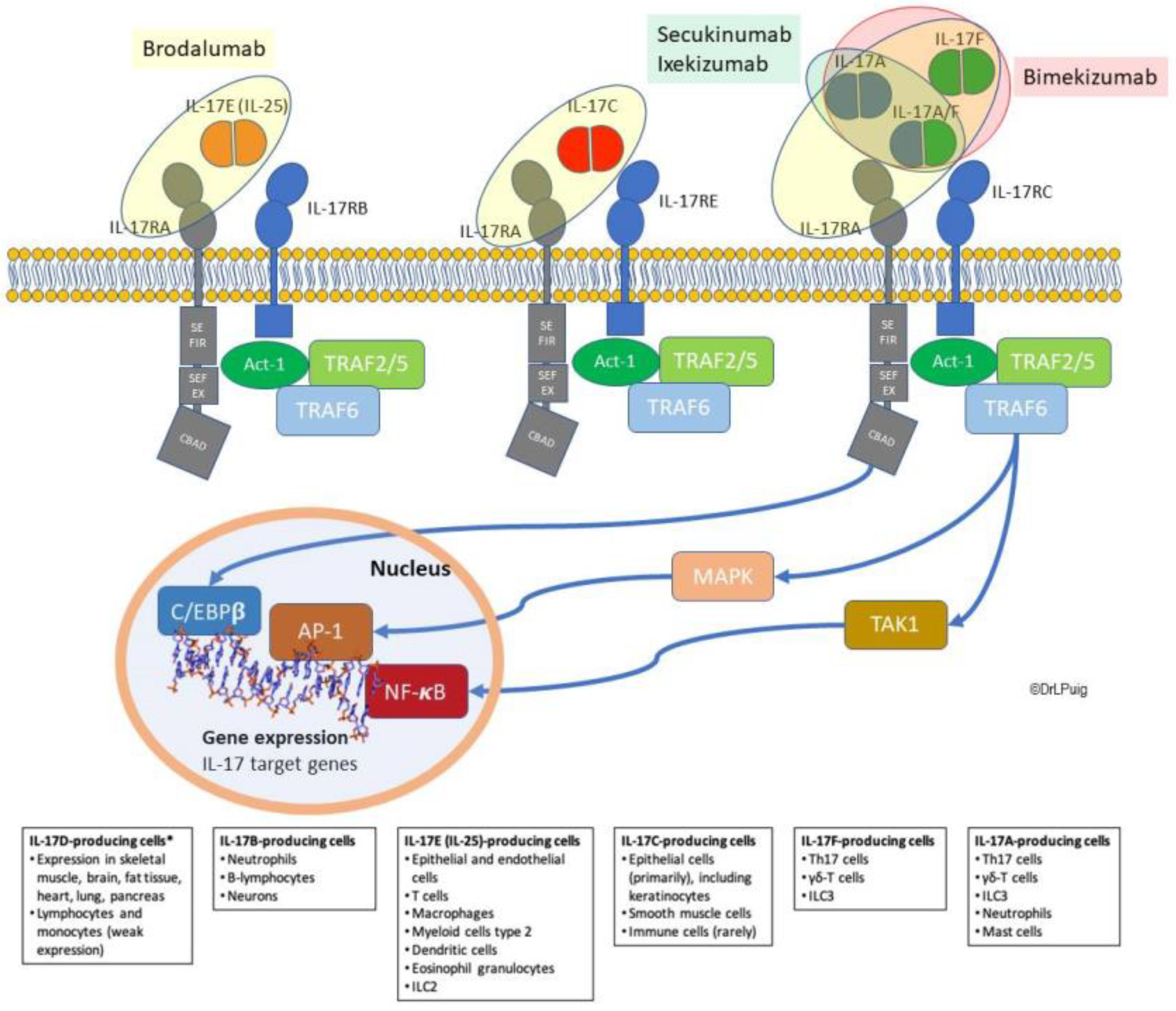
2.4. IL-17R as Unique Receptors in Cytokine Signaling: Focus on IL-17RA

The IL-17 receptor (IL-17R) family comprises five receptor subunits, correlatively named IL-17RA to IL-17RE, which are not homologous to any known receptors [27]. The receptor through which IL-17A, IL-17F, and IL-17AF signal is the IL-17RA/IL-17RC heteroreceptor. Interestingly, IL-17RA is a common subunit shared by other IL-17 receptors. Thus, IL-17C binds to the IL-17RA/IL-17RE heteroreceptor, and IL-17E (IL-25) to IL-17RA/IL-17RB. This notwithstanding, it has been recently shown that IL-17A also activates the IL-17RA/IL-17RD complex [78], inasmuch as IL-17B ligates IL-17RA/IL-17RB [35]. Unexpectedly, a recent in vitro study has reported that IL-17F, as well as IL-17A and IL-17A/F, can also bind two IL-17RC chains [79].
Structurally, all IL-17R subunits are single transmembrane domain-containing proteins in which certain conserved structural motifs are found, including an extracellular fibronectin type-III domain and a cytoplasmic SEF/IL-17R (SEFIR) domain, which is critical to mediate ligand signaling. A region at the C-terminal side of the SEFIR domain in IL-17RA has marked sequence homology to BB´ loops found in prototypical Toll/IL-1R (TIR) domains of TLRs from the innate immune system, therefore this motif is referred to as a TIR-like loop (TILL). It is presumed that these unique signaling properties of IL-17 receptors enable Th17 cells to act as a bridge between innate and adaptive immune cells. Of note, although all IL-17Rs have a SEFIR motif, the TILL domain seems to be unique to IL-17RA, which may explain why it is needed to partner with multiple IL-17R subunits [27,80].
Upon ligand binding, IL-17RA firstly recruits and activates the proximal adaptor protein ACT1, making another difference with Toll-like receptors. ACT1, which exhibits E3 ubiquitin ligase activity, also contains a SEFIR domain comprising a CC´ loop peptide that has been found to be critical for SEFIR-SEFIR interaction between IL-17RA and ACT1 [81]. ACT1 also bridges the interaction between IL-17RA and TRAF6, a signal transducer in the NF- κ B pathway with E3 ubiquitin ligase activity. ACT1-dependent signaling cascade induces the activation of NF- κ B, ERK, p38 MAPK, and JNK pathways. Interestingly, when IL-17A activates IL-17RA/IL-17RD instead of IL-17RA/IL-17RC heterodimers, p38 MAPK and JNK are preferentially induced [78]. Moreover, despite the crucial role of ACT-1, some ACT1-independent signaling in response to IL-17Rs activation has also been described [82,83].
Additional transcription factors are involved in response to IL-17A, including C/EBP β and C/EBP δ . The induction of C/EBPδ expression requires both ACT1 and the SEFIR and TILL domains of IL-17RA, which suggests the involvement of NF-κB, while C/EBPβ expression additionally needs a CBAD only found in IL-17RA. It must be mentioned that isoform LIP of C/EBP β is one of the few known inhibitory mechanisms of IL-17 signaling, activated by phosphorylation mediated by SEFIR, TILL, and CBAD domains of IL-17RA [27,49].
A fundamental aspect of IL-17 signaling is synergism and additive responsiveness, either between IL-17A and other cytokines (e.g., TNF-α, IL- 23, IL- 1β, IL- 6, and TGF- β) or between different IL-17 isoforms (e.g., IL-17F, IL-17C, and IL-17E), as the magnitude of the inflammatory effect cannot be explained as a mere sum of its components [39,50,84,85]. Regulation of mRNA stability is critical for coordinating cytokine expression and cytokine signaling, in which AU-rich elements (AREs) and GU-rich elements (GREs) as decay elements mediating rapid post-transcriptional degradation play a pivotal role [86]. It was initially found that activation of MAPKs by IL-17RA inhibited destabilization of proteins binding to AREs, thus blocking mRNA degradation and significantly increasing the concentration of related cytokines and chemokines [27]. Surprisingly, recent results have shown that ACT1 can also directly bind to mRNA, promoting its stability and translation [87]. Enhancement of mRNA stability may be one of the mechanisms underlying IL-17 synergism. Another mechanism for synergy involves C/EBP, as observed in functional cooperation between IL-17A and TNF-α [39].
Apart from characteristic signaling by activation of the aforementioned NF-κB, MAPK, and C/EBP pathways, IL-17RA can also signal through JAK/STAT. Upon IL-17E binding, the IL-17RA/IL-17RB complex has been found to activate STAT3 in an IL-6-independent, ACT1-dependent manner, resulting in upregulation of genes related to cell cycle progression and keratinocyte proliferation [53].
Additionally, IL-17RA interactions with a different type of receptor, such as epidermal growth factor receptor (EGFR), have been described [39]. Apart from known KRT16-mediated interplay, a recent study showed that IL-17RA, both functionally via ACT1, and physically by surface proximity, interacts with and transactivates EGFR in keratinocytes [88], suggesting a role in IL-17A-mediated keratinocyte hyperproliferation.
Expression of IL-17RA is ubiquitous, whereas its surface expression is widely variable [47,89,90]. IL-17RA mRNA has been found in murine spleen, kidney, liver, lung, brain, heart, skeletal muscle, and testes tissues, as well as in a wide variety of cell lines [91], although fibroblasts and endothelial cells have been reported to be the main responsive cells to IL-17A signal [89]. The broad distribution of IL-17RA, along with the large number of IL-17A-secreting cells and their migratory potential, underpin the IL-17A capacity to act on multiple cellular systems [47]. This contrasts with the localized expression of other IL-17 receptor subunits, such as IL-17RE, which is restricted to epithelial cells, thus limiting the activity of its ligand IL-17C to this cell type [47].
The expression of IL-17RA is dynamically modulated by the stimuli received by cells. IL-17RA expressed by CD8+ T-cells is induced by IL-15 and IL-21, and inhibited by IL-2 [92]. In dermal fibroblasts, UV irradiation induced a decrease in IL-6, IL-8, and CXCL-1 production simultaneously, with a decrease in IL-17RA and IL-17RC expression [93]. On the other hand, high levels of receptor expression are required for an effective IL-17RA response, implying a functional significance for receptor expression. Finally, IL-17RA internalization of its ligand IL-17 after binding is another mechanism by which IL-17RA may self-regulate its expression and signaling [89].

3. Targeting IL-17 Pathway in Psoriasis Treatment

3.1. Treatments Acting by Ligand Inhibition

3.1.1. Secukinumab

Secukinumab was the first IL-17-targeted therapy approved for psoriasis. Secukinumab is a recombinant, fully human monoclonal IgG1 κ antibody designed to target and neutralize the actions of IL-17A. It binds the IL-17A homodimer with high affinity (KD ~ 60—370 pM) and potency (0.14–0.4 nM), thus inhibiting its interaction with the IL-17RA receptor subunit. Although secukinumab is highly selective for IL-17A, it exhibits low cross-reactivity with the IL-17AF heterodimer (IC50 3.3 nM), and even lower with IL-17F (IC50 ∼ 2 μM). Considering that IL-17F’s affinity to its receptor is substantially higher than to secukinumab, it is presumed not to contribute to secukinumab activity in therapeutic doses. Secukinumab does not cross-react with the other IL-17 family members [94].
Gene expression patterns from psoriasis lesional skin were analyzed in the psoriasis proof-of-concept study [95] after a single secukinumab dose of 3 mg/kg i.v., using quantitative RT-PCR and microarrays. Significant down-regulation of IL-12B, IL-17A, IL-17F, IL-21, IL-22, IFN- γ , CCL20, and TNF- α (by RT-PCR) and IL-6, IL-8, KRT16, and DEFB4 (by microarray analysis) was observed [95]. Regarding IL-17F, additional results from a 3-study pooled analysis found no consistent changes over time in the median serum IL-17F concentrations after secukinumab treatment [94].
The clinical efficacy of IL-17A inhibition by secukinumab in psoriasis has been extensively studied in multiple clinical trials. Briefly, a phase III study of secukinumab (150 mg and 300 mg) versus placebo in patients with moderate-to-severe plaque psoriasis demonstrated a significantly higher proportion of patients with PASI 75 at week 12 in the secukinumab groups (71.6% (150 mg) and 81.6% (300 mg)) than in the placebo group (4.5%; p < 0.001). Additional phase III studies demonstrated superiority of secukinumab over etanercept (PASI 75 at week 12: 67.0% (150 mg) and 77.1% (300 mg) vs. 44.0%; p < 0.001) and ustekinumab (PASI 90 at week 16: 79.0% (300 mg) vs. 57.6%; p < 0.001). Other relevant endpoints, such as improvement in PASI and IGA mod 2011 score were also significantly better with secukinumab across studies. However, when compared to guselkumab, the efficacy of the latter was superior (PASI 90 at week 48: 70.0% (300 mg) vs. 84.0%; p < 0.001) [96,97].
Overall, the safety and tolerability profile of secukinumab is favorable. As observed with other IL-17A inhibitors, an adjusted higher rate of Candida infections, mostly mucosal or cutaneous candidiasis, have been reported, which is consistent with the important role of IL-17A against fungal infections [98]. Paradoxically, case series of cutaneous inflammatory eruptions have been described from secukinumab—and ixekizumab—real life use [99,100]. Although the mechanism is yet unclear, it has been hypothesized that a Th1/Th2 imbalance favoring Th2 pathway, induced by IL-17A inhibitors and/or a IL-22-mediated mechanism—as IL-17A inhibition do not decrease IL-22 levels—might underlie these inflammatory eruptions [99]. More recently, they have been proposed to be a consequence of the selective block of IL-17A, which might induce the overexpression of isoform IL-17C, which is mainly produced by epithelial cells and seems to be involved in the pathogenesis of atopic dermatitis [100].

3.1.2. Ixekizumab

Ixekizumab is a humanized IgG4 monoclonal antibody and the second approved drug designed to target and neutralize the actions of IL-17A. It selectively binds to IL-17A with high affinity (KD < 3 pM). Ixekizumab neither binds IL-17F nor any of the other members of the IL-17 cytokine family [101].
The effect of IL-17 neutralization on gene expression was analyzed in an exploratory phase I study [102]. Expression of cytokines from multiple T cell subsets (Il-17A, IL-17F, IL-22, IFN- γ , IL-23/p19, and IL12/IL23p40), chemokines and antimicrobial peptides from keratinocytes (LL37, β -defensin 2, S100A7, and S100A8), as well as markers of keratinocyte proliferation (Ki67 and KRT16) were found to decrease after treatment in lesional skin from psoriasis patients.
The clinical efficacy of ixekizumab in psoriasis has been demonstrated in multiple clinical trials. Briefly, a phase III study of ixekizumab (80 mg Q2W and 80 mg Q4W) versus placebo in patients with moderate-to-severe plaque psoriasis resulted in a significantly higher PASI 75 response rate at week 12 in the treatment groups (89.1% and 82.6%, respectively) than in the placebo group (3.9%; p < 0.001). The percentage of patients with sPGA score of 0 or 1 (sPGA 0/1) was also significantly higher in the treatment groups. Two clinical trials compared both dose regimens of ixekizumab with etanercept, showing superiority for ixekizumab (PASI 75 at week 12: 87.3–89.7% (Q2W) and 77.5–84.2% (Q4W) vs. 41.6–53.4%; p < 0.001). Results from sPGA 0/1 also showed ixekizumab superiority. Comparisons with ustekinumab were addressed in an additional phase III study, which also demonstrated a significantly higher PASI 90 response rate at week 12 with ixekizumab, than with the active comparator (72.8% vs. 42.9%; p < 0.001). PASI 75, PASI 100, and sPGA 0/1 were relevant endpoints with also significantly higher response rates for ixekizumab compared to ustekinumab [97,103].
Overall, ixekizumab has a favorable safety profile and is well tolerated. As found with secukinumab, higher adjusted rates of Candida infections were described in ixekizumab clinical trials [104], and ‘paradoxical’ cutaneous eruptions have been reported in real life use [99,100].

3.1.3. Bimekizumab

Bimekizumab (UCB4940) is an IL-17 inhibitor currently under phase III development for psoriasis. Unlike secukinumab and ixekizumab, which are specific for IL-17A, bimekizumab is a divalent humanized monoclonal IgG1 antibody designed to bind a similar site on both IL-17A (KD 3.2 pM) and IL-17F (KD 23 pM), thus conveying dual inhibition [105,106].
Transcriptional analysis of fibroblasts and synoviocytes from patients with psoriatic arthritis has been performed in an exploratory study. Bimekizumab, compared to an IL-17A inhibitor, induced a more profound anti-inflammatory effect in a large panel of inflammation-linked genes, including CCL20, CCL7, IL-6, IL-1b, and several chemokine ligands (CXCL), among others. The functional significance of dual blockade was confirmed by investigating the chemotactic potential of neutrophils towards Th17-stimulated fibroblasts. Dual IL-17A/IL-17F inhibition with bimekizumab achieved significantly more inhibition of neutrophil migration than neutralization of IL-17A or IL-17F alone. Additional results from the same study showed that, in the presence of TNF, both IL-17A and IL-17F (yet the latter with less potency) triggered a pro-inflammatory response with significantly increased production of IL-8 and IL-6. When these cells were exposed to bimekizumab as well as IL-17A, IL-17F, and TNF inhibitors, dual neutralization induced significantly greater down-regulation of IL-8, IL-6, and MMP3 than an IL-17A blockade alone, while an IL-17F blockade alone had no significant effect, and TNF inhibition had only a modest impact [106].
Results on clinical efficacy in plaque psoriasis are available from a phase I study in mild-to-moderate patients [105], and a phase IIb study in moderate-to-severe patients [107]. In the phase I study, bimekizumab doses ranging from 40 to 640 mg were compared to placebo [105], while in the phase IIb bimekizumab doses ranged from 64 mg to 480 mg, some of them preceded by a loading dose [107]. In mild-to-moderate patients, a mean reduction of >65% at week 2 from baseline PASI scores was observed in the 480 mg and 640 mg treatment groups. The maximum PASI improvement observed in these two groups was ≥94%. Bimekizumab 160 mg also improved PASI, though in a lesser magnitude. Comparable improvement was found in LSS and PGA scores. Responses to treatment were maintained to week 12. In moderate-to-severe patients, the percentage achieving PASI 90 at week 12 were significantly higher across all bimekizumab groups (46.2–79.1%) than in placebo group (0%; p < 0.0001). Other relevant endpoints, such as PASI 75, PASI 100, and PGA, were also significantly improved in bimekizumab groups compared to placebo [107].
Results from a phase III clinical trial comparing bimekizumab with adalimumab in moderate-to-severe plaque psoriasis patients have shown that bimekizumab was superior to adalimumab in reducing symptoms and signs of plaque psoriasis, but had a higher frequency of oral candidiasis and diarrhea [108]. Two more phase III studies with an active comparator have shown that bimekizumab is more effective than ustekinumab [109] and secukinumab [110].

3.2. Treatments Acting by Receptor Blockade

Brodalumab

Brodalumab is the first drug targeting a receptor instead of a ligand of the IL-17 pathway. Brodalumab is a human monoclonal IgG2 antibody that specifically binds IL-17RA with high affinity (KD 0.24 nM) and blocks downstream signaling of multiple IL-17 family isoforms, including IL-17A, IL-17F, IL-17A/F heterodimer, and IL-25 [111].
Molecular and histological analyses of skin biopsy specimens in studies of subjects with psoriasis were conducted as part of three different clinical trials. The first was a phase I study evaluating different brodalumab single doses (i.e., 140 mg SC, 350 mg SC, and 700 mg IV) versus placebo [112]. Molecular results after brodalumab administration evidenced rapid and significant improvements in lesional skin mRNA levels for a number of IL-17-modulated keratinocyte-derived factors, including β-defensin 4, cathelicidin, KRT16, CCL18, and CCL20. Moreover, the mRNA levels of IL-17A, IL-17C, and IL-17F were reduced to non-lesional levels over 6 weeks; those of IL-22 and both subunits of IL-23 were also reduced. Histologically, significant reductions in epidermal thickening, KRT16 levels, and Ki67-expressing cells were observed.
KRT16 and CD3 skin biomarkers were also measured in a biopsy sub-study of a phase II clinical trial evaluating brodalumab (doses of 70 mg, 140 mg, 210 mg, and 280 mg) versus placebo [113]. KRT16 staining of the upper epidermis was reduced in a dose-dependent manner across the 140 to 280 mg groups, and dermal CD3 counts decreased significantly from baseline across the 70 to 210 mg brodalumab groups. Additionally, epidermal thickness decreased significantly from baseline across the 140 to 280 mg groups.
The phase III study AMAGINE-1 [114], which evaluated brodalumab at doses of 140 mg and 210 mg versus placebo, also included a biopsy sub-study. Gene expression of IL-17 pathway cytokines (IL-17A, IL-17C, IL-17F, p19, and p40) decreased significantly at week 12 in a dose-dependent manner. Expression of IL-17C and both IL-23 subunits (IL-23p19 and IL-12p40) were reduced to near non-lesional levels, whereas IL-17A and IL-17F remained above non-lesional levels. From week 12 to 52, expression of all biomarkers in patients not re-randomized to placebo at week 12, continued decreasing, so that they were at or below non-lesional levels at week 52. Likewise, histological analysis evidenced that epidermal thickening, keratinocyte proliferation (Ki67, KRT16), and T-cell infiltrates (CD3 and CD8) were significantly reduced in samples from brodalumab groups at week 12 and continued improving up to week 52. Interestingly, samples from patients initially assigned to placebo and subsequently re-randomized to brodalumab showed no changes in biomarkers at week 12, but significant improvement at week 52, similar to that found in not re-randomized patients [114]. A mechanistic sub-study carried out with a subset of patients from three studies comprising the AMAGINE program was consistent with earlier results, showing a significant and progressive decrease in cellular infiltrates (CD31, CD81, CD11c1, and CD1631), markers of keratinocyte proliferation (Ki671 and KRT16), and inflammatory cytokines (IL-17A/C/F, p19, and p40), reaching close to non-lesional levels. Moreover, results confirmed that brodalumab induces transcriptional changes improving IL-17–dependent gene expression compared to ustekinumab, and such improvement correlated with PASI [115].
The clinical efficacy of brodalumab was thoroughly assessed over its clinical program, including one phase III clinical trial versus placebo (AMAGINE-1) and two phase III clinical trials versus ustekinumab (AMAGINE-2 and AMAGINE-3) [97,116]. Results from AMAGINE-1 indicated that, at week 12, a significantly greater proportion of patients in brodalumab groups achieved PASI 75 (60.3% (140 mg) and 83.3% (240 mg)) compared to placebo (2.7%; p < 0.001). Co-primary endpoint sPGA 0/1 was also significantly higher with brodalumab. The superiority of brodalumab over ustekinumab was also demonstrated. The AMAGINE-2 study showed that PASI 75 response at week 12 was greater with brodalumab (67% (140 mg) and 86% (240 mg)) than placebo (8%; p < 0.001). Co-primary endpoint sPGA 0/1 response was also significantly better with brodalumab than placebo. The second co-primary endpoint was PASI 100 response at week 12 compared to ustekinumab. Results demonstrated superiority of brodalumab 240 mg over ustekinumab (44% vs. 22%; p < 0.001). AMAGINE-3 reported similar results on PASI 75 at week 12 (69% (140 mg) and 85% (240 mg)) vs. 6% (placebo); p < 0.001) and sPGA 0/1, and met the PASI 100 co-primary endpoint at both doses (27% (140 mg) and 37% (240 mg) vs. 19%; p = 0.008 and p < 0.001, respectively). On the other hand, the uniqueness of brodalumab mechanism of action might explain its early onset of action, the fastest among biologics for psoriasis (<2 weeks) [117]. Furthermore, a recent systematic review and meta-analysis on long-term evidence available for the efficacy of systemic biologic and non-biologic drugs in moderate-to-severe psoriasis has estimated brodalumab as the most effective treatment in terms of both PASI response and probability of complete clearance [118].
Similar to secukinumab and ixekizumab, increased rates of Candida infections, mostly mucosal and cutaneous, and mild to moderate in severity, were reported from clinical trials [97]. In contrast, the risk for invasive fungal infections with these medications remains virtually close to zero to date, as no cases of deep fungal infections with IL-17-targeted treatments have been reported, according to a recent revision of randomized clinical trials including IL-12 and/or IL-23-targeted drugs [119]. The broad-targeting action of brodalumab rose concerns on its safety, based on the presumtion that blocking IL-17 isoforms besides IL-17A would increase the risk of impairing their additional physiological activity. Nevertheless, evidence from clinical trials and meta-analyses demonstrates that the safety profile of brodalumab is similar to those of other anti-IL17 biologics as well as ustekinumab [120,121].

3.3. Potential Role of IL-17A/IL-17RA Blockade in Psoriasis Comorbid Conditions

The potential effect of IL-17-targeted treatments on comorbidities, sharing with psoriasis a pathogenic role for the IL-17 pathway, is a field of growing interest to the available and upcoming anti-IL-17 biologics, considering some promising results observed earlier with anti-TNF α treatments [122].
In this regard, the impact of secukinumab has been assessed in an exploratory study including moderate-to-severe psoriasis patients without clinical CV disease and healthy volunteers [123]. Results indicated a significant improvement in endothelial function (determined as increased flow-mediated dilation) after 52 weeks of treatment with secukinumab compared to placebo. However, no significant changes in other relevant CV parameters were found. In the same vein, effects of ixekizumab on metabolic and CV-related markers and parameters have been evaluated, as well in moderate-to-severe psoriasis patients participating in three clinical trials for up to 60 weeks [124]. Results did not evidence consistent improvements over the study period.
Neutralization of IL-17 has shown promising results on NAFLD in mice, inducing a decrease in pro-inflammatory cytokine levels, attenuation of hepatic lipid accumulation, and improvement of liver function [125,126] by a mechanism of IL-17-related fatty acid metabolism suppression [126]. On the other hand, it has been found that IL-17RA−/− mice on a high-fat diet experience weight gain and hepatic triglyceride accumulation, but are protected from glucose metabolism dysfunction and progression of simple steatosis to steatohepatitis [76]. These findings suggest a potential role of IL-17A/IL-17RA blockade in comorbid NAFLD, but no clinical evidence in humans has been provided to date.

4. Conclusions

Psoriasis is a challenging systemic inflammatory disease that appears more complex with increasing knowledge of its pathogenic mechanisms, currently based on the critical involvement of IL-17 cytokines. Not surprisingly, a wide range of pharmacologic therapies are available for psoriasis treatment, including several biologics targeting the IL-17 pathway. Within this class of drugs, secukinumab and ixekizumab bind to and antagonize IL-17A and IL-17A/F, whereas brodalumab was developed to target and block the IL-17 receptor. By this unique mechanism, brodalumab binds IL-17RA and inhibits signaling of IL-17 isoforms, in addition to IL-17A and IL-17A/F, providing high efficacy, fast onset of action, and high probability of complete psoriasis clearance. Furthermore, the apparent pleiotropic effects of IL-17 isoforms signaling in psoriasis comorbidities is an exciting field of research that warrants future study.

Author Contributions

Conceptualization and writing—original draft preparation, all authors; writing—review and editing, all authors; All authors have read and agreed to the published version of the manuscript.

Funding

This work and the article processing charges (APC) was funded by LEO Pharma grant. The sponsors had no role in the design, execution, interpretation, or writing of the work.

Institutional Review Board Statement

Not applicable.

Informed Consent Statement

Not applicable.

Acknowledgments

The authors would like to thank LEO Pharma for their continued and constant support for this project, GOC Health consulting for their support and collaboration in the development of this document, and Esther Prieto for editorial assistance.

Conflicts of Interest

Silvia Vidal: Her institution has received grants or contracts from Novartis, BMS, Roche, Almirall, and Precigen, and she has received payment or honoraria for lectures, presentations, speaker’s bureaus, manuscript writing, or educational events from Sanofi, Eusa-pharma, Novartis, Roche, and Precigen. José-Manuel Carrascosa-Carrillo: His institution has received grants, has given research support, or participation in clinical trials from Abbvie, Almirall, Amgen, Boehringer Ingelheim, Celgene, Janssen, Leo-Pharma, Lilly, Novartis, Pfizer, Sanofi, and Sandoz. He has received honoraria or consultation fees from Abbvie, Almirall, Amgen, Biogen, Boehringer Ingelheim, Bristol Myers Squibb, Celgene, Fresenius-Kabi, Janssen, Leo-Pharma, Lilly, Mylan, Novartis, Pfizer, Sandoz, Samsung-Bioepis, Sanofi, and UCB. He has participated in a company sponsored speaker’s bureau: Celgene, Janssen, Lilly, Novartis, Pfizer, Almirall, and UCB. Álvaro González-Cantero: His institution has received grants or contracts from: Leo-Pharma, Almirall, and Celgene. He has received payment or honoraria for lectures, presentations, speaker’s bureaus, manuscript writing, or educational events from: Abbie, Janssen, Novartis, Almirall, Celgene, UCB, L´Oreal, MSD, and Leo Pharma. He has been given support for attending meetings and/or travel from: Abbvie, Janssen, Novartis, Almirall, Celgene, UCB, and Leo Pharma. He has participated on a Data Safety Monitoring Board or Advisory Board for: Abbie, Janssen, Novartis, Almirall, Celgene, UCB, L´Oreal, MSD, and Leo Pharma. Lluís Puig: His institution has received grants/research supports or participation in clinical trials from: Abbvie, Almirall, Amgen, Boehringer Ingelheim, Celgene, Janssen, Leo-Pharma, Lilly, Novartis, Pfizer, Regeneron, Roche, Sanofi, and UCB. He has received honoraria or consultation fees from: Abbvie, Almirall, Amgen, Baxalta, Biogen, Boehringer Ingelheim, Bristol Myers Squibb, Celgene, Fresenius-Kabi, Janssen, JS BIOCAD, Leo-Pharma, Lilly, Mylan, Novartis, Pfizer, Regeneron, Roche, Sandoz, Samsung-Bioepis, Sanofi, and UCB. He has participated in a company sponsored speaker’s bureau: Celgene, Janssen, Lilly, Novartis, and Pfizer. José-Carlos Ruiz-Carrascosa: The author declares no conflict of interest. Antonio-Manuel Velasco-Pastor has received fees for advice and speeches from Allmirall, Leo Pharma, Novartis, Janssen, Lilly, Abbvie, Gebro, Pfizer, Bristol, and Amgen.

Abbreviations

CBADC/EBPβ-activation domain
C/EBPCCAAT/enhancer-binding protein
CTLA8Cytotoxic T lymphocyte-associated antigen 8
CVCardiovascular
DCDendritic cells
DEFB4β-defensin 4
EGFREpidermal growth factor receptor
IGA mod 20112011 modified Investigator’s Global Assessment
ILC3Group 3 innate lymphoid cells
JNKc-Jun N-terminal kinase
LSSLesion Severity Score
NAFLDNon-alcoholic fatty liver disease
PASIPsoriasis Area and Severity Index
PGAPhysician´s Global Assessment
RORCRAR-related orphan receptor
SEFIRSEF (similar expression to fibroblast growth factor genes)/IL-17 receptor
TIRToll/IL-1 receptor
TLRToll-like receptor
TNFTumor necrosis factor
TRAF6TNFR-associated factor 6
VEGFVascular endothelial growth factor

References

  1. Parisi, R.; Iskandar, I.Y.K.; Kontopantelis, E.; Augustin, M.; Griffiths, C.E.M.; Ashcroft, D.M. Global Psoriasis Atlas National, Regional, and Worldwide Epidemiology of Psoriasis: Systematic Analysis and Modelling Study. BMJ 2020, 369, m1590. [Google Scholar] [CrossRef] [PubMed]
  2. Rendon, A.; Schäkel, K. Psoriasis Pathogenesis and Treatment. Int. J. Mol. Sci. 2019, 20, 1475. [Google Scholar] [CrossRef] [PubMed] [Green Version]
  3. Armstrong, A.W.; Read, C. Pathophysiology, Clinical Presentation, and Treatment of Psoriasis: A Review. JAMA 2020, 323, 1945–1960. [Google Scholar] [CrossRef]
  4. Shuster, S. Research into Psoriasis—The Last Decade. Br. Med. J. 1971, 3, 236–239. [Google Scholar] [CrossRef] [Green Version]
  5. Bjerke, J.R. In Situ Characterization and Counting of Mononuclear Cells in Lesions of Different Clinical Forms of Psoriasis. Acta Derm Venereol. 1982, 62, 93–100. [Google Scholar] [PubMed]
  6. Bjerke, J.R. Subpopulations of Mononuclear Cells in Lesions of Psoriasis, Lichen Planus and Discoid Lupus Erythematosus Studied Using Monoclonal Antibodies. Acta Derm. Venereol. 1982, 62, 477–483. [Google Scholar]
  7. Hammar, H.; Gu, S.Q.; Johannesson, A.; Sundkvist, K.G.; Biberfeld, P. Subpopulations of Mononuclear Cells in Microscopic Lesions of Psoriatic Patients. Selective Accumulation of Suppressor/Cytotoxic T Cells in Epidermis during the Evolution of the Lesion. J. Invest. Derm. 1984, 83, 416–420. [Google Scholar] [CrossRef] [Green Version]
  8. Mueller, W.; Herrmann, B. Cyclosporin A for Psoriasis. N. Engl. J. Med. 1979, 301, 555. [Google Scholar] [CrossRef]
  9. Ellis, C.N.; Gorsulowsky, D.C.; Hamilton, T.A.; Billings, J.K.; Brown, M.D.; Headington, J.T.; Cooper, K.D.; Baadsgaard, O.; Duell, E.A.; Annesley, T.M. Cyclosporine Improves Psoriasis in a Double-Blind Study. JAMA 1986, 256, 3110–3116. [Google Scholar] [CrossRef]
  10. Jegasothy, B.V.; Ackerman, C.D.; Todo, S.; Fung, J.J.; Abu-Elmagd, K.; Starzl, T.E. Tacrolimus (FK 506)—A New Therapeutic Agent for Severe Recalcitrant Psoriasis. Arch. Derm. 1992, 128, 781–785. [Google Scholar] [CrossRef] [Green Version]
  11. Gottlieb, S.L.; Gilleaudeau, P.; Johnson, R.; Estes, L.; Woodworth, T.G.; Gottlieb, A.B.; Krueger, J.G. Response of Psoriasis to a Lymphocyte-Selective Toxin (DAB389IL-2) Suggests a Primary Immune, but Not Keratinocyte, Pathogenic Basis. Nat. Med. 1995, 1, 442–447. [Google Scholar] [CrossRef]
  12. Mosmann, T.R.; Cherwinski, H.; Bond, M.W.; Giedlin, M.A.; Coffman, R.L. Two Types of Murine Helper T Cell Clone. I. Definition According to Profiles of Lymphokine Activities and Secreted Proteins. J. Immunol. 1986, 136, 2348–2357. [Google Scholar] [PubMed]
  13. Yawalkar, N.; Karlen, S.; Hunger, R.; Brand, C.U.; Braathen, L.R. Expression of Interleukin-12 Is Increased in Psoriatic Skin. J. Invest. Derm. 1998, 111, 1053–1057. [Google Scholar] [CrossRef]
  14. Mease, P.J.; Goffe, B.S.; Metz, J.; VanderStoep, A.; Finck, B.; Burge, D.J. Etanercept in the Treatment of Psoriatic Arthritis and Psoriasis: A Randomised Trial. Lancet 2000, 356, 385–390. [Google Scholar] [CrossRef]
  15. Chaudhari, U.; Romano, P.; Mulcahy, L.D.; Dooley, L.T.; Baker, D.G.; Gottlieb, A.B. Efficacy and Safety of Infliximab Monotherapy for Plaque-Type Psoriasis: A Randomised Trial. Lancet 2001, 357, 1842–1847. [Google Scholar] [CrossRef]
  16. Papoutsaki, M.; Chimenti, M.-S.; Costanzo, A.; Talamonti, M.; Zangrilli, A.; Giunta, A.; Bianchi, L.; Chimenti, S. Adalimumab for Severe Psoriasis and Psoriatic Arthritis: An Open-Label Study in 30 Patients Previously Treated with Other Biologics. J. Am. Acad. Derm. 2007, 57, 269–275. [Google Scholar] [CrossRef]
  17. Oppmann, B.; Lesley, R.; Blom, B.; Timans, J.C.; Xu, Y.; Hunte, B.; Vega, F.; Yu, N.; Wang, J.; Singh, K.; et al. Novel P19 Protein Engages IL-12p40 to Form a Cytokine, IL-23, with Biological Activities Similar as Well as Distinct from IL. Immunity 2000, 13, 715–725. [Google Scholar] [CrossRef] [Green Version]
  18. Aggarwal, S.; Ghilardi, N.; Xie, M.-H.; de Sauvage, F.J.; Gurney, A.L. Interleukin-23 Promotes a Distinct CD4 T Cell Activation State Characterized by the Production of Interleukin. J. Biol. Chem. 2003, 278, 1910–1914. [Google Scholar] [CrossRef] [Green Version]
  19. Wilson, N.J.; Boniface, K.; Chan, J.R.; McKenzie, B.S.; Blumenschein, W.M.; Mattson, J.D.; Basham, B.; Smith, K.; Chen, T.; Morel, F.; et al. Development, Cytokine Profile and Function of Human Interleukin 17-Producing Helper T Cells. Nat. Immunol. 2007, 8, 950–957. [Google Scholar] [CrossRef] [PubMed]
  20. Langrish, C.L.; Chen, Y.; Blumenschein, W.M.; Mattson, J.; Basham, B.; Sedgwick, J.D.; McClanahan, T.; Kastelein, R.A.; Cua, D.J. IL-23 Drives a Pathogenic T Cell Population That Induces Autoimmune Inflammation. J. Exp. Med. 2005, 201, 233–240. [Google Scholar] [CrossRef] [Green Version]
  21. Louten, J.; Boniface, K.; de Waal Malefyt, R. Development and Function of TH17 Cells in Health and Disease. J. Allergy Clin. Immunol. 2009, 123, 1004–1011. [Google Scholar] [CrossRef] [PubMed]
  22. Lee, E.; Trepicchio, W.L.; Oestreicher, J.L.; Pittman, D.; Wang, F.; Chamian, F.; Dhodapkar, M.; Krueger, J.G. Increased Expression of Interleukin 23 P19 and P40 in Lesional Skin of Patients with Psoriasis Vulgaris. J. Exp. Med. 2004, 199, 125–130. [Google Scholar] [CrossRef]
  23. Chen, X.; Tan, Z.; Yue, Q.; Liu, H.; Liu, Z.; Li, J. The Expression of Interleukin-23 (P19/P40) and Interleukin-12 (P35/P40) in Psoriasis Skin. J. Huazhong Univ. Sci. Technol. Med. Sci. 2006, 26, 750–752. [Google Scholar] [CrossRef] [PubMed]
  24. Piskin, G.; Sylva-Steenland, R.M.R.; Bos, J.D.; Teunissen, M.B.M. In Vitro and in Situ Expression of IL-23 by Keratinocytes in Healthy Skin and Psoriasis Lesions: Enhanced Expression in Psoriatic Skin. J. Immunol. 2006, 176, 1908–1915. [Google Scholar] [CrossRef] [PubMed]
  25. Schön, M.P.; Erpenbeck, L. The Interleukin-23/Interleukin-17 Axis Links Adaptive and Innate Immunity in Psoriasis. Front. Immunol. 2018, 9, 1323. [Google Scholar] [CrossRef] [Green Version]
  26. Lauffer, F.; Eyerich, K.; Boehncke, W.-H.; Asadullah, K.; Beissert, S.; Ghoreschi, K.; Schön, M.P. Cytokines of the IL-17 Family in Psoriasis. J. Dtsch. Derm. Ges. 2020, 18, 675–681. [Google Scholar] [CrossRef]
  27. Gaffen, S.L. Structure and Signalling in the IL-17 Receptor Family. Nat. Rev. Immunol. 2009, 9, 556–567. [Google Scholar] [CrossRef] [Green Version]
  28. Kolls, J.K.; Lindén, A. Interleukin-17 Family Members and Inflammation. Immunity 2004, 21, 467–476. [Google Scholar] [CrossRef] [Green Version]
  29. Rouvier, E.; Luciani, M.F.; Mattéi, M.G.; Denizot, F.; Golstein, P. CTLA-8, Cloned from an Activated T Cell, Bearing AU-Rich Messenger RNA Instability Sequences, and Homologous to a Herpesvirus Saimiri Gene. J. Immunol. 1993, 150, 5445–5456. [Google Scholar]
  30. McGeachy, M.J.; Cua, D.J.; Gaffen, S.L. The IL-17 Family of Cytokines in Health and Disease. Immunity 2019, 50, 892–906. [Google Scholar] [CrossRef]
  31. Hymowitz, S.G.; Filvaroff, E.H.; Yin, J.P.; Lee, J.; Cai, L.; Risser, P.; Maruoka, M.; Mao, W.; Foster, J.; Kelley, R.F.; et al. IL-17s Adopt a Cystine Knot Fold: Structure and Activity of a Novel Cytokine, IL-17F, and Implications for Receptor Binding. Embo J. 2001, 20, 5332–5341. [Google Scholar] [CrossRef] [PubMed]
  32. Liu, S.; Song, X.; Chrunyk, B.A.; Shanker, S.; Hoth, L.R.; Marr, E.S.; Griffor, M.C. Crystal Structures of Interleukin 17A and Its Complex with IL-17 Receptor, A. Nat. Commun. 2013, 4, 1888. [Google Scholar] [CrossRef] [PubMed]
  33. Brembilla, N.C.; Senra, L.; Boehncke, W.-H. The IL-17 Family of Cytokines in Psoriasis: IL-17A and Beyond. Front. Immunol. 2018, 9, 1682. [Google Scholar] [CrossRef] [Green Version]
  34. Monin, L.; Gaffen, S.L. Interleukin 17 Family Cytokines: Signaling Mechanisms, Biological Activities, and Therapeutic Implications. Cold Spring Harb. Perspect. Biol. 2018, 10. [Google Scholar] [CrossRef] [PubMed]
  35. Ramirez-Carrozzi, V.; Ota, N.; Sambandam, A.; Wong, K.; Hackney, J.; Martinez-Martin, N.; Ouyang, W.; Pappu, R. Cutting Edge: IL-17B Uses IL-17RA and IL-17RB to Induce Type 2 Inflammation from Human Lymphocytes. J. Immunol. 2019, 202, 1935–1941. [Google Scholar] [CrossRef] [Green Version]
  36. Swaidani, S.; Liu, C.; Zhao, J.; Bulek, K.; Li, X. TRAF Regulation of IL-17 Cytokine Signaling. Front. Immunol. 2019, 10, 1293. [Google Scholar] [CrossRef] [Green Version]
  37. Chen, K.; Kolls, J.K. Interleukin-17A (IL17A). Gene 2017, 614, 8–14. [Google Scholar] [CrossRef] [Green Version]
  38. Beringer, A.; Noack, M.; Miossec, P. IL-17 in Chronic Inflammation: From Discovery to Targeting. Trends Mol. Med. 2016, 22, 230–241. [Google Scholar] [CrossRef]
  39. Furue, M.; Furue, K.; Tsuji, G.; Nakahara, T. Interleukin-17A and Keratinocytes in Psoriasis. Int. J. Mol. Sci. 2020, 21, 1275. [Google Scholar] [CrossRef] [Green Version]
  40. Heidenreich, R.; Röcken, M.; Ghoreschi, K. Angiogenesis Drives Psoriasis Pathogenesis. Int. J. Exp. Pathol. 2009, 90, 232–248. [Google Scholar] [CrossRef]
  41. Numasaki, M.; Tsukamoto, H.; Tomioka, Y.; Nishioka, Y.; Ohrui, T. A Heterodimeric Cytokine, Consisting of IL-17A and IL-17F, Promotes Migration and Capillary-Like Tube Formation of Human Vascular Endothelial Cells. Tohoku J. Exp. Med. 2016, 240, 47–56. [Google Scholar] [CrossRef] [PubMed] [Green Version]
  42. Ishigame, H.; Kakuta, S.; Nagai, T.; Kadoki, M.; Nambu, A.; Komiyama, Y.; Fujikado, N.; Tanahashi, Y.; Akitsu, A.; Kotaki, H.; et al. Differential Roles of Interleukin-17A and -17F in Host Defense against Mucoepithelial Bacterial Infection and Allergic Responses. Immunity 2009, 30, 108–119. [Google Scholar] [CrossRef] [Green Version]
  43. Burns, L.A.; Maroof, A.; Marshall, D.; Steel, K.J.A.; Lalnunhlimi, S.; Cole, S.; Catrina, A.; Kirkham, B.; Taams, L.S. Presence, Function, and Regulation of IL-17F-Expressing Human CD4+ T Cells. Eur J. Immunol. 2020, 50, 568–580. [Google Scholar] [CrossRef] [Green Version]
  44. Johansen, C.; Usher, P.A.; Kjellerup, R.B.; Lundsgaard, D.; Iversen, L.; Kragballe, K. Characterization of the Interleukin-17 Isoforms and Receptors in Lesional Psoriatic Skin. Br. J. Dermatol. 2009, 160, 319–324. [Google Scholar] [CrossRef] [PubMed]
  45. Soderstrom, C.; Berstein, G.; Zhang, W.; Valdez, H.; Fitz, L.; Kuhn, M.; Fraser, S. Ultra-Sensitive Measurement of IL-17A and IL-17F in Psoriasis Patient Serum and Skin. AAPS J. 2017, 19, 1218–1222. [Google Scholar] [CrossRef] [PubMed]
  46. Pantelyushin, S.; Haak, S.; Ingold, B.; Kulig, P.; Heppner, F.L.; Navarini, A.A.; Becher, B. Rorγt+ Innate Lymphocytes and Γδ T Cells Initiate Psoriasiform Plaque Formation in Mice. J. Clin. Invest. 2012, 122, 2252–2256. [Google Scholar] [CrossRef] [PubMed] [Green Version]
  47. Ramirez-Carrozzi, V.; Sambandam, A.; Luis, E.; Lin, Z.; Jeet, S.; Lesch, J.; Hackney, J.; Kim, J.; Zhou, M.; Lai, J.; et al. IL-17C Regulates the Innate Immune Function of Epithelial Cells in an Autocrine Manner. Nat. Immunol. 2011, 12, 1159–1166. [Google Scholar] [CrossRef]
  48. Butcher, M.J.; Waseem, T.C.; Galkina, E.V. Smooth Muscle Cell-Derived Interleukin-17C Plays an Atherogenic Role via the Recruitment of Proinflammatory Interleukin-17A+ T Cells to the Aorta. Arter. Thromb Vasc. Biol. 2016, 36, 1496–1506. [Google Scholar] [CrossRef] [Green Version]
  49. Nies, J.F.; Panzer, U. IL-17C/IL-17RE: Emergence of a Unique Axis in TH17 Biology. Front. Immunol. 2020, 11, 341. [Google Scholar] [CrossRef] [PubMed] [Green Version]
  50. Johnston, A.; Fritz, Y.; Dawes, S.M.; Diaconu, D.; Al-Attar, P.M.; Guzman, A.M.; Chen, C.S.; Fu, W.; Gudjonsson, J.E.; McCormick, T.S.; et al. Keratinocyte Overexpression of IL-17C Promotes Psoriasiform Skin Inflammation. J. Immunol. 2013, 190, 2252–2262. [Google Scholar] [CrossRef] [Green Version]
  51. Vandeghinste, N.; Klattig, J.; Jagerschmidt, C.; Lavazais, S.; Marsais, F.; Haas, J.D.; Auberval, M.; Lauffer, F.; Moran, T.; Ongenaert, M.; et al. Neutralization of IL-17C Reduces Skin Inflammation in Mouse Models of Psoriasis and Atopic Dermatitis. J. Invest. Derm. 2018, 138, 1555–1563. [Google Scholar] [CrossRef] [PubMed] [Green Version]
  52. Borowczyk, J.; Shutova, M.; Brembilla, N.C.; Boehncke, W.-H. IL-25 (IL-17E) in Epithelial Immunology and Pathophysiology. J. Allergy Clin. Immunol. 2021. [Google Scholar] [CrossRef] [PubMed]
  53. Xu, M.; Lu, H.; Lee, Y.-H.; Wu, Y.; Liu, K.; Shi, Y.; An, H.; Zhang, J.; Wang, X.; Lai, Y.; et al. An Interleukin-25-Mediated Autoregulatory Circuit in Keratinocytes Plays a Pivotal Role in Psoriatic Skin Inflammation. Immunity 2018, 48, 787–798.e4. [Google Scholar] [CrossRef] [Green Version]
  54. Senra, L.; Stalder, R.; Martinez, D.A.; Chizzolini, C.; Boehncke, W.-H.; Brembilla, N.C. Keratinocyte-Derived IL-17E Contributes to Inflammation in Psoriasis. J. Invest. Derm. 2016, 136, 1970–1980. [Google Scholar] [CrossRef] [Green Version]
  55. Senra, L.; Mylonas, A.; Kavanagh, R.D.; Fallon, P.G.; Conrad, C.; Borowczyk-Michalowska, J.; Wrobel, L.J.; Kaya, G.; Yawalkar, N.; Boehncke, W.-H.; et al. IL-17E (IL-25) Enhances Innate Immune Responses during Skin Inflammation. J. Invest. Derm. 2019, 139, 1732–1742.e17. [Google Scholar] [CrossRef] [PubMed]
  56. Bie, Q.; Jin, C.; Zhang, B.; Dong, H. IL-17B: A New Area of Study in the IL-17 Family. Mol. Immunol. 2017, 90, 50–56. [Google Scholar] [CrossRef]
  57. Kouri, V.-P.; Olkkonen, J.; Ainola, M.; Li, T.-F.; Björkman, L.; Konttinen, Y.T.; Mandelin, J. Neutrophils Produce Interleukin-17B in Rheumatoid Synovial Tissue. Rheumatology 2014, 53, 39–47. [Google Scholar] [CrossRef] [PubMed] [Green Version]
  58. Liu, X.; Sun, S.; Liu, D. IL-17D: A Less Studied Cytokine of IL-17 Family. Int Arch. Allergy Immunol. 2020, 181, 618–623. [Google Scholar] [CrossRef]
  59. Boehncke, W.-H.; Schön, M.P. Psoriasis. Lancet 2015, 386, 983–994. [Google Scholar] [CrossRef]
  60. Boehncke, W.-H. Systemic Inflammation and Cardiovascular Comorbidity in Psoriasis Patients: Causes and Consequences. Front. Immunol. 2018, 9, 579. [Google Scholar] [CrossRef] [PubMed]
  61. Karbach, S.; Hobohm, L.; Wild, J.; Münzel, T.; Gori, T.; Wegner, J.; Steinbrink, K.; Wenzel, P.; Keller, K. Impact of Psoriasis on Mortality Rate and Outcome in Myocardial Infarction. J. Am. Heart Assoc. 2020, 9, e016956. [Google Scholar] [CrossRef]
  62. Menter, A.; Krueger, G.G.; Paek, S.Y.; Kivelevitch, D.; Adamopoulos, I.E.; Langley, R.G. Interleukin-17 and Interleukin-23: A Narrative Review of Mechanisms of Action in Psoriasis and Associated Comorbidities. Dermatology 2021. [Google Scholar] [CrossRef]
  63. Nordlohne, J.; von Vietinghoff, S. Interleukin 17A in Atherosclerosis—Regulation and Pathophysiologic Effector Function. Cytokine 2019, 122, 154089. [Google Scholar] [CrossRef] [PubMed]
  64. von Stebut, E.; Boehncke, W.-H.; Ghoreschi, K.; Gori, T.; Kaya, Z.; Thaci, D.; Schäffler, A. IL-17A in Psoriasis and Beyond: Cardiovascular and Metabolic Implications. Front. Immunol. 2019, 10, 3096. [Google Scholar] [CrossRef] [Green Version]
  65. Karbach, S.; Croxford, A.L.; Oelze, M.; Schüler, R.; Minwegen, D.; Wegner, J.; Koukes, L.; Yogev, N.; Nikolaev, A.; Reißig, S.; et al. Interleukin 17 Drives Vascular Inflammation, Endothelial Dysfunction, and Arterial Hypertension in Psoriasis-like Skin Disease. Arter. Thromb. Vasc. Biol. 2014, 34, 2658–2668. [Google Scholar] [CrossRef] [Green Version]
  66. Malecic, N.; Young, H.S. Excessive Angiogenesis Associated with Psoriasis as a Cause for Cardiovascular Ischaemia. Exp. Derm. 2017, 26, 299–304. [Google Scholar] [CrossRef] [PubMed]
  67. Mercurio, L.; Failla, C.M.; Capriotti, L.; Scarponi, C.; Facchiano, F.; Morelli, M.; Rossi, S.; Pagnanelli, G.; Albanesi, C.; Cavani, A.; et al. Interleukin (IL)-17/IL-36 Axis Participates to the Crosstalk between Endothelial Cells and Keratinocytes during Inflammatory Skin Responses. PLoS ONE 2020, 15, e0222969. [Google Scholar] [CrossRef] [PubMed]
  68. Schüler, R.; Brand, A.; Klebow, S.; Wild, J.; Veras, F.P.; Ullmann, E.; Roohani, S.; Kolbinger, F.; Kossmann, S.; Wohn, C.; et al. Antagonization of IL-17A Attenuates Skin Inflammation and Vascular Dysfunction in Mouse Models of Psoriasis. J. Invest. Derm. 2019, 139, 638–647. [Google Scholar] [CrossRef] [Green Version]
  69. Makavos, G.; Ikonomidis, I.; Andreadou, I.; Varoudi, M.; Kapniari, I.; Loukeri, E.; Theodoropoulos, K.; Pavlidis, G.; Triantafyllidi, H.; Thymis, J.; et al. Effects of Interleukin 17A Inhibition on Myocardial Deformation and Vascular Function in Psoriasis. Can. J. Cardiol. 2020, 36, 100–111. [Google Scholar] [CrossRef]
  70. Kotronen, A.; Yki-Järvinen, H. Fatty Liver: A Novel Component of the Metabolic Syndrome. Arter. Thromb Vasc Biol. 2008, 28, 27–38. [Google Scholar] [CrossRef]
  71. Mantovani, A.; Gisondi, P.; Lonardo, A.; Targher, G. Relationship between Non-Alcoholic Fatty Liver Disease and Psoriasis: A Novel Hepato-Dermal Axis? Int. J. Mol. Sci. 2016, 17, 217. [Google Scholar] [CrossRef] [PubMed] [Green Version]
  72. Carrascosa, J.M.; Bonanad, C.; Dauden, E.; Botella, R.; Olveira-Martín, A. En nombre del Grupo de Trabajo en Inflamación Sistémica en Psoriasis Psoriasis and Nonalcoholic Fatty Liver Disease. Actas Dermosifiliogr. 2017, 108, 506–514. [Google Scholar] [CrossRef] [PubMed]
  73. Candia, R.; Ruiz, A.; Torres-Robles, R.; Chávez-Tapia, N.; Méndez-Sánchez, N.; Arrese, M. Risk of Non-Alcoholic Fatty Liver Disease in Patients with Psoriasis: A Systematic Review and Meta-Analysis. J. Eur. Acad. Derm. Venereol. 2015, 29, 656–662. [Google Scholar] [CrossRef] [PubMed]
  74. Romero-Pérez, D.; Belinchón-Romero, I.; Bellot, P.; Francés, R.; Marco, F.; Ramos-Rincón, J.M. Nonalcoholic Fatty Liver Disease Puts Patients with Psoriasis at Greater Cardiovascular Risk. Australas J. Derm. 2019, 60, e304–e310. [Google Scholar] [CrossRef]
  75. Rau, M.; Schilling, A.-K.; Meertens, J.; Hering, I.; Weiss, J.; Jurowich, C.; Kudlich, T.; Hermanns, H.M.; Bantel, H.; Beyersdorf, N.; et al. Progression from Nonalcoholic Fatty Liver to Nonalcoholic Steatohepatitis Is Marked by a Higher Frequency of Th17 Cells in the Liver and an Increased Th17/Resting Regulatory T Cell Ratio in Peripheral Blood and in the Liver. J. Immunol. 2016, 196, 97–105. [Google Scholar] [CrossRef]
  76. Harley, I.T.W.; Stankiewicz, T.E.; Giles, D.A.; Softic, S.; Flick, L.M.; Cappelletti, M.; Sheridan, R.; Xanthakos, S.A.; Steinbrecher, K.A.; Sartor, R.B.; et al. IL-17 Signaling Accelerates the Progression of Nonalcoholic Fatty Liver Disease in Mice. Hepatology 2014, 59, 1830–1839. [Google Scholar] [CrossRef]
  77. Giles, D.A.; Moreno-Fernandez, M.E.; Divanovic, S. IL-17 Axis Driven Inflammation in Non-Alcoholic Fatty Liver Disease Progression. Curr. Drug Targets 2015, 16, 1315–1323. [Google Scholar] [CrossRef] [Green Version]
  78. Su, Y.; Huang, J.; Zhao, X.; Lu, H.; Wang, W.; Yang, X.O.; Shi, Y.; Wang, X.; Lai, Y.; Dong, C. Interleukin-17 Receptor D Constitutes an Alternative Receptor for Interleukin-17A Important in Psoriasis-like Skin Inflammation. Sci. Immunol. 2019, 4. [Google Scholar] [CrossRef]
  79. Goepfert, A.; Lehmann, S.; Blank, J.; Kolbinger, F.; Rondeau, J.-M. Structural Analysis Reveals That the Cytokine IL-17F Forms a Homodimeric Complex with Receptor IL-17RC to Drive IL-17RA-Independent Signaling. Immunity 2020, 52, 499–512.e5. [Google Scholar] [CrossRef]
  80. Ely, L.K.; Fischer, S.; Garcia, K.C. Structural Basis of Receptor Sharing by Interleukin 17 Cytokines. Nat. Immunol. 2009, 10, 1245–1251. [Google Scholar] [CrossRef] [Green Version]
  81. Liu, C.; Swaidani, S.; Qian, W.; Kang, Z.; Sun, P.; Han, Y.; Wang, C.; Gulen, M.F.; Yin, W.; Zhang, C.; et al. A CC’ Loop Decoy Peptide Blocks the Interaction between Act1 and IL-17RA to Attenuate IL-17- and IL-25-Induced Inflammation. Sci. Signal. 2011, 4, ra72. [Google Scholar] [CrossRef] [Green Version]
  82. Qian, Y.; Liu, C.; Hartupee, J.; Altuntas, C.Z.; Gulen, M.F.; Jane-Wit, D.; Xiao, J.; Lu, Y.; Giltiay, N.; Liu, J.; et al. The Adaptor Act1 Is Required for Interleukin 17-Dependent Signaling Associated with Autoimmune and Inflammatory Disease. Nat. Immunol. 2007, 8, 247–256. [Google Scholar] [CrossRef] [PubMed]
  83. Wu, L.; Zepp, J.A.; Qian, W.; Martin, B.N.; Ouyang, W.; Yin, W.; Bunting, K.D.; Aronica, M.; Erzurum, S.; Li, X. A Novel IL-25 Signaling Pathway through Stat5. J. Immunol. 2015, 194, 4528–4534. [Google Scholar] [CrossRef] [Green Version]
  84. Chiricozzi, A.; Guttman-Yassky, E.; Suárez-Fariñas, M.; Nograles, K.E.; Tian, S.; Cardinale, I.; Chimenti, S.; Krueger, J.G. Integrative Responses to IL-17 and TNF-α in Human Keratinocytes Account for Key Inflammatory Pathogenic Circuits in Psoriasis. J. Invest. Derm. 2011, 131, 677–687. [Google Scholar] [CrossRef]
  85. Krueger, J.G.; Brunner, P.M. Interleukin-17 Alters the Biology of Many Cell Types Involved in the Genesis of Psoriasis, Systemic Inflammation and Associated Comorbidities. Exp. Dermatol. 2018, 27, 115–123. [Google Scholar] [CrossRef] [Green Version]
  86. Guo, L.; Louis, I.V.-S.; Bohjanen, P.R. Post-Transcriptional Regulation of Cytokine Expression and Signaling. Curr. Trends Immunol. 2018, 19, 33–40. [Google Scholar] [PubMed]
  87. Herjan, T.; Hong, L.; Bubenik, J.; Bulek, K.; Qian, W.; Liu, C.; Li, X.; Chen, X.; Yang, H.; Ouyang, S.; et al. IL-17 Receptor-Associated Adaptor Act1 Directly Stabilizes MRNAs to Mediate IL-17 Inflammatory Signaling. Nat. Immunol. 2018, 19, 354–365. [Google Scholar] [CrossRef] [PubMed]
  88. Chen, X.; Cai, G.; Liu, C.; Zhao, J.; Gu, C.; Wu, L.; Hamilton, T.A.; Zhang, C.; Ko, J.; Zhu, L.; et al. IL-17R–EGFR Axis Links Wound Healing to Tumorigenesis in Lrig1+ Stem Cells. J. Exp. Med. 2018, 216, 195–214. [Google Scholar] [CrossRef] [PubMed] [Green Version]
  89. Kurte, M.; Luz-Crawford, P.; Vega-Letter, A.M.; Contreras, R.A.; Tejedor, G.; Elizondo-Vega, R.; Martinez-Viola, L.; Fernández-O’Ryan, C.; Figueroa, F.E.; Jorgensen, C.; et al. IL17/IL17RA as a Novel Signaling Axis Driving Mesenchymal Stem Cell Therapeutic Function in Experimental Autoimmune Encephalomyelitis. Front. Immunol. 2018, 9, 802. [Google Scholar] [CrossRef]
  90. Shen, F.; Hu, Z.; Goswami, J.; Gaffen, S.L. Identification of Common Transcriptional Regulatory Elements in Interleukin-17 Target Genes. J. Biol. Chem. 2006, 281, 24138–24148. [Google Scholar] [CrossRef] [Green Version]
  91. Yao, Z.; Fanslow, W.C.; Seldin, M.F.; Rousseau, A.-M.; Painter, S.L.; Comeau, M.R.; Cohen, J.I.; Spriggs, M.K. Herpesvirus Saimiri Encodes a New Cytokine, IL-17, Which Binds to a Novel Cytokine Receptor. J. Immunol. 2011, 187, 4392–4402. [Google Scholar] [CrossRef] [Green Version]
  92. Lindemann, M.J.; Hu, Z.; Benczik, M.; Liu, K.D.; Gaffen, S.L. Differential Regulation of the IL-17 Receptor by Gammac Cytokines: Inhibitory Signaling by the Phosphatidylinositol 3-Kinase Pathway. J. Biol. Chem. 2008, 283, 14100–14108. [Google Scholar] [CrossRef] [PubMed] [Green Version]
  93. Yin, L.; Hu, Y.; Xu, J.; Guo, J.; Tu, J.; Yin, Z. Ultraviolet B Inhibits IL-17A/TNF-α-Stimulated Activation of Human Dermal Fibroblasts by Decreasing the Expression of IL-17RA and IL-17RC on Fibroblasts. Front. Immunol. 2017, 8. [Google Scholar] [CrossRef] [Green Version]
  94. European Medicines Agency (EMA). Cosentyx (Secukinumab). European Public Assessment Report (EPAR); EMA: Brussels, Belgium, 2015. [Google Scholar]
  95. Hueber, W.; Patel, D.D.; Dryja, T.; Wright, A.M.; Koroleva, I.; Bruin, G.; Antoni, C.; Draelos, Z.; Gold, M.H.; Psoriasis Study Group; et al. Effects of AIN457, a Fully Human Antibody to Interleukin-17A, on Psoriasis, Rheumatoid Arthritis, and Uveitis. Sci. Transl. Med. 2010, 2, 52ra72. [Google Scholar] [CrossRef] [PubMed] [Green Version]
  96. Jaleel, T.; Elmets, C.; Weinkle, A.; Kassira, S.; Elewski, B. Secukinumab (AIN-457) for the Treatment of Psoriasis. Expert Rev. Clin. Pharm. 2016, 9, 187–202. [Google Scholar] [CrossRef]
  97. Ly, K.; Smith, M.P.; Thibodeaux, Q.; Reddy, V.; Liao, W.; Bhutani, T. Anti IL-17 in Psoriasis. Expert Rev. Clin. Immunol. 2019, 15, 1185–1194. [Google Scholar] [CrossRef] [PubMed]
  98. Langley, R.G.; Elewski, B.E.; Lebwohl, M.; Reich, K.; Griffiths, C.E.M.; Papp, K.; Puig, L.; Nakagawa, H.; Spelman, L.; Sigurgeirsson, B.; et al. Secukinumab in Plaque Psoriasis—Results of Two Phase 3 Trials. N. Engl. J. Med. 2014, 371, 326–338. [Google Scholar] [CrossRef] [Green Version]
  99. Napolitano, M.; Megna, M.; Fabbrocini, G.; Nisticò, S.P.; Balato, N.; Dastoli, S.; Patruno, C. Eczematous Eruption during Anti-Interleukin 17 Treatment of Psoriasis: An Emerging Condition. Br. J. Dermatol. 2019, 181, 604–606. [Google Scholar] [CrossRef] [PubMed]
  100. Caldarola, G.; Pirro, F.; Stefani, A.D.; Talamonti, M.; Galluzzo, M.; D’Adamio, S.; Magnano, M.; Bernardini, N.; Malagoli, P.; Bardazzi, F.; et al. Clinical and Histopathological Characterization of Eczematous Eruptions Occurring in Course of Anti IL-17 Treatment: A Case Series and Review of the Literature. Expert Opin. Biol. Ther. 2020, 20, 665–672. [Google Scholar] [CrossRef] [PubMed]
  101. European Medicines Agency (EMA). Taltz (Ixezumab). European Public Assessment Report (EPAR); EMA: Brussels, Belgium, 2016. [Google Scholar]
  102. Krueger, J.G.; Fretzin, S.; Suárez-Fariñas, M.; Haslett, P.A.; Phipps, K.M.; Cameron, G.S.; McColm, J.; Katcherian, A.; Cueto, I.; White, T.; et al. IL-17A Is Essential for Cell Activation and Inflammatory Gene Circuits in Subjects with Psoriasis. J. Allergy Clin. Immunol. 2012, 130, 145–154.e9. [Google Scholar] [CrossRef] [Green Version]
  103. Craig, S.; Warren, R.B. Ixekizumab for the Treatment of Psoriasis: Up-to-Date. Expert Opin. Biol. 2020, 20, 549–557. [Google Scholar] [CrossRef]
  104. Langley, R.G.; Kimball, A.B.; Nak, H.; Xu, W.; Pangallo, B.; Osuntokun, O.O.; Agada, N.; Reich, K. Long-Term Safety Profile of Ixekizumab in Patients with Moderate-to-Severe Plaque Psoriasis: An Integrated Analysis from 11 Clinical Trials. J. Eur Acad Derm. Venereol. 2019, 33, 333–339. [Google Scholar] [CrossRef]
  105. Glatt, S.; Helmer, E.; Haier, B.; Strimenopoulou, F.; Price, G.; Vajjah, P.; Harari, O.A.; Lambert, J.; Shaw, S. First-in-Human Randomized Study of Bimekizumab, a Humanized Monoclonal Antibody and Selective Dual Inhibitor of IL-17A and IL-17F, in Mild Psoriasis. Br. J. Clin. Pharm. 2017, 83, 991–1001. [Google Scholar] [CrossRef] [PubMed]
  106. Glatt, S.; Baeten, D.; Baker, T.; Griffiths, M.; Ionescu, L.; Lawson, A.D.G.; Maroof, A.; Oliver, R.; Popa, S.; Strimenopoulou, F.; et al. Dual IL-17A and IL-17F Neutralisation by Bimekizumab in Psoriatic Arthritis: Evidence from Preclinical Experiments and a Randomised Placebo-Controlled Clinical Trial That IL-17F Contributes to Human Chronic Tissue Inflammation. Ann. Rheum. Dis. 2018, 77, 523–532. [Google Scholar] [CrossRef] [PubMed]
  107. Papp, K.A.; Merola, J.F.; Gottlieb, A.B.; Griffiths, C.E.M.; Cross, N.; Peterson, L.; Cioffi, C.; Blauvelt, A. Dual Neutralization of Both Interleukin 17A and Interleukin 17F with Bimekizumab in Patients with Psoriasis: Results from BE ABLE 1, a 12-Week Randomized, Double-Blinded, Placebo-Controlled Phase 2b Trial. J. Am. Acad Derm. 2018, 79, 277–286.e10. [Google Scholar] [CrossRef] [Green Version]
  108. Warren, R.B.; Blauvelt, A.; Bagel, J.; Papp, K.A.; Yamauchi, P.; Armstrong, A.; Langley, R.G.; Vanvoorden, V.; De Cuyper, D.; Cioffi, C.; et al. Bimekizumab versus Adalimumab in Plaque Psoriasis. N. Engl. J. Med. 2021. [Google Scholar] [CrossRef] [PubMed]
  109. Reich, K.; Papp, K.A.; Blauvelt, A.; Langley, R.G.; Armstrong, A.; Warren, R.B.; Gordon, K.B.; Merola, J.F.; Okubo, Y.; Madden, C.; et al. Bimekizumab versus Ustekinumab for the Treatment of Moderate to Severe Plaque Psoriasis (BE VIVID): Efficacy and Safety from a 52-Week, Multicentre, Double-Blind, Active Comparator and Placebo Controlled Phase 3 Trial. Lancet 2021, 397, 487–498. [Google Scholar] [CrossRef]
  110. Reich, K.; Warren, R.B.; Lebwohl, M.; Gooderham, M.; Strober, B.; Langley, R.G.; Paul, C.; De Cuyper, D.; Vanvoorden, V.; Madden, C.; et al. Bimekizumab versus Secukinumab in Plaque Psoriasis. N. Engl. J. Med. 2021. [Google Scholar] [CrossRef]
  111. European Medicines Agency (EMA). Kyntheum (Brodalumab). European Public-Assessment Report (EPAR); EMA: Brussels, Belgium, 2017. [Google Scholar]
  112. Papp, K.A.; Reid, C.; Foley, P.; Sinclair, R.; Salinger, D.H.; Williams, G.; Dong, H.; Krueger, J.G.; Russell, C.B.; Martin, D.A. Anti-IL-17 Receptor Antibody AMG 827 Leads to Rapid Clinical Response in Subjects with Moderate to Severe Psoriasis: Results from a Phase I, Randomized, Placebo-Controlled Trial. J. Invest. Derm. 2012, 132, 2466–2469. [Google Scholar] [CrossRef] [PubMed] [Green Version]
  113. Papp, K.A.; Leonardi, C.; Menter, A.; Ortonne, J.-P.; Krueger, J.G.; Kricorian, G.; Aras, G.; Li, J.; Russell, C.B.; Thompson, E.H.Z.; et al. Brodalumab, an Anti-Interleukin-17-Receptor Antibody for Psoriasis. N. Engl. J. Med. 2012, 366, 1181–1189. [Google Scholar] [CrossRef]
  114. Papp, K.A.; Reich, K.; Paul, C.; Blauvelt, A.; Baran, W.; Bolduc, C.; Toth, D.; Langley, R.G.; Cather, J.; Gottlieb, A.B.; et al. A Prospective Phase III, Randomized, Double-Blind, Placebo-Controlled Study of Brodalumab in Patients with Moderate-to-Severe Plaque Psoriasis. Br. J. Dermatol. 2016, 175, 273–286. [Google Scholar] [CrossRef]
  115. Tomalin, L.E.; Russell, C.B.; Garcet, S.; Ewald, D.A.; Klekotka, P.; Nirula, A.; Norsgaard, H.; Suàrez-Fariñas, M.; Krueger, J.G. Short-Term Transcriptional Response to IL-17 Receptor-A Antagonism in the Treatment of Psoriasis. J. Allergy Clin. Immunol. 2020, 145, 922–932. [Google Scholar] [CrossRef] [Green Version]
  116. Foulkes, A.C.; Warren, R.B. Brodalumab in Psoriasis: Evidence to Date and Clinical Potential. Drugs Context 2019, 8, 212570. [Google Scholar] [CrossRef]
  117. Kim, H.J.; Lebwohl, M.G. Biologics and Psoriasis: The Beat Goes On. Derm. Clin. 2019, 37, 29–36. [Google Scholar] [CrossRef]
  118. Sawyer, L.M.; Cornic, L.; Levin, L.Å.; Gibbons, C.; Møller, A.H.; Jemec, G.B. Long-Term Efficacy of Novel Therapies in Moderate-to-Severe Plaque Psoriasis: A Systematic Review and Network Meta-Analysis of PASI Response. J. Eur. Acad. Derm. Venereol. 2019, 33, 355–366. [Google Scholar] [CrossRef] [PubMed] [Green Version]
  119. Lee, M.P.; Wu, K.K.; Lee, E.B.; Wu, J.J. Risk for Deep Fungal Infections during IL-17 and IL-23 Inhibitor Therapy for Psoriasis. Cutis 2020, 106, 199–205. [Google Scholar] [CrossRef] [PubMed]
  120. Rusta-Sallehy, S.; Gooderham, M.; Papp, K. Brodalumab: A Review of Safety. Ski. Ther. Lett. 2018, 23, 1–3. [Google Scholar]
  121. Iznardo, H.; Puig, L. The Safety of Brodalumab for the Treatment of Psoriasis. Expert Opin. Drug Saf. 2020, 19, 365–372. [Google Scholar] [CrossRef] [PubMed]
  122. Torres, T.; Raposo, I.; Selores, M. IL-17 Blockade in Psoriasis: Friend or Foe in Cardiovascular Risk? Am. J. Clin. Derm. 2016, 17, 107–112. [Google Scholar] [CrossRef] [PubMed]
  123. Von Stebut, E.; Reich, K.; Thaçi, D.; Koenig, W.; Pinter, A.; Körber, A.; Rassaf, T.; Waisman, A.; Mani, V.; Yates, D.; et al. Impact of Secukinumab on Endothelial Dysfunction and Other Cardiovascular Disease Parameters in Psoriasis Patients over 52 Weeks. J. Invest. Derm. 2019, 139, 1054–1062. [Google Scholar] [CrossRef] [Green Version]
  124. Egeberg, A.; Wu, J.J.; Korman, N.; Solomon, J.A.; Goldblum, O.; Zhao, F.; Mallbris, L. Ixekizumab Treatment Shows a Neutral Impact on Cardiovascular Parameters in Patients with Moderate-to-Severe Plaque Psoriasis: Results from UNCOVER-1, UNCOVER-2, and UNCOVER. J. Am. Acad Derm. 2018, 79, 104–109.e8. [Google Scholar] [CrossRef] [PubMed] [Green Version]
  125. Xu, R.; Tao, A.; Zhang, S.; Zhang, M. Neutralization of Interleukin-17 Attenuates High Fat Diet-Induced Non-Alcoholic Fatty Liver Disease in Mice. Acta Biochim. Biophys Sin. 2013, 45, 726–733. [Google Scholar] [CrossRef] [PubMed] [Green Version]
  126. Shi, W.; Zhu, Q.; Gu, J.; Liu, X.; Lu, L.; Qian, X.; Shen, J.; Zhang, F.; Li, G. Anti-IL-17 Antibody Improves Hepatic Steatosis by Suppressing Interleukin-17-Related Fatty Acid Synthesis and Metabolism. Clin. Dev. Immunol. 2013, 2013, 253046. [Google Scholar] [CrossRef] [PubMed] [Green Version]
Figure 1. IL-17 family and treatments acting on the IL-17 pathway. Illustration of the different IL-17 ligands and receptors, and scheme of the intracellular signaling cascade (using an example of the activation of the IL-17A receptor). * IL-17D is not represented as its receptor remains unidentified; neither is IL-17B for the sake of simplicity. SEFIR: SEF/IL-17 receptor; SEFEX: SEFIR extension; CBAD: C/EBPß activation domain; and TRAF: TNF-receptor associated factor. Circles mean the blocking target of each therapy: those within yellow circles blocked by Brodalumab, those within green circle blocked by Secukinumab and Ixekizumab and those within pink circle blocked by Bimekizumab. This illustration has been elaborated from information available in references [26,27,30,36].
Ijms 22 06740 g001
Table 1. Overview of IL-17 subtypes with characteristic length, encoding genes, effect on other cells, and association with diseases.
IL-17 SubtypeLengthChromosomal LocationEffect on Other CellsAssociation with Diseases
IL-17A1556p12Proinflammatory effect on epithelial cells
Synergy with IL-17F
Psoriasis
Atopic eczema
Multiple sclerosis
Rheumatoid arthritis
Psoriatic arthritis
Chronic inflammatory bowel diseases
Inflammation with acute coronary syndrome
IL-17F1536p12Proinflammatory effect on epithelial cellsPsoriasis
Synergy with IL-17AAtopic eczema
Multiple sclerosis
Rheumatoid arthritis
Psoriatic arthritis
Chronic inflammatory bowel diseases
IL-17C19716q24Autocrine stimulation of epithelial cellsPsoriasis
Proinflammatory effect on epithelial cells via the expression of cytokines, chemokines, and antimicrobial peptides
Synergy with TNF
Atopic eczema
Rheumatoid arthritis
Chronic inflammatory bowel diseases
IL-17E (IL-25)16114q11.2Induces the loss of cellular barrier function
Modulation of proinflammatory cytokines (e.g., IL-8, CCL-5, and GM-CSF)
Reduced IL-17E expression in chronic inflammatory intestinal diseases
Psoriasis
Atopic eczema
Allergic contact dermatitis
Bronchial asthma
Rheumatoid arthritis
Chronic inflammatory bowel diseases
IL-17B1805q32-34Increase in TNF-α production by fibroblastsRheumatoid arthritis
Poor prognosis in breast and stomach cancer
IL-17D20213q12.11Modulation of cytokine production by endothelial cellsRheumatoid arthritis
Release of proinflammatory cytokines (e.g., IL-6, IL-8, GM-CSF)
Publisher’s Note: MDPI stays neutral with regard to jurisdictional claims in published maps and institutional affiliations.

