Article

Reduced Endothelin-2 and Hypoxic Signaling Pathways in Granulosa-Lutein Cells of PCOS Women

1
Department of Animal Sciences, The Robert H. Smith Faculty of Agriculture, Food and Environment, The Hebrew University of Jerusalem, Rehovot 7610001, Israel
2
Institute of Animal Reproduction and Food Research of the Polish Academy of Sciences, Tuwima 10, 10-748 Olsztyn, Poland
3
UK Medical Center, Department of Obstetrics and Gynecology, University of Kentucky, Lexington, KY 40536, USA
4
Fertility and IVF Unit, Barzilai University Medical Center, Ashkelon 7830604, Israel
5
Faculty of Health Sciences, Ben-Gurion University of the Negev, Beer-Sheva 8410501, Israel
6
The Magda and Richard Hoffman Center for Human Placenta Research, Department of Obstetrics and Gynecology, Hadassah-Hebrew University Medical Center, Jerusalem 91240, Israel
7
Hadassah Ein Kerem Medical Center, Fertility Preservation Unit, Faculty of Medicine, Hebrew University of Jerusalem, Jerusalem 9112102, Israel
*
Authors to whom correspondence should be addressed.
Deceased.
Int. J. Mol. Sci. 2021, 22(15), 8216; https://doi.org/10.3390/ijms22158216
Submission received: 21 June 2021 / Revised: 25 July 2021 / Accepted: 26 July 2021 / Published: 30 July 2021
(This article belongs to the Special Issue Ovarian Diseases and Dysfunction)

Abstract

:
Granulosa-lutein cells (GLCs) from PCOS women display reduced HIF-1α and EDN2 levels, suggesting their role in PCOS etiology. Here, we investigated the mechanisms involved in aberrant EDN2 expression in PCOS, and its association with HIF-1α. Various HIF-1α-dependent factors were studied in GLCs from PCOS and compared to normally ovulating women. MicroRNA-210 (miR-210), its target genes (SDHD and GPD1L), and HIF-1α-responsive genes (EDN2 and VEGFA) differed in GLCs from PCOS, compared with those of healthy women. Levels of miR-210—designated hypoxiamiR—and EDN2 were reduced in the PCOS GLCs; concomitantly, GPD1L and SDHD levels were elevated. Cultured GLCs retained low EDN2 expression and had low HIF-1α levels, providing evidence for a disrupted hypoxic response in the PCOS GLCs. However, VEGFA expression was elevated in these cells. Next, miR-210 levels were manipulated. miR-210-mimic stimulated EDN2 twice as much as the miR-NC-transfected cells, whereas miR-210-inhibitor diminished EDN2, emphasizing the importance of hypoxiamiR for EDN2 induction. Intriguingly, VEGFA transcripts were reduced by both miR-210-mimic and -inhibitor, demonstrating that EDN2 and VEGFA are distinctly regulated. Disrupted hypoxic response in the GLCs of periovulatory follicles in PCOS women may play a role in ovulation failure, and in the reduced fertility prevalent in this syndrome.

1. Introduction

Polycystic ovary syndrome (PCOS) is a common reproductive and endocrine disorder affecting 5–20% of women of reproductive age worldwide [1]. It accounts for approximately 75% of the anovulatory infertility disorders [2], and is characterized by an- or oligo-ovulation; these women depend on ovulation-inducing drugs to conceive. Clinical or laboratory evidence of the appearance of hyperandrogenism and polycystic ovaries in gynecological ultrasound were described [3,4]. Current evidence suggests that granulosa cells (GCs) in the early growing follicles of PCOS women have increased proliferation [5], decreased apoptosis [6], and improper hormone production [7,8,9], which may contribute to the abnormal folliculogenesis and disordered ovulation. Previous reports have identified numerous differentially expressed genes in ovaries from women with PCOS compared with healthy controls; these genes are involved in numerous biological processes [10,11,12]. Among the differentially expressed genes was endothelin-2 (EDN2); we reported that EDN2 levels were lower in the granulosa-lutein cells (GLCs) of women with PCOS [8]. This is a highly relevant finding because EDN2, transiently elevated around the time of ovulation [13,14], plays a crucial role in follicular rupture, ovulation, and subsequent corpus luteum (CL) formation [14,15,16]. However, the possible mechanisms underlying this aberrant EDN2 expression in PCOS have not yet been elucidated.
The hypoxic mediator hypoxia-inducible factor 1 alpha (HIF-1α) is a potent stimulus of EDN2 transcription in the GCs of various species, including humans [14,17,18,19]. In vitro, hypoxic conditions (reduced oxygen tension, or a hypoxia-mimicking agent, such as cobalt chloride (CoCl2)) led to the simultaneous elevation of HIF-1α protein [20,21,22] and EDN2 transcripts [14,18,22]. Vascular endothelial growth factor A (VEGFA) and solute carrier family 2 member 1 (SLC2A1) were similarly induced [22], whereas HIF-1α knockdown in these cells abolished their expression, confirming that these genes are HIF-1α-responsive in GCs [17,19,22].
MicroRNA-210 (miR-210)—designated hypoxiamiR—is another direct transcriptional target of HIF-1α, as reported in both non-granulosa [23,24] and granulosa cells [19]. Our previous study showed the importance of miR-210 in the HIF-1α-mediated upregulation of EDN2 in human GLCs [19]. Two molecules—glycerol-3-phosphate dehydrogenase 1-like (GPD1L) and succinate dehydrogenase complex subunit D (SDHD)—were established as miR-210 targets, mediating the HIF-1α-stabilizing effect of this miR in GLCs [19,25,26,27]. Whether miR-210 has a distinct expression and function in the GCs of PCOS remains unknown.
The present study was undertaken to examine hypoxic pathways in the human GLCs of PCOS. Specifically, we compared the expression of miR-210, its target genes (GPD1L and SDHD), HIF-1α, and HIF-1α-responsive genes (EDN2, VEGFA, and SLC2A1) in GLCs from PCOS and normally ovulating women. Additionally, to critically determine the role of miR-210, we manipulated its levels in human GLCs using miR mimics and inhibitors.

2. Results

2.1. Clinical Characteristics of PCOS and Control Patients

The demographic and clinical characteristics of PCOS and normal ovulatory women that participated in this study are presented in Table 1. The age and follicle-stimulating hormone (FSH) levels during the early follicular phase were comparable between the groups; however, the body mass index (BMI) was significantly higher in the PCOS women than in the control group made of male factor infertility (MFI) patients (27.0 ± 5.26 vs. 23 ± 5.4). The average basal luteinizing hormone (LH) levels, the LH to FSH ratio, and the serum testosterone concentrations in PCOS patients were elevated more than in the control group (7.5 ± 3.59 vs. 5.4 ± 2.77, 1.4 ± 0.87 vs. 0.9 ± 0.55, and 2.2 ± 1.18 vs. 1.5 ± 0.85, respectively).
Cycle characteristics, including estradiol (E2) levels on the day of final oocyte maturation and the number of oocytes retrieved, were similar in the two IVF groups, indicating a similarly controlled ovarian hyperstimulation protocol.

2.2. Altered miRs and Gene Expression Levels in Freshly Isolated GLCs from PCOS and Healthy Women

miR profiling using the NanoString analysis of freshly isolated GLCs from PCOS patients and healthy women revealed that miR-210 was among the top five miRs, clearly exhibiting significantly reduced expression in the GLCs of PCOS patients, compared with those of normally ovulating women (Table 2). Aside from miR-210, we also identified miR-4284, miR-132-3p, miR-483-3p, and miR-1973 as the most underexpressed miRs in GLCs from the PCOS group. Since we previously identified miR-210 as a regulator of EDN2 in GLCs [19], we decided to investigate it further. MiR-210 counts were 3.2-fold lower in the GLCs from women with PCOS, compared with controls (408 vs. 1321 molecules).
Quantitative PCR analysis validated these findings, showing 3.4-fold lower levels of miR-210 in the GLCs from PCOS than from control women (Figure 1).
Next, we determined the GPD1L and SDHD expression levels, confirmed to be the gene targets of miR-210 in GLCs [19]. Indeed, concomitantly with the low levels of miR-210 (Table 2, Figure 1), we found that GPD1L (Figure 2A) and SDHD (Figure 2B) transcripts were significantly elevated in the GLCs from the PCOS group, compared with a healthy control group (a 2.1- and 3.6-fold difference, respectively). Additionally, along with a lower miR-210 level, freshly isolated cells derived from PCOS women exhibited significantly reduced levels of EDN2 (Figure 2C).
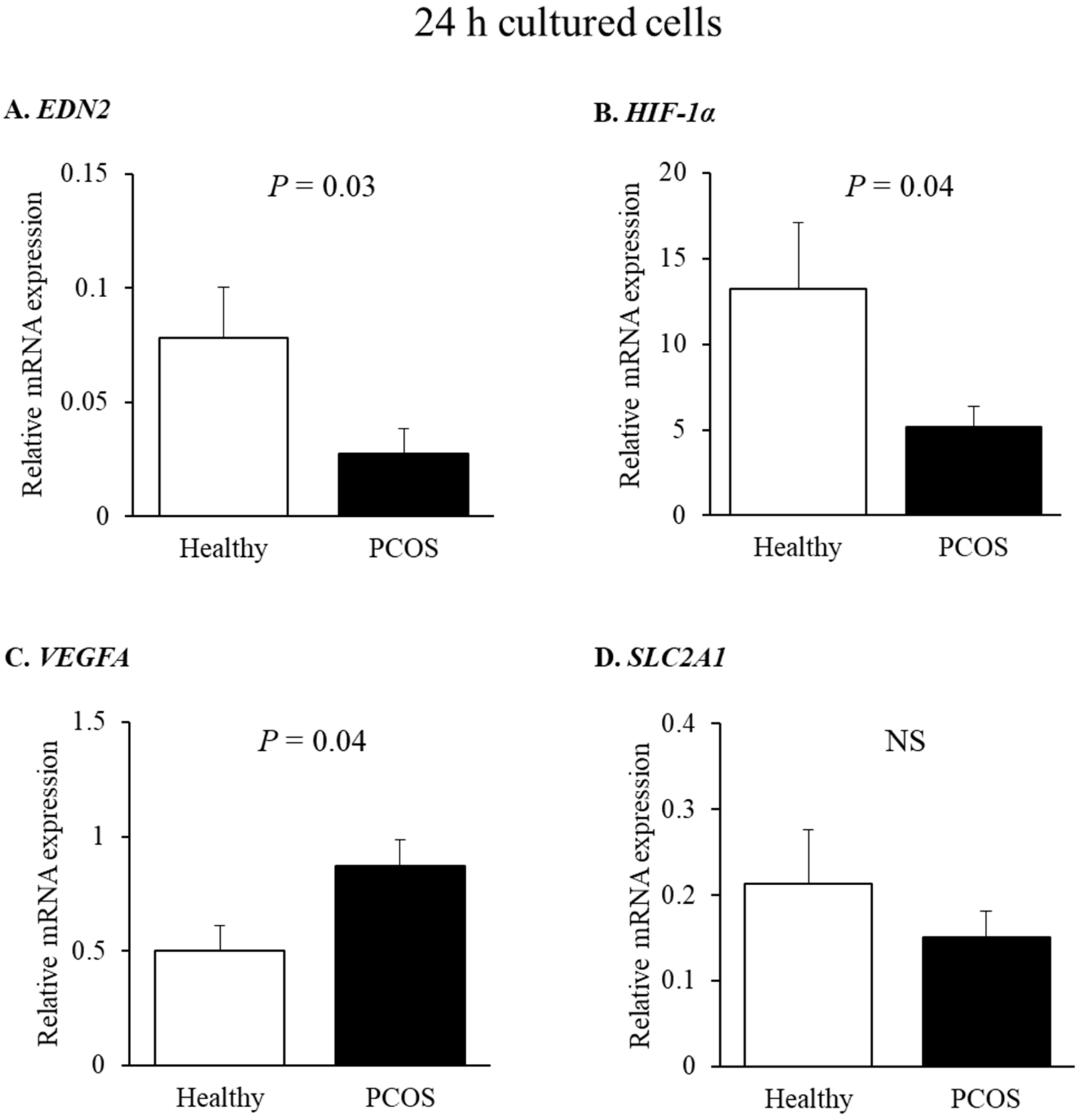
To further substantiate these findings, we determined the expression of HIF-1α and several HIF-1α-responsive genes—EDN2, VEGFA, and SLC2A1—in GLCs cultured for 24 h, derived from PCOS and healthy women (Figure 3). In accordance with what was observed in the freshly isolated cells (Figure 2), the expression of EDN2 was lower in cultured GLCs from the PCOS group (~2.6-fold less than in the GLCs of healthy controls (Figure 3A). HIF-1α also exhibited reduced expression in the GLCs of PCOS women (Figure 3B). Contrary to EDN2, VEGFA expression was significantly elevated in cells from PCOS women (Figure 3C). However, the expression of SLC2A1—another HIF-1α-responsive gene—remained similar in both groups (Figure 3D).

2.3. Effect of miR-210 on HIF-1α-Responsive Genes: EDN2 and VEGFA in Human GLCs

To better understand the role of miR-210, we manipulated its levels in human GLCs using miR-210-mimic or miR-210-inhibitor. The data presented in Figure 4A,B show that miR-210-mimic markedly elevated the miR-210 expression levels, whereas miR-210-inhibitor effectively reduced the miR-210 levels, compared with their respective negative controls. Next, we studied the role of miR-210 on HIF-1α-responsive genes—EDN2 and VEGFA—in GLCs. The overexpression of miR-210 stimulated EDN2 twice as much as the miR-NC-transfected cells did (Figure 4C), whereas miR-210-inhibitor had the opposite effect: it significantly diminished EDN2 expression (Figure 4D), thus confirming that miR-210 stimulates EDN2 in GLCs. Intriguingly, VEGFA transcripts were reduced by both miR-210-mimic (Figure 4C) and -inhibitor (Figure 4D).

3. Discussion

The present study shows that the HIF-1α-responsive factors in freshly isolated GLCs of PCOS patients resulted in an altered expression pattern, compared with healthy women; this suggests that a disrupted hypoxic response exists in these cells. Specifically, we found anomalous levels of miR-210, GPD1L, SDHD, VEGFA, and EDN2 in GLCs derived from PCOS. Cultured GLCs retained the pattern of low EDN2 expression observed in freshly isolated cells, and also exhibited low HIF-1α levels. In contrast to EDN2, VEGFA levels were higher in GLCs derived from PCOS. We found that EDN2 expression was dependent on miR-210 and its target genes in primary cells or in miR-210-transfected cells, whereas VEGFA did not show such a dependency. These results suggest that reduced miR-210 expression in GLCs obtained from PCOS women upregulates GPD1L and SDHD, and destabilizes HIF-1α, thus reducing EDN2.
One of the novel findings of this study is that the GLCs of PCOS women exhibited markedly reduced levels of miR-210, compared with cells of normally ovulating women.
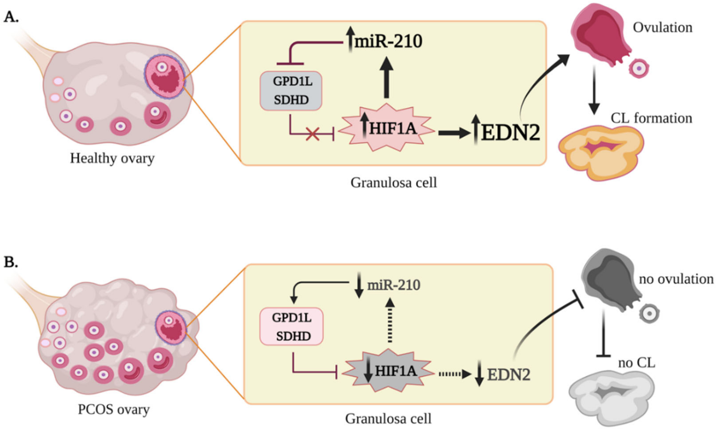
A positive feedback loop between miR-210, GPD1L, HIF-1α, and EDN2 was described in our previous study [19], and this was further substantiated here (Figure 5); hypoxia-induced or overexpression of miR-210 resulted in reduced GPD1L and SDHD expression, accompanied by higher HIF-1α protein and EDN2 levels in human GLCs. However, in PCOS cells, miR-210 was lower; this had functional consequences, causing reduced EDN2 and elevated levels of GPD1L and SDHD in these cells. GPD1L and SDHD are established miR-210 targets in human GLCs [19]. Regarding the role of miR-210 in hypoxia, these two genes destabilize HIF-1α by acting through different mechanisms—GPD1L mediates the hyperhydroxylation of HIF-1α by increasing the activity of the prolyl hydroxylase domain (PHD) enzymes [25], whereas reduced SDHD increases succinate accumulation, a natural inhibitor of PHD activity [26,27]; this subsequently causes a proteosomal degradation of HIF-1α [28]. In fact, suppressed GPD1L (by either elevated miR-210 or siRNA silencing) is required for HIF-1α accumulation, enabling the HIF-1α-responsive gene EDN2 to be stimulated [19]. However, in PCOS GLCs, this feedback loop is breached, since they express low miR-210 levels, resulting in the underexpression of HIF-1α and EDN2, compared with the cells of control women. Recently published data confirm these findings, demonstrating an abnormally lower expression of HIF-1α [29] and EDN2 [8] in these cells. It is noteworthy that the positive feedback loop between miR-210, HIF-1α, and EDN2 suggested here is supported by an in vivo profile of these factors: all three agents are markedly upregulated in early CL [14,15,17,30,31].
Avascular GCs survive in a hypoxic environment, and the O2 tension of follicular fluid negatively correlates with follicular size [32]. Such conditions support the transcription of hypoxia-dependent genes, including EDN2. EDN2 has been described as a trigger of follicular rupture; it acts by constricting the smooth muscle layer around periovulatory follicles [13]. As reported for mice and rats, blocking EDN2 action via a receptor antagonist or gene knockout inhibited follicle rupture, oocyte release, and CL formation [15,16,33]. EDN2 may also affect follicular rupture and ovulation by other mechanisms. Since it is a vasoactive hormone, EDN2 is likely to promote vascular changes that facilitate the ovulatory process [16]. Another HIF-regulated factor, ADAMTS-1—a matrix metalloprotease—may also participate in the remodeling of the extracellular matrix in the cumulus–oocyte complex, and in the disintegration of the follicular wall during the final stages of ovulation [34]. Owing to lagging angiogenesis, hypoxia is also prevalent during CL formation, favoring the upregulation of hypoxia-dependent genes [35]. In addition to follicular rupture, EDN2 may facilitate early CL development by promoting angiogenesis, cell proliferation, and differentiation. For example, in cattle, EDN2 increased GC numbers, as did PTGS2—a rate-limiting enzyme in prostaglandin synthesis, which is essential for ovulation [14]. These were also substantiated in ECE1 (EDN-converting enzyme 1) gene knockdown in bovine GCs, which resulted in reduced viable cell numbers and gene expression [14].
In contrast to EDN2, the mRNA levels of VEGFA—an established HIF-1α -dependent gene—were higher in PCOS GLCs than in healthy controls. These results are in accordance with the immunohistochemical evidence of extensive VEGFA staining in polycystic ovarian tissue [36,37], as well as with higher concentrations of VEGFA released to culture medium by the GLCs of PCOS women, compared with the non-PCOS group [38]. In addition, VEGFA was found to be elevated in the serum and follicular fluid of women with PCOS [29,39,40,41,42]. However, a few studies reported a reduction or no changes in the VEGFA levels in the GLCs of patients with PCOS [29,43]; the reason for this controversy is currently unclear. High VEGFA may contribute to the occurrence of PCOS by excess angiogenesis; inhibition of VEGFA in the rat ovaries of a PCOS model partially restored the accumulation of the small follicles observed, and reduced cyst formation, consequently improving ovulation and follicular development [44]. Both EDN2 and VEGFA are dependent on HIF-1α; however, only the relationship between EDN2 and HIF-1α is manifested in vivo around the time of ovulation [14,17,45]. VEGFA, which is elevated as the follicle matures, increases further during the early luteal phase, but remains elevated for the duration of the luteal phase [31,46,47]. Aside from hypoxia, there is a strong LH/hCG dependency on VEGFA transcription. In humans, macaques, and bovine GLCs, VEGFA expression [14,31,48,49,50]—as well as its secretion to the culture medium—was augmented by LH/hCG [38,41]. Of particular relevance, a strong correlation between VEGFA synthesis and hCG levels was observed in PCOS women, both in vivo and in vitro [38,41]. After hCG administration, PCOS women exhibited a rise in serum VEGFA that was not observed in the control patients [41]. Similarly, in vitro hCG treatment of GLCs derived from PCOS patients significantly increased VEGFA secretion; this production was greater in cells extracted from PCOS ovaries than from normal ones [38]. Because we showed an impaired hypoxic response of PCOS GLCs, we presume that an altered response to LH/hCG, rather than to hypoxia, results in increased VEGFA, as observed in GLCs derived from PCOS patients. However, additional research is needed in order to determine whether signals other than LH or hypoxia may be responsible for the aberrant high expression of VEGFA observed here in the GLCs of PCOS women.
In addition to GLCs, the follicular fluid aspirates contain non-steroidogenic cells—mainly leukocytes—to varying degrees [51,52,53]. However, although we did not remove these potentially contaminating cells from freshly isolated GLCs, we took advantage of a selective adherence of cultured cells to the surface of cell culture dishes; GLCs attach early, whereas lymphocytes—the large population of leukocytes in follicular fluid aspirates [54]—are non-adherent cells [53,55]. It should be noted that freshly isolated and cultured GLCs exhibited similar expression profiles, suggesting that the findings reported here represent the biological functions of GLCs. Nevertheless, further research using GLCs after purification steps—such as anti-CD45 magnetic immunobead, still remains to be carried out.
In summary, our study implies that the hypoxic responses of GLCs derived from women with PCOS are altered, suggesting a mechanism that may contribute to abnormal ovarian function. These findings are consistent with the notion that HIF-1α is a critical regulator of various ovarian processes, and that miR-210 and EDN2 are essential HIF-1α-responsive factors for follicular rupture, ovulation, and CL formation. Low miR-210, causing decreased EDN2 during the periovulatory stage, can result in the ovulation failure and reduced fertility observed in this syndrome (Figure 5). Elevated VEGFA in PCOS GLCs, irrespective of a lower hypoxic response, may further exacerbate the abnormal ovarian function.

4. Materials and Methods

4.1. Subjects

Forty-one women with PCOS and forty healthy, normally ovulating women were enrolled in this study. PCOS was diagnosed according to the Rotterdam revised criteria [3]. The control patients entered the IVF program with non-ovarian indications, which were limited to male factor infertility (MFI). For both the PCOS and control groups, women were subjected to the GnRH antagonist protocol [56]. In brief, controlled ovarian stimulation was initiated on day 2 or 3 of a spontaneous cycle. An initial dose of 150–225 IU recombinant FSH (rFSH; Gonal-F (Merck Serono, Darmstadt, Germany)), Puregon (MSD, Hertfordshire, UK), or highly purified hMG (Menopur (Ferring Pharmaceuticals, Saint-Prex, Switzerland)) was administered. From day 6 onward, the gonadotropin dose was estimated according to the serum E2 levels and a transvaginal ultrasound scan. When a leading follicle reached 13–14 mm, a GnRH antagonist (Cetrotide (Merck Serono) or Orgalutran (MSD)) was administered at 0.25 mg/d. Final oocyte maturation was triggered using 250 µg recombinant hCG (rhCG; Ovitrelle (Merck Serono)) as soon as the mean diameters of two follicles were ≥18 mm. Oocyte retrieval was scheduled 36 h after hCG injection. All accessible follicles were harvested, and oocytes were collected from follicular aspirates and subjected to fertilization. The residual follicular fluid aspirates containing GLCs were collected for further investigation.

4.2. Experiment Protocol

GLCs were obtained from follicular aspirates as previously described [57]. Briefly, the aspirates were centrifuged (3 min at 3000× g) and erythrocytes were removed using an ammonium-chloride–potassium buffer (0.15 mol/L NH4Cl, 1.0 mmol/L KHCO3, and 0.1 nmol/L EDTA). After having been washed with phosphate-buffered saline (PBS), cells were counted in a hemocytometer. A fraction of the freshly isolated cells (n = 16 from each group of patients) was taken and stored at −80 °C for further RNA extraction.
For in vitro experiments, cells were placed in 12- or 6-well plates (1.5 × 10⁵ cells/well) and cultured in a DMEM/F12 1:1 (v/v) nutrient mixture containing 10% fetal calf serum (FCS), 2 mM L-glutamine, and 100 mg/mL penicillin/ streptomycin (Biological Industries, Kibbutz Beit HaEmek, Israel). Cultures were maintained in a humidified 95% air 5% CO2 mixture at 37 °C. Following 24 h of incubation, cells were washed with PBS and collected for subsequent RNA isolation, or they were subjected to transfection as detailed below.

4.3. Cell Transfection

miR-210-mimic, miR-210-inhibitor, and the corresponding negative controls (miR-NC or miR-iNC, respectively) were purchased from Ambion (Thermo Fisher Scientific, Waltham, MA, USA) and Bioneer (Daejeo, Korea). Cells were transfected with miR-210-mimic (10 nmol/L), miR-210-inhibitor (60 nmol/L), or their respective NCs in 1% FCS for 24 h using Lipofectamine RNAiMAX reagent (Thermo Fisher Scientific) according to the manufacturer’s protocol. Forty-eight hours after transfection, the cells were harvested and total RNA was extracted.

4.4. Quantitative PCR Analysis of mRNA and miRNA

Total RNA was extracted using the TRI Reagent (Molecular Research Center, Cincinnati, OH, USA) in accordance with the manufacturer’s instructions. cDNA was synthesized from the total RNA (1000 ng) by using the qScript cDNA synthesis kit (Quantabio, Beverly, MA, USA). miRNA cDNA was obtained from the purified total RNA (700 ng) using the qScript microRNA Synthesis Kit (Quantabio). Quantitative polymerase chain reaction (qPCR) for mRNA expression was performed with LightCycler 480 SYBR Green I Master (Roche Diagnostics, Indianapolis, IN, USA) and for miR-210 with PerfeCTa SYBR Green SuperMix, Low ROX (Quantabio), as previously described [19,57]. Each PCR reaction was performed in duplicate. The PerfeCTa microRNA assay included Universal Primer, miR-210 Primer, and SNORD44 as positive control primers (Quantabio). The expression levels were normalized to beta-actin (ACTB; for mRNA) and ribosomal protein S18 (RPS18; for miRNA). The sequences of primers used for qPCR are listed in Table 3. The threshold cycle (Ct) values of each sample were generated, and the relative expression was calculated as 2-ΔCt=2-(Ct target gene − Ct housekeeping gene) [58].

4.5. MiRNA Expression Profiling

A global analysis approach was employed to reveal the miRNA expression profile using the nCounter miRNA expression assay at NanoString Technologies. About 800 human miRNAs were simultaneously assayed (miRBase v.18). A total of 100 ng of total GLC RNA pooled either from a control group (n = 8) or from patients with PCOS (n = 8) was assayed separately in technical duplicates using the Human nCounter miRNA Assay 2.0 Kit (NanoString Technologies, Seattle, WA, USA), following the manufacturer’s instructions. Briefly, mature miRNAs were ligated to a species-specific tag sequence (miRtag) via a thermally controlled splinted ligation. After enzymatic purification of unligated miRtags, prepared samples were hybridized with an nCounter Human miRNA Expression Assay CodeSet overnight at 65 °C. Unhybridized CodeSet was removed via automated purification performed on an nCounter Prep Station, and the resulting target–probe complexes were deposited and bound to an imaging surface as previously described [59]. Reporter counts were tabulated for each sample using the nCounter Digital Analyzer, and output as raw data was subsequently imported.

4.6. Statistical Analyses

Statistical analyses were performed using GraphPad PRISM v. 6.0 (GraphPad Software, Inc., San Diego, CA, USA). Student’s t-test with a two-tailed distribution, with two samples equaling variance was conducted. When appropriate, a non-parametric analysis was conducted using the Mann–Whitney U test. Numerical data are reported as the mean ± standard deviation (Table 1) or the mean ± standard error of the mean (SEM). A p-value < 0.05 was considered statistically significant.

5. Conclusions

Altered hypoxic responses and angiogenic potential in GLCs affect follicle development and maturation in human ovaries. Dysregulated hypoxic responses can lead to abnormal ovulation and, eventually, infertility, as commonly observed in PCOS patients. This study, together with our previous report [19], identified the molecular makeup of the hypoxic pathway in GLCs, highlighting the role of miR-210. miRs can be detected in bodily fluids such as blood plasma, saliva, urine, and follicular fluids, and may thus serve as biomarkers for cellular activity [60,61,62,63]. It is therefore anticipated that miR-210 levels could serve as a novel diagnostic tool, and also as a new therapeutic target to treat anovulation in PCOS patients.

Author Contributions

Study conceptualization, M.S., K.S. and R.M.; methodology, M.S., K.S., R.M. and E.G.; R.M., T.I., I.E., E.G. and A.H. prepared the documents for the ethics committee, allocated the women, and collected clinical samples. M.S. and K.S. carried out the majority of experiments and analyses, and interpreted the results together with R.M.; I.E. and T.I. performed NanoString analysis; manuscript writing—original draft preparation, M.S.; writing, review and editing, M.S., K.S. and R.M.; E.G., T.I. and I.E. inspected and revised sections relevant to clinical data; supervision, R.M.; project administration, R.M.; funding acquisition, R.M. All authors have read and agreed to the published version of the manuscript.

Funding

This research was supported by the German–Israeli Foundation for Scientific Research and Development (GIF), grant number I-1417-201.2/2017, and by the Robert H. Smith Faculty of Agriculture, Food, and Environment Research Fund for International Cooperation scholarship. The cost of Open Access publication was covered by the Society for Biology of Reproduction in Poland.

Institutional Review Board Statement

The study was conducted in accordance with the guidelines of the Declaration of Helsinki, and approved by the Institutional Review Board of Barzilai University Medical Center in Ashqelon (approval 0109-17) and Hadassah Hebrew University Medical Center in Jerusalem (approval HMO-0110-09), Israel.

Informed Consent Statement

Informed consent was obtained from all subjects involved in the study.

Data Availability Statement

The data presented in this study are available on request from the corresponding author. The data are not publicly available due to their containing information that could compromise the privacy of research participants.

Acknowledgments

The authors are grateful to the German–Israeli Foundation for Scientific Research and Development for funding this work, and to the International School of Agricultural Sciences, The Robert H. Smith Faculty of Agriculture, Food, and Environment for providing a fellowship to M. Szymanska, and to the Society for Biology of Reproduction in Poland for covering the cost of Open Access publication fees.

Conflicts of Interest

The authors declare no conflict of interest.

References

  1. Azziz, R. Polycystic Ovary Syndrome. Obstet. Gynecol. 2018, 132, 321–336. [Google Scholar] [CrossRef]
  2. Diao, F.-Y.; Xu, M.; Hu, Y.; Li, J.; Xu, Z.; Lin, M.; Wang, L.; Zhou, Y.; Zhou, Z.; Liu, J.; et al. The molecular characteristics of polycystic ovary syndrome (PCOS) ovary defined by human ovary cDNA microarray. J. Mol. Endocrinol. 2004, 33, 59–72. [Google Scholar] [CrossRef] [PubMed] [Green Version]
  3. The Rotterdam ESHRE/ASRM-Sponsored PCOS Consensus Workshop Group. Revised 2003 consensus on diagnostic criteria and long-term health risks related to polycystic ovary syndrome (PCOS). Hum. Reprod. 2004, 19, 41–47. [Google Scholar] [CrossRef] [Green Version]
  4. Barber, T.M.; Franks, S. Obesity and polycystic ovary syndrome. Clin. Endocrinol. 2021. [Google Scholar] [CrossRef]
  5. Stubbs, S.A.; Stark, J.; Dilworth, S.M.; Franks, S.; Hardy, K. Abnormal Preantral Folliculogenesis in Polycystic Ovaries Is Associated with Increased Granulosa Cell Division. J. Clin. Endocrinol. Metab. 2007, 92, 4418–4426. [Google Scholar] [CrossRef] [PubMed] [Green Version]
  6. Das, M.; Djahanbakhch, O.; Hacihanefioglu, B.; Saridogan, E.; Ikram, M.; Ghali, L.; Raveendran, M.; Storey, A. Granulosa cell survival and proliferation are altered in polycystic ovary syndrome. J. Clin. Endocrinol. Metab. 2008, 93, 881–887. [Google Scholar] [CrossRef] [Green Version]
  7. Li, Y.; Liu, Y.-D.; Zhou, X.-Y.; Chen, S.-L.; Chen, X.; Zhe, J.; Zhang, J.; Zhang, Q.-Y.; Chen, Y.-X. MiR-29a regulates the proliferation, aromatase expression, and estradiol biosynthesis of human granulosa cells in polycystic ovary syndrome. Mol. Cell. Endocrinol. 2019, 498, 110540. [Google Scholar] [CrossRef]
  8. Imbar, T.; Klipper, E.; Greenfield, C.; Hurwitz, A.; Haimov-Kochman, R.; Meidan, R. Altered endothelin expression in granulosa-lutein cells of women with polycystic ovary syndrome. Life Sci. 2012, 91, 703–709. [Google Scholar] [CrossRef] [Green Version]
  9. Erickson, G.F.; Magoffin, D.A.; Garzo, V.; Cheung, A.P.; Chang, R. Granulosa cells of polycystic ovaries: Are they normal or abnormal? Hum. Reprod. 1992, 7, 293–299. [Google Scholar] [CrossRef]
  10. Haouzi, D.; Assou, S.; Monzo, C.; Vincens, C.; Dechaud, H.; Hamamah, S. Altered gene expression profile in cumulus cells of mature mii oocytes from patients with polycystic ovary syndrome. Hum. Reprod. 2012, 27, 3523–3530. [Google Scholar] [CrossRef] [Green Version]
  11. Xu, B.; Zhang, Y.-W.; Tong, X.-H.; Liu, Y.-S. Characterization of microRNA profile in human cumulus granulosa cells: Identification of microRNAs that regulate Notch signaling and are associated with PCOS. Mol. Cell. Endocrinol. 2015, 404, 26–36. [Google Scholar] [CrossRef] [PubMed]
  12. Wood, J.R.; Ho, C.; Nelson-Degrave, V.L.; McAllister, J.M.; Strauss, J.F. The molecular signature of polycystic ovary syndrome (PCOS) theca cells defined by gene expression profiling. J. Reprod. Immunol. 2004, 63, 51–60. [Google Scholar] [CrossRef]
  13. Ko, C.; Gieske, M.C.; Al-Alem, L.; Hahn, Y.; Su, W.; Gong, M.C.; Iglarz, M.; Koo, Y. Endothelin-2 in Ovarian Follicle Rupture. Endocrinology 2006, 147, 1770–1779. [Google Scholar] [CrossRef] [PubMed] [Green Version]
  14. Klipper, E.; Levit, A.; Mastich, Y.; Berisha, B.; Schams, D.; Meidan, R. Induction of Endothelin-2 Expression by Luteinizing Hormone and Hypoxia: Possible Role in Bovine Corpus Luteum Formation. Endocrinology 2010, 151, 1914–1922. [Google Scholar] [CrossRef]
  15. Cacioppo, J.; Oh, S.W.; Kim, H.-Y.; Cho, J.; Lin, P.-C.P.; Yanagisawa, M.; Ko, C. Loss of Function of Endothelin-2 Leads to Reduced Ovulation and CL Formation. PLoS ONE 2014, 9, e96115. [Google Scholar] [CrossRef]
  16. Palanisamy, G.S.; Cheon, Y.-P.; Kim, J.; Kannan, A.; Li, Q.; Sato, M.; Mantena, S.R.; Sitruk-Ware, R.L.; Bagchi, M.K.; Bagchi, I.C. A Novel Pathway Involving Progesterone Receptor, Endothelin-2, and Endothelin Receptor B Controls Ovulation in Mice. Mol. Endocrinol. 2006, 20, 2784–2795. [Google Scholar] [CrossRef] [Green Version]
  17. Kim, J.; Bagchi, I.C.; Bagchi, M.K. Signaling by Hypoxia-Inducible Factors Is Critical for Ovulation in Mice. Endocrinology 2009, 150, 3392–3400. [Google Scholar] [CrossRef] [Green Version]
  18. Na, G.; Bridges, P.J.; Koo, Y.; Ko, C. Role of hypoxia in the regulation of periovulatory EDN2 expression in the mouse. Can. J. Physiol. Pharmacol. 2008, 86, 310–319. [Google Scholar] [CrossRef] [Green Version]
  19. Shrestha, K.; Onasanya, A.E.; Eisenberg, I.; Wigoda, N.; Yagel, S.; Yalu, R.; Meidan, R.; Imbar, T. miR-210 and GPD1L regulate EDN2 in primary and immortalized human granulosa-lutein cells. Reproduction 2018, 155, 197–205. [Google Scholar] [CrossRef] [Green Version]
  20. Tam, K.K.; Russell, D.L.; Peet, D.J.; Bracken, C.P.; Rodgers, R.J.; Thompson, J.G.; Kind, K.L. Hormonally regulated follicle differentiation and luteinization in the mouse is associated with hypoxia inducible factor activity. Mol. Cell. Endocrinol. 2010, 327, 47–55. [Google Scholar] [CrossRef]
  21. Zhang, J.; Zhang, Z.; Wu, Y.; Chen, L.; Luo, Q.; Chen, J.; Huang, X.; Cheng, Y.; Wang, Z. Regulatory Effect of Hypoxia-Inducible Factor-1α on hCG-Stimulated Endothelin-2 Expression in Granulosa Cells from the PMSG-Treated Rat Ovary. J. Reprod. Dev. 2012, 58, 678–684. [Google Scholar] [CrossRef] [Green Version]
  22. Yalu, R.; Oyesiji, A.E.; Eisenberg, I.; Imbar, T.; Meidan, R. HIF1A-dependent increase in endothelin 2 levels in granulosa cells: Role of hypoxia, LH/cAMP, and reactive oxygen species. Reproduction 2015, 149, 11–20. [Google Scholar] [CrossRef] [Green Version]
  23. Devlin, C.; Greco, S.; Martelli, F.; Ivan, M. miR-210: More than a silent player in hypoxia. IUBMB Life 2011, 63, 94–100. [Google Scholar] [CrossRef] [Green Version]
  24. Chan, Y.C.; Banerjee, J.; Choi, S.Y.; Sen, C.K. miR-210: The Master Hypoxamir. Microcirculation 2012, 19, 215–223. [Google Scholar] [CrossRef] [PubMed] [Green Version]
  25. Kelly, T.J.; Souza, A.L.; Clish, C.B.; Puigserver, P. A Hypoxia-Induced Positive Feedback Loop Promotes Hypoxia-Inducible Factor 1α Stability through miR-210 Suppression of Glycerol-3-Phosphate Dehydrogenase 1-Like. Mol. Cell. Biol. 2011, 31, 2696–2706. [Google Scholar] [CrossRef] [Green Version]
  26. Puissegur, M.-P.; Mazure, N.M.; Bertero, T.; Pradelli, L.; Grosso, S.; Robbe-Sermesant, K.; Maurin, T.; Lebrigand, K.; Cardinaud, B.; Hofman, V.; et al. miR-210 is overexpressed in late stages of lung cancer and mediates mitochondrial alterations associated with modulation of HIF-1 activity. Cell Death Differ. 2011, 18, 465–478. [Google Scholar] [CrossRef] [Green Version]
  27. Merlo, A.; de Quiros, S.B.; Secades, P.; Zambrano, I.; Balbín, M.; Astudillo, A.; Scola, B.; Arístegui, M.; Suarez, C.; Chiara, M.-D. Identification of a Signaling Axis HIF-1α/MicroRNA-210/ISCU Independent of SDH Mutation That Defines a Subgroup of Head and Neck Paragangliomas. J. Clin. Endocrinol. Metab. 2012, 97, E2194–E2200. [Google Scholar] [CrossRef] [Green Version]
  28. Semenza, G.L. Hypoxia-Inducible Factor 1 (HIF-1) Pathway. Sci. STKE 2007, 2007, cm8. [Google Scholar] [CrossRef]
  29. Patil, K.; Hinduja, I.; Mukherjee, S. Alteration in angiogenic potential of granulosa-lutein cells and follicular fluid contributes to luteal defects in polycystic ovary syndrome. Hum. Reprod. 2021, 36, 1052–1064. [Google Scholar] [CrossRef]
  30. Gecaj, R.; Schanzenbach, C.I.; Kirchner, B.; Pfaffl, M.; Riedmaier, I.; Tweedie-Cullen, R.Y.; Berisha, B. The Dynamics of microRNA Transcriptome in Bovine Corpus Luteum during Its Formation, Function, and Regression. Front. Genet. 2017, 8, 213. [Google Scholar] [CrossRef] [Green Version]
  31. Van den Driesche, S.; Myers, M.; Gay, E.; Thong, K.J.; Duncan, W.C. HCG up-regulates hypoxia inducible factor-1 alpha in luteinized granulosa cells: Implications for the hormonal regulation of vascular endothelial growth factor A in the human corpus luteum. Mol. Hum. Reprod. 2008, 14, 455–464. [Google Scholar] [CrossRef] [Green Version]
  32. Basini, G.; Bianco, F.; Grasselli, F.; Tirelli, M.; Bussolati, S.; Tamanini, C. The effects of reduced oxygen tension on swine granulosa cell. Regul. Pept. 2004, 120, 69–75. [Google Scholar] [CrossRef] [PubMed]
  33. Bridges, P.J.; Jo, M.; Al Alem, L.; Na, G.; Su, W.; Gong, M.C.; Jeoung, M.; Ko, C. Production and binding of endothelin-2 (EDN2) in the rat ovary: Endothelin receptor subtype A (EDNRA)-mediated contraction. Reprod. Fertil. Dev. 2010, 22, 780–787. [Google Scholar] [CrossRef] [PubMed] [Green Version]
  34. Shozu, M.; Minami, N.; Yokoyama, H.; Inoue, M.; Kurihara, H.; Matsushima, K.; Kuno, K. ADAMTS-1 is involved in normal follicular development, ovulatory process and organization of the medullary vascular network in the ovary. J. Mol. Endocrinol. 2005, 35, 343–355. [Google Scholar] [CrossRef]
  35. Shrestha, K.; Rodler, D.; Sinowatz, F.; Meidan, R. Corpus Luteum Formation. In The Ovary, 3rd ed.; Academic Press: Cambridge, MA, USA, 2019; pp. 255–267. [Google Scholar]
  36. Ferrara, N.; Frantz, G.; LeCouter, J.; Dillard-Telm, L.; Pham, T.; Draksharapu, A.; Giordano, T.; Peale, F. Differential Expression of the Angiogenic Factor Genes Vascular Endothelial Growth Factor (VEGF) and Endocrine Gland-Derived VEGF in Normal and Polycystic Human Ovaries. Am. J. Pathol. 2003, 162, 1881–1893. [Google Scholar] [CrossRef] [Green Version]
  37. Kamat, B.R.; Brown, L.F.; Manseau, E.J.; Senger, D.R.; Dvorak, H.F. Expression of vascular permeability factor/vascular endothelial growth factor by human granulosa and theca lutein cells. Role in corpus luteum development. Am. J. Pathol. 1995, 146, 157–165. [Google Scholar]
  38. Agrawal, R.; Jacobs, H.; Payne, N.; Conway, G. Concentration of vascular endothelial growth factor released by cultured human luteinized granulosa cells is higher in women with polycystic ovaries than in women with normal ovaries. Fertil. Steril. 2002, 78, 1164–1169. [Google Scholar] [CrossRef]
  39. Agrawal, R.; Sladkevicius, P.; Engmann, L.; Conway, G.S.; Payne, N.N.; Bekis, J.; Tan, S.L.; Campbell, S.; Jacobs, H.S. Serum vascular endothelial growth factor concentrations and ovarian stromal blood flow are increased in women with polycystic ovaries. Hum. Reprod. 1998, 13, 651–655. [Google Scholar] [CrossRef] [PubMed] [Green Version]
  40. Abd El Aal, D.E.; Mohamed, S.A.; Amine, A.F.; Meki, A.-R.M. Vascular endothelial growth factor and insulin-like growth factor-1 in polycystic ovary syndrome and their relation to ovarian blood flow. Eur. J. Obstet. Gynecol. Reprod. Biol. 2005, 118, 219–224. [Google Scholar] [CrossRef]
  41. Artini, P.G.; Monti, M.; Matteucci, C.; Valentino, V.; Cristello, F.; Genazzani, A.R. Vascular endothelial growth factor and basic fibroblast growth factor in polycystic ovary syndrome during controlled ovarian hyperstimulation. Gynecol. Endocrinol. 2006, 22, 465–470. [Google Scholar] [CrossRef] [PubMed]
  42. Artini, P.G.; Ruggiero, M.; Toldin, M.R.P.; Monteleone, P.; Monti, M.; Cela, V.; Genazzani, A.R. Vascular endothelial growth factor and its soluble receptor in patients with polycystic ovary syndrome undergoing IVF. Hum. Fertil. 2009, 12, 40–44. [Google Scholar] [CrossRef]
  43. Adams, J.; Liu, Z.; Ren, Y.A.; Wun, W.-S.; Zhou, W.; Kenigsberg, S.; Librach, C.; Valdes, C.; Gibbons, W.; Richards, J. Enhanced Inflammatory Transcriptome in the Granulosa Cells of Women with Polycystic Ovarian Syndrome. J. Clin. Endocrinol. Metab. 2016, 101, 3459–3468. [Google Scholar] [CrossRef] [PubMed] [Green Version]
  44. Abramovich, D.; Irusta, G.; Bas, D.; Cataldi, N.I.; Parborell, F.; Tesone, M. Angiopoietins/TIE2 System and VEGF Are Involved in Ovarian Function in a DHEA Rat Model of Polycystic Ovary Syndrome. Endocrinology 2012, 153, 3446–3456. [Google Scholar] [CrossRef] [PubMed] [Green Version]
  45. Choi, D.-H.; Kim, E.K.; Kim, K.-H.; Lee, K.-A.; Kang, D.-W.; Kim, H.Y.; Bridges, P.; Ko, C. Expression pattern of endothelin system components and localization of smooth muscle cells in the human pre-ovulatory follicle. Hum. Reprod. 2011, 26, 1171–1180. [Google Scholar] [CrossRef] [PubMed] [Green Version]
  46. Fraser, H.M.; Bell, J.; Wilson, H.; Taylor, P.D.; Morgan, K.; Anderson, R.A.; Duncan, W.C. Localization and Quantification of Cyclic Changes in the Expression of Endocrine Gland Vascular Endothelial Growth Factor in the Human Corpus Luteum. J. Clin. Endocrinol. Metab. 2005, 90, 427–434. [Google Scholar] [CrossRef] [Green Version]
  47. Berisha, B.; Schams, D.; Kosmann, M.; Amselgruber, W.; Einspanier, R. Expression and Tissue Concentration of Vascular Endothelial Growth Factor, Its Receptors, and Localization in the Bovine Corpus Luteum During Estrous Cycle and Pregnancy. Biol. Reprod. 2000, 63, 1106–1114. [Google Scholar] [CrossRef]
  48. Lee, A.; Christenson, L.K.; Patton, P.E.; Burry, K.A.; Stouffer, R.L. Vascular endothelial growth factor production by human luteinized granulosa cells in vitro. Hum. Reprod. 1997, 12, 2756–2761. [Google Scholar] [CrossRef] [Green Version]
  49. Meidan, R.; Klipper, E.; Zalman, Y.; Yalu, R. The role of hypoxia-induced genes in ovarian angiogenesis. Reprod. Fertil. Dev. 2013, 25, 343–350. [Google Scholar] [CrossRef]
  50. Christenson, L.K.; Stouffer, R.L. Follicle-Stimulating Hormone and Luteinizing Hormone/Chorionic Gonadotropin Stimulation of Vascular Endothelial Growth Factor Production by Macaque Granulosa Cells from Pre- and Periovulatory Follicles. J. Clin. Endocrinol. Metab. 1997, 82, 2135–2142. [Google Scholar] [CrossRef]
  51. Beckmann, M.W.; Polacek, D.; Seung, L.; Schreiber, J.R. Human ovarian granulosa cell culture: Determination of blood cell contamination and evaluation of possible culture purification steps. Fertil. Steril. 1991, 56, 881–887. [Google Scholar] [CrossRef]
  52. Ferrero, H.; Delgado-Rosas, F.; Garcia-Pascual, C.M.; Monterde, M.; Zimmermann, R.C.; Simón, C.; Pellicer, A.; Gómez, R. Efficiency and purity provided by the existing methods for the isolation of luteinized granulosa cells: A comparative study. Hum. Reprod. 2012, 27, 1781–1789. [Google Scholar] [CrossRef] [PubMed] [Green Version]
  53. Fedorcsák, P.; Ráki, M.; Storeng, R. Characterization and depletion of leukocytes from cells isolated from the pre-ovulatory ovarian follicle. Hum. Reprod. 2007, 22, 989–994. [Google Scholar] [CrossRef] [PubMed] [Green Version]
  54. Smith, M.; Flannery, G.; Randle, B.; Jenkins, J.; Holmes, C. Leukocyte origin and profile in follicular aspirates at oocyte retrieval. Hum. Reprod. 2005, 20, 3526–3531. [Google Scholar] [CrossRef]
  55. Sasson, R.; Rimon, E.; Dantes, A.; Cohen, T.; Shinder, V.; Land-Bracha, A.; Amsterdam, A. Gonadotrophin-induced gene regulation in human granulosa cells obtained from IVF patients. Modulation of steroidogenic genes, cytoskeletal genes and genes coding for apoptotic signalling and protein kinases. Mol. Hum. Reprod. 2004, 10, 299–311. [Google Scholar] [CrossRef]
  56. Fleming, R.; Haxton, M.; Hamilton, M.; Conaghan, C.; Black, W.; Yates, R.; Coutts, J. Combined gonadotropin-releasing hormone analog and exogenous gonadotropins for ovulation induction in infertile women: Efficacy related to ovarian function assessment. Am. J. Obstet. Gynecol. 1988, 159, 376–381. [Google Scholar] [CrossRef]
  57. Szymanska, M.; Manthe, S.; Shrestha, K.; Girsh, E.; Harlev, A.; Kisliouk, T.; Meidan, R. Sirtuin-1 inhibits endothelin-2 expression in human granulosa-lutein cells via hypoxia inducible factor 1 alpha and epigenetic modifications. Biol. Reprod. 2021, 104, 387–398. [Google Scholar] [CrossRef]
  58. Livak, K.J.; Schmittgen, T.D. Analysis of relative gene expression data using real-time quantitative PCR and the 2(-Delta Delta C(T)) Method. Methods 2001, 25, 402–408. [Google Scholar] [CrossRef] [PubMed]
  59. Geiss, G.K.; Bumgarner, R.; Birditt, B.; Dahl, T.; Dowidar, N.; Dunaway, D.L.; Fell, H.P.; Ferree, S.; George, R.D.; Grogan, T.; et al. Direct multiplexed measurement of gene expression with color-coded probe pairs. Nat. Biotechnol. 2008, 26, 317–325. [Google Scholar] [CrossRef] [PubMed]
  60. Cortez, M.A.; Bueso-Ramos, C.; Ferdin, J.; Lopez-Berestein, G.; Sood, A.K.; Calin, G.A. MicroRNAs in body fluids—The mix of hormones and biomarkers. Nat. Rev. Clin. Oncol. 2011, 8, 467–477. [Google Scholar] [CrossRef] [Green Version]
  61. Eisenberg, I.; Kotaja, N.; Goldman-Wohl, D.; Imbar, T. microRNA in Human Reproduction. Adv. Exp. Med. Biol. 2015, 888, 353–387. [Google Scholar] [CrossRef] [PubMed]
  62. Sang, Q.; Yao, Z.; Wang, H.; Feng, R.; Wang, H.; Zhao, X.; Xing, Q.; Jin, L.; He, L.; Wu, L.; et al. Identification of MicroRNAs in Human Follicular Fluid: Characterization of MicroRNAs That Govern Steroidogenesis in Vitro and Are Associated with Polycystic Ovary Syndrome in Vivo. J. Clin. Endocrinol. Metab. 2013, 98, 3068–3079. [Google Scholar] [CrossRef] [PubMed] [Green Version]
  63. Sørensen, S.S.; Nygaard, A.-B.; Christensen, T. miRNA expression profiles in cerebrospinal fluid and blood of patients with Alzheimer’s disease and other types of dementia—An exploratory study. Transl. Neurodegener. 2016, 5, 1–12. [Google Scholar] [CrossRef] [PubMed] [Green Version]
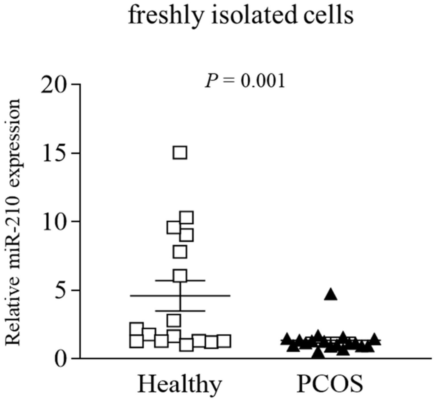
Figure 1. Relative expression of miR-210 in freshly isolated GLCs from PCOS and healthy normally ovulating women. miR-210 levels were measured in freshly isolated GLCs from PCOS and healthy control women by qPCR. Results are presented as a scatter plot with mean values ± SEM (n = 16 per group); squares denote values obtained in healthy controls, triangles denote values obtained in the PCOS group. The p-value indicates the statistically significant difference between groups.
Ijms 22 08216 g001
Figure 2. Relative expression of GPD1L, SDHD, and EDN2 in freshly isolated GLCs from PCOS and healthy, normally ovulating women. (A) GPD1L, (B) SDHD, and (C) EDN2 mRNA levels were measured in freshly isolated GLCs from PCOS and healthy control women by qPCR. Results are presented as scatter plots with mean values ± SEM (n = 9 per group); squares denote values obtained in healthy controls, triangles denote values obtained in the PCOS group. The p-values indicate statistically significant differences between groups.
Ijms 22 08216 g002
Figure 3. Relative expression of HIF-1α and selected HIF-1α-responsive genes in cultured GLCs derived from PCOS and healthy, normally ovulating women. GLCs derived from PCOS and healthy control women (n = 6 in both groups) were incubated with culture medium for 24 h. Then, RNA was extracted, and (A) EDN2, (B) HIF-1α, (C) VEGFA, and (D) SLC2A1 levels were determined by qPCR. The results are presented as the means ± SEM. The p-values indicate statistically significant differences between groups. NS: non-significant.
Ijms 22 08216 g003
Figure 4. miR-210 manipulation affects the expression of HIF-1α-responsive genes—EDN2 and VEGFA—in human GLCs. (A,C) Human GLCs were transfected with 10 nmol/L of either miR-210-mimic or miR-negative control (miR-NC). (B,D) Human GLCs were transfected with 60 nmol/L of either miR-210-inhibitor or miR-inhibitor negative control (miR-iNC). RNA was extracted at 48 h after transfection, and miR-210 (A,B), EDN2, and VEGFA (C,D) levels were determined by qPCR. The results are presented as the means ± SEM from three independent experiments. The p-values indicate statistically significant differences from respective NCs, designated as 1.
Ijms 22 08216 g004
Figure 5. Proposed role of hypoxia-related factors in PCOS ovulatory defects, compared with normally ovulating women. (A) In healthy human GLCs, HIF-1α transcriptionally upregulates miR-210 levels which, in turn, downregulates its target genes (GPD1L and SDHD), thereby stabilizing HIF-1α. This feed-forward loop between miR-210, the miR-210 target genes, and HIF-1α enhances EDN2 in GCs during ovulation and CL formation. (B) However, in the GLCs of PCOS women, the suppressed expression of HIF-1α leads to reduced miR-210 levels. Lower miR-210 elevates GPD1L and SDHD, which destabilize HIF-1α and reduce EDN2. Low levels of EDN2 may cause ovulatory defects in women with PCOS. The figure was created with BioRender.com.
Ijms 22 08216 g005
Table 1. Demographic and clinical characteristics of the study participants.
PCOS (n = 41)Control
(MFI; n = 40)
p-Value
Age (years)27.8 ± 4.127.6 ± 4.8NS
BMI (kg/m2)27.0 ± 5.2623 ± 5.40.001
Serum early follicular FSH (IU/L)5.7 ± 1.866.2 ± 2.26NS
Serum early follicular LH (IU/L)7.5 ± 3.595.4 ± 2.770.009
LH:FSH ratio1.4 ± 0.870.9 ± 0.550.007
Serum total testosterone (nmol/L)2.2 ± 1.181.5 ± 0.850.006
Peak E2 (pmol/L)7124.0 ± 35116965 ± 3663NS
Oocytes retrieved12.9 ± 8.014.3 ± 5.6NS
PCOS: polycystic ovary syndrome; MFI: male factor infertility; BMI: body mass index; FSH: follicle-stimulating hormone; LH: luteinizing hormone; E2: estradiol; NS: non-significant; values are reported as the mean ± standard deviation.
Table 2. The top five miRNAs with decreased expression in granulosa-lutein cells from patients with PCOS, compared to control/normally ovulating women. The miRNA expression profile was carried out using the nCounter miRNA expression assay from NanoString Technologies (see Section 4).
miRNAMolecule CountFold Changep-Value
PCOSControl
hsa-miR-428462262–4.20.009
hsa-miR-132-3p21937273–3.30.018
hsa-miR-2104081321–3.20.025
hsa-miR-483-3p2331022–3.10.032
hsa-miR-197363185–2.90.018
Table 3. Sequences of primers used for qPCR.
Gene NameSequence (5’-3’)Accession No.
GPD1LF: GATGCAGACACTGTTGAACTC
R: AGGTGGCTGTAGACACTTGG
NM_015141 [19]
SDHDF: TCCTTGCTCTGCGATGGAC
R: GCTTTGCAGATGCCCACAT
NM_003002 [19]
EDN2F: GCCAGCGTCCTCATCTAT
R: GCCGTAAGGAGCTGTCTGTTC
NM_001956 [19]
VEGFAF: ATCGAGACCCTGGTGGACA
R: CCTCGGCTTGTCACATCTGC
NM_001025366 [22]
HIF-1αF: ACTCATCCATGTGACCACG
R: TAGTTCTCCCCCGGCTAG
NM_001530 [19]
SLC2A1F: CGCTTCCTGCTCATTAACCG
R: CCTTCTTCTCCCGCATCAT
NM_006516 [22]
ACTBF: CGGGACCTGACGGACTACCTC
R: GCCATCTCCTGCTCGAAGTCC
NM_001100 [19]
RPS18F: CACCAAGAGGGCGGGAGA
R: CTTCTTCAGTCGCTCCAGG
NM_022551 [19]
Abbreviations—F: forward; R: reverse.
Publisher’s Note: MDPI stays neutral with regard to jurisdictional claims in published maps and institutional affiliations.