Sources of Variability in the Response of Labeled Microspheres and B Cells during the Analysis by a Flow Cytometer
Abstract
:1. Introduction
2. Results
2.1. Measurement of Multilevel Bead Response
2.2. Model for Fluorescence Response of Labeled Beads Transiting the Laser Beam of a Flow Cytometer
2.2.1. Calculation of the Total Optical Absorption Rate
2.2.2. Calculation of the Photon Emission Stream with the Stochastic Absorption–Emission Model
2.2.3. Optical Path of the Photon Stream to the Photomultiplier (PMT) Detector
2.2.4. Photomultiplier to Current-to-Voltage Converter (CVC)
2.2.5. From CVC to a Digital Representation of Fluorescence Intensity (FI)
2.3. Results of the Stochastic Model Calculations
2.3.1. Dependence of Variance on MFI
2.3.2. Interpretation of the Bead CV%
2.3.3. Interpretation of CV% Measured for Samples of Lymphocytes
3. Discussions
4. Materials and Methods
Author Contributions
Funding
Institutional Review Board Statement
Informed Consent Statement
Data Availability Statement
Disclaimer
Conflicts of Interest
Abbreviations
| FC | flow cytometer |
| FI | fluorescence intensity, height of a fluorescence pulse, volts or digital unit |
| MFI | mean fluorescence intensity |
| SD | standard deviation of the fluorescence signals |
| CV | coefficient of variation, SD divided by MFI. |
| CV% | the product of CV and 100 |
| PMT | photomultiplier tube |
| CVC | current-to-voltage converter circuit |
| ADC | analog-to-digital converter circuit |
| Gri | ground-state occupation index of the i-th label (1 if occupied, 0 if not occupied) |
| X, Y, Z | names of the axis used to describe the geometry of the flow, laser propagation, and detector |
| x, y, z | specific values of X-, Y-, Z-coordinates |
| L, M, H | names of three beads, with low, medium, and high label loadings |
| PBMCs | peripheral blood mononuclear cells |
| mAbs | monoclonal antibodies |
| FITC | fluorescein isothiocyanate, derivative of fluorescein, a fluorescent molecule |
| R-PE | R-phycoerythrin, fluorescent protein isolated from red algae |
| CD19 | receptor found on the surface of B cells |
Appendix A. Laser Beam Properties
Appendix A.1. Gaussian Beam
Appendix A.2. Flat-Top Beam
Appendix B. Survival Probability during the Transit of a Bead through the Laser Beam
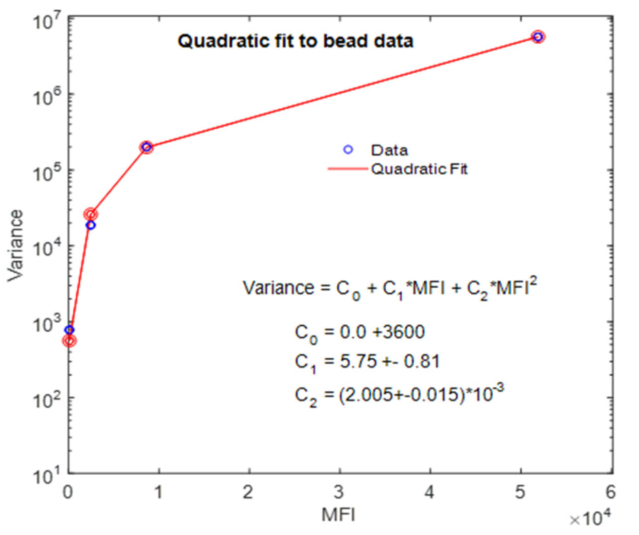
Appendix C. Predicted Coefficients c0, c1, and c2 for the Fit Shown in Figure 6
References
- Wang, L.; DeRose, P.C.; Inwood, S.L.; Gaigalas, A.K. Stochastic Reaction-Diffusion Model of the Binding of Monoclonal Antibodies to CD4 Receptors on the Surface of T Cells. Int. J. Mol. Sci. 2020, 21, 6086. [Google Scholar] [CrossRef] [PubMed]
- Chase, E.S.; Hoffman, R.A. Resolution of Dimly Fluorescent Particles: A Practical Measure of Fluorescence Sensitivity. Cytometry 1998, 33, 267–279. [Google Scholar] [CrossRef]
- Parks, D.R.; El Khettabi, F.; Chase, E.; Hoffman, R.A.; Perfetto, S.P.; Spidlen, J.; Wood, J.C.S.; Moore, W.A.; Brinkman, R.R. Evaluating Flow Cytometer Performance with Weighted Quadratic Least Squares Analysis of LED and Multi-Level Bead Data. Cytom. Part A 2017, 91, 232–249. [Google Scholar] [CrossRef] [PubMed] [Green Version]
- Ormerod, M.G. Flow Cytometry—A Basic Introduction; DeNovo Software: Pasadena, CA, USA, 2008. [Google Scholar]
- Wang, L.; Hoffman, R.A. Standardization, Calibration, and Control in Flow Cytometry. Curr. Protoc. Cytom. 2017, 79, 1.3.1–1.3.27. [Google Scholar] [CrossRef] [PubMed]
- O’Shea, D.C. Elements of Modern Optical Design; John Wiley Sons: New York, NY, USA, 1985; pp. 230–266. [Google Scholar]
- Tokmakoff, A. Time Dependent Quantum Mechanics and Spectroscopy. Physical & Theoretical Chemistry. 2020. Available online: https://chem.libretexts.org/Bookshelves (accessed on 1 June 2019).
- Photonics. Available online: https://www.rp-photonics.com/flat_top_beams.html (accessed on 1 January 2020).
- Song, L.; Varma, C.A.; Verhoeven, J.W.; Tanke, H.J. Influence of the Triplet Excited State on the Photobleaching Kinetics of Fluorescein in Microscopy. Biophys. J. 1996, 70, 2959–2968. [Google Scholar] [CrossRef] [Green Version]
- Loudon, R. The Quantum Theory of Light, 3rd ed.; Oxford University Press: New York, NY, USA, 2000; pp. 23–27. [Google Scholar]
- Gillespie, D.T. Exact Stochastic Simulation of Coupled Chemical Reactions. J. Phys. Chem. 1977, 81, 2340–2361. [Google Scholar] [CrossRef]
- Erban, R.; Chapman, S.J.; Maini, P.K. A Practical Guide to Stochastic Simulation of Reaction-Diffusion Processes. arXiv 2007, arXiv:0704.1908. [Google Scholar]
- Technologies, I.B. Photomultiplier Handbook. 1980. Available online: https://psec.uchicago.edu/links/Photomultiplier_Handbook.pdf (accessed on 30 July 2021).
- Ahn, S.; Fessler, J.A. Standard Errors of Mean, Variance, and Standard Deviation Estimators. 2003. Available online: web.eecs.umich.edu/~fessler/papers/files/tr/stderr.pdf (accessed on 1 March 2020).
- Ginaldi, L.; De Martinis, M.; Matutes, E.; Farahat, N.; Morilla, R.; Catovsky, D. Levels of expression of CD19 and CD20 in chrinic B cell leukaemias. J. Clin. Pathol. 1998, 51, 364–369. [Google Scholar] [CrossRef] [PubMed] [Green Version]
- Wang, L.; Bhardwaj, R.; Mostowski, H.; Patrone, P.N.; Kearsley, A.J.; Watson, J.; Lim, L.; Pichaandi, J.; Ornatsky, O.; Majonis, D.; et al. Establishing CD 19 B-cell reference control materials for comparable and quantitative cytometric expression analysis. PLoS ONE 2021, 16, e0248118. [Google Scholar]
- Coulter, B. Instructions for Use: CytoFLEX Platform, in B49006AP; Beckamn Coulter: Brea, CA, USA, 2019; pp. 1–42. [Google Scholar]
- Lehner, B.; Kaneko, K. Fluctuations and response in biology. Cell. Mol. Life Sci. 2011, 68, 1005–1010. [Google Scholar] [CrossRef] [PubMed] [Green Version]
- Sato, K.; Ito, Y.; Yomo, T.; Kaneko, K. On the relation between fluctuation and response in biological systems. Proc. Natl. Acad. Sci. USA 2003, 100, 14086–14090. [Google Scholar] [CrossRef] [PubMed] [Green Version]
- Roy, S.; Bagchi, B. Fluctuation theory of immune reponse: A statistical mechanical approach to understanding pathogen induced T-cell population dynamics. J. Chem. Phys. 2020, 153, 045107. [Google Scholar] [CrossRef] [PubMed]
- Balkay, L. fca_readfcs. In MATLAB Central File Exchange; MathWorks: Natick, MA, USA, 2021. [Google Scholar]
- Abad, E.; Yuste, S.B.; Lindenberg, K. Elucidating the Role of Subdiffusion and Evaneecence in the Target Problem: Some Recent Results. Math. Model. Nat. Phenom. 2013, 8, 100–113. [Google Scholar] [CrossRef] [Green Version]
- Rice, J.A. Mathematical Statistics and Data Analysis, 3rd ed.; Barnes and Noble: CENGAGE Learning: Boston, MA, USA, 2006. [Google Scholar]


| 100 µL/Min | 200 µL/Min | 500 µL/Min | ||||
|---|---|---|---|---|---|---|
| MFI | CV% | MFI | CV% | MFI | CV% | |
| B | 133 | 36.4 | 130 | 36.4 | 134 | 36.2 |
| L | 1861 | 7.2 | 1877 | 7.1 | 1889 | 7.3 |
| M | 6521 | 5.9 | 6568 | 6 | 6588 | 6.1 |
| H | 38,882 | 5.4 | 39,373 | 5.5 | 39,461 | 5.6 |
| 100 µL/Min | 200 µL/Min | 500 µL/Min | ||||
|---|---|---|---|---|---|---|
| MFI | CV% | MFI | CV% | MFI | CV% | |
| B | 95 | 29.4 | 88 | 43.2 | 85 | 39.7 |
| L | 2449 | 5.6 | 2455 | 6.5 | 2452 | 6.7 |
| M | 8615 | 5.2 | 8628 | 5.3 | 8626 | 5.3 |
| H | 51,882 | 4.6 | 51,899 | 4.8 | 51,880 | 4.8 |
| Name | Value | Location | Description |
|---|---|---|---|
| P0 | 0.04 W or 0.1 W | laser | Power of illuminating laser |
| λ | 488 × 10−9 m | laser | Wavelength of illuminating laser |
| wx wy | 25 µm, 10 µm | laser | Half-widths of the beam waist of the flaser beam |
| delx | varies | bead/cell | Deviation of path of bead/cell through laser beam |
| Nlb | varies | bead/cell | Number of labels on the surface of the bead/cell |
| Nbk | varies | bead/cell | Number of effective background labels |
| sig | 3.06 × 10⁻²⁰ m2 | bead/cell | Absorption cross-section of FITC label |
| QY | 0.95, varies | bead/cell | Fluorescence quantum yield of FITC label |
| τ | 4.3 × 10⁻⁹ s | bead/cell | Fluorescence decay lifetime of FITC label |
| Rb | 2.6 × 10⁻⁶ m | bead/cell | Radius of bead (diameter = 5.2 µm) |
| v0 | 0.80 m/s | bead/cell | Linear velocity of bead in sample stream |
| ster | 3.53 steradian | detector | Solid angle of detector aperture |
| QE | 0.8 varies | detector | Quantum efficiency of PMT photocathode |
| Gain | 1.0 × 10⁵ | detector | Photoelectron multiplication by PMT |
| Rf | 1.25 × 10⁵ | detector | Gain of the current-to-voltage converter |
| Resolution | 5/216 | detector | ADC amplitude resolution, 5 V into 216 bins |
| Time bin | 2 × 10⁻⁶ s | detector | ADC time bin width |
| c | 2.26 × 10⁸ m/s | constant | Speed of light in water (m/s) |
| h | 6.63 × 10⁻³⁴ J·s | constant | Planck constant (J·s) |
| Name | Measured Values, 100 μL/Min | Calculated Response | Bead Properties | ||||||
|---|---|---|---|---|---|---|---|---|---|
| Bead Name | MFI | CV% | Variance | MFI | CV% | Variance | Number of Labels | SD | CV% |
| 1 | 2 | 3 | 4 | 5 | 6 | 7 | 8 | 9 | 10 |
| B | 95 | 29.4 | 780 | 92 | 28.4 | 729 | 3.6 | 1.2 | 33 |
| L | 2449 | 5.6 | 18,808 | 2451 | 5.8 | 19,881 | 100 | 5.4 | 5.9 |
| M | 8615 | 5.2 | 200,686 | 8606 | 5.1 | 200,704 | 364 | 19 | 5.2 |
| H | 51,882 | 4.6 | 5,695,726 | 51,709 | 4.6 | 5,657,804 | 2115 | 91 | 4.3 |
| 100 µL/min | 200 µL/min | 500 µL/min | ||||
|---|---|---|---|---|---|---|
| MFI | CV% | MFI | CV% | MFI | CV% | |
| L | 2354 | 5.7 | 2367 | 6.5 | 2367 | 6.8 |
| M | 8520 | 5.2 | 8540 | 5.3 | 8541 | 5.3 |
| H | 51,787 | 4.6 | 51,811 | 4.8 | 51,814 | 4.8 |
| Cyto-Trol® | Vericell® | |||
|---|---|---|---|---|
| Uncorrected | Corrected | Uncorrected | Corrected | |
| MFI | 18,400 ± 245 | 18,300 ± 254 | 19,700 ± 350 | 19,600 ± 350 |
| SD | 10,600 ± 175 | 10,600 ± 175 | 7600 ± 250 | 7600 ± 250 |
| CV% | 57.6 ± 2.2 | 57.8 ± 2.2 | 38.6 ± 2.4 | 38.7 ± 2.4 |
Publisher’s Note: MDPI stays neutral with regard to jurisdictional claims in published maps and institutional affiliations. |
© 2021 by the authors. Licensee MDPI, Basel, Switzerland. This article is an open access article distributed under the terms and conditions of the Creative Commons Attribution (CC BY) license (https://creativecommons.org/licenses/by/4.0/).
Share and Cite
Gaigalas, A.K.; Zhang, Y.-Z.; Tian, L.; Wang, L. Sources of Variability in the Response of Labeled Microspheres and B Cells during the Analysis by a Flow Cytometer. Int. J. Mol. Sci. 2021, 22, 8256. https://doi.org/10.3390/ijms22158256
Gaigalas AK, Zhang Y-Z, Tian L, Wang L. Sources of Variability in the Response of Labeled Microspheres and B Cells during the Analysis by a Flow Cytometer. International Journal of Molecular Sciences. 2021; 22(15):8256. https://doi.org/10.3390/ijms22158256
Chicago/Turabian StyleGaigalas, Adolfas K., Yu-Zhong Zhang, Linhua Tian, and Lili Wang. 2021. "Sources of Variability in the Response of Labeled Microspheres and B Cells during the Analysis by a Flow Cytometer" International Journal of Molecular Sciences 22, no. 15: 8256. https://doi.org/10.3390/ijms22158256
APA StyleGaigalas, A. K., Zhang, Y.-Z., Tian, L., & Wang, L. (2021). Sources of Variability in the Response of Labeled Microspheres and B Cells during the Analysis by a Flow Cytometer. International Journal of Molecular Sciences, 22(15), 8256. https://doi.org/10.3390/ijms22158256


