
A Critical Assessment of the Association between HLA-G Expression by Carcinomas and Clinical Outcome

 , ,
, , 
Abstract
:1. Introduction
2. Association between HLA-G Expression and Clinicopathological Parameters in Carcinoma Patients
2.1. Breast Carcinoma
2.2. Cervical Carcinoma
2.3. Colorectal Carcinoma
2.4. Esophageal Carcinoma
2.5. Gastric Carcinoma
2.6. Hepatocellular Carcinoma
2.7. Lung Carcinoma
2.8. Oral Carcinoma
2.9. Ovarian Carcinoma
2.10. Pancreatic Carcinoma
2.11. Residual Carcinoma Types
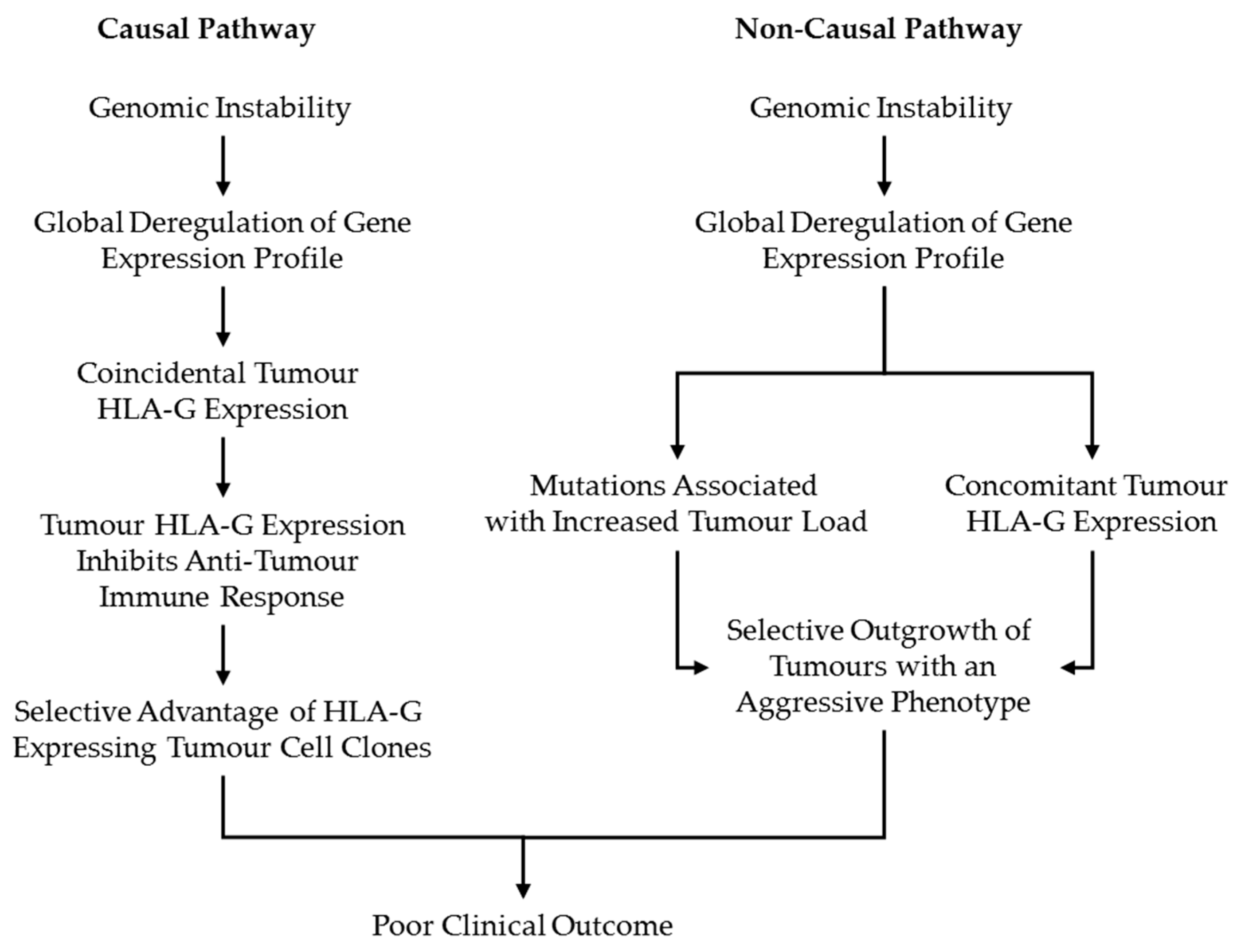
3. Discussion
4. Conclusions
Author Contributions
Funding
Institutional Review Board Statement
Informed Consent Statement
Data Availability Statement
Conflicts of Interest
References
- Ferreira, L.M.R.; Meissner, T.B.; Tilburgs, T.; Strominger, J.L. HLA-G: At the Interface of Maternal-Fetal Tolerance. Trends Immunol. 2017, 38, 272–286. [Google Scholar] [CrossRef]
- Kovats, S.; Main, E.K.; Librach, C.; Stubblebine, M.; Fisher, S.J.; DeMars, R. A class I antigen, HLA-G, expressed in human trophoblasts. Science 1990, 248, 220–223. [Google Scholar] [CrossRef]
- Xu, X.; Zhou, Y.; Wei, H. Roles of HLA-G in the Maternal-Fetal Immune Microenvironment. Front. Immunol. 2020, 11, 592010. [Google Scholar] [CrossRef] [PubMed]
- Colonna, M.; Navarro, F.; Bellón, T.; Llano, M.; García, P.; Samaridis, J.; Angman, L.; Cella, M.; López-Botet, M. A common inhibitory receptor for major histocompatibility complex class I molecules on human lymphoid and myelomonocytic cells. J. Exp. Med. 1997, 186, 1809–1818. [Google Scholar] [CrossRef] [PubMed] [Green Version]
- Colonna, M.; Samaridis, J.; Cella, M.; Angman, L.; Allen, R.L.; O’Callaghan, C.A.; Dunbar, R.; Ogg, G.S.; Cerundolo, V.; Rolink, A. Human myelomonocytic cells express an inhibitory receptor for classical and nonclassical MHC class I molecules. J. Immunol. 1998, 160, 3096–3100. [Google Scholar]
- Rajagopalan, S.; Long, E.O. A human histocompatibility leukocyte antigen (HLA)-G-specific receptor expressed on all natural killer cells. J. Exp. Med. 1999, 189, 1093–1100. [Google Scholar] [CrossRef] [PubMed] [Green Version]
- LeMaoult, J.; Zafaranloo, K.; Le Danff, C.; Carosella, E.D. HLA-G up-regulates ILT2, ILT3, ILT4, and KIR2DL4 in antigen presenting cells, NK cells, and T cells. FASEB J. Off. Publ. Fed. Am. Soc. Exp. Biol. 2005, 19, 662–664. [Google Scholar] [CrossRef] [PubMed]
- Ishitani, A.; Geraghty, D.E. Alternative splicing of HLA-G transcripts yields proteins with primary structures resembling both class I and class II antigens. Proc. Natl. Acad. Sci. USA 1992, 89, 3947–3951. [Google Scholar] [CrossRef] [Green Version]
- Djurisic, S.; Hviid, T.V. HLA Class Ib Molecules and Immune Cells in Pregnancy and Preeclampsia. Front. Immunol. 2014, 5, 652. [Google Scholar] [CrossRef] [Green Version]
- Morales, P.J.; Pace, J.L.; Platt, J.S.; Phillips, T.A.; Morgan, K.; Fazleabas, A.T.; Hunt, J.S. Placental cell expression of HLA-G2 isoforms is limited to the invasive trophoblast phenotype. J. Immunol. 2003, 171, 6215–6224. [Google Scholar] [CrossRef]
- Lin, A.; Yan, W.H. Heterogeneity of HLA-G Expression in Cancers: Facing the Challenges. Front. Immunol. 2018, 9, 2164. [Google Scholar] [CrossRef]
- Rouas-Freiss, N.; Moreau, P.; Menier, C.; Carosella, E.D. HLA-G in cancer: A way to turn off the immune system. Semin. Cancer Biol. 2003, 13, 325–336. [Google Scholar] [CrossRef]
- Rouas-Freiss, N.; Moreau, P.; Ferrone, S.; Carosella, E.D. HLA-G proteins in cancer: Do they provide tumor cells with an escape mechanism? Cancer Res. 2005, 65, 10139–10144. [Google Scholar] [CrossRef] [PubMed] [Green Version]
- Carosella, E.D.; Rouas-Freiss, N.; Tronik-Le Roux, D.; Moreau, P.; LeMaoult, J. HLA-G: An Immune Checkpoint Molecule. Adv. Immunol. 2015, 127, 33–144. [Google Scholar] [CrossRef]
- Dumont, C.; Jacquier, A.; Verine, J.; Noel, F.; Goujon, A.; Wu, C.L.; Hung, T.M.; Desgrandchamps, F.; Culine, S.; Carosella, E.D.; et al. CD8(+)PD-1(−)ILT2(+) T Cells Are an Intratumoral Cytotoxic Population Selectively Inhibited by the Immune-Checkpoint HLA-G. Cancer Immunol. Res. 2019, 7, 1619–1632. [Google Scholar] [CrossRef] [PubMed]
- Tronik-Le Roux, D.; Sautreuil, M.; Bentriou, M.; Verine, J.; Palma, M.B.; Daouya, M.; Bouhidel, F.; Lemler, S.; LeMaoult, J.; Desgrandchamps, F.; et al. Comprehensive landscape of immune-checkpoints uncovered in clear cell renal cell carcinoma reveals new and emerging therapeutic targets. Cancer Immunol. Immunother. CII 2020, 69, 1237–1252. [Google Scholar] [CrossRef] [PubMed]
- Ishibashi, K.; Kumai, T.; Ohkuri, T.; Kosaka, A.; Nagato, T.; Hirata, Y.; Ohara, K.; Oikawa, K.; Aoki, N.; Akiyama, N.; et al. Epigenetic modification augments the immunogenicity of human leukocyte antigen G serving as a tumor antigen for T cell-based immunotherapy. Oncoimmunology 2016, 5, e1169356. [Google Scholar] [CrossRef] [Green Version]
- Ramos, C.S.; Goncalves, A.S.; Marinho, L.C.; Gomes Avelino, M.A.; Saddi, V.A.; Lopes, A.C.; Simoes, R.T.; Wastowski, I.J. Analysis of HLA-G gene polymorphism and protein expression in invasive breast ductal carcinoma. Hum. Immunol. 2014, 75, 667–672. [Google Scholar] [CrossRef]
- He, X.; Dong, D.D.; Yie, S.M.; Yang, H.; Cao, M.; Ye, S.R.; Li, K.; Liu, J.; Chen, J. HLA-G expression in human breast cancer: Implications for diagnosis and prognosis, and effect on allocytotoxic lymphocyte response after hormone treatment in vitro. Ann. Surg. Oncol. 2010, 17, 1459–1469. [Google Scholar] [CrossRef]
- de Kruijf, E.M.; Sajet, A.; van Nes, J.G.; Natanov, R.; Putter, H.; Smit, V.T.; Liefers, G.J.; van den Elsen, P.J.; van de Velde, C.J.; Kuppen, P.J. HLA-E and HLA-G expression in classical HLA class I-negative tumors is of prognostic value for clinical outcome of early breast cancer patients. J. Immunol. 2010, 185, 7452–7459. [Google Scholar] [CrossRef] [Green Version]
- Engels, C.C.; Charehbili, A.; van de Velde, C.J.; Bastiaannet, E.; Sajet, A.; Putter, H.; van Vliet, E.A.; van Vlierberghe, R.L.; Smit, V.T.; Bartlett, J.M.; et al. The prognostic and predictive value of Tregs and tumor immune subtypes in postmenopausal, hormone receptor-positive breast cancer patients treated with adjuvant endocrine therapy: A Dutch TEAM study analysis. Breast Cancer Res. Treat. 2015, 149, 587–596. [Google Scholar] [CrossRef] [Green Version]
- Ferns, D.M.; Heeren, A.M.; Samuels, S.; Bleeker, M.C.G.; de Gruijl, T.D.; Kenter, G.G.; Jordanova, E.S. Classical and non-classical HLA class I aberrations in primary cervical squamous- and adenocarcinomas and paired lymph node metastases. J. Immunother. Cancer 2016, 4, 78. [Google Scholar] [CrossRef] [PubMed] [Green Version]
- Rodriguez, J.A.; Galeano, L.; Palacios, D.M.; Gomez, C.; Serrano, M.L.; Bravo, M.M.; Combita, A.L. Altered HLA class I and HLA-G expression is associated with IL-10 expression in patients with cervical cancer. Pathobiol. J. Immunopathol. Mol. Cell. Biol. 2012, 79, 72–83. [Google Scholar] [CrossRef] [PubMed]
- Ye, S.R.; Yang, H.; Li, K.; Dong, D.D.; Lin, X.M.; Yie, S.M. Human leukocyte antigen G expression: As a significant prognostic indicator for patients with colorectal cancer. Mod. Pathol. 2007, 20, 375–383. [Google Scholar] [CrossRef] [PubMed] [Green Version]
- Cai, Z.; Wang, L.; Han, Y.; Gao, W.; Wei, X.; Gong, R.; Zhu, M.; Sun, Y.; Yu, S. Immunoglobulinlike transcript 4 and human leukocyte antigenG interaction promotes the progression of human colorectal cancer. Int. J. Oncol. 2019, 54, 1943–1954. [Google Scholar] [CrossRef] [Green Version]
- Lin, A.; Zhang, X.; Zhang, R.L.; Zhang, J.G.; Zhou, W.J.; Yan, W.H. Clinical Significance of Potential Unidentified HLA-G Isoforms Without alpha1 Domain but Containing Intron 4 in Colorectal Cancer Patients. Front. Oncol. 2018, 8, 361. [Google Scholar] [CrossRef]
- Zhang, R.L.; Zhang, X.; Dong, S.S.; Hu, B.; Han, Q.Y.; Zhang, J.G.; Zhou, W.J.; Lin, A.; Yan, W.H. Predictive value of different proportion of lesion HLA-G expression in colorectal cancer. Oncotarget 2017, 8, 107441–107451. [Google Scholar] [CrossRef] [Green Version]
- Kirana, C.; Ruszkiewicz, A.; Stubbs, R.S.; Hardingham, J.E.; Hewett, P.J.; Maddern, G.J.; Hauben, E. Soluble HLA-G is a differential prognostic marker in sequential colorectal cancer disease stages. Int. J. Cancer 2017, 140, 2577–2586. [Google Scholar] [CrossRef]
- Guo, Z.Y.; Lv, Y.G.; Wang, L.; Shi, S.J.; Yang, F.; Zheng, G.X.; Wen, W.H.; Yang, A.G. Predictive value of HLA-G and HLA-E in the prognosis of colorectal cancer patients. Cell. Immunol. 2015, 293, 10–16. [Google Scholar] [CrossRef]
- Zeestraten, E.C.; Reimers, M.S.; Saadatmand, S.; Goossens-Beumer, I.J.; Dekker, J.W.; Liefers, G.J.; van den Elsen, P.J.; van de Velde, C.J.; Kuppen, P.J. Combined analysis of HLA class I, HLA-E and HLA-G predicts prognosis in colon cancer patients. Br. J. Cancer 2014, 110, 459–468. [Google Scholar] [CrossRef] [PubMed]
- Reimers, M.S.; Engels, C.C.; Putter, H.; Morreau, H.; Liefers, G.J.; van de Velde, C.J.; Kuppen, P.J. Prognostic value of HLA class I, HLA-E, HLA-G and Tregs in rectal cancer: A retrospective cohort study. BMC Cancer 2014, 14, 486. [Google Scholar] [CrossRef] [Green Version]
- Lin, A.; Zhang, X.; Zhou, W.J.; Ruan, Y.Y.; Xu, D.P.; Wang, Q.; Yan, W.H. Human leukocyte antigen-G expression is associated with a poor prognosis in patients with esophageal squamous cell carcinoma. Int. J. Cancer 2011, 129, 1382–1390. [Google Scholar] [CrossRef]
- Yie, S.M.; Yang, H.; Ye, S.R.; Li, K.; Dong, D.D.; Lin, X.M. Expression of HLA-G is associated with prognosis in esophageal squamous cell carcinoma. Am. J. Clin. Pathol. 2007, 128, 1002–1009. [Google Scholar] [CrossRef]
- Zheng, J.; Xu, C.; Chu, D.; Zhang, X.; Li, J.; Ji, G.; Hong, L.; Feng, Q.; Li, X.; Wu, G.; et al. Human leukocyte antigen G is associated with esophageal squamous cell carcinoma progression and poor prognosis. Immunol. Lett. 2014, 161, 13–19. [Google Scholar] [CrossRef] [PubMed]
- Du, L.; Xiao, X.; Wang, C.; Zhang, X.; Zheng, N.; Wang, L.; Zhang, X.; Li, W.; Wang, S.; Dong, Z. Human leukocyte antigen-G is closely associated with tumor immune escape in gastric cancer by increasing local regulatory T cells. Cancer Sci. 2011, 102, 1272–1280. [Google Scholar] [CrossRef]
- Ishigami, S.; Natsugoe, S.; Miyazono, F.; Nakajo, A.; Tokuda, K.; Matsumoto, M.; Okumura, H.; Douchi, T.; Hokita, S.; Aikou, T. HLA-G expression in gastric cancer. Anticancer Res. 2006, 26, 2467–2472. [Google Scholar] [PubMed]
- Murdaca, G.; Calamaro, P.; Lantieri, F.; Pigozzi, S.; Mastracci, L.; Grillo, F.; Magnani, O.; Ceppa, P.; Puppo, F.; Fiocca, R. HLA-G expression in gastric carcinoma: Clinicopathological correlations and prognostic impact. Virchows Arch. Int. J. Pathol. 2018, 473, 425–433. [Google Scholar] [CrossRef]
- Tuncel, T.; Karagoz, B.; Haholu, A.; Ozgun, A.; Emirzeoglu, L.; Bilgi, O.; Kandemir, E.G. Immunoregulatory function of HLA-G in gastric cancer. Asian Pac. J. Cancer Prev. 2013, 14, 7681–7684. [Google Scholar] [CrossRef] [PubMed] [Green Version]
- Wan, R.; Wang, Z.W.; Li, H.; Peng, X.D.; Liu, G.Y.; Ou, J.M.; Cheng, A.Q. Human Leukocyte Antigen-G Inhibits the Anti-Tumor Effect of Natural Killer Cells via Immunoglobulin-Like Transcript 2 in Gastric Cancer. Cell. Physiol. Biochem. Int. J. Exp. Cell. Physiol. Biochem. Pharmacol. 2017, 44, 1828–1841. [Google Scholar] [CrossRef]
- Yie, S.M.; Yang, H.; Ye, S.R.; Li, K.; Dong, D.D.; Lin, X.M. Expression of human leukocyte antigen G (HLA-G) correlates with poor prognosis in gastric carcinoma. Ann. Surg. Oncol. 2007, 14, 2721–2729. [Google Scholar] [CrossRef] [PubMed]
- Wang, Y.; Ye, Z.; Meng, X.Q.; Zheng, S.S. Expression of HLA-G in patients with hepatocellular carcinoma. Hepatobiliary Pancreat. Dis. Int. 2011, 10, 158–163. [Google Scholar] [CrossRef]
- Cai, M.Y.; Xu, Y.F.; Qiu, S.J.; Ju, M.J.; Gao, Q.; Li, Y.W.; Zhang, B.H.; Zhou, J.; Fan, J. Human leukocyte antigen-G protein expression is an unfavorable prognostic predictor of hepatocellular carcinoma following curative resection. Clin. Cancer Res. Off. J. Am. Assoc. Cancer Res. 2009, 15, 4686–4693. [Google Scholar] [CrossRef] [PubMed] [Green Version]
- Lin, A.; Zhu, C.C.; Chen, H.X.; Chen, B.F.; Zhang, X.; Zhang, J.G.; Wang, Q.; Zhou, W.J.; Hu, W.; Yang, H.H.; et al. Clinical relevance and functional implications for human leucocyte antigen-g expression in non-small-cell lung cancer. J. Cell. Mol. Med. 2010, 14, 2318–2329. [Google Scholar] [CrossRef] [PubMed] [Green Version]
- Yan, W.H.; Liu, D.; Lu, H.Y.; Li, Y.Y.; Zhang, X.; Lin, A. Significance of tumour cell HLA-G5/-G6 isoform expression in discrimination for adenocarcinoma from squamous cell carcinoma in lung cancer patients. J. Cell. Mol. Med. 2015, 19, 778–785. [Google Scholar] [CrossRef] [PubMed]
- Yie, S.M.; Yang, H.; Ye, S.R.; Li, K.; Dong, D.D.; Lin, X.M. Expression of human leucocyte antigen G (HLA-G) is associated with prognosis in non-small cell lung cancer. Lung Cancer 2007, 58, 267–274. [Google Scholar] [CrossRef] [PubMed]
- Zhang, Y.; Zhao, J.; Qiu, L.; Zhang, P.; Li, J.; Yang, D.; Wei, X.; Han, Y.; Nie, S.; Sun, Y. Co-expression of ILT4/HLA-G in human non-small cell lung cancer correlates with poor prognosis and ILT4-HLA-G interaction activates ERK signaling. Tumour Biol. J. Int. Soc. Oncodevelopmental Biol. Med. 2016, 37, 11187–11198. [Google Scholar] [CrossRef]
- Goncalves, A.S.; Wastowski, I.J.; Capeletti, L.R.; Sacono, N.T.; Cortez, A.P.; Valadares, M.C.; Silva, T.A.; Batista, A.C. The clinicopathologic significance of the expression of HLA-G in oral squamous cell carcinoma. Oral Surg. Oral Med. Oral Pathol. Oral Radiol. 2014, 117, 361–368. [Google Scholar] [CrossRef]
- Imani, R.; Seyedmajidi, M.; Ghasemi, N.; Moslemi, D.; Shafaee, S.; Bijani, A. HLA-G Expression is Associated with an Unfavorable Prognosis of Oral Squamous Cell Carcinoma. Asian Pac. J. Cancer Prev. 2018, 19, 2527–2533. [Google Scholar] [CrossRef] [PubMed]
- Mosconi, C.; Arantes, D.A.C.; Goncalves, A.S.; Alencar, R.C.G.; Oliveira, J.C.; Silva, T.A.; Mendonca, E.F.; Batista, A.C. Immunohistochemical investigations on the expression of programmed cell death ligand 1, human leukocyte antigens G and E, and granzyme B in intraoral mucoepidermoid carcinoma. Arch. Oral Biol. 2017, 83, 55–62. [Google Scholar] [CrossRef] [PubMed]
- Andersson, E.; Poschke, I.; Villabona, L.; Carlson, J.W.; Lundqvist, A.; Kiessling, R.; Seliger, B.; Masucci, G.V. Non-classical HLA-class I expression in serous ovarian carcinoma: Correlation with the HLA-genotype, tumor infiltrating immune cells and prognosis. Oncoimmunology 2016, 5, e1052213. [Google Scholar] [CrossRef] [Green Version]
- Babay, W.; Ben Yahia, H.; Boujelbene, N.; Zidi, N.; Laaribi, A.B.; Kacem, D.; Ben Ghorbel, R.; Boudabous, A.; Ouzari, H.I.; Rizzo, R.; et al. Clinicopathologic significance of HLA-G and HLA-E molecules in Tunisian patients with ovarian carcinoma. Hum. Immunol. 2018, 79, 463–470. [Google Scholar] [CrossRef]
- Jung, Y.W.; Kim, Y.T.; Kim, S.W.; Kim, S.; Kim, J.H.; Cho, N.H.; Kim, J.W. Correlation of human leukocyte antigen-G (HLA-G) expression and disease progression in epithelial ovarian cancer. Reprod. Sci. 2009, 16, 1103–1111. [Google Scholar] [CrossRef] [PubMed]
- Rutten, M.J.; Dijk, F.; Savci-Heijink, C.D.; Buist, M.R.; Kenter, G.G.; van de Vijver, M.J.; Jordanova, E.S. HLA-G expression is an independent predictor for improved survival in high grade ovarian carcinomas. J. Immunol. Res. 2014, 2014, 274584. [Google Scholar] [CrossRef]
- Zhang, X.; Han, Q.Y.; Li, J.B.; Ruan, Y.Y.; Yan, W.H.; Lin, A. Lesion HLA-G5/-G6 isoforms expression in patients with ovarian cancer. Hum. Immunol. 2016, 77, 780–784. [Google Scholar] [CrossRef] [PubMed]
- Seitz, C.; Uchanska-Ziegler, B.; Zank, A.; Ziegler, A. The monoclonal antibody HCA2 recognises a broadly shared epitope on selected classical as well as several non-classical HLA class I molecules. Mol. Immunol. 1998, 35, 819–827. [Google Scholar] [CrossRef]
- Hiraoka, N.; Ino, Y.; Hori, S.; Yamazaki-Itoh, R.; Naito, C.; Shimasaki, M.; Esaki, M.; Nara, S.; Kishi, Y.; Shimada, K.; et al. Expression of classical HLA class I antigens, HLA-E, and HLA-G is adversely prognostic in pancreatic cancer patients. Cancer Sci. 2020. [Google Scholar] [CrossRef]
- Sideras, K.; Biermann, K.; Yap, K.; Mancham, S.; Boor, P.P.C.; Hansen, B.E.; Stoop, H.J.A.; Peppelenbosch, M.P.; van Eijck, C.H.; Sleijfer, S.; et al. Tumor cell expression of immune inhibitory molecules and tumor-infiltrating lymphocyte count predict cancer-specific survival in pancreatic and ampullary cancer. Int. J. Cancer 2017, 141, 572–582. [Google Scholar] [CrossRef] [PubMed]
- Xu, Y.F.; Lu, Y.; Cheng, H.; Jiang, J.; Xu, J.; Long, J.; Liu, L.; Ni, Q.; Liu, C.; Yu, X.J. High Expression of Human Leukocyte Antigen-G is Associated with a Poor Prognosis in Patients with PDAC. Curr. Mol. Med. 2015, 15, 360–367. [Google Scholar] [CrossRef] [PubMed]
- Zhou, L.; Niu, Z.Y.; Liang, Z.Y.; Zhou, W.X.; You, L.; Wang, M.Y.; Yao, L.T.; Liao, Q.; Zhao, Y.P. HLA-G impairs host immune response and predicts poor prognosis in pancreatic cancer. Am. J. Transl. Res. 2015, 7, 2036–2044. [Google Scholar] [PubMed]
- Bijen, C.B.; Bantema-Joppe, E.J.; de Jong, R.A.; Leffers, N.; Mourits, M.J.; Eggink, H.F.; van der Zee, A.G.; Hollema, H.; de Bock, G.H.; Nijman, H.W. The prognostic role of classical and nonclassical MHC class I expression in endometrial cancer. Int. J. Cancer 2010, 126, 1417–1427. [Google Scholar] [CrossRef] [PubMed]
- Lopes, M.; Gonzaga, A.K.G.; Mosconi, C.; Palomino, G.M.; Mendonca, E.F.; Batista, A.C.; Silveira, E. Immune response and evasion mechanisms in lip carcinogenesis: An immunohistochemical study. Arch. Oral Biol. 2019, 98, 99–107. [Google Scholar] [CrossRef] [PubMed]
- de Figueiredo Feitosa, N.L.; Crispim, J.C.; Zanetti, B.R.; Magalhaes, P.K.; Soares, C.P.; Soares, E.G.; Neder, L.; Donadi, E.A.; Maciel, L.M. HLA-G is differentially expressed in thyroid tissues. Thyroid Off. J. Am. Thyroid Assoc. 2014, 24, 585–592. [Google Scholar] [CrossRef] [PubMed]
- Jasinski-Bergner, S.; Stoehr, C.; Bukur, J.; Massa, C.; Braun, J.; Huttelmaier, S.; Spath, V.; Wartenberg, R.; Legal, W.; Taubert, H.; et al. Clinical relevance of miR-mediated HLA-G regulation and the associated immune cell infiltration in renal cell carcinoma. Oncoimmunology 2015, 4, e1008805. [Google Scholar] [CrossRef] [PubMed] [Green Version]
- Friedrich, M.; Stoehr, C.; Jasinski-Bergner, S.; Hartmann, A.; Wach, S.; Wullich, B.; Steven, A.; Seliger, B. Characterization of the expression and immunological impact of the transcriptional activator CREB in renal cell carcinoma. J. Transl. Med. 2020, 18, 371. [Google Scholar] [CrossRef] [PubMed]
- Zhao, L.; Teklemariam, T.; Hantash, B.M. Reassessment of HLA-G isoform specificity of MEM-G/9 and 4H84 monoclonal antibodies. Tissue Antigens 2012, 80, 231–238. [Google Scholar] [CrossRef] [PubMed]
- Polakova, K.; Kuba, D.; Russ, G. The 4H84 monoclonal antibody detecting beta2m free nonclassical HLA-G molecules also binds to free heavy chains of classical HLA class I antigens present on activated lymphocytes. Hum. Immunol. 2004, 65, 157–162. [Google Scholar] [CrossRef]
- Polakova, K.; Bennink, J.R.; Yewdell, J.W.; Bystricka, M.; Bandzuchova, E.; Russ, G. Mild acid treatment induces cross-reactivity of 4H84 monoclonal antibody specific to nonclassical HLA-G antigen with classical HLA class I molecules. Hum. Immunol. 2003, 64, 256–264. [Google Scholar] [CrossRef]
- Swets, M.; Konig, M.H.; Zaalberg, A.; Dekker-Ensink, N.G.; Gelderblom, H.; van de Velde, C.J.; van den Elsen, P.J.; Kuppen, P.J. HLA-G and classical HLA class I expression in primary colorectal cancer and associated liver metastases. Hum. Immunol. 2016, 77, 773–779. [Google Scholar] [CrossRef]
- Shiroishi, M.; Kuroki, K.; Ose, T.; Rasubala, L.; Shiratori, I.; Arase, H.; Tsumoto, K.; Kumagai, I.; Kohda, D.; Maenaka, K. Efficient leukocyte Ig-like receptor signaling and crystal structure of disulfide-linked HLA-G dimer. J. Biol. Chem. 2006, 281, 10439–10447. [Google Scholar] [CrossRef] [Green Version]
- Attia, J.V.D.; Dessens, C.E.; van de Water, R.; Houvast, R.D.; Kuppen, P.J.K.; Krijgsman, D. The Molecular and Functional Characteristics of HLA-G and the Interaction with Its Receptors: Where to Intervene for Cancer Immunotherapy? Int. J. Mol. Sci. 2020, 21, 8678. [Google Scholar] [CrossRef]
- Krijgsman, D.; Roelands, J.; Hendrickx, W.; Bedognetti, D.; Kuppen, P.J.K. HLA-G: A New Immune Checkpoint in Cancer? Int. J. Mol. Sci. 2020, 21, 4528. [Google Scholar] [CrossRef]
- Swets, M.; Seneby, L.; Boot, A.; van Wezel, T.; Gelderblom, H.; van de Velde, C.J.; van den Elsen, P.J.; Kuppen, P.J. Promoter methylation and mRNA expression of HLA-G in relation to HLA-G protein expression in colorectal cancer. Hum. Immunol. 2016, 77, 764–772. [Google Scholar] [CrossRef] [PubMed]
- Castelli, E.C.; Veiga-Castelli, L.C.; Yaghi, L.; Moreau, P.; Donadi, E.A. Transcriptional and posttranscriptional regulations of the HLA-G gene. J. Immunol. Res. 2014, 2014, 734068. [Google Scholar] [CrossRef] [PubMed] [Green Version]
- Zhang, Y.; Jin, X.; Wang, J. miR-148a modulates the viability, migration and invasion of oral squamous cell carcinoma cells by regulating HLA-G expression. Mol. Med. Rep. 2019, 20, 795–801. [Google Scholar] [CrossRef] [PubMed] [Green Version]
- Seliger, B. Role of microRNAs on HLA-G expression in human tumors. Hum. Immunol. 2016, 77, 760–763. [Google Scholar] [CrossRef]
- Swets, M.; Wouters, A.; Krijgsman, D.; van Vlierberghe, R.L.P.; Boot, A.; van Eendenburg, J.D.; van Wezel, T.; Gelderblom, H.; van de Velde, C.J.H.; van den Elsen, P.J.; et al. HLA-G protein expression in colorectal cancer evaluated by immunohistochemistry and western blot analysis: Its expression characteristics remain enigmatic. Clin. Immunol. 2018, 194, 80–86. [Google Scholar] [CrossRef]
- Raulet, D.H. Missing self recognition and self tolerance of natural killer (NK) cells. Semin Immunol 2006, 18, 145–150. [Google Scholar] [CrossRef]
- Rouas-Freiss, N.; Goncalves, R.M.; Menier, C.; Dausset, J.; Carosella, E.D. Direct evidence to support the role of HLA-G in protecting the fetus from maternal uterine natural killer cytolysis. Proc. Natl. Acad. Sci. USA 1997, 94, 11520–11525. [Google Scholar] [CrossRef] [PubMed] [Green Version]
- Amiot, L.; Ferrone, S.; Grosse-Wilde, H.; Seliger, B. Biology of HLA-G in cancer: A candidate molecule for therapeutic intervention? Cell. Mol. Life Sci. 2011, 68, 417–431. [Google Scholar] [CrossRef] [PubMed] [Green Version]
- Rouas-Freiss, N.; LeMaoult, J.; Verine, J.; Tronik-Le Roux, D.; Culine, S.; Hennequin, C.; Desgrandchamps, F.; Carosella, E.D. Intratumor heterogeneity of immune checkpoints in primary renal cell cancer: Focus on HLA-G/ILT2/ILT4. Oncoimmunology 2017, 6, e1342023. [Google Scholar] [CrossRef] [Green Version]
- Zhang, X.; Lin, A.; Han, Q.Y.; Zhang, J.G.; Chen, Q.Y.; Ye, Y.H.; Zhou, W.J.; Xu, H.H.; Gan, J.; Yan, W.H. Intratumor Heterogeneity of HLA-G Expression in Cancer Lesions. Front. Immunol. 2020, 11, 565759. [Google Scholar] [CrossRef] [PubMed]
- Anna, F.; Bole-Richard, E.; LeMaoult, J.; Escande, M.; Lecomte, M.; Certoux, J.M.; Souque, P.; Garnache, F.; Adotevi, O.; Langlade-Demoyen, P.; et al. First immunotherapeutic CAR-T cells against the immune checkpoint protein HLA-G. J. Immunother. Cancer 2021, 9. [Google Scholar] [CrossRef] [PubMed]


| First Author [Ref.] | mAb and Included Patient Cohort | HLA-G Quantification Method | HLA-G+ Samples (%) | Association with Tumour HLA-G Expression | |
|---|---|---|---|---|---|
| Clinico-Pathological Parameters with p-Values ≤ 0.05 | Clinical Outcome (p-Value) | ||||
| * Engels [21] | 4H84 Post-meno-pausa, hormone receptor positive BC patients | Tumour was considered HLA-G-positive when >1% of tumour cells were stained Tumour immune-susceptibility was expressed in IS and was generated by adding up regression coefficients of HLA-G, HLA-class I, HLA-E and FoxP3 expression, thereby creating three groups; low, intermediate and high IS | Low IS: 817/1636 (50) Intermediate IS: 318/1636 (19) High IS: 501/1636 (31) Total cohort: 484/2042 (24) | Increased tumour grade. | Cox univariate analysis: OS (ns); RFP (ns); CSS (ns). Cox proportional hazard analysis, intermediate vs. high IS: OS, HR = 1.471 (none provided); RFP, HR = 1.539 (none provided); CSS, HR = 2.119 (none provided). Cox proportional hazard analysis, low vs. high IS: Shorter OS, HR = 1.602 (0.002); Shorter RFP, HR = 1.634 (0.002); Shorter CSS, HR = 2.103 (<0.001). |
| * De Kruijf [20] | 4H84 Early BC patients | Tumour was considered HLA-G-positive when >1% of tumour cells were stained | 201/501 (40) | HLA-class I expression; Her2 over-expression; Type of received systemic therapy. | KM analysis: OS (ns); RFP (ns). KM analysis, stratified for HLA-class I expression, n = 361: OS (ns); RFP (ns). KM analysis, stratified for loss of HLA-class I expression, n = 106: OS (ns); Shorter RFP (0.035). |
| Ishibashi [17] | 4H84 BC patients, random cohort | Low (absent (-) or weak (+)) staining vs. high staining (moderate (++) or strong (+++)) | +: 58/102 (57) ++: 32/102 (31) +++: 6/102 (6) High staining: 38/102 (37) | Tumour ER down-regulation; Tumour PR down-regulation. | KM analysis: Shorter OS (0.006); Shorter DFS (0.049). |
| Ramos [18] | MEM-G/2 Patients with invasive ductal BC | Based on ROC-curve analysis | 28/45 (62) | Increased LNM | KM analysis: Shorter OS (0.03) Cox multivariate analysis: Shorter OS, HR = 8.8 (0.04) |
| He [19] | HGY BC patients, random cohort | Absent (0%) and weak (1–25%) staining vs. moderate (25–50%) and strong (>50%) staining | Cohort with available follow-up Weak staining: 42/84 (50) Moderate/ strong staining: 25/84 (30) | Increased tumour size; Increased LNM; Advanced disease stage; Tumour ER over-expression; Tumour PR over-expression. | KM analysis: Shorter OS (0.028) Cox multivariate analysis: Shorter OS, HR = 10.2 (0.006) |
| First Author [Ref.] | mAb and Included Patient Cohort | HLA-G Quantification Method | HLA-G+ Samples (%) | Association with Tumour HLA-G Expression | |
|---|---|---|---|---|---|
| Clinico-Pathological Parameters with p-Values ≤ 0.05 | Clinical Outcome (p-Value) | ||||
| Ferns [22] | 4H84 Patients with cervical SCC and AC | Representation of percentage and intensity scores: No expression (0–4) vs. positive expression (5–8) | SSC patients: 23/103 (22) AC patients: 10/33 (31) Total cohort: 33/136 (24) | None declared | KM analysis, in SCC patients with loss of HLA-A expression, n = 31: Shorter DFS (0.001); n = 30: Shorter CSS (0.004). KM analysis, in patients with loss of classical HLA-class I, n = 19: Shorter DFS (0.002); Shorter CSS (0.003). KM analysis, in AC patients: (ns) |
| * Rodriguez [23] | 4H84 CINIII and invasive stage IBI-IVB patients | No expression (=0), focal/weak (=1) or >75% expression (=2) Not defined when a tumour sample was considered as HLA-G positive | 16/58 (28) | Decreased HLA-class I expression; IL-10 overexpression. | KM analysis: OS (ns) |
| First Author [Ref.] | mAb and Included Patient Cohort | HLA-G Quantification Method | HLA-G+ Samples (%) | Association with Tumour HLA-G Expression | |
|---|---|---|---|---|---|
| Clinico-Pathological Parameters with p-Values ≤ 0.05 | Clinical Outcome Associated with HLA-G (p-Value) | ||||
| * Reimers [31] | 4H84 CRC patients, random cohort | Staining intensity: Weak staining (undetectable to faint staining in <70% of cells) vs. strong staining (weak to moderate staining in >70% of cells) | Weak staining: 350/484 (72) Strong staining: 134/484 (28) | Weak staining: Advanced TNM stage; Increased LNM. Strong staining: Increased number of infiltrating Tregs; Increased HLA-class I expression. | KM analysis: OS (ns); Prolonged DFS (0.040). Cox univariate analysis: OS, HR = 0.76 (ns); Prolonged DFS, HR = 0.75 (0.042). Cox multivariate analysis: OS, HR = 0.88 (ns); DFS, HR = 0.85 (ns). |
| Cai [25] | 4H84 CRC patients, random cohort | Representation of percentage and intensity scores: No expression (0–3) vs. positive expression (4–9) | HLA-G+/ILT4+: 44/88 (50) HLA-G+/ILT4-: 8/88 (9) HLA-G-/ILT4+: 16/88 (18) HLA-G-/ILT4-: 20/88 (23) Total cohort: 52/88 (59) | Advanced TNM stage; Increased ILT4 expression. | KM analysis, HLA-G+/ILT4+ vs. HLA-G-/ILT4-: Shorter OS (0.032) KM analysis, HLA-G+/ILT4+ vs. HLA-G-/ILT4+: Shorter OS (0.043) KM analysis, HLA-G+/ILT4+ vs. HLA-G+/ILT4-: OS (ns) |
| Zhang [27] | 4H84 CRC patients, random cohort | Staining was considered as positive at >5% or >55% | Cohort with available follow-up, >5% staining: 296/417 (71) | More prevalently observed in colon than rectal carcinoma patients | KM analysis: OS (ns) Cox univariate analysis: OS, HR = 1.348 (ns) Cox multivariate analysis: OS, HR = 1.423 (ns) |
| Cohort with available follow-up, >55% staining: 273/417 (65) | More prevalently observed in colon than rectal carcinoma patients | KM analysis: Shorter OS (0.042) Cox univariate analysis: Shorter OS, HR = 1.428 (0.044) Cox multivariate analysis: Shorter OS, HR = 1.481 (0.028) | |||
| *, † Kirana [28] | 4H84 CRC patients, random cohort. | Staining intensity: No staining vs. moderate or strong staining | Moderate staining: 206/255 (81) Strong staining: 12/255 (5) Total cohort: 218/255 (86) | Strong staining: More prevalently observed in female than male patients | KM analysis, strong vs. no staining, n = 48: CSS (ns) KM analysis, strong vs. moderate staining, n = 215: Shorter CSS (0.04) KM analysis, strong vs. no/moderate staining, n = 251: CCS, HR = 0.571 (ns) KM analysis, moderate vs. no staining, n = 239: CSS (ns) KM analysis, strong vs. no/moderate staining in patients with tumour stage II-III, n = 167: Shorter CSS (0.01) |
| † Lin [26] | 4H84 CRC patients, random cohort | Any staining >5% was considered as positive | 268/379 (71) | Lower TNM stage | KM analysis, n = 339: OS (ns) Cox univariate analysis, n = 339: OS, HR = 1.267 (ns) |
| 5A6G7 CRC patients, random cohort | Any staining >5% was considered as positive | 229/379 (60) | No association between clinico-pathological variables and HLA-G expression was found | KM analysis, n = 339: OS (ns) Cox univariate analysis, n = 339: OS, HR = 0.812 (ns) | |
| * Zeestraten [30] | 4H84 Only colon carcinoma patients | Any staining is considered as positive (1–100%) | 51/251 (20) | No significant correlations | KM analysis: OS (ns); DFS (ns). Cox univariate analysis: OS, HR = 1.2 (ns); DFS, HR = 1.3 (ns). |
| Guo [29] | MEM-G/2 CRC patients, random cohort | General presence of staining, not further specified | 72/102 (71) | Most prevalently observed in adenocarcinoma patients | KM analysis: Shorter OS (0.0243) Cox univariate analysis: Shorter OS, HR = 0.461 (0.029) Cox multivariate analysis: Shorter OS, HR = 0.311 (0.008) |
| † Ye [24] | HGY CRC patients, random cohort | No staining (0%) vs. weak (1–25%), moderate (25–50%) and strong (>50%) staining | Weak staining: 65/201 (32) Moderate staining: 41/201 (20) Strong staining: 24/201 (12) Total cohort: 130/201 (65) | Advanced TNM stage; Advanced histological grade; Increased tumour depth; Weak immune response; More prevalently observed proximally in colon carcinoma than distally in rectal carcinoma patients. | KM analysis, n = 85: Shorter OS (0.001) Cox univariate analysis, n = 85: Shorter OS, HR = 6.40 (0.001) Cox multivariate analysis, n = 85: Shorter OS, HR = 3.14 (0.021) |
| First Author [Ref.] | mAb and Included Patient Cohort | HLA-G Quantification Method | HLA-G+ Samples (%) | Association with Tumour HLA-G Expression | |
|---|---|---|---|---|---|
| Clinico-Pathological Parameters with p-Values ≤ 0.05 | Clinical Outcome Associated with HLA-G (p-Value) | ||||
| Lin [32] | 4H84 ESCC patients, random cohort | 0% (0) vs. 1–25% (1+), 26–50% (2+), 51–75% (3+) or >75% (4+) staining | Cohort with available follow-up: 1+/2+: 14/40 (35) 3+/4+: 9/40 (23) 1+/2+/3+/4+: 23/40 (58) | Advanced TNM stage | KM analysis: Shorter OS (<0.001) Cox univariate analysis: Shorter OS, HR = 3.76 (0.001) Cox multivariate analysis: Shorter OS, HR = 3.83 (0.001) KM analysis, 1+/2+ vs. 0: Shorter OS (0.005) Cox univariate analysis, 1+/2+ vs. 0: Shorter OS, HR = 2.02 (0.01) KM analysis, 3+/4+ vs. 0: Shorter OS (<0.001) Cox univariate analysis, 3+/4+ vs. 0: Shorter OS, HR = 3.02 (<0.001) KM analysis, 3+/4+ vs. 1+/2+: Shorter OS (<0.029) |
| Zheng [34] | MEM-G/1 ESCC patients, random cohort | <25% vs. >25% staining | 42/60 (70) | Advanced differentiation grade; Increased LNM. | KM analysis: Shorter OS (0.01) |
| Yie [33] | HGY ESCC patients, random cohort | 0% (-) vs. 1–25% (1+), 25–50% (2+) or >50% (3+) staining | Cohort with available follow-up: 1+: 27/70 (39) 2+/3+: 32/70 (46) 1+/2+/3+: 59/70 (84) | Advanced tumour grade; Nodal status; Advanced TNM stage; Increased tumour depth; Weak immune response. | KM analysis: Shorter OS (0.001) Cox univariate analysis: Shorter OS, HR = 3.33 (0.001) Cox multivariate analysis: Shorter OS, HR = 2.99 (0.002) |
| First Author [Ref.] | mAb and Included Patient Cohort | HLA-G Quantification Method | HLA-G+ Samples (%) | Association with Tumour HLA-G Expression | |
|---|---|---|---|---|---|
| Clinico-Pathological Parameters with p-Values ≤ 0.05 | Clinical Outcome Associated with HLA-G (p-Value) | ||||
| Murdaca [37] | 4H84 Gastric adeno-carcinoma patients, random cohort | No staining vs. weak/strong staining | Within stage I patients: 4/14 (29) Within stage II patients: 7/40 (18) Within stage III patients: 13/40 (33) Total cohort: 24/94 (26) | No significant correlations | KM analysis: Shorter OS (<0.0001) Cox proportional hazard analysis: Shorter OS, HR = 4.41 (<0.0001) KM analysis, in stage I patients: OS (ns) KM analysis, in stage II patients: Shorter OS (0.0065) KM analysis, in stage III patients: Shorter OS (<0.0001) |
| Wan [39] | 4H84 GC patients, random cohort | <10% (−) vs. 10–30% (+), 30–50% (++) or >50% (+++) staining | +: 4/49 (8) ++: 17/49 (37) +++: 9/49 (18) Total cohort: 30/49 (61) | Increased preoperative anaemia; Increased tumour depth; Increased LNM; Advanced TNM stage; Decreased number of infiltrating NK cells. | KM analysis: Shorter OS (0.0359); Shorter DFS (0.0438). Cox univariate analysis: Shorter OS, (0.050); DFS (ns). Cox multivariate analysis: OS, 95%CI: 0.500–6.886 (ns); DFS, 95%CI: 0.549–4.307 (ns). |
| *, † Du [35] | 4H84 GC patients, random cohort | 0% (−) vs. 1–25% (+), 26–50% (++) or >50% (+++) staining | +: 26/179 (15) ++: 35/179 (20) +++: 28/179 (16) Total cohort: 89/179 (50) | Increased tumour depth; More invaded adjacent organs; Advanced tumour stage; Increased number of infiltrating Tregs. | KM analysis: Shorter OS (<0.001); Shorter DFS (<0.001); n = 150 Shorter CSS (<0.001). Cox multivariate analysis: Shorter OS, 95%CI: 1.094–3.040 (0.021); Shorter DFS, 95%CI: 1.187–3.445 (0.010); n = 150 Shorter CSS, 95%CI: 1.041–3.192 (0.036). |
| Ishigami [36] | MEM-G/1 GC patients, random cohort | No staining vs. weak, moderate or strong staining | Weak staining: 16/115 (14) Moderate staining: 19/115 (17) Strong staining: 17/115 (15) Total cohort: 52/115 (45) | Less tumour depth; Decreased LNM; Earlier clinical stage. | KM analysis: Prolonged OS (<0.05) |
| Yie [40] | HGY GC patients, random cohort | No staining (0%) vs. weak (1–25%), moderate (25–50%) or strong (>50%) staining | Weak staining: 30/160 (19) Moderate staining: 32/160 (20) Strong staining: 51/160 (32) Total cohort: 113/160 (71) | Advanced tumour grade; Increased tumour depth; Increased LNM; Advanced clinical stage; Weak immune response. | KM analysis: Shorter OS (0.001) Cox univariate analysis: Shorter OS, HR = 5.72 (0.0001) Cox multivariate analysis: Shorter OS, HR = 9.08 (0.0001) KM analysis, in patients with disease stage I/II, n = 101: Shorter OS (0.001) KM analysis, in patients with disease stage III/IV, n = 59: Shorter OS (0.001) |
| Tuncel [38] | 5A6G7 GC patients, random cohort | Any staining >10% was considered as positive | 16/52 (31) | Increased LNM; Worse differentiation stage; Tumour type; Advanced TNM stage; Increased number of infiltrating Tregs; Decreased number of CD8+ T cells. | KM analysis: Shorter OS (0.008) Cox univariate analysis: Shorter OS, HR = 3.122 (0.008) Cox multivariate analysis: Shorter OS, HR = 2.662 (0.012) |
| First Author [Ref.] | mAb and Included Patient Cohort | HLA-G Quantification Method | HLA-G+ Samples (%) | Association with Tumour HLA-G Expression | |
|---|---|---|---|---|---|
| Clinico-Pathological Parameters with p-Values ≤ 0.05 | Clinical Outcome Associated with HLA-G (p-Value) | ||||
| * Cai [42] | MEM-G/1 HCC patient, random cohort | Mean density calculation as determined by a computerized imaging system | Within early stage HCC patients: 48/76 (63) Total cohort: 99/173 (57) | More prevalently observed in male than female patients; Tregs/CD8+ ratio. | KM analysis: Shorter OS (0.024); RFP (ns). Cox multivariate analysis: Shorter OS, HR = 1.987 (0.004) KM analysis, in patients with early stage HCC: Shorter OS, (0.012); Shorter RFP, (0.038). Cox multivariate analysis, in patients with early stage HCC: Shorter OS, HR 3.145 (0.041); Shorter RFP, HR = 3.208 (0.023). |
| † Wang [41] | MEM-G/1 HCC patient, random cohort | The appearance of a 39 kDa band corresponding to HLA-G1 | 24/36 (67) | No significant correlations | KM analysis: Shorter OS (0.027); Shorter RFP (0.035). Cox univariate analysis: Shorter OS, HR = 4.565 (0.044); Shorter RFP, HR = 3.503 (0.048). |
| First Author [Ref.] | mAb and Included Patient Cohort | HLA-G Quantification Method | HLA-G+ Samples (%) | Association with Tumour HLA-G Expression | |
|---|---|---|---|---|---|
| Clinico-Pathological Parameters with p-Values ≤ 0.05 | Clinical Outcome Associated with HLA-G (p-Value) | ||||
| * Lin [43] | 4H84 NSCLC patients, random cohort | 0% (0) vs. 1–25% (1), 26–50% (2) or >50% (3) staining | 1: 13/101 (13) 2: 16/101 (16) 3: 13/101 (13) Total cohort: 42/101 (42) | Advanced disease stage | KM analysis, n = 51: OS (ns) |
| Zhang [46] | 4H84 NSCLC patients, random cohort | Representation of percentage and intensity scores: No expression (<4) vs. positive expression (≥4) | HLA-G+/ILT4+: 29/81 (36) HLA-G+/ILT4-: 13/81 (16) HLA-G-/ILT4+: 9/81 (11) HLA-G-/ILT4-: 30/81 (37) Total cohort: 42/81 (52) | Increased LNM; Advanced disease stage; Worse differentiation stage; ILT4 overexpression. | KM analysis, HLA-G+/ILT4+ vs. HLA-G-/ILT4+: Shorter OS (0.021) KM analysis, HLA-G+/ILT4+ vs. HLA-G-/ILT4-: Shorter OS (0.048) KM analysis, HLA-G+/ILT4+ vs. HLA-G+/ILT4-: OS (ns) |
| Yie [45] | HGY NSCLC patients, random cohort | 0% (−) vs. 1–25% (+), 26–50% (++) and >50% (+++) staining | Percentage of HLA-G-positive samples within specific staining groups was unattainable Total cohort with available follow-up: 23/39 (59) | Increased LNM; Advanced disease stage; Weak immune response. | KM analysis: Shorter OS (0.001) Cox univariate analysis: Shorter OS, HR = 4.01 (0.003) Cox multivariate analysis: Shorter OS, HR = 4.09 (0.010) |
| * Yan [44] | 5A6G7 NSCLC patients, random cohort | Any staining >5% was considered as positive | SSC patients: 4/66 (6) AC patients: 40/55 (73) ASC patients: 1/10 (10) Total cohort: 41/123 (34) | More prevalently observed in adenocarcinoma than squamous or adenosquamous carcinoma patients; More prevalently observed in female than male patients. | Cox univariate analysis: OS, HR = 1.15 (ns) KM analysis, in SSC patients, n = 62: OS (ns) Cox univariate analysis, in SSC patients, n = 62: OS, HR = 2.76 (ns) Cox multivariate analysis, in SSC patients, n = 62: OS, HR = 4.05 (ns) KM analysis, in AC patients, n = 51: OS (ns) Cox univariate analysis, in AC patients, n = 51: OS, HR = 1.04 (ns) Cox univariate analysis, in ASC patients: OS, HR = 0.03 (ns) |
| First Author [Ref.] | mAb and Included Patient Cohort | HLA-G Quantification Method | HLA-G+ Samples (%) | Association with Tumour HLA-G Expression | |
|---|---|---|---|---|---|
| Clinico-Pathological Parameters with p-Values ≤ 0.05 | Clinical Outcome Associated with HLA-G (p-Value) | ||||
| Imani [48] | 4H84 Oral SCC patients, random cohort | IRS (representation of percentage and intensity): No expression (0) vs. low (≤2) vs. high (≥2) expression | 0: 0/33 (0%) ≤2: 6/33 (18%) ≥2: 27/33 (82%) | Advanced tumour stage; Increased LNM; Increased distant metastasis. | KM analysis, groups compared unverifiable: OS (ns) Spearman’s CC: OS, CC = −0.374 (0.018) |
| Goncalves [47] | MEM-G/2 Oral SCC patients, random cohort | IRS (representation of percentage and intensity): No expression (0) vs. low (≤2) or high (≥2) expression | 0: 0/60 (0) ≤2: 30/60 (50) ≥2: 30/60 (50) | Increased tumour depth | KM analysis: OS (ns) |
| Mosconi [49] | MEM-G/2 Intraoral muco-epidermoid carcinoma patients, random cohort | Low expression (<50% staining) vs. high expression (>50% staining) | Unverifiable | Advanced histological grade | KM analysis, n = 30: OS (ns) |
| First Author [Ref.] | mAb and Included Patient Cohort | HLA-G Quantification Method | HLA-G+ Samples (%) | Association with Tumour HLA-G Expression | |
|---|---|---|---|---|---|
| Clinico-Pathological Parameters with p-Values ≤ 0.05 | Clinical Outcome Associated with HLA-G (p-Value) | ||||
| * Rutten [53] | 4H84 Type II, high grade ovarian carcinoma patients | Representation of percentage and intensity; Normal expression (<3) vs. upregulated expression (>3) | 81/152 (53) | More residual tumour after debulking surgery; Increased platinum sensitivity. | KM analysis: Prolonged OS (0.001); Prolonged CSS (0.008); Prolonged PFS (0.036). Cox univariate analysis: Prolonged CSS, HR = 1.69 (0.009) Cox multivariate analysis: Prolonged CSS, HR = 1.62 (0.020) KM analysis, in patients with tissue collected prior to chemotherapy, n = 108: Prolonged CSS (0.011); Prolonged PFS (0.027). KM analysis, in patients with downregulated HLA-A, n = 137: CSS (ns) |
| † Babay [51] | 4H84 Ovarian carcinoma patients, random cohort | <1% staining (0) vs. 1–5% (1), 6–25% (2), 26–50% (3) or >50% (4) staining | Percentage of HLA-G-positive samples within specific staining groups was unattainable Cohort with available follow-up: 36/51 (71) | No significant correlations | KM analysis: OS (ns) Multivariate binomial logistic regression analysis: Recurrence, HR = 4.115 (ns) |
| Jung [52] | 4H84 Ovarian carcinoma patients, random cohort | Mild (0–25%; 1+), moderate (25–50%; 2+) and strong (>50%; 3+) staining Optimal cut-off value was determined by ROC curve analysis at 17% for HLA-G detection | 1+: 22/40 (55) 2+: 8/40 (20) 3+: 10/40 (25) > 17%: 24/40 (60) | Advanced disease stage | KM analysis: Shorter OS (0.04); PFS (ns). Cox univariate analysis: Shorter OS, HR = 3.00 (0.04) Cox multivariate analysis: OS (ns) KM analysis, in patients with specific cancer stages: OS (ns) |
| ‡ 4H84 Ovarian carcinoma patients, random cohort | Optimal cut-off value was determined by ROC curve analysis at DV 1.14 for HLA-G detection | DV >1.14: 18/40 (45) | Advanced disease stage; CA125 over-expression. | KM analysis: OS (ns); PFS (ns). Cox univariate analysis: OS, HR = 1.48 (ns) | |
| Andersson [50] | MEM-G/1 Advanced stage III/IV, serous ovarian adeno-carcinoma patients | No staining (0) vs. 1–25% (1), 26–50% (2), 51–75% (3) or >75% (4) staining | Percentage of HLA-G-positive samples within specific staining groups was unattainable Total cohort: 14/72 (20) | Absence of infiltrating Tregs and CD8+ T cells | KM analysis: OS (ns) KM analysis, in patients with HLA-A*02, without CD8+ cells and with HLA-G expression vs. patients with HLA-A otherwise, presence of CD8+ cells and without HLA-G expression, n = 42: Shorter OS (0.006) |
| Zhang [54] | 5A6G7 Ovarian carcinoma patients, random cohort | Any staining >5% was considered as positive | Cohort with available follow-up: 14/17 (82) | No significant correlations | KM analysis: OS (ns) Cox univariate analysis: OS, HR = 0.58 (ns) Cox multivariate analysis: OS, HR = 0.48 (ns) |
| First Author [Ref.] | mAb and Included Patient Cohort | HLA-G Quantification Method | HLA-G+ Samples (%) | Association with Tumour HLA-G Expression | |
|---|---|---|---|---|---|
| Clinico-Pathological Parameters with p-Values ≤ 0.05 | Clinical Outcome Associated with HLA-G (p-Value) | ||||
| Hiraoka [56] | 4H84 PDAC patients, random cohort | Any staining >5% was considered as positive | 36/98 (37) | No significant correlations | KM analysis: Shorter OS (0.005); Shorter DFS (0.009). Cox univariate analysis: Shorter OS, HR = 2.026 (0.006); Shorter DFS, HR = 1.867 (0.011). Cox multivariate analysis: Shorter OS, HR = 1.824 (0.021); Shorter DFS, HR = 1.828 (0.015). |
| * Sideras [57] | MEM-G/1 Pancreatic and ampullary carcinoma patients | Any staining was considered as positive Lowest −2 log likelihood was chosen as cut-off value for survival analyses | Percentages of HLA-G-positive samples within pancreatic (n = 148) and ampullary (n = 76) carcinoma patient groups was unattainable Cohort with available follow-up: 32/217 (15) | More peri-neural invasion; Increased HVEM expression. | KM analysis: Prolonged OS (0.004); Prolonged DFS (0.008). Cox univariate analysis: Prolonged CSS, HR = 0.43 (0.004); Prolonged DFS, HR = 0.51 (0.008). Cox multivariate analysis: CSS, HR = 0.53 (ns) Cox univariate analysis, in patients with pancreas carcinomas: CSS, HR = 0.66 (ns) Cox univariate analysis, in patients with ampulla carcinomas: Prolonged CSS, HR = 0.38 (0.021) |
| * Zhou [59] | None specified Pancreatic carcinoma patients, random cohort | Negative (<5%) or local (5–75%) staining vs. diffuse (>75%) staining | Percentage of HLA-G-positive samples with available follow-up data with local staining was unattainable Cohort with available follow-up, with diffuse staining: 20/143 (14) | Advanced tumour stage; Decreased TIL number. | KM analysis: Shorter OS (<0.001) Cox univariate analysis: Shorter OS (<0.001) Cox multivariate analysis: Shorter OS, HR = 2.135 (0.011) |
| † Xu [58] | Polyclonal Rabbit Ab PDAC patients, cohort uncertain | Sum of proportion and intensity | 78/122 (64) | Advanced TNM; Increased LNM; Worse differentiation. | Cox multivariate analysis, n = unknown: Shorter OS, HR = 3.894 (<0.001) |
| First Author [Ref.] | mAb and Included Patient Cohort | HLA-G Quantification Method | HLA-G+ Samples (%) | Association with Tumour HLA-G Expression | |
|---|---|---|---|---|---|
| Clinico-Pathological Parameters with p-Values ≤ 0.05 | Clinical Outcome Associated with HLA-G (p-Value) | ||||
| *,† Bijen [60] | 4H84 Endo-metrial carcinoma patients, random cohort | Representation of percentage and intensity: No expression (≤2.5) vs. low (2.5–6.5) or strong (≥6.5) expression | Percentage of HLA-G-positive samples within specific scoring groups was unattainable Total cohort: 209/525 (40) | HLA-class I overexpression | Cox univariate analysis, n = 111: DFS, HR = 0.85 (ns); n = 73 CSS, HR = 1.01 (ns). Cox univariate analysis, in patients with cancer stage type I, n = 71: DFS, HR = 0.80 (ns); n = 35 CSS, HR = 0.81 (ns). Cox univariate analysis, in patients with cancer stage type II, n = 40: DFS, HR = 0.86 (ns); n = 38 CSS, HR = 1.08 (ns). |
| Lopes [61] | MEM-G/2 LSCC patients, random cohort | 0% (0) and 1–25% (1) staining vs. 26–50% (2) and >50% (3) staining | Percentage of HLA-G-positive samples within specific staining groups was unattainable All LSCC samples showed HLA-G staining: 40/40 (100) | Distant metastasis | KM analysis: OS (ns) |
| De Figueiredo-Feitosa [62] | 5A6G7 PTC and FTC patients, random cohort | No (0%, -), mild (1–25%, +) or moderate (26%-50%, ++) expression vs. strong (>50%, +++) expression | PTC patients: +: 2/72 (3) ++: 12/72 (17) +++: 58/72 (80) | Increased tumour size | KM analysis: DFS (ns) |
| FTC patients: +: 1/19 (5) ++: 3/19 (16) +++: 15/19 (79) | No significant correlations | n/a | |||
| * Jasinski-Bergner [63] | 4H84 RCC patients, random cohort | IRS (product of percentage and intensity). Staining intensity; negative (0), weak (1), moderate (2) and strong (3) Not defined when a tumour sample was considered HLA-G positive | Percentage of HLA-G-positive samples within specific IRS groups was unattainable Total cohort: 186/367 (51) | Strong cytoplasmic HLA-G expression: Advanced tumour grade | KM analysis: OS (ns) |
Publisher’s Note: MDPI stays neutral with regard to jurisdictional claims in published maps and institutional affiliations. |
© 2021 by the authors. Licensee MDPI, Basel, Switzerland. This article is an open access article distributed under the terms and conditions of the Creative Commons Attribution (CC BY) license (https://creativecommons.org/licenses/by/4.0/).
Share and Cite
van de Water, R.B.; Krijgsman, D.; Houvast, R.D.; Vahrmeijer, A.L.; Kuppen, P.J.K. A Critical Assessment of the Association between HLA-G Expression by Carcinomas and Clinical Outcome. Int. J. Mol. Sci. 2021, 22, 8265. https://doi.org/10.3390/ijms22158265
van de Water RB, Krijgsman D, Houvast RD, Vahrmeijer AL, Kuppen PJK. A Critical Assessment of the Association between HLA-G Expression by Carcinomas and Clinical Outcome. International Journal of Molecular Sciences. 2021; 22(15):8265. https://doi.org/10.3390/ijms22158265
Chicago/Turabian Stylevan de Water, Ricky B., Daniëlle Krijgsman, Ruben D. Houvast, Alexander L. Vahrmeijer, and Peter J. K. Kuppen. 2021. "A Critical Assessment of the Association between HLA-G Expression by Carcinomas and Clinical Outcome" International Journal of Molecular Sciences 22, no. 15: 8265. https://doi.org/10.3390/ijms22158265