Article

In Silico Analysis of Fatty Acid Desaturases Structures in Camelina sativa, and Functional Evaluation of Csafad7 and Csafad8 on Seed Oil Formation and Seed Morphology

Department of Biotechnology, College of Life Science and Technology, Huazhong University of Science and Technology, Wuhan 430074, China
*
Author to whom correspondence should be addressed.
Contributed equally to this work.
Int. J. Mol. Sci. 2021, 22(19), 10857; https://doi.org/10.3390/ijms221910857
Submission received: 14 September 2021 / Revised: 1 October 2021 / Accepted: 5 October 2021 / Published: 8 October 2021
(This article belongs to the Section Molecular Plant Sciences)

Abstract

:
Fatty acid desaturases add a second bond into a single bond of carbon atoms in fatty acid chains, resulting in an unsaturated bond between the two carbons. They are classified into soluble and membrane-bound desaturases, according to their structure, subcellular location, and function. The orthologous genes in Camelina sativa were identified and analyzed, and a total of 62 desaturase genes were identified. It was revealed that they had the common fatty acid desaturase domain, which has evolved separately, and the proteins of the same family also originated from the same ancestry. A mix of conserved, gained, or lost intron structure was obvious. Besides, conserved histidine motifs were found in each family, and transmembrane domains were exclusively revealed in the membrane-bound desaturases. The expression profile analysis of C. sativa desaturases revealed an increase in young leaves, seeds, and flowers. C. sativa ω3-fatty acid desaturases CsaFAD7 and CsaDAF8 were cloned and the subcellular localization analysis showed their location in the chloroplast. They were transferred into Arabidopsis thaliana to obtain transgenic lines. It was revealed that the ω3-fatty acid desaturase could increase the C18:3 level at the expense of C18:2, but decreases in oil content and seed weight, and wrinkled phenotypes were observed in transgenic CsaFAD7 lines, while no significant change was observed in transgenic CsaFAD8 lines in comparison to the wild-type. These findings gave insights into the characteristics of desaturase genes, which could provide an excellent basis for further investigation for C. sativa improvement, and overexpression of ω3-fatty acid desaturases in seeds could be useful in genetic engineering strategies, which are aimed at modifying the fatty acid composition of seed oil.

1. Introduction

Fatty acid desaturases are enzymes, which turn a single bond of carbon atom into a double bond at specific positions of fatty acid hydrocarbon chains, resulting in an unsaturated bond between the two carbon atoms [1,2,3,4]. Desaturation occurs in the plastid and the endoplasmic reticulum (ER) with two different pathways [5,6]. Two classes of fatty acid desaturases (the soluble and the membrane-bound desaturase) have been identified [7]. Soluble desaturases (stearoyl-ACP [acyl-carrier-protein] desaturase SADs or FAB2) are located in the plastid and act on desaturation of stearoyl-ACP to oleoyl-ACP by adding a Cis-double bond between C9 and C10 of the carbon chain. Besides, membrane-bound desaturases are located in both plastid and ER, and they work on the desaturation of fatty acids, which are either turned into acyl-CoA via esterification or are bound to the glycerol part of glycerolipids [3]. Those membrane-bound desaturases are responsible for membrane lipid alteration and adjustment [8]. They include the ADS family (Acyl-lipid Δ9-desaturase) [9], the DES family (sphingolipids Δ4-desaturase) [10], and the FAD family. The FAD family includes the Δ12-desaturase/ω6-fatty acid desaturases FAD2 [11] and FAD6 [12], the Δ15-desaturase/ω3-fatty acid desaturases FAD3 [13], FAD7 [14] and FAD8 [15], and the Δ3-desaturase/palmitate desaturase FAD4 [16]. The SLD family (sphingolipids Δ8-desaturase) is also a membrane-bound desaturase, which acts on the desaturation of sphingolipids [17]. Otherwise, a common characteristic found in desaturases is the presence of conserved histidine motifs, which consist of a diiron binding site supporting the separation of the C-H bond to form water during the fatty acid desaturation [18,19]. Those genes encoding fatty acid desaturases are among the 600 genes encoding the proteins, which are implied in acyl-lipid formation in A. thaliana [20].
Camelina sativa is an allohexaploid Brassicaceae species (n = 20) [21,22,23], arising from the hybridization between C. hispida (diploid genome, n = 7) and C. neglecta (allotetraploid genome, n = 13) [24]. The model plant Arabidopsis thaliana also belongs to the Brassicaceae family, and it was estimated that the progenitors of A. thaliana and C. sativa separated ~17 MYA [23]. Both the A. thaliana and C. sativa genomes originated from the Ancestral Crucifer Karyotype (ACK), and both of them have experienced a genome rearrangement to develop their current genome structure [24,25,26,27]. C. sativa seeds contain about 36–47% oil, which is comparable to the oil content in rapeseed seeds, but of a higher quantity in comparison to that of soybean [28]. C. sativa provides biofuels for aviation, which reduces carbon dioxide emissions by 75% in comparison to petroleum [29]. With an abundance of long-chain hydrocarbons in its oil profile, C. sativa is qualified as a model crop for biofuels development [30].
Research findings have reported the improvement of oil content in C. sativa via genetic engineering. For instance, overexpression of DGAT1 could increase the seed oil by 24% [31]; when being co-expressed with GPD1, the DGAT1 oil increase was up to 13% [32], and co-expression with MYB96A resulted in an increase of 21% in fatty acid levels [33]. Other genes could also enhance the seed oil content in C. sativa, such as AGG3 [34], HMA3 (under heavy metal stress) [35], WRI1 [36], the patatin-related phospholipase AIIId [37], FAX1 and ABCA9 [38] and ZmLEC1 [39]. Otherwise, earlier research works on fatty acid desaturases were mostly focused on soluble desaturases [40] and FAD2, especially on their structure and expression [22,41] and function related to fatty acid alteration in C. sativa [42,43,44], and response to variations in temperature [45]. Previously, our team analyzed the function of CsaFAD3 on seed phenotype and oil accumulation [46]. Particularly, in silico analysis of C. sativa desaturase genes, coupled with the study of expression and function associated with stress response, has been performed recently, using the NCBI gene bank for the identification of 24 desaturase genes [47]. However, a database of Camelina gene regulation, named CamRegBase, was established recently [48], which offers a broader view on Camelina genes, including their expression and regulation, and which is worth using to perform in silico analysis of desaturase genes in C. sativa, to compare them with the previous findings. Moreover, the gene and protein structures of the fatty acid desaturases in C. sativa have not been studied previously. Besides, the implications of the ω3-fatty acid desaturases CsaFAD7 and CsaFAD8 genes on seed oil and seed size have not clearly been understood until now, as CsaFAD2 was often the focus of the previous research [22,41,42,43,44,45].
The purpose of this study was to identify and investigate the soluble and membrane-bound fatty acid desaturases in C. sativa, with the focus on their evolution and structure, and the implications of ω3-fatty acid desaturases on seed oil biosynthesis and seed size. Therefore, the following schemes were conducted: (1) the genes encoding fatty acid desaturases were identified in C. sativa, and the synteny and the evolutionary relationship of the fatty acid desaturases in C. sativa and A. thaliana were studied; (2) the structures of the fatty acid desaturase genes and proteins were examined; (3) the subcellular locations of the C. sativa fatty acid desaturases were uncovered; (4) the impact of modifying the genes encoding the ω3-fatty acid desaturases CsaFAD7 and CsaFAD8 on seed oil and seed morphology was reported. The current study would increase our knowledge on C. sativa fatty acid desaturases, which would serve as the basis for future research works on C. sativa breeding improvement.

2. Results

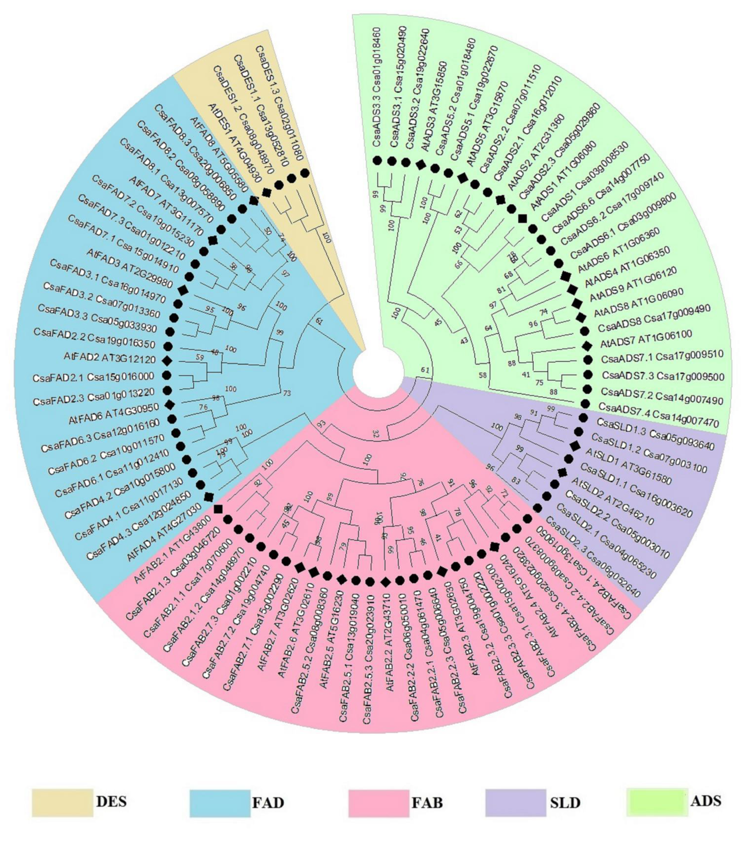
2.1. Synteny and Evolutionary Relationship of Fatty Acid Desaturase Genes in C. Sativa and A. thaliana

In total, 62 fatty acid desaturase genes were identified, with 18 soluble CsaFAB2 and 44 membrane-bound desaturase genes (17 CsaADS, 18 CsaFAD, 6 CsaSLD and 3 CsaDES1) (Table 1). The synteny of fatty acid desaturase genes in A. thaliana and C. sativa is illustrated in Figure 1. The copy number of fatty acid desaturase genes in C. sativa was higher in comparison to that of A. thaliana. Fatty acid desaturase genes were present in 18 among the 20 chromosomes of C. sativa (excluding chromosomes 9 and 18). The size of the proteins was larger in C. sativa, which might explain the difference in amino acid sequence identity.
The evolutionary relationship of the fatty acid desaturases was made based on the amino acid sequences. Five clusters represented the five groups of fatty acid desaturases (Figure 2). Each group of fatty acid desaturase genes had moderately (bootstrap BS = 52%) to well (BS 100%) supported clades, which indicated the possible evolution of each group from a common origin. The divergence within each group occurred due to an alteration in amino acid sequences—some proteins were shorter or longer than the others—resulting in their evolution into separate clades. For example, ADS5 and ADS6 had 361 AA and 299 AA, respectively, which had led them to evolve into two distinct clades. It was also obvious that the longer proteins in C. sativa seemed to have evolved separately from the other members, which were closer to A. thaliana proteins rather than that other C. sativa proteins, as seen in CsaSLD1-Csa16g003620, which had 766 AA and was rather distant from the other CsaSLD1 (Csa07g003100 and Csa05g093640) in the phylogenetic tree. It was observed that some proteins were longer than other proteins of the same clade, for example, CsaADS7 (Csa14g007470) contained 898 AA, but its homolog in A. thaliana contained 299 AA, and other copies of CsaADS7 had 259-347 AA. There might be an annotation error with the gene ID Csa14g007470 (CsaADS7) on CamRegBase, which should be revised. This gene might be divided into three separate genes of about 300 AA, each. Nevertheless, it shared 80% similarity with AtADS7 and was found with the ADS7 clade in the phylogenetic tree.

2.2. Diversity in Gene and Protein Structure in C. sativa Fatty Acid Desaturases

Gene structural diversity could be characterized with the arrangement of introns–exons in individual fatty acid desaturase genes, and the type of introns (i.e., the phase in which they belonged). In phase 0 introns, no codon is disrupted; in phase 1 introns, a disruption between the first and second base of a codon occurs; and in phase 2 introns, a disruption between the second and third base of a codon occurs. It is very important to know which exon might be the target of alternative splicing since it might occur when introns of the same phase surround the exon, leading to what is called a symmetrical exon [53]. C. sativa desaturase genes experienced a mixture of gain and loss of intron–exon structure in comparison to those of A. thaliana (Figure 3). For example, AtADS contained five exons, but some CsaADS (e.g., CsaADS3.2-Csa19g022640) had gained one exon structure. Conversely, the CsaFAD8.1-Csa13g007570 and CsaFAD8.2-Csa08g058890 had lost all introns compared to their homolog AtFAD8. The SLD family seemed to be intronless. In all groups of fatty acid desaturase genes, most of the introns were of phase 0, and then phase 2, but phase 1 introns were few in number. Meanwhile, most of the genes contained exons, which were surrounded by two introns of the same type, such as in CsaFAB2.1.1-Csa17g070600, and might be a target for alternative splicing. The genes that had experienced a gain or loss of intron/exon were detected, possibly due to the pressure of evolution.
Protein domains are the essential structure that gives an identity to a protein and an indication of the protein’s function [54]. In the current study, 87 proteins of fatty acid desaturase from A. thaliana and C. sativa were subjected to a domain architecture analysis (Supplementary Table S1 and Figure S1). All of the analyzed proteins contained at least one fatty acid desaturase domain (FA_desaturase), except for the FAD4 class in A. thaliana and C. sativa, of which one or two B domains of TMEM were found. AtFAD4 (AT4G27030) was, though, recognized as a fatty acid (palmitate) desaturase in previous studies, despite lacking this FA_desaturase domain. The domain architecture of C. sativa desaturase differed with the number and family of the domain in each protein: FAB2 contained one lengthy FA_desaturase_2 domain (Ferritin-like, pfam03405), ADS had one to three FA_desaturase domains (pfam00487 and/or cl37993), and DES1 had one C-terminal FA_desaturase domain and one Lipid_DES domain in their N-terminal side. Similarly, the FAD family contained one C-terminal FA_desaturase domain (pfam00487), except for CsaFAD2.2 (Csa19g016350), which had two FA_desaturase domains, and an N-terminal DUF domain (cl13407 or cl15288), except for AtFAD6 (AT4G30950), CsaFAD6.2 (Csa10g011570), and CsaFAD6.3 (Csa12g016160), which lacked DUF domain. Finally, the SLD family contained a C-terminal FA_desaturase domain (pfam00487) and an N-terminal Cyt-b5 domain (pfam00173). CsaSLD1.1 (Csa16g003620) was an exception that it contained two FA_desaturase domains (pfam00487) and two Cyt-b5 domains (pfam00173). The FA_desaturase domains existed as three different types in the fatty acid desaturases (pfam03405, pfam00487, and cl37993). Pfam03405 is a Ferritin-like family and was exclusively found in the soluble fatty acid desaturase FAB2, whereas pfam00487 is a family, which belongs to the cl37993 superfamily, and they were solely found in the membrane-bound fatty acid desaturases.
Besides, two to ten and two to 14 transmembrane domains were found in membrane-bound desaturase by using SOSUI and TMHMM, respectively (Supplementary Figure S2). The FAD4 clade lacked any transmembrane domain in both A. thaliana and C. sativa. The numbers of transmembrane domains in each family varied from two to 14 in the ADS family, four to five in the DES family, one to nine in the FAD family, and in the SLD family, respectively. Besides, three histidine-enriched motifs were conserved in the soluble FAB2, while two to five histidine enriched motifs were found in membrane-bound desaturases (Table 2). It was remarkable to note the presence of motifs enriched with three consecutive histidines (HHHxH), exclusively in the ω3-fatty acid desaturases CsaFAD3, CsaFAD7, and CsaFAD8. Likewise, a motif that consists of HPMAGHFISEH was only found in CsaDES1. The divergence in gene structure, protein domain architecture, and histidine motifs were obvious and might explain the separate evolution of the fatty acid desaturases in C. sativa.

2.3. Expression Profile Analysis of C. sativa Desaturases Revealed an Increase in Expression in Young Leaves, Seeds, and Flowers

The expression profile of the five families of C. sativa desaturases was analyzed in a wild-type whole plant, stem, seed, root, embryo, leaf, young leaf, flower, cotyledon, and bud during the development phase (Figure 4). It was revealed that four FAB2 genes (Csa06g050010, Csa05g006640, Csa04g061470, and Csa15g002300) were increased in young leaves and seeds, while ADS2 genes’ expressions were increased in both young leaves and cotyledons, and ADS6 (Csa14g007750) was increased in buds. FAD2 genes were only increased in young leaves. One DES1 (Csa08g048970) and two SLD2 (Csa06g052640 and Csa05g003010) had increased expression in the flowers, while one SLD1 (Csa05g093640) was increased in the stem, flower, and bud. This indicated that the accumulation of desaturase genes was associated with different tissues, and the expression differed among the desaturase genes families, which might add a further reason for their divergence in function, even within the same family with close a phylogenetic relationship.

2.4. C. sativa Fatty Acid Desaturases Were Detected in Three Intracellular Compartments

In silico analysis predicted that the soluble CsaFAB2 was located in chloroplast, which is similar to the membrane-bound CsaFAD4, CsaFAD6, CsaFAD7, and CsaFAD8. However, CsaFAD2 and CsaFAD3 were found in ER. In turn, CsaADS and CsaSLD were found in the plasma membrane (Supplementary Table S2). In our previous study, CsaFAD3 was located in the ER [46]. In the current study, the ω3-fatty acid desaturases CsaFAD7 and CsaFAD8 were transiently expressed in A. thaliana protoplast. As expected, the CsaFAD7 and CsaFAD8 were located in the chloroplast (Figure 5), which suggested that the ω3-fatty acid desaturases CsaFAD3, CsaFAD7, and CsaFAD8, which produced C18:3CisΔ9,12,15, existed in a functionally non-redundant form and played different roles in the evolutionary process.

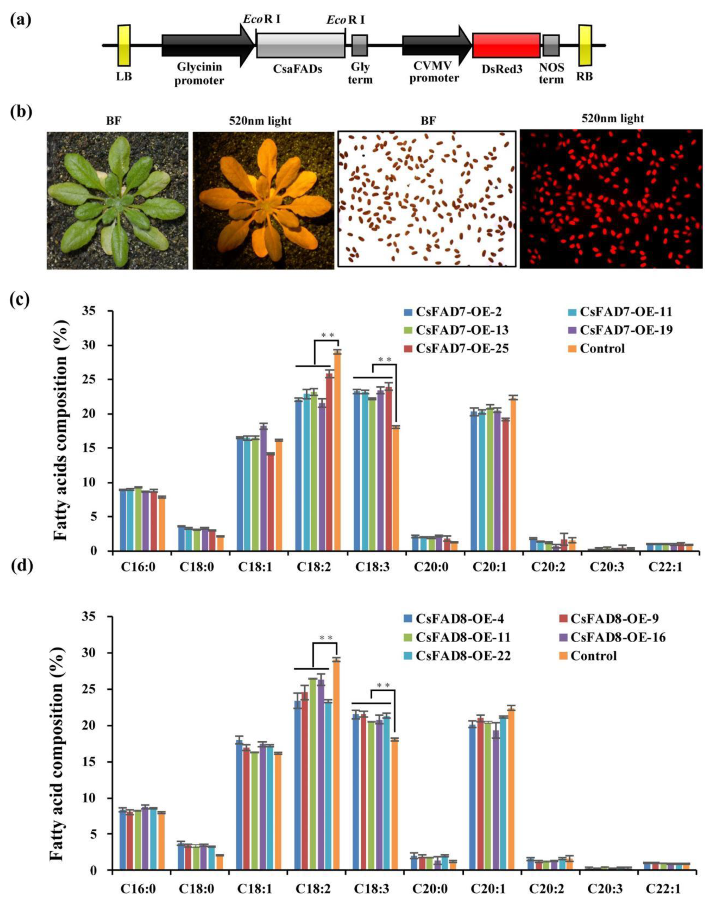
2.5. Overexpression of CsaFAD7 Caused Seed Size and Oil Content Reduction in A. thaliana Seed but Not CsaFAD8

Previously, we revealed that the C18:3 production was negatively correlated with seed size and roundness, via overexpression of the CsaFAD3 in A. thaliana seeds [46]. The same seed phenotypes were analyzed in the current study. Thus, the cDNA encoding the ω-3 fatty acid desaturases CsaFAD7 and CsaFAD8 were overexpressed in A. thaliana Col-0. The CsaFAD7 and CsaFAD8 coding sequences were driven by a seed-specific expression promoter of Glycinin (Figure 6a). Red fluorescent protein DsRed3 was used as a selection marker, for screening the positive transgenic seed (Figure 6b). More than 20 T1 transgenic lines were screened according to the red fluorescent marker of each group. For further fatty acid analysis, five homozygous transgenic lines were selected from CsaFAD7 and CsaFAD8 overexpression lines, respectively. The expression levels of CsaFAD7 and CsaFAD8 genes in the transgenic lines were detected by q-PCR in the developing siliques, five days after flowering, and revealed a significantly higher level in the transgenic lines, i.e., three to seven times higher than those of the wild-type and the empty vector control group (Supplementary Figure S3). However, no significant difference was observed between the expression level of CsaFAD7 and CsaFAD8 in the transgenic lines.
A significant increase in C18:3CisΔ9,12,15 and a significant decrease in C18:2Cis∆9,12 in all lines transformed with CsaFAD3 were reported previously [46]. In the current study, all of the transgenic lines transformed with CsaFAD7 and CsaFAD8 displayed a slight increase in C18:3 (~5.89% and ~3.25%, respectively, Supplementary Figure S4) and a decrease in C18:2 (~3.22% and ~5.80%, respectively) (Figure 6c,d). All these indicate that the ER located protein CsaFAD3 contributed more in C18:3 production rather than the chloroplastic located proteins CsaFAD7 and CsaFAD8. The C18:3 composition was higher in the CsaFAD7 overexpression line than in CsaFAD8.
Besides, the seed oil content significantly decreased in lines being transformed with CsaFAD7 (~3.88%), while those transformed with CsaFAD8 did not display any significant change in comparison with the wild-type (Figure 7a). Moreover, the thousand-seed weight (TSW) in CsaFAD7 overexpression lines was also found lower than WT control and CsaFAD8 overexpression lines. Those findings were similar to the CsaFAD3 overexpression lines that have been reported previously [46], while the TSW of CsaFAD8 overexpression lines did not show a significant decrease compared with the WT control (Figure 7b). The seed length to width value of the CsaFAD7 transgenic lines showed a significant increase, while those of CsaFAD8 were similar to the wild-type (Supplementary Figure S5). The seed of CsaFAD7 transgenic lines displayed a wrinkled phenotype (Figure 7c), which was previously seldom found in CsaFAD3 [46]. These results confirmed that overexpression of CsaFAD7 caused abnormal oil accumulation while CsaFAD8 did not. Moreover, the negative correlation between C18:3 accumulation and seed normalcy confirmed our previous studies. These findings suggest that C. sativa ω3-fatty acid desaturases play different roles in fatty acid accumulation and embryo development during the evolutionary process.

3. Discussion

3.1. Evolution of A. thaliana and C. sativa and their Respective Fatty Acid Desaturases

C. sativa fatty acid desaturase genes were identified based on homology with A. thaliana genes, and it was obvious that one gene in A. thaliana might have one to four copies of homologs in C. sativa, other genes had no homolog. A total of 62 fatty acid desaturase genes were found in CamRegBase during the current study, which was more numerous than the 24 genes found in NCBI [47]. In addition, some gene copies in C. sativa were longer than in A. thaliana—probably due to some errors, so this needs some revision—but they still had the FA_desaturase domain, which is common in fatty acid desaturases. The fact that A. thaliana and C. sativa both belong to the Brassicaceae family indicated that they both inherited the genomic blocks (GB) from the ACK, but the genome arrangement was different. A. thaliana, which has a genome size of 135Mb [55], has inherited a single copy of the 24 GB of the ACK [26]. However, C. sativa has been reported to have undergone a whole-genome triplication event [22], which indicated that C. sativa had a genome size and GB at least three times bigger in the size than those of A. thaliana [23]. The genome size of C. sativa was estimated to be ~750 to 785Mb [22,23,56], which was much larger than that of A. thaliana. Plant genome enlargement is the consequence of polyploidization and the expansion of transposable elements [57,58]. In general, during a polyploidization process, genome duplication/triplication, hybridization, and rearrangement occur, which increase the offspring genome size (gene copies) but also put pressure on chromosomes, which might lead to some gene losses. This phenomenon was reported in the polyploid genome of rapeseed (Brassica napus) [59,60,61,62], of which a mixture of retained, duplicated, lost, and converted genes (homologous exchange) or chromosomal segments were found in comparison to its progenitors [63]. Similarly, the genome of C. sativa experienced two polyploidy events from progenitors, of which repeated hybridization of closely related ancestry occurred, almost without any subgenome rearrangement in comparison to the parental genomes, conferring general stability [24]. A previous study reported that ~70% of the annotated genes in Camelina were syntenic to A. thaliana [23]. Particularly, it was estimated that genes involved in lipid metabolism were 217% higher in C. sativa in comparison to A. thaliana, and three copies of oil genes were generally retained in C. sativa [23], which was consistent with our findings, which showed that the majority of fatty acid desaturase genes were present as three copies in C. sativa. Seven duplicated genes were also found in fatty acid desaturase genes, which also contributed to the expansion of desaturase genes in C. sativa.
Then, a phylogenetic tree displayed five clusters corresponding to the five families of desaturase, indicating that they have evolved separately. The profile of the phylogenetic tree was comparable to those found in soybean [6], sunflower [64], cotton [65], and wheat [66]. Within the individual clade, orthologous genes of A. thaliana and C. sativa were clustered together. Focusing on the evolutionary relationship of each family of desaturase, diverse classes of the gene have evolved separately. In the soluble FAB2, the FAB2.1 clade seemed to separate earlier before the simultaneous emergence of the other members of the clade (FAB2.2-FAB2.7). Similarly, ADS3 appeared to diverge before the emergence of ADS5, and then ADS1 and ADS2. The clade of ADS4, ADS6 to ADS9 seemed to appear later. Additionally, the Δ12 fatty acid desaturases FAD6 and FAD2 seemed to separate earlier from the other clades and the Δ15 fatty acid desaturases FAD3, FAD7, and FAD8 emerged later. The Δ3 desaturase FAD4, which lacked FA_desaturase and transmembrane domains in our analysis, seemed to have evolved separately from the rest of the FAD family. Then, the two classes of the SLD family just diverged from each other and had evolved separately. In a broad view, some gene classes had more than one gene copy in those C. sativa desaturases, possibly due to duplication events during polyploidization, resulting in genes with different sequences [67]. Furthermore, the emergence of paralogous genes allows the growth of a subfunctionalization or a neofunctionalization [68,69]. Conversely, some genes were lost and this process is common during genome rearrangement, due to the pressure of evolution [70].

3.2. Conservation and Diversification in Structure of the Fatty Acid Desaturases

Structural analysis of fatty acid desaturases would help to unravel the previous evolutionary relationship between them. The conserved structure might assure the stability of the maintained function, while divergence might silence the original function or cause a new function to arise [71]. Gene structure analysis revealed a mixture of conservation, gain, and loss of intron in C. sativa fatty acid desaturases genes. The single intron gain or loss might be the outcome of a long evolution process [62]. Particularly, some genes, including CsaSLD, CsaFAD4, and some CsaFAD2 and CsaFAD8, were intronless. Lack of intron was also observed in SLD and FAD2 of rice [72] and grasses [73], and in SLD, FAD2, and FAD4 of wheat [66], which is rather common in SLD genes [74]. In eukaryotes, intronless genes originate from horizontal gene transfer, from retrotransposon, or duplication of another intronless gene [75]. Besides, stress response genes such as FAD2 [76] and FAD8 [15,77] were reported to contain very few introns [78,79]. The presence of seven introns in AtFAD8 and CsaFAD8.3 was surprising in this analysis.
The protein domain of C. sativa desaturases mostly displayed a conserved structure, similar to what is found in A. thaliana. Only eight proteins displayed additional domains, which were different from those of A. thaliana. Protein domains are masterpieces that specify a protein structure and function [54,80,81], they are formed from an association of short polypeptides conglomerates [82] and evolve independently [83,84,85]. Protein domains were suggested to be more conserved during evolution than protein sequences [86,87]. Paralogous proteins often emerge from domain duplication [88,89], but also from divergence and recombination [54,90], as seen in the FAD family of which dissimilar domain architecture arose. Those domain recombinations lead to the emergence of more functions [89]. FA_desaturase was commonly seen in all desaturase genes under three different classes, which are specific to each family of desaturase. Even the same class of FA_desaturase showed divergence in the amino acid sequences in different desaturase families. Additional domains were found in some desaturases, such as DUF domains in the FAD family, which correspond to functionally uncharacterized proteins [91], except for FAD4, which was enriched with TMEM domains corresponding to a transmembrane protein in the Pfam database [92]. The SLD family also had an additional cytochrome-b5 domain on their structure. Cyt-B5 are electron transporters/donors for the desaturases [93,94]. Likewise, the DES family had a Lipid_DES domain, which is exclusively found in sphingolipid Δ4-desaturase proteins [95]. The lack of Lipid_DES in CsaDES1.3 was surprising as by homology, it was much closer to AtDES1 rather than other desaturase families; the sequence 1 to 24 AA in CsaDES1.3 was much different from AtDES1, CsaDES1.1, and CsaDES1.2, and the missing amino acids were also found leading to a shorter protein in CsaDES1.3, which was possibly the cause of the Lip_DES domain loss in this protein. A protein domain might change within its structure and function, and changes might also occur in the genome of different species [85,96]. Protein evolution is then the result of domain duplication, divergence, convergence, or fusion [89,97,98,99,100]. The earlier phylogenetic tree of desaturase genes was based on protein domains. The clear separation between the five families of desaturase was explained, the fact that the soluble and the membrane-bound desaturases are unrelated despite their similarities [18,101], and that they might originate from different ancestors [6]; those points were confirmed in our study.
A common characteristic in the soluble and the membrane-bound desaturases is the presence of histidine motif [18,102], and membrane-bound desaturases have a transmembrane domain in their structure [3,7]. Between two and 14 transmembrane domains were found in membrane-bound desaturases, except for FAD4, which lacked this structure in TMHMM and SOSUI analysis. Protein domain analysis revealed through the presence of TMEM domain was discovered, which indicates a transmembrane protein in Pfam database [92]. Transmembrane domains adopt an alpha-helix shape [103]. In eukaryotes, the length and amino acid sequence of the transmembrane domain was strongly linked to the intracellular compartmentation of the membrane proteins [104]. CsaADS, CsaDES1, and CsaSLD were located in the plasma membrane and they displayed a comparable amount of transmembrane domains unless for longer proteins, which had richer domains. Three histidine motifs were found in the soluble FAB2, and two of them, which were ENRHG and DEKRHE, were similar to the motifs found in peanut [6], rice [72], and wheat [66]. Likewise, the two histidine motifs which were found in the CsaDES family (HLEHH and HNEHH) were also found in peanut [6], rice [72], and wheat [66], whereas the motifs found in CsaFAD (HRTHH and HVIHH) were similar to those found in rice [72]. How well those motifs are conserved in different species indicated their important involvement in protein and enzyme functions [105].

3.3. C. sativa Desaturases Accumulation Was Associated with Different Cellular Compartmentations and Tissues

Fatty acid desaturases in C. sativa were accumulated in three subcellular compartments (chloroplast, plasma membrane, and ER), and in five different tissues (young leaves, flowers, buds, seeds, and cotyledons). Previously, Chi et al. analyzed the expression of desaturases in A. thaliana and soybean and revealed the expression of AtSLD1 in flowers [6], similar to CsaSLD1 in our findings. Similarly, AtSLD1 was expressed in all organs, particularly in flowers, while AtSLD2 was highly accumulated in flowers and siliques, but displayed a low expression level in leaves, stems, and roots [106]. By contrast, AtFAB2 was reported to be highly expressed in flowers, root, stem, adult leaves, and cotyledons, while CsaFAB2 was increased only in young leaves and seeds. AtADS was also reported to accumulate in seeds and flowers, while in our study CsaADS were expressed in young leaves, cotyledons, and buds. The other desaturase genes had low expression or no expression at all in C. sativa; however, Chi et al. found AtFAD5, AtFAD6, AtFAD7 were highly expressed in cotyledons, stems, and leaves [6]. In a recent study, AtFAD7 and AtFAD8 were found to be expressed in leaves [107]. Besides, the difference in gene expression levels between C. sativa and A. thaliana might be due to differences in tissue age and environmental factors. A study on gene expression of desaturase in C. sativa revealed their variation according to stress conditions [47]. The accumulation of desaturase genes in C. sativa seemed to differ from those of A. thaliana, which indicated a possible divergence of biological function between them.

3.4. Seed-Specific Expression of the cDNA Encoding CsaFAD7 and CsaFAD8 Affected the Fatty Acid Composition of Seed Oil and the Seed Size in A. thaliana

FAD are key enzymes in the regulation of polyunsaturated fatty acid biosynthesis. Particularly, ω-3 fatty acid desaturases have been proven to use C18:2 as substrates to produce 18:3 in the plastid, where FAD7 and FAD8 are located, and in the ER where FAD3 are found [5]. In plants, the accumulation of 18:3 under the activity of ω-3 fatty acid desaturases, particularly FAD3 has been demonstrated in several studies, such as in soybean [108,109], in rice [110], in olive [111,112,113], in peanut [114], in flax [115], in walnuts [116], in cowpea [117], in alfalfa [118], in peach aphid [119] and now in C. sativa, according to the current study.
Earlier, we could demonstrate that the overexpression of CsaFAD3 caused seed abnormality and high accumulation of C18:3 [46], but this was not enough to understand the function of C. sativa ω-3 fatty acid desaturases on seed phenotypes. Thus, CsaFAD7 and CsaFAD8 functions were also analyzed to ensure our understanding. Moreover, CsaFAD7 and CsaFAD8 were more found to be located in different compartments than CsaFAD3, which increased our interest in uncovering the consequent phenotype. Although all three ω-3 fatty acid desaturases of C. sativa enhanced C18:3, CsaFAD3 remarkably produced more C18:3 [46], in comparison to CsaFAD7 and CsaFAD8. Differences between those three desaturases were found in their structure (amino acid sequences) of which CsaFAD7 and CsaFAD8 were very much similar in our analysis (~80% of identity), but also in their intracellular compartments, where CsaFAD7 and CsaFAD8 were located in the chloroplast, while CsaFAD3 were located in the ER. This might explain the comparable performance of CsaFAD7 and CsaFAD8 on producing C18:3 accumulation. Moreover, desaturation of C18:2 to produce C18:3 occurs in the ER [5]. It might be possible that CsaFAD3 promoted more accumulation of C18:3 due to their presence in the main location where C18:3 is produced, and CsaFAD7 and CsaFAD8 in the chloroplast might indirectly affect C18:3 production. Conversely, the seed of transgenic CsaFAD3 [46] and CsaFAD7 lines displayed a decrease in seed size, seed weight, and a wrinkled seed surface, while no obvious alteration was found in CsaFAD8 transgenic lines. The underlying reason why the seed showed decreased oil content and seed size was still unclear. When the unusual fatty acid was produced in heterologous seeds, the seed oil content was also decreased according to the previous reports [120,121,122,123,124,125]. There were two hypotheses associated with this phenotype including the fatty acid β-oxidation [126] and the inhibition of fatty acid synthesis [121]. Those reports also suggested that different plant embryos have various concentrations of tolerance to unusual fatty acids. Recently, seed-specific overexpression of Lesquerella FAD3 dramatically increased the C18:3 content and increased the seed size in soybean [127], while heterologous seed-specific overexpression of CsaFAD7 in the A. thaliana embryo showed the reverse phenotype. Those reports and findings imply that the production and accumulation of C18:3 showed various tolerance in A. thaliana and soybean. Certainly, ω3-fatty acid desaturases play different roles in embryo development and oil accumulation, but the dissimilarity found between transgenic CsaFAD7 and CsaFAD8 on the seed surface was intriguing. Previously, the expression and activity of FAD7 and FAD8 in A. thaliana were compared to investigate their response to stress conditions [107]. FAD7 had high promoter activity and was accumulated more in leaves, whereas FAD8 displayed lower promoter activity, and was less expressed in leaves. Additionally, FAD7 accumulated in response to wound and decreased with abscisic acid treatment, while FAD8 concentrated in response to cold or jasmonate treatment, and attenuated at high temperature. In the current study, expressions and activities of CsaFAD7 and CsaFAD8 were also different; the reason why one caused abnormal phenotype might be found in the 20% identity difference between them, or due to some other factors. This needs further investigation.
In conclusion, the present study focused on soluble and membrane-bound fatty acid desaturases in C. sativa. Their evolution, structure, and subcellular location were studied, and their implication in oil biosynthesis was investigated. The close similarity between A. thaliana and C. sativa reflected their common origins, and the expansion of orthologous genes in C. sativa was the consequence of polyploidization. Despite acting as desaturases, the analyses of gene and protein domain structures confirmed that soluble and membrane-bound proteins were unrelated in our study, but paralogous genes within each family originated from a common ancestry. The histidine motifs, which are common in desaturases, were conserved in C. sativa. The subcellular location of fatty acid desaturases was similar to those of A. thaliana, which might suggest similar activity of proteins, but divergence might occur since protein function also depends on key amino acids sequences. As of now, it is clear that similarly to other ω-3 fatty acid desaturases of the other species, those of C. sativa could effectively accumulate alpha-linolenic acid. To the best of our knowledge, the current study was the first one to analyze both the soluble and membrane-bound desaturases in C. sativa, exploring the evolutionary, structural, and functional aspects. Our findings would be an excellent foundation for an in-depth understanding of the characteristics of fatty acid desaturases in C. sativa, and could be subjected to many aspects of investigations by using genetic engineering to discover new interesting functions for C. sativa trait improvement.

4. Materials and Methods

4.1. Plant Materials and Growth Conditions

The Columbia wild-type A. thaliana (Col-0) and transgenic lines were cultured at 16 h light/8 h dark (100 μm/m2/s) under constant temperature 23 °C. The position of control and transgenic lines used for lipid analysis were changed in the same tray every 5 days to make the plants grow in the same conditions.

4.2. Identification of Desaturase Genes in C. sativa

Identification of FAD genes in C. sativa was based on homology with the 25 genes identified in A. thaliana [6,20,65], using Camelina Gene Regulation Database or “CamRegBase” (http://camregbase.org/ accessed on 14 September 2021) [48]. The chromosomes’ location, the sequence, and the size of the genes were kindly provided by Professor Danny Schnell and Mr. Eric Maina of Michigan State University, USA. Gene synteny was drawn using TBtools software (https://github.com/CJ-Chen/TBtools accessed on 14 September 2021) [49].

4.3. Phylogenetic Analysis of Desaturase Genes

Phylogenetic analysis was made with protein sequences from A. thaliana and C. sativa. Alignment was performed with ClustalX software (http://www.clustal.org accessed on 14 September 2021) [128]. The phylogenic tree was unrooted and was inferred with Neighborhood Joining (NJ) [50]. The bootstrap consensus tree inferred from 1000 replicates was taken to represent the evolutionary history of the taxa being analyzed [129]. The evolutionary distances were computed using the Poisson Correction method [130] and were in the units of the number of amino acid substitutions per site. All ambiguous positions were removed for each sequence pair (pairwise deletion) [131,132]. Evolutionary analyses were conducted in MEGA X (https://www.megasoftware.net accessed on 14 September 2021) [52].

4.4. Gene Structure Analysis

The intron/exon structures of the desaturase genes in C. sativa were identified based on alignments of their coding sequences with their genomic sequences, and a diagram was obtained using the GSDS-Gene structure display server (https://gsds.cbi.pku.edu.cn/ accessed on 14 September 2021) [133].

4.5. Protein Domain Structure and Conserved Motif Analysis

Protein conserved domains in C. sativa desaturases were analyzed with the Batch CD-search tool in the NCBI database (http://www.ncbi.nlm.nih.gov/Structure/bwrpsb/bwrpsb.cgi accessed on 14 September 2021) [134], with CDSEARCH/oasis_pfam v3 as a source and an e-cut off value of 0.10. Analysis of transmembrane helix motif in membrane-bound desaturases was made using TMHMM 2.0 server based on a hidden Markov model (http://www.cbs.dtu.dk/services/TMHMM accessed on 14 September 2021) [135] and SOSUI (https://harrier.nagahama-i-bio.ac.jp/sosui/sosuiG/sosuigsubmit.html accessed on 14 September 2021) [136]. The conserved histidine motif of membrane-bound desaturases was observed via proteins alignment with Vector NTI Advanced 11.5.1 software. (Thermo Fisher Scientific, Waltham, MA, USA).

4.6. Expression Pattern of Desaturase Genes

The expression of each gene in the five families of desaturases was acquired from CamRegBase gene expression search tool (http://camregbase.org/data_search/gene accessed on 14 September 2021) [48], using gene ID as a query. The inquiry was made in normal tissues (whole plant, stem, seed, root, embryo, leaf, young leaf, flower, cotyledon, and bud) during development.

4.7. Subcellular Localization Analysis

The subcellular localization of fatty acid desaturase proteins was predicted using CELLO v.2.5 (subCELlular Localization predictor) server (http://cello.life.nctu.edu.tw/ accessed on 14 September 2021) [137]. Then, the CsaFADs coding sequence fusing with GFP tag was inserted into pCambia1303 vector between Spe I and BstE II promoted by 35S promoter. The reconstructed vector and ER marker fused with cyan fluorescence protein was co-transformed into A. thaliana protoplasts as described previously [138,139]. The A. thaliana protoplasts were imaged by a confocal laser scanning microscopy FV1000 (Olympus, Japan). The images were analyzed with FV10-ASW software (Olympus, Japan).

4.8. Construction of CsaFAD7 and Csafad8 Seed-Specific Expression Vectors and Expression in A. thaliana

The coding sequences of CsaFAD7 and CsaFAD8 were inserted into the pBinGlyRed3 vector in the EcoRI site, under the control of a seed-specific Glycinin promoter. The reconstructed vectors were transformed into Agrobacterium tumefaciens GV3101 and transfected to A. thaliana with the dip floral method. T3 generation homozygous transgenic A. thaliana lines were collected for the lipid analysis. The developing siliques were collected five days after flowering in the transgenic lines and the wild-type for the q-PCR analysis, and Actin7 was used as an internal control.

4.9. Transgenic Seed Size and Lipid Analysis

For the seed size analysis, 5 to 10 mg T3 dry seeds were weighted and then spread out on a plate separately to make sure the individual grains could be distinguished without overlapping. The seed grain image was acquired by the scanner (Unis D6810, Beijing, China). The grain shape characters were calculated by the SC-G grain appearance quality image analysis system (WSeen, Hangzhou, China) developed by Hangzhou WSeen Detection Technology Co., Ltd., China [140,141,142].
Fatty acid composition and oil content analyses were performed according to the previous description [46]. Briefly, 5 mg of dried seeds were collected following 1.5 mL of 2.5% H2SO4 methanol solution (Sinopharm, Beijing, China) with 0.01% BHT (Sigma-Aldrich, Germany), 0.4 mL methylbenzene, and 200 µL of 2 mg/mL C17:0 (Sigma-Aldrich, Hamburg, Germany) as internal standard. The mixture was incubated at 90 °C for 1 h, 1.8 mL of deionized water, and 1 mL of hexane was added to the mixture. The supernatant was transferred for gas chromatography (GC) with flame ionization detection analysis (Agilent, CA, USA). The resolution of fatty acid methyl esters (FAMEs) in the supernatant was achieved with a DB-23 column (30 m length with 0.25 mm inner diameter and 0.25 µm thickness film; Agilent, CA, USA). The injection volume is 1 µL with a split ratio of 10:1. The carrier gas was with a flow rate of 1 mL/min with a pressure of 17.39 psi. The oven temperature was initiated from 150 °C for 3 min, then increased at 10 °C per minute to 240 ℃ and held for 8 min.

Supplementary Materials

The following are available online at https://www.mdpi.com/article/10.3390/ijms221910857/s1.

Author Contributions

Conceptualization, N.R., Y.Y. and M.L.; methodology, N.R., Y.Y., K.C. and J.H.; validation, N.R., Y.Y. and M.L.; formal analysis, N.R., Y.Y., K.C. and J.H.; investigation, N.R. and Y.Y.; resources, N.R. and Y.Y.; data curation, N.R. and Y.Y.; writing—original draft preparation, N.R. and Y.Y.; writing—review and editing, M.L.; visualization, M.L.; supervision, L.Y. and M.L.; project administration, M.L.; funding acquisition, N.R., Y.Y. and M.L. All authors have read and agreed to the published version of the manuscript.

Funding

The work was funded by the National Natural Science Foundation of China (32172087, 31871656, 32072098 and 31701456), and the China Postdoctoral Science Foundation (2016M602309).

Institutional Review Board Statement

Not applicable.

Informed Consent Statement

Not applicable.

Data Availability Statement

Not applicable.

Acknowledgments

We are thankful to Danny Schnell and Eric Maina of Michigan State University, USA, for providing the gene and protein sequences of C. sativa.

Conflicts of Interest

The authors declare no conflict of interest.

References

  1. Harwood, J.L. Plant acyl lipids: Structure, distribution, and analysis. In Biochemistry of Plants; Stumpf, P.K., Conn, E.E., Eds.; Academic Press: New York, NY, USA, 1980; Volume 4, pp. 1–55. [Google Scholar]
  2. Stumpf, P.K. Biosynthesis of saturated and unsaturated fatty acids. In Biochemistry of Plants; Stumpf, P.K., Conn, E.E., Eds.; Academic Press: New York, NY, USA, 1980; Volume 4, pp. 177–204. [Google Scholar]
  3. Los, D.A.; Murata, N. Structure and expression of fatty acid desaturases. Biochim. Biophys. Acta 1998, 1394, 3–15. [Google Scholar] [CrossRef]
  4. Nakamura, M.T.; Nara, T.Y. Structure, function, and dietary regulation of delta6, delta5, and delta9 desaturases. Annu. Rev. Nutr. 2004, 24, 345–376. [Google Scholar] [CrossRef]
  5. Ohlrogge, J.; Browse, J. Lipid biosynthesis. Plant Cell 1995, 7, 957–970. [Google Scholar] [CrossRef] [PubMed] [Green Version]
  6. Chi, X.Y.; Yang, Q.L.; Lu, Y.D.; Wang, J.Y.; Zhang, Q.F.; Pan, L.J.; Yu, S. Genome-wide analysis of fatty acid desaturases in soybean (Glycine max). Plant Mol. Biol. Rep. 2011, 29, 769–783. [Google Scholar] [CrossRef] [Green Version]
  7. Murphy, D.J. Production of novel oils in plants. Curr. Opin. Biotechnol. 1999, 10, 175–180. [Google Scholar] [CrossRef]
  8. Kargiotidou, A.; Deli, D.; Galanopoulou, D.; Tsaftaris, A.; Farmaki, T. Low temperature and light regulate delta 12 fatty acid desaturases (FAD2) at a transcriptional level in cotton (Gossypium hirsutum). J. Exp. Bot. 2008, 59, 2043–2056. [Google Scholar] [CrossRef] [Green Version]
  9. Smith, M.A.; Dauk, M.; Ramadan, H.; Yang, H.; Seamons, L.E.; Haslam, R.P.; Forseille, L. Involvement of Arabidopsis ACYL-COENZYME A DESATURASE-LIKE2 (At2g31360) in the biosynthesis of the very-long-chain monounsaturated fatty acid components of membrane lipids. Plant Physiol. 2013, 161, 81–96. [Google Scholar] [CrossRef] [Green Version]
  10. Kachroo, A.; Shanklin, J.; Whittle, E.; Lapchyk, L.; Hildebrand, D.; Kachroo, P. The Arabidopsis stearoyl-acyl carrier protein-desaturase family and the contribution of leaf isoforms to oleic acid synthesis. Plant Mol. Biol. 2007, 63, 257–271. [Google Scholar] [CrossRef] [PubMed]
  11. Okuley, J.; Lightner, J.; Feldmann, K.; Yadav, N.; Lark, E. Arabidopsis FAD2 gene encodes the enzyme that is essential for polyunsaturated lipid synthesis. Plant Cell 1994, 6, 147–158. [Google Scholar] [CrossRef]
  12. Falcone, D.L.; Gibson, S.; Lemieux, B.; Somerville, C. Identification of a gene that complements an Arabidopsis mutant deficient in chloroplast ω6 desaturase activity. Plant Physiol. 1994, 106, 1453–1459. [Google Scholar] [CrossRef] [Green Version]
  13. Arondel, V.; Lemieux, I.; Hwang, S.; Gibson, H.M. Goodman and C.R. Somerville, Map-based cloning of a gene controlling omega-3 fatty acid desaturation in Arabidopsis. Science 1992, 258, 1353–1355. [Google Scholar] [CrossRef] [PubMed]
  14. Iba, K.; Gibson, S.; Nishiuchi, T.; Fuse, T.; Nishimura, M.; Arondel, V.; Somerville, C. A gene encoding a chloroplast omega-3 fatty acid desaturase complements alterations in fatty acid desaturation and chloroplast copy number of the fad7 mutant of Arabidopsis thaliana. J. Biol. Chem. 1993, 268, 24099–24105. [Google Scholar] [CrossRef]
  15. Gibson, S.; Arondel, V.; Iba, K.; Somerville, C. Cloning of a temperature-regulated gene encoding a chloroplast ω-3 desaturase from Arabidopsis thaliana. Plant Physiol. 1994, 106, 1615–1621. [Google Scholar] [CrossRef] [PubMed] [Green Version]
  16. Gao, J.; Ajjawi, I.; Manoli, A.; Sawin, A.; Xu, C.; Froehlich, J.E.; Benning, C. Fatty acid desaturase4 of Arabidopsis encodes a protein distinct from characterized fatty acid desaturases. Plant J. 2009, 60, 832–839. [Google Scholar] [CrossRef]
  17. Ryan, P.R.; Liu, Q.; Sperling, P.; Dong, B.; Franke, S.; Delhaize, E. A higher plant Δ8 sphingolipid desaturase with a preference for (Z)-isomer formation confers aluminum tolerance to yeast and plants. Plant Physiol. 2007, 144, 1968–1977. [Google Scholar] [CrossRef] [Green Version]
  18. Shanklin, J.; Cahoon, E. Desaturation and related modifications of fatty acids. Ann. Rev. Plant Physiol. Plant Mol. Biol. 1998, 49, 611–641. [Google Scholar] [CrossRef] [Green Version]
  19. López Alonso, D.; García-Maroto, F.; Rodríguez-Ruiz, J.; Garrido, J.; Vilches, M. Evolution of the membrane-bound fatty acid desaturases. Biochem. Syst. Ecol. 2003, 31, 1111–1124. [Google Scholar] [CrossRef]
  20. Li-Beisson, Y.; Shorrosh, B.; Beisson, F.; Andersson, M.X.; Arondel, V.; Bates, P.D.; Ohlrogge, J. Acyl-lipid metabolism. Arabidopsis Book 2013, 11, e161. [Google Scholar] [CrossRef] [Green Version]
  21. Gehringer, A.; Friedt, W.; Luhs, W.; Snowdon, R.J. Genetic mapping of agronomic traits in false flax (Camelina sativa subsp. sativa). Genome 2006, 49, 1555–1563. [Google Scholar] [CrossRef]
  22. Hutcheon, C.; Ditt, R.F.; Beilstein, M.; Comai, L.; Schroeder, J.; Goldstein, E.; Kiser, J. Polyploid genome of Camelina sativa revealed by isolation of fatty acid synthesis genes. BMC Plant Biol. 2010, 10, 233. [Google Scholar] [CrossRef] [Green Version]
  23. Kagale, S.; Koh, C.; Nixon, J.; Bollina, V.; Clarke, W.E.; Tuteja, R.; Parkin, I.A. The emerging biofuel crop Camelina sativa retains a highly undifferentiated hexaploid genome structure. Nat. Commun. 2014, 5, 3706. [Google Scholar] [CrossRef] [Green Version]
  24. Mandáková, T.; Pouch, M.; Brock, J.R.; Al-Shehbaz, I.A.; Lysak, M.A. Origin and evolution of diploid and allopolyploid Camelina genomes were accompanied by chromosome shattering. Plant Cell 2019, 31, 2596–2612. [Google Scholar] [CrossRef]
  25. Lysak, M.A.; Berr, A.; Pecinka, A.; Schmidt, R.; McBreen, K.; Schubert, I. Mechanisms of chromosome number reduction in Arabidopsis thaliana and related Brassicaceae species. Proc. Natl. Acad. Sci. USA 2006, 103, 5224–5229. [Google Scholar] [CrossRef] [Green Version]
  26. Schranz, M.E.; Lysak, M.A.; Mitchell-Olds, T. The ABC’s of comparative genomics in the Brassicaceae: Building blocks of crucifer genomes. Trends Plant Sci. 2006, 11, 535–542. [Google Scholar] [CrossRef]
  27. Lysak, M.A.; Mandáková, T.; Schranz, M.E. Comparative paleogenomics of crucifers: Ancestral genomic blocks revisited. Curr. Opin. Plant Biol. 2016, 30, 108–115. [Google Scholar] [CrossRef]
  28. Moser, B.R. Biodiesel from alternative oilseed feedstocks: Camelina and field pennycress. Biofuels 2012, 3, 193–209. [Google Scholar] [CrossRef] [Green Version]
  29. Shonnard, D.R.; Williams, L.; Kalnes, T.M. Camelina derived jet fuel and diesel: Sustainable advanced biofuels. Environ. Prog. Sustain. Energy 2010, 29, 382–392. [Google Scholar] [CrossRef]
  30. Iskandarov, U.; Kim, H.J.; Cahoon, E.B. Camelina: An emerging oilseed platform for advanced biofuels and bio-based materials. In Plants and Bioenergy; McCann, M.C., Buckeridge, M.S., Carpita, N.C., Eds.; Springer: Berlin, Germany, 2014; pp. 131–140. [Google Scholar] [CrossRef] [Green Version]
  31. Kim, H.; Park, J.H.; Kim, D.J.; Kim, A.Y.; Su, M.C. Functional analysis of diacylglycerol acyltransferase1 genes from Camelina sativa and effects of CsDGAT1B overexpression on seed mass and storage oil content in C. sativa. Plant Biotechnol. Rep. 2016, 10, 141–153. [Google Scholar] [CrossRef]
  32. Chhikara, S.; Abdullah, H.M.; Akbari, P.; Schnell, D.; Dhankher, O.P. Engineering Camelina sativa (L.) Crantz for enhanced oil and seed yields by combining diacylglycerol acyltransferase1 and glycerol-3-phosphate dehydrogenase expression. Plant Biotechnol. J. 2017, 16, 1–12. [Google Scholar] [CrossRef] [Green Version]
  33. Kim, R.J.; Kim, H.U.; Suh, M.C. Development of Camelina enhanced with drought stress resistance and seed oil production by co-overexpression of MYB96A and DGAT1C. Ind. Crops Prod. 2019, 138, 111475. [Google Scholar] [CrossRef]
  34. Choudhury, S.R.; Riesselman, A.J.; Sona Pandey, S. Constitutive or seed-specific overexpression of Arabidopsis G-protein c subunit 3 (AGG3) results in increased seed and oil production and improved stress tolerance in Camelina sativa. Plant Biotechnol. J. 2014, 12, 49–59. [Google Scholar] [CrossRef] [PubMed]
  35. Park, W.; Feng, Y.; Kim, H.; Suh, M.C.; Ahn, S.J. Changes in fatty acid content and composition between wild type and CsHMA3 overexpressing Camelina sativa under heavy-metal stress. Plant Cell Rep. 2015, 34, 1489–1498. [Google Scholar] [CrossRef]
  36. An, D.; Suh, M.C. Overexpression of Arabidopsis WRI1 enhanced seed mass and storage oil content in Camelina sativa. Plant Biotechnol. Rep. 2015, 9, 137–148. [Google Scholar] [CrossRef]
  37. Li, M.; Wei, F.; Tawfall, A.; Tang, M.; Saettele, A.; Wang, X. Overexpression of patatin-related phospholipase AIIIδ altered plant growth and increased seed oil content in Camelina. Plant Biotechnol. J. 2015, 13, 766–778. [Google Scholar] [CrossRef] [PubMed] [Green Version]
  38. Cai, G.; Wang, G.; Kim, S.C.; Li, J.; Zhou, Y.; Wang, X. Increased expression of fatty acid and ABC transporters enhances seed oil production in Camelina. Biotechnol. Biofuels 2021, 14, 49. [Google Scholar] [CrossRef] [PubMed]
  39. Zhu, Y.; Xie, L.; Chen, G.Q.; Lee, M.Y.; Loque, D.; Scheller, H.V. A transgene design for enhancing oil content in Arabidopsis and Camelina seeds. Biotechnol. Biofuels 2018, 11, 46. [Google Scholar] [CrossRef] [PubMed] [Green Version]
  40. Rodríguez, M.F.; Sánchez-García, A.; Salas, J.J.; Garcés, R.; Martínez-Force, E. Characterization of soluble acyl-ACP desaturases from Camelina sativa, Macadamia tetraphylla and Dolichandra unguis-cati. J. Plant Physiol. 2015, 178, 35–42. [Google Scholar] [CrossRef] [Green Version]
  41. Kang, J.; Snapp, A.R.; Lu, C. Identification of three genes encoding microsomal oleate desaturases (FAD2) from the oilseed crop Camelina sativa. Plant Physiol. Biochem. 2011, 49, 223–229. [Google Scholar] [CrossRef]
  42. Kim, H.; Go, Y.S.; Kim, A.Y.; Lee, S.; Kim, K.N.; Lee, G.J.; Suh, M.C. Isolation and functional analysis of three microsomal delta-12 fatty acid desaturase genes from Camelina sativa (L.) cv. CAME. J. Plant Biotechnol. 2014, 41, 146–158. [Google Scholar] [CrossRef] [Green Version]
  43. Jiang, W.Z.; Henry, I.M.; Lynagh, P.G.; Comai, L.; Cahoon, E.B.; Weeks, D.P. Significant enhancement of fatty acid composition in seeds of the allohexaploid, Camelina sativa, using CRISPR/Cas9 gene editing. Plant Biotechnol. J. 2017, 15, 648–657. [Google Scholar] [CrossRef] [Green Version]
  44. Morineau, C.; Bellec, Y.; Tellier, F.; Gissot, L.; Kelemen, Z.; Nogué, F.; Faure, J.D. Selective gene dosage by CRISPR-Cas9 genome editing in hexaploid Camelina sativa. Plant Biotechnol. J. 2017, 15, 729–739. [Google Scholar] [CrossRef] [PubMed] [Green Version]
  45. Rodríguez-Rodríguez, M.F.; Salas, J.J.; Venegas-Calerón, M.; Garcés, R.; Martínez-Force, E. Molecular cloning and characterization of the genes encoding a microsomal oleate Δ12 desaturase (CsFAD2) and linoleate Δ15 desaturase (CsFAD3) from Camelina sativa. Ind. Crops Prod. 2016, 89, 405–415. [Google Scholar] [CrossRef] [Green Version]
  46. Yin, Y.; Guo, Z.; Chen, K.; Tian, T.; Tan, J.; Chen, X.; Li, M. Ultra-high α-linolenic acid accumulating developmental defective embryo was rescued by lysophosphatidic acid acyltransferase 2. Plant J. 2020, 103, 2151–2167. [Google Scholar] [CrossRef] [PubMed]
  47. Ahmadizadeh, M.; Rezaee, S.; Heidari, P. Genome-wide characterization and expression analysis of fatty acid desaturase gene family in Camelina sativa. Gene Rep. 2020, 21, 100894. [Google Scholar] [CrossRef]
  48. Gomez-Cano, F.; Carey, L.; Lucas, K.; García Navarrete, T.; Mukundi, E.; Lundback, S.; Grotewold, E. CamRegBase: A gene regulation database for the biofuel crop, Camelina sativa. Database 2020. [Google Scholar] [CrossRef] [PubMed]
  49. Chen, C.; Chen, H.; Zhang, Y.; Thomas, H.R.; Frank, M.H.; He, Y.; Xia, R. TBtools: An integrative toolkit developed for interactive analyses of big biological data. Mol. Plant 2020, 13, 1194–1202. [Google Scholar] [CrossRef] [PubMed]
  50. Saitou, N.; Nei, M. The neighbor-joining method: A new method for reconstructing phylogenetic trees. Mol. Biol. Evol. 1987, 4, 406–425. [Google Scholar] [CrossRef]
  51. Nei, M.; Kumar, S. Molecular Evolution and Phylogenics; Oxford University Press: New York, NY, USA, 2000. [Google Scholar]
  52. Kumar, S.; Stecher, G.; Li, M.; Knyaz, C.; Tamura, K. MEGA X: Molecular evolutionary genetics analysis across computing platforms. Mol. Biol. Evol. 2018, 35, 1547–1549. [Google Scholar] [CrossRef]
  53. Choudhuri, S. Chapter 1-Fundamentals of Genes and Genomes. Bioinformatics for Beginners; Academic Press: Cambridge, MA, USA, 2014; pp. 1–25. [Google Scholar] [CrossRef]
  54. Itoh, M.; Nacher, J.C.; Kuma, K.; Goto, S.; Kanehisa, M. Evolutionary history and functional implications of protein domains and their combinations in eukaryotes. Genome Biol. 2007, 8, R121. [Google Scholar] [CrossRef] [Green Version]
  55. The Arabidopsis Genome Initiative. Analysis of the genome sequence of the flowering plant Arabidopsis thaliana. Nature 2000, 408, 796–815. [Google Scholar] [CrossRef] [Green Version]
  56. Berti, M.; Gesch, R.; Eynck, C.; Anderson, J.; Cermak, S. Camelina uses, genetics, genomics, production, and management. Ind. Crops Prod. 2016, 94, 690–710. [Google Scholar] [CrossRef]
  57. Kumar, A.; Bennetzen, J.L. Plant retrotransposons. Annu. Rev. Genet. 1999, 33, 479–532. [Google Scholar] [CrossRef] [PubMed] [Green Version]
  58. Bennetzen, J.L. Mechanisms and rates of genome expansion and contraction in flowering plants. Genetica 2002, 115, 29–36. [Google Scholar] [CrossRef] [PubMed]
  59. Lagercrantz, U. Comparative mapping between Arabidopsis thaliana and Brassica nigra indicates that Brassica genomes have evolved through extensive genome replication accompanied by chromosome fusions and frequent rearrangements. Genetics 1998, 150, 1217–1228. [Google Scholar] [CrossRef]
  60. Lysak, M.A.; Koch, M.A.; Pecinka, A.; Schubert, I. Chromosome triplication found across the tribe Brassiceae. Genome Res. 2005, 15, 516–525. [Google Scholar] [CrossRef] [Green Version]
  61. Parkin, I.A.; Gulden, S.M.; Sharpe, A.G.; Lukens, L.; Trick, M.; Osborn, T.C.; Lydiate, D.J. Segmental structure of the Brassica napus genome based on comparative analysis with Arabidopsis thaliana. Genetics 2005, 71, 765–781. [Google Scholar] [CrossRef] [Green Version]
  62. Cheng, F.; Wu, J.; Wang, X. Genome triplication drove the diversification of Brassica plants. Hortic. Res. 2014, 1, 14024. [Google Scholar] [CrossRef] [Green Version]
  63. Chalhoub, B.; Denoeud, F.; Liu, S.; Parkin, I.A.; Tang, H.; Wang, X.; Chiquet, J.; Belcram, H.; Tong, C.; Samans, B.; et al. Early allopolyploid evolution in the post-Neolithic Brassica napus oilseed genome. Science 2014, 345, 950–953. [Google Scholar] [CrossRef] [Green Version]
  64. Celik Altunoglu, Y.; Unel, N.M.; Baloglu, M.C.; Ulu, F.; Can, T.H.; Cetinkaya, R. Comparative identification and evolutionary relationship of fatty acid desaturase (FAD) genes in some oil crops: The sunflower model for evaluation of gene expression pattern under drought stress. Biotechnol. Biotechnol. Equip. 2018, 32, 846–857. [Google Scholar] [CrossRef] [Green Version]
  65. Liu, W.; Li, W.; He, Q.; Daud, M.K.; Chen, J.; Zhu, S. Characterization of 19 genes encoding membrane-bound fatty acid desaturases and their expression profiles in Gossypium raimondii under low temperature. PLoS ONE 2015, 10, e0123281. [Google Scholar] [CrossRef] [Green Version]
  66. Hajiahmadi, Z.; Abedi, A.; Wei, H.; Sun, W.; Ruan, H.; Zhuge, Q.; Movahedi, A. Identification, evolution, expression, and docking studies of fatty acid desaturase genes in wheat (Triticum aestivum L.). BMC genomics 2020, 21, 778. [Google Scholar] [CrossRef]
  67. Zhang, J.Z. Evolution by gene duplication: An update. Trends Ecol. Evol. 2003, 18, 292–298. [Google Scholar] [CrossRef] [Green Version]
  68. Rastogi, S.; Liberles, D.A. Subfunctionalization of duplicated genes as a transition state to neofunctionalization. BMC Evol. Biol. 2005, 5, 28. [Google Scholar] [CrossRef] [Green Version]
  69. Freeling, M.; Scanlon, M.J.; Fowler, J.E. Fractionation and subfunctionalization following genome duplications: Mechanisms that drive gene content and their consequences. Curr. Opin. Genet. Dev. 2015, 5, 110–118. [Google Scholar] [CrossRef]
  70. Hu, T.T.; Pattyn, P.; Bakker, E.G.; Cao, J.; Cheng, J.F.; Clark, R.M.; Guo, Y.L. The Arabidopsis lyrata genome sequence and the basis of rapid genome size change. Nat. Genet. 2011, 43, 476–481. [Google Scholar] [CrossRef] [PubMed]
  71. Cao, J.; Shi, F. Evolution of the RALF gene family in plants: Gene duplication and selection patterns. Evol. Bioinf. 2012, 8, S9652. [Google Scholar] [CrossRef]
  72. Chen, C.; Yang, J.; Tong, H.; Li, T.; Wang, L.; Chen, H. Genome-wide analysis of fatty acid desaturase genes in rice (Oryza sativa L.). Sci. Rep. 2019, 9, 19445. [Google Scholar] [CrossRef] [Green Version]
  73. Díaz, M.L.; Cuppari, S.; Soresi, D.; Carrera, A. In silico analysis of fatty acid desaturase genes and proteins in grasses. Appl. Biochem. Biotechnol. 2018, 184, 484–499. [Google Scholar] [CrossRef]
  74. Sperling, P.; Zahringer, U.; Heinz, E. A sphingolipid desaturase from higher plants. Identification of a new cytochrome b5 fusion protein. J. Biol. Chem. 1998, 273, 28590–28596. [Google Scholar] [CrossRef] [Green Version]
  75. Huang, W.; Xian, Z.; Kang, X.; Tang, N.; Li, Z. Genome-wide identification, phylogeny and expression analysis of GRAS gene family in tomato. BMC Plant Biol. 2015, 15, 209. [Google Scholar] [CrossRef] [Green Version]
  76. Zhang, J.; Liu, H.; Sun, J.; Li, B.; Zhu, Q.; Chen, S.; Zhang, H. Arabidopsis fatty acid desaturase fad2 is required for salt tolerance during seed germination and early seedling growth. PLoS ONE 2012, 7, e30355. [Google Scholar] [CrossRef] [PubMed]
  77. Zhang, M.; Barg, R.; Yin, M.; Gueta-Dahan, Y.; Leikin-Frenkel, A.; Salts, Y.; Ben-Hayyim, G. Modulated fatty acid desaturation via overexpression of two distinct ω-3 desaturases differentially alters tolerance to various abiotic stresses in transgenic tobacco cells and plants. Plant J. 2005, 44, 361–371. [Google Scholar] [CrossRef]
  78. Lan, T.; Gao, J.; Zeng, Q.Y. Genome-wide analysis of the LEA (late embryogenesis abundant) protein gene family in Populus trichocarpa. Tree Genet. Genom. 2013, 9, 253–264. [Google Scholar] [CrossRef]
  79. Xie, D.W.; Wang, X.N.; Fu, L.S.; Sun, J.; Zheng, W.; Li, Z.F. Identification of the trehalose-6-phosphate synthase gene family in winter wheat and expression analysis under conditions of freezing stress. J Genet. 2015, 94, 55–65. [Google Scholar] [CrossRef] [PubMed]
  80. Janin, J.; Wodak, S.J. Structural domains in proteins and their role in the dynamics of protein function. Prog. Biophys. Mol. Biol. 1983, 42, 21–78. [Google Scholar] [CrossRef]
  81. Han, J.H.; Batey, S.; Nickson, A.A.; Teichmann, S.A.; Clarke, J. The folding and evolution of multidomain proteins. Nat. Rev. Mol. Cell Biol. 2007, 8, 319–330. [Google Scholar] [CrossRef]
  82. Lupas, A.N.; Ponting, C.P.; Russell, R.B. On the evolution of protein folds: Are similar motifs in different protein folds the result of convergence, insertion, or relics of an ancient peptide world? J. Struct. Biol. 2001, 134, 191–203. [Google Scholar] [CrossRef]
  83. Ponting, C.P.; Russell, R.R. The natural history of protein domains. Annu. Rev. Biophys. Biomol. Struct. 2002, 31, 45–71. [Google Scholar] [CrossRef]
  84. Vogel, C.; Bashton, M.; Kerrison, N.D.; Chothia, C.; Teichmann, S.A. Structure, function and evolution of multidomain proteins. Curr. Opin. Struct. Biol. 2004, 14, 208–216. [Google Scholar] [CrossRef]
  85. Yang, S.; Bourne, P.E. The evolutionary history of protein domains viewed by species phylogeny. PLoS ONE 2009, 4, e8378. [Google Scholar] [CrossRef]
  86. Wang, M.; Caetano-Anollés, G. The evolutionary mechanics of domain organization in proteomes and the rise of modularity in the protein world. Structure 2009, 17, 66–78. [Google Scholar] [CrossRef] [Green Version]
  87. Illergård, K.; Ardell, D.H.; Elofsson, A. Structure is three to ten times more conserved than sequence—a study of structural response in protein cores. Proteins Struct. Funct. Bioinforma. 2009, 77, 499–508. [Google Scholar] [CrossRef]
  88. Lynch, M.; Conery, J.S. The evolutionary fate and consequences of duplicate genes. Science 2000, 290, 1151–1155. [Google Scholar] [CrossRef] [Green Version]
  89. Vogel, C.; Teichmann, S.A.; Pereira-Leal, J. The relationship between domain duplication and recombination. J. Mol. Biol. 2005, 346, 355–365. [Google Scholar] [CrossRef] [PubMed]
  90. Grishin, N.V. Fold change in evolution of protein structures. J. Struct. Biol. 2001, 134, 167–185. [Google Scholar] [CrossRef] [PubMed] [Green Version]
  91. Bateman, A.; Coggill, P.; Finn, R.D. DUFs: Families in search of function. Acta Crystallogr. Sect. F Struct. Biol. Cryst. Commun. 2010, 66, 1148–1152. [Google Scholar] [CrossRef] [Green Version]
  92. Lodish, H.; Berk, A.; Zipursky, S. Insertion of Membrane Proteins into the ER Membrane, 4th ed.; Freeman, W.H., Ed.; W.H. Freeman & Company: New York, NY, USA, 2000. [Google Scholar]
  93. Dailey, H.A.; Strittmatter, P. Characterization of the interaction of amphipathic cytochrome b5 with stearoyl coenzyme A desaturase and NADPH:cytochrome P-450 reductase. J. Biol. Chem. 1980, 255, 5184–5189. [Google Scholar] [CrossRef]
  94. Mitchell, A.G.; Martin, C.E. A novel cytochrome b5-like domain is linked to the carboxyl terminus of the Saccharomyces cerevisiae v-9 fatty acid desaturase. J. Biol. Chem. 1995, 270, 29766–29772. [Google Scholar] [CrossRef] [PubMed] [Green Version]
  95. Ternes, P.; Franke, S.; Zähringer, U.; Sperling, P.; Heinz, E. Identification and characterization of a sphingolipid delta 4-desaturase family. J. Biol. Chem. 2002, 277, 25512–25518. [Google Scholar] [CrossRef] [PubMed] [Green Version]
  96. Scheeff, E.D.; Bourne, P.E. Structural evolution of the protein kinase-like superfamily. PLoS Comput. Biol. 2005, 1, e49. [Google Scholar] [CrossRef]
  97. Gough, J. Convergent evolution of domain architectures (is rare). Bioinformatics 2005, 21, 1464–1471. [Google Scholar] [CrossRef] [PubMed] [Green Version]
  98. Kummerfeld, S.K.; Teichmann, S.A. Relative rates of gene fusion and fission in multi-domain proteins. Trends Genet. 2005, 21, 25–30. [Google Scholar] [CrossRef] [PubMed]
  99. Björklund, A.K.; Ekman, D.; Elofsson, A. Expansion of protein domain repeats. PLoS Comput. Biol. 2006, 2, e114. [Google Scholar] [CrossRef] [PubMed] [Green Version]
  100. Vogel, C.; Morea, V. Duplication, divergence and formation of novel protein topologies. Bioessays 2006, 28, 973–978. [Google Scholar] [CrossRef]
  101. Somerville, C.; Browse, J. Dissecting desaturation: Plants prove advantageous. Trends Cell Biol. 1996, 6, 148–153. [Google Scholar] [CrossRef]
  102. Xue, Y.; Chen, B.; Win, A.N.; Fu, C.; Lian, J.; Liu, X.; Wang, R.; Zhang, X.; Chai, Y. Omega-3 fatty acid desaturase gene family from two ω-3 sources, Salvia hispanica and Perilla frutescens: Cloning, characterization and expression. PLoS ONE 2018, 13, e0191432. [Google Scholar] [CrossRef]
  103. Engelman, D.M.; Steitz, T.A.; Goldman, A. Identifying nonpolar transbilayer helices in amino acid sequences of membrane proteins. Annu. Rev. Biophys. Biophys. Chem. 1986, 15, 321–353. [Google Scholar] [CrossRef]
  104. Sharpe, H.J.; Stevens, T.J.; Munro, S. A comprehensive comparison of transmembrane domains reveals organelle-specific properties. Cell 2010, 142, 158–169. [Google Scholar] [CrossRef] [Green Version]
  105. Hashimoto, K.; Yoshizawa, A.C.; Okuda, S.; Kuma, K.; Goto, S.; Kanehisa, M. The repertoire of desaturases and elongases reveals fatty acid variations in 56 eukaryotic genomes. J. Lipid Res. 2008, 49, 183–191. [Google Scholar] [CrossRef] [PubMed] [Green Version]
  106. Chen, M.; Markham, J.E.; Cahoon, E.B. Sphingolipid Δ8 unsaturation is important for glucosylceramide biosynthesis and low-temperature performance in Arabidopsis. Plant J. 2012, 69, 769–781. [Google Scholar] [CrossRef] [PubMed]
  107. Soria-Garcïa, Ï.N.; Rubio, M.A.C.; Lagunas, B.; Lï Pez-Gomollï, N.S.; Lujï, N.M.A.L.Ï.N.; Dï Az-Guerra, R.L. Tissue Distribution and Specific Contribution of Arabidopsis FAD7 and FAD8 plastid desaturases to the JA- and ABA-mediated cold stress or defense responses. Plant Cell Physiol. 2019, 60, 1025–1040. [Google Scholar] [CrossRef] [Green Version]
  108. Damude, H.G.; Zhang, H.; Farrall, L.; Ripp, K.G.; Tomb, J.F.; Hollerbach, D.; Yadav, N.S. Identification of bifunctional Δ12/ω-3 fatty acid desaturases for improving the ratio of ω-3 to ω-6 fatty acid in microbes and plants. Proc. Nat. Acad. Sci. USA 2006, 103, 9446–9451. [Google Scholar] [CrossRef] [Green Version]
  109. Eckert, H.; LaVallee, B.; Schweiger, B.J.; Kinney, A.J.; Cahoon, E.B.; Clemente, T. Co-expression of the borage Δ6 desaturase and the Arabidopsis Δ15 desaturase results in high accumulation of stearidonic acid in the seeds of transgenic soybean. Planta 2006, 224, 1050–1057. [Google Scholar] [CrossRef]
  110. Liu, H.L.; Yin, Z.J.; Xiao, L.; Xu, Y.N.; Qu, L.Q. Identification and evaluation of ω-3 fatty acid desaturase genes for hyperfortifying α-linolenic acid in transgenic rice seed. J. Exp. Bot. 2012, 63, 3279–3287. [Google Scholar] [CrossRef] [Green Version]
  111. D’Angeli, S.; Matteucci, M.; Fattorini, L.; Gismondi, A.; Ludovici, M.; Canini, A.; Altamura, M.M. OeFAD8, OeLIP and OeOSM expression and activity in cold-acclimation of Olea europaea, a perennial dicot without winter-dormancy. Planta 2016, 243, 1279–1296. [Google Scholar] [CrossRef] [PubMed] [Green Version]
  112. Hernandez, M.L.; Sicardo, M.D.; Martinez-Rivas, J.M. Differential contribution of endoplasmic reticulum and chloroplast ω-3 fatty acid desaturase genes to the linolenic acid content of olive (Olea europaea) fruit. Plant Cell Physiol. 2016, 57, 138–151. [Google Scholar] [CrossRef] [PubMed] [Green Version]
  113. Hernandez, M.L.; Lima-Cabello, E.; Alché, J.D.; Martínez-Rivas, J.M.; Castro, A.J. Lipid Composition and Associated Gene Expression Patterns during Pollen Germination and Pollen Tube Growth in Olive (Olea europaea L.). Plant Cell Physiol. 2020, 6, 1348–1364. [Google Scholar] [CrossRef] [PubMed]
  114. Peng, Z.Y.; Ruan, J.; Tian, H.Y.; Shan, L.; Meng, J.J.; Guo, F.; Li, X. The family of peanut fatty acid desaturase genes and a functional analysis of four ω-3 AhFAD3 members. Plant Mol. Biol. Rep. 2020, 38, 209–221. [Google Scholar] [CrossRef] [Green Version]
  115. Dmitriev, A.A.; Kezimana, P.; Rozhmina, T.A.; Zhuchenko, A.A.; Povkhova, L.V.; Pushkova, E.N.; Melnikova, N.V. Genetic diversity of SAD and FAD genes responsible for the fatty acid composition in flax cultivars and lines. BMC Plant Biol. 2020, 20, 301. [Google Scholar] [CrossRef]
  116. Liu, K.; Zhao, S.; Wang, S.; Wang, H.; Zhang, Z. Identification and analysis of the FAD gene family in walnuts (Juglans regia L.) based on transcriptome data. BMC Genom. 2020, 21, 299. [Google Scholar] [CrossRef] [Green Version]
  117. Torres-Franklin, M.L.; Repellin, A.; Huynh, V.B.; d’Arcy-Lameta, A.; Zuily-Fodil, Y.; Pham-Thi, A.T. Omega-3 fatty acid desaturase (FAD3, FAD7, FAD8) gene expression and linolenic acid content in cowpea leaves submitted to drought and after rehydration. Environ. Exp. Bot. 2009, 65, 162–169. [Google Scholar] [CrossRef]
  118. Zhang, Z.; Jin, X.; Liu, Z.; Zhang, J.; Liu., W. Genome-wide identification of FAD gene family and functional analysis of MsFAD3.1 involved in the accumulation of α-linolenic acid in alfalfa. Crop Sci. 2020, 1–14. [Google Scholar] [CrossRef]
  119. Li, J.; Galla, A.L.; Avila, C.A.; Flattmann, K.; Vaughn, K.L.; Goggin, F. Fatty acid desaturases in the chloroplast and endoplasmic reticulum promote susceptibility to the green peach aphid, myzus persicae, in Arabidopsis thaliana. Mol. Plant Microbe Interact 2021. [Google Scholar] [CrossRef] [PubMed]
  120. Adhikari, N.D.; Bates, P.D.; Browse, J. Wrinkled1 rescues feedback inhibition of fatty acid synthesis in hydroxylase-expressing seeds. Plant Physiol. 2016, 171, 179–191. [Google Scholar] [CrossRef] [PubMed] [Green Version]
  121. Bates, P.D.; Johnson, S.R.; Cao, X.; Li, J.; Nam, J.W.; Jaworski, J.G.; Ohlrogge, J.B.; Browse, J. Fatty acid synthesis is inhibited by inefficient utilization of unusual fatty acids for glycerolipid assembly. Proc. Natl. Acad. Sci. USA 2014, 111, 1204–1209. [Google Scholar] [CrossRef] [Green Version]
  122. Bhattacharya, S.; Sinha, S.; Das, N.; Maiti, M.K. Increasing the stearate content in seed oil of Brassica juncea by heterologous expression of MlFatB affects lipid content and germination frequency of transgenic seeds. Plant Physiol. Biochem. 2015, 96, 345–355. [Google Scholar] [CrossRef] [PubMed]
  123. Cahoon, E.B.; Dietrich, C.R.; Meyer, K.; Damude, H.G.; Dyer, J.M.; Kinney, A.J. Conjugated fatty acids accumulate to high levels in phospholipids of metabolically engineered soybean and Arabidopsis seeds. Phytochemistry 2006, 67, 1166–1176. [Google Scholar] [CrossRef]
  124. Cahoon, E.B.; Carlson, T.J.; Ripp, K.G.; Schweiger, B.J.; Cook, G.A.; Hall, S.E.; Kinney, A.J. Biosynthetic origin of conjugated double bonds: Production of fatty acid components of high-value drying oils in transgenic soybean embryos. Proc. Natl. Acad. Sci. USA 1999, 96, 12935–12940. [Google Scholar] [CrossRef] [Green Version]
  125. Li, R.; Yu, K.; Wu, Y.; Tateno, M.; Hatanaka, T.; Hildebrand, D.F. Vernonia DGATs can complement the disrupted oil and protein metabolism in epoxygenase-expressing soybean seeds. Metab. Eng. 2012, 14, 29–38. [Google Scholar] [CrossRef]
  126. Eccleston, V.S.; Ohlrogge, J.B. Expression of lauroyl-acyl carrier protein thioesterase in Brassica napus seeds induces pathways for both fatty acid oxidation and biosynthesis and implies a set point for triacylglycerol accumulation. Plant Cell 1998, 10, 613–622. [Google Scholar] [CrossRef] [Green Version]
  127. Yeom, W.W.; Kim, H.J.; Lee, K.; Cho, H.S.; Kim, J.; Jung, H.W.; Oh, S.; Jun, S.E.; Kim, H.U.; Chung, Y. Increased Production of α-Linolenic Acid in Soybean Seeds by Overexpression of Lesquerella FAD3-1. Front. Plant Sci. 2020, 10. [Google Scholar] [CrossRef]
  128. Thompson, J.D.; Gibson, T.J.; Plewniak, F.; Jeanmougin, F.; Higgins, D.G. The CLUSTAL_X windows interface: Flexible strategies for multiple sequence alignment aided by quality analysis tools. Nucl. Acids Res. 1997, 25, 4876–4882. [Google Scholar] [CrossRef] [Green Version]
  129. Felsenstein, J. Confidence limits on phylogenies: An approach using the bootstrap. Evolution 1985, 39, 783–791. [Google Scholar] [CrossRef]
  130. Zuckerkandl, E.; Pauling, L. Evolutionary divergence and convergence in proteins. In Evolving Genes and Proteins; Bryson, V., Vogel, H.J., Eds.; Academic Press: New York, NY, USA, 1965; pp. 97–166. [Google Scholar]
  131. Higgins, D.G.; Sharp, P.M. CLUSTAL: A package for performing multiple sequence alignment on a microcomputer. Gene 1988, 73, 237–244. [Google Scholar] [CrossRef]
  132. Tamura, K.; Stecher, G.; Peterson, D.; Filipski, A.; Kumar, S. MEGA6: Molecular evolutionary genetics analysis version 6.0. Mol. Biol. Evol. 2013, 30, 2725–2729. [Google Scholar] [CrossRef] [PubMed] [Green Version]
  133. Hu, B.; Jin, J.; Guo, A.Y.; Zhang, H.; Luo, J.; Gao, G. GSDS 2.0: An upgraded gene feature visualization server. Bioinformatics 2015, 31, 1296–1297. [Google Scholar] [CrossRef] [PubMed] [Green Version]
  134. Marchler-Bauer, A.; Bryant, S.H. CD-search: Protein domain annotations on the fly. Nucl. Acids Res. 2004, 32, W327–W331. [Google Scholar] [CrossRef]
  135. Krogh, A.; von Heijne, B.L.; Sonnhammer, E.L. Predicting transmembrane protein topology with a hidden Markov model: Application to complete genomes. J. Mol. Biol. 2001, 305, 567–580. [Google Scholar] [CrossRef] [PubMed] [Green Version]
  136. Hirokawa, T.; Boon-Chieng, S.; Mitaku, S. SOSUI: Classification and secondary structure prediction system for membrane proteins. Bioinformatics 1998, 14, 378–379. [Google Scholar] [CrossRef] [Green Version]
  137. Yu, C.S.; Chen, Y.C.; Lu, C.H.; Hwang, J.K. Prediction of protein subcellular localization. Proteins Struct. Funct. Bioinf. 2006, 64, 643–651. [Google Scholar] [CrossRef] [PubMed]
  138. Yoo, S.; Cho, Y.; Sheen, J. Arabidopsis mesophyll protoplasts: A versatile cell system for transient gene expression analysis. Nat. Prot. 2007, 2, 1565–1572. [Google Scholar] [CrossRef] [Green Version]
  139. Nelson, B.K.; Cai, X.; Nebenführ, A. A multicolored set of in vivo organelle markers for colocalization studies in Arabidopsis and other plants. Plant J. 2007, 51, 1126–1136. [Google Scholar] [CrossRef]
  140. Shao, G.; Wei, X.; Chen, M.; Tang, S.; Luo, J.; Jiao, G.; Hu, P. Allelic variation for a candidate gene for GS7, responsible for grain shape in rice. Theor. Appl. Genet. 2012, 125, 1303–1312. [Google Scholar] [CrossRef] [PubMed]
  141. Tang, S.Q.; Shao, G.N.; Wei, X.J.; Chen, M.L.; Sheng, Z.H.; Luo, J.; Hu, P.S. QTL mapping of grain weight in rice and the validation of the QTL qTGW3.2. Gene 2013, 527, 201–206. [Google Scholar] [CrossRef] [PubMed]
  142. Yin, C.; Li, H.; Li, S.; Xu, L.; Zhao, Z.; Wang, J. Genetic dissection on rice grain shape by the two-dimensional image analysis in one japonica × indica population consisting of recombinant inbred lines. Theor. Appl. Genet. 2015, 128, 1969–1986. [Google Scholar] [CrossRef] [PubMed] [Green Version]
Figure 1. Synteny analysis of fatty acid desaturase genes in A. thaliana and C. sativa. The map was developed with TBtools software [49]. AtChr and Csa represent the chromosomes in A. thaliana and C. sativa, respectively. Gene names are arranged according to their position in the chromosomes, outside the circle.
Ijms 22 10857 g001
Figure 2. Evolutionary relationships of fatty acid desaturases. The tree was inferred using the Neighbor-Joining method [50]. The evolutionary distances were computed using the p-distance method [51] and are in the units of the number of amino acid differences per site. The analysis involved 87 amino acid sequences. Desaturase families were clustered into five classes, indicated by different colors. Numbers indicate the bootstrap value (%). Evolutionary analyses were conducted in MEGAX [52].
Ijms 22 10857 g002
Figure 3. Gene structure of fatty acid desaturases in A. thaliana and C. sativa. Numbers indicate the intron phase.
Ijms 22 10857 g003
Figure 4. Expression pattern of fatty acid desaturase genes in C. sativa.
Ijms 22 10857 g004
Figure 5. Subcellular location of CsaFAD7 and CsaFAD8. The CsaFAD7 and CsaFAD8 coding sequences were fused with EGFP and transiently transformed into A. thaliana mesophyll protoplast. GFP, the green fluorescent detection channel signal. Chloroplast-auto, the chlorophyll autofluorescence. Bar = 10 μm.
Ijms 22 10857 g005
Figure 6. Transgenic screen of CsaFAD7 and CsaFAD8 seed-specific overexpression lines and fatty acid composition. (a) CsaFADs overexpression cassettes structure. LB, left border; RB, right border; Gly term, Glycinin terminator; NOS term, nopaline synthase terminator. (b) Screening of transgenic lines according to red fluorescence DsRed3 marker. BF, bright field; 520 nm light, seedlings and seeds were activated by wavelength 520 nm light. (c,d) Fatty acid composition in CsaFAD7 and CsaFAD8 overexpression lines. T3 generation dry mature seeds were collected for GC analysis with 5 times repeat for each line. Asterisk represents a significant difference (p < 0.01, n = 5).
Ijms 22 10857 g006
Figure 7. Oil content and seed morphology of CsaFAD7 and CsaFAD8 transgenic lines. (a) Oil content of CsaFAD7 and CsaFAD8 overexpression lines dry seeds. DW, dry weight. T3 generation dry mature seeds were collected for GC analysis with 5 repeats for each line. Asterisk represents a significant difference (p < 0.01, n = 5). (b) Thousand seed weight of CsaFAD7 and CsaFAD8 overexpression lines dry seeds. The dry mature seed size was measured with 5 repeats for each line. Asterisk represents a significant difference (p < 0.01, n = 5). Vector control, pBinGlyRed3 transgenic lines under wild type background as control. (c) Seed appearance of CsaFAD7, CsaFAD8 overexpression lines, and WT dry seeds.
Ijms 22 10857 g007
Table 1. List of FAD genes in A. thaliana and C. sativa.
GroupA. thaliana (n = 5) C. sativa (n = 20)
Gene NameGene IDChrSize (AA)Gene IDChrSize (AA)Identity (%)
SolubleFAB2.1AT1G438001391Csa17g0706001739192
Csa14g0489701439192
Csa03g046720339487
FAB2.2AT2G437102401Csa04g061470440198
Csa06g050010640198
Csa05g006640546697
FAB2.3AT3G026303396Csa15g0023001539696
Csa19g0047501939697
Csa01g002220154496
FAB2.4AT5G162405394Csa13g0190501333394
Csa08g008370840688
Csa20g0239202042789
FAB2.5AT5G162305401Csa13g0190401340295
Csa08g008360842595
Csa20g0239102040396
FAB2.6AT3G026103411*
FAB2.7AT3G026203403Csa15g0022901541290
Csa19g0047401940989
Csa01g002210141189
Membrane-boundADS1AT1G060801305Csa03g008530392397
ADS2AT2G313602307Csa16g0120101630796
Csa07g011510730796
Csa05g029860530596
ADS3/FAD5AT3G158503371Csa15g0204901537295
Csa19g0226401953795
Csa01g018460137395
ADS4AT1G063501300*
ADS5AT3G158703361Csa19g0226701958480
Csa01g018480136181
ADS6AT1G063601299Csa03g009800329992
Csa17g0097401742289
Csa14g0077501429992
ADS7AT1G061001299Csa17g0095101734785
Csa14g0074901425981
Csa17g0095001728880
Csa14g0074701489880
ADS8AT1G060901299Csa17g0094901730091
ADS9AT1G061201299*
DES1AT4G049304332Csa13g0528101326687
Csa08g048970833794
Csa02g011080230787
FAD2AT3G121203383Csa15g0160001538596
Csa19g0163501950296
Csa01g013220138497
FAD3AT2G299802386Csa16g0149701643996
Csa07g013360738797
Csa05g033930538796
FAD4AT4G270304323Csa11g0171301132190
Csa10g0158001055591
Csa12g0248501232391
FAD6AT4G309504448Csa11g0124101155895
Csa10g0115701044595
Csa12g0161601249695
FAD7AT3G111703446Csa15g0149101544890
Csa19g0152301944890
Csa01g012210144890
FAD8AT5G055805435Csa13g0075701343394
Csa08g058890843394
Csa20g0068502043394
SLD1AT3G615803449Csa16g0036201676695
Csa07g003100744991
Csa05g093640545093
SLD2AT2G462102449Csa04g065230444995
Csa06g052640644996
Csa05g003010550895
* The gene is absent or lost in the species.
Table 2. Histidine motifs of fatty acid desaturases in A. thaliana and C. sativa.
MotifsLocation (AA)
FAB2HxxxxH133–136
ENRHG283–287
DEKRHE394–400
ADS1HRNLAH83–88
HRYHH120–124
HNNHH252–256
ADS2HRNLAH85–90
HRYHH122–126
HNNHH254–258
ADS3HRYHH354–358
HNNHH486–490
ADS5HRNLSH361–366
HRNLSH398–403
HNNHH530–534
ADS6HRFHH114–118
HNNHH369–373
ADS7HRFHH714–718
HNNHH951–955
DES1HELSH106-110
HLEHH143–147
HPMAGHFISEH238–248
FAD2HECGHH107–112
HRRHH143–147
HVAHH435–439
FAD3HDCGH154–158
HHQNH193–197
HHHGH314–318
HVIHH357–361
FAD4HAWAH465–469
HAEHH494–498
FAD6HDCAH319–323
HDRHH355–359
HHTAPH472–477
HIPHH515–519
FAD7HDCGH170–174
HHQNH209–213
HHHGH330–334
HVIHH373–377
FAD8HDCGH158–162
HRTHH198–198
HHHGH318–322
HVIHH361–365
SLDHPGTAWHH182–189
HIKDFH398–403
HDSGH477–481
HNAHH573–577
HDPDLQH585–591
Publisher’s Note: MDPI stays neutral with regard to jurisdictional claims in published maps and institutional affiliations.

