Abstract
Patients with Brugada syndrome (BrS) can show a leftward deviation of the frontal QRS-axis upon provocation with sodium channel blockers. The cause of this axis change is unclear. In this study, we aimed to determine (1) the prevalence of this left axis deviation and (2) to evaluate its cause, using the insights that could be derived from vectorcardiograms. Hence, from a large cohort of patients who underwent ajmaline provocation testing (n = 1430), we selected patients in whom a type-1 BrS-ECG was evoked (n = 345). Depolarization and repolarization parameters were analyzed for reconstructed vectorcardiograms and were compared between patients with and without a >30° leftward axis shift. We found (1) that the prevalence of a left axis deviation during provocation testing was 18% and (2) that this left axis deviation was not explained by terminal conduction slowing in the right ventricular outflow tract (4th QRS-loop quartile: +17 ± 14 ms versus +13 ± 15 ms, nonsignificant) but was associated with a more proximal conduction slowing (1st QRS-loop quartile: +12[8;18] ms versus +8[4;12] ms, p < 0.001 and 3rd QRS-loop quartile: +12 ± 10 ms versus +5 ± 7 ms, p < 0.001). There was no important heterogeneity of the action potential morphology (no difference in the ventricular gradient), but a left axis deviation did result in a discordant repolarization (spatial QRS-T angle: 122[59;147]° versus 44[25;91]°, p < 0.001). Thus, although the development of the type-1 BrS-ECG is characterized by a terminal conduction delay in the right ventricle, BrS-patients with a left axis deviation upon sodium channel blocker provocation have an additional proximal conduction slowing, which is associated with a subsequent discordant repolarization. Whether this has implications for risk stratification is still undetermined.
1. Introduction
In patients suspected of Brugada syndrome (BrS), documenting the spontaneous type-1 BrS-ECG or, after, provocation testing with a cardiac sodium channel blocker is a required criterion for the BrS-diagnosis [1]. During such provocation testing, the QRS axis may deviate towards the left [2,3]. As the right ventricular outflow tract (RVOT) is a critical area in the development of the type-1 BrS-ECG and its associated malignant arrhythmias [1,4], this left axis deviation may be caused, among others, by exaggerated conduction slowing in the RVOT exceeding the conduction slowing that is already associated with the development of the type-1 ECG [1,4,5]. This could result in a diminished rightward vector and consequently a more pronounced and leftward vector, resulting in a leftward axis deviation [1]. Alternatively, this leftward deviation may be due to more proximal conduction abnormalities. Currently, the prevalence of a left axis deviation in drug-induced BrS and its origin are unknown. Importantly, when the underlying pathophysiological mechanisms underlying the ECG variations in BrS are unraveled, this could contribute to risk stratification.
While the 12-lead ECG is unsuitable for determining the origin of a leftward axis deviation, vectorcardiography provides more three-dimensional electrophysiological data and consequently more spatiotemporal information [4,6,7]. For this reason, the vectorcardiogram is potentially able to distinguish between conduction slowing in the RVOT and more proximal conduction abnormalities, thereby providing the opportunity to discover the origin of the left axis deviation. Furthermore, the vectorcardiogram is also able to provide additional information on repolarization characteristics [4,8,9], which could provide additional insights on the electrophysiological effects of axis deviations.
In this study, we evaluated vectorcardiograms of a large cohort of patients suspected of BrS who underwent provocation testing in order to (1) determine the prevalence of a left axis deviation in patients with a positive ajmaline test result and (2) to evaluate the cause of this left axis deviation.
2. Results
2.1. Baseline Characteristics
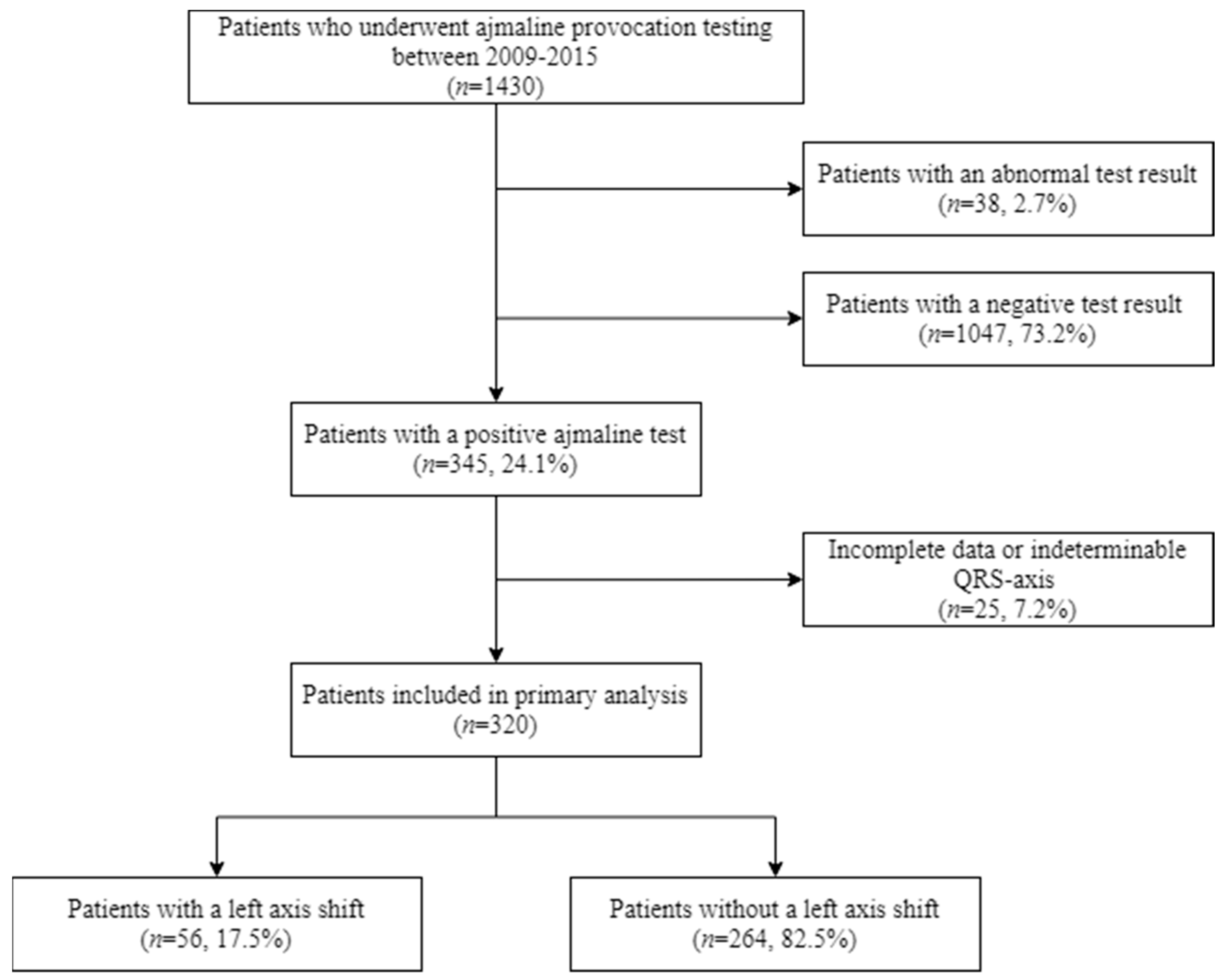
Figure 1 shows the flowchart of the selection of patients. In total, 345 patients in whom ajmaline testing elicited a type-1 BrS-ECG (positive test) were identified from a total cohort of 1430 patients who underwent provocation testing (24.1%). In total, 320 of these 345 (92.8%) patients were included for this study, and 25 (7.2%) patients were excluded due to incomplete data or an indeterminable axis. The prevalence of a left axis deviation in patients with a positive test and determinable QRS-axis was 17.5% (n = 56/320). Patients with a left axis deviation at baseline were more often female and were significantly shorter and lighter; the BMI, however, did not significantly differ (Table 1). The age, histories of (possible) arrhythmias, the indication for testing and the administered ajmaline dose, as well as the percentage of the ajmaline maximal target dose, did not differ (Table 1). The presence of an SCN5A mutation was also not significantly different between the groups. Please note that genetic testing was not performed in all patients, in particular when genetic testing in a family member had already revealed the absence of a potentially causative mutation. The baseline ECG parameters are presented in Table 1.

Figure 1.
Flowchart of the patient selection.

Table 1.
Baseline characteristics.
2.2. Baseline Vectorcardiogram Parameters
Patients with a positive test and a left axis deviation upon provocation testing had, at baseline, shorter QRS-durations when compared to patients without a left axis deviation upon provocation testing (100 ± 14 ms versus 105 ± 15 ms, p < 0.05). In the 3rd quartile of the QRS-loop, conduction was slightly faster in patients with a left axis deviation upon provocation (10[8;12] ms versus 12[10;14] ms, p < 0.05). There was no difference in the depolarization–repolarization interaction in both groups (Table 2, QRS-T angle and ventricular gradient).

Table 2.
Vectorcardiographic parameters in patients with a left axis deviation of >30° at baseline, at ajmaline peak, and the change between the baseline and ajmaline peak.
2.3. Vectorcardiogram Parameters at Peak Ajmaline Dose
2.3.1. Depolarization Abnormalities
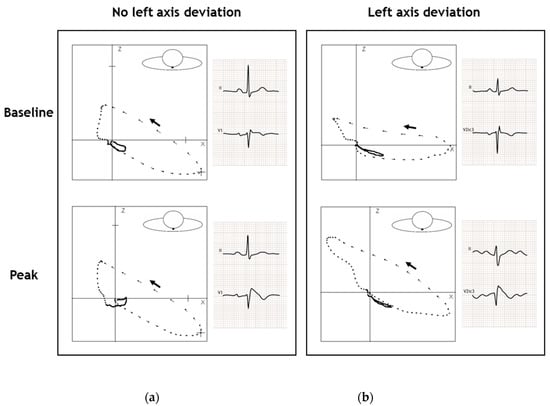
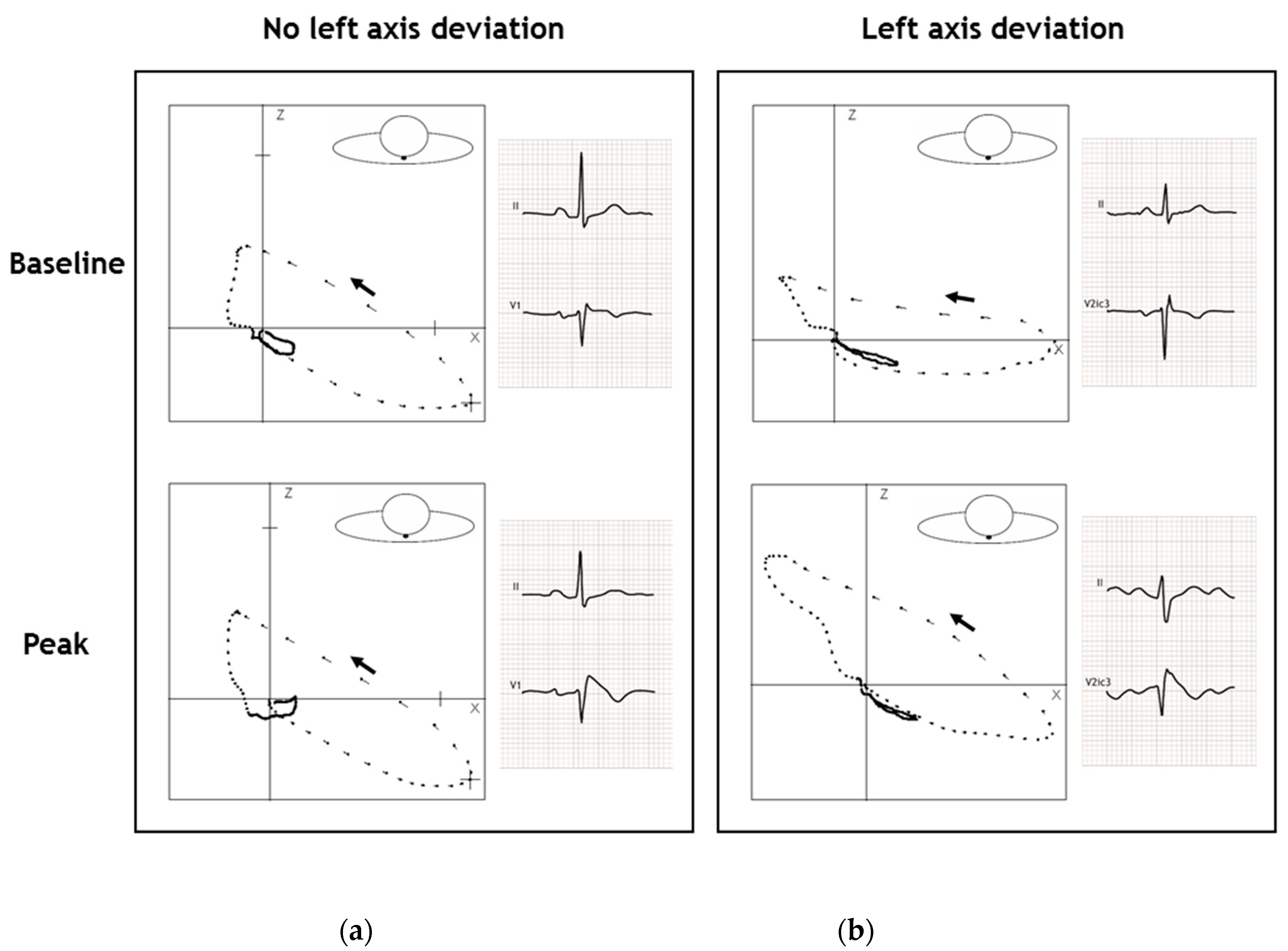
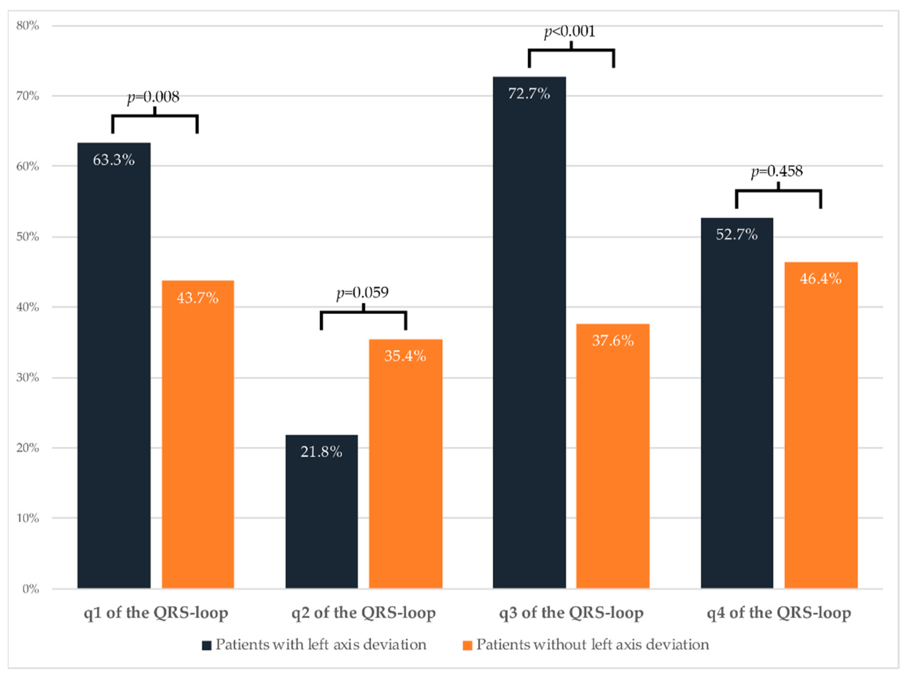
At the peak ajmaline dose, progressive conduction slowing had occurred in all four quartiles in both groups (Table 2). Patients with a left axis deviation showed more of a conduction delay when compared to patients without a left axis deviation (QRS duration: +43 ± 17 ms versus +30 ± 21 ms, p < 0.001). This conduction delay in patients with a left axis deviation occurred primarily in the first (+12[8;18] ms versus +8[4;12] ms, p < 0.001) and third (+12 ± 10 ms versus + 5 ± 7 ms, p < 0.001) quartiles. In the second quartile, conduction slowing was less pronounced in patients with a left axis deviation (+2[−2;6] ms versus +6[2;8] ms, p <0.001). Noticeably, the conduction delay in the fourth quartile was similar in both groups (Table 2). In addition, there were no differences in the amount of maximal right-precordial ST-elevation (measured at the J-point) between the two groups. In Figure 2, the QRS-T loops of two representative patients at baseline and ajmaline peak are shown; from this figure, the conduction slowing at the peak ajmaline dose when compared to baseline (especially in the patient with left axis deviation) can be appreciated. Figure 3 shows the proportion of patients with a left axis deviation and an increase of the conduction intervals above the median value of the cohort. For the first (63.3% versus 43.7%, p < 0.05) and third quartiles (72.7% versus 37.6%, p < 0.001) this proportion was significantly higher compared to patients without a left axis deviation (Figure 3). For the second quartile, in contrast, the proportion of patients with conduction slowing was larger in the patients without a left axis deviation (21.8% versus 35.4%, p = 0.059). The proportion of patients with conduction slowing above the median of the entire cohort in the fourth quartile was similar in both groups (52.7% versus 46.4%, p = 0.458) (Figure 3).

Figure 2.
The QRS-T loop in the transverse plane of two representative patients at baseline and ajmaline peak. (a) Patient without a left axis deviation. The QRS-axis in the frontal plane changed 3° to the left. At baseline, the QRS-duration was 94 ms and increased to 106 ms at ajmaline peak; the changes in the duration of the quartiles of the QRS-loops were: q1 +0 ms, q2 +2 ms, q3 +2 ms and q4 +8 ms. The QRS-T angle changed with –19° from 27° at baseline to 8° at ajmaline peak; (b) Patient with a left axis deviation. The QRS-axis in the frontal plane changed 60° to the left. At baseline, the QRS-duration was 92 ms and increased to 138 ms at ajmaline peak; the changes in the duration of the quartiles of the QRS-loops were: q1 +14 ms, q2 -2 ms, q3 +18 ms and q4 +16 ms. The QRS-T angle changed with +60° from 40° at baseline to 100° at ajmaline peak. Dashes of the QRS-T loop: 2-ms intervals. The black arrow indicates the QRS-loop direction.

Figure 3.
Proportion of patients with conduction slowing above the median value of the entire cohort.
2.3.2. Repolarization Abnormalities
The spatial QRS-T angle significantly differed at the peak ajmaline dose (Table 2): in patients with a left axis deviation, the spatial QRS-T angle rose to borderline abnormal values (>105–135°) whilst remaining normal in the patients without a left axis deviation (122[59;147]° versus 44[25;91]°, p < 0.001). The heterogeneity of the action potential morphology, as expressed by the ventricular gradient, did not significantly change or differ between the two groups at the peak ajmaline dose (Table 2).
3. Discussion
3.1. Main Findings
In this study, we show that (1) the prevalence of a left axis deviation during ajmaline provocation testing in patients with a positive ajmaline test is 17.5% and (2) that this left axis deviation is not caused by conduction slowing in the RVOT but is due to additional conduction slowing in the more proximal conduction system.
The observed left axis deviation could possibly occur due to excessive conduction slowing in the RVOT, as this is the most commonly affected region in BrS patients [1,4]. Our results, however, show that the RVOT conduction, as mirrored by the conduction slowing in the 4th quartile of the QRS-loop, slows in equal amounts between patients with a left axis deviation and patients without a left axis deviation while they develop the type-1 ECG. This demonstrates that the RVOT region is equally affected in both groups and that the cause of the left axis deviation must originate more proximally in the conduction system. Conduction slowing in BrS patients who developed a left axis deviation was indeed most prominent in the 1st and 3rd quartiles of the QRS-loop, most likely representing additional septal and free wall depolarization abnormalities, respectively [7,10,11,12]. Apparently, in these patients with a left axis deviation, conduction seems to be more extensively affected. This could potentially explain some part of the varying arrhythmogenic risk in BrS-patients [1]. These conductional differences cannot be explained from our results by the currently known (likely) pathogenic SCN5A mutation, as the mutation status did not differ significantly between groups. Still, loss-of-function SCN5A mutations will often result in a more general conduction disease. Surprisingly, at baseline, conduction in the 3rd quartile is slightly faster in patients with a left axis deviation, whilst out of the four quarters, this quartile showed the most conduction slowing at the peak ajmaline. If conduction in the free wall actually is the most extensively affected region, this faster conduction at baseline is remarkable and against our expectations. The underlying mechanism and the clinical implications of this finding are unclear and could be the focus of future research. In addition, global repolarization appeared to be more discordant from depolarization in patients with a left axis deviation, as indicated by increased, borderline abnormal (>105–135°), QRST-angles. In the general population, abnormal (>135°) and borderline abnormal (105–135°) QRS-T angles are strong predictors for cardiac death. One could therefore hypothesize that patients with a positive test with a concomitant left axis deviation might have a higher arrhythmogenic risk.
As to the origin of the characteristic ST-elevation (or J-point elevation) that is associated with the development of a type-1 BrS-ECG, previous studies suggested that excitation failure at the RVOT was elementary in this process [13,14]. In the current study, the amount of ST-elevation did not differ between those ajmaline-positive patients with or without a left axis deviation upon ajmaline provocation. Whether vectorcardiography, which is dependent on spatial vectors, would be able to add insights to the occurrence and localization of an excitation failure in the development of the type-1 ECG during the simultaneous slowing of conduction during type-1 ECG development is currently uncertain.
3.2. Future Perspective
Future studies may investigate whether the occurrence of a left axis deviation during ajmaline testing in BrS-patients partly determines their arrhythmogenic risk. Clearly, a greater ability to stratify the arrhythmogenic risk in BrS patients would make it possible to optimize treatments accordingly.
3.3. Limitations
Vectorcardiograms were not recorded using additional orthogonal Frank XYZ leads but were reconstructed from 12-lead ECGs. Despite the fact that the reconstruction process using matrix multiplication has been validated, we cannot exclude some degree of variability from Frank leads [15]. However, we used the same lead setup in every patient and compared within-patient changes. In addition, by using the reconstructed vectorcardiograms, we enabled a future comparison of our data with other cohorts for whom digital 12-lead ECG data is available.
In addition, as this was a retrospective exploratory study, we were not informed about the follow-up data of these patients so as to determine potential differences in arrhythmogenic risk or differences in the progression of the clinical phenotype as indicated by the presence or absence of a (ajmaline-induced) left axis deviation.
4. Materials and Methods
4.1. Patients and Sodium Channel Provocation Testing
In this study, out of a cohort of 1430 patients who underwent provocation testing during the period from 2009 to 2015, a selection of patients was made based on the provocation test result and the cardiac axis. All patients for whom the provocation test was positive (see below) and whose electrocardiogram were available for a vectorcardiogram reconstruction were selected for this study; those with an abnormal or negative test were excluded. Tests were defined as (1) positive if a type-1 BrS-ECG occurred [1], (2) abnormal if arrhythmias or an excessive QRS-widening of ≥40% occurred, and (3) negative if the target dose was reached and none of the criteria above were met. Ajmaline was used as the sodium channel blocker and was infused intravenously in boluses of 10 mg/min until the maximum dose (1 mg/kg) was reached or until a positive or abnormal test result was obtained. None of the patients had exhibited a spontaneous type-1 BrS ECG before the provocation test. These patients underwent provocation testing because of symptoms (e.g., unexplained syncope or documented ventricular arrhythmias), a baseline ECG that raised the suspicion of BrS, family screening for BrS or family screening in the context of a sudden cardiac death or sudden unexplained death.
4.2. Electrocardiographic Recordings, Analysis and Definitions
4.2.1. Electrocardiographic Recordings
Modified 12-lead ECGs—with V3 and V5 placed cranially to V1 and V2 over the third intercostal space—were recorded at baseline and at one minute after each ajmaline bolus.
4.2.2. Electrocardiographic Analysis
ECGs were analyzed with the electrocardiographic analysis system MEANS, and its markers settings (e.g., P-wave onset, P-wave offset, QRS onset, etc.) were manually inspected and adjusted if deemed necessary [16]. Vectorcardiograms were reconstructed from the 12-lead ECG using the matrix multiplication as previously described [15]. The vectorcardiographic analysis consisted of dividing the spatial QRS-loop in four quartiles of equal length. Subsequently, the durations (ms) in the transverse plane were measured out of these four quartiles. Furthermore, to study the depolarization and repolarization interaction, the spatial QRS-T angle (°) and the vector magnitude (mV·ms) (i.e., ventricular gradient) of the spatial QRS-T integral were determined. An abnormally increased QRS-T angle indicates global discordant repolarization, values of 105–135° (borderline abnormal) and >135 (abnormal) are associated with fatal cardiac arrhythmias in the general population [8]. The ventricular gradient is considered a three-dimensional measure of the heterogeneity of the action potential morphology [9]. In order to calculate the change in the conduction intervals, the vectorcardiographic parameters at baseline were deducted from the parameters at the peak ajmaline dose. To further evaluate the four quartiles of the QRS-loop between patients with a left axis deviation and patients without a left axis deviation, the proportion of patients with an increase above the median value of the entire cohort was compared between the two groups.
4.2.3. Electrocardiographic Definitions
A left axis deviation was defined in this study as a leftward shift of the frontal QRS axis of >30° at the peak ajmaline dose as compared to the baseline. We defined the fourth quartile of the QRS-loop—in patients with a positive ajmaline test—as representative of RVOT conduction [4].
4.3. Statistical Analysis
The statistical analysis was performed with SPSS Statistics (version 25.0, IBM Corporation, Armonk, New York, USA). Categorical variables are presented as frequencies and group percentages. To compare such variables, the Fisher-exact test was used. Continuous variables are expressed as the mean ± standard deviation in the case of a normal distribution or the median [interquartile range] in the case of a skewed distribution. Histograms and Q–Q plots were used to evaluate the distribution of variables with continuous data. The unpaired two-tailed t-test was used to compare normally distributed variables; in the case of a skewed distribution, the Mann–Whitney U test was used. A p-value of <0.05 was accepted as the level of statistical significance.
5. Conclusions
In BrS-patients, a left axis deviation during ajmaline testing occurs in a significant number of patients. This leftward shift in axis is not caused by an additional conduction slowing of the RVOT but appears to occur as a consequence of an additional, more proximal conduction slowing. Whether a left axis deviation can also be used for the stratification of arrhythmia risk is currently undetermined.
Author Contributions
Conceptualization, P.G.P. and A.A.M.W. and M.H.v.d.R.; methodology, P.G.P. and A.A.M.W. and M.H.v.d.R.; software, J.A.K.; validation, J.A.K.; formal analysis, M.H.v.d.R. and J.V.; investigation, P.G.P., A.A.M.W., A.S.A. and H.L.T.; resources P.G.P., A.A.M.W. and H.L.T.; data curation, M.H.v.d.R., J.V. and J.A.K.; writing—original draft preparation, M.H.v.d.R., J.V. and P.G.P.; writing—review and editing, A.A.M.W., A.S.A. and H.L.T.; visualization, M.H.v.d.R., P.G.P.; supervision, P.G.P.; project administration, M.H.v.d.R. and J.V.; funding acquisition, A.A.M.W. and H.L.T. All authors have read and agreed to the published version of the manuscript.
Funding
This work has received funding from the European Union’s Horizon 2020 research and innovation programme under acronym ESCAPE-NET, registered under grant agreement No. 733381, and the COST Action PARQ (grant agreement No CA19137) supported by COST (European Cooperation in Science and Technology).
Institutional Review Board Statement
Ethical review and approval were waived for this study, the sodium channel provocation tests were performed as part of routine clinical care.
Informed Consent Statement
Patient consent was waived, the sodium channel provocation tests were performed as part of routine clinical care. The data was subsequently collected and anonymized by the treating physician.
Data Availability Statement
The data presented in this study are available on request from the corresponding author. The data are not publicly available due to privacy reasons.
Acknowledgments
We acknowledge the support from the Netherlands CardioVascular Research Initiative, the Dutch Heart Foundation, Dutch Federation of University Medical Centres, the Netherlands Organisation for Health Research and Development and the Royal Netherlands Academy of Sciences (Predict2).
Conflicts of Interest
The authors declare no conflict of interest.
Abbreviations
| BrS | Brugada Syndrome |
| ECG | Electrocardiogram |
References
- Antzelevitch, C.; Yan, G.-X.; Ackerman, M.J.; Borggrefe, M.; Corrado, D.; Guo, J.; Gussak, I.; Hasdemir, C.; Horie, M.; Huikuri, H.; et al. J-Wave syndromes expert consensus conference report: Emerging concepts and gaps in knowledge. Europace 2016, 13, 665–694. [Google Scholar] [CrossRef]
- Morita, H.; Morita, S.T.; Nagase, S.; Banba, K.; Nishii, N.; Tani, Y.; Watanabe, A.; Nakamura, K.; Kusano, K.F.; Emori, T.; et al. Ventricular Arrhythmia Induced by Sodium Channel Blocker in Patients with Brugada Syndrome. J. Am. Coll. Cardiol. 2003, 42, 1624–1631. [Google Scholar] [CrossRef] [PubMed]
- Wilde, A.A.M.; Antzelevitch, C.; Borggrefe, M.; Brugada, J.; Brugada, R.; Brugada, P.; Corrado, D.; Hauer, R.N.W.; Kass, R.S.; Nademanee, K.; et al. Proposed diagnostic criteria for the Brugada syndrome: Consensus report. Circulation 2002, 106, 2514–2519. [Google Scholar] [CrossRef] [PubMed]
- Postema, P.G.; van Dessel, P.F.H.M.; Kors, J.A.; Linnenbank, A.C.; van Herpen, G.; Ritsema van Eck, H.J.; van Geloven, N.; de Bakker, J.M.T.; Wilde, A.A.M.; Tan, H.L. Local Depolarization Abnormalities Are the Dominant Pathophysiologic Mechanism for Type 1 Electrocardiogram in Brugada Syndrome. A Study of Electrocardiograms, Vectorcardiograms, and Body Surface Potential Maps During Ajmaline Provocation. J. Am. Coll. Cardiol. 2010, 55, 789–797. [Google Scholar] [CrossRef] [PubMed]
- Pappone, C.; Mecarocci, V.; Manguso, F.; Ciconte, G.; Vicedomini, G.; Sturla, F.; Votta, E.; Mazza, B.; Pozzi, P.; Borrelli, V.; et al. New electromechanical substrate abnormalities in high-risk patients with Brugada syndrome. Heart Rhythm 2020, 17, 637–645. [Google Scholar] [CrossRef] [PubMed]
- Pérez Riera, A.R.; Uchida, A.H.; Filho, C.F.; Meneghini, A.; Ferreira, C.; Schapacknik, E.; Dubner, S.; Moffa, P. Significance of Vectorcardiogram in the Cardiological Diagnosis of the 21st Century. Clin. Cardiol. 2007, 30, 319–323. [Google Scholar] [CrossRef] [PubMed]
- Pérez-Riera, A.R.; Ferreira Filho, C.; de Abreu, L.C.; Ferreira, C.; Yanowitz, F.G.; Femenia, F.; Brugada, P.; Baranchuk, A. Do patients with electrocardiographic Brugada type 1 pattern have associated right bundle branch block? A comparative vectorcardiographic study. Europace 2012, 14, 889–897. [Google Scholar] [CrossRef] [PubMed]
- Kardys, I.; Kors, J.A.; Van der Meer, I.M.; Hofman, A.; Van der Kuip, D.A.M.; Witteman, J.C.M. Spatial QRS-T angle predicts cardiac death in a general population. Eur. Heart J. 2003, 24, 1357–1364. [Google Scholar] [CrossRef]
- Draisma, H.H.M.; Schalij, M.J.; van der Wall, E.E.; Swenne, C.A. Elucidation of the spatial ventricular gradient and its link with dispersion of repolarization. Heart Rhythm 2006, 3, 1092–1099. [Google Scholar] [CrossRef] [PubMed]
- Durrer, D.; van Dam, R.T.; Freud, G.E.; Janse, M.J.; Meijler, F.L.; Arzbaecher, R.C. Total excitation of the isolated human heart. Circulation 1970, 41, 899–912. [Google Scholar] [CrossRef]
- Lambiase, P.D.; Rinaldi, A.; Hauck, J.; Mobb, M.; Elliott, D.; Mohammad, S.; Gill, J.S.; Bucknall, C.A. Non-contact left ventricular endocardial mapping in cardiac resynchronisation therapy. Heart 2004, 90, 44–51. [Google Scholar] [CrossRef] [PubMed]
- Postema, P.G.; van Dessel, P.F.H.M.; de Bakker, J.M.T.; Dekker, L.R.C.; Linnenbank, A.C.; Hoogendijk, M.G.; Coronel, R.; Tijssen, J.G.P.; Wilde, A.A.M.; Tan, H.L. Slow and discontinuous conduction conspire in Brugada syndrome: A right ventricular mapping and stimulation study. Circ. Arrhythm. Electrophysiol. 2008, 1, 379–386. [Google Scholar] [CrossRef] [PubMed]
- Hoogendijk, M.G.; Potse, M.; Vinet, A.; de Bakker, J.M.T.; Coronel, R. ST segment elevation by current-to-load mismatch: An experimental and computational study. Heart Rhythm 2011, 8, 111–118. [Google Scholar] [CrossRef] [PubMed]
- Hoogendijk, M.G.; Opthof, T.; Postema, P.G.; Wilde, A.A.M.; De Bakker, J.M.T.; Coronel, R. The Brugada ECG pattern a marker of channelopathy, structural heart disease, or neither? Toward a unifying mechanism of the Brugada syndrome. Circ. Arrhythmia Electrophysiol. 2010, 3, 283–290. [Google Scholar] [CrossRef] [PubMed]
- Kors, J.A.; Van Herpen, G.; Sittig, A.C.; Van Bemmel, J.H. Reconstruction of the frank vectorcardiogram from standard electrocardiographic leads: Diagnostic comparison of different methods. Eur. Heart J. 1990, 11, 1083–1092. [Google Scholar] [CrossRef] [PubMed]
- Van Bemmel, J.H.; Kors, J.A.; van Herpen, G. Methodology of the modular ECG analysis system MEANS. Methods Inf. Med. 1990, 29, 346–353. [Google Scholar] [PubMed]
Publisher’s Note: MDPI stays neutral with regard to jurisdictional claims in published maps and institutional affiliations. |
© 2021 by the authors. Licensee MDPI, Basel, Switzerland. This article is an open access article distributed under the terms and conditions of the Creative Commons Attribution (CC BY) license (http://creativecommons.org/licenses/by/4.0/).


