Environmentally Relevant Iron Oxide Nanoparticles Produce Limited Acute Pulmonary Effects in Rats at Realistic Exposure Levels

 ,
,  , and
, and
Abstract
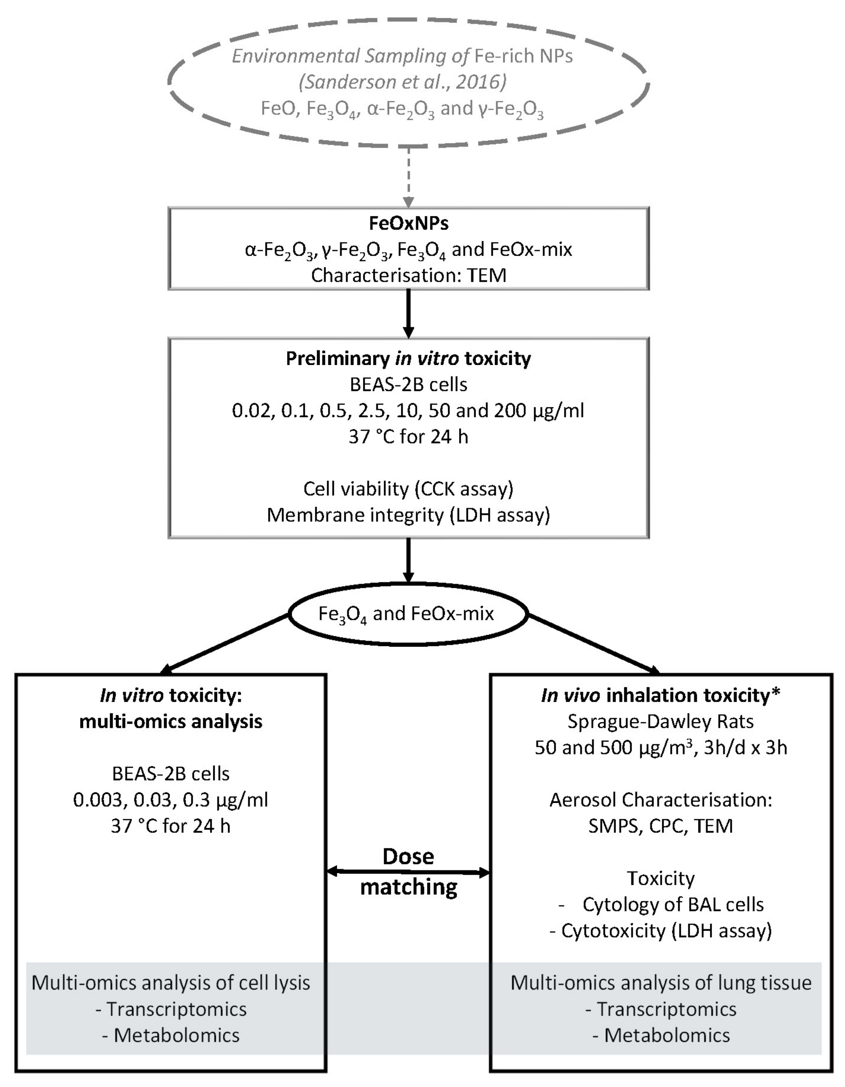
:1. Introduction
2. Results
2.1. Toxicity Assessment of FeOxNPs in BEAS-2B Cells
2.1.1. Characterisation of FeOxNPs
2.1.2. Cytotoxic Effects of FeOxNPs on Human Bronchial Epithelial Cells (BEAS-2B)
2.1.3. Multi-Omics Analysis of BEAS-2B Cells
2.2. Nose-Only Inhalation Study of FeOxNPs in Sprague Dawley Rats
2.2.1. Characterisation of FeOxNP Aerosols and Deposited Doses
2.2.2. Assessment of Gross Toxicity
2.2.3. Multi-Omics Analysis of Lung Tissues
3. Discussion
4. Materials and Methods
4.1. Iron Oxide Nanoparticles
4.2. Characterisation of FeOxNPs in Dispersions
4.3. Cell Culture and FeOxNPs Preparation
4.4. Cell Cytotoxicity Assays
4.5. Cell Exposure Study for Transcriptomics and Metabolomics
4.6. In Vivo Exposure of Sprague–Dawley Rats (Nose-Only Inhalation)
4.7. Aerosol Exposure System
4.8. Characterisation of FeOx Nanoparticle Aerosols
4.9. Dose Estimation
4.10. Lung Tissue Samples and Bronchoalveolar Lavage (BAL)
4.11. BALF Analysis
4.12. Metabolite and RNA Extraction from BEAS-2B Cells and Lung Tissues
4.13. Microarray-Based Gene Expression Profiling of BEAS-2B Cells and Lung Tissue Extracts
4.14. Metabolomics of BEAS-2B Cells and Lung Tissue Extracts
5. Conclusions
Supplementary Materials
Author Contributions
Funding
Institutional Review Board Statement
Informed Consent Statement
Data Availability Statement
Acknowledgments
Conflicts of Interest
References
- WHO. Ambient Air Pollution: A Global Assessment of Exposure and Burden of Disease; World Health Organization: Geneva, Switzerland, 2016; Available online: https://apps.who.int/iris/bitstream/handle/10665/250141/9789241511353-eng.pdf?sequence=1 (accessed on 1 March 2017).
- Cassee, F.R.; Heroux, M.E.; Gerlofs-Nijland, M.E.; Kelly, F.J. Particulate matter beyond mass: Recent health evidence on the role of fractions, chemical constituents and sources of emission. Inhal. Toxicol. 2013, 25, 802–812. [Google Scholar] [CrossRef]
- COMEAP. Statement on the Evidence for Differential Health Effects of Particulate Matter According to Source or Components. Available online: https://assets.publishing.service.gov.uk/government/uploads/system/uploads/attachment_data/file/411762/COMEAP_The_evidence_for_differential_health_effects_of_particulate_matter_according_to_source_or_components.pdf2015 (accessed on 1 October 2016).
- HEI Review Panel on Ultrafine Particles. HEI Perspectives 3. Understanding the Health Effects of Ambient Ultrafine Particles; Health Effects Institute: Boston, MA, USA, 2013; Available online: https://www.healtheffects.org/system/files/Perspectives3.pdf (accessed on 1 October 2013).
- Wilson, M.R.; Lightbody, J.H.; Donaldson, K.; Sales, J.; Stone, V. Interactions between ultrafine particles and transition metals in vivo and in vitro. Toxicol. Appl. Pharmacol. 2002, 184, 172–179. [Google Scholar] [CrossRef]
- Rejman, J.; Oberle, V.; Zuhorn, I.S.; Hoekstra, D. Size-dependent internalization of particles via the pathways of clathrin- and caveolae-mediated endocytosis. Biochem. J. 2004, 377 Pt 1, 159–169. [Google Scholar] [CrossRef] [PubMed]
- Kumar, P.; Robins, A.; Vardoulakis, S.; Britter, R. A review of characteristics of nanoparticles in the urban atmosphere and the prospects for developing regulatory controls. Atmos. Environ. 2010, 44, 5035–5052. [Google Scholar] [CrossRef] [Green Version]
- Goddard, S.; Williams, K.; Robins, C.; Butterfield, D.; Brown, R. Concentration trends of metals in ambient air in the UK: A review. Environ. Monit. Assess. 2019, 191, 683. [Google Scholar] [CrossRef] [PubMed]
- Smith, J.D.; Barratt, B.M.; Fuller, G.W.; Kelly, F.J.; Loxham, M.; Nicolosi, E.; Priestman, M.; Tremper, A.H.; Green, D.C. PM2.5 on the London Underground. Environ. Int. 2020, 134, 105188. [Google Scholar] [CrossRef] [PubMed]
- Sitzmann, B.; Kendall, M.; Watt, J.; Williams, I. Characterisation of airborne particles in London by computer-controlled scanning electron microscopy. J. Sci. Total Environ. 1999, 241, 63. [Google Scholar] [CrossRef]
- Seaton, A.; Cherrie, J.; Dennekamp, M.; Donaldson, K.; Hurley, J.F.; Tran, C.L. The London Underground: Dust and hazards to health. Occup. Environ. Med. 2005, 62, 355–362. [Google Scholar] [CrossRef] [PubMed] [Green Version]
- COMEAP. Particulate Air Pollution on London Underground: Health Effects. Available online: https://www.gov.uk/government/publications/particulate-air-pollution-on-london-underground-health-effects2019 (accessed on 1 February 2019).
- Sanderson, P.; Delgado-Saborit, J.M.; Harrison, R. A review of chemical and physical characterisation of atmospheric metallic nanoparticles. Atmos. Environ. 2014, 94, 353–365. [Google Scholar] [CrossRef] [Green Version]
- Yang, Z.; Huang, W.; Tianqi, C.; Fang, D.; Wang, Y.; Song, J.; Hu, M.; Zhang, Y. Concentrations and chemical compositions of fine particles (PM2.5) during haze and non-haze days in Beijing. Atmos. Res. 2016, 174, 62–69. [Google Scholar]
- Adachi, K.; Buseck, P. Hosted and Free-Floating Metal-Bearing Atmospheric Nanoparticles in Mexico City. Environ. Sci. Technol. 2010, 44, 2299–2304. [Google Scholar] [CrossRef] [PubMed]
- Sanderson, P.N.; Su, D.S.; Chang, I.T.H.; Delgado Saborit, J.M.; Kepaptsoglou, D.M.; Weber, R.J.M.; Harrison, R.M. Characterisation of iron-rich atmospheric submicrometre particles in the roadside environment. Atmos. Environ. 2016, 140, 167–175. [Google Scholar] [CrossRef]
- Adachi, K.; Moteki, N.; Kondo, Y.; Igarashi, Y. Mixing states of light-absorbing particles measured using a transmission electron microscope and a single-particle soot photometer in Tokyo, Japan: LIGHT-ABSORBING PARTICLES BY TEM AND SP2. J. Geophys. Res. Atmos. 2016, 121, 9153–9164. [Google Scholar] [CrossRef] [Green Version]
- Smith, S.; Ward, M.; Lin, R.; Brydson, R.; Dall´osto, M.; Harrison, R. Comparative study of single particle characterisation by Transmission Electron Microscopy and time-of-flight aerosol mass spectrometry in the London atmosphere. Atmos. Environ. 2012, 62, 400–407. [Google Scholar] [CrossRef]
- Gasser, M.; Riediker, M.; Mueller, L.; Perrenoud, A.; Blank, F.; Gehr, P.; Rothen-Rutishauser, B. Toxic effects of brake wear particles on epithelial lung cells in vitro. Part. Fibre Toxicol. 2009, 6, 30. [Google Scholar] [CrossRef] [Green Version]
- Steiner, S.; Czerwinski, J.; Comte, P.; Heeb, N.V.; Mayer, A.; Petri-Fink, A.; Rothen-Rutishauser, B. Effects of an iron-based fuel-borne catalyst and a diesel particle filter on exhaust toxicity in lung cells in vitro. Anal. Bioanal. Chem. 2015, 407, 5977–5986. [Google Scholar] [CrossRef] [Green Version]
- Grigg, J.; Tellabati, A.; Jones, G.D.; Howes, P. DNA damage of macrophages induced by metal nanoparticulates using an air-liquid interface exposure model. Nanotoxicology 2013, 7, 961–962. [Google Scholar] [CrossRef]
- Xie, Y.; Worth Longest, P.; Xu, Y.H.; Wang, J.P.; Wiedmann, T.S. In Vitro and In Vivo Lung Deposition of Coated Magnetic Aerosol Particles. J. Pharm. Sci. 2010, 99, 4658–4668. [Google Scholar] [CrossRef]
- Sutunkova, M.P.; Katsnelson, B.A.; Privalova, L.I.; Gurvich, V.B.; Konysheva, L.K.; Shur, V.Y.; Shishkina, E.V.; Minigalieva, I.A.; Solovjeva, S.N.; Grebenkina, S.V.; et al. On the contribution of the phagocytosis and the solubilization to the iron oxide nanoparticles retention in and elimination from lungs under long-term inhalation exposure. Toxicology 2016, 363–364, 19–28. [Google Scholar] [CrossRef]
- Watson, A.Y.; Brain, J.D. Uptake of iron aerosols by mouse airway epithelium. Lab. Investig. 1979, 40, 450–459. [Google Scholar]
- Kwon, J.-T.; Hwang, S.-K.; Jin, H.; Kim, D.-S.; Minai-Tehrani, A.; Yoon, H.-J.; Choi, M.; Yoon, T.-J.; Han, D.-Y.; Kang, Y.-W.; et al. Body Distribution of Inhaled Fluorescent Magnetic Nanoparticles in the Mice. J. Occup. Health 2008, 50, 1–6. [Google Scholar] [CrossRef] [PubMed]
- Hopkins, L.E.; Laing, E.A.; Peake, J.L.; Uyeminami, D.; Mack, S.M.; Li, X.; Smiley-Jewell, S.; Pinkerton, K.E. Repeated Iron-Soot Exposure and Nose-to-brain Transport of Inhaled Ultrafine Particles. Toxicol. Pathol. 2018, 46, 75–84. [Google Scholar] [CrossRef] [PubMed] [Green Version]
- Zhou, Y.-M.; Zhong, C.-Y.; Kennedy, I.M.; Pinkerton, K.E. Pulmonary responses of acute exposure to ultrafine iron particles in healthy adult rats. Environ. Toxicol. 2003, 18, 227–235. [Google Scholar] [CrossRef] [PubMed]
- Zhong, C.-Y.; Zhou, Y.-M.; Smith, K.R.; Kennedy, I.M.; Chen, C.-Y.; Aust, A.E.; Pinkerton, K.E. Oxidative Injury in The Lungs of Neonatal Rats Following Short-Term Exposure to Ultrafine Iron and Soot Particles. J. Toxicol. Environ. Health Part A 2010, 73, 837–847. [Google Scholar] [CrossRef]
- Srinivas, A.; Rao, P.J.; Selvam, G.; Goparaju, A.; Murthy, P.B.; Reddy, P.N. Oxidative stress and inflammatory responses of rat following acute inhalation exposure to iron oxide nanoparticles. Hum. Exp. Toxicol. 2012, 31, 1113–1131. [Google Scholar] [CrossRef]
- Sotiriou, G.A.; Diaz, E.; Long, M.S.; Godleski, J.; Brain, J.; Pratsinis, S.E.; Demokritou, P. A novel platform for pulmonary and cardiovascular toxicological characterization of inhaled engineered nanomaterials. Nanotoxicology 2012, 6, 680–690. [Google Scholar] [CrossRef]
- Teeguarden, J.G.; Mikheev, V.B.; Minard, K.R.; Forsythe, W.C.; Wang, W.; Sharma, G.; Karin, N.; Tilton, S.C.; Waters, K.M.; Asgharian, B.; et al. Comparative iron oxide nanoparticle cellular dosimetry and response in mice by the inhalation and liquid cell culture exposure routes. Part. Fibre Toxicol. 2014, 11, 46. [Google Scholar] [CrossRef]
- Pettibone, J.M.; Adamcakova-Dodd, A.; Thorne, P.S.; O’Shaughnessy, P.T.; Weydert, J.A.; Grassian, V.H. Inflammatory response of mice following inhalation exposure to iron and copper nanoparticles. Nanotoxicology 2008, 2, 189–204. [Google Scholar] [CrossRef]
- Pauluhn, J.; Wiemann, M. Siderite (FeCO3) and magnetite (Fe3O4) overload-dependent pulmonary toxicity is determined by the poorly soluble particle not the iron content. Inhal. Toxicol. 2011, 23, 763–783. [Google Scholar] [CrossRef]
- Hofmann, T.; Ma-Hock, L.; Strauss, V.; Treumann, S.; Rey Moreno, M.; Neubauer, N.; Wohlleben, W.; Gröters, S.; Wiench, K.; Veith, U.; et al. Comparative short-term inhalation toxicity of five organic diketopyrrolopyrrole pigments and two inorganic iron-oxide-based pigments. Inhal. Toxicol. 2016, 28, 463–479. [Google Scholar] [CrossRef]
- Grigoratos, T.; Martini, G. Brake wear particle emissions: A review. Environ. Sci. Pollut. Res. Int. 2015, 22, 2491–2504. [Google Scholar] [CrossRef] [PubMed] [Green Version]
- Timmers, V.; Achten, P. Non-exhaust PM emissions from electric vehicles. Atmos. Environ. 2016, 134, 10–17. [Google Scholar] [CrossRef]
- Krarnes, J.; Buettner, H.; Ebert, F. Submicron particle generation by evaporation of water droplets. J. Aerosol Sci. 1991, 22, S15–S18. [Google Scholar] [CrossRef]
- LaFranchi, B.W.; Knight, M.; Petrucci, G.A. Leaching as a source of residual particles from nebulization of deionized water. J. Aerosol Sci. 2003, 34, 1589–1594. [Google Scholar] [CrossRef]
- Park, E.J.; Umh, H.N.; Choi, D.H.; Cho, M.H.; Choi, W.; Kim, S.W.; Kim, Y.; Kim, J.H. Magnetite- and maghemite-induced different toxicity in murine alveolar macrophage cells. Arch. Toxicol. 2014, 88, 1607–1618. [Google Scholar] [CrossRef]
- Kornberg, T.G.; Stueckle, T.A.; Antonini, J.A.; Rojanasakul, Y.; Castranova, V.; Yang, Y.; Wang, L. Potential Toxicity and Underlying Mechanisms Associated with Pulmonary Exposure to Iron Oxide Nanoparticles: Conflicting Literature and Unclear Risk. Nanomaterials 2017, 7, 307. [Google Scholar] [CrossRef] [Green Version]
- Chen, Z.; Yin, J.J.; Zhou, Y.T.; Zhang, Y.; Song, L.; Song, M.; Hu, S.; Gu, N. Dual enzyme-like activities of iron oxide nanoparticles and their implication for diminishing cytotoxicity. ACS Nano 2012, 6, 4001–4012. [Google Scholar] [CrossRef]
- Auffan, M.; Rose, J.; Orsiere, T.; De Meo, M.; Thill, A.; Zeyons, O.; Proux, O.; Masion, A.; Chaurand, P.; Spalla, O.; et al. CeO2 nanoparticles induce DNA damage towards human dermal fibroblasts in vitro. Nanotoxicology 2009, 3, 161–171. [Google Scholar] [CrossRef]
- Feng, Q.; Liu, Y.; Huang, J.; Chen, K.; Huang, J.; Xiao, K. Uptake, distribution, clearance, and toxicity of iron oxide nanoparticles with different sizes and coatings. Sci. Rep. 2018, 8, 1–13. [Google Scholar] [CrossRef]
- Malhotra, N.; Lee, J.-S.; Liman, R.A.D.; Ruallo, J.M.S.; Villaflores, O.B.; Ger, T.-R.; Hsiao, C.-D. Potential Toxicity of Iron Oxide Magnetic Nanoparticles: A Review. Molecules 2020, 25, 3159. [Google Scholar] [CrossRef]
- Billing, A.M.; Knudsen, K.B.; Chetwynd, A.J.; Ellis, L.A.; Tang, S.V.Y.; Berthing, T.; Wallin, H.; Lynch, I.; Vogel, U.; Kjeldsen, F. Fast and Robust Proteome Screening Platform Identifies Neutrophil Extracellular Trap Formation in the Lung in Response to Cobalt Ferrite Nanoparticles. ACS Nano 2020, 14, 4096–4110. [Google Scholar] [CrossRef] [PubMed]
- Guo, C.; Buckley, A.; Marczylo, T.; Seiffert, J.; Römer, I.; Warren, J.; Hodgson, A.; Chung, K.F.; Gant, T.W.; Smith, R.; et al. The small airway epithelium as a target for the adverse pulmonary effects of silver nanoparticle inhalation. Nanotoxicology 2018, 12, 539–553. [Google Scholar] [CrossRef] [PubMed] [Green Version]
- Oishi, K.; Machida, K. Different effects of immobilization stress on the mRNA expression of antioxidant enzymes in rat peripheral organs. Scand. J. Clin. Lab. Investig. 2002, 62, 115–121. [Google Scholar] [CrossRef] [PubMed]
- Kwon, J.-T.; Kim, D.-S.; Minai-Tehrani, A.; Hwang, S.-K.; Chang, S.-H.; Lee, E.-S.; Xu, C.-X.; Lim, H.T.; Kim, J.-E.; Yoon, B.-I.; et al. Inhaled Fluorescent Magnetic Nanoparticles Induced Extramedullary Hematopoiesis in the Spleen of Mice. J. Occup. Health 2009, 51, 423–431. [Google Scholar] [CrossRef] [PubMed]
- Pham, H.; Bonham, A.C.; Pinkerton, K.E.; Chen, C.-Y. Central neuroplasticity and decreased heart rate variability after particulate matter exposure in mice. Environ. Health Perspect. 2009, 117, 1448–1453. [Google Scholar] [CrossRef] [Green Version]
- Maher, B.A.; Gonzalez-Maciel, A.; Reynoso-Robles, R.; Torres-Jardon, R.; Calderon-Garciduenas, L. Iron-rich air pollution nanoparticles: An unrecognised environmental risk factor for myocardial mitochondrial dysfunction and cardiac oxidative stress. Environ. Res. 2020, 188, 109816. [Google Scholar] [CrossRef]
- Rowan, W.H.; Campen, M.J.; Wichers, L.B.; Watkinson, W.P. Heart rate variability in rodents: Uses and caveats in toxicological studies. Cardiovasc. Toxicol. 2007, 7, 28–51. [Google Scholar] [CrossRef]
- Hinderliter, P.M.; Minard, K.R.; Orr, G.; Chrisler, W.B.; Thrall, B.D.; Pounds, J.G.; Teeguarden, J.G. ISDD: A computational model of particle sedimentation, diffusion and target cell dosimetry for in vitro toxicity studies. Part. Fibre Toxicol. 2010, 7, 36. [Google Scholar] [CrossRef] [Green Version]
- Demokritou, P.; Gass, S.; Pyrgiotakis, G.; Cohen, J.M.; Goldsmith, W.; McKinney, W.; Frazer, D.; Ma, J.; Schwegler-Berry, D.; Brain, J.; et al. An in vivo and in vitro toxicological characterisation of realistic nanoscale CeO(2) inhalation exposures. Nanotoxicology 2013, 7, 1338–1350. [Google Scholar] [CrossRef] [Green Version]
- Oberdörster, G.; Oberdörster, E.; Oberdörster, J. Nanotoxicology: An emerging discipline evolving from studies of ultrafine particles. Environ. Health Perspect. 2005, 113, 823–839. [Google Scholar] [CrossRef]
- Schmid, O.; Stöger, T. Surface area is the biologically most effective dose metric for acute nanoparticle toxicity in the lung. J. Aerosol Sci. 2016, 99, 133–143. [Google Scholar] [CrossRef] [Green Version]
- Schmid, O.; Cassee, F.R. On the pivotal role of dose for particle toxicology and risk assessment: Exposure is a poor surrogate for delivered dose. Part. Fibre Toxicol. 2017, 14, 52. [Google Scholar] [CrossRef] [PubMed] [Green Version]
- Pauluhn, J. Retrospective analysis of 4-week inhalation studies in rats with focus on fate and pulmonary toxicity of two nanosized aluminum oxyhydroxides (boehmite) and pigment-grade iron oxide (magnetite): The key metric of dose is particle mass and not particle surface area. Toxicology 2009, 259, 140–148. [Google Scholar] [PubMed]
- Hadrup, N.; Saber, T.; Kyjovska, Z.; Jacobsen, N.; Vippola, M.; Sarlin, E.; Ding, Y.; Schmid, O.; Wallin, H.; Jensen, K.; et al. Pulmonary toxicity of Fe2O3, ZnFe2O4, NiFe2O4 and NiZnFe4O8 nanomaterials: Inflammation and DNA strand breaks. Environ. Toxicol. Pharmacol. 2020, 74, 103303. [Google Scholar] [CrossRef] [PubMed]
- Marvanová, S.; Kulich, P.; Skoupý, R.; Hubatka, F.; Ciganek, M.; Bendl, J.; Hovorka, J.; Machala, M. Size-segregated urban aerosol characterization by electron microscopy and dynamic light scattering and influence of sample preparation. Atmos. Environ. 2018, 178, 181–190. [Google Scholar] [CrossRef]
- Li, T.; Chen, X.; Yan, Z. ComParison of fine particles emissions of light-duty gasoline vehicles from chassis dynamometer tests and on-road measurements. Atmos. Environ. 2013, 68, 82–91. [Google Scholar] [CrossRef]
- Li, W.; Shao, L.; Zhang, D.; Ro, C.-U.; Hu, M.; Bi, X.; Geng, H.; Matsuki, A.; Niu, H.; Chen, J.-M. A review of single aerosol particle studies in the atmosphere of East Asia: Morphology, mixing state, source, and heterogeneous reactions. J. Clean. Prod. 2016, 112, 1330–1349. [Google Scholar] [CrossRef]
- Harrison, R.; Jones, A.; Lawrence, R. Major component composition of PM10 and PM2.5 from roadside and urban background sites. Atmos. Environ. 2004, 38, 4531–4538. [Google Scholar] [CrossRef]
- Guo, C.; Robertson, S.; Weber, R.J.M.; Buckley, A.; Warren, J.; Hodgson, A.; Rappoport, J.Z.; Ignatyev, K.; Meldrum, K.; Römer, I.; et al. Pulmonary toxicity of inhaled nano-sized cerium oxide aerosols in Sprague-Dawley rats. Nanotoxicology 2019, 13, 733–750. [Google Scholar] [CrossRef] [Green Version]
- Buckley, A.; Warren, J.; Hodgson, A.; Marczylo, T.; Ignatyev, K.; Guo, C.; Smith, R. Slow lung clearance and limited translocation of four sizes of inhaled iridium nanoparticles. Part. Fibre Toxicol. 2017, 14, 5. [Google Scholar] [CrossRef] [Green Version]
- Mauderly, J.L. Respiration of F344 rats in nose-only inhalation exposure tubes. J. Appl. Toxicol. 1986, 6, 25–30. [Google Scholar] [CrossRef] [PubMed]
- Whalen, F.X.; Gajic, O.; Thompson, G.B.; Kendrick, M.L.; Que, F.L.; Williams, B.A.; Joyner, M.J.; Hubmayr, R.D.; Warner, D.O.; Sprung, J. The effects of the alveolar recruitment maneuver and positive end-expiratory pressure on arterial oxygenation during laparoscopic bariatric surgery. Anesth. Analg. 2006, 102, 298–305. [Google Scholar] [CrossRef] [PubMed]
- Semmler-Behnke, M.; Kreyling, W.; Schulz, H.; Takenaka, S.; Butler, J.; Henry, F.; Tsuda, A. Nanoparticle delivery in infant lungs. Proc. Natl. Acad. Sci. USA 2012, 109, 5092–5097. [Google Scholar] [CrossRef] [PubMed] [Green Version]
- Filho, W.; Fontinele, R.; Souza, R. Reference Database of Lung Volumes and Capacities in Wistar Rats from 2 to 24 Months. Curr. Aging Sci. 2014, 7, 220–228. [Google Scholar] [CrossRef]
- U.S. EPA. Methods For Derivation Of Inhalation Reference Concentrations (RfCs) And Application Of Inhalation Dosimetry; U.S. Environmental Protection Agency, Office of Research and Development, Office of Health and Environmental Assessment: Washington, DC, USA, 1994.
- Wu, H.; Southam, A.D.; Hines, A.; Viant, M.R. High-throughput tissue extraction protocol for NMR- and MS-based metabolomics. Anal. Biochem. 2008, 372, 204–212. [Google Scholar] [CrossRef]
- Southam, A.D.; Weber, R.J.; Engel, J.; Jones, M.R.; Viant, M.R. A complete workflow for high-resolution spectral-stitching nanoelectrospray direct-infusion mass-spectrometry-based metabolomics and lipidomics. Nat. Protoc. 2016, 12, 310–328. [Google Scholar] [CrossRef]
- Xu, X.; Jiang, S.Y.; Wang, T.-Y.; Bai, Y.; Zhong, M.; Wang, A.; Lippmann, M.; Chen, L.-C.; Rajagopalan, S.; Sun, Q. Inflammatory response to fine particulate air pollution exposure: Neutrophil versus monocyte. PLoS ONE 2013, 8, e71414. [Google Scholar] [CrossRef] [Green Version]
- Yang, B.; Guo, J.; Xiao, C. Effect of PM2.5 environmental pollution on rat lung. Environ. Sci. Pollut. Res. 2018, 25, 36136–36146. [Google Scholar] [CrossRef]
- Wahsner, J.; Gale, E.M.; Rodríguez-Rodríguez, A.; Caravan, P. Chemistry of MRI Contrast Agents: Current Challenges and New Frontiers. Chem. Rev. 2019, 119, 957–1057. [Google Scholar] [CrossRef]
- Gerlofs-Nijland, M.E.; Bokkers, B.G.H.; Sachse, H.; Reijnders, J.J.E.; Gustafsson, M.; Boere, A.J.F.; Fokkens, P.F.H.; Leseman, D.; Augsburg, K.; Cassee, F.R. Inhalation toxicity profiles of particulate matter: A comparison between brake wear with other sources of emission. Inha. Toxicol. 2019, 31, 89–98. [Google Scholar] [CrossRef]
- Selley, L.; Schuster, L.; Marbach, H.; Forsthuber, T.; Forbes, B.; Gant, T.W.; Sandström, T.; Camiña, N.; Athersuch, T.J.; Mudway, I.; et al. Brake dust exposure exacerbates inflammation and transiently compromises phagocytosis in macrophages. Metallomics 2020, 12, 371–386. [Google Scholar] [CrossRef] [PubMed] [Green Version]

 ) or exposed to Fe3O4 B (triangle
  ), FeOx-mix A (square
  ), FeOx-mix B (plus
  ), and FeOx-mix C (plus
  ).
) or exposed to Fe3O4 B (triangle ), FeOx-mix A (square ), FeOx-mix B (plus ), and FeOx-mix C (plus ).
).
) or exposed to Fe3O4 B (triangle ), FeOx-mix A (square ), FeOx-mix B (plus ), and FeOx-mix C (plus ).


 ) or 7 days (triangle
  ) post-exposure, Fe3O4 (low dose) at one day (square
  ) or seven days (plus
  ) post-exposure, Fe3O4 (high dose) at one day (square with cross
  ) or seven days (astrix
  ) post-exposure, and FeOx-mix (high dose) at one day (diamond
  ) and seven days (inverted triangle
  ) post-exposure.
) or 7 days (triangle ) post-exposure, Fe3O4 (low dose) at one day (square ) or seven days (plus ) post-exposure, Fe3O4 (high dose) at one day (square with cross ) or seven days (astrix ) post-exposure, and FeOx-mix (high dose) at one day (diamond ) and seven days (inverted triangle ) post-exposure.
) post-exposure.
) or 7 days (triangle ) post-exposure, Fe3O4 (low dose) at one day (square ) or seven days (plus ) post-exposure, Fe3O4 (high dose) at one day (square with cross ) or seven days (astrix ) post-exposure, and FeOx-mix (high dose) at one day (diamond ) and seven days (inverted triangle ) post-exposure.

| Characteristic | Iron Oxide | ||
|---|---|---|---|
| Fe3O4 | α-Fe2O3 | γ-Fe2O3 | |
| Primary particle size (TEM) (nm) Mean ± SD (sample size) | 19.42 ± 16.37 (n = 40) | 10.61 ± 8.82 (n = 40) | 5.47 ± 2.15 (n = 40) |
| Size (nm) of primary spherules in High-Fe airborne nanoparticle clusters from environmental samples (a) Mean ± SD (sample size) | 26.97 ± 20.88 (n = 8) | 13.14 ± 11.61 (n = 5) | 32.28 ± 22.34 (n = 11) |
| Aerosol Parameter | Fe3O4 Low Dose | Fe3O4 High Dose | FeOx-mix |
|---|---|---|---|
| Count Median Diameter (SMPS) (nm) | 130.5 ± 2.0 | 143.3 ± 1.7 | 139.2 ± 3.3 |
| Geometric Standard Deviation | 1.54 ± 0.02 | 1.60 ± 0.01 | 1.60 ± 0.02 |
| Number Concentration (particles/cm3) | 7.15 ± 0.95 × 10³ | 7.16 ± 0.74 × 10⁴ | 7.48 ± 1.56 × 10⁴ |
| Mass Concentration (µg/m3) | 47.6 ± 3.5 | 487.4 ± 3.7 | 507.8 ± 4.0 |
| Group | Treatment Conc. (µg/mL) | Aerosol Conc. (µg/m3) | Deposited Dose Lung (µg) | Deposited Dose Alveolar (µg) | Deposited Dose Tracheobronchial (µg) | Dose Per Unit Area † (ng/cm2) |
|---|---|---|---|---|---|---|
| In vitro | ||||||
| FeOx-mix A | 0.003 | - | - | - | - | 0.99 |
| FeOx-mix B | 0.03 | - | - | - | - | 9.9 |
| FeOx-mix C | 0.3 | - | - | - | - | 99 |
| Fe3O4 | 0.03 | - | - | - | - | 9.9 |
| In vivo | ||||||
| FeOx-mix High Dose | - | 508 | 8.4 | 5.8 | 2.6 | 110 |
| Fe3O4 High Dose | - | 487 | 8.0 | 5.6 | 2.5 | 110 |
| Fe3O4 Low Dose | - | 48 | 0.8 | 0.6 | 0.24 | 11 |
Publisher’s Note: MDPI stays neutral with regard to jurisdictional claims in published maps and institutional affiliations. |
© 2021 by the authors. Licensee MDPI, Basel, Switzerland. This article is an open access article distributed under the terms and conditions of the Creative Commons Attribution (CC BY) license (http://creativecommons.org/licenses/by/4.0/).
Share and Cite
Guo, C.; Weber, R.J.M.; Buckley, A.; Mazzolini, J.; Robertson, S.; Delgado-Saborit, J.M.; Rappoport, J.Z.; Warren, J.; Hodgson, A.; Sanderson, P.; et al. Environmentally Relevant Iron Oxide Nanoparticles Produce Limited Acute Pulmonary Effects in Rats at Realistic Exposure Levels. Int. J. Mol. Sci. 2021, 22, 556. https://doi.org/10.3390/ijms22020556
Guo C, Weber RJM, Buckley A, Mazzolini J, Robertson S, Delgado-Saborit JM, Rappoport JZ, Warren J, Hodgson A, Sanderson P, et al. Environmentally Relevant Iron Oxide Nanoparticles Produce Limited Acute Pulmonary Effects in Rats at Realistic Exposure Levels. International Journal of Molecular Sciences. 2021; 22(2):556. https://doi.org/10.3390/ijms22020556
Chicago/Turabian StyleGuo, Chang, Ralf J. M. Weber, Alison Buckley, Julie Mazzolini, Sarah Robertson, Juana Maria Delgado-Saborit, Joshua Z. Rappoport, James Warren, Alan Hodgson, Paul Sanderson, and et al. 2021. "Environmentally Relevant Iron Oxide Nanoparticles Produce Limited Acute Pulmonary Effects in Rats at Realistic Exposure Levels" International Journal of Molecular Sciences 22, no. 2: 556. https://doi.org/10.3390/ijms22020556