Article

Plastid Genomes of the Early Vascular Plant Genus Selaginella Have Unusual Direct Repeat Structures and Drastically Reduced Gene Numbers

1
Department of Agriculture, Forestry and Bioresources, Plant Genomics & Breeding Institute, Research Institute of Agriculture and Life Sciences, College of Agriculture & Life Sciences, Seoul National University, 1 Gwanak-ro, Gwanak-gu, Seoul 08826, Korea
2
Phyzen Genomics Institute, Seongnam 13558, Korea
3
Department of Horticulture, Kangwon National University, Chuncheon 24341, Korea
4
Department of Molecular Bioscience, Kangwon National University, Chuncheon 24341, Korea
*
Authors to whom correspondence should be addressed.
Int. J. Mol. Sci. 2021, 22(2), 641; https://doi.org/10.3390/ijms22020641
Submission received: 16 November 2020 / Revised: 6 January 2021 / Accepted: 8 January 2021 / Published: 11 January 2021
(This article belongs to the Special Issue Chloroplast Development and Function)

Abstract

:
The early vascular plants in the genus Selaginella, which is the sole genus of the Selaginellaceae family, have an important place in evolutionary history, along with ferns, as such plants are valuable resources for deciphering plant evolution. In this study, we sequenced and assembled the plastid genome (plastome) sequences of two Selaginella tamariscina individuals, as well as Selaginella stauntoniana and Selaginella involvens. Unlike the inverted repeat (IR) structures typically found in plant plastomes, Selaginella species had direct repeat (DR) structures, which were confirmed by Oxford Nanopore long-read sequence assembly. Comparative analyses of 19 lycophytes, including two Huperzia and one Isoetes species, revealed unique phylogenetic relationships between Selaginella species and related lycophytes, reflected by structural rearrangements involving two rounds of large inversions that resulted in dynamic changes between IR and DR blocks in the plastome sequence. Furthermore, we present other uncommon characteristics, including a small genome size, drastic reductions in gene and intron numbers, a high GC content, and extensive RNA editing. Although the 16 Selaginella species examined may not fully represent the genus, our findings suggest that Selaginella plastomes have undergone unique evolutionary events yielding genomic features unparalleled in other lycophytes, ferns, or seed plants.

1. Introduction

Chloroplasts, representing the most typical form of plastids, are semiautonomous cellular organelles found in photosynthetic plants and algae that contain their own genomes. Plastid genomes (plastomes) are typically 120–160 kb long, with a quadripartite architecture comprising one long single-copy (LSC) region and a short single-copy (SSC) region separated by two inverted repeats (IRA and IRB) [1]. Plastomes contain approximately 120 genes, in which most encoding proteins function in photosynthesis, protein synthesis, and DNA replication [2,3]. Although the gene order and genome architecture have been broadly preserved across taxa, plastomes have undergone remarkable genome reduction and rearrangements over the course of plant evolution. Classic examples include the almost complete loss of IRs in conifers [4,5], IR expansion and contraction in some monilophyte ferns [6], several inversions and the loss of IR regions in some legumes [7] and ferns [8], and the loss of most or all ndh genes in distantly related fern lineages [9] and in seed plants including both gymnosperms [10,11] and angiosperms [12,13,14,15].
Pteridophytes are free-sporing vascular plants comprising two classes—Lycopodiopsida (lycophytes) and Polypodiopsida (ferns)—which form distinct evolutionary lineages in the tracheophyte phylogenetic tree [16]. Lycopodiopsida is an ancient lineage that diverged shortly after land plants evolved to acquire vascular tissues [17]. Although lycophytes were abundant and dominant in land flora during the Carboniferous era [18], only three orders are currently recognized within Lycopodiopsida, including Lycopodiales, Isoëtales, and Selaginellales [16]. Selaginellales contains the single family Selaginellaceae, which consists of the single genus Selaginella [19,20,21]. The Selaginella genus contains over 700 species distributed in a diverse range of habitats, including deserts, tropical rain forests, and alpine and arctic regions.
Analyses of plastome sequences have revealed that considerable genomic changes have occurred in some lineages of pteridophyte species. Comparative analyses with moss plastomes revealed five inversions in the fern plastomes [8]. One ~3.3 kb inversion in the LSC region existed in all ferns [22]. A pair of partially overlapping inversions was mapped to the IR region in the common ancestor of most fern species and a second pair of overlapping inversions was found in core leptosporangiate ferns [23]. Gene losses were also prominent events in the plastomes in some lineages, including the loss of chlB, chlL, and chlN in Psilotum SW and Tmesipteris [6,24] and the complete loss of ndh genes and reduction of the small single-copy length in Schizaeceae [9]. Therefore, comparing the components of plastomes with those of whole genomes or proteome sequences provides important insights into plant phylogeny and evolution [9,22,25,26].
Since the release of the first plastome sequence for the lycophyte Huperzia lucidula [27], complete plastome sequences have been made available for various Huperzia species (Lycopodiales) [28,29], Isoetes flaccida (Isoëtales) [22], and Selaginella species (Selaginellales) [30,31,32,33,34,35,36]. A comparative analysis of the plastomes of H. lucidula with those of bryophytes and seed plants revealed a 30 kb inversion in H. lucidula and seed plants [27]. Karol et al. compared the plastome structures of mosses, a hornwort (Anthoceros formosae), and four lycophytes (H. lucidula, I. flaccida, Selaginella moellendorffii, and Selaginella uncinata) and identified an inversion between the plastomes of I. flaccida and Selaginella species and a microinversion between H. lucidula and I. flaccida. Furthermore, the gene ycf2 is present in Huperzia, but has been deleted in Isoetes [22]. Tsuji et al. showed that the gene order and arrangement are almost identical between the plastomes of H. lucidula and bryophytes, but the plastome of S. uncinata is considerably different from those of bryophytes, which were derived from a unique inversion event, transpositions, and many gene losses [30].
One notable feature of Selaginella plastomes is the direction of the repeat blocks. Although the near or complete loss of IRs has been reported in some plant lineages of conifers and Fabaceae [4,5], the two repeats are inversely oriented as IRA and IRB in most plastomes [37,38]. Recent studies, including the current report, however, have revealed that the repeat blocks are direct repeats (DRA and DRB) in most plastomes in the genus Selaginella [33,34,35,36]. Other distinct features of the plastomes of Selaginella species are a genus-wide GC bias and the high occurrence of RNA editing. An analysis of the 3507 plastome sequences from algae to seed plants at the National Center for Biotechnology Information (NCBI) as of November 2019 [39] revealed an average GC content of 37.38 ± 2.26%. However, the average GC content of the five Selaginella plastomes was 52.8%, ranging from 51.0% in S. moellendorffii to 54.8% in S. uncinata [34]. RNA editing is a prominent feature of organelle genes, including Selaginella plastid genes. Whereas about 200–500 sites of C-to-U RNA editing have been detected in flowering plant plastomes, more than 3400 C-to-U RNA editing events were discovered in S. uncinata plastomes [32]. This extensive RNA editing is thought to be related to the high GC bias due to a combination of the reduced AT-mutation pressure and the high number of C-to-U RNA editing sites in the Selaginella genus [31].
In the current study, we sequenced the plastomes of three Selaginella species—Selaginella tamariscina, Selaginella stauntoniana, and Selaginella involvens—via whole-genome sequencing (WGS) using both the Illumina sequencing platform and the Oxford Nanopore long-read platform and assembled their complete plastomes. We performed a genus-wide comparison of genomic features, including GC contents, structural changes in the genome, and gene losses. This analysis uncovered remarkably dynamic features of the plastomes of plants of the Selaginella genus in the Division Lycopodiophyta.

2. Results

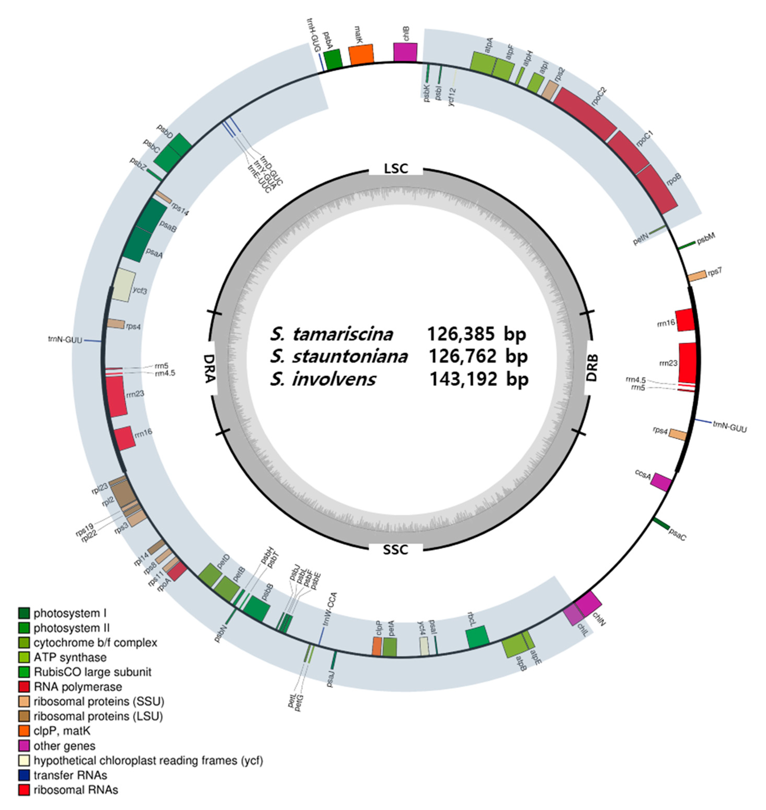
2.1. Selaginella Plastomes Contain Reduced LSCs and Unusual DR

In the current study, we sequenced, assembled, and annotated the plastomes of three Selaginella species, including S. tamariscina, S. stauntoniana, and S. involvens (Figure 1). We analyzed these assembled plastome sequences, along with the sequences of 13 other species in the Selaginella genus and three species from other orders (Isoetales and Lycopodiales) in the Lycopodiopsida, which were obtained from NCBI (Table 1). The plastome sizes of Selaginella species ranged from 110,411 to 147,148 bp, with an average of 132,571 ± 11,612, while the plastomes of non-Selaginella lycophytes ranged from 145,303 to 154,373 bp. Overall, Selaginella species had reduced LSC regions, with some species having LSCs that were almost half the size of those of non-Selaginella lycophytes. Most Selaginella species had longer SSCs than LSCs, except for the three species of Selaginella lepidophylla, Selaginella hainanensis, and S. uncinata (Table 1).
Most Selaginella species shared a unique plastome structure consisting of a set of direct repeats (DRs) instead of the inverted repeats (IRs) found in most plastomes. This DR structure was confirmed by PCR amplification using primer combinations designed based on the junction sites for the hypothetical DR and IR structures (Figure 2a). The primer combinations only differed in the sequence of the first repeat block (the inverted form of DRB in Figure 2a). PCR amplification and gel electrophoresis showed that only primer combinations for the direct repeat structure were amplified, suggesting that the direct repeat structure was present in the plastome (Figure 2b). We confirmed the unusual DR structure through the assembly of the S. tamariscina plastome using Oxford Nanopore Sequencing Technology and an assembly pipeline we established. The plastome was thereby assembled into two contigs of 61,029 and 71,944 bp in size (Figure 2c). These contigs covered the junction sites between the single-copy regions and the DR regions without inversions, confirming the direct repeat structure of the S. tamariscina plastome.

2.2. Selaginella Plastomes Contain Fewer Genes than Other Lycophyte Plastomes

The number of plastome genes differed significantly among lycophyte species, pointing to the frequent occurrence of gene loss events. In particular, the plastomes of Selaginella species generally had fewer protein-coding and transfer RNA (tRNA) genes than those of non-Selaginella species (Table 1). Within the genus, S. hainanensis had the highest number of plastome genes (103 genes), whereas S. tamariscina had the fewest (75 genes). Huperzia and Isoetes species contained almost all plastid genes. All 19 lycophyte species analyzed carried two copies of the four ribosomal RNA (rRNA) genes located in the repeat regions.
Figure 3 shows gene polymorphisms (presence/absence/pseudogenes) among the plastomes of the 19 lycophyte species. Isoetes and Huperzia species contained most plastid genes, except for a few that were lost or pseudogenized. In Selaginella species, many genes were absent or pseudogenized, specifically tRNA genes, ribosomal protein genes, and ndh genes. While non-Selaginella species had an average of 29.7 tRNA genes, Selaginella species averaged 11.6 tRNA genes, i.e., less than half the number in non-Selaginella species. The S. stauntoniana and S. tamariscina plastomes each contained only six tRNA genes. There were also many losses of protein-coding genes, such as the loss or pseudogenization of most ndh genes in S. lepidophylla, Selaginella lyallii, Selaginella indica, S. stauntoniana, S. tamariscina, Selaginella vardei, and Selaginella bisulcata, as well as losses of many ribosomal subunit genes throughout the genus. Other genes lost in Selaginella species included genes related to lipid biosynthesis (accD), translation initiation (infA), and other miscellaneous functions (ycf4, ycf66, cemA, and matK).

2.3. Phylogenetic Analysis Shows Two Main Lineages with Dynamic Structural Variations

Phylogenetic analysis using the BEAST software separated the 19 lycophyte species into three major groups, including one group for non-Selaginella species and two lineages within the Selaginella genus, all divided by two main divergence events (Figure 3). The first group (labeled 1 in Figure 3) consists of H. lucidula, H. serrata, and I. flaccida, which contain IRs. The Selaginella species were divided into two lineages. One lineage (labeled 2 in Figure 3) was divided into two subgroups, with one group containing S. lyallii, Selaginella kraussiana, and Selaginella remotifolia and the other containing S. indica, S. vardei, and S. lepidophylla. S. lepidophylla contains IRs, while the five other species contain DRs. Another lineage (labeled 2′ in Figure 3) contains 10 Selaginella species without apparent subgrouping. Among these 10 species, eight contain DRs, whereas S. hainanensis and S. uncinata contain IRs.
As a result of the plastome structure comparison of all 19 species, species within the same lineages were shown to share similar structures (Figure 4). In the first group, comprising non-Selaginella species, the plastomes had typical plastome structures, containing IRs. Selaginella species exhibited a different structure by a block inversion spanning from trnF in the LSC to trnN at the junction between IRB and the SSC. This inversion caused part of the LSC to be repositioned in the SSC region, resulting in the shortening of the LSC and the expansion of the SSC seen in Selaginella species. The block that took part in this inversion event contained one of the repeat regions, IRB, which ended up in the same orientation as the unaffected repeat region, thus creating a set of DRs in Selaginella species. The second inversion event occurred in both lineages independently (red diamonds in Figure 3). Because DRB was involved in these second inversions, DRB was converted back to IRB in species that underwent the second inversion event in each lineage, meaning S. lepidophylla in lineage 2 and S. hainanensis and S. uncinata in lineage 2′ (Figure 3).

2.4. Plastome Diversity among Five S. tamariscina Collections Reflects the Geographical Diversity

We explored the intraspecific diversity within five S. tamariscina individuals collected from different regions: Two from China and three from Korea. The plastome size of S. tamariscina ranged from 126,365 to 126,700 bp. We compared the five plastome sequences by aligning them in a pairwise fashion. We examined the number of single-nucleotide polymorphisms (SNPs) and insertions/deletions (InDels) to analyze the intraspecies diversity in the S. tamariscina collections. The collections from regions within Korea showed relatively little variation, harboring up to 1 SNP and 15 InDels. However, a comparison of SNP and InDel counts in collections from Korea vs. China revealed much more variation. Plastomes of Korean collections showed approximately 1218 SNPs when compared to those of two Chinese collections: China2018 and China2019. Furthermore, between the two Chinese collections [33,35], 1246 SNPs and 401 InDels were detected (Table 2). Among the variations, insertions and deletions were located within two genes related to chlorophyll biosynthesis (chlB and chlN) that induced frameshifts resulting in premature stop codons. While China2019 retained all three chl genes, the other four collections contained the remains of chlB and chlN as pseudogenes that are likely nonfunctional. A BEAST phylogenetic tree of five S. tamariscina individuals with S. stauntoniana, which is the closest relative, as the outgroup showed that SNU2014, SNU2018, Korea2020, and China2018 diverged from each other around 90 thousand years ago, but diverged from China2019 around 900 thousand years ago (Figure A1).

2.5. Selaginella Plastomes Exhibit Many Intron Losses and GC Bias

We identified 19 genes that contain introns (13 protein-coding genes and 6 tRNA genes) in the plastomes of the 19 lycophytes (Table 3). Of the protein-coding genes, two—clpP and ycf3—contained two introns and the other 17 contained a single intron between two exons. Huperzia and Isoetes species retained introns in most of these genes. However, the numbers of introns in the Selaginella plastomes was highly reduced due to either the deletion of intron-containing genes or the absence of introns in specific Selaginella species. For instance, rps12, ycf66, and rps16 were not present in Selaginella species, and tRNA genes containing introns in other lycophytes were either absent or intron-less in Selaginella. The clpP gene in Huperzia and Isoetes species contained two introns: clpP-1 and clpP-2. clpP-1 was lost in species in lineage 2, but was retained in most species in lineage 2′, except for S. stauntoniana and S. tamariscina. ycf3 also contained two introns: ycf3-1 and ycf3-2. While ycf3-1 was present in all lycophyte species analyzed, ycf3-2 was not present in the species of Selaginella lineage 2.
Analysis of the GC contents of the plastomes revealed that the Selaginella species had considerably higher GC ratios than non-Selaginella species (Table 1). The GC contents of Selaginella species ranged from 50.75% for S. lyallii to 56.49% for S. remotifolia, whereas those of non-Selaginella species were significantly lower (ranging from 36.25% to 37.94%). There was no correlation between the orientation of the repeats and the GC content. Overall, Selaginella species lost more intron sequences compared to non-Selaginella lycophytes, and the intron pattern was shared in each lineage.

2.6. RNA Editing Is Commonly Found in Selaginella Plastomes

The annotation of the three plastomes was hindered by the frequent absence of authentic ATG start codons for protein-coding genes, as many genes contained ACG nucleotides instead of ATGs at their start sites. Stop codons were also missing in some genes. After a final manual curation of the gene annotations, we determined that 76.8%, 77.2%, and 53.3% of protein-coding genes in S. tamariscina, S. stauntoniana, and S. involvens, respectively, started with ACG instead of ATG.
To validate our hypothesis about RNA editing in these sequences, we conducted a transcriptome analysis of the S. tamariscina plastome. We mapped raw RNA sequencing data from S. tamariscina onto the complete plastome sequence of the same species to determine whether the potential RNA editing sites were indeed edited in the mRNA sequences. We calculated the ratios of reads aligned to the start and stop codons in the unedited (C nucleotide) vs. edited (T nucleotide) forms using CLC find variations (ver. 4.3.0). In the S. tamariscina plastome sequence, 43 of the 56 genes contained potential editing sites for start codons, and four genes contained potential editing sites for stop codons (Figure 5). The coexistence of reads aligned to the same start and stop codons with either the edited or unedited codon indicated that these genes had undergone C-to-U RNA editing. Forty-two of the 43 potential editing sites were confirmed to have undergone C-to-U RNA editing. In contrast, there was a lower rate of potential RNA editing sites in stop codons, but three out of the four potentially edited sites were indeed edited. For start codons, most of the genes (except atpE, chlL, psbN, and rpoB) had a higher ratio of RNA-edited reads with the T nucleotide (switched from the U nucleotide during RNA sequencing) than unedited reads with the C nucleotide.
Overall, Selaginella species had a higher rate of abnormal start codons (ranging from 23.33% of the genes for S. lyallii to 77.19% in S. stauntoniana), while the Huperzia species had very low percentages (<10%), and I. flaccida was on the lower end of the range for Selaginella species (Figure 6). In regard to abnormal stop codons, Selaginella sanguinolenta had the highest percentage (41.79%), and the levels for most other Selaginella species were similar; the exceptions were S. lyallii, S. kraussiana, S. lepidophylla, S. stauntoniana, and S. tamariscina, with percentages < 10%. Some codons (marked as ‘others’) did not fall into either category, as they lacked authentic start or stop codons, but did not contain codons that were potential targets for RNA editing (Figure 6). A survey of start and stop codons in all 19 lycophyte plastid genes revealed a generally higher frequency of potential editing sites in start codons compared to stop codons (Figure 6). The percentages of potential RNA editing sites in start codons were 29.27%, 8.14%, and 4.71% for I. flaccida, H. lucidula, and H. serrata, respectively, and among the Selaginella, they ranged from 23.33% for S. lyallii to 77.19% for S. stauntoniana. Within the genus, S. stauntoniana and S. tamariscina had the highest frequency of potential RNA editing sites (77.19% and 76.79%, respectively). In stop codons, the proportions of potential RNA editing sites ranged from 4.71% to 5.81% for H. serrata and H. lucidula, respectively, while those for Selaginella species ranged from 0% for S. kraussiana to 41.79% for S. sanguinolenta (Figure 6).

3. Discussion

3.1. Unique Features of Selaginella Plastomes

Overall, our results support previous findings that the plastome sequences of Selaginella species were smaller than those of non-Selaginella and typical land plants [34]. As of February 2019, 2364 complete plastome sequences of land plants were registered in NCBI, with an average size of 151,167 ± 34,672 bp. In contrast, the average size of the 16 Selaginella plastomes we assessed was 132,571 ± 11,612 bp. Although the canonical quadripartite plastid genomic structure was retained in these Selaginella species, directional change occurred in most Selaginella species with SSC expansion and LSC contraction. The relative length of the SSC as a proportion of the total plastome ranged from 24.96% (S. moellendorffii) to 42.65% (S. involvens), averaging 35.73%. These values are higher than those in non-Selaginella species (12.73–18.72%) (Table 1), as well as those in eight selected monilophyte ferns (ranging from 11.7% in Psilotum nudum to 15.57% in Adiantum capillus-veneris, averaging 13.84%) [6]. The expansion of the SSC region in Selaginella species contrasts with the SSC contraction in Schizaeceae ferns, in which the SSC was reduced by as much as 2255 bp to contain only two genes in Schizaea elegans [9]. In the Geraniaceae family, the SSC has contracted in the genera Viviania, Hypseocharis, and Pelargonium, but expanded in the genera Melianthus, Francoa, and California [41]. The overall statistics revealed unique features of the Selaginella genus.

3.2. One Common and Two Independent Inversions Caused the Appearance of DR and IR Structures during Selaginella Species Evolution

Canonical plastomes are arranged in a quadripartite structure composed of an LSC and SSC region separated by two inverted repeats [37]. This is true for the plastomes of Isoetes and Huperzia. However, the Selaginella genus contains both inverted and direct repeats. A large inversion occurred just after the divergence of Selaginella from other lycophyte genera, which gave rise to DRB in the Selaginella plastomes. DRs have also been reported in S. tamariscina [33], S. vardei, and S. indica [36]. Zhang et al. compared I. flaccida with S. vardei, proposing that this inversion might have occurred between 142.5 and 281.5 mya during the Late Triassic [36], which is earlier than the 64.24 mya that was estimated in this study. Nevertheless, this inversion resulted in the distinctive DRs found in the plastomes of the Selaginella genus, except for the three species of S. lepidophylla, S. hainanensis, and S. uncinata. Further inversions involving DRB occurred independently in S. lepidophylla and in S. hainanensis and S. uncinata, in both cases restoring IRB and lengthened LSCs. S. lepidophylla split from its sister species S. indica and S. vardei approximately 25.64 mya. Therefore, the inversion in S. lepidophylla must not be older than 25.64 million years old. S. hainanensis and S. uncinata diverged from each other approximately 1.03 mya, and from their sister species S. bisulcata and S. pennata approximately 4.51 mya. Therefore, this inversion must have occurred between 4.51 and 1.03 mya. The grouping of Selaginella species is generally in agreement with the reported phylogeny of Selaginellaceae [20,21]. However, the estimated divergence times of Selaginella species are relatively younger than those from previous reports [35]. These differences could be due to the fact that divergence times estimated from previous studies were obtained by utilizing a few universal genes, such as rbcL [20], leading to possible underestimation or overestimation. Nonetheless, the inversion events were estimated to have occurred around 246 and 23 mya [35], which is similar to the time frame our results.
IRs are an interesting feature of circular plastomes. Although one missing IR has been reported in eudicots in Fabaceae [2,42] and in some species of Geraniaceae [41], directional changes rarely occur. Other than in Selaginella species, only one instance of DRs has been reported, in the red alga Porphyra purpurea [43]. IR and DR regions contain ribosomal RNA genes and a few tRNA genes, in duplicates. Cyanobacteria and other eubacteria have one copy. It is possible that in the ancestral plastid, these genes were present in duplicates in the same orientation, but were reversed in the plastids of extant species from algae to seed plants [44]. Aside from the high demand of ribosomal RNAs for efficient translation, the IR serves to stabilize the plastome, enabling mutations to be repaired by homologous recombination between repeats through homologous sequence synapsis [37]. The synapsis of two inverted repeats can lead to the formation of a dumbbell-shaped plastid structure, as confirmed in the plastids of the common bean [37]. Alternatively, pairing between DR sequences might impose structural constraints, rather than stabilizing the circular genome. Further studies are needed to confirm the circular plastome structures of Selaginella species.

3.3. Gene and Intron Losses

Along with dynamic structural rearrangement events, Selaginella species showed evidence of severe gene losses that resulted in their overall small plastomes. The most noticeable losses involved ndh genes, encoding for NADH dehydrogenase that function in electron transfer during photosynthesis. ndh genes were lost or pseudogenized in several Selaginella species. The ndh genes are also absent or pseudogenized in some species of green algae and almost all gymnosperms [39]. Rhulman et al. demonstrated that the loss of plastid ndh genes is compensated for by the nucleus-encoded ndh genes in gymnosperms [45], but there are no nuclear ndh genes in S. tamariscina [33]. Zhang et al. noted that Selaginella species adapted to dry environments have lost their ndh genes, suggesting that this gene loss might be related to their adaptation to these habitats [35]. The loss of ndh genes was recent, as revealed by the pseudogenization of all ndh genes in S. bisulcata, whereas its sister species S. pennata retained the complete set: These two species only diverged 3.61 mya (Figure 3). Moreover, homologous genes to the missing ribosomal protein genes in S. tamariscina were found in the nuclear genome [33]. Therefore, homologous genes in the nuclear genome may have replaced the functions of some of the genes missing from the plastomes of Selaginella species, which could also be true for other genes.
Sixty-one codons encode 20 amino acids, but only 37 plastid tRNAs have been recognized [39]. Selaginella might be the genus with the most reduced number of tRNA genes in eukaryotes, as we determined in the current study. To compensate for this apparent lack of tRNA genes, Wolf et al. have proposed that post-transcriptional editing alters anticodons to code for different tRNAs [46]. However, there is no evidence for RNA editing in tRNAs from S. uncinata [32]. The importation of tRNAs from the nucleus has been proposed for S. uncinata [30] and the non-photosynthetic parasitic angiosperm Epifagus virginiana [47]. Alternatively, the superwobbling theory has been put forward to explain the insufficient number of plastid tRNAs [48]. This “two out of three” mechanism could allow the reading of all four nucleotides in the third codon position so that a single tRNA gene could read the corresponding codons for one four-codon family [48,49]. We also support this superwobbling theory due to the highly insufficient number of tRNAs in Selaginella plastomes, given the lack of clear evidence for nucleus-derived tRNA species in the plastid.
Introns have been attracting increasing attention due to the various functions that they might have exerted during eukaryotic evolution [50]. The introns in plastid genes are self-splicing, with most being group II introns [51], except for the intron in trnL-UAA [52]. Nineteen plastome introns have been recognized, including 13 in protein-coding genes and 6 in tRNA genes in plastomes from green algae to seed plants [39]. Most of these introns are present in Isoetes and Huperzia species, as can be found in monilophyte ferns [6], but their numbers have been reduced by approximately half in Selaginella species. The deletion of intron-containing genes appears to account for the reduced number of introns in the Selaginella plastomes. For instance, rps12, ycf66, rps16, and many tRNA genes containing introns were not present in the Selaginella plastomes. Moreover, we observed a Selaginella genus-specific absence of clpP gene introns and the second intron of the ycf3 gene.

3.4. Intraspecies Diversity

There was a wide range of intraspecies diversity within S. tamariscina individuals collected in different regions of Korea and China. Unlike the little variation detected among Korean collections, Chinese collections were very different at the nucleotide level (Table 2). Considering that the rate of intraspecific variation is lower in the plastome than in nuclear DNA due to the typical uniparental mode of inheritance [53], the variation detected within S. tamariscina individuals was very high, such as a Chinese collection—China 2019—which showed a divergence time of approximately 0.9 MYA with other S. tamariscina collections in China and Korea (Figure A1). Intraspecific variation may reflect the adaptation of plants to changing environmental conditions [54,55], perhaps explaining the high level of intraspecific diversity within the S. tamariscina collections from different regions. The morphological classification of these small primitive plants may be difficult. Therefore, with the availability of additional characteristics derived from morphological keys and molecular data, species nomenclature could become more specific.

3.5. A High GC Content and Abundant RNA Editing

Unlike nuclear genomes, organellar genomes are AT biased, with an average GC content of 36.45 ± 2.81% and 36.87 ± 8.23% detected in 11,542 mitochondrial genomes and plastomes, respectively [39]. Therefore, the GC contents of >50% observed in Selaginella plastomes are higher than those of other eukaryotic organellar genomes. AT-mutation pressure or AT-biased gene conversion (or both) is thought to have driven most plastomes to become AT biased [56,57]. Based on the nucleotide compositions of the four-fold degenerate sites in protein-coding and noncoding regions in the S. uncinata and S. moellendorffii plastomes, Smith proposed that unbiased mutation/gene conversion equilibrated the nucleotide composition to ~50% GC contents in Selaginella plastomes. Alternatively, he suggested that RNA editing has influenced the GC contents in these plastomes, as numerous GC-rich codons were changed into AT-rich codons through RNA editing [31].
Selaginella plastomes are subject to extensive RNA editing. C-to-U RNA editing is a post-transcriptional modification mechanism that grants gene diversity. There are 30–50 in plant plastomes, but S. uncinata contains 3415 RNA editing sites [32]. According to our results, there are numerous potential RNA editing sites in Selaginella plastomes. The frequency of RNA editing at the start codons of plastid genes was >50% in some Selaginella species. Furthermore, S. tamariscina RNA sequencing reads confirmed these editing sites. This high number of editing sites coincides with the exceptionally high number of pentatricopeptide repeat (PPR) protein gene families (>800) in the S. moellendorffii nuclear genome, representing proteins essential for plastid RNA editing [17,58]. Hecht et al. identified 2139 C-to-U RNA editing sites in the mitochondrial genome of S. moellendorffii [59]. Grewe et al. detected 1782 RNA editing sites in the mitochondrial genome and identified U-to-C editing in Isoetes engelmannii [60], indicating that the organelle genomes of lycophytes contain many RNA editing sites. If all potential sites were checked in S. tamariscina plastomes, the number of total events observed could be even greater than that in S. uncinata [32].

4. Materials and Methods

4.1. Plant Materials and Publicly Available Plastome Sequences

One S. tamariscina (SNU2014) individual was collected from Baegunsan, Gwangyang-si, Jeollanam-do, Korea. A second S. tamariscina individual (SNU2018) was collected from Jeju Island. S. stauntoniana from Danyang-gun, Chungcheongbuk-do, Korea, and S. involvens from Jeju Island were provided by Hantaek Botanical Garden, Yongin, Gyeonggi-do, 17183, Republic of Korea.
The complete plastome sequences of 13 Selaginella species and three non-Selaginella lycophytes were obtained from NCBI (Table 1). To analyze the intraspecies diversity in S. tamariscina, two plastome sequences obtained in the current study (SNU2014 and SNU2018) were used, along with assembled and annotated sequences for another Korean collection reported by Park et al. collected from Wolsong-ri, Jijeong-myeon, Wonju-si, Korea [40] and two Chinese collections reported by Xu et al. and Zhang et al. (China2018 and China2019, respectively) [33,35].

4.2. DNA Extraction, Sequencing, Plastome Assembly, and Annotation

Leaves were collected from the plants and ground in liquid nitrogen with a mortar and pestle. A modified cetyltrimethylammonium bromide (CTAB) method [61] was used to extract total genomic DNA from S. tamariscina (SNU2014), S. stauntoniana, and S. involvens. The DNA quality and concentration were assessed by gel agarose electrophoresis and UV-spectrophotometry (NanoDrop ND-1000, Thermo Fisher Scientific, Waltham, MA, USA). The extracted DNA was sequenced on the Illumina NextSeq platform, generating 7.68, 2.2, and 2.0 Gb of raw data from S. tamariscina (SNU2014), S. stauntoniana, and S. involvens, respectively.
The plastomes of the three species were assembled using the de novo assembly of Low-Coverage Whole genome sequence (dnaLCW) approach with the CLC genome assembler program (ver. 4.6 beta, CLC Inc., Aarhus, Denmark) [62,63]. In short, raw paired-end reads were trimmed with an offset value of 33, and trimmed reads were assembled with overlap distances set to 150 to 500 bp and the window size set to 32. Initial contigs were extracted from the assembled reads using MUMmer [64], with the reference sequence of a related species as a guide.
For the second S. tamariscina individual (SNU2018), total genomic DNA was extracted using an Exgene Plant SV midi kit (GeneAll Biotechnology, Seoul, Korea). The DNA quality and concentration were assessed via agarose gel electrophoresis and UV-spectrophotometry. The extracted DNA was sequenced on a MinION sequencer from Oxford Nanopore Technologies. Library preparation was performed with a Ligation Sequencing Kit (SQK-LSK108) following the manufacturer’s instructions. One flow cell was run for 48 h. In total, 10 Gb of raw sequencing reads was filtered by removing adaptor sequences using Porechop (v0.2.0, https://github.com/rrwick/Porechop) and corrected with Canu using default parameters [65]. The Canu-corrected reads were assembled using SMARTdenovo (https://github.com/ruanjue/smartdenovo). The complete plastome sequence assembled using Illumina sequencing data was used as a reference to extract plastome-related contigs assembled by Nanopore sequencing using BLASTN.
The plastomes assembled in the previous steps were annotated using GeSeq (https://chlorobox.mpimp-golm.mpg.de/geseq.html) with complete plastome references of other species in the Selaginella genus and non-Selaginella species registered in NCBI. The annotated sequences were manually curated for details such as start and stop codons and correct coding frames using Artemis [66] For further validation of annotation, complete CDS sequences of reference sequences were collected and converted into amino acid sequences, and searched in the three assembled sequences using TBLASTN (https://blast.ncbi.nlm.nih.gov/Blast.cgi?PROGRAM=tblastn&PAGE_TYPE=BlastSearch&LINK_LOC=blasthome) for sequence homology. Furthermore, the aligned sequence was searched in the Conserved domain search provided by NCBI for complete gene structures. Genes that have sequence homology detected by TBLASTN but have incomplete gene structures due to the lack of sequences of internal stop codons were assigned as pseudogenes. Genes that were not searched at all through TBLASTN were marked as missing genes. This validation step for the pseudogene/missing gene assignment was conducted for other already published sequences used in this study.

4.3. Confirmation of Direct Repeat Structures by PCR Amplification

To confirm the direct repeat structure of S. tamariscina, primers were designed based on the junction regions between the repeat regions and the two single copies (LSC and SSC). Primers were used in different combinations to amplify the junction sites of the plastome. Hypothetical direct repeat junctions were amplified by primer combinations 1/2, 3/4, 5/2, and 3/6, while hypothetical inverted repeat junctions were amplified by different primer combinations of 1/3 and 2/4 for the first repeat block (IRB) (Table A1).
PCR amplification was performed in a 25 µL volume containing 1U Taq polymerase, 2.5 µL of 10× reaction buffer, 0.2 mM dNTPs (Inclone Biotech, Yongin, Korea), 10 ng genomic DNA, and 10 pmol of each primer. The PCR was carried out in a thermocycler with the following parameters: 5 min at 94 °C; 28 cycles of 20 s at 94 °C, 20 s at 62 °C, and 20 s at 72 °C; and 7 min at 72 °C for the final extension step. The PCR products were visualized by electrophoresis using a 1% agarose gel with a 100 bp ladder.

4.4. Gene Contents and Phylogenetic Analysis

CDS, tRNA, and rRNA sequences for all 19 species of interest were extracted using FeatureExtract 1.2 L (http://www.cbs.dtu.dk/services/FeatureExtract/). Existing genes and pseudogenes were marked. Among the extracted CDS, 45 genes that were common to all 19 species were combined into one long fasta sequence for each species. These sequences were aligned using MAFFT (https://mafft.cbrc.jp/alignment/server/). The aligned sequences were trimmed using Gblocks [67] so that conserved blocks from the aligned sequences could be extracted for more accurate phylogenetic analysis. The aligned file was used for phylogenetic analysis performed using BEAST version 2.5.2. The general Time-Reversible (GTR) substitution model and a gamma category count of 4 were used for the Gamma Site Model. A random local clock and Yule tree prior were applied. A normal distribution root calibration of 375 mya representing the split between Isoetes and Selaginella families reported by Wikstrom and Kenrick [68] was used. The chain length was set to 1 million generations with a sampling frequency of 1000. The BEAST analysis was repeated four times, and the results were checked for the effective sample size (ESS) via Tracer v1.7.1. Results from all four repetitions were combined with LogCombiner implemented in the BEAST software. Moreover, a maximum clade credibility tree was drawn utilizing TreeAnnotator with a burnin percentage of 15. The final tree was visualized with FigTree v1.4.4, marking the height 95% HPD with blue node bars and node median values near the node bars.

4.5. Analysis of Intron Variation and RNA Editing Sites

Total cellular RNAs were prepared from hydrated and 50% dehydrated leaf tissues with the RNeasy Plant Mini Kit (Qiagen, Hilden, Germany) by following the manufacturer’s instructions. The RNA quality and concentration were assessed using the Agilent Bioanalyzer (Agilent Technology, Santa Clara, CA, USA) and Nanodrop spectrophotometer (Thermo Fisher Scientific, Waltham, MA, USA) with parameters RIN ≥ 7, 28S:18S > 1, and OD260/280 ≥ 2.
For RNA sequencing, the cDNA libraries were constructed using TruSeq Stranded mRNA (RS-122-2101, Illumina, USA) and samples were processed with next-generation sequencing (NGS) procedures on an Illumina Hiseq3000 with a paired-end method. The NGS reads were filtered by the threshold of >20% ‘N’ bases, ≤Q20 in Phred quality, and a length > 50 bp. De novo assembly was performed using TRINITY (https://github.com/trinityrnaseq; [69]) and CD-HIT (http://weizhongli-lab.org/cd-hit/; [70]).
Genes in the plastomes of all 19 species were surveyed to check for the presence of intron regions. Potential C-to-U RNA editing sites were surveyed in the start and stop codons of the CDS from the plastomes of ten lycophyte species. Start codons with an ACG sequence instead of ATG and stop codons with CAA, CAG, and CGA sequences instead of TAA, TAG, and TGA were counted as potential RNA editing sites. Potential RNA editing sites were marked for all ten species and analyzed.
To determine whether the potential RNA editing sites had gone through the editing process, raw RNA sequencing data from S. tamariscina_2 were mapped onto the reference genome produced from the assembly of S. tamariscina (SNU2014) sequencing data. The start and stop codon positions of the CDS were obtained, and CLC find variations software (ver. 4.3.0) was used to examine the distribution of unedited mRNA reads with the C nucleotide and edited mRNA reads with the T nucleotide (previously U in RNA that changed to T during the process of RNA sequencing).

Author Contributions

Conceptualization, H.S., T.-J.Y., and N.-S.K.; methodology, H.J.L. and H.S.; validation, H.S., J.L., and T.-J.Y.; formal analysis, H.S.; investigation, H.J.L.; resources, J.-H.K.; data curation, H.S., J.L., and H.-O.L.; writing—original draft preparation, H.S. and N.-S.K.; writing—review and editing, H.S., N.-S.K., and T.-J.Y.; supervision, N.-S.K. and T.-J.Y.; project administration, N.-S.K. and T.-J.Y. All authors have read and agreed to the published version of the manuscript.

Funding

This work was carried out with the support of “Cooperative Research Program for Agriculture Science & Technology Development (Project No. PJ013238)” Rural Development Administration, Republic of Korea. This research was supported by the Bio & Medical Technology Development Program of the NRF funded by the Korean government, MSIP (NRF-2015M3A9A5030733). Part of this work was supported by the Kangwon National University Professor Grant (Grant ID 520170398) to NSK.

Institutional Review Board Statement

Not applicable.

Informed Consent Statement

Not applicable.

Data Availability Statement

The data presented in this study are openly available in the National Center for Biotechnology Information under the accession numbers presented in Table 1.

Conflicts of Interest

The authors declare no conflict of interest.

Abbreviations

IRInverted repeat
DRDirect repeat
SCSingle-copy
LSCLong single-copy
SSCShort single-copy
NCBINational Center for Biotechnology Information
WGSWhole genome sequencing
PCRPolymerase Chain Reaction
SNPSingle Nucleotide Polymorphisms
InDelsInsertions/deletions
myaMillion years ago
CTABCetyltrimethylammonium bromide
dnaLCWDe novo assembly of Low-Coverage Whole genome sequence
CDSCoding sequence
tRNATransfer RNA
rRNARibosomal RNA
HPDHighest Posterior Density
GTRGeneral Time-Reversible

Appendix A

Figure A1. Phylogenetic tree of five Selaginella tamariscina plastid genome sequences with Selaginella stauntoniana as the outgroup. Phylogenetic analysis was performed with protein coding sequences that all six plastome sequences share with BEAST v2.5.2 software. The HYK substitution model and a gamma category count of 4 were used in the Gamma Site Model. For this analysis, a strict clock with a clock rate of 1, as well as the coalescent constant population tree prior, were applied. 2.66 mya, representing the split between S. tamariscina and S. stauntoniana derived from the phylogenetic analysis in this study (Figure 3), was used as the secondary calibration value with a normal distribution. The five S. tamariscina species were grouped into one monophyletic group, and the chain length was set to 1 M, sampling every 1000 chains. All other values were left at default parameters. Results were checked for the effective sample size (ESS) via Tracer v1.7.1, and the maximum clade credibility tree was drawn by TreeAnnotator with a burnin of 15%. Visualization of the final tree was conducted with FigTree v1.4.4. Blue bars show node bars representing the height 95% HPD, and numbers near the nodes show the median values.
Ijms 22 00641 g0a1
Table A1. Primer sequences used in combination to confirm the direct repeat structure of the Selaginella tamariscina plastid genome.
Primer NamePrimer Sequence
Primer 1AGCCGACATTCGTTACGGTT
Primer 2GGATTCGCATTACCGACAGTG
Primer 3GCATTTAATCGGCGCGAAGT
Primer 4CACCACTCCCCTTGAACCTC
Primer 5TATGCGCCGCGATTAGGC
Primer 6GTCGGTTCAAAACCCGTGC

References

  1. Palmer, J.D. Comparative organization of chloroplast genomes. Annu. Rev. Genet. 1985, 19, 325–354. [Google Scholar] [CrossRef]
  2. Ruhlman, T.A.; Jansen, R.K. The Plastid Genomes of Flowering Plants. In Computational Biology; Springer: Berlin/Heidelberg, Germany, 2014; pp. 3–38. [Google Scholar]
  3. Daniell, H.; Lin, C.-S.; Yu, M.; Chang, W.-J. Chloroplast genomes: Diversity, evolution, and applications in genetic engineering. Genome Biol. 2016, 17, 1–29. [Google Scholar] [CrossRef] [PubMed] [Green Version]
  4. Wakasugi, T.; Tsudzuki, J.; Ito, S.; Nakashima, K.; Sugiura, M.; Tsudzuki, T. Loss of all ndh genes as determined by sequencing the entire chloroplast genome of the black pine Pinus thunbergii. Proc. Natl. Acad. Sci. USA 1994, 91, 9794–9798. [Google Scholar] [CrossRef] [PubMed] [Green Version]
  5. Wu, C.-S.; Wang, Y.-N.; Hsu, C.-Y.; Lin, C.-P.; Chaw, S.-M. Loss of Different Inverted Repeat Copies from the Chloroplast Genomes of Pinaceae and Cupressophytes and Influence of Heterotachy on the Evaluation of Gymnosperm Phylogeny. Genome Biol. Evol. 2011, 3, 1284–1295. [Google Scholar] [CrossRef] [PubMed] [Green Version]
  6. Grewe, F.; Guo, W.; Gubbels, E.A.; Hansen, A.K.; Mower, J.P. Complete plastid genomes from Ophioglossum californicum, Psilotum nudum, and Equisetum hyemale reveal an ancestral land plant genome structure and resolve the position of Equisetales among monilophytes. BMC Evol. Biol. 2013, 13, 8. [Google Scholar] [CrossRef] [Green Version]
  7. Palmer, J.D.; Osorio, B.; Aldrich, J.; Thompson, W.F. Chloroplast DNA evolution among legumes: Loss of a large inverted repeat occurred prior to other sequence rearrangements. Curr. Genet. 1987, 11, 275–286. [Google Scholar] [CrossRef] [Green Version]
  8. Wolf, P.G.; Der, J.P.; Duffy, A.M.; Davidson, J.B.; Grusz, A.L.; Pryer, K.M. The evolution of chloroplast genes and genomes in ferns. Plant Mol. Biol. 2011, 76, 251–261. [Google Scholar] [CrossRef] [Green Version]
  9. Labiak, P.H.; Karol, K.G.K.G. Plastome sequences of an ancient fern lineage reveal remarkable changes in gene content and architecture. Am. J. Bot. 2017, 104, 1008–1018. [Google Scholar] [CrossRef]
  10. Cronn, R.C.; Liston, A.; Parks, M.; Gernandt, D.S.; Shen, R.; Mockler, T. Multiplex sequencing of plant chloroplast genomes using Solexa sequencing-by-synthesis technology. Nucleic Acids Res. 2008, 36, e122. [Google Scholar] [CrossRef] [Green Version]
  11. McCoy, S.R.; Kuehl, J.V.; Boore, J.L.; Raubeson, L.A. The complete plastid genome sequence of Welwitschia mirabilis: An unusually compact plastome with accelerated divergence rates. BMC Evol. Biol. 2008, 8, 130. [Google Scholar] [CrossRef] [Green Version]
  12. Wicke, S.; Müller, K.F.; De Pamphilis, C.W.; Quandt, D.; Wickett, N.J.; Zhang, Y.; Renner, S.S.; Schneeweiss, G.M. Mechanisms of Functional and Physical Genome Reduction in Photosynthetic and Nonphotosynthetic Parasitic Plants of the Broomrape Family. Plant Cell 2013, 25, 3711–3725. [Google Scholar] [CrossRef] [Green Version]
  13. Yang, J.-B.; Tang, M.; Li, H.; Zhang, Z.; Li, D.Z. Complete chloroplast genome of the genus Cymbidium: Lights into the species identification, phylogenetic implications and population genetic analyses. BMC Evol. Biol. 2013, 13, 84. [Google Scholar] [CrossRef] [PubMed] [Green Version]
  14. Luo, J.; Hou, B.-W.; Niu, Z.-T.; Liu, W.; Xue, Q.-Y.; Ding, X.-Y. Comparative Chloroplast Genomes of Photosynthetic Orchids: Insights into Evolution of the Orchidaceae and Development of Molecular Markers for Phylogenetic Applications. PLoS ONE 2014, 9, e99016. [Google Scholar] [CrossRef] [PubMed]
  15. Wicke, S.; Schäferhoff, B.; Depamphilis, C.W.; Müller, K.F. Disproportional Plastome-Wide Increase of Substitution Rates and Relaxed Purifying Selection in Genes of Carnivorous Lentibulariaceae. Mol. Biol. Evol. 2014, 31, 529–545. [Google Scholar] [CrossRef] [Green Version]
  16. Shmakov, A. A community-derived classification for extant lycophytes and ferns. J. Syst. Evol. 2016, 54, 563–603. [Google Scholar] [CrossRef]
  17. Banks, J.A.; Nishiyama, T.; Hasebe, M.; Bowman, J.L.; Gribskov, M.; Depamphilis, C.W.; Albert, V.A.; Aono, N.; Aoyama, T.; Ambrose, B.A.; et al. The Selaginella Genome Identifies Genetic Changes Associated with the Evolution of Vascular Plants. Sci. 2011, 332, 960–963. [Google Scholar] [CrossRef] [Green Version]
  18. Kenrick, P.; Crane, P.R. The origin and early evolution of plants on land. Nat. Cell Biol. 1997, 389, 33–39. [Google Scholar] [CrossRef]
  19. Zhou, X.-M.; Zhang, L. A classification of Selaginella (Selaginellaceae) based on molecular (chloroplast and nuclear), macromorphological, and spore features. Taxon 2015, 64, 1117–1140. [Google Scholar] [CrossRef]
  20. Weststrand, S.; Korall, P. Phylogeny of Selaginellaceae: There is value in morphology after all! Am. J. Bot. 2016, 103, 2136–2159. [Google Scholar] [CrossRef] [Green Version]
  21. Weststrand, S.; Korall, P. A subgeneric classification of Selaginella (Selaginellaceae). Am. J. Bot. 2016, 103, 2160–2169. [Google Scholar] [CrossRef] [Green Version]
  22. Karol, K.G.; Arumuganathan, K.; Boore, J.L.; Duffy, A.M.; Everett, K.D.; Hall, J.D.; Hansen, S.K.; Kuehl, J.V.; Mandoli, D.F.; Mishler, B.D.; et al. Complete plastome sequences of Equisetum arvense and Isoetes flaccida: Implications for phylogeny and plastid genome evolution of early land plant lineages. BMC Evol. Biol. 2010, 10, 321. [Google Scholar] [CrossRef] [PubMed] [Green Version]
  23. Wolf, P.G.; Roper, J.M.; Duffy, A.M. The evolution of chloroplast genome structure in ferns. Genome 2010, 53, 731–738. [Google Scholar] [CrossRef] [PubMed] [Green Version]
  24. Zhong, B.; Fong, R.; Collins, L.J.; McLenachan, P.A.; Penny, D. Two New Fern Chloroplasts and Decelerated Evolution Linked to the Long Generation Time in Tree Ferns. Genome Biol. Evol. 2014, 6, 1166–1173. [Google Scholar] [CrossRef] [PubMed] [Green Version]
  25. Jansen, R.K.; Cai, Z.; Raubeson, L.A.; Daniell, H.; Depamphilis, C.W.; Leebens-Mack, J.; Müller, K.F.; Guisinger-Bellian, M.; Haberle, R.C.; Hansen, A.K.; et al. Analysis of 81 genes from 64 plastid genomes resolves relationships in angiosperms and identifies genome-scale evolutionary patterns. Proc. Natl. Acad. Sci. USA 2007, 104, 19369–19374. [Google Scholar] [CrossRef] [Green Version]
  26. Turmel, M.; Otis, C.; Lemieux, C. Dynamic Evolution of the Chloroplast Genome in the Green Algal Classes Pedinophyceae and Trebouxiophyceae. Genome Biol. Evol. 2015, 7, 2062–2082. [Google Scholar] [CrossRef] [Green Version]
  27. Wolf, P.G.; Karol, K.G.; Mandoli, D.F.; Kuehl, J.V.; Arumuganathan, K.; Ellis, M.W.; Mishler, B.D.; Kelch, D.G.; Olmstead, R.G.; Boore, J.L. The first complete chloroplast genome sequence of a lycophyte, Huperzia lucidula (Lycopodiaceae). Gene 2005, 350, 117–128. [Google Scholar] [CrossRef] [Green Version]
  28. Guo, Z.-Y.; Zhang, H.-R.; Shrestha, N.; Zhang, X.-C. Complete Chloroplast Genome of a Valuable Medicinal Plant, Huperzia serrata (Lycopodiaceae), and Comparison with Its Congener. Appl. Plant Sci. 2016, 4, 1600071. [Google Scholar] [CrossRef]
  29. Zhang, H.-R.; Kang, J.-S.; Viane, R.L.L.; Zhang, X.-C. The complete chloroplast genome sequence of Huperzia javanica (sw.) C. Y. Yang in Lycopodiaceae. Mitochondrial DNA Part B 2017, 2, 216–218. [Google Scholar] [CrossRef] [Green Version]
  30. Tsuji, S.; Ueda, K.; Nishiyama, T.; Hasebe, M.; Yoshikawa, S.; Konagaya, A.; Nishiuchi, T.; Yamaguchi, K. The chloroplast genome from a lycophyte (microphyllophyte), Selaginella uncinata, has a unique inversion, transpositions and many gene losses. J. Plant Res. 2007, 120, 281–290. [Google Scholar] [CrossRef]
  31. Smith, D.R. Unparalleled GC content in the plastid DNA of Selaginella. Plant Mol. Biol. 2009, 71, 627–639. [Google Scholar] [CrossRef]
  32. Oldenkott, B.; Yamaguchi, K.; Tsuji-Tsukinoki, S.; Knie, N.; Knoop, V. Chloroplast RNA editing going extreme: More than 3400 events of C-to-U editing in the chloroplast transcriptome of the lycophyte Selaginella uncinata. RNA 2014, 20, 1499–1506. [Google Scholar] [CrossRef] [PubMed] [Green Version]
  33. Xu, Z.; Xin, T.; Bartels, D.; Li, Y.; Gu, W.; Yao, H.; Liu, S.; Yu, H.; Pu, X.; Zhou, J.G.; et al. Genome Analysis of the Ancient Tracheophyte Selaginella tamariscina Reveals Evolutionary Features Relevant to the Acquisition of Desiccation Tolerance. Mol. Plant 2018, 11, 983–994. [Google Scholar] [CrossRef] [PubMed] [Green Version]
  34. Mower, J.P.; Ma, P.F.; Grewe, F.; Taylor, A.; Michael, T.P.; VanBuren, R.; Qiu, Y.L. Lycophyte plastid genomics: Extreme variation in GC, gene and intron content and multiple inversions between a direct and inverted orientation of the rRNA repeat. New Phytol. 2019, 222, 1061–1075. [Google Scholar] [CrossRef] [PubMed] [Green Version]
  35. Zhang, H.-R.; Qiao-Ping, X.; Zhang, X.-C. The Unique Evolutionary Trajectory and Dynamic Conformations of DR and IR/DR-Coexisting Plastomes of the Early Vascular Plant Selaginellaceae (Lycophyte). Genome Biol. Evol. 2019, 11, 1258–1274. [Google Scholar] [CrossRef]
  36. Zhang, H.-R.; Zhang, X.-C.; Xiang, Q.-P. Directed repeats co-occur with few short-dispersed repeats in plastid genome of a Spikemoss, Selaginella vardei (Selaginellaceae, Lycopodiopsida). BMC Genom. 2019, 20, 1–11. [Google Scholar]
  37. Palmer, J.D. Chloroplast DNA exists in two orientations. Nat. Cell Biol. 1983, 301, 92–93. [Google Scholar] [CrossRef]
  38. Wicke, S.; Schneeweiss, G.M.; Depamphilis, C.W.; Müller, K.F.; Quandt, D. The evolution of the plastid chromosome in land plants: Gene content, gene order, gene function. Plant Mol. Biol. 2011, 76, 273–297. [Google Scholar] [CrossRef] [Green Version]
  39. Kwon, E.-C.; Kim, J.-H.; Kim, N.-S. Comprehensive genomic analyses with 115 plastomes from algae to seed plants: Structure, gene contents, GC contents, and introns. Genes Genom. 2020, 42, 553–570. [Google Scholar] [CrossRef]
  40. Park, J.; Kim, Y.; Lee, G.-H.; Park, C.-H. The complete chloroplast genome of Selaginella tamariscina (Beauv.) Spring (Selaginellaceae) isolated in Korea. Mitochondrial DNA Part B 2020, 5, 1654–1656. [Google Scholar] [CrossRef] [Green Version]
  41. Weng, M.-L.; Blazier, J.C.; Govindu, M.; Jansen, R.K. Reconstruction of the Ancestral Plastid Genome in Geraniaceae Reveals a Correlation between Genome Rearrangements, Repeats, and Nucleotide Substitution Rates. Mol. Biol. Evol. 2014, 31, 645–659. [Google Scholar] [CrossRef] [Green Version]
  42. Palmer, J.D.; Thompson, W.F. Chloroplast DNA rearrangements are more frequent when a large inverted repeat sequence is lost. Cell 1982, 29, 537–550. [Google Scholar] [CrossRef]
  43. Reith, M.; Munholland, J. The ribosomal RNA repeats are non-identical and directly oriented in the chloroplast genome of the red alga Porphyra purpurea. Curr. Genet. 1993, 24, 443–450. [Google Scholar] [CrossRef] [PubMed]
  44. Douglas, S.E. Chloroplast origins and evolution. In The Molecular Biology of Cyanobacteria; Springer: Berlin/Heidelberg, Germany, 1994; pp. 91–118. [Google Scholar]
  45. Ruhlman, T.; Chang, W.-J.; Chen, J.J.; Huang, Y.-T.; Chan, M.-T.; Zhang, J.; Liao, D.-C.; Blazier, J.C.; Jin, X.-H.; Shih, M.-C.; et al. NDH expression marks major transitions in plant evolution and reveals coordinate intracellular gene loss. BMC Plant Biol. 2015, 15, 100. [Google Scholar] [CrossRef] [PubMed] [Green Version]
  46. Wolf, P.G.; Rowe, C.A.; Sinclair, R.B.; Hasebe, M. Complete Nucleotide Sequence of the Chloroplast Genome from a Leptosporangiate Fern, Adiantum capillus-veneris L. DNA Res. 2003, 10, 59–65. [Google Scholar] [CrossRef] [Green Version]
  47. Morden, C.W.; Wolfe, K.; DePamphilis, C.; Palmer, J. Plastid translation and transcription genes in a non-photosynthetic plant: Intact, missing and pseudo genes. EMBO J. 1991, 10, 3281–3288. [Google Scholar] [CrossRef]
  48. Rogalski, M.; Karcher, D.; Bock, R. Superwobbling facilitates translation with reduced tRNA sets. Nat. Struct. Mol. Biol. 2008, 15, 192–198. [Google Scholar] [CrossRef]
  49. Pfitzinger, H.; Weil, J.; Pillay, D.; Guillemaut, P. Codon recognition mechanisms in plant chloroplasts. Plant Mol. Biol. 1990, 14, 805–814. [Google Scholar] [CrossRef]
  50. Chorev, M.; Carmel, L. The Function of Introns. Front. Genet. 2012, 3, 55. [Google Scholar] [CrossRef] [Green Version]
  51. Dabbagh, N.; Bennett, M.S.; Triemer, R.E.; Preisfeld, A. Chloroplast genome expansion by intron multiplication in the basal psychrophilic euglenoid Eutreptiella pomquetensis. PeerJ 2017, 5, e3725. [Google Scholar] [CrossRef] [Green Version]
  52. Vogel, J.; Börner, T.; Hess, W.R. Comparative analysis of splicing of the complete set of chloroplast group II introns in three higher plant mutants. Nucleic Acids Res. 1999, 27, 3866–3874. [Google Scholar] [CrossRef] [Green Version]
  53. Zhang, R.; Hipp, A.L.; Gailing, O. Sharing of chloroplast haplotypes among red oak species suggests interspecific gene flow between neighboring populations. Botany 2015, 93, 691–700. [Google Scholar] [CrossRef] [Green Version]
  54. Norberg, J.; Swaney, D.P.; Dushoff, J.; Lin, J.; Casagrandi, R.; Levin, S.A. Phenotypic diversity and ecosystem functioning in changing environments: A theoretical framework. Proc. Natl. Acad. Sci. USA 2001, 98, 11376–11381. [Google Scholar] [CrossRef] [PubMed] [Green Version]
  55. Björklund, M.; Ranta, E.; Kaitala, V.; Bach, L.A.; Lundberg, P.; Stenseth, N.C. Quantitative Trait Evolution and Environmental Change. PLoS ONE 2009, 4, e4521. [Google Scholar] [CrossRef] [PubMed]
  56. Kusumi, J.; Tachida, H. Compositional Properties of Green-Plant Plastid Genomes. J. Mol. Evol. 2005, 60, 417–425. [Google Scholar] [CrossRef] [PubMed]
  57. Khakhlova, O.; Bock, R. Elimination of deleterious mutations in plastid genomes by gene conversion. Plant J. 2006, 46, 85–94. [Google Scholar] [CrossRef] [PubMed]
  58. Kotera, E.; Tasaka, M.; Shikanai, T. A pentatricopeptide repeat protein is essential for RNA editing in chloroplasts. Nature 2005, 433, 326–330. [Google Scholar] [CrossRef]
  59. Hecht, J.; Grewe, F.; Knoop, V. Extreme RNA Editing in Coding Islands and Abundant Microsatellites in Repeat Sequences of Selaginella moellendorffii Mitochondria: The Root of Frequent Plant mtDNA Recombination in Early Tracheophytes. Genome Biol. Evol. 2011, 3, 344–358. [Google Scholar] [CrossRef] [Green Version]
  60. Grewe, F.; Herres, S.; Viehöver, P.; Polsakiewicz, M.; Weisshaar, B.; Knoop, V. A unique transcriptome: 1782 positions of RNA editing alter 1406 codon identities in mitochondrial mRNAs of the lycophyte Isoetes engelmannii. Nucleic Acids Res. 2011, 39, 2890–2902. [Google Scholar] [CrossRef]
  61. Allen, G.C.; Flores-Vergara, M.A.; Krasynanski, S.; Kumar, S.; Thompson, W.F. A modified protocol for rapid DNA isolation from plant tissues using cetyltrimethylammonium bromide. Nat. Protoc. 2006, 1, 2320–2325. [Google Scholar] [CrossRef]
  62. Kim, K.; Lee, S.-C.; Lee, J.; Lee, H.O.; Joh, H.J.; Kim, N.-H.; Park, H.-S.; Yang, T.-J. Comprehensive Survey of Genetic Diversity in Chloroplast Genomes and 45S nrDNAs within Panax ginseng Species. PLoS ONE 2015, 10, e0117159. [Google Scholar] [CrossRef]
  63. Kim, K.; Lee, S.-C.; Lee, J.; Yu, Y.; Yang, K.; Choi, B.-S.; Koh, H.-J.; Waminal, N.E.; Choi, H.-I.; Kim, N.-H.; et al. Complete chloroplast and ribosomal sequences for 30 accessions elucidate evolution of Oryza AA genome species. Sci. Rep. 2015, 5, 15655. [Google Scholar] [CrossRef] [PubMed] [Green Version]
  64. Kurtz, S.; Phillippy, A.M.; Delcher, A.L.; Smoot, M.; Shumway, M.; Antonescu, C.; Salzberg, S.L. Versatile and open software for comparing large genomes. Genome Biol. 2004, 5, R12. [Google Scholar] [CrossRef] [PubMed] [Green Version]
  65. Koren, S.; Walenz, B.P.; Berlin, K.; Miller, J.R.; Bergman, N.H.; Phillippy, A.M. Canu: Scalable and accurate long-read assembly via adaptive k-mer weighting and repeat separation. Genome Res. 2017, 27, 722–736. [Google Scholar] [CrossRef] [PubMed] [Green Version]
  66. Rutherford, K.; Parkhill, J.; Crook, J.; Horsnell, T.; Rice, P.; Rajandream, M.-A.; Barrell, B. Artemis: Sequence visualization and annotation. Bioinformatics 2000, 16, 944–945. [Google Scholar] [CrossRef] [Green Version]
  67. Castresana, J. Selection of Conserved Blocks from Multiple Alignments for Their Use in Phylogenetic Analysis. Mol. Biol. Evol. 2000, 17, 540–552. [Google Scholar] [CrossRef] [Green Version]
  68. Wikström, N.; Kenrick, P. Evolution of Lycopodiaceae (Lycopsida): Estimating Divergence Times from rbcL Gene Sequences by Use of Nonparametric Rate Smoothing. Mol. Phylogenetics Evol. 2001, 19, 177–186. [Google Scholar] [CrossRef]
  69. Grabherr, M.G.; Haas, B.J.; Yassour, M.; Levin, J.Z.; Thompson, D.A.; Amit, I.; Adiconis, X.; Fan, L.; Raychowdhury, R.; Zeng, Q.; et al. Full-length transcriptome assembly from RNA-Seq data without a reference genome. Nat. Biotechnol. 2011, 29, 644–652. [Google Scholar] [CrossRef] [Green Version]
  70. Fu, L.; Niu, B.; Zhu, Z.; Wu, S.; Li, W. CD-HIT: Accelerated for clustering the next-generation sequencing data. Bioinformatics 2012, 28, 3150–3152. [Google Scholar] [CrossRef]
Figure 1. Map of complete plastid genomes of the three Selaginella species sequenced and assembled in this study. Shaded areas indicate regions involved in the inversion event.
Ijms 22 00641 g001
Figure 2. Validation of the direct repeat structures of Selaginella tamariscina via PCR and long-read sequencing. (a) Primers 1–6 were designed based on the junction regions between the single copies and repeat regions (the primer sequences are listed in Appendix A Table A1). The primers were used in various combinations to amplify hypothetical direct repeat structures and indirect repeat structures. (b) 1% agarose gel electrophoresis of the PCR products. (c) Confirmation of the direct repeat structure in assembled contigs spanning single-copy regions and the repeat region using Oxford Nanopore Sequencing technology.
Ijms 22 00641 g002
Figure 3. Phylogenetic analysis of the 19 lycophyte species examined in this study. The coding sequence (CDS) of 45 shared genes were used in the phylogenetic analysis with BEAST software. The calibration node was set to 375 mya for the split of Isoetes and Selaginella species. Left, phylogenetic tree. The gray box labeled 1 highlights non-Selaginella species. Lineages 2 and 2′ in the Selaginella genus are highlighted with yellow and green boxes, respectively. The red diamond labeled 1 represents the first inversion event, which differentiates non-Selaginella species from Selaginella species. Red diamonds labeled 2 and 2′ represent the second inversion events, which occurred independently in each lineage and converted the direct repeat structures back into inverted repeats. Blue bars on the branch of the phylogenetic tree indicate height 95% highest posterior density (HPD) and numbers near the nodes represent median values. Right, corresponding gene contents for each species. Blue boxes indicate genes that are present, gray boxes indicate pseudogenes, and white boxes indicate missing genes.
Ijms 22 00641 g003
Figure 4. Structural rearrangements in the 19 lycophytes that diverged from non-Selaginella species (gray circle labeled 1) in two major lineages labeled 2 (yellow circle) and 2′ (green circle). Major inversion events containing part of the single-copy (SC) regions and one repeat region are marked with red diamonds. Each junction site is labeled with gene names to the left and right boundaries of the inversion block junction sites. All structures are drawn to scale.
Ijms 22 00641 g004
Figure 5. C-to-U RNA editing sites in the start and stop codons of the S. tamariscina chloroplast genome. RNA sequencing data from S. tamariscina were mapped onto the chloroplast genome sequence to identify C-to-T nucleotide distributions in the raw reads. Blue indicates reads containing C in the codon site, and orange indicates reads containing T. For genes in which both the start and stop codons were edited, the ratios of edited to non-edited reads are represented by two bars (with the bar for the start codon on top).
Ijms 22 00641 g005
Figure 6. Potential C-to-U RNA editing sites in the start codons (a) and stop codons (b) of the 19 lycophyte species. Sites were checked based on the chloroplast genome sequences. Blue bars (‘abnormal’) represent the percentages of genes with the start codon ACG and stop codon CAA, CAG, or CGA, which are potential RNA editing sites. Orange bars (‘normal’) represent the percentages of genes with the normal start codon ATG and stop codons TAA, TAG, and TGA. Gray bars (‘others’) indicate the percentages of genes with start or stop codons that do not fall into either category. Complete plastomes sequenced, assembled, and annotated in this study.
Ijms 22 00641 g006
Table 1. Plastome information about the 19 lycophyte species examined in this study.
OrderScientific NameStructure Size (bp)Gene Contents (Repeated Genes) GC Content (%)GenBank ID
Total LengthLSCSSCIRs or DRsTotalProteinrRNAtRNARepeat
LycopodialesHuperzia lucidula154,373104,08819,65715,314124854(4)27(4)IR36.25NC_006861.1
Huperzia serrata154,176104,08019,65815,21913085(2)4(4)30(5)IR36.28NC_033874.1
IsoetalesIsoetes flaccida145,30391,86227,20513,11812882(1)4(4)32(5)IR37.94GU191333.1
SelaginellalesS. lyallii110,41144,94345,27610,0968360(1)4(4)12(2)DR50.75NC_041556.1
S. kraussiana129,97146,04954,72814,5979270(3)4(4)10(1)DR52.33NC_040926.1
S. remotifolia131,86746,35155,84414,8369570(3)4(4)12(2)DR56.49NC_041644.1
S. indica122,46045,71148,39514,1778661(3)4(4)12(2)DR53.55MK156801.1
S. vardei121,25445,79247,67613,8938461(3)4(4)10(2)DR53.21MG272482.1
S. lepidophylla114,69380,62519,452730885644(4)12(1)IR51.94NC_040927.1
S. sanguinolenta147,14854,43659,65016,53110267(2)4(4)22(3)DR50.78NC_041645.1
S. stauntoniana126,76254,23147,74512,3937660(1)4(4)6(1)DR54.06MK460598
S. tamariscina126,38553,21947,60012,7837559(1)4(4)6(1)DR53.98MK460597
S. doederleinii142,75257,84162,86511,02310075(1)4(4)14(2)DR51.13NC_041641.1
S. involvens143,19258,19361,07511,96210275(1)4(4)14(4)DR50.82MK460599
S. moellendorffii143,52558,19861,12912,0999975(1)4(4)12(3)DR51.00MG272484.1
S. bisulcata140,50955,59859,65912,6268559(3)4(4)12(3)DR52.77NC_041640.1
S. pennata138,02454,97959,84711,5999371(3)4(4)9(2)DR52.91NC_041643.1
S. hainanensis144,20177,78040,81912,80110377(3)4(4)12(3)IR54.83NC_041642.1
S. uncinata144,17077,70640,88612,78910175(3)4(4)11(4)IR54.85AB197035.2
Complete plastomes sequenced, assembled, and annotated in this study; LSC, long-single copy; SSC, short-single copy; IR, inverted repeats; DR, direct repeats; tRNA, transfer RNA; rRNA, ribosomal RNA. Numbers in parentheses represent the number of duplicated genes due to their position in the inverted repeat region.
Table 2. Information about single-nucleotide polymorphisms (SNPs) and insertions/deletions (InDels) among the plastomes of the five S. tamariscina collections examined in this study.
SNP
SNU
2014
SNU
2018
Korea
2020
China
2018
China
2019
InDelSNU
2014
-111511219
SNU
2018
8-01501218
Korea
2020
1415-1501218
China
2018
113113116-1246
China
2019
396394386401-
Sources: SNU2014, Gwangyang-si, Jeollanam-do, Korea; SNU2018, Jeju Island, Korea; Korea2020, Wolsong-ri, Jijeong-myeon, Wonju-si, Korea, [40]; China2018, [32]; China2019, [34]. Complete plastomes sequenced, assembled, and annotated in this study. The gray gradient color scheme displays the increasing SNP/InDel numbers.
Table 3. Presence and absence of introns in intron-containing genes in the 19 lycophyte species.
FunctionGeneH. luH. seI. flS. lyS. kraS. remS. indS. vaS. lepS. sanS. stS. taS. doeS. invS. moS. bisS. penS. haiS. un
ATP synthaseatpF+++++++++++++++++
OtherclpP-1+++++++++++
clpP-2+++++
NADH dehydrogenasendhA+++ ++ +++ +++
ndhB+++ ++ +++ +++
CytochromepetB+++++++++++++++++++
petD+++++++++++++++++++
Large subunit ribosomal proteinrpl16++++++++++ +++++++
rpl2+++++++++++++++++++
RNA polymeraserpoC1++++++++++++++ +++
Small subunit ribosomal proteinrps12+++
rps16++
Unknownycf3–1+++++++++++++++++++
ycf3–2+++++++++++++
ycf66++
tRNAtrnL+++
trnG+
trnI ++
trnA+++
trnK ++
trnV+++
Complete plastomes sequenced, assembled, and annotated in this study; ‘+’ indicates intron-containing genes; ‘−‘ indicates genes without introns; gray boxes indicate missing genes or pseudogenes; H. lu, Huperzia lucidula; H. se, Huperzia serrata; I. fl, Isoetes flaccida; S. ly, Selaginella lyallii; S. kra, S. kraussiana; S. rem, S. remotifolia; S. ind, S. indica; S. va, S. vardei; S. lep, S. lepidophylla; S. san, S. sanguinolenta; S. st, S. stauntoniana; S. ta, S. tamariscina; S. doe, S. doederleinii; S. inv, S. involvens; S. mo, S. moellendorffii; S. bis, S. bisulcata; S. pen, S. pennata; S. hai, S. hainanensis; and S. un, S. uncinata.
Publisher’s Note: MDPI stays neutral with regard to jurisdictional claims in published maps and institutional affiliations.

