Article

Central Carbon Metabolism, Sodium-Motive Electron Transfer, and Ammonium Formation by the Vaginal Pathogen Prevotella bivia

1
Institute of Biology, University of Hohenheim, Garbenstraße 30, 70599 Stuttgart, Germany
2
HoLMiR-Hohenheim Center for Livestock Microbiome Research, University of Hohenheim, Leonore-Blosser-Reisen-Weg 3, 70599 Stuttgart, Germany
3
Institute of Animal Science, University of Hohenheim, Emil-Wolff-Straße 8, 70599 Stuttgart, Germany
*
Author to whom correspondence should be addressed.
Int. J. Mol. Sci. 2021, 22(21), 11925; https://doi.org/10.3390/ijms222111925
Submission received: 13 October 2021 / Revised: 26 October 2021 / Accepted: 28 October 2021 / Published: 3 November 2021
(This article belongs to the Special Issue Molecular Advances in Microbial Metabolism)

Abstract

:
Replacement of the Lactobacillus dominated vaginal microbiome by a mixed bacterial population including Prevotella bivia is associated with bacterial vaginosis (BV). To understand the impact of P. bivia on this microbiome, its growth requirements and mode of energy production were studied. Anoxic growth with glucose depended on CO2 and resulted in succinate formation, indicating phosphoenolpyruvate carboxylation and fumarate reduction as critical steps. The reductive branch of fermentation relied on two highly active, membrane-bound enzymes, namely the quinol:fumarate reductase (QFR) and Na+-translocating NADH:quinone oxidoreductase (NQR). Both enzymes were characterized by activity measurements, in-gel fluorography, and VIS difference spectroscopy, and the Na+-dependent build-up of a transmembrane voltage was demonstrated. NQR is a potential drug target for BV treatment since it is neither found in humans nor in Lactobacillus. In P. bivia, the highly active enzymes L-asparaginase and aspartate ammonia lyase catalyze the conversion of asparagine to the electron acceptor fumarate. However, the by-product ammonium is highly toxic. It has been proposed that P. bivia depends on ammonium-utilizing Gardnerella vaginalis, another typical pathogen associated with BV, and provides key nutrients to it. The product pattern of P. bivia growing on glucose in the presence of mixed amino acids substantiates this notion.

Graphical Abstract

1. Introduction

The most commonly reported microbiological syndrome among women in reproductive age is bacterial vaginosis (BV) [1]. This infection is associated with a variety of health issues, such as increased susceptibility to sexual transmitted pathogens, higher risk of pelvic inflammatory disease, or preterm births [1]. BV is characterized by a drastic change of the vaginal microbiome [2]. A healthy vagina is dominated by Gram-positive Lactobacillus, which maintain a vaginal pH of ~4.5 due to the degradation of sugars to lactic acid [3,4]. In BV, the vaginal microbiota is dominated by opportunistic pathogens such as Gardnerella vaginalis (earlier Haemophilus vaginalis), Prevotella bivia, or Peptostreptococcus anaerobuis [3,5,6].
P. bivia is a Gram-negative obligate anaerobic bacterium which, together with other Prevotella sp., accounts for up to 44% of bacterial species identified in BV patients [7]. It has the ability to invade the human cervix [8] and cause intrauterine infections [9]. G. vaginalis is another marker strain for BV and both G. vaginalis [10] and P. bivia [11] trigger BV phenotypes in mice models. It was proposed that amino acids released by G. vaginalis are metabolized by P. bivia, leading to a rise in ammonium concentration in a biofilm established by G. vaginalis and P. bivia. This increases the pH and might promote the formation of a microbial community characteristic for BV [2,12].
Recent studies with P. copri [13] and P. bryantii [14] reveal important catabolic roles of the Na+-translocating NADH:quinone oxidoreductase (NQR) and the quinol:fumarate oxidoreductase (QFR) in these Prevotella species found in the intestinal tract. Here, we study growth, membrane potential formation, and ammonia production by P. bivia. It is demonstrated that the energy metabolism of P. bivia relies on NQR and QFR for the recycling of NAD+ during growth on glucose. P. bivia readily converts asparagine to ammonium, providing endogenous fumarate as an electron sink. The relevance of these findings for the vaginal microenvironment is discussed.

2. Results

2.1. CO2-Dependent Succinate Formation by P. bivia

P. bivia was grown in a synthetic, carbonate-buffered medium (pH 7.5) containing short-chain fatty acids (SCFA’s), glucose, and mixed amino acids (tryptone) [15]. Searching the genome of P. bivia DSM 20514 (NCBI accession number: NZ_AJVZ00000000) revealed a putative metabolic route for glucose fermentation involving phosphoenolpyruvate (PEP) carboxykinase and pyruvate oxidoreductase (POR), ultimately leading to succinate and acetate. To test this assumption, P. bivia was cultivated and, at indicated times (t = 0, 5, 18, and 48 h), aliquots were retrieved for metabolite analysis by 1D 1H NMR. During two days of growth, cell density increased to an OD600 of 1.4 ± 0.2 and the glucose concentration decreased from 13 mM to 5 mM (Figure 1), indicating that glucose (8 mM) was utilized as a carbon source by P. bivia. Notably, growth during the first 5 h was not accompanied by a decrease in glucose concentration. As previously reported for P. bivia grown in vaginal-defined medium [16], succinate (6 mM) was formed as a major product together with malate (5 mM). This indicated a metabolic pathway leading from PEP to oxaloacetate, malate, fumarate, and, finally, succinate.
Short-chain fatty acids including acetate are important growth supplements of P. bryantii [15] and were also added to the synthetic growth medium used here. The acetate concentration at the timepoint of inoculation was 36 mM. We observed an increase of acetate by 3 mM during the first 5 h of growth, followed by a decrease to a concentration of 32 mM acetate after 2 d when the cells approached the stationary phase (Figure 1, upper panel). This indicated an initial formation and later an uptake as well as consumption of acetate by P. bivia. Formation of acetate starts from PEP, which is converted to pyruvate. Oxidation of pyruvate to acetyl-CoA, conversion to acetyl phosphate, and its reaction with ADP leads to ATP and acetate. Degradation of acetate requires its activation to acetyl-CoA. In E. coli [17], this is achieved with the help of the AMP-forming acetyl-CoA synthase. A homolog of this enzyme is found in P. bivia (Supplementary Table S1).
P. bivia did not grow in the medium prepared with N2 instead of CO2 (Figure 2). This indicates that glucose utilization by P. bivia critically depends on carboxylation of PEP by PEP carboxykinase. This CO2-dependent step yields oxaloacetate, which is further converted in consecutive steps to malate and fumarate. Fumarate is then reduced to succinate by quinol:fumarate reductase (QFR), as described below. PEP conversion to succinate is a major route in glucose degradation by P. bivia. Enzymes catalyzing these reactions, as predicted by genome analysis, are listed with their UNIPROT accession numbers in the electronic Supplementary Material (Table S1).

2.2. Ammonia Formation from L-Asparagine by P. bivia

Another important metabolic reaction in P. bivia is the conversion of amino acids. A genome search suggested that P. bivia might degrade L-asparagine to ammonia with the help of L-asparaginase and aspartate ammonia lyase (Supplementary Material Table S1). L-asparaginase converts L-asparagine into NH3 and L-aspartate, and the latter is converted to fumarate and NH3 by the aspartate ammonia lyase. High activities of both enzymes were detected in the soluble fraction of P. bivia, exhibiting L-asparaginase activity of 951.4 ± 22.3 nmol min−1 mg−1 and aspartate ammonia lyase activity of 994.9 ± 5.6 nmol min−1 mg−1.
The effect and conversion of L-asparagine (50 mM) was also studied with growing P. bivia cells in medium that was adjusted to pH 5.0, 6.0, and 7.0 at the timepoint of inoculation. In the controls, asparagine was omitted (Figure 3 and Table 1). The highest growth yield and lowest doubling time was observed at neutral pH without added L- asparagine, with OD600 of 1.8 ± 0.2 and 8 h. The addition of Asn had a moderate effect on the final yield (OD600 of 1.6 ± 0.4) and doubling time (10 h). This was in marked contrast to the growth at pH 6.0, where the Asn addition led to a decrease in the final yield from OD600 = 1.1 ± 0.4 to OD600 = 0.4 ± 0.1, and to an increase in doubling time from 13 h to 17 h (Table 1). Very low yield (OD600 = 0.5 ± 0.1) and high doubling time (101 h) was observed at pH 5. Here, the Asn addition had no significant effect (Table 1) and yields did not increase further when cells were incubated for two additional days (Figure S1).
Considering all pH conditions tested, the net formation of ammonium (NH4+) when 50 mM of asparagine was added to the medium was ~83 mM at pH 7, 75 mM at pH 6, and 48 mM at pH 5 after 7 days (Table 1). Note that at neutral and acidic pH, NH4+ was the dominant species (>99%). When biomass was taken into account, the highest ammonium formation rate of cells (416.4 ± 31.4 nmol min−1 mg−1) was observed at pH 6, followed by 265.7 ± 24.1 nmol min−1 mg−1 at pH 5 and 119.1 ± 2.5 nmol min−1 mg−1 at pH 7. Without asparagine added, the ammonium concentration in the cultures increased by ~21 mM (pH 7) and ~13 mM (pH 6 and pH 5), suggesting the conversion of amino acids such as asparagine from tryptone, which is a component of the medium. We speculated that the observed reduction of growth at pH 7 and pH 6 in the presence of L-asparagine was caused by the intoxication of cells with NH3/NH4+ [18] formed by P. bivia. To test this, P. bivia was cultivated at pH 6.0 in the standard growth medium containing 7 mM (NH4)2SO4 or 80 mM (NH4)2SO4. At high (160 mM) NH4+ concentration, the final cell yields decreased by approximately 50% compared with the cells grown in the presence of 14 mM NH4+ (Figure 4).
This finding supports the notion that a reduced growth was observed with asparagine at pH 7 and 6, which is caused by the ammonia/ammonium formed from L-asparagine. Notably, asparagine did not influence growth behavior at pH 5.0, although the ammonium formation rate (per mg of cell protein) was higher than at pH 7 (Table 1).
The concentration of succinate in cell-free supernatants from cultures (pH 7.5) in stationary phase (t = 170 h) with and without supplementation of 50 mM of L-asparagine was determined. With 50 mM of L-asparagine added, 38.2 ± 1.5 mM succinate was formed, corresponding to a formation rate of 241.1 ± 9.6 nmol min−1 mg−1. Without L-asparagine added, 16.1 ± 0.8 mM of succinate was formed, corresponding to a formation rate of 81.1 ± 4.1 nmol min−1 mg−1. These results indicated that the L-asparagine present in the medium was taken up by P. bivia and converted to fumarate, which acted as an electron acceptor by QFR under the formation of succinate.

2.3. Membrane-Bound Electron Transfer Complexes in P. bivia

The analysis of the P. bivia growth medium revealed that succinate is a major product under the chosen conditions, suggesting a reduction of fumarate under the participation of a membrane-bound QFR. The genome of P. bivia encodes the FrdABC complex, which is related to the QFR, found in fumarate-respiring anaerobes such as Wolinella succinogenes (Figure 5 and Figure S2 in the electronic Supplementary Material). The hydrophilic FrdA subunit is comprised of the fumarate catalytic site and contains one covalently bound FAD [19]. Subunit FrdB, which, similar to FrdA, is oriented towards the cytoplasm, harbors three iron–sulfur centers and interacts with the membrane-bound, quinol-binding FrdC subunit containing two b hemes [19]. Electrons from quinol are transferred from FrdC via FrdB to FrdA, which reduces fumarate to succinate. In-gel fluorography and the subsequent mass spectrometric analysis confirmed the presence of flavinylated FrdA, with an apparent molecular mass of ca. 75 kDa in membranes, and DDM-solubilized membranes of P. bivia (Figure 6 and Supplementary Material Tables S2 and S3).
Besides flavins, hemes assigned to QFR and the cytochrome bd quinol oxidase were detected in the VIS redox difference spectrum (dithionite-reduced minus air-oxidized) of solubilized membranes of P. bivia (Figure 7). Based on sequence comparison (Figure S2), the FrdC subunit of QFR was predicted to contain two b hemes with absorption maxima at 560 nm, 527 nm, and 427 nm in the reduced state. These typical maxima were detected in the solubilized membranes of P. bivia. The maximum at 630 nm (Figure 7) was assigned to heme d of cytochrome bd quinol oxidase [20].

2.4. NADH:Quinone and Quinol:Fumarate Oxidoreduction Activities of P. bivia Membranes

As expected from succinate formation and in accord with redox cofactor analyses of membrane proteins, native and DDM-solubilized membranes of P. bivia exhibited fumarate reduction activities of 30 ± 2 nmol min−1 mg−1 and 101 ± 14 nmol min−1 mg−1, respectively. This raised the question for the redox enzyme providing quinol to QFR. The related P. bryantii operates the Na+-translocating NADH:quinone oxidoreductase (NQR), feeding redox equivalents from NADH to the quinone pool [14,21]. The NQR is a membrane-bound protein complex composed of six subunits (NqrABCDEF) harboring one FAD, two iron–sulfur centers, one riboflavin, and two covalently bound FMNs [22,23]. The P. bivia NQR encoded by the nqr operon (Figure 5) is related to the enzyme from V. cholerae (electronic Supplementary Material, Figure S3). In-gel fluorography of P. bivia solubilisates revealed two flavinylated proteins running at ~25 kDa (Figure 6). These proteins were assigned to subunits NqrB and NqrC of NQR by mass spectroscopic analysis of the corresponding bands (Tables S2 and S3, Supplementary Material).
P. bivia NqrC and NqrB subunits exhibit 50% and 55% sequence identity to the corresponding subunits from V. cholerae NQR, including the conserved threonine residues Thr209 (NqrC) and Thr204 (NqrB, P. bivia numbering) for covalent attachment of FMNs [24]. P. bivia membranes exhibited specific activities of 170 ± 5 nmol min−1 mg−1 NADH oxidation and 74 ± 6 nmol min−1 mg−1 ubiquinone-1 (Q1) reduction. DDM-solubilized membranes of P. bivia exhibited specific activities of 244 ± 4 nmol min−1 mg−1 NADH oxidation and 106 ± 20 nmol min−1 mg−1 Q1 reduction. P. bivia harbors genes coding for enzymes that are required for menaquinone synthesis (Men pathway, [25]) but lacks the pathway for ubiquinone synthesis. With 2,3-dimethyl-1,4-naphthoquinone (DMN) as an electron acceptor, P. bivia membranes exhibited specific activities of 150 ± 4 nmol min−1 mg−1 NADH oxidation and 28 ± 10 nmol min−1 mg−1 DMN reduction. DDM-solubilized membranes of P. bivia exhibited specific activities of 287 ± 7 nmol min−1 mg−1 (NADH oxidation) and 40 ± 1 nmol min−1 mg−1 (DMN reduction). In P. bivia, ORFs assigned to nuo genes suggested the presence of the 11-subunit complex related to the NUO complex (NADH dehydrogenase I, or complex I; Figure 5) [26]. The 11-subunit complex of P. bivia lacks the NADH-oxidizing part of the bona fide NUO complex and does not catalyze NADH oxidation. In contrast, the non-electrogenic NADH dehydrogenase (NDH2) [27] encoded by ndh2 (Figure 5) exhibits NADH:Q oxidoreduction activity. To estimate the contribution of NQR and NDH-2 to the overall NADH oxidation activity, the effects of Ag+ (an inhibitor of NQR) [14,28] and Na+ (the coupling cation of NQR) [23] on NADH:Q oxidoreduction activity were studied. Half-maximal inhibition of NADH oxidation activity was observed at 1 µM Ag+, which is reminiscent of the inhibition profile observed with membrane-bound NQR from Vibrio alginolyticus [28,29].
NADH:Q oxidoreduction activity was stimulated by Na+, whereas the addition of K+ did not lead to increased activity (Figure 8). It is concluded that respiratory NADH oxidation in P. bivia is predominantly catalyzed by the Na+-translocating NQR. This raised the question regarding whether the formation of a membrane potential in P. bivia is stimulated by Na+.

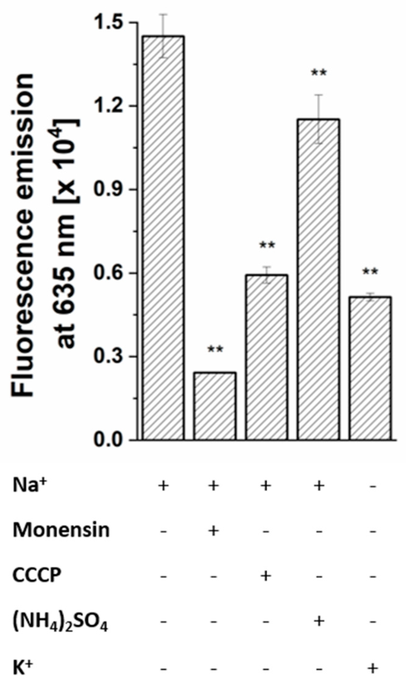
2.5. Sodium Dependent Membrane Potential in P. bivia

The membrane potential (ΔΨ, inside negative) was estimated using the fluorescent dye DiOC2 (3,3′-diethyloxacarbocyanine iodide), which exhibits increased emission at 635 nm in cells with high ΔΨ. The membrane potential established by P. bivia was strongly diminished when cells were depleted for Na+ by repeated washing with K+ (Figure 9). The sodium ionophore monensin, the protonophore carbonyl cyanide m-chlorophenylhydrazone (CCCP), and NH4+ diminished the membrane potential with decreasing efficiency. This indicates that P. bivia maintains an electrochemical Na+ gradient (sodium motive force (SMF)). In addition, an electrochemical proton potential (proton motive force (PMF)) was established in P. bivia, as indicated by the partial dissipation of the membrane potential by a protonophore, specifically CCCP. In P. bivia, ammonium (10 mM) acted as uncoupling agent. This was unexpected given that ammonium in the millimolar concentration range is usually added as a nitrogen source the to bacterial growth media. These findings were in line with the observed reduced growth when Asn or NH4+ was added to P. bivia cultures.

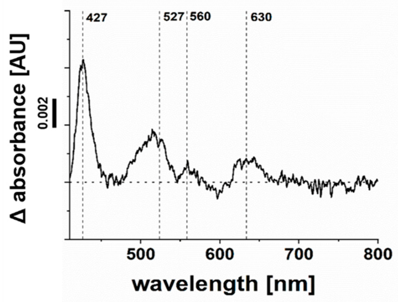
2.6. Cytochrome bd Quinol Oxidase of P. bivia
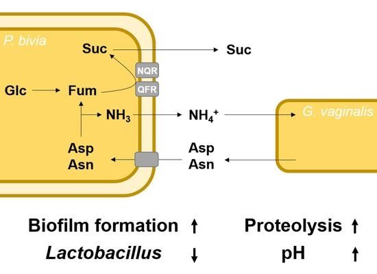
Absorbances in the VIS difference spectrum assigned to heme d and the presence of both cyd-1 and cyd-2 genes suggested the presence of cytochrome bd quinol oxidase in P. bivia. This was analyzed by monitoring the peroxidase activity of this enzyme. Solubilized membranes catalyzed the oxidation of the DMNH2 with H2O2 as an electron acceptor with a specific activity of 0.6 nmol min−1 mg−1. Solubilized membranes from P. bryantii, which lack the bd quinol oxidase, exhibited only residual DMNH2 oxidation activity at rates similar to protein-free controls (Figure 10).

3. Discussion

Key metabolic features of P. bivia are succinate production, the generation of an electrochemical sodium gradient, the operation of a terminal oxidase, and the conversion of asparagine under the formation of ammonium. From the genome, P. bivia is predicted to operate both the typical and atypical Embden-Meyerhoff-Parnas (EMP) pathways, yielding PEP as a central intermediate [30] (Figure S4). The CO2-dependent growth indicates that carboxylation of PEP to oxaloacetate by the carboxykinase is crucial for P. bivia. This reaction ultimately provides endogenous fumarate, acting as an acceptor for an electron transport chain, which generates a membrane potential (Figure 11). Fumarate is reduced to succinate by the membrane-bound quinol:fumarate oxidoreductase (QFR), which uses menaquinol as a substrate. The product pattern observed with P. bivia compares favorably with the results of a transcriptome study of the vaginal microbiota of BV patients, which identified pathways leading to succinate and short-chain fatty acids [31].
Besides QFR, P. bivia operates a membrane-bound NADH:quinone oxidoreductase (NQR), which provides menaquinol for the fumarate reduction and regenerates the NAD+ required for glycolysis. The similarity of P. bivia NQR to the Na+-translocating V. cholerae NQR [23] and the stimulation of NADH:quinone oxidoreduction activity by Na+ indicate that the P. bivia NQR acts as a Na+ pump. In accordance with this notion, the membrane potential established by P. bivia cells critically depends on Na+ and collapses in the presence of the sodium ionophore monensin. It is proposed that the build-up of a membrane potential by the Na+-translocating NQR is crucial for the energy conservation in P. bivia. NQR is widely distributed in Prevotella sp. [14] but it is neither found in the Lactobacilli of the vaginal microbiota, nor in the human host. This makes NQR an attractive target for the development of antibacterial compounds, as demonstrated for the case of Chlamydia trachomatis [32].
Partial dissipation of the transmembrane voltage by the protonophore CCCP indicates that P. bivia also establishes a PMF, probably with the help of the F1FO ATPase. Considering critical, conserved residues in the cation-binding site [33], the F1FO ATPase of P. bivia is a proton rather than a sodium-dependent enzyme. To regulate cytoplasmic proton and Na+ concentrations, P. bivia operates Na+/H+ antiporters related to NhaA and NhaD (Supplementary Material Table S1; Figure 11).
The formation of H2O2 and other reactive oxygen species by Lactobacilli colonizing the vaginal epithelium [34] prohibits the growth of strict anaerobes, which typically lack enzymes protecting against oxidative stress. P. bivia is an exception since it operates a superoxide dismutase [35] and possesses an active bd oxidase utilizing quinol as an electron donor for the reduction of O2 or H2O2. Moreover, P. bivia exhibits robust growth at acidic pH, producing ammonium from asparagine. Thus, it is capable of thriving in a vaginal environment dominated by Lactobacilli.
BV is characterized by a biofilm [36] established by a microbial consortium, with Gardnerella vaginalis and Prevotella bivia as prominent strains. In metabolic cross-feeding, ammonium released by P. bivia was utilized by G. vaginalis [12], followed by the degradation of the vaginal mucus layer by sialidases and adherence of other BV-associated bacteria [37]. G. vaginalis lacks metabolic routes for amino acid synthesis with the exception of pathways for the synthesis of L-aspartate and L-asparagine [38]. P. bivia possesses L-asparaginase and aspartate ammonia lyase, producing [NH3 + NH4+] at high rates in vitro and in vivo. NH4+ (160 mM) inhibited the growth of P. bivia, most likely due to the partial dissipation of the membrane potential. This is in marked contrast to the situation in E. coli, where no detrimental effect on growth was observed up to 500 mM NH4+ [18]. In a consortium with G. vaginalis consuming ammonium, high turnover of Asn by P. bivia under the formation of fumarate is possible and P. bivia will benefit from fumarate acting as an electron acceptor. This could facilitate the colonization of the vaginal epithelium by G. vaginalis and P. bivia at an early stage of BV.

4. Materials and Methods

4.1. Bacterial Strains and Growth Conditions

Prevotella bivia DSM 20514 and Prevotella bryantii B14 were cultivated anaerobically at 39 °C in a synthetic medium composed of 1% tryptone (w/v), 13 mM of glucose, 50 mM of NaHCO3, 15% (by volume) mineral solution 1 (17 mM K2HPO4), 15% (by volume) mineral solution 2 (17 mM KH2PO4, 45 mM of (NH4)2SO4, 100 mM of NaCl, 5 mM of MgSO4, and 5.4 mM CaCl2), and 0.44 μM of resazurin (sodium salt). The redox potential was adjusted with 8 mM of L-Cysteine HCl. In addition, the medium contained (by volume) 0.17% acetic acid, 0.01% n-valeric acid, 0.01% iso-valeric acid, 0.03% n-butyric acid, 0.01% iso-butyric acid, and 0.06% propionic acid [15]. Hungate tubes (7 mL volume) and serum bottles (0.1 L or 1 L volume) with gas-tight caps were used.

4.2. Isolation and Solubilization of Membranes

Cells were harvested at an OD600 of 1.5–2.0 (P. bivia) or at an OD600 of 2.5–3.0 (P. bryantii) by centrifugation at 9000× g for 30 min (4 °C). The cells were washed twice in 20 mM of Tris-H2SO4 (pH 7.5) and 50 mM of K2SO4. Cells (10 g wet weight) were resuspended in 30 mL of 20 mM Tris-H2SO4 (pH 7.5) containing 50 mM of K2SO4, 5 mM of MgSO4, 1 mM of dithiothreitol, 1 mM of phenylmethyl sulfonylfluoride (PMSF), 0.1 mM of diisopropyl fluorophosphate, and traces of DNase I (Roche Diagnostics GmbH, Mannheim, Germany). The suspension was passed three times through an EmulsiFlex®-C3 high-pressure homogenizer (Avestin Europe GmbH, Mannheim, Germany) at 20,000 psi. Cell debris and unbroken cells were removed by centrifugation at 27,000 g for 30 min at 4 °C. Membranes were collected by ultracentrifugation (50,000 rpm, Beckman Ti70 rotor; Beckman Coulter GmbH, Krefeld, Germany) for 90 min at 4 °C; washed once in 20 mM of Tris-H2SO4 (pH 7.5), 50 mM of K2SO4, and 5% (v/v) glycerol; and resuspended in the same buffer. The membrane suspension (5–10 mg protein/mL) was frozen by pipetting aliquots of 30 μL into liquid N2. The frozen droplets were collected and stored in liquid N2 until further use. For the solubilization of the membranes, protein and n-dodecyl-ß-D-maltoside (DDM; 7.5 µM final concentration) were incubated in a 1:3.75 (protein:detergent) ratio in a buffer containing 20 mM of Tris-H2SO4 (pH 7.5), 50 mM of K2SO4, and 5% (v/v) glycerol, with a total volume of 1.5 mL, for 2 h at 6 °C under gentle shaking (350 rpm). The membrane suspensions were ultracentrifuged (50,000 rpm, Beckman Ti70 rotor; Beckman Coulter GmbH, Krefeld, Germany) for 45 min at 4 °C. Supernatants containing solubilized membrane proteins were frozen and stored in liquid N2 as described above.

4.3. Bacterial Growth

Growth was followed in Hungate tubes with 7 mL of medium inoculated with 500 µL of P. bivia overnight culture grown at pH ~7. Turbidity of the cultures in tubes was measured with a cell density meter (WPA biowave CO8000, Biochrom Ltd., Cambridge, UK) at 600 nm. To analyze the CO2 dependency of the growth of P. bivia, triplicate growth experiments with medium prepared with CO2 [15] were conducted. In the controls, CO2 was replaced with N2. To analyze the medium during growth under a chosen condition, six tubes per experiment were inoculated at t = 0 h and turbidity was monitored in parallel. At indicated times, the culture from one tube was retrieved. To study the effect of pH on the growth of P. bivia, the growth medium was adjusted to pH 5, 6, or 7 by adding NaOH. Growth was monitored for 7 days (pH 6 and pH 7) or 9 days (pH 5) in triplicate experiments. At indicated times, cells were harvested by centrifugation at 16,000 g for 5 min at 4 °C and both pH and ammonium concentration of supernatants were determined. In parallel growth experiments, the media contained 50 mM of L-asparagine. To study the effect of NH4+ on growth, medium (pH 6.0) was supplemented with 7 mM of (NH4)2SO4 and 14 mM of K2SO4, or with 80 mM of (NH4)2SO4 and 20 mM of K2SO4. To identify and quantify organic compounds in cultures by 1H-NMR, experiments in Hungate tubes were performed in triplicates. After 5 h, 18 h, and 48 h of growth, the ODs were determined and one culture was harvested by centrifugation at 16,000 g for 5 min at 4 °C to obtain supernatants for NMR analysis.

4.4. Analytical Methods

The protein concentration was determined with the bicinchoninic acid method [39] using the reagent from PierceTM (ThermoFisher Scientific, Waltham, MA, USA). To determine the protein content of the cell suspensions, cells from 1 mL of culture were washed in 300 mM of sucrose. The cell pellet was resuspended in 5% (v/v) trichloroacetic acid and heated (100 °C) for 10 min [40]. [NH3 + NH4+] in supernatants of cell cultures was determined spectrophotometrically with the Nessler’s reagent [41]. Ammonium sulfate was used as the standard.
Glucose, acetate, succinate, and malate in cultures of P. bivia were determined by 1D 1H NMR spectroscopy. Supernatants from cell cultures were dried with a vacuum concentrator (program V-AQ; Eppendorf SE, Hamburg, Germany,). The pellets were resuspended in 50 mM of Na2HPO4 (pH 7) in D2O containing 5 mM of 3-trimethylsilyl propionic-2,2,3,3 acid sodium salt (TSP) as an internal reference for the 1H chemical shift calibration and the suspensions were filled into NMR tubes. 1D 1H NMR spectra were recorded using a Bruker Avance III HD NMR 600 MHz spectrometer equipped with a 5 mm BBO Prodigy cryo-probe (Bruker BioSpin GmbH, Ettlingen, Germany). For structural identification of the metabolites, 1D 1H heteronuclear NMR experiments (gHSQC and gHMBC) [42] were recorded at 298 K. For acquisition, processing, and evaluation of NMR spectra, the software TopSpin 3.5pl7 (Bruker BioSpin GmbH, Ettlingen, Germany) was used. To quantify succinate in medium, to which 50 mM of L-asparagine was added at the start of the growth, cells were cultivated for 50 h in Hungate tubes. Cell-free supernatants were analyzed using the Sigma -AldrichTM Succinate Assay Kit (Merck KGaA, Darmstadt, Germany).
Denaturing polyacrylamide gel electrophoresis (SDS-PAGE) was performed with a 12% polyacrylamide gel [43]. Protein and membrane suspensions were diluted in 5x SDS sample buffer (500 mM of DTT; 1 M of Tris-HCl, pH 6.8; 5% SDS; and 28.8% glycerol (w/v), bromophenol blue) and boiled for 5 min before loading on the gel. In-gel fluorescence of covalently bound flavins in NqrB, NqrC, and FrdA, separated by SDS-PAGE, was detected using the ImageQuant LAS 4000 imager (λexcitation = 460, emission filter = Y515 CyTM2; Cytiva, Marlborough, MA, USA). As a positive control, the purified NqrC’ subunit was used. This protein is a truncated variant of the NqrC subunit of the V. cholerae NQR comprising the covalently attached FMN but lacking the N-terminal transmembrane helix [44]. The molecular mass of NqrC’ was 25.38 kDa. Proteolysis of proteins separated by SDS-PAGE, followed by mass spectrometric analysis of the peptides, was performed as described previously [21].

4.5. UV/Vis Difference Spectra of Redox Cofactors in P. bivia

The absorption spectrum of dithionite-reduced redox cofactors in solubilized membranes of P. bivia was compared with an aliquot of the same sample with cofactors in their air-oxidized state using a double-beam UV/VIS spectrophotometer (UV-2600i; Shimadzu GmbH, Berlin, Deutschland). Light is split by a half mirror passing separately through the reference sample (beam 1) and through the test sample (beam 2). The light intensities passing through the sample and reference were compared. The range of 220–800 nm was monitored with an interval of 0.5 nm and with medium scan speed. The difference in absorbance of beam 2 minus beam 1 at a given wavelength was calculated using the software LabSolutions UV-Vis (Shimadzu GmbH, Berlin, Deutschland). DDM-solubilized membranes of P. bivia were analyzed at a concentration of ~0.8 mg of protein per mL in 20 mM of potassium phosphate buffer, pH 7.5. Beam 1 contained air-oxidized solubilisate, whereas in beam 2, an aliquot of solubilisate mixed with a few crystals of sodium dithionite was analyzed. The difference spectrum of dithionite-reduced minus air-oxidized membranes was recorded.

4.6. Enzymatic Assays

NADH oxidation and quinone reduction activities were monitored simultaneously in a quartz cuvette (d = 1 cm) in a total volume of 1 mL at 25 °C using a Hewlett Packard 8452A diode-array spectrophotometer (Agilent Technologies, Santa Clara, CA, USA). NADH oxidation was followed at 340 nm (εNADH = 6.22 mM−1 cm−1) and ubiquinone-1 (Q1) or 2,3-dimethyl-1,4-naphthoquinone (DMN) reduction at 280 nm (εQ1 = 14.5 mM−1 cm−1 and εDMN = 15.2 mM−1 cm−1) [45]. Solubilized membranes of P. bivia (50 µg of protein in 20 mM of Tris-H2SO4, pH 7.5; 50 mM of K2SO4; 5% (v/v) glycerol; and 7.5 µM of DDM) were incubated with varying amounts of AgNO3 (0–3.0 µM) for 5 min at 4 °C and were added to a cuvette with buffer (20 mM of Tris H2SO4, pH 7.5; 100 mM of Na2SO4; 100 µM of NADH; and 100 µM of Q1) to start the enzymatic reaction. In buffers, chloride was replaced with sulfate to avoid precipitation of AgCl. To study the effect of Na+ on NADH dehydrogenase activity of solubilized membranes (50 µg), NaCl (0–1000 µM) or corresponding amounts of KCl (0–1000 µM) were added to the assay buffer (20 mM of Tris H2SO4, pH 7.5; 100 µM of NADH; and 100 µM of Q1). The residual Na+ concentration in the assay without the added NaCl was ~10 µM, as determined by atomic absorption spectroscopy (AA240, Agilent Technologies, Santa Clara, CA, USA).
Quinol:fumarate oxidoreductase (QFR) activity was determined with anoxic materials and buffer (20 mM of potassium phosphate, pH 7.5) containing benzyl viologen (~0.5 mM) in the anaerobic chamber. Benzyl viologen was reduced by adding sodium dithionite crystals to achieve an absorbance of 1 at 564 nm [46]. Then, 100–200 μg of protein was added. The reaction was started by adding 10 mM of fumarate. Decrease in absorbance of benzyl viologen was monitored at 564 nm (ε = 19.5 mM−1 cm−1) in a cuvette at 20 °C using a diode array spectrophotometer (Black-comet, StellarNet Inc., Tampa, FL, USA). The cuvette holder was placed inside the anaerobic chamber. The detector and light source (SL5 UV + VIS lamp, StellarNet Inc., Tampa, FL, USA) were placed outside of the anaerobic chamber and the components were connected with fiber optic cables.
The cytochrome quinol bd oxidase activity in solubilized membranes was determined by following the oxidation of quinol with H2O2 [47]. 2,3-dimethyl-1,4-naphthoquinol (DMNH2), obtained as described in [48], was used as an electron donor. DMNH2 oxidation with H2O2 as an electron acceptor was monitored from the formation of DMN (εDMN = 15.2 mM−1 cm−1) [49] under anoxic conditions by recording difference spectra over 16 min in a double-beam photometer (Lambda 16, PerkinElmer, Waltham, MA, USA). Buffers and reagents were made anoxic by flushing with N2 and were mixed in cuvettes inside the anaerobic chamber. Cuvettes were sealed gas-tight and difference spectra were recorded immediately outside of the chamber. In beam 1, the reference cuvette was analyzed, containing 1 mL of assay solution (180 µg of solubilized membrane protein from P. bivia or P. bryantii; 50 mM of MOPS, pH 7.0; 100 mM of NaCl; 0.1% v/v DDM; and 200 µM of DMNH2). A cuvette with 1 mL of assay solution mixed with 10 µL of 30% (by volume) H2O2 was placed in beam 2. To calculate the rates of DMNH2 oxidation, the difference in absorbance from 270 nm to 290 nm obtained by subtraction of the spectrum of beam 1 from the spectrum of beam 2 at a given timepoint was determined. In the control, H2O2 was omitted.
For L-asparaginase and aspartate lyase activity determinations, the soluble protein fraction of P. bivia was obtained by ultracentrifugation of crude cell extracts. L-asparaginase activity was determined as described in [41]. Aspartate ammonia lyase activity was determined as described in [50] and modified as follows. After incubation at 30 °C for 30 min, the assay solution (1 mL) was heated at 80 °C for 5 min to stop the reaction. The (NH3 + NH4+) concentration was determined photometrically with the Nessler’s reagent [51].

4.7. Membrane Potential

Membrane potential of P. bivia was estimated with the BacLightTM Bacterial Membrane Potential Kit ((ThermoFisher Scientific, Waltham, MA, USA)) using a Infinite F200 Pro plate reader (Tecan Deutschland GmbH, Crailsheim, Germany) [52]. P. bivia cells were cultivated in Hungate tubes until an OD600 of 0.6 was reached. The following steps were performed in the anaerobic chamber. Cells were harvested, diluted in sodium buffer (10 mM of sodium buffer, pH 7.4, and 145 mM of NaCl) or potassium buffer (10 mM of potassium buffer, pH 7.4, and 145 mM of KCl), and adjusted to OD600 = 0.25. Cells in 800 µL of this suspension were sedimented by centrifugation (16,000 g, 5 min), washed twice, and resuspended in 800 µL of the corresponding buffer. To analyze the effect of ionophores on the membrane potential, 2.5 µM of carbonylcyanid-m-chlorphenylhydrazon (CCCP), 2.5 µM of monensin, or 5 mM of (NH4)2SO4 were added to cell suspensions, as indicated. After incubation for 10 min (20 °C), the fluorescence dye 3,3′-diethyloxacarbocyanine iodide (DiOC2, 15 µM) was added and cells were further incubated for 60 min in the dark. Outside of the anaerobic chamber, three aliquots (200 µM) of each sample were applied to a black, flat-bottom 96-well plate (polysterene; 4titude Ltd., Berlin, Germany). To determine red fluorescence intensities, excitation was set to 480 nm (band width, 9 nm) and emission to 635 nm (band width, 20 nm; gain, 117). To determine green fluorescence intensities, the emission was changed to 535 nm (band width, 25 nm; gain, 107). Fluorescence emission intensities were in the linear range of the fluorescence detector. Background fluorescence intensities of buffer with dye and of cell suspensions were determined for background corrections. As expected, an increase of red fluorescence intensity, indicating a transmembrane voltage, was accompanied by a decrease in green fluorescence intensity. Mean values of red fluorescence intensities are presented.

Supplementary Materials

Supplementary Materials are available online at https://www.mdpi.com/article/10.3390/ijms222111925/s1.

Author Contributions

Conceptualization, J.S. (Julia Steuber) and L.S.; methodology, L.S. and G.F.; software, L.S.; validation, L.S. and A.T.; formal analysis, L.S. and A.T.; investigation, L.S. and S.H.; resources, J.S. (Julia Steuber) and J.S. (Jana Seifert); data curation, L.S.; writing—original draft preparation, L.S.; writing—review and editing, J.S. (Julia Steuber), J.S. (Jana Seifert), A.T. and L.S.; visualization, L.S.; supervision, J.S. (Julia Steuber); project administration, J.S. (Julia Steuber) and J.S. (Jana Seifert); funding acquisition, J.S. (Julia Steuber) and J.S. (Jana Seifert). All authors have read and agreed to the published version of the manuscript.

Funding

This research study was funded by the Deutsche Forschungsgemeinschaft to J. Seifert and J. Steuber (project number 327953272).

Data Availability Statement

Additional data are provided in the Supplementary Materials.

Acknowledgments

We thank Johannes Günther, Jens Pfannstiel, and Berit Würtz of the Core Facility University of Hohenheim for their support in the NMR spectroscopy and mass spectrometry.

Conflicts of Interest

The authors declare no conflict of interest.

References

  1. Onderdonk, A.B.; Delaney, M.L.; Fichorova, R.N. The human microbiome during bacterial vaginosis. Clin. Microbiol. Rev. 2016, 29, 223–238. [Google Scholar] [CrossRef] [Green Version]
  2. Muzny, C.A.; Taylor, C.M.; Swords, W.E.; Tamhane, A.; Chattopadhyay, D.; Cerca, N.; Schwebke, J.R. An updated conceptual model on the pathogenesis of bacterial vaginosis. J. Infect. Dis. 2019, 220, 1399–1405. [Google Scholar] [CrossRef]
  3. Ravel, J.; Gajer, P.; Abdo, Z.; Schneider, G.M.; Koenig, S.S.K.; McCulle, S.L.; Karlebach, S.; Gorle, R.; Russell, J.; Tacket, C.O.; et al. Vaginal microbiome of reproductive-age women. Proc. Natl. Acad. Sci. USA 2011, 108 (Suppl. S1), 4680–4687. [Google Scholar] [CrossRef] [Green Version]
  4. Nunn, K.L.; Clair, G.C.; Adkins, J.N.; Engbrecht, K.; Fillmore, T.; Forney, L.J. Amylases in the human vagina. mSphere 2020, 5, e00943-20. [Google Scholar] [CrossRef]
  5. Zozaya-Hinchliffe, M.; Lillis, R.; Martin, D.H.; Ferris, M.J. Quantitative PCR assessments of bacterial species in women with and without bacterial vaginosis. J. Clin. Microbiol. 2010, 48, 1812–1819. [Google Scholar] [CrossRef] [PubMed] [Green Version]
  6. Delaney, M. Nugent score related to vaginal culture in pregnant women. Obstet. Gynecol. 2001, 98, 79–84. [Google Scholar] [CrossRef] [PubMed]
  7. Aroutcheva, A.; Ling, Z.; Faro, S. Prevotella bivia as a source of lipopolysaccharide in the vagina. Anaerobe 2008, 14, 256–260. [Google Scholar] [CrossRef] [Green Version]
  8. Strömbeck, L.; Sandros, J.; Holst, E.; Madianos, P.; Nannmark, U.; Papapanou, P.; Mattsby-Baltzer, I. Prevotella bivia can invade human cervix epithelial (HeLa) cells. APMIS 2007, 115, 241–251. [Google Scholar] [CrossRef] [PubMed]
  9. Mikamo, H.; Kawazoe, K.; Sato, Y.; Imai, A.; Tamaya, T. Preterm labor and bacterial intra-amniotic infection: Arachidonic acid liberation by phospholipase A2 of Prevotella bivia. Anaerobe 1998, 4, 209–212. [Google Scholar] [CrossRef]
  10. Gilbert, N.M.; Lewis, W.G.; Lewis, A.L. Clinical features of bacterial vaginosis in a murine model of vaginal infection with Gardnerella vaginalis. PLoS ONE. 2013, 8, e59539. [Google Scholar] [CrossRef] [Green Version]
  11. Gilbert, N.M.; Lewis, W.G.; Li, G.; Sojka, D.K.; Lubin, J.B.; Lewis, A.L. Gardnerella vaginalis and Prevotella bivia trigger distinct and overlapping phenotypes in a mouse model of bacterial vaginosis. J. Infect. Dis. 2019, 220, 1099–1108. [Google Scholar] [CrossRef]
  12. Pybus, V.; Onderdonk, A.B. Evidence for a commensal, symbiotic relationship between Gardnerella vaginalis and Prevotella bivia involving ammonia: Potential significance for bacterial vaginosis. J. Infect. Dis. 1997, 175, 406–413. [Google Scholar] [CrossRef]
  13. Franke, T.; Deppenmeier, U. Physiology and central carbon metabolism of the gut bacterium Prevotella copri. Mol. Microbiol. 2018, 109, 528–540. [Google Scholar] [CrossRef] [PubMed] [Green Version]
  14. Deusch, S.; Bok, E.; Schleicher, L.; Seifert, J.; Steuber, J. Occurrence and function of the Na+-translocating NADH:quinone oxidoreductase in Prevotella spp. Microorganisms 2019, 7, 117. [Google Scholar] [CrossRef] [PubMed] [Green Version]
  15. Trautmann, A.; Schleicher, L.; Deusch, S.; Gätgens, J.; Steuber, J.; Seifert, J. Short-chain fatty acids modulate metabolic pathways and membrane lipids in Prevotella bryantii B14. Proteomes 2020, 8, 28. [Google Scholar] [CrossRef]
  16. Pybus, V.; Onderdonk, A.B. The effect of pH on growth and succinate production by Prevotella bivia. Microb. Ecol. Health Dis. 1996, 9, 19–25. [Google Scholar] [CrossRef]
  17. Wolfe, A.J. The acetate switch. Microbiol. Mol. Biol. Rev. 2005, 69, 12–50. [Google Scholar] [CrossRef] [Green Version]
  18. Müller, T.; Walter, B.; Wirtz, A.; Burkovski, A. Ammonium toxicity in bacteria. Curr. Microbiol. 2006, 52, 400–406. [Google Scholar] [CrossRef] [PubMed] [Green Version]
  19. Lancaster, C.D. Wolinella succinogenes quinol:fumarate reductase—2.2-Å resolution crystal structure and the E-pathway hypothesis of coupled transmembrane proton and electron transfer. Biochim. Biophys. Acta 2002, 1565, 215–231. [Google Scholar] [CrossRef] [Green Version]
  20. Borisov, V.B.; Gennis, R.B.; Hemp, J.; Verkhovsky, M.I. The cytochrome bd respiratory oxygen reductases. Biochim. Biophys. Acta. 2011, 1807, 1398–1413. [Google Scholar] [CrossRef] [PubMed] [Green Version]
  21. Schleicher, L.; Trautmann, A.; Stegmann, D.P.; Fritz, G.; Gätgens, J.; Bott, M.; Hein, S.; Simon, J.; Seifert, J.; Steuber, J. A sodium-translocating module linking succinate production to formation of a membrane potential in Prevotella bryantii. Appl. Environ. Microbiol. 2021, 87, AEM0121121. [Google Scholar] [CrossRef]
  22. Steuber, J.; Vohl, G.; Casutt, M.S.; Vorburger, T.; Diederichs, K.; Fritz, G. Structure of the V. cholerae Na+-pumping NADH:quinone oxidoreductase. Nature 2014, 516, 62–67. [Google Scholar] [CrossRef] [Green Version]
  23. Steuber, J.; Vohl, G.; Muras, V.; Toulouse, C.; Claußen, B.; Vorburger, T.; Fritz, G. The structure of Na+-translocating NADH:ubiquinone oxidoreductase of Vibrio cholerae: Implications on coupling between electron transfer and Na+ transport. Biol. Chem. 2015, 396, 1015–1030. [Google Scholar] [CrossRef]
  24. Casutt, M.S.; Wendelspiess, S.; Steuber, J.; Fritz, G. Crystallization of the Na+-translocating NADH:quinone oxidoreductase from Vibrio cholerae. Acta Crystallogr. Sect. F Struct. Biol. Cryst. Commun. 2010, 66, 1677–1679. [Google Scholar] [CrossRef]
  25. Nowicka, B.; Kruk, J. Occurrence, biosynthesis and function of isoprenoid quinones. Biochim. Biophys. Acta 2010, 1797, 1587–1605. [Google Scholar] [CrossRef] [Green Version]
  26. Moparthi, V.K.; Hägerhäll, C. The evolution of respiratory chain complex I from a smaller last common ancestor consisting of 11 protein subunits. J. Mol. Evol. 2011, 72, 484–497. [Google Scholar] [CrossRef] [Green Version]
  27. Heikal, A.; Nakatani, Y.; Dunn, E.; Weimar, M.R.; Day, C.L.; Baker, E.N.; Lott, J.S.; Sazanov, L.A.; Cook, G.M. Structure of the bacterial type II NADH dehydrogenase: A monotopic membrane protein with an essential role in energy generation. Mol. Microbiol. 2014, 91, 950–964. [Google Scholar] [CrossRef]
  28. Steuber, J.; Krebs, W.; Dimroth, P. The Na+-translocating NADH:ubiquinone oxidoreductase from Vibrio alginolyticus--redox states of the FAD prosthetic group and mechanism of Ag+ inhibition. Eur. J. Biochem. 1997, 249, 770–776. [Google Scholar] [CrossRef] [Green Version]
  29. Asano, M.; Hayashi, M.; Unemoto, T.; Tokuda, H. Ag+ -sensitive NADH dehydrogenas in the Na+ -motive respiratory chain of the marine bacterium Vibrio alginolyticus. Agric. Biol. Chem. 1985, 49, 2813–2817. [Google Scholar] [CrossRef]
  30. Hackmann, T.J.; Ngugi, D.K.; Firkins, J.L.; Tao, J. Genomes of rumen bacteria encode atypical pathways for fermenting hexoses to short-chain fatty acids. Environ. Microbiol. 2017, 19, 4670–4683. [Google Scholar] [CrossRef] [Green Version]
  31. Macklaim, J.M.; Fernandes, A.D.; Di Bella, J.M.; Hammond, J.; Reid, G.; Gloor, G.B. Comparative meta-RNA-seq of the vaginal microbiota and differential expression by Lactobacillus iners in health and dysbiosis. Microbiome 2013, 1, 12. [Google Scholar] [CrossRef] [Green Version]
  32. Dibrov, P.; Dibrov, E.; Maddaford, T.G.; Kenneth, M.; Nelson, J.; Resch, C.; Pierce, G.N. Development of a novel rationally designed antibiotic to inhibit a nontraditional bacterial target. Can. J. Physiol. Pharmacol. 2017, 95, 595–603. [Google Scholar] [CrossRef] [Green Version]
  33. Meier, T.; Polzer, P.; Diederichs, K.; Welte, W.; Dimroth, P. Structure of the rotor ring of F-Type Na+-ATPase from Ilyobacter tartaricus. Science 2005, 308, 659–662. [Google Scholar] [CrossRef] [Green Version]
  34. Amabebe, E.; Anumba, D.O.C. The vaginal microenvironment: The physiologic role of Lactobacilli. Front. Med. 2018, 5, 181. [Google Scholar] [CrossRef] [Green Version]
  35. Schleicher, L.; Fritz, G.; Seifert, J.; Steuber, J. Anoxic cell rupture of Prevotella bryantii by high-pressure homogenization protects the Na+-translocating NADH:quinone oxidoreductase from oxidative damage. Arch. Microbiol. 2020, 202, 1263–1266. [Google Scholar] [CrossRef] [Green Version]
  36. Jung, H.; Ehlers, M.M.; Lombaard, H.; Redelinghuys, M.J.; Kock, M.M. Etiology of bacterial vaginosis and polymicrobial biofilm formation. Crit. Rev. Microbiol. 2017, 43, 651–667. [Google Scholar] [CrossRef] [Green Version]
  37. Schwebke, J.R.; Muzny, C.A.; Josey, W.E. Role of Gardnerella vaginalis in the pathogenesis of bacterial vaginosis: A conceptual model. J. Infect. Dis. 2014, 210, 338–343. [Google Scholar] [CrossRef]
  38. Harwich, M.D.; Alves, J.M.; Buck, G.A.; Strauss, J.F.; Patterson, J.L.; Oki, A.T.; Girerd, P.H.; Jefferson, K.K. Drawing the line between commensal and pathogenic Gardnerella vaginalis through genome analysis and virulence studies. BMC Genom. 2010, 11, 375. [Google Scholar] [CrossRef] [Green Version]
  39. Smith, P.K.; Krohn, R.I.; Hermanson, G.T.; Mallia, A.K.; Gartner, F.H.; Provenzano, M.D.; Fujimoto, E.K.; Goeke, N.M.; Olson, B.J.; Klenk, D.C. Measurement of protein using bicinchoninic acid. Anal. Biochem. 1985, 150, 76–85. [Google Scholar] [CrossRef]
  40. Ito, M.; Cooperberg, B.; Krulwich, T.A. Diverse genes of alkaliphilic Bacillus firmus OF4 that complement K+-uptake-deficient Escherichia coli include an ftsH homologue. Extremophiles 1997, 1, 22–28. [Google Scholar] [CrossRef]
  41. Mohamed, S.A.; Elshal, M.F.; Kumosani, T.A.; Aldahlawi, A.M. Purification and characterization of asparaginase from Phaseolus vulgaris seeds. Evid.-Based Complement. Alternat. Med. 2015, 2015, 309214. [Google Scholar] [CrossRef] [Green Version]
  42. Leutbecher, H.; Greiner, G.; Amann, R.; Stolz, A.; Beifuss, U.; Conrad, J. Laccase-catalyzed phenol oxidation. Rapid assignment of ring-proton deficient polycyclic benzofuran regioisomers by experimental 1H-13C long-range coupling constants and DFT-predicted product formation. Org. Biomol. Chem. 2011, 9, 2667–2673. [Google Scholar] [CrossRef] [PubMed]
  43. Schägger, H.; von Jagow, G. Tricine-sodium dodecyl sulfate-polyacrylamide gel electrophoresis for the separation of proteins in the range from 1 to 100 kDa. Anal. Biochem. 1987, 166, 368–379. [Google Scholar] [CrossRef]
  44. Vohl, G.; Nedielkov, R.; Claussen, B.; Casutt, M.S.; Vorburger, T.; Diederichs, K.; Möller, H.M.; Steuber, J.; Fritz, G. Crystallization and preliminary analysis of the NqrA and NqrC subunits of the Na+-translocating NADH:ubiquinone oxidoreductase from Vibrio cholerae. Acta Crystallogr. Sect. F Struct. Biol. Cryst. Commun. 2014, 70, 987–992. [Google Scholar] [CrossRef] [PubMed] [Green Version]
  45. Juárez, O.; Athearn, K.; Gillespie, P.; Barquera, B. Acid residues in the transmembrane helices of the Na+-pumping NADH:quinone oxidoreductase from Vibrio cholerae involved in sodium translocation. Biochemistry 2009, 48, 9516–9524. [Google Scholar] [CrossRef] [Green Version]
  46. Kröger, A.; Innerhofer, A. The function of menaquinone, covalently bound FAD and iron-sulfur protein in the electron transport from formate to fumarate of Vibrio succinogenes. Eur. J. Biochem. 1976, 69, 487–495. [Google Scholar] [CrossRef]
  47. Al-Attar, S.; Yu, Y.; Pinkse, M.; Hoeser, J.; Friedrich, T.; Bald, D.; de Vries, S. Cytochrome bd displays significant quinol peroxidase activity. Sci. Rep. 2016, 6, 27631. [Google Scholar] [CrossRef] [Green Version]
  48. Trumpower, B.L.; Edwards, C.A. Purification of a reconstitutively active iron-sulfur protein (oxidation factor) from succinate cytochrome c reductase complex of bovine heart mitochondria. J. Biol. Chem. 1979, 254, 8697–8706. [Google Scholar] [CrossRef]
  49. Geisler, V.; Ullmann, R.; Kröger, A. The direction of the proton exchange associated with the redox reactions of menaquinone during electron transport in Wolinella succinogenes. Biochim. Biophys. Acta 1994, 1184, 219–226. [Google Scholar] [CrossRef]
  50. Blasco, L.; Kahala, M.; Tupasela, T.; Joutsjoki, V. Determination of aspartase activity in dairy Propionibacterium strains. FEMS Microbiol. Lett. 2011, 321, 10–13. [Google Scholar] [CrossRef] [PubMed]
  51. Bzura, J.; Koncki, R. A mechanized urease activity assay. Enzyme Microb. Technol. 2019, 123, 1–7. [Google Scholar] [CrossRef] [PubMed]
  52. Vorburger, T.; Nedielkov, R.; Brosig, A.; Bok, E.; Schunke, E.; Steffen, W.; Mayer, S.; Götz, F.; Möller, H.M.; Steuber, J. Role of the Na+-translocating NADH:quinone oxidoreductase in voltage generation and Na+ extrusion in Vibrio cholerae. Biochim. Biophys. Acta 2016, 1857, 473–482. [Google Scholar] [CrossRef] [PubMed]
Figure 1. Consumption of glucose and formation of carboxylic acids during anaerobic growth of P. bivia. Upper panel: concentrations of glucose (closed circles), acetate (open circles), succinate (closed triangles), and malate (open triangles). Lower panel: optical density at 600 nm. Average and standard deviations of three biological replicates are shown.
Ijms 22 11925 g001
Figure 2. Growth of P. bivia is dependent on CO2. Cells of P. bivia were cultivated in medium prepared with CO2 (back circles) or with N2 (white circles). Average and standard deviation of three biological replicates are shown.
Ijms 22 11925 g002
Figure 3. Effect of L-asparagine on the growth of P. bivia at varying pH. P. bivia was cultivated in medium with (black symbols) or without (white symbols) supplementation of 50 mM of L-asparagine. The initial pH of the medium was 7.0 (circles; top panel), 6.0 (squares; middle panel), and 5.0 (triangles; lower panel). Average and standard deviations of three biological replicates are shown.
Ijms 22 11925 g003
Figure 4. Effect of (NH4)2SO4 on the growth of P. bivia. Cells were cultivated with 7 mM of (NH4)2SO4 (white circles) or 80 mM of (NH4)2SO4 (black circles) at pH 6. Average and standard deviations of three biological replicates are shown.
Ijms 22 11925 g004
Figure 5. Genes coding for membrane-bound electron transfer complexes of P. bivia. ORF (PrebiDRAFT) and NCBI accession numbers (EIM) are given on the left. The fumarate reductase (QFR) was encoded by frdA (1983 bp), frdB (675 bp), and frdC (756 bp). The non-electrogenic NADH dehydrogenase (Ndh2) is a membrane-associated enzyme encoded by ndh2 (1308 bp; black frame). The nuoA (351 bp), nuoB (906 bp), nuoCD (1578 bp), nuoH (1098 bp), nuoI (534 bp), nuoJ (534 bp), nuoK (309 bp), nuoL (2061 bp), nuoM (1512 bp), and nuoN (1440 bp) genes are similar to the genes coding for the corresponding subunits of the 11-subunit complex related to the NUO complex. The Na+-translocating NADH:quinone oxidoreductase (NQR) was encoded by nqrA (1359 bp), nqrB (1161 bp), nqrC (714 bp), nqrD (633 bp), nqrE (627 bp), and nqrF (1263 bp). The cytochrome bd quinol oxidase was encoded by cyd-2 (1143 bp) and cyd-1 (1536 bp). Red frames correspond to genes coding for hydrophobic (membrane-bound) subunits. Blue frames correspond to genes coding for hydrophilic (peri or cytoplasmatic) subunits.
Ijms 22 11925 g005
Figure 6. Detection of flavinylated subunits from QFR and NQR in P. bivia membranes. Membranes (100 µg; membr.) and membranes solubilized with DDM (100 µg; solub.) of P. bivia were separated by SDS PAGE. Proteins were stained with Coomassie (upper panel) and analyzed by in-gel fluorography (lower panel) to detect flavinylated proteins. NqrC’ (25 kDa), the FMN-containing domain of subunit NqrC from V. cholerae NQR, served as the control (2 µg). Black boxes (1 and 2) indicate bands subjected to tryptic digestion and mass spectrometry analysis.
Ijms 22 11925 g006
Figure 7. Detection of hemes b and d in solubilized membranes of P. bivia. VIS difference spectrum of dithionite-reduced minus air-oxidized DDM solubilisates (0.8 mg protein/mL) with the maxima of reduced heme b (560 nm, 527 nm, and 427 nm) and heme d (630 nm). A typical trace from three biological replicates is presented.
Ijms 22 11925 g007
Figure 8. The Na+ -translocating NADH:quinone oxidoreductase is the major membrane-bound NADH dehydrogenase in P. bivia. Assays were performed with solubilized membranes (50 µg of protein). Upper panel: NADH dehydrogenase activity at increasing [Ag+] in chloride-free assay buffer. Lower panel: Q1 reduction activities at increasing [K+] (open circles) or [Na+] (closed circles). Residual Na+ concentration of buffer was at ~10 µM. Average and standard deviations from two technical replicates are shown.
Ijms 22 11925 g008
Figure 9. Effect of Na+ and uncouplers on the membrane potential of P. bivia. The fluorescence emission of cells incubated under the indicated conditions was corrected by the emission of fluorophore in buffer. CCCP, carbonyl cyanide m-chlorophenylhydrazone. Mean values and averages from four technical replicates are shown. Asterisks (**) indicate significant differences from cells incubated with Na+ in the absence of inhibitors with p < 0.05.
Ijms 22 11925 g009
Figure 10. Cytochrome bd quinol oxidase activity of P. bivia. Oxidation of DMNH2 in the presence of H2O2 (peroxidase activity) was followed with solubilized membranes from P. bivia (closed circles) or P. bryantii (open circles). The control reaction was performed in the absence of solubilized membranes (closed squares).
Ijms 22 11925 g010
Figure 11. Energy-converting complexes and central carbon metabolism of P. bivia. Important products are highlighted by black boxes. Red arrows indicate reactions for the assimilation of acetate. Gluc = glucose; Gluc 6-P = glucose 6-phosphate; and PEP = phosphoenolpyruvate. Blue = fumarate reductase (QFR); green = Na+-translocating NADH:quinone oxidoreductase (NQR); dotted = F1FO ATPase; striped = Na+/H+ antiporter; wavy = pyrophosphatase; yellow = cytochrome bd quinol oxidase; red = NDH2; and grey = 11-subunit complex related to NDHI (complex I). Subunits of NQR (A–F) and QFR (A–C) are indicated. Colored symbols in the protein complexes represent cofactors. Yellow triangle = flavin; black circle = iron-sulfur center; red diamond = heme b; and MK = menaquinone. UNIPROT numbers of proteins are listed in Table S1 (electronic Supplementary Material).
Ijms 22 11925 g011
Table 1. Effect of pH and L-asparagine on the growth and formation of (NH3 + NH4+) by P. bivia. OD600 and [NH3 + NH4+] were determined in the stationary phase (t = 7 d) of P. bivia cultures grown at pH 5, 6, or 7 with or without supplementation of 50 mM of L-asparagine.
Growth ConditionOD600
(t = 7 d)
Doubling Time
(h)
NH4+ (mM; t = 0)NH4+ (mM; t = 7)Net (NH3 + NH4+) Formed (mM)Rate of Net (NH3 + NH4+) Formation (nmol min−1 mg−1)
pH 5 + Asn0.5 ± 0.19114.7 ± 0.262.9 ± 0.848.2 ± 4.4265.7 ± 24.1
pH 5 − Asn 0.5 ± 0.110114.8 ± 0.127.9 ± 0.213.1 ± 0.159.7 ± 0.4
pH 6 + Asn0.4 ± 0.11716.9 ± 2.192.4 ± 1.175.5 ± 5.6416.4 ± 31.4
pH 6 − Asn1.1 ± 0.41312.1 ± 0.525.5 ± 0.213.1 ± 0.621.7 ± 2.1
pH 7 + Asn1.6 ± 0.41018.1 ± 0.8101.5 ± 0.583.4 ± 4.1119.1 ± 2.5
pH 7 − Asn1.8 ± 0.2816.5 ± 0.737.6 ± 0.0321.1 ± 0.2525.2 ± 0.5
Publisher’s Note: MDPI stays neutral with regard to jurisdictional claims in published maps and institutional affiliations.

