Review

Pathophysiological Potentials of NRF3-Regulated Transcriptional Axes in Protein and Lipid Homeostasis

1
Laboratory for Genetic Code, Department of Medical Life Systems, Faculty of Life and Medical Sciences, Doshisha University, Kyotanabe 610-0394, Japan
2
Laboratory for Genetic Code, Graduate School of Life and Medical Sciences, Doshisha University, Kyotanabe 610-0394, Japan
*
Author to whom correspondence should be addressed.
Int. J. Mol. Sci. 2021, 22(23), 12686; https://doi.org/10.3390/ijms222312686
Submission received: 4 November 2021 / Revised: 22 November 2021 / Accepted: 22 November 2021 / Published: 24 November 2021
(This article belongs to the Special Issue Transcriptional Regulation and Its Misregulation in Human Diseases)

Abstract

:
NRF3 (NFE2L3) belongs to the CNC-basic leucine zipper transcription factor family. An NRF3 homolog, NRF1 (NFE2L1), induces the expression of proteasome-related genes in response to proteasome inhibition. Another homolog, NRF2 (NFE2L2), induces the expression of genes related to antioxidant responses and encodes metabolic enzymes in response to oxidative stress. Dysfunction of each homolog causes several diseases, such as neurodegenerative diseases and cancer development. However, NRF3 target genes and their biological roles remain unknown. This review summarizes our recent reports that showed NRF3-regulated transcriptional axes for protein and lipid homeostasis. NRF3 induces the gene expression of POMP for 20S proteasome assembly and CPEB3 for NRF1 translational repression, inhibiting tumor suppression responses, including cell-cycle arrest and apoptosis, with resistance to a proteasome inhibitor anticancer agent bortezomib. NRF3 also promotes mevalonate biosynthesis by inducing SREBP2 and HMGCR gene expression, and reduces the intracellular levels of neural fatty acids by inducing GGPS1 gene expression. In parallel, NRF3 induces macropinocytosis for cholesterol uptake by inducing RAB5 gene expression. Finally, this review mentions not only the pathophysiological aspects of these NRF3-regulated axes for cancer cell growth and anti-obesity potential but also their possible role in obesity-induced cancer development.

1. Introduction

Protein and lipid homeostasis is crucial for cell survival and proliferation, and the defects interfere with several diseases, such as neurodegeneration, cancer development, metabolic disorder, and obesity [1,2,3,4]. NRF3 (nuclear factor erythroid 2-like 3; NFE2L3) belongs to the cap‘n’collar (CNC)-basic leucine zipper transcription factor family, and has two homologs: NRF1 (nuclear factor erythroid 2-like 1; NFE2L1) and NRF2 (nuclear factor erythroid 2-like 2; NFE2L2) [5,6]. NRF1 mainly maintains the proteasome activity by comprehensively inducing the expression of most proteasome-related genes [7]. Nrf1-null mice suffer from embryonic lethality [8]. Thus, neuron, liver, or osteoblast-specific Nrf1 knockout mice have been generated and show tissue defects, such as neurodegeneration, nonalcoholic steatohepatitis, and bone loss [9,10,11,12]. NRF2 is crucial for the cytoprotective mechanisms against xenobiotic and oxidative stress [13]. NRF2 also activates genes encoding enzymes for glutaminolysis, shifting the metabolic flux of glutamine to the glutathione synthesis pathway [14,15]. Nrf2-null mice do not respond to oxidative stress [16], whereas Kelch-like ECH-associated protein 1 (Keap1)-null mice demonstrate postnatal lethality by the constitutive activation of Nrf2 [17]. Meanwhile, the biological function of NRF3 has long remained unclear because Nrf3-null mice develop and grow normally under physiological conditions [18,19,20]. However, studies on NRF3 have recently increased [21,22,23]. For example, we reported that the NRF3 gene is highly expressed in several cancers [24].
NRF3 and NRF1 proteins are anchored to the endoplasmic reticulum (ER), and are degraded through ER-associated degradation. Proteotoxic stress, such as proteasome inhibition, leads to the cleavage of these proteins by the aspartic protease DNA damage-inducible 1 homolog 2 (DDI2), resulting in the nuclear translocation of cleaved NRF3 and NRF1 proteins for transcription activation [25,26,27]. Meanwhile, NRF2 proteins are negatively regulated by a cytosolic E3 ligase adaptor protein KEAP1, and are activated in response to oxidative stress [28]. In the nucleus, activated NRF proteins heterodimerize with small musculoaponeurotic fibrosarcoma (sMAF) proteins, including MAFF, MAFG, and MAFK, and bind to a consensus sequence called antioxidant response element (ARE; TGA[G/C]NNNGC) [29,30]. To date, we have identified several NRF3 target genes that coordinate protein and lipid homeostasis by gene expression analysis based on DNA microarray, real-time quantitative PCR, and chromatin immunoprecipitation (ChIP) experiments.
This review first introduces that NRF3 promotes cancer development through proteasome regulation, by inducing the gene regulation of proteasome maturation protein (POMP) [31] and cytoplasmic polyadenylation element-binding protein 3 (CPEB3) [32]. Then, this review describes the gene expression network of NRF3-regulated lipid metabolism, including sterol regulatory element-binding protein 2 (SREBP2) and hydroxy-methylglutaryl-CoA reductase (HMGCR) [33]. NRF3 also induces the gene expression of GGP synthase 1 (GGPS1) for geranylgeranyl pyrophosphate (GGPP)-mediated lipogenesis inhibition and ras-related small GTPase protein (RAB5) for macropinocytic cholesterol uptake [33]. Finally, this review remarks on the pathophysiological potential of these NRF3-regulated axes for cancer and obesity.

2. Assembly of the Ubiquitin-Independent 20S Proteasome

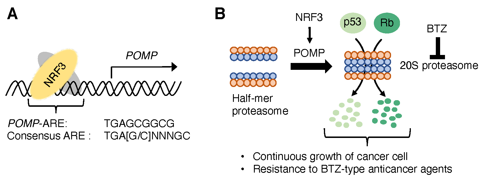
2.1. POMP, a 20S Proteasome Assembly Factor

The 26S proteasome is essential for ubiquitin-dependent protein degradation and consists of two subcomplexes: a 20S proteasome and a 19S-regulatory particle (RP) [34]. Several chaperones strictly coordinate the assembly of 20S proteasome and 19S-RP [35,36]. NRF1 induces the expression of almost all proteasome-related genes required for 26S proteasome [7]. Meanwhile, NRF3 does not affect the expression of almost all proteasome-related genes, but induces the gene expression of POMP [31], a chaperone of the 20S proteasome assembly [37]. ChIP experiments showed an ARE-like sequence (TGAGCGGCG) near the transcription start site of the POMP gene as the NRF3 binding region [31] (Figure 1A). Furthermore, POMP-ARE mutations using CRISPR/Cas9-based genome editing reduced not only NRF3 recruitment on POMP-ARE but also POMP gene expression induced by NRF3 [31]. Proteasome activity assays using a fluorogenic substrate showed that NRF3 increases the amount and activity of 20S proteasome [31] (Figure 1B). These results provided direct evidence that POMP is an NRF3 target gene for enhancing the 20S proteasome activity.

2.2. NRF3-POMP-20S Proteasome Assembly Axis for Cancer Development

The 20S proteasome, a homodimer of a half-mer proteasome composed of an outer α-ring and an inner β-ring, contains proteolytic sites with different specificities: chymotrypsin-, caspase-, and trypsin-like activities. Meanwhile, the 20S proteasome lacks the 19S-RP that selects and unfolds ubiquitin substrates. Previous studies suggested that the 20S proteasome contributes to the ubiquitin-independent degradation of several tumor suppressor proteins, such as p53 and retinoblastoma (Rb) [38]. Surprisingly, NRF3 decreases p53 and Rb proteins without alteration of their mRNA levels under treatment with a ubiquitin-activating enzyme E1 inhibitor TAK-243, which inhibits 26S proteasome-mediated protein degradation by covalently binding with ubiquitin proteins [39] (Figure 1B). Furthermore, POMP-ARE mutation impairs the NRF3-mediated reduction in p53 and Rb protein, irrespective of TAK-243 treatment. More importantly, p53 and Rb inhibit cancer cell proliferation by inducing cell-cycle arrest or apoptosis in response to DNA damage [40]. NRF3 suppresses the expression of p53 target genes, including the cell-cycle inhibitory effector gene p21 [41] and the proapoptotic gene PUMA (p53 upregulated modulator of apoptosis) [42]. NRF3 further inhibits p53-dependent cell-cycle arrest and apoptosis induction, leading to continuous cancer cell growth [31] (Figure 1B). These results indicated that the NRF3-POMP-20S proteasome assembly axis affects the ubiquitin-independent degradation of endogenous p53 and Rb proteins.
The proteasome is a target for cancer chemotherapy, and several proteasome inhibitors have been developed as anticancer agents. Among proteasome inhibitor anticancer agents, bortezomib (BTZ) inhibits both 20S and 26S proteolytic activities by binding to catalytic sites within the 20S proteasome [43]. Expectedly, upregulation of the NRF3-POMP-20S proteasome axis confers resistance to BTZ [31] (Figure 1B). Furthermore, xenograft and hepatic metastatic mouse models showed that NRF3 increases tumorigenesis and metastasis, whereas POMP-ARE mutation inhibits this tumor burden [31]. More importantly, clinical analyses indicated a negative correlation between POMP/NRF3 mRNA levels and the survival rates of patients with colorectal adenocarcinoma, where NRF3 is highly expressed [31]. These insights shed light on the crucial function of the NRF3-POMP-20S proteasome axis on cancer development, by inhibiting tumor suppression signals of p53 and Rb through ubiquitin-independent degradation. The upregulation of the axis also confers resistance to a BTZ-type proteasome inhibitor [44].

3. Complementary Maintenance of Proteasome with NRF1

3.1. CPEB3, a Translational Repressor of NRF1

NRF3 increases 20S proteasome activity through POMP expression [31], whereas NRF1 maintains 26S proteasome activity by inducing the expression of almost all proteasome-related genes under proteasome inhibition [7], implying the biological relevance of NRF1 and NRF3 for proteasome activity. In fact, the double knockdown of NRF1 and NRF3 impairs basal proteasome activity in living cells [32]. Compared to the single knockdown of NRF1 or NRF3, double knockdown of NRF1 and NRF3 reduces several proteasome-related genes, including PSMB3, PSMB7, PSMC2, PSMD3, PSMG2, PSMG3, and POMP [32]. ChIP experiments showed an ARE sequence near the transcription start site of each gene. These results indicated that NRF1 and NRF3 complementarily induce the expression of several proteasome-related genes to maintain the proteasome activity [32].
Interestingly, NRF3 represses the translation of NRF1 proteins by decreasing the amount of NRF1 mRNA in polysomes, although NRF3 does not only affect the levels of NRF1 mRNA but also the degradation of NRF1 proteins [32]. Gene expression analysis identified CPEB3 as the candidate NRF3 target gene for this NRF1 translation repression [32] (Figure 2A). CPEB family proteins recognize a CPEB recognition motif (5′-UUUUA-3′, CPE) in the 3′-untranslated region (UTR) of a target gene for translation regulation [45]. CPEB3 interacts with the NRF1–3′-UTR which contains five CPEs, decreasing NRF1 protein levels and the amount of NRF1 mRNA in polysomes [32] (Figure 2A). Meanwhile, NRF3 deficiency or CPE mutation of the NRF1–3′-UTR increases NRF1 translation [32] (Figure 2B). These results indicate that NRF3 directly induces CPEB3 gene expression, and then CPEB3 inhibits ribosome recruitment to NRF1 mRNA, resulting in the repression of NRF1 translation.

3.2. Clinical Significance of the NRF3-CPEB3-NRF1 Translational Repression Axis

In NRF3-deficienct cells, CPEB3 represses NRF1 translation and then reduces the expression levels of PSMB3, PSMB7, PSMC2, PSMG2, and POMP genes, resulting in the suppression of 26S proteasome activity [32]. CPEB3 also confers resistance to BTZ in NRF3-deficient cells [32]. Furthermore, colorectal cancer patients with higher CPEB3/NRF3-expressing tumors exhibited shorter overall survival rates, but higher CPEB3/NRF1 expression was not associated with poor prognosis [32]. These results suggested that the NRF3-CPEB3-NRF1 translational repression axis is involved in cancer development by shunting ubiquitin-dependent protein degradation through the NRF1–26S proteasome regulatory axis to ubiquitin-independent protein degradation through the POMP-20S proteasome axis (Figure 2).

4. Reprogramming of Lipid Metabolism

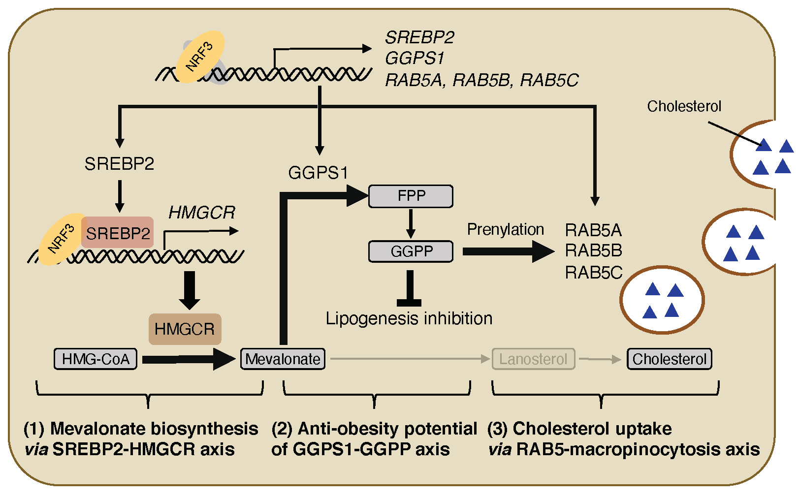
4.1. NRF3-SREBP2-HMGCR Axis for Mevalonate Biosynthesis

Lipids, such as cholesterol and fatty acids, influence cell signaling, energy storage, and membrane formation. SREBPs are membrane-bound transcription factors crucial for lipid metabolism [46]. In response to cholesterol depletion, SREBP1 and SREBP2 proteins are cleaved in the Golgi apparatus, resulting in the translocation to the nucleus. SREBP1 induces the gene expression of enzymes required for fatty acid biosynthesis and adipocyte differentiation, whereas SREBP2 induces the gene expression of enzymes required for mevalonate/cholesterol biosynthesis.
NRF3 induces the expression of several SREBP2 target genes, such as hydroxy-methylglutaryl-CoA synthase 1 (HMGCS1) and HMGCR, encoding a rate-limiting enzyme in mevalonate/cholesterol biosynthesis [47] (Figure 3(1)). ChIP experiments with previously published ChIP sequencing data [48] indicated that NRF3 binds to both AREs in SREBP2 and HMGCR promoters, and that SREBP2 binds to the site nearby HMGCR-ARE [33]. Moreover, NRF3 interacts with the active form of SREBP2 [33] (Figure 3(1)), implying that NRF3 and SREBP2 form a transcriptional complex for HMGCR gene expression. Luciferase reporter assays containing both ARE and SREBP2 binding sites showed a synergistic transcriptional activity of NRF3 and SREBP2 through the HMGCR promoter [33]. Taken together, NRF3 promotes mevalonate biosynthesis by upregulating the SREBP2-HMGCR axis (Figure 3(1)).

4.2. NRF3-GGPS1-GGPP Production Axis for Lipogenesis Inhibition

Interestingly, NRF3 does not affect the intracellular levels of cholesterol, even if NRF3 increases the expression levels and enzymatic activity of HMGCR. Meanwhile, NRF3 reduces that of lanosterol [33]. Lanosterol is not only a precursor of cholesterol but also a downstream metabolite of farnesyl pyrophosphate, which is also metabolized to GGPP in a reaction catalyzed by GGPS1 (Figure 3(2)). Furthermore, NRF3 directly induces GGPS1 expression [33], implying that NRF3 reprograms cholesterol biogenesis to the production of GGPP rather than lanosterol. GGPP suppresses SREBP1-dependent fatty acid biosynthesis and intracellular lipid accumulation [49,50]. In fact, DNA microarray analysis showed a negative correlation between the expression levels of NRF3 and genes related to fatty acid metabolism [33]. More directly, intracellular levels of neutral lipids are increased by NRF3 knockdown and reduced by GGPP treatment [33] (Figure 3(2)). Consistently, a few body mass index-associated genomic loci near the NRF3 gene have been identified previously [51,52]. These results indicated the potential role of the NRF3-GGPS1-GGPP production axis (Figure 3(2)).

4.3. NRF3-RAB5-Macropincytosis Induction Axis for Cholesterol Uptake

Intracellular cholesterol is derived from not only de novo biosynthesis but also endocytic uptake [53]. NRF3 decreases lanosterol levels, but it does not change cholesterol levels in cells [33] (Figure 3(3)), implying that NRF3 enhances endocytosis for cholesterol uptake to compensate for the potential depletion in cholesterol levels following lanosterol reduction. Low-density lipoprotein receptor (LDLR) is a key endocytosis regulator of LDL [54]. However, NRF3 does not induce LDLR gene expression. Meanwhile, NRF3 induces the gene expression of three isoforms of RAB5A, RAB5B, and RAB5C [33] (Figure 3(3)). These RAB5 proteins act as early endocytosis regulators [55] and are involved in macropinocytosis, a bulk and fluid-phase endocytosis process [56]. NRF3 further increases posttranslational prenylation RAB5 proteins [33] essential for proper localization and function in membranes [57]. The previous section (Figure 3(2)) described that NRF3 induces the production of GGPP, which functions as a required substrate for protein prenylation [58]. Altogether, NRF3 enhances RAB5-mediated endocytosis rather than LDLR-mediated endocytosis for cholesterol uptake through GGPP production (Figure 3, (3)). NRF3 enhances the uptake of fluorogenic LDL in a RAB5-dependent manner. Moreover, NRF3-enhanced uptake of other fluorogenic macropinocytosis indicators based on 70 kDa dextran and bovine serum albumin is abolished by treatment with 5-(N-ethyl-N-isopropyl)amiloride [33], also known as an inhibitor of macropinocytosis and a selective blocker of Na+/H+ exchanger [59]. Similarly, NRF3 enhances the uptake of two fluorogenic cholesterols through macropinocytosis [33]. These results indicated the crucial function of the NRF3-RAB5-macropinocysosis induction (NRF3-RAB5-macropinocysosis) axis on cholesterol uptake (Figure 3(3)). The next section discusses the pathophysiological potential of this axis.

5. Concluding Remarks

Increased ubiquitin-independent proteasomal activity causes tumor growth, metastasis, and resistance to the proteasome inhibitor BTZ. NRF3 induces POMP expression, leading to ubiquitin-independent protein degradation of tumor suppressors Rb and p53 (Figure 1). Upregulation of the POMP-20S proteasome axis further results in poor prognosis of colorectal cancer patients [31]. NRF3 also induces the expression of proteasome-related genes in parallel with NRF1 translational repression by inducing CPEB3 expression [32] (Figure 2A). If the NRF3 gene is deficient, NRF1 escapes from CPEB3-mediated translational repression, and complementarily plays a transcriptional role for the robust maintenance of basal proteasome activity in cancer cells (Figure 2B). Although NRF3 shares several target genes with NRF1 and NRF2 on ARE [30,60], this review showed the translation-mediated crosstalk between NRF3 and NRF1.
Nrf1 is ubiquitously expressed in normal tissues, and Nrf1 knockout mice suffer from embryonic lethality [8]. Meanwhile, Nrf3 expression levels are low, except in several mouse tissues, such as the placenta [5,6], and Nrf3 knockout mice do not exhibit any obvious abnormalities under normal physiological conditions [18,19,20]. However, NRF3 is highly expressed in many cancer cells [31], implying that the proteasome in cancer or normal cells is maintained through the CPEB3-NRF1 axis or the negative feedback regulation of NRF1. Higher CPEB3/NRF3 expression, but not higher CPEB3/NRF1 expression, is associated with poor prognosis of cancer patients [32]. Therefore, NRF1 maintains a 26S proteasome activity for normal development, whereas NRF3 alternatively maintains 20S proteasome activity for cancer development through both POMP-20S proteasome and CPEB3-NRF1 axes.
Furthermore, NRF3 is involved in lipid metabolism through three regulatory axes [33] (Figure 3): (1) NRF3 induces the gene expression of SREBP2 required for cholesterol biosynthesis through the mevalonate pathway. NRF3 also leads to SREBP2 activation through direct induction of gene expression. NRF3 and SREBP2 synergistically induce HMGCR expression and the following mevalonate biosynthesis. (2) NRF3 then upregulates GGPS1-mediated GGPP production for lipogenesis inhibition. (3) In parallel, NRF3 confers RAB5-mediated induction of macropinocytosis for cholesterol uptake. This gene expression is induced in colon and/or rectal tissue of newly generated NRF3-transgenic mice [33]. Dietary cholesterol in the blood is absorbed in the intestine [61], and its dysregulation is associated with obesity, resulting in an increased risk of cardiovascular diseases (CVD) and colorectal cancer [62,63]. The gut microbiota has been identified as a CVD risk factor and regulates host cholesterol homeostasis [64,65,66], suggesting the pathophysiological potential of the NRF3-regulated host lipid metabolism to the gut–heart connection through the gut microbiota.
Epidemiological studies have associated obesity with a range of cancers [67,68]. Furthermore, many efforts have been made to identify the key factor for obesity-induced cancer, including insulin resistance, increased steroid hormones and adipokine, and aberrant inflammation [69]. However, these findings implied possible opposite roles for NRF3 in obesity-induced cancer development (Figure 4): NRF3-SREBP2-HMGCR and the following NRF3-GGPS1-GGPS axes regulate lipid homeostasis and confer resistance to obesity through lipogenesis inhibition, while the NRF3-POMP-20S proteasome and NRF3-CPEB3-NRF1 axes regulate protein homeostasis and confer ubiquitin-independent protein degradation for continuous cancer cell growth. This review further showed that NRF3 maintains cholesterol homeostasis through the RAB5-macropinocytosis axis (Figure 3, (3)). Interestingly, macropinocytosis is associated with obesity-related disorders, such as increased diabetic mouse macrophages and chronic inflammation [70,71]. Furthermore, NRF2 induces macropinocytosis and contributes to the escape of autophagy-deficient cancer cells from metabolic decline and anticancer drugs, such as gemcitabine and doxorubicin, which target the anabolic dependencies of cancer cells [72,73]. These insights implied the possibility that the NRF3-RAB5-macropinocytosis axis paradoxically interferes with obesity-induced cancer development through attenuation of obesity-induced inflammation and resistance to therapy targeting cancer anabolism (Figure 4).
A big issue of the NRF3 study is identifying the endogenous cue of NRF3 activation, although NRF3 is experimentally activated by treatment with a proteasome inhibitor. Recently, NRF1 senses cholesterol levels in the ER membrane through the cholesterol recognition amino acid consensus motif domain (CRAC), and it is activated in response to cholesterol depletion [74]. Because the CRAC domain is conserved in NRF3 proteins [27], NRF3 acts as a cholesterol sensor in the ER membrane similarly to NRF1.

Author Contributions

T.W. and A.K. were involved in the literature survey and wrote the paper. All authors have read and agreed to the published version of the manuscript.

Funding

This work was supported by the JSPS (grant nos. 17K18234 (T.W.), 19K07650 (T.W.), 16H03265 (A.K.), 19K22826 (A.K.), 20H04135 (A.K.), and 21K19743 (A.K.)), the Uehara Memorial Foundation (T.W.), the Harris Research Institute of Doshisha University (T.W.), and the Mitsubishi Foundation (A.K.).

Institutional Review Board Statement

Not applicable.

Informed Consent Statement

Not applicable.

Data Availability Statement

Not applicable.

Acknowledgments

The authors thank all members of our laboratory and the collaborators for their contributions to the research.

Conflicts of Interest

The authors declare no conflict of interest.

References

  1. Guang, M.H.Z.; Kavanagh, E.; Dunne, L.; Dowling, P.; Zhang, L.; Lindsay, S.; Bazou, D.; Goh, C.; Hanley, C.; Bianchi, G.; et al. Targeting Proteotoxic Stress in Cancer: A Review of the Role that Protein Quality Control Pathways Play in Oncogenesis. Cancers 2019, 11, 66. [Google Scholar] [CrossRef] [Green Version]
  2. Su, K.-H.; Dai, C. Metabolic control of the proteotoxic stress response: Implications in diabetes mellitus and neurodegenerative disorders. Cell. Mol. Life Sci. 2016, 73, 4231–4248. [Google Scholar] [CrossRef] [Green Version]
  3. Munir, R.; Lisec, J.; Swinnen, J.V.; Zaidi, N. Lipid metabolism in cancer cells under metabolic stress. Br. J. Cancer 2019, 120, 1090–1098. [Google Scholar] [CrossRef]
  4. Yadav, R.S.; Tiwari, N.K. Lipid Integration in Neurodegeneration: An Overview of Alzheimer’s Disease. Mol. Neurobiol. 2014, 50, 168–176. [Google Scholar] [CrossRef]
  5. Kobayashi, A.; Ito, E.; Toki, T.; Kogame, K.; Takahashi, S.; Igarashi, K.; Hayashi, N.; Yamamoto, M. Molecular Cloning and Functional Characterization of a New Cap’n’ Collar Family Transcription Factor Nrf3. J. Biol. Chem. 1999, 274, 6443–6452. [Google Scholar] [CrossRef] [Green Version]
  6. Chénais, B.; Derjuga, A.; Massrieh, W.; Red-Horse, K.; Bellingard, V.; Fisher, S.J.; Blank, V. Functional and Placental Expression Analysis of the Human NRF3 Transcription Factor. Mol. Endocrinol. 2005, 19, 125–137. [Google Scholar] [CrossRef] [Green Version]
  7. Radhakrishnan, S.K.; Lee, C.S.; Young, P.; Beskow, A.; Chan, J.Y.; Deshaies, R.J. Transcription Factor Nrf1 Mediates the Proteasome Recovery Pathway after Proteasome Inhibition in Mammalian Cells. Mol. Cell 2010, 38, 17–28. [Google Scholar] [CrossRef] [Green Version]
  8. Chan, J.Y.; Kwong, M.; Lu, R.; Chang, J.; Wang, B.; Yen, T.; Kan, Y.W. Targeted disruption of the ubiquitous CNC-bZIP transcription factor, Nrf-1, results in anemia and embryonic lethality in mice. EMBO J. 1998, 17, 1779–1787. [Google Scholar] [CrossRef] [Green Version]
  9. Kobayashi, A.; Tsukide, T.; Miyasaka, T.; Morita, T.; Mizoroki, T.; Saito, Y.; Ihara, Y.; Takashima, A.; Noguchi, N.; Fukamizu, A.; et al. Central nervous system-specific deletion of transcription factor Nrf1 causes progressive motor neuronal dysfunction. Genes Cells 2011, 16, 692–703. [Google Scholar] [CrossRef]
  10. Kim, J.; Xing, W.; Wergedal, J.; Chan, J.Y.; Mohan, S. Targeted disruption of nuclear factor erythroid-derived 2-like 1 in osteoblasts reduces bone size and bone formation in mice. Physiol. Genom. 2010, 40, 100–110. [Google Scholar] [CrossRef] [Green Version]
  11. Lee, C.S.; Hu, T.; Nguyen, J.M.; Zhang, J.; Martin, M.V.; Vawter, M.P.; Huang, E.J.; Chan, J.Y. Loss of nuclear factor E2-related factor 1 in the brain leads to dysregulation of proteasome gene expression and neurodegeneration. Proc. Natl. Acad. Sci. USA 2011, 108, 8408–8413. [Google Scholar] [CrossRef] [Green Version]
  12. Xu, Z.; Chen, L.; Leung, L.; Yen, T.S.B.; Lee, C.; Chan, J.Y. Liver-specific inactivation of the Nrf1 gene in adult mouse leads to nonalcoholic steatohepatitis and hepatic neoplasia. Proc. Natl. Acad. Sci. USA 2005, 102, 4120–4125. [Google Scholar] [CrossRef] [Green Version]
  13. Yamamoto, T.; Suzuki, T.; Kobayashi, A.; Wakabayashi, J.; Maher, J.; Motohashi, H.; Yamamoto, M. Physiological Significance of Reactive Cysteine Residues of Keap1 in Determining Nrf2 Activity. Mol. Cell. Biol. 2008, 28, 2758–2770. [Google Scholar] [CrossRef] [Green Version]
  14. Romero, R.; Sayin, V.I.; Davidson, S.M.; Bauer, M.R.; Singh, S.X.; Leboeuf, S.E.; Karakousi, T.R.; Ellis, D.C.; Bhutkar, A.; Sánchez-Rivera, F.J.; et al. Keap1 loss promotes Kras-driven lung cancer and results in dependence on glutaminolysis. Nat. Med. 2017, 23, 1362–1368. [Google Scholar] [CrossRef] [Green Version]
  15. Mitsuishi, Y.; Taguchi, K.; Kawatani, Y.; Shibata, T.; Nukiwa, T.; Aburatani, H.; Yamamoto, M.; Motohashi, H. Nrf2 Redirects Glucose and Glutamine into Anabolic Pathways in Metabolic Reprogramming. Cancer Cell 2012, 22, 66–79. [Google Scholar] [CrossRef] [Green Version]
  16. Itoh, K.; Chiba, T.; Takahashi, S.; Ishii, T.; Igarashi, K.; Katoh, Y.; Oyake, T.; Hayashi, N.; Satoh, K.; Hatayama, I.; et al. An Nrf2/Small Maf Heterodimer Mediates the Induction of Phase II Detoxifying Enzyme Genes through Antioxidant Response Elements. Biochem. Biophys. Res. Commun. 1997, 236, 313–322. [Google Scholar] [CrossRef]
  17. Wakabayashi, N.; Itoh, K.; Wakabayashi, J.; Motohashi, H.; Noda, S.; Takahashi, S.; Imakado, S.; Kotsuji, T.; Otsuka, F.; Roop, D.R.; et al. Keap1-null mutation leads to postnatal lethality due to constitutive Nrf2 activation. Nat. Genet. 2003, 35, 238–245. [Google Scholar] [CrossRef]
  18. Derjuga, A.; Gourley, T.S.; Holm, T.M.; Heng, H.H.Q.; Shivdasani, R.A.; Ahmed, R.; Andrews, N.; Blank, V. Complexity of CNC Transcription Factors As Revealed by Gene Targeting of the Nrf3 Locus. Mol. Cell. Biol. 2004, 24, 3286–3294. [Google Scholar] [CrossRef] [Green Version]
  19. Kobayashi, A.; Ohta, T.; Yamamoto, M. Unique Function of the Nrf2–Keap1 Pathway in the Inducible Expression of Antioxidant and Detoxifying Enzymes. Methods Enzymol. 2004, 378, 273–286. [Google Scholar] [CrossRef]
  20. Chevillard, G.; Blank, V. NFE2L3 (NRF3): The Cinderella of the Cap‘n’Collar transcription factors. Cell. Mol. Life Sci. 2011, 68, 3337–3348. [Google Scholar] [CrossRef]
  21. Bury, M.; Le Calvé, B.; Lessard, F.; Maso, T.D.; Saliba, J.; Michiels, C.; Ferbeyre, G.; Blank, V. NFE2L3 Controls Colon Cancer Cell Growth through Regulation of DUX4, a CDK1 Inhibitor. Cell Rep. 2019, 29, 1469–1481.e9. [Google Scholar] [CrossRef]
  22. Wang, H.; Zhan, M.; Yang, R.; Shi, Y.; Liu, Q.; Wang, J. Elevated expression of NFE2L3 predicts the poor prognosis of pancreatic cancer patients. Cell Cycle 2018, 17, 2164–2174. [Google Scholar] [CrossRef]
  23. Siegenthaler, B.; Defila, C.; Muzumdar, S.; Beer, H.-D.; Meyer, M.; Tanner, S.; Bloch, W.; Blank, V.; Schäfer, M.; Werner, S. Nrf3 promotes UV-induced keratinocyte apoptosis through suppression of cell adhesion. Cell Death Differ. 2018, 25, 1749–1765. [Google Scholar] [CrossRef] [Green Version]
  24. Aono, S.; Hatanaka, A.; Hatanaka, A.; Gao, Y.; Hippo, Y.; Taketo, M.M.; Waku, T.; Kobayashi, A. β-Catenin/TCF4 Complex-Mediated Induction of the NRF3 (NFE2L3) Gene in Cancer Cells. Int. J. Mol. Sci. 2019, 20, 3344. [Google Scholar] [CrossRef] [Green Version]
  25. Chowdhury, A.M.M.A.; Katoh, H.; Hatanaka, A.; Iwanari, H.; Nakamura, N.; Hamakubo, T.; Natsume, T.; Waku, T.; Kobayashi, A. Multiple regulatory mechanisms of the biological function of NRF3 (NFE2L3) control cancer cell proliferation. Sci. Rep. 2017, 7, 1–14. [Google Scholar] [CrossRef]
  26. Koizumi, S.; Irie, T.; Hirayama, S.; Sakurai, Y.; Yashiroda, H.; Naguro, I.; Ichijo, H.; Hamazaki, J.; Murata, S. The aspartyl protease DDI2 activates Nrf1 to compensate for proteasome dysfunction. eLife 2016, 5, 1–10. [Google Scholar] [CrossRef]
  27. Zhang, Y.; Kobayashi, A.; Yamamoto, M.; Hayes, J. The Nrf3 Transcription Factor Is a Membrane-bound Glycoprotein Targeted to the Endoplasmic Reticulum through Its N-terminal Homology Box 1 Sequence. J. Biol. Chem. 2009, 284, 3195–3210. [Google Scholar] [CrossRef] [Green Version]
  28. Yamamoto, M.; Kensler, T.W.; Motohashi, H. The KEAP1-NRF2 System: A Thiol-Based Sensor-Effector Apparatus for Maintaining Redox Homeostasis. Physiol. Rev. 2018, 98, 1169–1203. [Google Scholar] [CrossRef] [Green Version]
  29. Motohashi, H.; O’Connor, T.; Katsuoka, F.; Engel, J.D.; Yamamoto, M. Integration and diversity of the regulatory network composed of Maf and CNC families of transcription factors. Gene 2002, 294, 1–12. [Google Scholar] [CrossRef]
  30. Liu, P.; Kerins, M.J.; Tian, W.; Neupane, D.; Zhang, D.D.; Ooi, A. Differential and overlapping targets of the transcriptional regulators NRF1, NRF2, and NRF3 in human cells. J. Biol. Chem. 2019, 294, 18131–18149. [Google Scholar] [CrossRef]
  31. Waku, T.; Nakamura, N.; Koji, M.; Watanabe, H.; Katoh, H.; Tatsumi, C.; Tamura, N.; Hatanaka, A.; Hirose, S.; Katayama, H.; et al. NRF3-POMP-20S Proteasome Assembly Axis Promotes Cancer Development via Ubiquitin-Independent Proteolysis of p53 and Retinoblastoma Protein. Mol. Cell. Biol. 2020, 40. [Google Scholar] [CrossRef] [Green Version]
  32. Waku, T.; Katayama, H.; Hiraoka, M.; Hatanaka, A.; Nakamura, N.; Tanaka, Y.; Tamura, N.; Watanabe, A.; Kobayashi, A. NFE2L1 and NFE2L3 Complementarily Maintain Basal Proteasome Activity in Cancer Cells through CPEB3-Mediated Translational Repression. Mol. Cell. Biol. 2020, 40. [Google Scholar] [CrossRef]
  33. Waku, T.; Hagiwara, T.; Tamura, N.; Atsumi, Y.; Urano, Y.; Suzuki, M.; Iwami, T.; Sato, K.; Yamamoto, M.; Noguchi, N.; et al. NRF3 upregulates gene expression in SREBP2-dependent mevalonate pathway with cholesterol uptake and lipogenesis inhibition. iScience 2021, 24, 103180. [Google Scholar] [CrossRef]
  34. Coux, O.; Tanaka, K.; Goldberg, A.L. Structure and functions of the 20s and 26s proteasomes. Annu. Rev. Biochem. 1996, 65, 801–847. [Google Scholar] [CrossRef]
  35. Murata, S.; Yashiroda, H.; Tanaka, K. Molecular mechanisms of proteasome assembly. Nat. Rev. Mol. Cell Biol. 2009, 10, 104–115. [Google Scholar] [CrossRef]
  36. Gallastegui, N.; Groll, M. The 26S proteasome: Assembly and function of a destructive machine. Trends Biochem. Sci. 2010, 35, 634–642. [Google Scholar] [CrossRef]
  37. Witt, E.; Zantopf, D.; Schmidt, M.; Kraft, R.; Kloetzel, P.-M.; Krüger, E. Characterisation of the newly identified human Ump1 homologue POMP and analysis of LMP7(β5i) incorporation into 20 S proteasomes. J. Mol. Biol. 2000, 301, 1–9. [Google Scholar] [CrossRef]
  38. Ben-Nissan, G.; Sharon, M. Regulating the 20S Proteasome Ubiquitin-Independent Degradation Pathway. Biomolecules 2014, 4, 862–884. [Google Scholar] [CrossRef] [Green Version]
  39. Hyer, M.L.; A Milhollen, M.; Ciavarri, J.; Fleming, P.; Traore, T.; Sappal, D.; Huck, J.; Shi, J.; Gavin, J.; Brownell, J.; et al. A small-molecule inhibitor of the ubiquitin activating enzyme for cancer treatment. Nat. Med. 2018, 24, 186–193. [Google Scholar] [CrossRef]
  40. Sherr, C.J.; McCormick, F. The RB and p53 pathways in cancer. Cancer Cell 2002, 2, 103–112. [Google Scholar] [CrossRef] [Green Version]
  41. Waldman, T.; Kinzler, K.W.; Vogelstein, B. p21 is necessary for the p53-mediated G1 arrest in human cancer cells. Cancer Res. 1995, 55. [Google Scholar]
  42. Nakano, K.; Vousden, K.H. PUMA, a Novel Proapoptotic Gene, Is Induced by p53. Mol. Cell 2001, 7, 683–694. [Google Scholar] [CrossRef]
  43. Groll, M.; Berkers, C.R.; Ploegh, H.L.; Ovaa, H. Crystal Structure of the Boronic Acid-Based Proteasome Inhibitor Bortezomib in Complex with the Yeast 20S Proteasome. Structure 2006, 14, 451–456. [Google Scholar] [CrossRef] [Green Version]
  44. Kobayashi, A. Roles of NRF3 in the Hallmarks of Cancer: Proteasomal Inactivation of Tumor Suppressors. Cancers 2020, 12, 2681. [Google Scholar] [CrossRef]
  45. Fernández-Miranda, G.; Méndez, R. The CPEB-family of proteins, translational control in senescence and cancer. Ageing Res. Rev. 2012, 11, 460–472. [Google Scholar] [CrossRef]
  46. Horton, J.D.; Goldstein, J.L.; Brown, M.S. SREBPs: Activators of the complete program of cholesterol and fatty acid synthesis in the liver. J. Clin. Investig. 2002, 109, 1125–1131. [Google Scholar] [CrossRef]
  47. Xue, L.; Qi, H.; Zhang, H.; Ding, L.; Huang, Q.; Zhao, D.; Wu, B.J.; Li, X. Targeting SREBP-2-Regulated Mevalonate Metabolism for Cancer Therapy. Front. Oncol. 2020, 10, 1510. [Google Scholar] [CrossRef]
  48. Landt, S.G.; Marinov, G.; Kundaje, A.; Kheradpour, P.; Pauli, F.; Batzoglou, S.; Bernstein, B.E.; Bickel, P.; Brown, J.B.; Cayting, P.; et al. ChIP-seq guidelines and practices of the ENCODE and modENCODE consortia. Genome Res. 2012, 22, 1813–1831. [Google Scholar] [CrossRef] [Green Version]
  49. Bertolio, R.; Napoletano, F.; Mano, M.; Maurer-Stroh, S.; Fantuz, M.; Zannini, A.; Bicciato, S.; Sorrentino, G.; Del Sal, G. Sterol regulatory element binding protein 1 couples mechanical cues and lipid metabolism. Nat. Commun. 2019, 10, 1–11. [Google Scholar] [CrossRef] [Green Version]
  50. Yeh, Y.-S.; Goto, T.; Takahashi, N.; Egawa, K.; Takahashi, H.; Jheng, H.-F.; Kim, Y.-I.; Kawada, T. Geranylgeranyl pyrophosphate performs as an endogenous regulator of adipocyte function via suppressing the LXR pathway. Biochem. Biophys. Res. Commun. 2016, 478, 1317–1322. [Google Scholar] [CrossRef]
  51. Lamiquiz-Moneo, I.; Mateo-Gallego, R.; Bea, A.M.; Dehesa-García, B.; Pérez-Calahorra, S.; Marco-Benedí, V.; Baila-Rueda, L.; Laclaustra, M.; Civeira, F.; Cenarro, A. Genetic predictors of weight loss in overweight and obese subjects. Sci. Rep. 2019, 9, 1–9. [Google Scholar] [CrossRef] [Green Version]
  52. Monda, K.L.; Chen, G.K.; Taylor, K.C.; Palmer, C.; Edwards, T.L.; A Lange, L.; Ng, M.C.Y.; A Adeyemo, A.; A Allison, M.; et al.; NABEC Consortium A meta-analysis identifies new loci associated with body mass index in individuals of African ancestry. Nat. Genet. 2013, 45, 690–696. [Google Scholar] [CrossRef] [Green Version]
  53. Steck, T.L.; Lange, Y. Cell cholesterol homeostasis: Mediation by active cholesterol. Trends Cell Biol. 2010, 20, 680–687. [Google Scholar] [CrossRef] [Green Version]
  54. Goldstein, J.L.; Brown, M.S. The LDL Receptor. Arter. Thromb. Vasc. Biol. 2009, 29, 431–438. [Google Scholar] [CrossRef] [Green Version]
  55. Zeigerer, A.; Gilleron, J.; Bogorad, R.L.; Marsico, G.; Nonaka, H.; Seifert, S.; Epstein-Barash, H.; Kuchimanchi, S.; Peng, C.G.; Ruda, V.M.; et al. Rab5 is necessary for the biogenesis of the endolysosomal system in vivo. Nat. Cell Biol. 2012, 485, 465–470. [Google Scholar] [CrossRef]
  56. Kruth, H.S.; Jones, N.L.; Huang, W.; Zhao, B.; Ishii, I.; Chang, J.; Combs, C.A.; Malide, D.; Zhang, W.-Y. Macropinocytosis Is the Endocytic Pathway That Mediates Macrophage Foam Cell Formation with Native Low Density Lipoprotein. J. Biol. Chem. 2005, 280, 2352–2360. [Google Scholar] [CrossRef] [Green Version]
  57. Barbieri, M.A.; Hoffenberg, S.; Roberts, R.; Mukhopadhyay, A.; Pomrehn, A.; Dickey, B.F.; Stahl, P.D. Evidence for a Symmetrical Requirement for Rab5-GTP in in Vitro Endosome-Endosome Fusion. J. Biol. Chem. 1998, 273, 25850–25855. [Google Scholar] [CrossRef] [Green Version]
  58. Wang, M.; Casey, P. Protein prenylation: Unique fats make their mark on biology. Nat. Rev. Mol. Cell Biol. 2016, 17, 110–122. [Google Scholar] [CrossRef]
  59. Commisso, C.; Flinn, R.J.; Bar-Sagi, D. Determining the macropinocytic index of cells through a quantitative image-based assay. Nat. Protoc. 2014, 9, 182–192. [Google Scholar] [CrossRef] [Green Version]
  60. Sankaranarayanan, K.; Jaiswal, A.K. Nrf3 Negatively Regulates Antioxidant-response Element-mediated Expression and Antioxidant Induction of NAD(P)H:Quinone Oxidoreductase1 Gene. J. Biol. Chem. 2004, 279, 50810–50817. [Google Scholar] [CrossRef] [Green Version]
  61. Lecerf, J.-M.; de Lorgeril, M. Dietary cholesterol: From physiology to cardiovascular risk. Br. J. Nutr. 2011, 106, 6–14. [Google Scholar] [CrossRef] [Green Version]
  62. Nelson, R.H. Hyperlipidemia as a Risk Factor for Cardiovascular Disease. Prim. Care Clin. Off. Pr. 2013, 40, 195–211. [Google Scholar] [CrossRef] [Green Version]
  63. Yao, X.; Tian, Z. Dyslipidemia and colorectal cancer risk: A meta-analysis of prospective studies. Cancer Causes Control. 2015, 26, 257–268. [Google Scholar] [CrossRef]
  64. Vourakis, M.; Mayer, G.; Rousseau, G. The Role of Gut Microbiota on Cholesterol Metabolism in Atherosclerosis. Int. J. Mol. Sci. 2021, 22, 8074. [Google Scholar] [CrossRef]
  65. Kenny, D.J.; Plichta, D.R.; Shungin, D.; Koppel, N.; Hall, A.B.; Fu, B.; Vasan, R.S.; Shaw, S.Y.; Vlamakis, H.; Balskus, E.P.; et al. Cholesterol Metabolism by Uncultured Human Gut Bacteria Influences Host Cholesterol Level. Cell Host Microbe 2020, 28, 245–257.e6. [Google Scholar] [CrossRef]
  66. Le Roy, T.; Lécuyer, E.; Chassaing, B.; Rhimi, M.; Lhomme, M.; Boudebbouze, S.; Ichou, F.; Haro Barceló, J.; Huby, T.; Guerin, M.; et al. The Intestinal Microbiota Regulates Host Cholesterol Homeostasis. BMC Biol. 2019, 17, 94. [Google Scholar] [CrossRef] [Green Version]
  67. Bianchini, F.; Kaaks, R.; Vainio, H. Overweight, obesity, and cancer risk. Lancet Oncol. 2002, 3, 565–574. [Google Scholar] [CrossRef]
  68. Calle, E.E.; Rodriguez, C.; Walker-Thurmond, K.; Thun, M.J. Overweight, Obesity, and Mortality from Cancer in a Prospectively Studied Cohort of U.S. Adults. N. Engl. J. Med. 2003, 348, 1625–1638. [Google Scholar] [CrossRef] [Green Version]
  69. Khandekar, M.J.; Cohen, P.; Spiegelman, B.M. Molecular mechanisms of cancer development in obesity. Nat. Rev. Cancer 2011, 11, 886–895. [Google Scholar] [CrossRef]
  70. Guest, C.B.; Chakour, K.S.; Freund, G.G. Macropinocytosis is decreased in diabetic mouse macrophages and is regulated by AMPK. BMC Immunol. 2008, 9, 42–48. [Google Scholar] [CrossRef] [Green Version]
  71. Wang, Y.; Tang, B.; Long, L.; Luo, P.; Xiang, W.; Li, X.; Wang, H.; Jiang, Q.; Tan, X.; Luo, S.; et al. Improvement of obesity-associated disorders by a small-molecule drug targeting mitochondria of adipose tissue macrophages. Nat. Commun. 2021, 12, 1–16. [Google Scholar] [CrossRef]
  72. Jayashankar, V.; Edinger, A.L. Macropinocytosis confers resistance to therapies targeting cancer anabolism. Nat. Commun. 2020, 11, 1–15. [Google Scholar] [CrossRef] [PubMed]
  73. Su, H.; Yang, F.; Fu, R.; Li, X.; French, R.; Mose, E.; Pu, X.; Trinh, B.; Kumar, A.; Liu, J.; et al. Cancer cells escape autophagy inhibition via NRF2-induced macropinocytosis. Cancer Cell 2021, 39, 678–693.e11. [Google Scholar] [CrossRef]
  74. Widenmaier, S.; Snyder, N.A.; Nguyen, T.B.; Arduini, A.; Lee, G.Y.; Arruda, A.P.; Saksi, J.; Bartelt, A.; Hotamisligil, G.S. NRF1 Is an ER Membrane Sensor that Is Central to Cholesterol Homeostasis. Cell 2017, 171, 1094–1109.e15. [Google Scholar] [CrossRef] [Green Version]
Figure 1. NRF3-POMP-20S proteasome assembly axis. (A) NRF3 directly induces POMP expression by binding to POMP-ARE, which is slightly different from consensus ARE. (B) Upregulation of the NRF3-POMP axis enhances a dimerization of a half-mer proteasome (known as the 20S proteasome assembly). The increased 20S proteasome confers the ubiquitin-independent degradation of p53 and Rb proteins, resulting in the rapid and continuous growth of cancer cells. Aberrant upregulation of the axis also confers resistance to a BTZ-type anticancer agent.
Ijms 22 12686 g001
Figure 2. NRF3-CPEB3-NRF1 translational repression axis. (A) In NRF3-abundant cells, NRF3 directly induces CPEB3 expression, resulting in the repression of NRF1 translation. In parallel, NRF3 confers ubiquitin (Ub)-independent protein degradation through the POMP-20S proteasome axis. (B) In NRF3-deficient cells, NRF1 escapes from CPEB3-mediated translational repression and confers ubiquitin (Ub)-dependent protein degradation.
Ijms 22 12686 g002
Figure 3. NRF3-regulated lipid metabolism through three axes. (1) NRF3 activates SREBP2 by directly inducing gene expression. NRF3 and SREBP2 synergistically induce HMGCR gene expression, promoting mevalonate biosynthesis. (2) In parallel, NRF3 induces GGPS1 expression and then reprograms cholesterol biosynthesis to GGPP production, resulting in lipogenesis inhibition. (3) NRF3 also induces the gene expression of three RAB5 isoforms, resulting in cholesterol uptake through macropinocytosis.
Ijms 22 12686 g003
Figure 4. Possible roles for NRF3 in obesity-induced cancer development.
Ijms 22 12686 g004
Publisher’s Note: MDPI stays neutral with regard to jurisdictional claims in published maps and institutional affiliations.

