Distal Lung Microenvironment Triggers Release of Mediators Recognized as Potential Systemic Biomarkers for Idiopathic Pulmonary Fibrosis
Abstract
:1. Introduction
2. Results
2.1. Proteins Released in Ex Vivo Model
2.2. Characteristics of IPF Patients and Controls
2.3. Proteins in Baseline Serum from IPF Patients Compared to Controls
2.4. Proteins in IPF Serum at Follow Up
2.5. Protein Expression and Disease Severity
2.6. Comparison between Stable and Progressive IPF Patients
2.7. Outcome of Treatment Effects in Protein Expression
3. Discussion
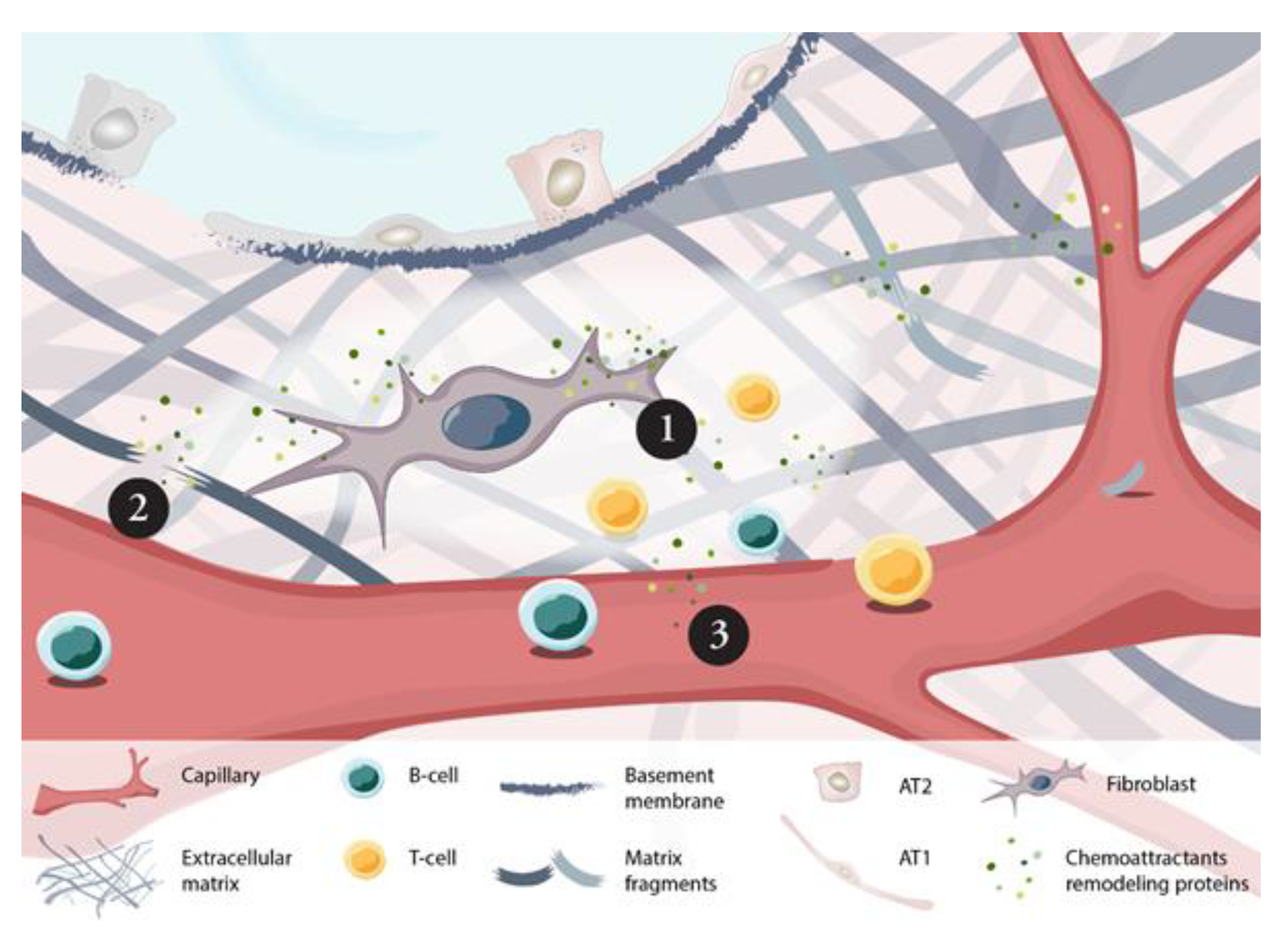
3.1. Inherent ECM Properties Trigger Fibroblast Activation
3.2. Remodeling Processes Linked to Inflammatory Processes
3.3. Inflammatory Processes Linked to Remodeling
3.4. Protein Association to Disease Severity and Progression
3.5. Progressors vs. Stable Patients
3.6. Treatment Effects
4. Materials and Methods
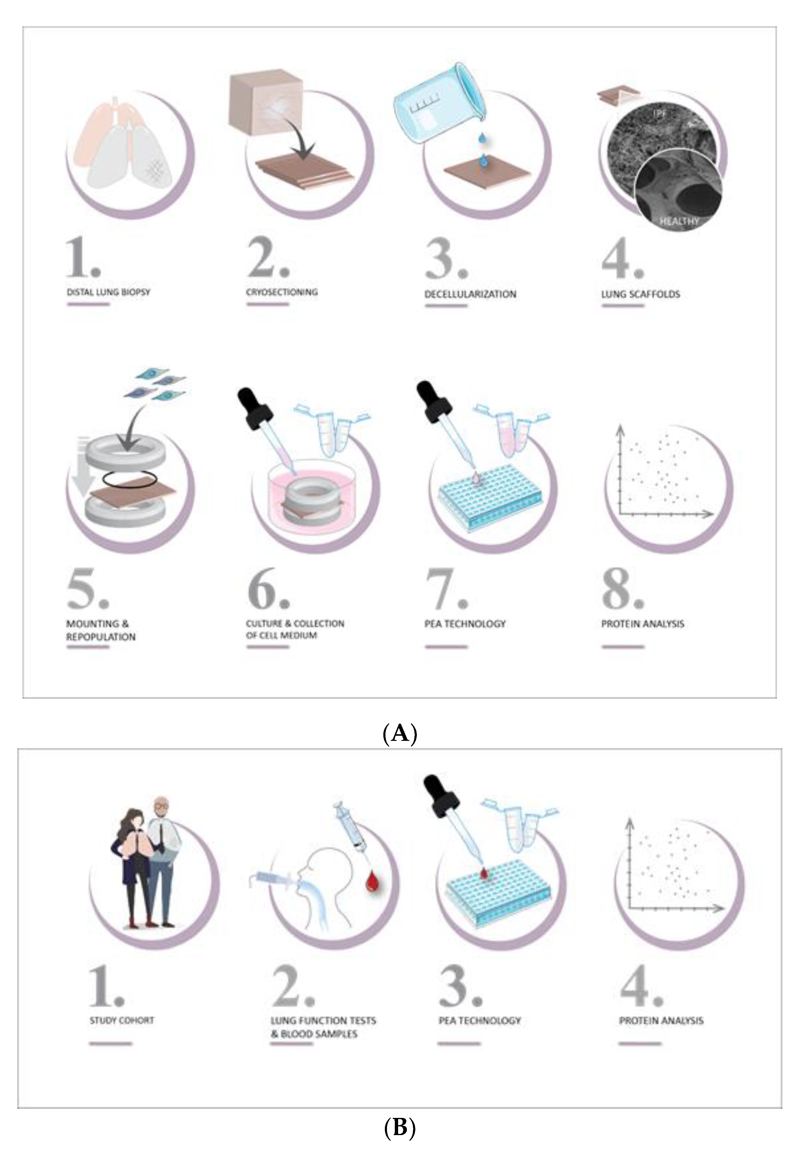
4.1. Study Design
4.2. Ex Vivo Model
4.3. Serum from IPF Patients and Controls
4.4. Protein Analysis
4.5. Statistical Analyses
5. Conclusions
Supplementary Materials
Author Contributions
Funding
Institutional Review Board Statement
Informed Consent Statement
Data Availability Statement
Acknowledgments
Conflicts of Interest
References
- Raghu, G.; Remy-Jardin, M.; Myers, J.L.; Richeldi, L.; Ryerson, C.J.; Lederer, D.J.; Behr, J.; Cottin, V.; Danoff, S.K.; Morell, F.; et al. Diagnosis of Idiopathic Pulmonary Fibrosis. An Official ATS/ERS/JRS/ALAT Clinical Practice Guideline. Am. J. Respir. Crit. Care Med. 2018, 198, e44–e68. [Google Scholar] [CrossRef]
- Ley, B.; Collard, H.R.; King, T.E., Jr. Clinical course and prediction of survival in idiopathic pulmonary fibrosis. Am. J. Respir. Crit. Care Med. 2011, 183, 431–440. [Google Scholar] [CrossRef] [PubMed]
- Stainer, A.; Faverio, P.; Busnelli, S.; Catalano, M.; Della Zoppa, M.; Marruchella, A.; Pesci, A.; Luppi, F. Molecular Biomarkers in Idiopathic Pulmonary Fibrosis: State of the Art and Future Directions. Int. J. Mol. Sci. 2021, 22, 6255. [Google Scholar] [CrossRef] [PubMed]
- Simler, N.R.; Brenchley, P.E.; Horrocks, A.W.; Greaves, S.M.; Hasleton, P.S.; Egan, J.J. Angiogenic cytokines in patients with idiopathic interstitial pneumonia. Thorax 2004, 59, 581–585. [Google Scholar] [CrossRef] [Green Version]
- Hilberg, F.; Roth, G.J.; Krssak, M.; Kautschitsch, S.; Sommergruber, W.; Tontsch-Grunt, U.; Garin-Chesa, P.; Bader, G.; Zoephel, A.; Quant, J.; et al. BIBF 1120: Triple angiokinase inhibitor with sustained receptor blockade and good antitumor efficacy. Cancer Res. 2008, 68, 4774–4782. [Google Scholar] [CrossRef] [Green Version]
- Richeldi, L.; Costabel, U.; Selman, M.; Kim, D.S.; Hansell, D.M.; Nicholson, A.G.; Brown, K.K.; Flaherty, K.R.; Noble, P.W.; Raghu, G.; et al. Efficacy of a tyrosine kinase inhibitor in idiopathic pulmonary fibrosis. N. Engl. J. Med. 2011, 365, 1079–1087. [Google Scholar] [CrossRef] [PubMed] [Green Version]
- Kotani, I.; Sato, A.; Hayakawa, H.; Urano, T.; Takada, Y.; Takada, A. Increased procoagulant and antifibrinolytic activities in the lungs with idiopathic pulmonary fibrosis. Thromb. Res. 1995, 77, 493–504. [Google Scholar] [CrossRef] [Green Version]
- Magro, C.M.; Waldman, W.J.; Knight, D.A.; Allen, J.N.; Nadasdy, T.; Frambach, G.E.; Ross, P.; Marsh, C.B. Idiopathic pulmonary fibrosis related to endothelial injury and antiendothelial cell antibodies. Hum. Immunol. 2006, 67, 284–297. [Google Scholar] [CrossRef]
- Prasad, S.; Hogaboam, C.M.; Jarai, G. Deficient repair response of IPF fibroblasts in a co-culture model of epithelial injury and repair. Fibrogenesis Tissue Repair. 2014, 7, 7. [Google Scholar] [CrossRef] [Green Version]
- Wynn, T.A. Integrating mechanisms of pulmonary fibrosis. J. Exp. Med. 2011, 208, 1339–1350. [Google Scholar] [CrossRef] [Green Version]
- King, T.E., Jr.; Pardo, A.; Selman, M. Idiopathic pulmonary fibrosis. Lancet 2011, 378, 1949–1961. [Google Scholar] [CrossRef]
- Barkauskas, C.E.; Noble, P.W. Cellular mechanisms of tissue fibrosis. 7. New insights into the cellular mechanisms of pulmonary fibrosis. Am. J. Physiol. Cell Physiol. 2014, 306, C987–C996. [Google Scholar] [CrossRef] [PubMed] [Green Version]
- Elowsson Rendin, L.; Lofdahl, A.; Ahrman, E.; Muller, C.; Notermans, T.; Michalikova, B.; Rosmark, O.; Zhou, X.H.; Dellgren, G.; Silverborn, M.; et al. Matrisome Properties of Scaffolds Direct Fibroblasts in Idiopathic Pulmonary Fibrosis. Int. J. Mol. Sci. 2019, 20, 4013. [Google Scholar] [CrossRef] [PubMed] [Green Version]
- Philp, C.J.; Siebeke, I.; Clements, D.; Miller, S.; Habgood, A.; John, A.E.; Navaratnam, V.; Hubbard, R.B.; Jenkins, G.; Johnson, S.R. Extracellular Matrix Cross-Linking Enhances Fibroblast Growth and Protects against Matrix Proteolysis in Lung Fibrosis. Am. J. Respir. Cell Mol. Biol. 2018, 58, 594–603. [Google Scholar] [CrossRef] [PubMed]
- Plataki, M.; Koutsopoulos, A.V.; Darivianaki, K.; Delides, G.; Siafakas, N.M.; Bouros, D. Expression of apoptotic and antiapoptotic markers in epithelial cells in idiopathic pulmonary fibrosis. Chest 2005, 127, 266–274. [Google Scholar] [CrossRef] [PubMed] [Green Version]
- Haak, A.J.; Tan, Q.; Tschumperlin, D.J. Matrix biomechanics and dynamics in pulmonary fibrosis. Matrix Biol. 2018, 73, 64–76. [Google Scholar] [CrossRef] [PubMed]
- Asano, S.; Ito, S.; Takahashi, K.; Furuya, K.; Kondo, M.; Sokabe, M.; Hasegawa, Y. Matrix stiffness regulates migration of human lung fibroblasts. Physiol. Rep. 2017, 5, e13281. [Google Scholar] [CrossRef] [PubMed]
- Berhan, A.; Harris, T.; Jaffar, J.; Jativa, F.; Langenbach, S.; Lonnstedt, I.; Alhamdoosh, M.; Ng, M.; Lee, P.; Westall, G.; et al. Cellular Microenvironment Stiffness Regulates Eicosanoid Production and Signaling Pathways. Am. J. Respir. Cell Mol. Biol. 2020, 63, 819–830. [Google Scholar] [CrossRef] [PubMed]
- Herrera, J.; Forster, C.; Pengo, T.; Montero, A.; Swift, J.; Schwartz, M.A.; Henke, C.A.; Bitterman, P.B. Registration of the extracellular matrix components constituting the fibroblastic focus in idiopathic pulmonary fibrosis. JCI Insight 2019, 4, e125185. [Google Scholar] [CrossRef] [PubMed] [Green Version]
- Liu, F.; Lagares, D.; Choi, K.M.; Stopfer, L.; Marinkovic, A.; Vrbanac, V.; Probst, C.K.; Hiemer, S.E.; Sisson, T.H.; Horowitz, J.C.; et al. Mechanosignaling through YAP and TAZ drives fibroblast activation and fibrosis. Am. J. Physiol. Lung Cell. Mol. Physiol. 2015, 308, L344–L357. [Google Scholar] [CrossRef] [Green Version]
- Parker, M.W.; Rossi, D.; Peterson, M.; Smith, K.; Sikstrom, K.; White, E.S.; Connett, J.E.; Henke, C.A.; Larsson, O.; Bitterman, P.B. Fibrotic extracellular matrix activates a profibrotic positive feedback loop. J. Clin. Investig. 2014, 124, 1622–1635. [Google Scholar] [CrossRef] [PubMed] [Green Version]
- Ley, B.; Bradford, W.Z.; Weycker, D.; Vittinghoff, E.; du Bois, R.M.; Collard, H.R. Unified baseline and longitudinal mortality prediction in idiopathic pulmonary fibrosis. Eur. Respir. J. 2015, 45, 1374–1381. [Google Scholar] [CrossRef] [Green Version]
- Lofdahl, A.; Rydell-Tormanen, K.; Muller, C.; Martina Holst, C.; Thiman, L.; Ekstrom, G.; Wenglen, C.; Larsson-Callerfelt, A.K.; Westergren-Thorsson, G. 5-HT2B receptor antagonists attenuate myofibroblast differentiation and subsequent fibrotic responses in vitro and in vivo. Physiol. Rep. 2016, 4, e12873. [Google Scholar] [CrossRef] [PubMed]
- Rydell-Tormanen, K.; Andreasson, K.; Hesselstrand, R.; Risteli, J.; Heinegard, D.; Saxne, T.; Westergren-Thorsson, G. Extracellular matrix alterations and acute inflammation; developing in parallel during early induction of pulmonary fibrosis. Lab. Investig. 2012, 92, 917–925. [Google Scholar] [CrossRef] [PubMed] [Green Version]
- Fraser, E.; Denney, L.; Antanaviciute, A.; Blirando, K.; Vuppusetty, C.; Zheng, Y.; Repapi, E.; Iotchkova, V.; Taylor, S.; Ashley, N.; et al. Multi-Modal Characterization of Monocytes in Idiopathic Pulmonary Fibrosis Reveals a Primed Type I Interferon Immune Phenotype. Front. Immunol. 2021, 12, 623430. [Google Scholar] [CrossRef]
- Moore, B.B.; Fry, C.; Zhou, Y.; Murray, S.; Han, M.K.; Martinez, F.J.; Flaherty, K.R.; The, C.I. Inflammatory leukocyte phenotypes correlate with disease progression in idiopathic pulmonary fibrosis. Front. Med. 2014, 1, 56. [Google Scholar] [CrossRef] [PubMed] [Green Version]
- Andersson-Sjoland, A.; de Alba, C.G.; Nihlberg, K.; Becerril, C.; Ramirez, R.; Pardo, A.; Westergren-Thorsson, G.; Selman, M. Fibrocytes are a potential source of lung fibroblasts in idiopathic pulmonary fibrosis. Int. J. Biochem. Cell Biol. 2008, 40, 2129–2140. [Google Scholar] [CrossRef] [PubMed]
- Sand, J.M.; Larsen, L.; Hogaboam, C.; Martinez, F.; Han, M.; Rossel Larsen, M.; Nawrocki, A.; Zheng, Q.; Karsdal, M.A.; Leeming, D.J. MMP mediated degradation of type IV collagen alpha 1 and alpha 3 chains reflects basement membrane remodeling in experimental and clinical fibrosis--validation of two novel biomarker assays. PLoS ONE 2013, 8, e84934. [Google Scholar] [CrossRef]
- Dempsey, S.G.; Miller, C.H.; Schueler, J.; Veale, R.W.F.; Day, D.J.; May, B.C.H. A novel chemotactic factor derived from the extracellular matrix protein decorin recruits mesenchymal stromal cells in vitro and in vivo. PLoS ONE 2020, 15, e0235784. [Google Scholar] [CrossRef]
- Tufvesson, E.; Westergren-Thorsson, G. Tumour necrosis factor-alpha interacts with biglycan and decorin. FEBS Lett. 2002, 530, 124–128. [Google Scholar] [CrossRef]
- Ziche, M.; Maglione, D.; Ribatti, D.; Morbidelli, L.; Lago, C.T.; Battisti, M.; Paoletti, I.; Barra, A.; Tucci, M.; Parise, G.; et al. Placenta growth factor-1 is chemotactic, mitogenic, and angiogenic. Lab. Investig. 1997, 76, 517–531. [Google Scholar]
- Li, X.; Jin, Q.; Yao, Q.; Zhou, Y.; Zou, Y.; Li, Z.; Zhang, S.; Tu, C. Placental Growth Factor Contributes to Liver Inflammation, Angiogenesis, Fibrosis in Mice by Promoting Hepatic Macrophage Recruitment and Activation. Front. Immunol. 2017, 8, 801. [Google Scholar] [CrossRef] [Green Version]
- Yamamoto, H.; Yun, E.J.; Gerber, H.P.; Ferrara, N.; Whitsett, J.A.; Vu, T.H. Epithelial-vascular cross talk mediated by VEGF-A and HGF signaling directs primary septae formation during distal lung morphogenesis. Dev. Biol. 2007, 308, 44–53. [Google Scholar] [CrossRef] [Green Version]
- Lefere, S.; Van de Velde, F.; Hoorens, A.; Raevens, S.; Van Campenhout, S.; Vandierendonck, A.; Neyt, S.; Vandeghinste, B.; Vanhove, C.; Debbaut, C.; et al. Angiopoietin-2 Promotes Pathological Angiogenesis and Is a Therapeutic Target in Murine Nonalcoholic Fatty Liver Disease. Hepatology 2019, 69, 1087–1104. [Google Scholar] [CrossRef] [PubMed]
- Carmeliet, P.; Moons, L.; Luttun, A.; Vincenti, V.; Compernolle, V.; De Mol, M.; Wu, Y.; Bono, F.; Devy, L.; Beck, H.; et al. Synergism between vascular endothelial growth factor and placental growth factor contributes to angiogenesis and plasma extravasation in pathological conditions. Nat. Med. 2001, 7, 575–583. [Google Scholar] [CrossRef]
- Li, Q.; Park, P.W.; Wilson, C.L.; Parks, W.C. Matrilysin shedding of syndecan-1 regulates chemokine mobilization and transepithelial efflux of neutrophils in acute lung injury. Cell 2002, 111, 635–646. [Google Scholar] [CrossRef] [Green Version]
- Matute-Bello, G.; Wurfel, M.M.; Lee, J.S.; Park, D.R.; Frevert, C.W.; Madtes, D.K.; Shapiro, S.D.; Martin, T.R. Essential role of MMP-12 in Fas-induced lung fibrosis. Am. J. Respir. Cell Mol. Biol. 2007, 37, 210–221. [Google Scholar] [CrossRef] [PubMed] [Green Version]
- Ito, T.K.; Ishii, G.; Saito, S.; Yano, K.; Hoshino, A.; Suzuki, T.; Ochiai, A. Degradation of soluble VEGF receptor-1 by MMP-7 allows VEGF access to endothelial cells. Blood 2009, 113, 2363–2369. [Google Scholar] [CrossRef] [PubMed]
- Bauer, Y.; White, E.S.; de Bernard, S.; Cornelisse, P.; Leconte, I.; Morganti, A.; Roux, S.; Nayler, O. MMP-7 is a predictive biomarker of disease progression in patients with idiopathic pulmonary fibrosis. ERJ Open Res. 2017, 3. [Google Scholar] [CrossRef] [Green Version]
- Tzouvelekis, A.; Herazo-Maya, J.D.; Slade, M.; Chu, J.H.; Deiuliis, G.; Ryu, C.; Li, Q.; Sakamoto, K.; Ibarra, G.; Pan, H.; et al. Validation of the prognostic value of MMP-7 in idiopathic pulmonary fibrosis. Respirology 2017, 22, 486–493. [Google Scholar] [CrossRef] [Green Version]
- Imai, K.; Hiramatsu, A.; Fukushima, D.; Pierschbacher, M.D.; Okada, Y. Degradation of decorin by matrix metalloproteinases: Identification of the cleavage sites, kinetic analyses and transforming growth factor-beta1 release. Biochem. J. 1997, 322 Pt 3, 809–814. [Google Scholar] [CrossRef] [PubMed]
- Zhang, Z.; Garron, T.M.; Li, X.J.; Liu, Y.; Zhang, X.; Li, Y.Y.; Xu, W.S. Recombinant human decorin inhibits TGF-beta1-induced contraction of collagen lattice by hypertrophic scar fibroblasts. Burns 2009, 35, 527–537. [Google Scholar] [CrossRef] [PubMed]
- Schonherr, E.; Broszat, M.; Brandan, E.; Bruckner, P.; Kresse, H. Decorin core protein fragment Leu155-Val260 interacts with TGF-beta but does not compete for decorin binding to type I collagen. Arch. Biochem. Biophys. 1998, 355, 241–248. [Google Scholar] [CrossRef]
- Blobe, G.C.; Schiemann, W.P.; Lodish, H.F. Role of transforming growth factor beta in human disease. N. Engl. J. Med. 2000, 342, 1350–1358. [Google Scholar] [CrossRef]
- Nikaido, T.; Tanino, Y.; Wang, X.; Sato, Y.; Togawa, R.; Kikuchi, M.; Misa, K.; Saito, K.; Fukuhara, N.; Kawamata, T.; et al. Serum decorin is a potential prognostic biomarker in patients with acute exacerbation of idiopathic pulmonary fibrosis. J. Thorac. Dis. 2018, 10, 5346–5358. [Google Scholar] [CrossRef] [PubMed]
- Ke, B.; Fan, C.; Yang, L.; Fang, X. Matrix Metalloproteinases-7 and Kidney Fibrosis. Front. Physiol. 2017, 8, 21. [Google Scholar] [CrossRef] [Green Version]
- Ackermann, M.; Verleden, S.E.; Kuehnel, M.; Haverich, A.; Welte, T.; Laenger, F.; Vanstapel, A.; Werlein, C.; Stark, H.; Tzankov, A.; et al. Pulmonary Vascular Endothelialitis, Thrombosis, and Angiogenesis in Covid-19. N. Engl. J. Med. 2020, 383, 120–128. [Google Scholar] [CrossRef] [PubMed]
- Eapen, M.S.; Lu, W.; Gaikwad, A.V.; Bhattarai, P.; Chia, C.; Hardikar, A.; Haug, G.; Sohal, S.S. Endothelial to mesenchymal transition: A precursor to post-COVID-19 interstitial pulmonary fibrosis and vascular obliteration? Eur. Respir. J. 2020, 56. [Google Scholar] [CrossRef] [PubMed]
- Ansel, K.M.; Ngo, V.N.; Hyman, P.L.; Luther, S.A.; Forster, R.; Sedgwick, J.D.; Browning, J.L.; Lipp, M.; Cyster, J.G. A chemokine-driven positive feedback loop organizes lymphoid follicles. Nature 2000, 406, 309–314. [Google Scholar] [CrossRef]
- Cheng, H.W.; Onder, L.; Cupovic, J.; Boesch, M.; Novkovic, M.; Pikor, N.; Tarantino, I.; Rodriguez, R.; Schneider, T.; Jochum, W.; et al. CCL19-producing fibroblastic stromal cells restrain lung carcinoma growth by promoting local antitumor T-cell responses. J. Allergy Clin. Immunol. 2018, 142, 1257–1271.e1254. [Google Scholar] [CrossRef] [PubMed] [Green Version]
- Vuga, L.J.; Tedrow, J.R.; Pandit, K.V.; Tan, J.; Kass, D.J.; Xue, J.; Chandra, D.; Leader, J.K.; Gibson, K.F.; Kaminski, N.; et al. C-X-C motif chemokine 13 (CXCL13) is a prognostic biomarker of idiopathic pulmonary fibrosis. Am. J. Respir. Crit. Care Med. 2014, 189, 966–974. [Google Scholar] [CrossRef]
- Marchal-Somme, J.; Uzunhan, Y.; Marchand-Adam, S.; Valeyre, D.; Soumelis, V.; Crestani, B.; Soler, P. Cutting edge: Nonproliferating mature immune cells form a novel type of organized lymphoid structure in idiopathic pulmonary fibrosis. J. Immunol. 2006, 176, 5735–5739. [Google Scholar] [CrossRef] [Green Version]
- Phillips, R.J.; Burdick, M.D.; Hong, K.; Lutz, M.A.; Murray, L.A.; Xue, Y.Y.; Belperio, J.A.; Keane, M.P.; Strieter, R.M. Circulating fibrocytes traffic to the lungs in response to CXCL12 and mediate fibrosis. J. Clin. Investig. 2004, 114, 438–446. [Google Scholar] [CrossRef] [Green Version]
- Strieter, R.M.; Belperio, J.A.; Keane, M.P. CXC chemokines in angiogenesis related to pulmonary fibrosis. Chest 2002, 122, 298S–301S. [Google Scholar] [CrossRef]
- Kayakabe, K.; Kuroiwa, T.; Sakurai, N.; Ikeuchi, H.; Kadiombo, A.T.; Sakairi, T.; Matsumoto, T.; Maeshima, A.; Hiromura, K.; Nojima, Y. Interleukin-6 promotes destabilized angiogenesis by modulating angiopoietin expression in rheumatoid arthritis. Rheumatology 2012, 51, 1571–1579. [Google Scholar] [CrossRef] [PubMed] [Green Version]
- Zhou, L.; Isenberg, J.S.; Cao, Z.; Roberts, D.D. Type I collagen is a molecular target for inhibition of angiogenesis by endogenous thrombospondin-1. Oncogene 2006, 25, 536–545. [Google Scholar] [CrossRef]
- Crestani, B.; Dehoux, M.; Hayem, G.; Lecon, V.; Hochedez, F.; Marchal, J.; Jaffre, S.; Stern, J.B.; Durand, G.; Valeyre, D.; et al. Differential role of neutrophils and alveolar macrophages in hepatocyte growth factor production in pulmonary fibrosis. Lab. Investig. 2002, 82, 1015–1022. [Google Scholar] [CrossRef] [Green Version]
- Yamanouchi, H.; Fujita, J.; Yoshinouchi, T.; Hojo, S.; Kamei, T.; Yamadori, I.; Ohtsuki, Y.; Ueda, N.; Takahara, J. Measurement of hepatocyte growth factor in serum and bronchoalveolar lavage fluid in patients with pulmonary fibrosis. Respir. Med. 1998, 92, 273–278. [Google Scholar] [CrossRef] [Green Version]
- Willems, S.; Verleden, S.E.; Vanaudenaerde, B.M.; Wynants, M.; Dooms, C.; Yserbyt, J.; Somers, J.; Verbeken, E.K.; Verleden, G.M.; Wuyts, W.A. Multiplex protein profiling of bronchoalveolar lavage in idiopathic pulmonary fibrosis and hypersensitivity pneumonitis. Ann. Thorac. Med. 2013, 8, 38–45. [Google Scholar] [CrossRef]
- Przybylski, G.; Chorostowska-Wynimko, J.; Dyczek, A.; Wedrowska, E.; Jankowski, M.; Szpechcinski, A.; Gizycka, A.; Golinska, J.; Kopinski, P. Studies of hepatocyte growth factor in bronchoalveolar lavage fluid in chronic interstitial lung diseases. Pol. Arch. Med. Wewn. 2015, 125, 260–271. [Google Scholar] [CrossRef] [PubMed]
- Ziora, D.; Adamek, M.; Czuba, Z.; Jastrzębski, D.D.; Zeleznik, K.; Kasperczyk, S.; Kozielski, J.; Krol, W. Increased Serum Hepatocyte Growth Factor (HGF) Levels in Patients with Idiopathic Pulmonary Fibrosis (IPF) or Progressive Sarcoidosis. J. Mol. Biomark. Diagn. 2014, 5, 2. [Google Scholar] [CrossRef]
- Ware, L.B.; Matthay, M.A. Keratinocyte and hepatocyte growth factors in the lung: Roles in lung development, inflammation, and repair. Am. J. Physiol. Lung Cell Mol. Physiol. 2002, 282, L924–L940. [Google Scholar] [CrossRef] [Green Version]
- Gazdhar, A.; Fachinger, P.; van Leer, C.; Pierog, J.; Gugger, M.; Friis, R.; Schmid, R.A.; Geiser, T. Gene transfer of hepatocyte growth factor by electroporation reduces bleomycin-induced lung fibrosis. Am. J. Physiol. Lung Cell Mol. Physiol. 2007, 292, L529–L536. [Google Scholar] [CrossRef] [PubMed] [Green Version]
- Dohi, M.; Hasegawa, T.; Yamamoto, K.; Marshall, B.C. Hepatocyte growth factor attenuates collagen accumulation in a murine model of pulmonary fibrosis. Am. J. Respir. Crit. Care Med. 2000, 162, 2302–2307. [Google Scholar] [CrossRef] [PubMed]
- Mizuno, S.; Matsumoto, K.; Li, M.Y.; Nakamura, T. HGF reduces advancing lung fibrosis in mice: A potential role for MMP-dependent myofibroblast apoptosis. FASEB J. 2005, 19, 580–582. [Google Scholar] [CrossRef]
- Yaekashiwa, M.; Nakayama, S.; Ohnuma, K.; Sakai, T.; Abe, T.; Satoh, K.; Matsumoto, K.; Nakamura, T.; Takahashi, T.; Nukiwa, T. Simultaneous or delayed administration of hepatocyte growth factor equally represses the fibrotic changes in murine lung injury induced by bleomycin. A morphologic study. Am. J. Respir. Crit. Care Med. 1997, 156, 1937–1944. [Google Scholar] [CrossRef] [PubMed] [Green Version]
- Wiley, S.R.; Cassiano, L.; Lofton, T.; Davis-Smith, T.; Winkles, J.A.; Lindner, V.; Liu, H.; Daniel, T.O.; Smith, C.A.; Fanslow, W.C. A novel TNF receptor family member binds TWEAK and is implicated in angiogenesis. Immunity 2001, 15, 837–846. [Google Scholar] [CrossRef] [Green Version]
- Chen, S.; Liu, J.; Yang, M.; Lai, W.; Ye, L.; Chen, J.; Hou, X.; Ding, H.; Zhang, W.; Wu, Y.; et al. Fn14, a Downstream Target of the TGF-beta Signaling Pathway, Regulates Fibroblast Activation. PLoS ONE 2015, 10, e0143802. [Google Scholar] [CrossRef]
- Gomez, I.G.; Roach, A.M.; Nakagawa, N.; Amatucci, A.; Johnson, B.G.; Dunn, K.; Kelly, M.C.; Karaca, G.; Zheng, T.S.; Szak, S.; et al. TWEAK-Fn14 Signaling Activates Myofibroblasts to Drive Progression of Fibrotic Kidney Disease. J. Am. Soc. Nephrol. 2016, 27, 3639–3652. [Google Scholar] [CrossRef] [PubMed]
- Choi, E.S.; Jakubzick, C.; Carpenter, K.J.; Kunkel, S.L.; Evanoff, H.; Martinez, F.J.; Flaherty, K.R.; Toews, G.B.; Colby, T.V.; Kazerooni, E.A.; et al. Enhanced monocyte chemoattractant protein-3/CC chemokine ligand-7 in usual interstitial pneumonia. Am. J. Respir. Crit. Care Med. 2004, 170, 508–515. [Google Scholar] [CrossRef] [PubMed]
- Ong, V.H.; Evans, L.A.; Shiwen, X.; Fisher, I.B.; Rajkumar, V.; Abraham, D.J.; Black, C.M.; Denton, C.P. Monocyte chemoattractant protein 3 as a mediator of fibrosis: Overexpression in systemic sclerosis and the type 1 tight-skin mouse. Arthritis Rheum. 2003, 48, 1979–1991. [Google Scholar] [CrossRef]
- Tsuneyama, K.; Harada, K.; Yasoshima, M.; Hiramatsu, K.; Mackay, C.R.; Mackay, I.R.; Gershwin, M.E.; Nakanuma, Y. Monocyte chemotactic protein-1, -2, and -3 are distinctively expressed in portal tracts and granulomata in primary biliary cirrhosis: Implications for pathogenesis. J. Pathol. 2001, 193, 102–109. [Google Scholar] [CrossRef]
- Yanaba, K.; Komura, K.; Kodera, M.; Matsushita, T.; Hasegawa, M.; Takehara, K.; Sato, S. Serum levels of monocyte chemotactic protein-3/CCL7 are raised in patients with systemic sclerosis: Association with extent of skin sclerosis and severity of pulmonary fibrosis. Ann. Rheum. Dis. 2006, 65, 124–126. [Google Scholar] [CrossRef] [Green Version]
- da Silva Antunes, R.; Mehta, A.K.; Madge, L.; Tocker, J.; Croft, M. TNFSF14 (LIGHT) Exhibits Inflammatory Activities in Lung Fibroblasts Complementary to IL-13 and TGF-beta. Front. Immunol. 2018, 9, 576. [Google Scholar] [CrossRef] [PubMed]
- Herro, R.; Da Silva Antunes, R.; Aguilera, A.R.; Tamada, K.; Croft, M. Tumor necrosis factor superfamily 14 (LIGHT) controls thymic stromal lymphopoietin to drive pulmonary fibrosis. J. Allergy Clin. Immunol. 2015, 136, 757–768. [Google Scholar] [CrossRef] [PubMed] [Green Version]
- Herro, R.; Croft, M. The control of tissue fibrosis by the inflammatory molecule LIGHT (TNF Superfamily member 14). Pharmacol. Res. 2016, 104, 151–155. [Google Scholar] [CrossRef] [PubMed] [Green Version]
- Liang, Q.S.; Xie, J.G.; Yu, C.; Feng, Z.; Ma, J.; Zhang, Y.; Wang, D.; Lu, J.; Zhuang, R.; Yin, J. Splenectomy improves liver fibrosis via tumor necrosis factor superfamily 14 (LIGHT) through the JNK/TGF-beta1 signaling pathway. Exp. Mol. Med. 2021, 53, 393–406. [Google Scholar] [CrossRef] [PubMed]
- Herro, R.; Antunes, R.D.S.; Aguilera, A.R.; Tamada, K.; Croft, M. The Tumor Necrosis Factor Superfamily Molecule LIGHT Promotes Keratinocyte Activity and Skin Fibrosis. J. Investig. Dermatol. 2015, 135, 2109–2118. [Google Scholar] [CrossRef] [Green Version]
- Li, Y.; Tang, M.; Han, B.; Wu, S.; Li, S.J.; He, Q.H.; Xu, F.; Li, G.Q.; Zhang, K.; Cao, X.; et al. Tumor necrosis factor superfamily 14 is critical for the development of renal fibrosis. Aging 2020, 12, 25469–25486. [Google Scholar] [CrossRef]
- Chi, Y.; Ge, Y.; Wu, B.; Zhang, W.; Wu, T.; Wen, T.; Liu, J.; Guo, X.; Huang, C.; Jiao, Y.; et al. Serum Cytokine and Chemokine Profile in Relation to the Severity of Coronavirus Disease 2019 in China. J. Infect. Dis. 2020, 222, 746–754. [Google Scholar] [CrossRef]
- Yang, Y.; Shen, C.; Li, J.; Yuan, J.; Wei, J.; Huang, F.; Wang, F.; Li, G.; Li, Y.; Xing, L.; et al. Plasma IP-10 and MCP-3 levels are highly associated with disease severity and predict the progression of COVID-19. J. Allergy Clin. Immunol. 2020, 146, 119–127 e114. [Google Scholar] [CrossRef] [PubMed]
- Haljasmagi, L.; Salumets, A.; Rumm, A.P.; Jurgenson, M.; Krassohhina, E.; Remm, A.; Sein, H.; Kareinen, L.; Vapalahti, O.; Sironen, T.; et al. Longitudinal proteomic profiling reveals increased early inflammation and sustained apoptosis proteins in severe COVID-19. Sci. Rep. 2020, 10, 20533. [Google Scholar] [CrossRef] [PubMed]
- Schaefer, C.J.; Ruhrmund, D.W.; Pan, L.; Seiwert, S.D.; Kossen, K. Antifibrotic activities of pirfenidone in animal models. Eur. Respir. Rev. 2011, 20, 85–97. [Google Scholar] [CrossRef] [PubMed] [Green Version]
- Grimminger, F.; Gunther, A.; Vancheri, C. The role of tyrosine kinases in the pathogenesis of idiopathic pulmonary fibrosis. Eur. Respir. J. 2015, 45, 1426–1433. [Google Scholar] [CrossRef] [Green Version]
- Wollin, L.; Maillet, I.; Quesniaux, V.; Holweg, A.; Ryffel, B. Antifibrotic and anti-inflammatory activity of the tyrosine kinase inhibitor nintedanib in experimental models of lung fibrosis. J. Pharmacol. Exp. Ther. 2014, 349, 209–220. [Google Scholar] [CrossRef] [PubMed] [Green Version]
- Rosmark, O.; Ahrman, E.; Muller, C.; Elowsson Rendin, L.; Eriksson, L.; Malmstrom, A.; Hallgren, O.; Larsson-Callerfelt, A.K.; Westergren-Thorsson, G.; Malmstrom, J. Quantifying extracellular matrix turnover in human lung scaffold cultures. Sci. Rep. 2018, 8, 5409. [Google Scholar] [CrossRef] [PubMed] [Green Version]
- Ferrara, G.; Carlson, L.; Palm, A.; Einarsson, J.; Olivesten, C.; Skold, M. Idiopathic pulmonary fibrosis in Sweden: Report from the first year of activity of the Swedish IPF-Registry. Eur. Clin. Respir. J. 2016, 3, 31090. [Google Scholar] [CrossRef] [Green Version]
- Gao, J.; Kalafatis, D.; Carlson, L.; Pesonen, I.H.A.; Li, C.X.; Wheelock, A.; Magnusson, J.M.; Skold, C.M. Baseline characteristics and survival of patients of idiopathic pulmonary fibrosis: A longitudinal analysis of the Swedish IPF Registry. Respir. Res. 2021, 22, 40. [Google Scholar] [CrossRef] [PubMed]
- Förening, S.L. Idiopatisk Lungfibros Vårdprogram. Available online: http://slmf.se/wp-content/uploads/2019/03/vp_ipf_19_web.pdf (accessed on 15 November 2021).
- Raghu, G.; Collard, H.R.; Egan, J.J.; Martinez, F.J.; Behr, J.; Brown, K.K.; Colby, T.V.; Cordier, J.F.; Flaherty, K.R.; Lasky, J.A.; et al. An official ATS/ERS/JRS/ALAT statement: Idiopathic pulmonary fibrosis: Evidence-based guidelines for diagnosis and management. Am. J. Respir. Crit. Care Med. 2011, 183, 788–824. [Google Scholar] [CrossRef] [PubMed]
- Raghu, G.; Rochwerg, B.; Zhang, Y.; Garcia, C.A.; Azuma, A.; Behr, J.; Brozek, J.L.; Collard, H.R.; Cunningham, W.; Homma, S.; et al. An Official ATS/ERS/JRS/ALAT Clinical Practice Guideline: Treatment of Idiopathic Pulmonary Fibrosis. An Update of the 2011 Clinical Practice Guideline. Am. J. Respir. Crit. Care Med. 2015, 192, e3–e19. [Google Scholar] [CrossRef]
- Wells, A.U.; Desai, S.R.; Rubens, M.B.; Goh, N.S.; Cramer, D.; Nicholson, A.G.; Colby, T.V.; du Bois, R.M.; Hansell, D.M. Idiopathic pulmonary fibrosis: A composite physiologic index derived from disease extent observed by computed tomography. Am. J. Respir. Crit. Care Med. 2003, 167, 962–969. [Google Scholar] [CrossRef] [PubMed]
- Forsslund, H.; Mikko, M.; Karimi, R.; Grunewald, J.; Wheelock, A.M.; Wahlstrom, J.; Skold, C.M. Distribution of T-cell subsets in BAL fluid of patients with mild to moderate COPD depends on current smoking status and not airway obstruction. Chest 2014, 145, 711–722. [Google Scholar] [CrossRef]
- Karimi, R.; Tornling, G.; Forsslund, H.; Mikko, M.; Wheelock, A.M.; Nyren, S.; Skold, C.M. Differences in regional air trapping in current smokers with normal spirometry. Eur. Respir. J. 2017, 49. [Google Scholar] [CrossRef] [PubMed] [Green Version]
- Kohler, M.; Sandberg, A.; Kjellqvist, S.; Thomas, A.; Karimi, R.; Nyren, S.; Eklund, A.; Thevis, M.; Skold, C.M.; Wheelock, A.M. Gender differences in the bronchoalveolar lavage cell proteome of patients with chronic obstructive pulmonary disease. J. Allergy Clin. Immunol. 2013, 131, 743–751. [Google Scholar] [CrossRef] [PubMed]
- Assarsson, E.; Lundberg, M.; Holmquist, G.; Bjorkesten, J.; Thorsen, S.B.; Ekman, D.; Eriksson, A.; Rennel Dickens, E.; Ohlsson, S.; Edfeldt, G.; et al. Homogenous 96-plex PEA immunoassay exhibiting high sensitivity, specificity, and excellent scalability. PLoS ONE 2014, 9, e95192. [Google Scholar] [CrossRef] [PubMed] [Green Version]
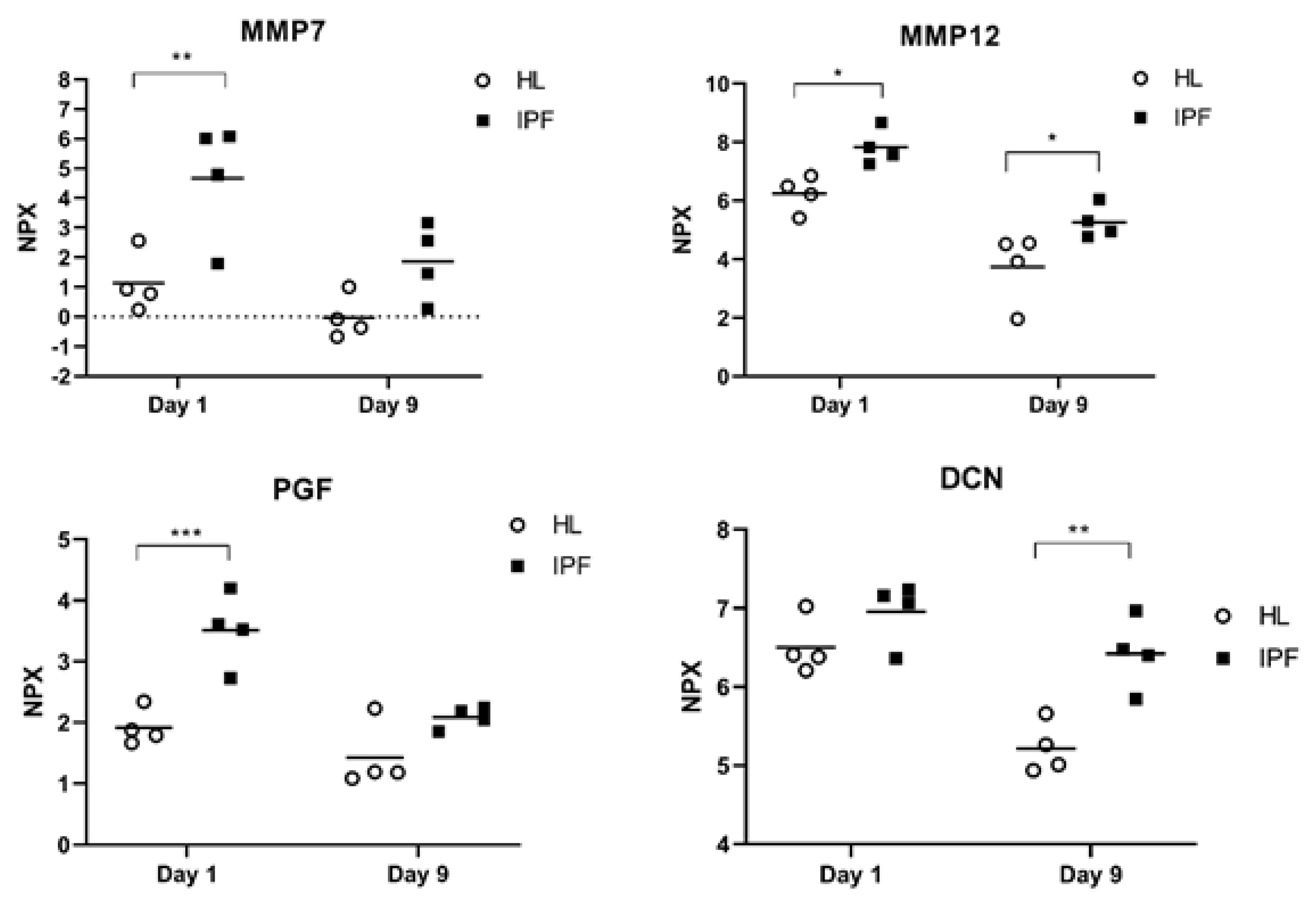

| Protein | Mean IPF Day 1 log2FC ± SD | Mean HL Day 1 log2FC ± (SD) | Mean Difference (IPF−HL) Day 1 | p−Value (Day 1) | Mean IPF Day 9 log2FC ± SD | Mean HL Day 9 log2FC ± (SD) | Mean Difference (IPF−HL) Day 9 | p−Value (Day 9) |
|---|---|---|---|---|---|---|---|---|
| MMP7 | 4.67 (2.01) | 1.13 (1.00) | 3.54 * | 5.7 × 10⁻³ | 1.87 (1.28) | −0.03 (0.73) | 1.89 | 1.3 × 10⁻¹ |
| MMP12 | 7.83 (0.61) | 6.24 (0.62) | 1.59 * | 3.1 × 10⁻² | 5.23 (0.56) | 3.74 (1.22) | 1.53 * | 3.8 × 10⁻² |
| PGF | 3.51 (0.60) | 1.92 (0.30) | 1.56 * | 5.0 × 10⁻⁴ | 2.09 (0.18) | 1.42 (0.54) | 0.66 | 1.1 × 10⁻¹ |
| DCN | 6.96 (0.40) | 6.51 (0.36) | 0.45 | 2.4 × 10⁻¹ | 6.42 (0.46) | 5.22 (0.33) | 1.20 * | 1.8 × 10⁻³ |
| Inflammation/Chemotaxis | ||||||||
| CXCL13 | 9.94 (1.83) | 3.59 (0.70) | 6.35 * | 1.0 × 10⁻⁴ | 8.01 (2.00) | 2.62 (1.12) | 5.38 * | 6.0 × 10⁻⁴ |
| GAL9 | 9.01 (0.93) | 5.69 (0.56) | 3.31 * | <1 × 10⁻⁴ | 5.84 (0.46) | 3.01 (0.40) | 2.84 * | <1 × 10⁻⁴ |
| GZMA | 2.57 (1.91) | 0.10 (0.37) | 2.48 * | 8.9 × 10⁻³ | −0.16 (0.43 | −0.49 (0.23) | 0.33 | 8.8 × 10⁻¹ |
| CD40 | 8.17 (1.49) | 6.22 (1.58) | 1.96 | 6.6 × 10⁻² | 6.40 (0.45) | 4.18 (0.61) | 2.22 * | 3.6 × 10⁻² |
| CCL19 | 3.96 (1.05) | 2.24 (0.71) | 1.73 * | 2.7 × 10⁻² | 2.45 (0.87) | 1.12 (0.71) | 1.35 | 8.6 × 10⁻² |
| CD4 | 1.85 (1.14) | 0.25 (0.76) | 1.60 * | 1.6 × 10⁻² | −0.26 (0.14) | −1.01 (0.38) | 0.74 | 3.0 × 10⁻¹ |
| TNFRSF9 | 1.35 (0.60) | −0.20 (0.41) | 1.55 * | 2.0 × 10⁻⁴ | −0.04 (0.21) | −0.71 (0.19) | 0.68 | 6.0 × 10⁻² |
| TNFRSF21 | 2.09 (0.33) | 1.56 (0.09) | 0.52 * | 7.6 × 10⁻³ | 1.36 (0.15) | 1.33 (0.20) | 0.03 | 9.7 × 10⁻¹ |
| IPF (n = 38) | Controls (n = 77) | |
|---|---|---|
| Age (Mean ± SD) | 73.8 ±7.83 | 55.6 ± 6.7 |
| Male/Female, n (%) | 29/9 (76%/24%) | 39/38 (51%/49%) |
| Smoking history | ||
| - Never smokers (n, %) | 8 (21%) | 37 (48%) |
| - Ex-smokers (n, %) | 29 (76%) | - |
| - Current smokers (n, %) | 1 (3%) | 40 (52%) |
| Lung function | ||
| - FVC (% predicted) | 80.8 ± 20.2 | 113 ± 14.4 |
| - FEV1 (% predicted) | 81.2 ± 17.9 | 103 ± 13.9 |
| - DLCO (% predicted) | 50.4 ± 11.8 | 86.5 ± 14.0 |
| - TLC (% predicted) | 64.3 ± 11.2 | 107 ± 11.0 |
| CPI (Mean ± SD) | 43.0 ± 10.7 | NA |
| GAP stage (n, %) | NA | |
| 1 | 21 (55%) | |
| 2 | 17 (45%) | |
| 3 | 0 (0%) | |
| Treatment with antifibrotics at serum sampling | NA | |
| - Treated baseline and treated follow-up | 12 (32%) | |
| - Untreated baseline and treated follow-up | 13 (34%) | |
| - Untreated baseline and untreated follow-up | 11 (29%) | |
| - Treated baseline and untreated follow-up | 2 (5%) |
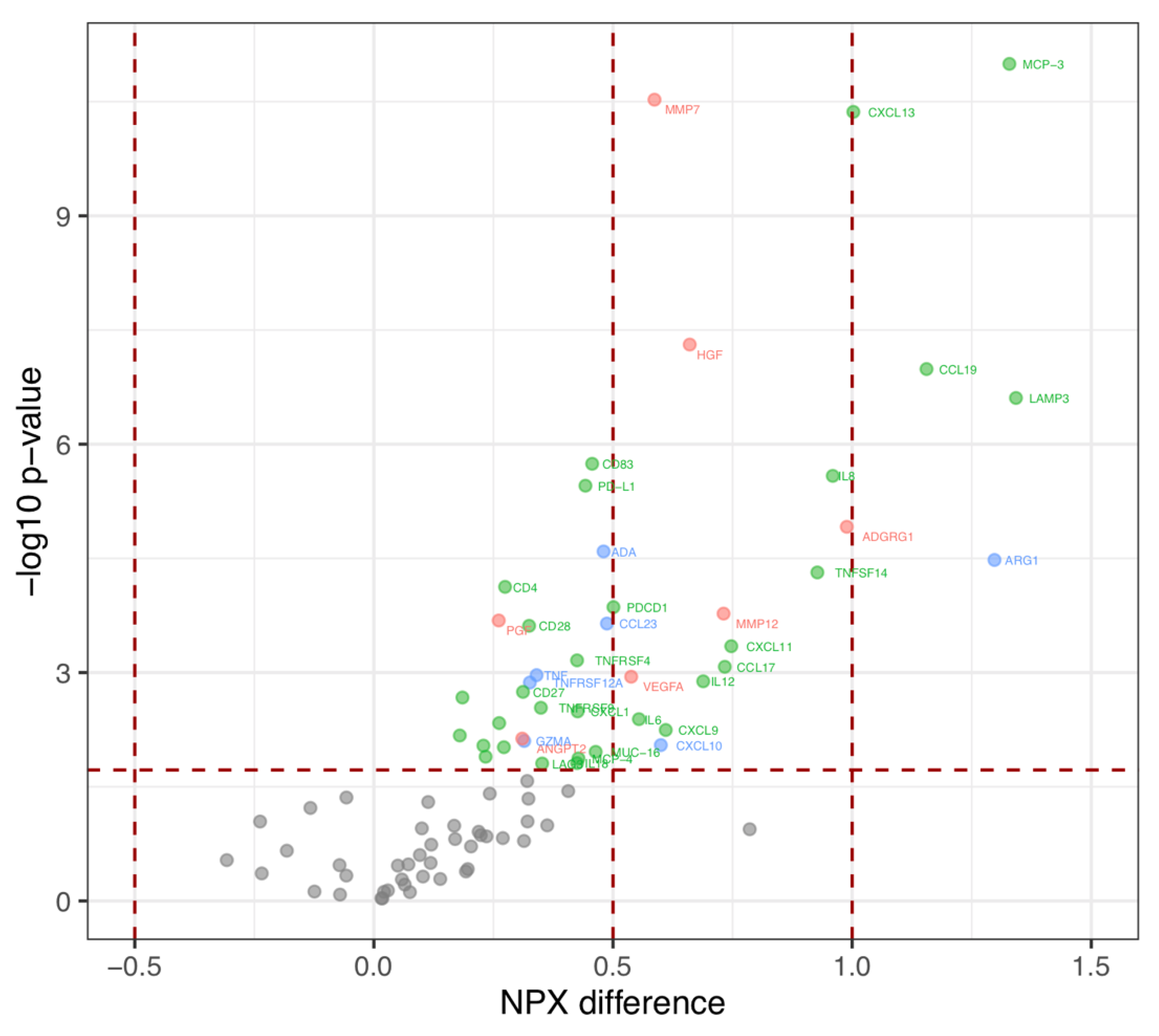
| Protein | NPX−Difference | p-Value | FDR Adjusted p-Value |
|---|---|---|---|
| Tissue remodeling | |||
| ADGRG1 | 0.99 | 1.22 × 10⁻⁵ | 5.75 × 10⁻³ |
| MMP12 a | 0.73 | 1.68 × 10⁻⁴ | 9.20 × 10⁻³ |
| HGF | 0.66 | 4.91 × 10⁻⁸ | 2.30 × 10⁻³ |
| MMP7 a | 0.59 | 2.98 × 10⁻¹¹ | 1.15 × 10⁻³ |
| VEGFA | 0.54 | 1.13 × 10⁻³ | 1.38 × 10⁻² |
| ANGPT2 | 0.31 | 7.37 × 10⁻³ | 2.01 × 10⁻² |
| PGF a | 0.26 | 2.07 × 10⁻⁴ | 9.77 × 10⁻³ |
| Inflammation/Chemotaxis | |||
| LAMP3 | 1.34 | 2.47 × 10⁻⁷ | 3.45 × 10⁻³ |
| MCP−3 | 1.33 | 1.01 × 10⁻¹¹ | 5.75 × 10⁻⁴ |
| CCL19 a | 1.16 | 1.03 × 10⁻⁷ | 2.87 × 10⁻³ |
| CXCL13 a | 1.00 | 4.31 × 10⁻¹¹ | 1.72 × 10⁻³ |
| IL8 | 0.96 | 2.61 × 10⁻⁶ | 4.60 × 10⁻³ |
| TNFSF14 | 0.93 | 4.84 × 10⁻⁵ | 7.47 × 10⁻³ |
| CXCL11 | 0.75 | 4.51 × 10⁻⁴ | 1.15 × 10⁻² |
| CCL17 | 0.73 | 8.39 × 10⁻⁴ | 1.26 × 10⁻² |
| IL12 | 0.69 | 1.30 × 10⁻³ | 1.44 × 10⁻² |
| CXCL9 | 0.61 | 5.68 × 10⁻³ | 1.90 × 10⁻² |
| IL6 | 0.55 | 4.09 × 10⁻³ | 1.78 × 10⁻² |
| PDCD1 | 0.50 | 1.38 × 10⁻⁴ | 8.62 × 10⁻³ |
| MUC−16 | 0.46 | 1.10 × 10⁻² | 2.30 × 10⁻² |
| CD83 | 0.46 | 1.81 × 10⁻⁶ | 4.02 × 10⁻³ |
| PD−L1 | 0.44 | 3.51 × 10⁻⁶ | 5.17 × 10⁻³ |
| MCP−4 | 0.43 | 1.34 × 10⁻² | 2.41 × 10⁻² |
| CXCL1 | 0.43 | 3.24 × 10⁻³ | 1.72 × 10⁻² |
| TNFRSF4 | 0.42 | 6.91 × 10⁻⁴ | 1.21 × 10⁻² |
| IL18 | 0.42 | 1.55 × 10⁻² | 2.47 × 10⁻² |
| LAG3 | 0.35 | 1.56 × 10⁻² | 2.53 × 10⁻² |
| TNFRSF9 a | 0.35 | 2.90 × 10⁻³ | 1.67 × 10⁻² |
| CD28 | 0.32 | 2.43 × 10⁻⁴ | 1.09 × 10⁻² |
| CD27 | 0.31 | 1.79 × 10⁻³ | 1.55 × 10⁻² |
| CD4 a | 0.27 | 7.48 × 10⁻⁵ | 8.05 × 10⁻³ |
| CD5 | 0.27 | 9.57 × 10⁻³ | 2.24 × 10⁻² |
| CD40 a | 0.26 | 4.61 × 10⁻³ | 1.84 × 10⁻² |
| Gal−9 a | 0.23 | 1.27 × 10⁻² | 2.36 × 10⁻² |
| CX3CL1 | 0.23 | 9.11 × 10⁻³ | 2.18 × 10⁻² |
| CSF−1 | 0.19 | 2.13 × 10⁻³ | 1.61 × 10⁻² |
| Gal−1 | 0.18 | 6.71 × 10⁻³ | 1.95 × 10⁻² |
| Overlapping functions | |||
| ARG1 | 1.30 | 3.32 × 10⁻⁵ | 6.90 × 10⁻³ |
| CXCL10 | 0.60 | 8.96 × 10⁻³ | 2.13 × 10⁻² |
| CCL23 | 0.49 | 2.27 × 10⁻⁴ | 1.03 × 10⁻² |
| ADA | 0.48 | 2.56 × 10⁻⁵ | 6.32 × 10⁻³ |
| TNF | 0.34 | 1.08 × 10⁻³ | 1.32 × 10⁻² |
| TNFRSF12A | 0.33 | 1.35 × 10⁻³ | 1.49 × 10⁻² |
| GZMA a | 0.31 | 7.92 × 10⁻³ | 2.07 × 10⁻² |
| Protein | Baseline Mean NPX (±SD) | Follow Up Mean NPX (±SD) | Mean Difference (±SD) | p-Value |
|---|---|---|---|---|
| Tissue remodeling | ||||
| EGF | 10.49 ± 0.84 | 10.08 ± 0.83 | −0.39 ± 0.97 | 0.02 |
| FGF2 | 0.93 ± 0.59 | 0.63 ± 0.55 | −0.31 ± 0.49 | 0.0001 |
| LAP TGF-beta-1 | 9.99 ± 0.37 | 9.81 ± 0.51 | −0.16 ± 0.38 | 0.023 |
| VEGFR-2 | 8.48 ± 0.22 | 8.35 ± 0.21 | −0.12 ± 0.18 | 0.0002 |
| HO-1 | 11.18 ± 0.40 | 11.07 ± 0.36 | −0.1 ± 0.28 | 0.020 |
| TIE2 | 7.70 ± 0.26 | 7.60 ± 0.24 | −0.09 ± 0.16 | 0.0008 |
| PTN | 3.28 ± 1.38 | 3.57 ± 1.36 | 0.21 ± 0.59 | 0.046 |
| Inflammation/Chemotaxis | ||||
| CCL3 | 6.62 ± 0.65 | 6.42 ± 0.50 | −0.19 ± 0.54 | 0.042 |
| CD28 b | 1.42 ± 0.31 | 1.27 ± 0.38 | −0.15 ± 0.22 | 0.0002 |
| CD244 | 6.26 ± 0.26 | 6.11 ± 0.31 | −0.14 ± 0.2 | 0.0001 |
| LAMP3 b | 6.85 ± 0.47 | 6.70 ± 0.46 | −0.14 ± 0.39 | 0.015 |
| IL12RB1 | 2.08 ± 0.30 | 1.96 ± 0.25 | −0.12 ± 0.29 | 0.026 |
| ADA b | 5.18 ± 0.36 | 5.06 ± 0.34 | −0.11 ± 0.23 | 0.01 |
| IL18 b | 8.96 ± 0.49 | 8.83 ± 0.45 | −0.11 ± 0.29 | 0.027 |
| MIC-A/B | 4.62 ± 1.62 | 4.48 ± 1.60 | −0.1 ± 0.21 | 0.009 |
| CCL4 | 7.29 ± 0.54 | 7.20 ± 0.55 | −0.09 ± 0.34 | 0.040 |
| CD40 b | 10.03 ± 0.32 | 9.94 ± 0.35 | −0.08 ± 0.25 | 0.033 |
| ICOSLG | 6.14 ± 0.19 | 6.07 ± 0.19 | −0.07 ± 0.14 | 0.003 |
| PD-L2 | 2.27 ± 0.25 | 2.23 ± 0.24 | −0.05 ± 0.17 | 0.03 |
| Overlapping functions | ||||
| CASP-8 | 4.36 ± 0.59 | 4.08 ± 0.57 | −0.24 ± 0.62 | 0.02 |
| TRAIL | 8.57 ± 0.30 | 8.45 ± 0.32 | −0.12 ± 0.2 | 0.002 |
| TWEAK | 9.13 ± 0.30 | 9.02 ± 0.33 | −0.1 ± 0.26 | 0.01 |
| FVC % | Rho Coefficient | p-Value | Biological Function |
|---|---|---|---|
| MMP7 a,b | −0.51 | 0.0006 | R |
| HGF b | −0.48 | 0.003 | R |
| ANGPT1 | −0.44 | 0.005 | R |
| EGF | −0.43 | 0.007 | R |
| LAP TGF-beta 1 | −0.40 | 0.012 | R |
| PDGF subunit B | −0.33 | 0.041 | R |
| DCN | 0.34 | 0.037 | R |
| PTN | 0.46 | 0.004 | R |
| TNFSF14 b | −0.54 | 0.0005 | I |
| MCP-3 b | −0.53 | 0.0006 | I |
| LAMP3 b | −0.46 | 0.004 | I |
| CXCL1 b | −0.43 | 0.008 | I |
| CD40-L | −0.42 | 0.009 | I |
| CXCL5 | −0.33 | 0.042 | I |
| MIC-A/B | −0.33 | 0.047 | I |
| TWEAK | −0.35 | 0.03 | O |
| MCP-1 | −0.33 | 0.044 | O |
| TLC % | |||
| EGF | −0.52 | 0.0009 | R |
| MMP7 a,b | −0.47 | 0.003 | R |
| ANGPT1 | −0.47 | 0.003 | R |
| HGF b | −0.37 | 0.022 | R |
| LAP TGF-beta 1 | −0.34 | 0.040 | R |
| PTN | 0.33 | 0.042 | R |
| TNFSF14 b | −0.53 | 0.0007 | I |
| CD40-L | −0.50 | 0.001 | I |
| MCP-3 b | −0.50 | 0.001 | I |
| CXCL1 b | −0.47 | 0.003 | I |
| MCP-4 b | −0.41 | 0.011 | I |
| CCL4 | −0.33 | 0.044 | I |
| MCP-1 | −0.42 | 0.008 | O |
| DLCO % | |||
| DCN a | 0.37 | 0.023 | R |
| CPI | |||
| HGF b | 0.40 | 0.013 | R |
| DCN a | −0.38 | 0.018 | R |
| LAMP3 b | 0.40 | 0.013 | I |
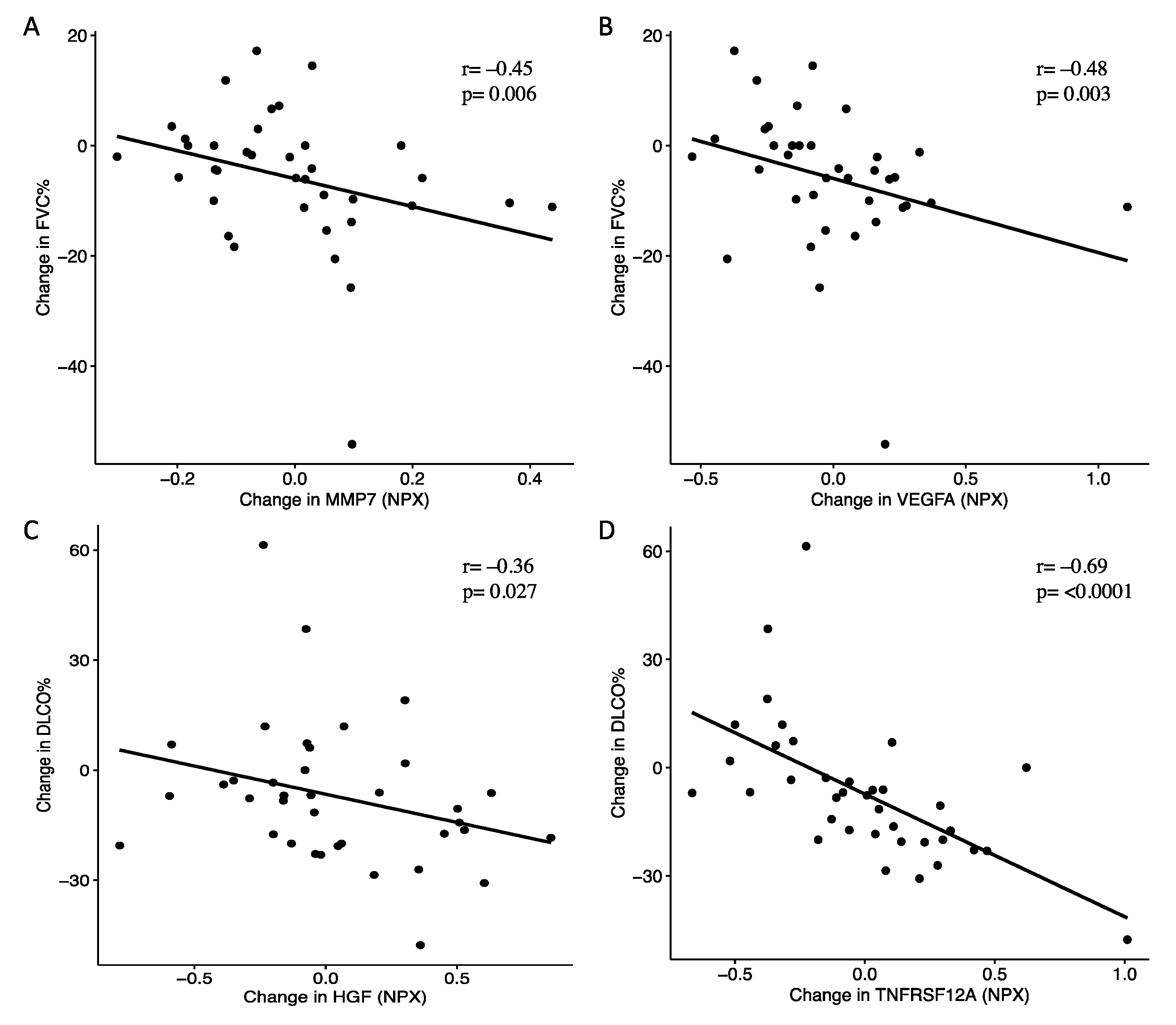
| Change in FVC % | Protein | Rho Coefficient | p-Value | Biological Function |
|---|---|---|---|---|
| VEGFA b | −0.48 | 0.003 | R | |
| PDGF subunit B | −0.45 | 0.005 | R | |
| MMP7 a,b | −0.45 | 0.006 | R | |
| ANGPT1 | −0.38 | 0.022 | R | |
| NOS3 | −0.35 | 0.033 | R | |
| PD-L1 b | −0.39 | 0.017 | I | |
| CXCL12 | −0.36 | 0.029 | I | |
| KIR3DL1 | −0.35 | 0.035 | I | |
| CXCL9 b | 0.49 | 0.002 | I | |
| IL12 b | 0.45 | 0.006 | I | |
| PDCD1 b | 0.39 | 0.018 | I | |
| CXCL10 b | 0.39 | 0.019 | O | |
| Change in DLCO % | ||||
| NOS3 | −0.38 | 0.021 | R | |
| HGF b | −0.36 | 0.027 | R | |
| MMP12 a,b | −0.36 | 0.028 | R | |
| MMP7 a,b | −0.35 | 0.035 | R | |
| VEGFA b | −0.33 | 0.045 | R | |
| CD2 7 b | −0.44 | 0.006 | I | |
| Gal-1 b | −0.38 | 0.019 | I | |
| IL18 b | −0.35 | 0.035 | I | |
| CCL4 | −0.35 | 0.035 | I | |
| TNFRSF21 b | −0.33 | 0.043 | I | |
| KIR3DL1 | −0.33 | 0.045 | I | |
| TNFRSF12A b | −0.69 | <0.0001 | O | |
| Change in TLC % | ||||
| LAP TGF-beta-1 | −0.36 | 0.029 | R | |
| IL12 b | 0.41 | 0.012 | I | |
| CCL20 | 0.38 | 0.021 | I | |
| NCR1 | 0.35 | 0.036 | I | |
| CXCL9 b | 0.35 | 0.034 | I |
| Protein | Mean Change (±SD) | Minimum | Maximum | p-Value |
|---|---|---|---|---|
| Untreated baseline and untreated follow-up | ||||
| TRAIL | −0.16 ± 0.19 | −0.61 | 0.08 | 0.014 |
| Untreated baseline and treated follow-up | ||||
| EGF | −0.92 ± 0.85 | −2.21 | 0.85 | 0.003 |
| TNFSF14 | −0.75 ± 0.86 | −1.87 | 1.47 | 0.011 |
| CD40-L | −0.67 ± 0.72 | −1.88 | 0.59 | 0.011 |
| CASP-8 | −0.64 ± 0.56 | −1.58 | 0.16 | 0.003 |
| FGF2 | −0.56 ± 0.45 | −1.22 | 0.3 | 0.002 |
| CCL3 | −0.4 ± 0.38 | −1.02 | 0.13 | 0.001 |
| IL6 | −0.35 ± 0.5 | −1.47 | 0.25 | 0.033 |
| MUC−16 | −0.29 ± 0.38 | −1.2 | 0.3 | 0.013 |
| CD244 | −0.27 ± 0.2 | −0.58 | 0.14 | 0.001 |
| GZMA | −0.27 ± 0.28 | −0.77 | 0.14 | 0.003 |
| CD40 | −0.26 ± 0.12 | −0.46 | −0.03 | 0.0002 |
| LAMP3 | −0.26 ± 0.37 | −0.86 | 0.32 | 0.04 |
| MCP-2 | −0.25 ± 0.28 | −0.95 | 0.07 | 0.006 |
| KLRD1 | −0.25 ± 0.24 | −0.59 | 0.19 | 0.003 |
| VEGFR-2 | −0.24 ± 0.11 | −0.41 | −0.08 | 0.0002 |
| IL18 | −0.24 ± 0.29 | −0.71 | 0.22 | 0.013 |
| CCL4 | −0.23 ± 0.24 | −0.67 | 0.27 | 0.006 |
| ADA | −0.22 ± 0.22 | −0.53 | 0.3 | 0.011 |
| TWEAK | −0.21 ± 0.15 | −0.44 | 0.01 | 0.0005 |
| CD28 | −0.21 ± 0.27 | −0.69 | 0.25 | 0.017 |
| MIC-A/B | −0.21 ± 0.23 | −0.59 | 0.15 | 0.005 |
| CCL23 | −0.19 ± 0.28 | −0.66 | 0.34 | 0.04 |
| TIE2 | −0.18 ± 0.12 | −0.42 | 0 | 0.0005 |
| FASLG | −0.17 ± 0.13 | −0.45 | 0 | 0.0005 |
| TRAIL | −0.14 ± 0.21 | −0.62 | 0.14 | 0.022 |
| KIR3DL1 | −0.14 ± 0.18 | −0.45 | 0.15 | 0.017 |
| ICOSLG | −0.1 ± 0.15 | −0.3 | 0.17 | 0.04 |
| PD-L2 | −0.1 ± 0.17 | −0.34 | 0.32 | 0.048 |
| TNFRSF21 | −0.08 ± 0.13 | −0.33 | 0.17 | 0.027 |
| IL10 | 0.21 ± 0.32 | −0.39 | 0.75 | 0.033 |
| Treated baseline and treated follow-up | ||||
| FGF2 | −0.31 ± 0.28 | −0.83 | 0.08 | 0.003 |
| CD28 | −0.09 ± 0.15 | −0.32 | 0.11 | 0.043 |
| CD244 | −0.08 ± 0.12 | −0.23 | 0.12 | 0.027 |
| ICOSLG | −0.06 ± 0.09 | −0.2 | 0.14 | 0.043 |
| CD8A | 0.18 ± 0.19 | −0.11 | 0.58 | 0.009 |
| MMP12 | 0.27 ± 0.45 | −0.68 | 1.05 | 0.043 |
| GZMB | 0.33 ± 0.47 | −0.12 | 1.37 | 0.021 |
| PTN | 0.53 ± 0.53 | −0.42 | 1.28 | 0.007 |
Publisher’s Note: MDPI stays neutral with regard to jurisdictional claims in published maps and institutional affiliations. |
© 2021 by the authors. Licensee MDPI, Basel, Switzerland. This article is an open access article distributed under the terms and conditions of the Creative Commons Attribution (CC BY) license (https://creativecommons.org/licenses/by/4.0/).
Share and Cite
Kalafatis, D.; Löfdahl, A.; Näsman, P.; Dellgren, G.; Wheelock, Å.M.; Elowsson Rendin, L.; Sköld, M.; Westergren-Thorsson, G. Distal Lung Microenvironment Triggers Release of Mediators Recognized as Potential Systemic Biomarkers for Idiopathic Pulmonary Fibrosis. Int. J. Mol. Sci. 2021, 22, 13421. https://doi.org/10.3390/ijms222413421
Kalafatis D, Löfdahl A, Näsman P, Dellgren G, Wheelock ÅM, Elowsson Rendin L, Sköld M, Westergren-Thorsson G. Distal Lung Microenvironment Triggers Release of Mediators Recognized as Potential Systemic Biomarkers for Idiopathic Pulmonary Fibrosis. International Journal of Molecular Sciences. 2021; 22(24):13421. https://doi.org/10.3390/ijms222413421
Chicago/Turabian StyleKalafatis, Dimitrios, Anna Löfdahl, Per Näsman, Göran Dellgren, Åsa M. Wheelock, Linda Elowsson Rendin, Magnus Sköld, and Gunilla Westergren-Thorsson. 2021. "Distal Lung Microenvironment Triggers Release of Mediators Recognized as Potential Systemic Biomarkers for Idiopathic Pulmonary Fibrosis" International Journal of Molecular Sciences 22, no. 24: 13421. https://doi.org/10.3390/ijms222413421
APA StyleKalafatis, D., Löfdahl, A., Näsman, P., Dellgren, G., Wheelock, Å. M., Elowsson Rendin, L., Sköld, M., & Westergren-Thorsson, G. (2021). Distal Lung Microenvironment Triggers Release of Mediators Recognized as Potential Systemic Biomarkers for Idiopathic Pulmonary Fibrosis. International Journal of Molecular Sciences, 22(24), 13421. https://doi.org/10.3390/ijms222413421


