Article

Low Protein Expression of both ATRX and ZNRF3 as Novel Negative Prognostic Markers of Adult Adrenocortical Carcinoma

by
Vania Balderrama Brondani
1,*,
Amanda Meneses Ferreira Lacombe
1,
Beatriz Marinho de Paula Mariani
2,
Luciana Montenegro
2,3,
Iberê Cauduro Soares
4,5,
João Evangelista Bezerra-Neto
6,
Fabio Yoshiaki Tanno
7,
Victor Srougi
7,
José Luis Chambo
7,
Berenice Bilharinho Mendonca
1,2,3,
Madson Q. Almeida
1,2,8,
Maria Claudia Nogueira Zerbini
4 and
Maria Candida Barisson Villares Fragoso
1,2,8,*
1
Unidade de Suprarrenal, Serviço de Endocrinologia e Metabologia, Hospital das Clínicas, Faculdade de Medicina da Universidade de São Paulo, Av. Dr. Enéas de Carvalho Aguiar, 155, São Paulo 05403-900, Brazil
2
Laboratório de Hormônios e Genética Molecular LIM/42, Hospital das Clínicas, Faculdade de Medicina da Universidade de São Paulo, Av. Dr. Enéas de Carvalho Aguiar, 255, São Paulo 05403-900, Brazil
3
Laboratório de Sequenciamento em Larga Escala (SELA), Faculdade de Medicina da Universidade de São Paulo, Av. Dr. Arnaldo, 455, São Paulo 01246-903, Brazil
4
Departamento de Patologia, Faculdade de Medicina da Universidade de São Paulo, Av. Dr. Arnaldo, 455, São Paulo 01246-903, Brazil
5
Serviço de Anatomia Patológica, Instituto do Câncer do Estado de São Paulo (ICESP), Faculdade de Medicina da Universidade de São Paulo, Av. Dr. Arnaldo, 251, São Paulo 01246-000, Brazil
6
Serviço de Oncologia, Instituto do Câncer do Estado de São Paulo (ICESP), Faculdade de Medicina da Universidade de São Paulo, Av. Dr. Arnaldo, 251, São Paulo 01246-000, Brazil
7
Serviço de Urologia, Hospital das Clínicas, Faculdade de Medicina da Universidade de São Paulo, Av. Dr. Enéas de Carvalho Aguiar, 155, São Paulo 05403-900, Brazil
8
Serviço de Endocrinologia, Instituto do Câncer do Estado de São Paulo (ICESP), Faculdade de Medicina da Universidade de São Paulo, Av. Dr. Arnaldo, 251, São Paulo 01246-000, Brazil
*
Authors to whom correspondence should be addressed.
Int. J. Mol. Sci. 2021, 22(3), 1238; https://doi.org/10.3390/ijms22031238
Submission received: 24 November 2020 / Revised: 5 January 2021 / Accepted: 22 January 2021 / Published: 27 January 2021

Abstract

:
Adrenocortical carcinoma (ACC) is a rare malignancy that is associated with a dismal prognosis. Pan-genomic studies have demonstrated the involvement of ATRX and ZNRF3 genes in adrenocortical tumorigenesis. Our aims were to evaluate the protein expression of ATRX and ZNRF3 in a cohort of 82 adults with ACC and to establish their prognostic value. Two pathologists analyzed immuno-stained slides of a tissue microarray. The low protein expression of ATRX and ZNRF3 was associated with a decrease in overall survival (OS) (p = 0.045, p = 0.012, respectively). The Cox regression for ATRX protein expression of >1.5 showed a hazard ratio (HR) for OS of 0.521 (95% CI 0.273–0.997; p = 0.049) when compared with ≤1.5; for ZNRF3 expression >2, the HR for OS was 0.441 (95% CI, 0.229–0.852; p = 0.015) when compared with ≤2. High ATRX and ZNRF3 protein expressions were associated with optimistic recurrence-free survival (RFS) (p = 0.027 and p = 0.005, respectively). The Cox regression of RFS showed an HR of 0.332 (95%CI, 0.111–0.932) for ATRX expression >2.7 (p = 0.037), and an HR of 0.333 (95%CI, 0.140–0.790) for ZNRF3 expression >2 (p = 0.013). In conclusion, low protein expression of ATRX and ZNRF3 are negative prognostic markers of ACC; however, different cohorts should be evaluated to validate these findings.

1. Introduction

Adrenocortical carcinoma (ACC) is a rare and aggressive cancer deriving from the adrenal cortex gland with an incidence of 0.7–2 cases/million/year [1,2,3]. Despite a dismal prognosis, the five-year overall survival (OS) rate varies, ranging from 60 to 80% when the tumor is restricted to the adrenal gland and from 0 to 28% in the advanced stages of the disease [3,4,5,6,7,8]. These data suggest that ACC is a heterogeneous adrenal disorder, presenting different behaviors, probably due to its different biology and genetic signatures [2,8,9].
In previous years, considerable advances in the genetic field of ACC tumorigenesis investigation have occurred, allowing patients to be stratified according to the combination of clinical–hormonal–pathological and genetic alterations.
Prognostic factors have an essential role in determining the aggressiveness of ACC. We can classify tumors according to (A) clinical data (age, hormone-related symptoms, and tumor stage); (B) pathological findings, genetic/protein background, and category of surgical procedure (Weiss system, modified Weiss system, Helsinki score, mitotic count, Ki-67 proliferation marker, SF-1, P53, β-catenin, and resection status); (C) clinical and pathological parameter associations (staging system of European Network for the Study of Adrenal Tumors modified by grade, resection status, age and symptoms—mENSAT plus GRAS); and (D) molecular classifications (chromosomal aberrations, methylome, gene expression alterations, microRNAs dysregulation, and gene mutations) [2,3,4,6,10,11,12,13,14,15,16,17,18,19,20,21,22,23,24,25,26,27,28,29,30,31,32,33,34,35].
Pan genomic studies have identified new genes related to adrenocortical carcinogenesis, providing a refinement of ACC taxonomy, and improving the development of new biomarkers for early diagnosis and staging, conducive to the development of molecularly targeted therapies.
ATRX is a chromatin structure regulator and telomere structure maintainer. Somatic pathogenic variants and structural variations, which cause the deletion of multiple exons of ATRX were associated with germline TP53 pathogenic variants in a pediatric cohort [36]. Pinto et al. reported alterations in TP53 and ATRX in 32% of their cohort, and these alterations were associated with advanced disease and poor event-free survival in pediatric patients with adrenocortical tumor [36]. Assie et al. identified somatic ATRX alterations in only 4% of their adult ACC cohort. In contrast, ZNRF3 was the most frequently altered gene, corresponding to 21% of cases [27]. This gene encodes a cell surface transmembrane E3 ubiquitin ligase that acts as a negative feedback regulator of the canonical Wnt signaling (Wnt/β-catenin signaling). Zheng et al. studied an adult ACC cohort and reported a frequency of 19.3% for ACCs with deletions or non-silent somatic pathogenic variants in the ZNRF3 gene. In comparison, ATRX alterations were present in only four cases (4.3%) [31].
The Cancer Genome Atlas (TCGA) ACC Research Network and the European Network for the Study of Adrenal Tumors (ENSAT) including the cohort of Assie et al. cohort [27], are two critical datasets for the study of ACC. TCGA-ACC, from National Cancer Institute, includes a cohort of 184 cases of ACC with a median age 48.5 years (14–83 years), 120 (65%) female, and a frequency of ENSAT staging system as follows: stage 1, 10% (18 of 180 cases); stage 2, 49% (88 cases); stage 3, 21% (38 cases); and stage 4, 20% (36 cases). TCGA-ACC registered 68 (37%) deaths (the data shown in the present study are based upon data generated by TCGA Research Network) [37]. The ENSAT and Assie et al. cohorts include a total of 130 ACC cases, with median age 46 years (18–86 years), 90 (69.2%) female, with a frequency of ENSAT stage as follows: stage 1, 7.2% (9 of 125 cases); stage 2, 53.6% (67 cases); stage 3, 16% (20 cases); and stage 4, 23.2% (29 cases). There were 46 (35.4%) deaths among 130 cases [27]. TCGA-ACC dataset registered the most mutated genes in ACC, including TP53 (21%), ZNRF3 (19%), CDKN2A (15%), CTNNB1 (16%), TERT (14%), and PPKAR1A (11%), while the ATRX gene had a frequency of 4.3% of cohort [31]. ENSAT with Assie et al. showed the most frequent mutated genes as ZNRF3 (21%), CTNNB1 (16%), TP53 (16%), and CDKN2A (11%). Alterations on ATRX were present in 4% of cases [27]. Both cohorts present similar clinical and molecular data.
In this study, we aimed to evaluate the protein expression of the ATRX and ZNRF3 genes in a cohort of 82 adult ACC patients from a unique Complex tertiary center to investigate their potential role as prognostic markers.

2. Results

2.1. ATRX Protein

Patients with ACC showing high ATRX expression, defined as tumor scored 2+ or higher, represented 47.5% of cohort (Figure 1). ATRX high expression group had a median age at diagnosis 52.5 years. In contrast, patients with ACC with low ATRX expression had a median age of 34.7 years (Z = −3.349; p = 0.001; Mann–Whitney U test). ATRX was highly expressed in 90% of cases without hypercortisolism at initial presentation, while it was expressed at a low level in 59.2% of cases with hypercortisolism (X2(2) = 8.759; p = 0.013; Chi-squared test). The group with hypercortisolism presented a median ATRX expression score of 0.5 (ranging from 0 to 4), while the non-hypercortisolism group presented a median score of 3.08 (ranging from 0 to 4) (X2(2) = 6.728; p = 0.035; Kruskal–Wallis test).
The quantitative analysis of ATRX expression showed a positive correlation with age at diagnosis (coefficient 0.391; p < 0.001; Spearman’s rank correlation coefficient) and negative correlations with tumor size (coefficient -0.263; p = 0.02; Spearman’s rank correlation coefficient) and Weiss score (coefficient -0.245; p = 0.033; Spearman’s rank correlation coefficient).
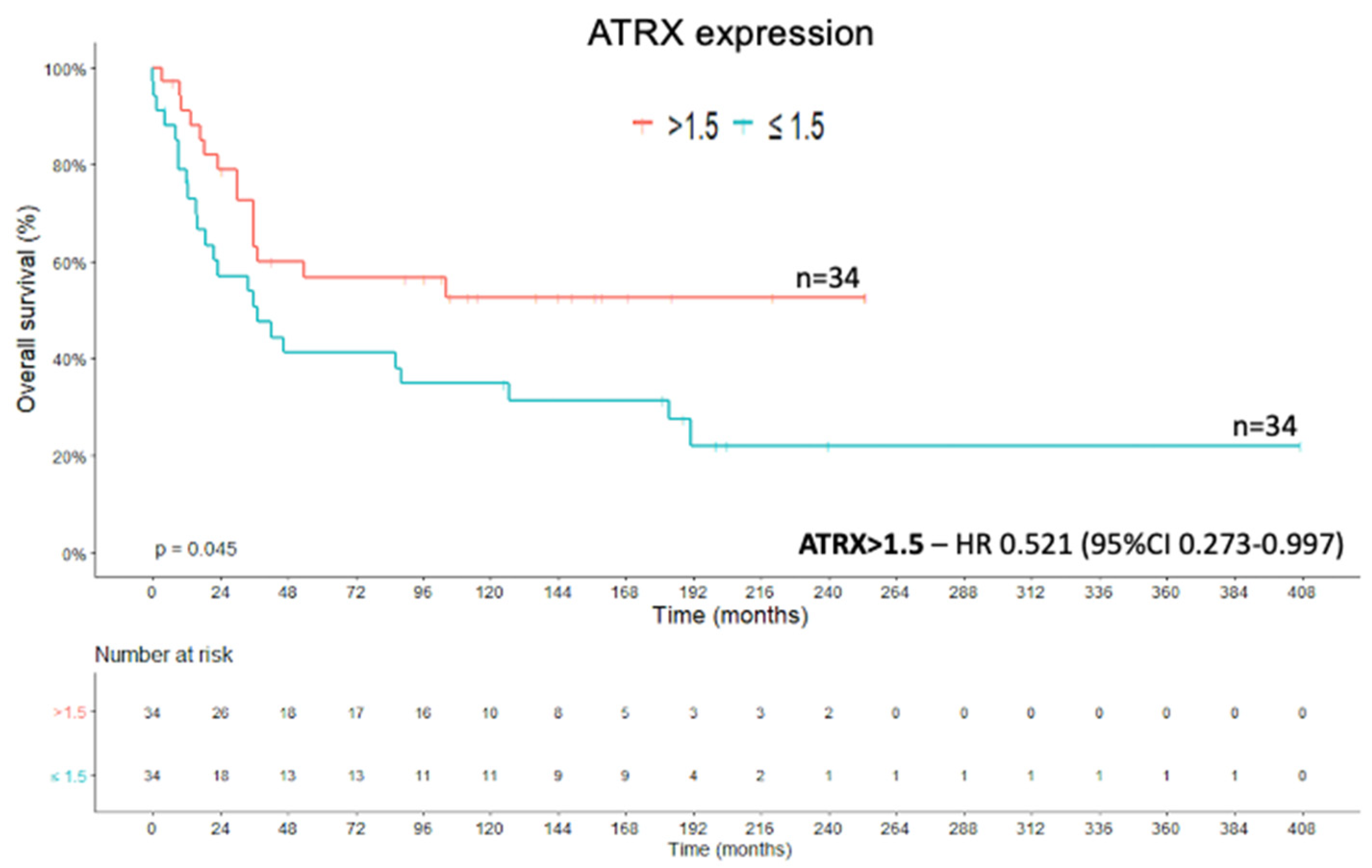
Thirty-nine patients from the cohort died due to ACC or its clinical complications during the study period. The ATRX expression cut-off point was statistically determined and used for the OS analysis. The cut-off value ≤1.5 for ATRX expression had an impact on the Kaplan–Meier survival curve (X2(1) = 4.021; p = 0.045; Kaplan–Meier method and log rank test) (Figure 2). In addition, the Cox regression of OS showed a hazard ratio (HR) of 0.521 (95%CI, 0.273–0.997) for ATRX expression of >1.5 when compared with expression of ≤1.5 (Wald = 3.881; df1; p = 0.049; Cox’s proportional hazards model).

2.2. ZNRF3 Protein

The high expression of ZNRF3 protein was defined as the sum of extent and intensity parameters equaling ≥3+ (Figure 1). High ZNRF3 expression was present in 46.25% of the cohort and was associated with a small tumor size (average size of 9.7 cm), while low expression was associated with a large tumor size (average size of 13.3 cm) at diagnosis (Z = −2.665; p = 0.008; Mann–Whitney U test). Tumors with high ZNRF3 expression presented with lower Weiss score (average 4.7) than the group with low ZNRF3 expression (average 6.1) (Z = −2.942; p = 0.003; Mann–Whitney U test). ZNRF3 expression was related to the ENSAT staging system. High ZNRF3 expression was present in almost 89 % of patients with an ENSAT staging system score of 1 at diagnosis, 42.9% with an ENSAT score of 2, 29.4% with an ENSAT score of 3, and 44.4% with an ENSAT score of 4 (p = 0.033, Fisher’s exact test). ZNRF3 protein expression had no relationship with hypercortisolism at clinical presentation (X2(2) = 2.035; p > 0.05, Chi-squared test). Regarding the outcome analysis, 64.1% of patients with ACC who progressed to death presented with a low level of ZNRF3 protein expression and had a median survival of 19 months (X2(1) = 4.716; p = 0.030; Chi-squared test).
The quantitative expression of ZNRF3 showed negative correlations with tumor weight (coefficient −0.282; p = 0.026, Spearman’s rank correlation coefficient), tumor size (coefficient −0.313; p = 0.005; Spearman’s rank correlation coefficient), and the Weiss score (coefficient −0.345; p = 0.002; Spearman’s rank correlation coefficient). The ZNRF3 expression cut-off of ≤2 had an impact on the OS analysis ((X2(1) = 6.277; p = 0.012; Kaplan–Meier method and log rank test) (Figure 3). The Cox regression of OS showed an HR of 0.441 (95% CI, 0.229–0.852) for ZNRF3 expression >2 compared with the value for an expression level of ≤2 (Wald = 5.942; df1; p = 0.015; Cox’s proportional hazards model).
ZNRF3 is considered a suppressor of the Wnt/β-catenin pathway. Due to this known function, we analyzed the status of β-catenin in the tumoral cells. Active Wnt/β-catenin signaling was characterized by a cytoplasmic/nuclear staining for β-catenin [37]. The analysis demonstrated a positive correlation between ZNRF3 expression and β-catenin expression in the cell membrane (coefficient 0.202; p = 0.004; Spearman’s rank correlation coefficient). These data support the function of ZNRF3 as a tumor suppressor, keeping β-catenin attached in the cell membrane (Figure 4).

2.3. Ki-67 Proliferation Marker and Combined Analysis

The Ki-67 proliferation marker showed positive correlation with tumor weight (coefficient 0.377; p = 0.01, Spearman’s rank correlation coefficient), tumor size (coefficient 0.344; p = 0.007, Spearman’s rank correlation coefficient), and the Weiss score (coefficient 0.572; p < 0.001, Spearman’s rank correlation coefficient). Furthermore, the categorization of Ki-67 into groups (<10%, ≥10% and <20%, ≥20%) impacted the OS analysis (X2(2) = 29.363; p < 0.001; Kaplan–Meier method and log rank test) (Figure 5). The Cox regression of OS from the analysis of Ki-67 expression showed an HR of 5.802 (95% CI, 2.447–13.726) for the Ki-67 expression grouped in ≥10% and <20% (Wald = 15.925; df1; p < 0.001; Cox’s proportional hazards model), and we calculated an HR of 9.041 (95% CI, 3.543–23.073) for the Ki-67 expression grouped in ≥20% (Wald = 21.217; df1; p < 0.001; Cox’s proportional hazards model).
Combined analysis was done in groups classified according to the Ki-67 value (>10% or ≤10%) and ATRX expression (>1.5 or ≤1.5), and according to the Ki-67 value (>10% or ≤10%) and ZNRF3 expression (>2 or ≤2). The combination of Ki-67 with ATRX and ZNRF3 did not impact the Kaplan–Meier survival curves (X2(3) = 5.206; p > 0.05; and X2(3) = 6.619; p > 0.05, respectively. Kaplan–Meier method and log rank test for both). However, the combination of low ATRX expression and low ZNRF3 expression had an effect on the OS curve (X2(3) = 9.867; p = 0.02; Kaplan–Meier method and log rank test) (Figure 6). The Cox regression of OS from the combined analysis of ATRX and ZNRF3 expressions showed an HR of 0.314 (95% CI, 0.136–0.725) for the ATRX (>1.5) and ZNRF3 (>2) expression levels (Wald = 7.363; df1; p = 0.007; Cox’s proportional hazards model).

2.4. Recurrence-Free Survival

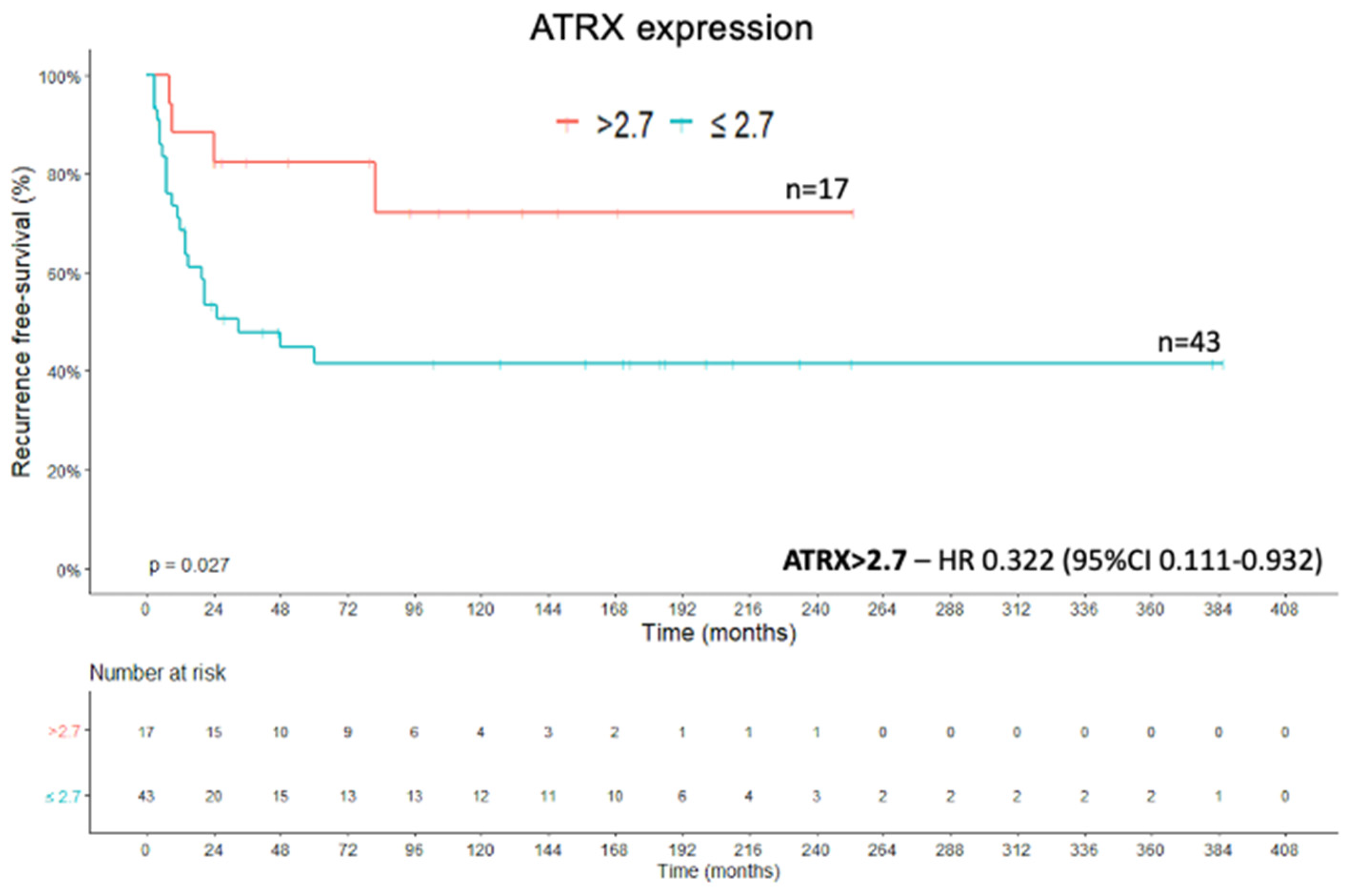
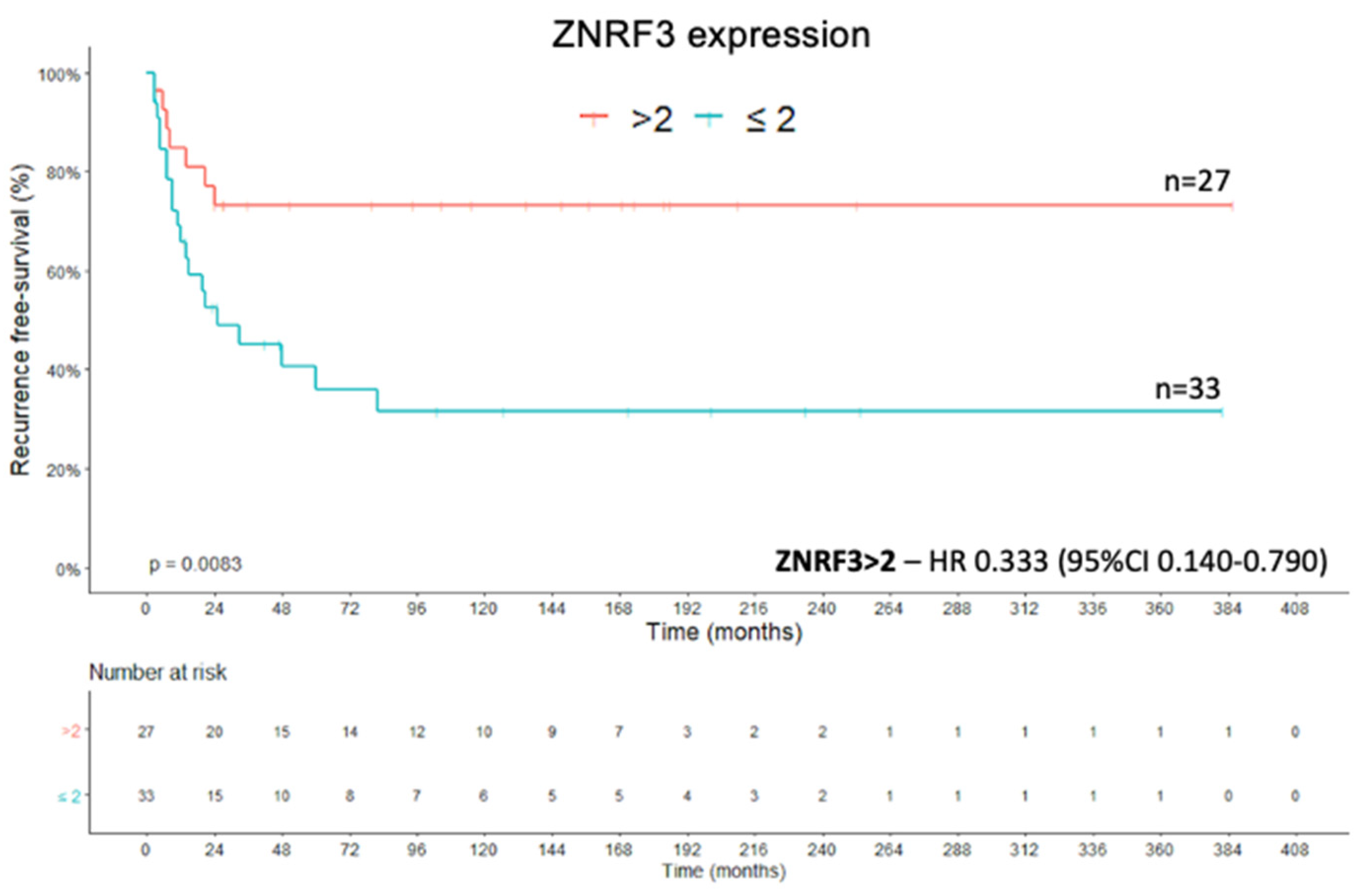
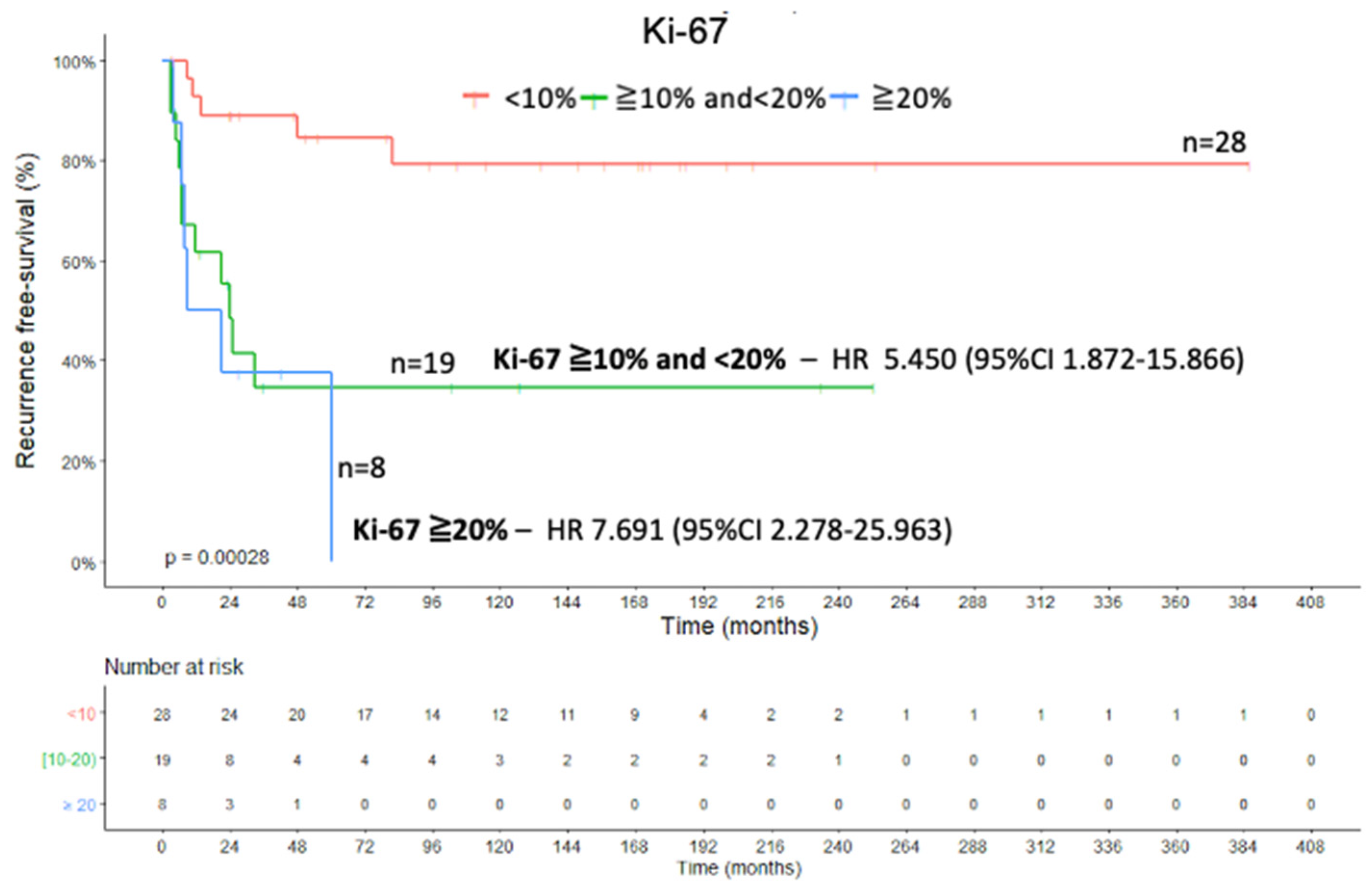
The recurrence-free survival (RFS) was defined as the period between the date of complete tumor resection and the date of the first radiological evidence of local or distant recurrence [38]. ATRX protein expression was associated with a better RFS when a cut-off >2.7 was statistically established (X2(1) = 4.920; p = 0.027; Kaplan–Meier method and log rank test) (Figure 7). The Cox regression of RFS showed an HR of 0.332 (95% CI, 0.111–0.932) for ATRX expression >2.7 compared with the value for an expression level of ≤2.7 (Wald = 4.365; df1; p = 0.037; Cox’s proportional hazards model). The presence of high ZNRF3 protein expression (score > 2) was positively associated with RFS (X2(1) = 6.963; p = 0.008; Kaplan–Meier method and log rank test) (Figure 8), which means that the cases with high protein expression presented a greater recurrence-free survival than the group with low protein expression. This corroborates to ZNRF3 function of tumor suppressor. The Cox regression of RFS showed an HR of 0.333 (95% CI, 0.140–0.790) for ZNRF3 expression >2 compared with the value for an expression level of ≤2 (Wald = 6.222; df1; p = 0.013; Cox’s proportional hazards model). Categorization of the Ki-67 proliferation marker into groups (<10%, ≥10% and <20%, ≥20%) impacted the RFS curve (X2(2) = 16.357; p < 0.001; Kaplan–Meier method and log rank test), as seen in Figure 9. The Cox regression of RFS from the analysis of Ki-67 expression showed an HR of 5.450 (95% CI, 1.872–15.866) for the Ki-67 expression grouped in ≥10% and <20% (Wald = 9.674; df1; p = 0.002; Cox’s proportional hazards model), and we had an HR of 7.691 (95% CI, 2.278–25.963) for the Ki-67 expression grouped in ≥20% (Wald = 10.801; df1; p = 0.001; Cox’s proportional hazards model).

2.5. Other Variables with Prognostic Value

Overall survival was associated with a tumor weight of > 55 g (X2(1) = 13.534; p < 0.001; Kaplan–Meier method and log rank test), a tumor size of >7 cm (X2(1) = 11.462; p = 0.001; Kaplan–Meier method and log rank test), a Weiss score of ≥ 5 (X2(1) = 15.231; p < 0.001; Kaplan–Meier method and log rank test) and Ki-67 of > 8% (X2(1) = 18.294; p < 0.001; Kaplan–Meier method and log rank test). The ENSAT staging system (X2(3) = 11.799; p = 0.008; Kaplan–Meier method and log rank test), the presence of metastasis at diagnosis (X2(1) = 9.111; p = 0.003; Kaplan–Meier method and log rank test), and the presence of local (X2(1) = 6.775; p = 0.009; Kaplan–Meier method and log rank test), or distant recurrence (X2(1) = 8.688; p = 0.003; Kaplan–Meier method and log rank test) also impacted the OS curves. The presence of hypercortisolism at the initial presentation did not impact the OS or RFS curves (X2(1) = 3.103; p > 0.05; and X2(1) = 3.060; p > 0.05, respectively. Kaplan–Meier method and log rank test for both).
The Cox regression analysis revealed the following independent variables for death due to ACC: tumor weight (HR 39.052; 95% CI 1.357–1123.525; Wald = 4.572; df1; p = 0.032; Cox’s proportional hazards model), tumor size (HR 4.451; 95% CI 1.729–11.458′ Wald = 9.577; df1; p = 0.002; Cox’s proportional hazards model), Weiss score (HR 4.040; 95 %CI 1.895–8.611; Wald = 13.073; df1; p < 0.001; Cox’s proportional hazards model), Ki-67 proliferation marker (HR 6.360; 95% CI 2.434–16.618; Wald = 14.256; df1; p < 0.001; Cox’s proportional hazards model), presence of metastasis at diagnosis (HR 2.971; 95% CI 1.415–6.237; Wald = 8.282; df1; p = 0.004; Cox’s proportional hazards model), and disease recurrence (HR 14.985; 95% CI 1.358–165.390; Wald = 4.882; df1; p = 0.027; Cox’s proportional hazards model). The principal Cox regression results for OS are presented in the Supplementary Material (Table S1).

3. Discussion

Malignancies develop protector mechanisms for self-survival. Generally, defects are harbored in evolutionarily conserved pathways, such as the Wnt/β-catenin pathway [39]. However, nine acquired biological capabilities, known as cancer hallmarks, are involved in a multistep tumorigenesis process [40].
Telomeres are responsible for protecting genetic material and avoiding end-to-end fusion due to the repair process [41]. Telomeric shortening results from ineffective replication due to a problem in the DNA replication machinery [41,42,43]. When telomeres reach a critical length, the cell enters a state of senescence, which is considered to be a natural tumor suppressor mechanism in humans [44,45]. Some malignancies present an alternative lengthening of telomeres (ALT) pathway to avoid senescence and apoptosis [41,46]. The ATRX gene encodes a chromatin remodeler (ATRX protein), which functions in nucleosome stability, DNA replication, transcription, maintenance of telomere and heterochromatin structure and stability [46,47,48,49,50]. Lovejoy et al. suggested that the inactivation of ATRX is a significant step in the ALT pathway [51]. Bower et al. showed that the loss of wild-type ATRX expression in somatic cell hybrids results in the development of the ALT pathway [52]. Barthel et al. analyzed 9127 patients and 31 cancer types and showed that inactivation of ATRX is associated with telomere length elongation, reinforcing the idea of an association between ATRX and ALT [53]. ATRX is suggested to be a suppressor of the ALT pathway [47]. Cohorts of different malignancies have shown that ATRX alterations have prognostic value [54,55,56,57,58,59,60,61,62,63,64,65].
The Wnt/β-catenin pathway is one of the central molecular mechanisms of embryonic development [66]. Although it is essential for the embryonic phase, the Wnt/β-catenin has a role in various malignancies. ZNRF3 is a transmembrane E3 ubiquitin ligase molecular target of R-spondin that is localized to the plasma membrane [67]. Hao et al. suggested that ZNRF3 inhibits Wnt/β-catenin signaling by promoting decreases in the membrane levels of frizzled and LRP6 [67]. Basham et al. reported ZNRF3 homeostatic regulation of the adrenal cortex, showing that its loss leads to severe adrenocortical hyperplasia [68]. In the same way as the ATRX gene, multiple cohorts have reported the role of ZNRF3 in tumorigenesis and its prognostic value in different malignancies [69,70,71,72,73,74,75,76,77].
We presented a younger cohort with median age 38.17 years (range 15.38–85.46 years), with more female patients (63, 76.8%), and more deaths (39, 55%), compared to TCGA-ACC and ENSAT and Assie et al. cohorts [27,37]. However, the frequency of ENSAT system in the cohort was very similar with those of both cohorts. Unfortunately, we did not have access to genetic samples to enable determination of the frequency of alterations in these genes or to calculate correlations between molecular alterations and protein expression levels. Nevertheless, we were able to show the protein expression profiles of ATRX and ZNRF3 in our cohort.
Our objective was to analyze the ATRX and ZNRF3 protein expression in an ACC cohort; we chose the immunohistochemistry technique because of its availability and ease of use, aiming to bring pan-genomic studies closer to the clinical practice. The ATRX analysis considered the extent of cells. Unlike Mete et al. [32] who registered only the global loss of ATRX expression, we adapted the ATRX analysis from other neoplasia studies, intending to be more descriptive [78,79]. The ZNRF3 evaluation was based on studies from Qiu et al. and Yu et al. [71,73], based their characterization of the extent and intensity of ZNRF3 cytoplasmic staining.
ATRX protein expression is seen in the nuclear compartment of ACC cells (Figure 1). Mete et al. studied ATRX protein expression in 43 ACCs in a tissue microarray (TMA) and showed a global loss of nuclear ATRX expression in 48% of the cohort [32]. We registered the global loss of ATRX expression (score 0) in 47.5% (n = 38) of our cohort. The loss of ATRX protein expression and its association with disease-free survival or with the presence of adverse outcomes was not proven in the study by Mete et al. [32]. We demonstrated that low expression of ATRX, present in 52.5% of our cohort, impacted negatively on OS and RFS. Moreover, we proved the positive effect of ATRX expression (score >1.5) on prognosis with an HR of 0.521 (95% CI, 0.273–0.997) (p = 0.049).
Hypercortisolism has a negative impact on the OS and RFS of ACC, acting as a predictor of recurrence and death [26,80,81,82]. Although cortisol-secreting ACCs have been associated with a worse OS [83], this was not shown in our cohort. ATRX was highly expressed (median score of 3.08) in the non-hypercortisolism group, and it showed a low expression (median score of 0.5) in the hypercortisolism group. The role of ATRX in cortisol secretion is unknown.
The occurrence of Wnt/β-catenin signaling has been established in different malignancies [37,84,85]. ZNRF3 acts as a tumor suppressor of the Wnt/β-catenin pathway [39,68,69,86,87,88]. Tissier et al. have already demonstrated the role of Wnt/β-catenin in adrenal tumorigenesis [89]. They reported that 77% of an ACC cohort presented with active Wnt/β-catenin signaling through diffuse cytoplasmic/nuclear β-catenin accumulation in tumor cells [89]. We demonstrated that the presence of ZNRF3 expression is correlated with β-catenin expression in the cell membrane (p = 0.004), supporting the idea that ZNRF3 keeps β-catenin attached in the cell membrane (Figure 4).
The high expression of ZNRF3 was associated with smaller tumors in our cohort. ZNRF3 expression was negatively correlated with tumor weight (p = 0.026) and tumor size (p = 0.005). Qin et al. assessed ZNRF3 expression in a cohort of gastric cancer patients and showed an association of protein expression with smaller tumor size [70]. They also revealed that ZNRF3 overexpression causes significantly more apoptosis and lowered proliferation of cancer cells by reducing the level of Lgr5, a component of Wnt/β-catenin signaling, while also reducing Gli1, a component of Hedgehog signaling (SHH) [70]. Gomes et al. showed that the SHH pathway is upregulated in adult ACC and downregulated in pediatric adrenocortical tumors [90]. If ZNRF3 has a role in the adrenal SHH pathway, it remains unclear.
Qiu et al. demonstrated the downregulation of ZNRF3 protein expression in human papillary thyroid carcinoma cell lines [73]. They showed that ZNRF3 overexpression strongly inhibits the migratory and invasive capacities of the cancer cells [73]. Yu et al. revealed that strong expression of ZNRF3 had a significant association with disease-free survival and OS in a colorectal carcinoma cohort [71]. We showed that low ZNRF3 expression impacted negatively on OS analysis in this ACC cohort. The Cox regression showed an HR of 0.441 (95% CI, 0.229–0.852) for tumors with high ZNRF3 expression; furthermore, this factor was positively associated with a better RFS than the loss of its expression.
Ki-67 proliferation marker is considered a prognostic factor in localized ACC and is a powerful tool for predicting recurrence after complete surgical resection [28]. Some studies have documented the importance of Ki-67 as a marker of cellular proliferation, providing a value in the ACC scenario [13,20,91,92]. We categorized the Ki-67 proliferation marker into groups (<10%, ≥10% and <20%, ≥20%), as suggested by Beuschlein et al. [28], and we confirmed the association of Ki-67 with OS and RFS. However, the expressions levels of ATRX and ZNRF3 were not shown to be associated with the Ki-67 proliferation marker.
Since ATRX and ZNRF3 are present in independent pathways, they were considered prognostic markers in this cohort, so we analyzed the association of low expression levels of both. When low expression levels were combined, a stronger negative prognostic value was found for this cohort (HR 0.314; 95% CI, 0.136–0.725; p = 0.007).
Tumors with active Wnt/β-catenin signaling due to CTNNB1, ZNRF3, or APC mutations present a dismal prognosis, according to TCGA Research Network. TCGA reported a high expression of ZNRF3 transcript by RNA sequencing (RNA-seq) levels is associated with a low survival rate in the ACC cohort. On the contrary, we showed that a low ZNRF3 protein expression is linked to poor survival.
The protein and transcript levels are in agreement in most cases, and some hypotheses may explain the difference in the ZNRF3 and ACC context. ZNRF3 is a target gene of Wnt/β-catenin signaling, and the levels of ZNRF3 transcript can reflect the level of Wnt/β-catenin signaling activity. However, ZNRF3 deletions may influence its transcript levels, which could be undetectable due to biallelic deletion; simultaneously, the Wnt/β-catenin signaling could be highly active due to the biallelic loss of its ZNRF3 suppressor. The TCGA analyzed the ZNRF3 transcript levels without excluding the biallelic deletions cases, and this could be a reason for the difference between transcript and protein expression. Another hypothesis is based on the function of ZNRF3 negative regulator by R-spondin proteins in the Wnt/β-catenin signaling. ZNRF3 protein expression may be more dependent on R-spondin proteins activity, through LGR-dependent and LGR-independent mechanisms, reflecting their activity, than the absolute quantity of its transcript.
TCGA also reported ATRX transcript expression by RNA-seq levels in the ACC cohort; however, the ATRX expression did not impact the survival rate.
Patients classified as having a low or moderate risk of recurrence (ENSAT 1 or 2, presenting microscopically complete resection and Ki-67 ≤10%), according to Fassnacht et al. [8], may benefit from the use of this novel prognostic tool. The presence of low expression levels of ATRX and ZNRF3, which presented a negative prognostic value, could guide the medical approach used for these particular patients to early adjuvant treatment, since these prognostic markers showed associations with OS and RFS.
Regarding germline TP53 pathogenic variants, two patients presented TP53 p.R337H variant (cases 51 and 58), and one patient presented TP53 p.R273H (case 68) (Table S2). Although all of them presented with hypercortisolism at initial presentation and died due to ACC (OS 7, 36, and 12 months, respectively), they presented with variable expression of ATRX (score 2, 0, and 0, respectively) and variable expression of ZNRF3 (score 3, 2, and 5, respectively). It remains unclear as to whether an association between the loss of ATRX expression and germline TP53 pathogenic variants exists and could impact OS in the adult ACC population. However, the association between genes has already been published in the pediatric adrenocortical tumor cohort and in other malignancy cohorts [36,57,93,94,95].
Tumor biology knowledge is an important step in the delivery of personalized medicine, allowing the design of better clinical approaches and targeted therapies. This study brought pan-genomic studies closer to clinical practice with an easily accessible tool to refine prognostication in adult patients with ACC. In conclusion, we demonstrated that low levels of protein expression of both ATRX and ZNRF3 are negative prognostic markers of ACC; however, different cohorts should be evaluated in future studies to validate these findings.

4. Materials and Methods

The Ethics Committees of the Hospital das Clínicas—University of São Paulo approved this study (n.2.394.934/2017). Informed written consent was obtained from all patients.
All patients underwent an adrenalectomy due to ACC, and they were followed in a unique tertiary center by a team specialized in adrenal disorders. Detailed clinical data, including clinical and hormonal presentations, treatments, follow-up and survival data, imaging, and histopathological results, were collected from medical records. Laboratory results were adjusted according to the delta of reference value, aimed at carrying out a comparison since the laboratory tests were not performed in the same laboratory. ACCs were diagnosed according to Weiss Score (Weiss ≥ 3) [10]. The tumor stage was classified according to the ENSAT and Helsinki systems [5]. Enrolled patients were followed up in the Adrenal Ambulatory-HC/FMUSP and in the Instituto do Cancer do Estado de Sao Paulo—ICESP for an average time of 85.35 months (1.4–406.83 months) (Table 1).
A total of 82 adrenocortical carcinomas were evaluated in terms of the protein expression of ATRX, ZNRF3, and β-catenin. Detailed information about the cohort is presented in the Supplemental Material (Table S2).
ACCs present a large degree of intratumor heterogeneity [9,96], and therefore, two expert adrenal pathologists (I.C.S., M.C.N.Z.) selected the most representative areas of ACC tissues to construct a tissue microarray (TMA). The internal validation of the TMA process has already been published [22,38,97,98,99].

4.1. Tissue Microarrays (TMA) and Immunohistochemistry

The tumor series included a total of 82 formalin-fixed paraffin-embedded surgical cases of ACC. Representative areas of the ACCs (viable tumor tissue without necrosis) were identified on hematoxylin- and eosin-stained slides and marked on paraffin donor blocks by two experienced pathologists (I.C.S., M.C.N.Z.). The spotted areas of the donor blocks were punched (1.0 mm punch) and mounted into recipient paraffin blocks using a precision microarray instrument (Beecher Instruments, Sun Prairie, WI, USA), with a total of 3 cores per ACC case. The organization of cores was guided by a cartesian map constituting the three TMAs. Control samples (kidney and liver) were included for TMA orientation. One set of three slides was selected (one slide from each of the three TMA paraffin blocks of the triplicate) for staining with anti-ATRX rabbit polyclonal antibody (titer 1:1000; code HPA001906; lot BH115094; 100 μL (0.2 mg/mL); Sigma-Aldrich, Darmstadt, Germany, anti-ZNRF3 rabbit polyclonal antibody (titer 1:100; code HPA036703; lot A86511; 100 μL (0.1 mg/mL); Sigma-Aldrich, Germany), and anti-β-catenin mouse monoclonal antibody (titer 1:200; clone 14/β-catenin; code 610154; lot 4351813, 150 μg (0.6 mL, 250 μg/mL) BD Transduction Laboratories). The positive control for immunostaining with ATRX and ZNRF3 antibodies was an ACC sample with preserved ATRX immunoexpression and with a known wild type ATRX status, and a colon carcinoma, respectively. Tonsil tissue was used as a positive control for b-catenin and Ki-67 antibodies. A modified immunoperoxidase immunohistochemical method with humid heat antigen retrieval was used as previously described and validated [38,100]. The immunostaining evaluations were performed by two independent observers (I.C.S. and M.C.N.Z.), who were unaware of clinical data, who independently evaluated ATRX, ZNRF3, and β-catenin staining. The average value of the two evaluations was taken for statistical analysis [22,97]. TMA samples were included in the analysis only if two or more evaluable cores were available after the staining procedure. Two ACC cases were excluded from analysis due to the loss of tissue sample (1.62%). The interobserver agreement coefficient (kappa) for the staining evaluation was 0.854 (T = 11.76; p < 0.0001; Cohen’s kappa coefficient). A kappa coefficient >0.6 is considered to represent substantial agreement [101].
Nuclear staining for Ki-67 was also evaluated, and another set of three slides were stained with mouse monoclonal anti-human Ki-67 antigen (titer 1:40; clone MIB-1; code M7240; lot 95324; 0.2 mL (46 mg/L); Dako, Glostrup, Denmark). The stained slides were scanned by the Scanner of histological slides through Panoramic Viewer 1:15 software (3DHISTECH, Budapest, Hungary). Image Pro Plus 4.5 software (MediaCybernetics, Rockville, MD, USA) was used to process the images, and the positive and total nuclei were automatically counted.

Analysis of Immunohistochemistry

ATRX and ZNRF3 expression levels were characterized by nuclear staining and cytoplasmic staining, respectively. Sections were scored semi-quantitatively for extent of ATRX, ZNRF3, and β-catenin expression in ACC cells as follows: 0, no immunoreactive cells; 1, <25% immunoreactive cells; 2, 25–50% immunoreactive cells; 3, 51–75% immunoreactive cells, and 4, >75% immunoreactive cells. A low expression level of ATRX was considered when the presence of <25% immunoreactive cells was identified. The intensity of staining for ZNRF3 and β-catenin was also scored semi-qualitatively, as follows: 0, negative; 1, weak; 2, intermediate; and 3, strong. The final scores for ZNRF3 and β-catenin expressions levels were taken as the sum of both parameters (extent and intensity) and grouped into low expression levels (scores of 0–2) and high expression levels (scores of 3–7) as previously reported [99,102]. β-catenin expression in the different cellular localizations (membranous versus cytoplasmic and/or nuclear) was evaluated separately [102,103,104] (Table 2).

4.2. Statistical Analysis

The baseline patient characteristics were expressed as absolute and relative frequencies for qualitative variables and as the average, standard deviation, median, minimum, and maximum for quantitative variables. The association among qualitative variables were evaluated by the chi-squared test or Fisher’s exact test. The Spearman’s rank correlation coefficient was calculated for the quantitative variables. The Kaplan–Meier estimator of the survival function was applied for survival analysis, and the log-rank test was used to compare the survival function between groups. Regarding the expression of ZNRF3 and ATRX, the determination of two groups of observations for a simple cut-off point was estimated using the maximum of the standardized log-rank statistic proposed by Lausen and Schumacher in 1992 [105]. The Cox semiparametric proportional hazards model was fitted to describe the relationship between OS or RFS and the covariates [106]. OS was defined as the time from the date of ACC diagnosis to the date of disease-related death or the last follow-up visit. RFS was defined as the time from the date of complete tumor resection to the date of the first radiological evidence of local and/or distant recurrence [38]. The assumption of proportional hazards was assessed based on the so-called Schoenfeld residuals [107,108]. There was evidence that covariates had a constant effect over time in all cases. The significance level was fixed at 5% for all tests. Statistical analyses were performed using IBM SPSS Statistics version 23.0 (IBM Corp., Armonk, NY, USA) and R software version 3.6 (R Foundation for Statistical Computing, Vienna, Austria).

Supplementary Materials

Supplementary materials can be found at https://www.mdpi.com/1422-0067/22/3/1238/s1.

Author Contributions

V.B.B. and M.C.B.V.F. designed the study. V.B.B. and A.M.F.L. acquired the clinical data and patient information. V.B.B., M.C.N.Z. and I.C.S. performed the pathological review of all cases and selected representative areas of the ACCs for tissue microarray construction. M.C.N.Z. and I.C.S. performed the immunohistochemical scoring. V.B.B. and M.C.B.V.F. wrote the manuscript. V.B.B., A.M.F.L., B.M.d.P.M., L.M., I.C.S., J.E.B.-N., F.Y.T., V.S., J.L.C., B.B.M., M.Q.A., M.C.N.Z., and M.C.B.V.F. performed critical revision of the article. All authors have read and agreed to the published version of the manuscript.

Funding

This research was supported in part by Fundação de Amparo à Pesquisa do Estado de São Paulo (2017/26345-5) to MCBVF.

Institutional Review Board Statement

The study was conducted according to the guidelines of the Declaration of Helsinki, and it was approved by the Ethics Committee of the Hospital das Clínicas—University of São Paulo (protocol code n.2.394.934, date of approval 27/09/2017).

Informed Consent Statement

Informed consent was obtained from all subjects involved in the study.

Data Availability Statement

The data presented in this study are available in Supplementary Material (Tables S1 and S2).

Conflicts of Interest

The authors declare no conflict of interest.

References

  1. Dackiw, A.P.B.; Lee, J.E.; Gagel, R.F.; Evans, D.B. Adrenal cortical carcinoma. World J. Surg. 2001, 25, 914–926. [Google Scholar] [CrossRef] [PubMed]
  2. Kebebew, E.; Reiff, E.; Duh, Q.Y.; Clark, O.H.; McMillan, A. Extent of disease at presentation and outcome for adrenocortical carcinoma: Have we made progress? World J. Surg. 2006, 30, 872–878. [Google Scholar] [CrossRef] [PubMed]
  3. Bilimoria, K.Y.; Shen, W.T.; Elaraj, D.; Bentrern, D.J.; Winchester, D.J.; Kebebew, E. Adrenocortical Carcinoma in the United States Treatment Utilization and Prognostic Factors. Cancer 2008, 113, 3130–3136. [Google Scholar] [CrossRef]
  4. Icard, P.; Goudet, P.; Charpenay, C.; Andreassian, B.; Carnaille, B.; Chapuis, Y.; Cougard, P.; Henry, J.-F.; Proye, C. Adrenocortical carcinomas: Surgical trends and results of a 253-patient series from the French Association of Endocrine Surgeons study group. World J. Surg. 2000, 25, 891–897. [Google Scholar] [CrossRef] [PubMed]
  5. Fassnacht, M.; Johanssen, S.; Quinkler, M.; Bucsky, P.; Willenberg, H.S.; Beuschlein, F.; Terzolo, M.; Mueller, H.-H.; Hahner, S.; Allolio, B.; et al. Limited Prognostic Value of the 2004 International Union Against Cancer Staging Classification for Adrenocortical Carcinomas. Cancer 2009, 115, 243–250. [Google Scholar] [CrossRef]
  6. Fassnacht, M.; Libe, R.; Kroiss, M.; Allolio, B. Adrenocortical carcinoma: A clinician’s update. Nat. Rev. Endocrinol. 2011, 7, 323–335. [Google Scholar] [CrossRef] [PubMed]
  7. Fassnacht, M.; Terzolo, M.; Allolio, B.; Baudin, E.; Haak, H.; Berruti, A.; Welin, S.; Schade-Brittinger, C.; Lacroix, A.; Jarzab, B.; et al. Combination Chemotherapy in Advanced Adrenocortical Carcinoma. N. Engl. J. Med. 2012, 366, 2189–2197. [Google Scholar] [CrossRef]
  8. Fassnacht, M.; Dekkers, O.M.; Else, T.; Baudin, E.; Berruti, A.; De Krijger, R.R.; Haak, H.R.; Mihai, R.; Assie, G.; Terzolo, M. European Society of Endocrinology Clinical Practice Guidelines on the management of adrenocortical carcinoma in adults, in collaboration with the European Network for the Study of Adrenal Tumors. Eur. J. Endocrinol. 2018, 179, G1–G46. [Google Scholar] [CrossRef]
  9. Else, T.; Kim, A.C.; Sabolch, A.; Raymond, V.M.; Kandathil, A.; Caoili, E.M.; Jolly, S.; Miller, B.S.; Giordano, T.J.; Hammer, G.D. Adrenocortical Carcinoma. Endocr. Rev. 2014, 35, 282–326. [Google Scholar] [CrossRef] [Green Version]
  10. Weiss, L.M. Comparative histologic-study of 43 metastasizing and nonmetastasizing adrenocortical tumors. Am. J. Surg. Pathol. 1984, 8, 163–169. [Google Scholar] [CrossRef]
  11. Luton, J.P.; Cerdas, S.; Billaud, L.; Thomas, G.; Guilhaume, B.; Bertagna, X.; Laudat, M.-H.; Louvel, A.; Chapuis, Y.; Blondeau, P.; et al. Clinical-features of adrenocortical carcinoma, prognostic factors, and the effect of mitotane therapy. N. Engl. J. Med. 1990, 322, 1195–1201. [Google Scholar] [CrossRef] [PubMed]
  12. Medeiros, L.J.; Weiss, L.M. New developments in the pathological diagnosis of adrenal-cortical neoplasms—A review. Am. J. Clin. Pathol. 1992, 97, 73–83. [Google Scholar] [CrossRef] [PubMed]
  13. McNicol, A.M.; Struthers, A.J.; Nolan, C.E.; Hermans, J.; Haak, H.R. Proliferation in adrenocortical tumors: Correlation with clinical outcome and p53 status. Endocr. Pathol. 1997, 8, 29–36. [Google Scholar] [CrossRef]
  14. Vassilopoulou-Sellin, R.; Schultz, P.N. Adrenocortical carcinoma—Clinical outcome at the end of the 20th century. Cancer 2001, 92, 1113–1121. [Google Scholar] [CrossRef]
  15. Stojadinovic, A.; Ghossein, R.A.; Hoos, A.; Nissan, A.; Marshall, D.; Dudas, M.; Cordon-Cardo, C.; Jaques, D.P.; Brennan, M.F. Adrenocortical carcinoma: Clinical, morphologic, and molecular characterization. J. Clin. Oncol. 2002, 20, 941–950. [Google Scholar] [CrossRef]
  16. Aubert, S.; Wacrenier, A.; Leroy, X.; Devos, P.; Carnaille, B.; Proye, C.; Wemeau, J.L.; Lecomte-Houcke, M.; Leteurtre, E. Weiss system revisited—A clinicopathologic and immunohistochemical study of 49 adrenocortical tumors. Am. J. Surg. Pathol. 2002, 26, 1612–1619. [Google Scholar] [CrossRef]
  17. Stojadinovic, A.; Brennan, M.F.; Hoos, A.; Omeroglu, A.; Leung, D.H.Y.; Dudas, M.E.; Nissan, A.; Cordon-Cardo, C.; Ghossein, R.A. Adrenocortical adenoma and carcinoma: Histopathological and molecular comparative analysis. Mod. Pathol. 2003, 16, 742–751. [Google Scholar] [CrossRef]
  18. Sturgeon, C.; Shen, W.T.; Clark, O.H.; Duh, Q.Y.; Kebebew, E. Risk assessment in 457 adrenal cortical carcinomas: How much does tumor size predict the likelihood of malignancy? J. Am. Coll. Surg. 2006, 202, 423–430. [Google Scholar] [CrossRef]
  19. Assie, G.; Antoni, G.; Tissier, F.; Caillou, B.; Abiven, G.; Gicquel, C.; Leboulleux, S.; Travagli, J.-P.; Dromain, C.; Bertagna, X.; et al. Prognostic parameters of metastatic adrenocortical carcinoma. J. Clin. Endocrinol. Metab. 2007, 92, 148–154. [Google Scholar] [CrossRef]
  20. Morimoto, R.; Satoh, F.; Murakami, O.; Suzuki, T.; Abe, T.; Tanemoto, M.; Abe, M.; Urno, A.; Ishidoya, S.; Arai, Y.; et al. Immunohistochemistry of a proliferation marker Ki-67/MIB1 in adrenocortical carcinomas: Ki-67/MIB1 Labeling index is a predictor for recurrence of adrenocortical carcinomas. Endocr. J. 2008, 55, 49–55. [Google Scholar] [CrossRef] [Green Version]
  21. Lau, S.K.; Weiss, L.M. The Weiss system for evaluating adrenocortical neoplasms: 25 years later. Hum. Pathol. 2009, 40, 757–768. [Google Scholar] [CrossRef] [PubMed]
  22. Almeida, M.Q.; Soares, I.C.; Ribeiro, T.C.; Fragoso, M.; Marins, L.V.; Wakamatsu, A.; Ressio, R.A.; Nishi, M.Y.; Jorge, A.A.L.; Lerario, A.M.; et al. Steroidogenic Factor 1 Overexpression and Gene Amplification Are More Frequent in Adrenocortical Tumors from Children than from Adults. J. Clin. Endocrinol. Metab. 2010, 95, 1458–1462. [Google Scholar] [CrossRef] [PubMed] [Green Version]
  23. Durand, J.; Lampron, A.; Mazzuco, T.L.; Chapman, A.; Bourdeau, I. Characterization of Differential Gene Expression in Adrenocortical Tumors Harboring β-Catenin (CTNNB1) Mutations. J. Clin. Endocrinol. Metab. 2011, 96, E1206–E1211. [Google Scholar] [CrossRef] [PubMed] [Green Version]
  24. Gaujoux, S.; Grabar, S.; Fassnacht, M.; Ragazzon, B.; Launay, P.; Libe, R.; Chokri, I.; Audebourg, A.; Royer, B.; Sbiera, S.; et al. β-Catenin Activation Is Associated with Specific Clinical and Pathologic Characteristics and a Poor Outcome in Adrenocortical Carcinoma. Clin. Cancer Res. 2011, 17, 328–336. [Google Scholar] [CrossRef] [PubMed] [Green Version]
  25. Ayala-Ramirez, M.; Jasim, S.; Feng, L.; Ejaz, S.; Deniz, F.; Busaidy, N.; Waguespack, S.G.; Naing, A.; Sircar, K.; Wood, C.G.; et al. Adrenocortical carcinoma: Clinical outcomes and prognosis of 330 patients at a tertiary care center. Eur. J. Endocrinol. 2013, 169, 891–899. [Google Scholar] [CrossRef] [PubMed]
  26. Berruti, A.; Fassnacht, M.; Haak, H.; Else, T.; Baudin, E.; Sperone, P.; Kroiss, M.; Kerkhofs, T.; Williams, A.R.; Ardito, A.; et al. Prognostic Role of Overt Hypercortisolism in Completely Operated Patients with Adrenocortical Cancer. Eur. Urol. 2014, 65, 832–838. [Google Scholar] [CrossRef]
  27. Assié, G.; Letouzé, E.; Fassnacht, M.; Jouinot, A.; Luscap, W.; Barreau, O.; Omeiri, H.; Rodriguez, S.; Perlemoine, K.; René-Corail, F.; et al. Integrated genomic characterization of adrenocortical carcinoma. Nat. Genet. 2014, 46, 607–612. [Google Scholar] [CrossRef]
  28. Beuschlein, F.; Weigel, J.; Saeger, W.; Kroiss, M.; Wild, V.; Daffara, F.; Libé, R.; Ardito, A.; Al Ghuzlan, A.; Quinkler, M.; et al. Major Prognostic Role of Ki-67 in Localized Adrenocortical Carcinoma After Complete Resection. J. Clin. Endocrinol. Metab. 2015, 100, 841–849. [Google Scholar] [CrossRef]
  29. Libé, R.; Borget, I.; Ronchi, C.L.; Zaggia, B.; Kroiss, M.; Kerkhofs, T.; Bertherat, J.; Volante, M.; Quinkler, M.; Chabre, O.; et al. Prognostic factors in stage III-IV adrenocortical carcinomas (ACC): An European Network for the Study of Adrenal Tumor (ENSAT) study. Ann. Oncol. 2015, 26, 2119–2125. [Google Scholar] [CrossRef]
  30. Pennanen, M.; Heiskanen, I.; Sane, T.; Remes, S.; Mustonen, H.; Haglund, C.; Arola, J. Helsinki score-a novel model for prediction of metastases in adrenocortical carcinomas. Hum. Pathol. 2015, 46, 404–410. [Google Scholar] [CrossRef]
  31. Zheng, S.; Cherniack, A.D.; Dewal, N.; Moffitt, R.A.; Danilova, L.; Murray, B.A.; Lerario, A.M.; Else, T.; Knijnenburg, T.A.; Ciriello, G.; et al. Comprehensive Pan-Genomic Characterization of Adrenocortical Carcinoma. Cancer Cell. 2016, 29, 723–736. [Google Scholar] [CrossRef] [PubMed] [Green Version]
  32. Mete, O.; Gucer, H.; Kefeli, M.; Asa, S.L. Diagnostic and Prognostic Biomarkers of Adrenal Cortical Carcinoma. Am. J. Surg. Pathol. 2018, 42, 201–213. [Google Scholar] [CrossRef]
  33. Mete, O.; Asa, S.L.; Giordano, T.J.; Papotti, M.; Sasano, H.; Volante, M. Immunohistochemical Biomarkers of Adrenal Cortical Neoplasms. Endocr. Pathol. 2018, 29, 137–149. [Google Scholar] [CrossRef] [PubMed]
  34. Libe, R. Clinical and molecular prognostic factors in adrenocortical carcinoma. Minerva Endocrinol. 2019, 44, 58–69. [Google Scholar] [CrossRef] [PubMed]
  35. Ahmed, A.A.; Thomas, A.J.; Ganeshan, D.M.; Blair, K.J.; Lall, C.; Lee, J.T.; Morshid, A.I.; Habra, M.A.; Elsayes, K.M. Adrenal cortical carcinoma: Pathology, genomics, prognosis, imaging features, and mimics with impact on management. Abdom. Radiol. 2020, 45, 945–963. [Google Scholar] [CrossRef] [PubMed]
  36. Pinto, E.M.; Chen, X.; Easton, J.; Finkelstein, D.; Liu, Z.; Pounds, S.; Rodriguez-Galindo, C.; Lund, T.C.; Mardis, E.R.; Wilson, R.K.; et al. Genomic landscape of paediatric adrenocortical tumours. Nat. Commun. 2015, 6, 10. [Google Scholar] [CrossRef] [Green Version]
  37. The Cancer Genome Atlas Program. Available online: https://www.cancer.gov/tcga (accessed on 3 January 2021).
  38. Giles, R.H.; van Es, J.H.; Clevers, H. Caught up in a Wnt storm: Wnt signaling in cancer. Biochim. Biophys. Acta 2003, 1653, 1–24. [Google Scholar] [CrossRef]
  39. Lacombe, A.M.F.; Soares, I.C.; Mariani, B.M.D.P.; Nishi, M.Y.; Bezerra-Neto, J.E.; Charchar, H.D.S.; Brondani, V.B.; Tanno, F.Y.; Srougi, V.; Chambo, J.L.; et al. Sterol O-Acyl Transferase 1 as a Prognostic Marker of Adrenocortical Carcinoma. Cancers 2020, 12, 247. [Google Scholar] [CrossRef] [Green Version]
  40. Fearon, E.R.; Spence, J.R. Cancer Biology: A New RING to Wnt Signaling. Curr. Biol. 2012, 22, R849–R851. [Google Scholar] [CrossRef] [Green Version]
  41. Hanahan, D.; Weinberg, R.A. Hallmarks of Cancer: The Next Generation. Cell 2011, 144, 646–674. [Google Scholar] [CrossRef] [Green Version]
  42. Cesare, A.J.; Reddel, R.R. Alternative lengthening of telomeres: Models, mechanisms and implications. Nat. Rev. Genet. 2010, 11, 319–330. [Google Scholar] [CrossRef]
  43. Watson, J.D. Origin of concatemeric T7 DNA. Nat. New Biol. 1972, 239, 197. [Google Scholar] [CrossRef]
  44. Olovnikov, A.M. Theory of marginotomy—Incomplete copying of template margin in enzymic-synthesis of polynucleotides and biological significance of phenomenon. J. Theor. Biol. 1973, 41, 181–190. [Google Scholar] [CrossRef]
  45. Shay, J.W. Role of Telomeres and Telomerase in Aging and Cancer. Cancer Discov. 2016, 6, 584–593. [Google Scholar] [CrossRef] [Green Version]
  46. Der-Sarkissian, H.; Bacchetti, S.; Cazes, L.; Londono-Vallejo, J.A. The shortest telomeres drive karyotype evolution in transformed cells. Oncogene 2004, 23, 1221–1228. [Google Scholar] [CrossRef] [Green Version]
  47. Watson, L.A.; Goldberg, H.; Berube, N.G. Emerging roles of ATRX in cancer. Epigenomics 2015, 7, 1365–1378. [Google Scholar] [CrossRef]
  48. Clynes, D.; Higgs, D.R.; Gibbons, R.J. The chromatin remodeller ATRX: A repeat offender in human disease. Trends Biochem. Sci. 2013, 38, 461–466. [Google Scholar] [CrossRef]
  49. Drane, P.; Ouararhni, K.; Depaux, A.; Shuaib, M.; Hamiche, A. The death-associated protein DAXX is a novel histone chaperone involved in the replication-independent deposition of H3.3. Genes Dev. 2010, 24, 1253–1265. [Google Scholar] [CrossRef] [Green Version]
  50. Elsasser, S.J.; Huang, H.D.; Lewis, P.W.; Chin, J.W.; Allis, C.D.; Patel, D.J. DAXX envelops a histone H3.3-H4 dimer for H3.3-specific recognition. Nature 2012, 491, 560. [Google Scholar] [CrossRef] [PubMed] [Green Version]
  51. Goldberg, A.D.; Banaszynski, L.A.; Noh, K.-M.; Lewis, P.W.; Elsaesser, S.J.; Stadler, S.; Dewell, S.; Law, M.; Guo, X.; Li, X.; et al. Distinct Factors Control Histone Variant H3.3 Localization at Specific Genomic Regions. Cell 2010, 140, 678–691. [Google Scholar] [CrossRef] [Green Version]
  52. Lovejoy, C.A.; Li, W.; Reisenweber, S.; Thongthip, S.; Bruno, J.; De Lange, T.; De, S.; Petrini, J.H.J.; Sung, P.A.; Jasin, M.; et al. Loss of ATRX, Genome Instability, and an Altered DNA Damage Response Are Hallmarks of the Alternative Lengthening of Telomeres Pathway. PLoS Genet. 2012, 8, 16. [Google Scholar] [CrossRef] [PubMed]
  53. Bower, K.; Napier, C.E.; Cole, S.L.; Dagg, R.A.; Lau, L.M.S.; Duncan, E.L.; Moy, E.L.; Reddel, R.R. Loss of Wild-Type ATRX Expression in Somatic Cell Hybrids Segregates with Activation of Alternative Lengthening of Telomeres. PLoS ONE 2012, 7, 10. [Google Scholar] [CrossRef] [PubMed] [Green Version]
  54. Barthel, F.P.; Wei, W.; Tang, M.; Martinez-Ledesma, E.; Hu, X.; Amin, S.B.; Akdemir, K.C.; Seth, S.; Song, X.; Wang, Q.; et al. Systematic analysis of telomere length and somatic alterations in 31 cancer types. Nat. Genet. 2017, 49, 349–357. [Google Scholar] [CrossRef] [PubMed]
  55. Wiestler, B.; Capper, D.; Holland-Letz, T.; Von Deimling, A.; Pfister, S.M.; Platten, M.; Weller, M.; Wick, W. ATRX loss refines the classification of anaplastic glioma and is a favorable prognostic marker. Cancer Res. 2013, 73, 1. [Google Scholar]
  56. Marinoni, I.; Kurrer, A.S.; Vassella, E.; Dettmer, M.; Rudolph, T.; Banz, V.M.; Hunger, F.; Pasquinelli, S.; Speel, E.; Perren, A. Loss of DAXX and ATRX Are Associated With Chromosome Instability and Reduced Survival of Patients With Pancreatic Neuroendocrine Tumors. Gastroenterology 2014, 146, 453. [Google Scholar] [CrossRef] [PubMed]
  57. Liau, J.-Y.; Lee, J.-C.; Tsai, J.-H.; Yang, C.-Y.; Liu, T.-L.; Ke, Z.-L.; Hsu, H.-H.; Jeng, Y.-M. Comprehensive screening of alternative lengthening of telomeres phenotype and loss of ATRX expression in sarcomas. Mod. Pathol. 2015, 28, 1545–1554. [Google Scholar] [CrossRef] [Green Version]
  58. Yang, C.-Y.; Liau, J.-Y.; Huang, W.-J.; Chang, Y.-T.; Chang, M.-C.; Lee, J.-C.; Tsai, J.-H.; Su, Y.-N.; Hung, C.-C.; Jeng, Y.-M. Targeted next-generation sequencing of cancer genes identified frequent TP53 and ATRX mutations in leiomyosarcoma. Am. J. Transl. Res. 2015, 7, 2072–2081. [Google Scholar]
  59. Cai, J.; Chen, J.; Zhang, W.; Yang, P.; Zhang, C.; Li, M.; Yao, K.; Wang, H.; Li, Q.; Jiang, C.; et al. Loss of ATRX, associated with DNA methylation pattern of chromosome end, impacted biological behaviors of astrocytic tumors. Oncotarget 2015, 6, 18105–18115. [Google Scholar] [CrossRef]
  60. Cai, J.; Zhu, P.; Zhang, C.; Li, Q.; Wang, Z.; Li, G.; Wang, G.; Yang, P.; Li, J.; Han, B.; et al. Detection of ATRX and IDH1-R132H immunohistochemistry in the progression of 211 paired gliomas. Oncotarget 2016, 7, 16384–16395. [Google Scholar] [CrossRef] [Green Version]
  61. Singhi, A.D.; Liu, T.-C.; Roncaioli, J.L.; Cao, D.; Zeh, H.J.; Zureikat, A.H.; Tsung, A.; Marsh, J.W.; Lee, K.K.; Hogg, M.E.; et al. Alternative Lengthening of Telomeres and Loss of DAXX/ATRX Expression Predicts Metastatic Disease and Poor Survival in Patients with Pancreatic Neuroendocrine Tumors. Clin. Cancer Res. 2017, 23, 600–609. [Google Scholar] [CrossRef] [Green Version]
  62. Ren, X.L.; Tu, C.; Tang, Z.C.; Ma, R.F.; Li, Z.H. Alternative lengthening of telomeres phenotype and loss of ATRX expression in sarcomas (Review). Oncol. Lett. 2018, 15, 7489–7496. [Google Scholar] [CrossRef] [PubMed] [Green Version]
  63. Chou, A.; Itchins, M.; De Reuver, P.R.; Arena, J.; Clarkson, A.; Sheen, A.; Sioson, L.; Cheung, V.; Perren, A.; Nahm, C.; et al. ATRX loss is an independent predictor of poor survival in pancreatic neuroendocrine tumors. Hum. Pathol. 2018, 82, 249–257. [Google Scholar] [CrossRef] [PubMed] [Green Version]
  64. Lu, H.C.; Apicelli, A.J.; Eulo, V.; Pekmezci, M.; Hirbe, A.C.; Dahiya, S. Aberrant ATRX Protein Expression is Associated with Worse Survival in NF1-Associated Malignant Peripheral Nerve Sheath Tumors. Mod. Pathol. 2018, 31, 661. [Google Scholar]
  65. Terra, S.; Xie, H.; Boland, J.M.; Mansfield, A.S.; Molina, J.R.; Roden, A.C. Loss of ATRX expression predicts worse prognosis in pulmonary carcinoid tumors. Hum. Pathol. 2019, 94, 78–85. [Google Scholar] [CrossRef]
  66. Hong, X.; Qiao, S.; Li, F.; Wang, W.; Jiang, R.; Wu, H.; Chen, H.; Liu, L.; Peng, J.; Wang, J.; et al. Whole-genome sequencing reveals distinct genetic bases for insulinomas and non-functional pancreatic neuroendocrine tumours: Leading to a new classification system. Gut 2020, 69, 877–887. [Google Scholar] [CrossRef] [Green Version]
  67. Klaus, A.; Birchmeier, W. Wnt signalling and its impact on development and cancer. Nat. Rev. Cancer 2008, 8, 387–398. [Google Scholar] [CrossRef]
  68. Hao, H.-X.; Xie, Y.; Zhang, Y.; Charlat, O.; Oster, E.; Avello, M.; Lei, H.; Mickanin, C.; Liu, D.; Ruffner, H.; et al. ZNRF3 promotes Wnt receptor turnover in an R-spondin-sensitive manner. Nat. Cell Biol. 2012, 485, 195–200. [Google Scholar] [CrossRef]
  69. Basham, K.J.; Rodriguez, S.; Turcu, A.F.; Lerario, A.M.; Logan, C.Y.; Rysztak, M.R.; Gomez-Sanchez, C.E.; Breault, D.T.; Koo, B.K.; Clevers, H.; et al. A ZNRF3-dependent Wnt/β-catenin signaling gradient is required for adrenal homeostasis. Genes. Dev. 2019, 33, 209–220. [Google Scholar] [CrossRef] [Green Version]
  70. Zhou, Y.; Lan, J.; Wang, W.; Shi, Q.; Lan, Y.; Cheng, Z.; Guan, H. ZNRF3 acts as a tumour suppressor by the Wnt signalling pathway in human gastric adenocarcinoma. J. Mol. Histol. 2013, 44, 555–563. [Google Scholar] [CrossRef]
  71. Qin, H.Z.; Cai, A.Z.; Xi, H.Q.; Yuan, J.; Chen, L. ZNRF3 Induces Apoptosis of Gastric Cancer Cells by Antagonizing Wnt and Hedgehog Signaling. Cell Biochem. Biophys. 2015, 73, 361–367. [Google Scholar] [CrossRef]
  72. Yu, N.; Zhu, H.; Tao, Y.; Huang, Y.; Song, X.; Zhou, Y.; Li, Y.; Pei, Q.; Tan, Q.; Pei, H. Association between prognostic survival of human colorectal carcinoma and ZNRF3 expression. Onco Targets Ther. 2016, 9, 6679–6687. [Google Scholar] [CrossRef] [PubMed] [Green Version]
  73. Bond, C.E.; McKeone, D.M.; Kalimutho, M.; Bettington, M.L.; Pearson, S.-A.; Dumenil, T.D.; Wockner, L.F.; Burge, M.; Leggett, B.A.; Whitehall, V.L. RNF43 and ZNRF3 are commonly altered in serrated pathway colorectal tumorigenesis. Oncotarget 2016, 7, 70589–70600. [Google Scholar] [CrossRef] [PubMed]
  74. Qiu, W.W.; Yang, Z.L.; Fan, Y.B.; Zheng, Q. ZNRF3 is downregulated in papillary thyroid carcinoma and suppresses the proliferation and invasion of papillary thyroid cancer cells. Tumor Biol. 2016, 37, 12665–12672. [Google Scholar] [CrossRef] [PubMed]
  75. Shi, J.C.; Jiang, X.G.; Yu, Z.J.; He, G.Q.; Ning, H.Y.; Wu, Z.X.; Cai, Y.; Yu, H.; Chen, A. ZNRF3 contributes to the growth of lung carcinoma via inhibiting Wnt/β-catenin pathway and is regulated by miR-93. Tumor Biol. 2016, 37, 3051–3057. [Google Scholar] [CrossRef] [PubMed]
  76. Xu, E.; Zhao, J.; Ma, J.; Wang, C.; Zhang, C.; Jiang, H.; Cheng, J.; Gao, R.; Zhou, X. miR-146b-5p promotes invasion and metastasis contributing to chemoresistance in osteosarcoma by targeting zinc and ring finger 3. Oncol. Rep. 2016, 35, 275–283. [Google Scholar] [CrossRef] [Green Version]
  77. Wang, Z.W.; Wang, Y.L.; Ren, H.T.; Jin, Y.Y.; Guo, Y. ZNRF3 Inhibits the Invasion and Tumorigenesis in Nasopharyngeal Carcinoma Cells by Inactivating the Wnt/β-Catenin Pathway. Oncol. Res. 2017, 25, 571–577. [Google Scholar] [CrossRef]
  78. Qiao, G.E.; Dai, C.G.; He, Y.; Shi, J.J.; Xu, C.F. Effects of miR-106b-3p on cell proliferation and epithelial-mesenchymal transition, and targeting of ZNRF3 in esophageal squamous cell carcinoma. Int. J. Mol. Med. 2019, 43, 1817–1829. [Google Scholar] [CrossRef]
  79. Berbegall, A.P.; Villamen, E.; Tadeo, I.; Martinsson, T.; Canete, A.; Castel, V.; Navarro, S.; Noguera, R. Neuroblastoma after Childhood: Prognostic Relevance of Segmental Chromosome Aberrations, ATRX Protein Status, and Immune Cell Infiltration. Neoplasia 2014, 16, 471–480. [Google Scholar] [CrossRef] [Green Version]
  80. Davidson, B.; McFadden, E.; Holth, A.; Brunetti, M.; Florenes, V.A. Death domain-associated protein (DAXX) expression is associated with poor survival in metastatic high-grade serous carcinoma. Virchows Arch. 2020, 477, 857–864. [Google Scholar] [CrossRef]
  81. Berruti, A.; Terzolo, M.; Sperone, P.; Pia, A.; Della Casa, S.; Gross, D.J.; Carnaghi, C.; Casali, P.; Porpiglia, F.; Mantero, F.; et al. Etoposide, doxorubicin and cisplatin plus mitotane in the treatment of advanced adrenocortical carcinoma: A large prospective phase II trial. Endocr. Relat. Cancer 2005, 12, 657–666. [Google Scholar] [CrossRef]
  82. Abiven, G.; Coste, J.; Groussin, L.; Anract, P.; Tissier, F.; Legmann, P.; Dousset, B.; Bertagna, X.; Bertherat, J. Clinical and biological features in the prognosis of adrenocortical cancer: Poor outcome of cortisol-secreting tumors in a series of 202 consecutive patients. J. Clin. Endocrinol. Metab. 2006, 91, 2650–2655. [Google Scholar] [CrossRef] [PubMed]
  83. Margonis, G.A.; Kim, Y.; Tran, T.B.; Postlewait, L.M.; Maithel, S.K.; Wang, T.S.; Glenn, J.A.; Hatzaras, I.; Shenoy, R.; Phay, J.E.; et al. Outcomes after resection of cortisol-secreting adrenocortical carcinoma. Am. J. Surg. 2016, 211, 1106–1113. [Google Scholar] [CrossRef] [PubMed] [Green Version]
  84. Vanbrabant, T.; Fassnacht, M.; Assie, G.; Dekkers, O.M. Influence of hormonal functional status on survival in adrenocortical carcinoma: Systematic review and meta-analysis. Eur. J. Endocrinol. 2018, 179, 429–436. [Google Scholar] [CrossRef] [PubMed] [Green Version]
  85. Clements, W.M.; Wang, J.; Sarnaik, A.; Kim, O.J.; MacDonald, J.; Fenoglio-Preiser, C.; Groden, J.; Lowy, A.M. β-catenin mutation is a frequent cause of Wnt pathway activation in gastric cancer. Cancer Res. 2002, 62, 3503–3506. [Google Scholar]
  86. De La Coste, A.; Romagnolo, B.; Billuart, P.; Renard, C.A.; Buendia, M.A.; Soubrane, O.; Fabre, M.; Chelly, J.; Beldjord, C.; Khan, A.; et al. Somatic mutations of the β-catenin gene are frequent in mouse and human hepatocellular carcinomas. Proc. Natl. Acad. Sci. USA 1998, 95, 8847–8851. [Google Scholar] [CrossRef] [Green Version]
  87. Hao, H.-X.; Jiang, X.; Cong, F. Control of Wnt Receptor Turnover by R-spondin-ZNRF3/RNF43 Signaling Module and Its Dysregulation in Cancer. Cancers 2016, 8, 25. [Google Scholar] [CrossRef] [Green Version]
  88. Faillot, S.; Assie, G. The genomics of adrenocortical tumors. Eur. J. Endocrinol. 2016, 174, R249–R265. [Google Scholar] [CrossRef] [Green Version]
  89. Espiard, S.; Bertherat, J. The genetics of adrenocortical tumors. Endocrinol. Metab. Clin. N. Am. 2015, 44, 311–334. [Google Scholar] [CrossRef]
  90. Tissier, F.; Cavard, C.; Groussin, L.; Perlemoine, K.; Fumey, G.; Hagnere, A.M.; Rene-Corail, F.; Jullian, E.; Gicquel, C.; Bertagna, X.; et al. Mutations of β-catenin in adrenocortical tumors: Activation of the wnt signaling pathway is a frequent event in both benign and malignant adrenocortical tumors. Cancer Res. 2005, 65, 7622–7627. [Google Scholar] [CrossRef] [Green Version]
  91. Gomes, D.C.; Leal, L.F.; Mermejo, L.M.; Scrideli, C.A.; Martinelli, C.E.; Fragoso, M.C.B.V.; Latronico, A.C.; Tone, L.G.; Tucci, S.; Yunes, J.A.; et al. Sonic Hedgehog Signaling Is Active in Human Adrenal Cortex Development and Deregulated in Adrenocortical Tumors. J. Clin. Endocrinol. Metab. 2014, 99, E1209–E1216. [Google Scholar] [CrossRef] [Green Version]
  92. Terzolo, M.; Boccuzzi, A.; Bovio, S.; Cappia, S.; De Giuli, P.; Alì, A.; Paccotti, P.; Porpiglia, F.; Fontana, D.; Angeli, A. Immunohistochemical assessment of Ki-67 in the differential diagnosis of adrenocortical tumors. Urology 2001, 57, 176–182. [Google Scholar] [CrossRef]
  93. Sasano, H.; Suzuki, T.; Moriya, T. Recent advances in histopathology and immunohistochemistry of adrenocortical carcinoma. Endocr. Pathol. 2007, 17, 345–354. [Google Scholar] [CrossRef]
  94. Xie, Y.; Tan, Y.; Yang, C.; Zhang, X.; Xu, C.; Qiao, X.; Xu, J.; Tian, S.; Fang, C.; Kang, C. Omics-based integrated analysis identified ATRX as a biomarker associated with glioma diagnosis and prognosis. Cancer Biol. Med. 2019, 16, 784–796. [Google Scholar]
  95. Ikemura, M.; Shibahara, J.; Mukasa, A.; Takayanagi, S.; Aihara, K.; Saito, N.; Aburatani, H.; Fukayama, M. Utility of ATRX immunohistochemistry in diagnosis of adult diffuse gliomas. Histopathology 2016, 69, 260–267. [Google Scholar] [CrossRef]
  96. Liu, X.-Y.; Gerges, N.; Korshunov, A.; Sabha, N.; Khuong-Quang, D.-A.; Fontebasso, A.M.; Fleming, A.; Hadjadj, D.; Schwartzentruber, J.; Majewski, J.; et al. Frequent ATRX mutations and loss of expression in adult diffuse astrocytic tumors carrying IDH1/IDH2 and TP53 mutations. Acta Neuropathol. 2012, 124, 615–625. [Google Scholar] [CrossRef]
  97. Duregon, E.; Volante, M.; Bollito, E.; Goia, M.; Buttigliero, C.; Zaggia, B.; Berruti, A.; Scagliotti, G.V.; Papotti, M. Pitfalls in the diagnosis of adrenocortical tumors: A lesson from 300 consultation cases. Hum. Pathol. 2015, 46, 1799–1807. [Google Scholar] [CrossRef]
  98. Faria, A.M.; Sbiera, S.; Ribeiro, T.C.; Soares, I.C.; Mariani, B.M.; Freire, D.S.; De Sousa, G.R.; Lerário, A.M.; Ronchi, C.L.; Deutschbein, T.; et al. Expression of LIN28 and its regulatory microRNAs in adult adrenocortical cancer. Clin. Endocrinol. 2015, 82, 481–488. [Google Scholar] [CrossRef]
  99. De Sousa, G.R.V.; Soares, I.C.; Faria, A.M.; Domingues, V.B.; Wakamatsu, A.; Lerário, A.M.; Alves, V.A.F.; Zerbini, M.C.N.; De Mendonça, B.B.; Fragoso, M.C.B.V.; et al. DAX1 Overexpression in Pediatric Adrenocortical Tumors: A Synergic Role with SF1 in Tumorigenesis. Horm. Metab. Res. 2015, 47, 656–661. [Google Scholar] [CrossRef]
  100. De Sousa, G.R.V.; Ribeiro, T.C.; Faria, A.M.; Mariani, B.M.P.; Lerario, A.M.; Zerbini, M.C.N.; Soares, I.C.; Wakamatsu, A.; Alves, V.A.F.; Mendonca, B.B.; et al. Low DICER1 expression is associated with poor clinical outcome in adrenocortical carcinoma. Oncotarget 2015, 6, 22724–22733. [Google Scholar] [CrossRef]
  101. Shi, S.R.; Key, M.E.; Kalra, K.L. Antigen retrieval in formalin-fixed, paraffin-embedded tissues—An enhancement method for immunohistochemical staining based on microwave-oven heating of tissue-sections. J. Histochem. Cytochem. 1991, 39, 741–748. [Google Scholar] [CrossRef] [Green Version]
  102. Landis, J.R.; Koch, G.G. Measurement of observer agreement for categorical data. Biometrics 1977, 33, 159–174. [Google Scholar] [CrossRef] [PubMed] [Green Version]
  103. Pinheiro, C.; Granja, S.; Longatto, A.; Faria, A.M.; Fragoso, M.; Lovisolo, S.M.; Bonatelli, M.; Costa, R.F.A.; Lerario, A.M.; Almeida, M.Q.; et al. GLUT1 expression in pediatric adrenocortical tumors: A promising candidate to predict clinical behavior. Oncotarget 2017, 8, 63835–63845. [Google Scholar] [CrossRef] [PubMed] [Green Version]
  104. Heaton, J.H.; Wood, M.A.; Kim, A.C.; Lima, L.O.; Barlaskar, F.M.; Almeida, M.Q.; Fragoso, M.C.B.V.; Kuick, R.; Lerario, A.M.; Simon, D.P.; et al. Progression to Adrenocortical Tumorigenesis in Mice and Humans through Insulin-Like Growth Factor 2 and β-Catenin. Am. J. Pathol. 2012, 181, 1017–1033. [Google Scholar] [CrossRef]
  105. Tadjine, M.; Lampron, A.; Ouadi, L.; Horvath, A.; Stratakis, C.A.; Bourdeau, I. Detection of somatic β-catenin mutations in primary pigmented nodular adrenocortical disease (PPNAD). Clin. Endocrinol. 2008, 69, 367–373. [Google Scholar] [CrossRef] [Green Version]
  106. Lausen, B.; Schumacher, M. Maximally selected rank statistics. Biometrics 1992, 48, 73–85. [Google Scholar] [CrossRef]
  107. Cox, D.R. Regression models and life-tables. J. R. Stat. Soc. 1972, 34, 187. [Google Scholar] [CrossRef]
  108. Schoenfeld, D. Partial residuals for the proportional hazards regression-model. Biometrika 1982, 69, 239–241. [Google Scholar] [CrossRef]
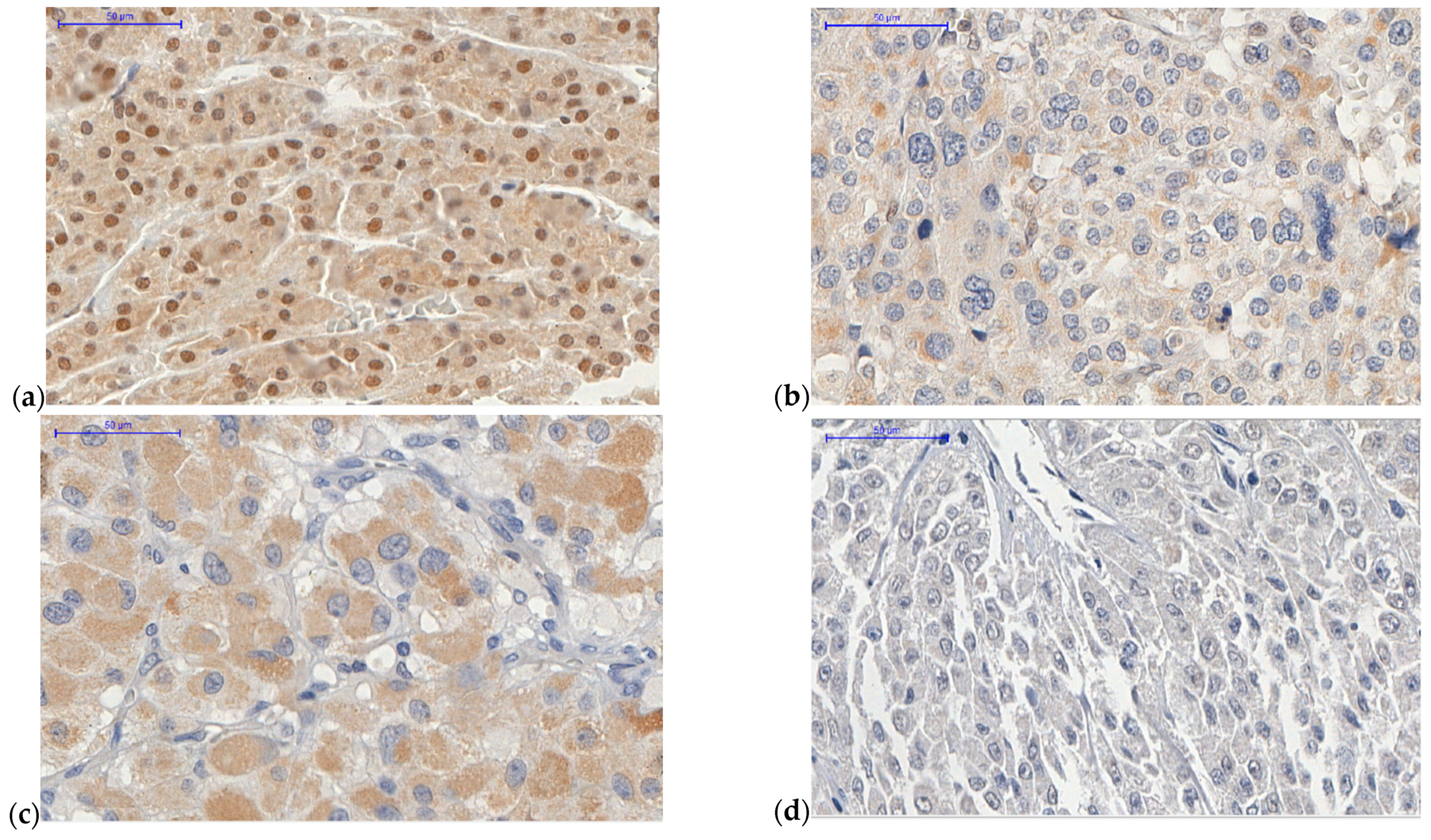
Figure 1. Adrenocortical carcinoma (ACC) samples of tissue microarray (TMA). Obj 40×. (a) ACC case 13 presenting high ATRX nuclear expression (score 4). Female patient, aged 28.7 y with virilizing syndrome, a Weiss score 3, and a European Network for the Study of Adrenal Tumors (ENSAT) system score of 2. (b) ACC case 32 with low ATRX nuclear expression (score 0). Female patient, aged 26.5 y with mixed syndrome (hypercortisolism and androgen excess), a Weiss score of 7, and an ENSAT system score of 4. (c) ACC case 77 with high ZNRF3 cytoplasmic expression (score 5; extent = 4, intensity = 1). Male patient, aged 65.6 y with mixed syndrome (hypercortisolism and androgen excess), a Weiss score of 6, and an ENSAT system score of 4. (d) ACC case 5 with low ZNRF3 cytoplasmic expression (score 0). Female patient, aged 85.4 y with hypercortisolism syndrome, a Weiss score of 7, and an ENSAT system score of 3. Scale bar = 50 μm.
Ijms 22 01238 g001
Figure 2. ATRX protein expression—overall survival (OS) Kaplan–Meier curve.
Ijms 22 01238 g002
Figure 3. ZNRF3 protein expression—OS Kaplan–Meier curve.
Ijms 22 01238 g003
Figure 4. Figure of TMA: (a) ACC case 22 with high ZNRF3 cytoplasmic expression (score 5). Female patient, aged 19.3 y with hypercortisolism syndrome, a Weiss score of 4, and an ENSAT system score of 1. (b) ACC case 22 with membranous β-catenin expression. (c) ACC case 50 with low ZNRF3 cytoplasmic expression (score 0). Male patient, aged 36.1y with a silent tumor, a Weiss score of 8, and an ENSAT system score of 2. (d) ACC case 50 with cytoplasmic and nuclear β-catenin expression. Scale bar = 50 μm.
Ijms 22 01238 g004aIjms 22 01238 g004b
Figure 5. Ki-67 proliferation marker—OS Kaplan–Meier curve.
Ijms 22 01238 g005
Figure 6. Overall survival curve of combined ATRX and ZNRF3 protein expressions levels.
Ijms 22 01238 g006
Figure 7. ATRX expression—Recurrence-free survival Kaplan–Meier curve.
Ijms 22 01238 g007
Figure 8. ZNRF3 expression—Recurrence-free survival Kaplan–Meier curve.
Ijms 22 01238 g008
Figure 9. Ki-67 proliferation marker—Recurrence-free survival Kaplan–Meier curve.
Ijms 22 01238 g009
Table 1. Summary of features of 82 adult patients with adrenocortical carcinoma (ACC) which tumors were used on TMA.
VariablesParametersValues—N (%)
GenderFemale63 (76.8%)
Male19 (23.2%)
AgeMedian38.17 y
Average42.05 y
Range15.38–85.46 y
Clinical presentationHypercortisolism51 (66.2%)
No hypercortisolism10 (13%)
Silent tumor16 (20.8%)
Not available5
ENSAT * staging system19 (11.2%)
236 (44.4%)
318 (22.2%)
418 (22.2%)
Not available1
Tumor weightMedian 445 g
Average707.46 g
Range10–2600 g
Tumor sizeMedian 11 cm
Average11.59 cm
Range2.2–23 cm
Weiss system315 (19.5%)
416 (20.8%)
56 (7.8%)
612 (15.6%)
711 (14.3%)
815 (19.5%)
92 (2.5%)
Modified Weiss system<315 (22.4%)
313 (19.4%)
46 (8.9%)
511 (16.4%)
68 (11.9%)
714 (20.8%)
Not available15
Helsinki system≤8.521 (32.3%)
>8.544 (67.7%)
Not available17
Ki-67 proliferation marker<10%31 (43%)
≥10 and <20%26 (36.1%)
≥20%15 (20.9%)
Not available10
OutcomeDeath39
Alive31
Not available12
Global survivalMedian 39.58 months
Average85.35 months
Range1.4–406.83 months
* ENSAT: The stage classification proposed by the European Network for the Study of Adrenal Tumors.
Table 2. Summary of analysis of immunohistochemistry used on TMA for ATRX and ZNRF3 expressions.
Protein ExpressionCompartment of ACC CellsParametersValuesDescription
ATRXNuclearExtent0No immunoreactive cells
1<25% immunoreactive cells
225–50% immunoreactive cells
351–75% immunoreactive cells
4>75% immunoreactive cells
ZNRF3CytoplasmicExtent0No immunoreactive cells
1<25% immunoreactive cells
225–50% immunoreactive cells
351–75% immunoreactive cells
4>75% immunoreactive cells
Intensity0Negative
1Weak
2Intermediate
3Strong
Publisher’s Note: MDPI stays neutral with regard to jurisdictional claims in published maps and institutional affiliations.