Article

TaAP2-15, An AP2/ERF Transcription Factor, Is Positively Involved in Wheat Resistance to Puccinia striiformis f. sp. tritici

State Key Laboratory of Crop Stress Biology for Arid Areas, College of Plant Protection, Northwest A&F University, Yangling 712100, China
*
Authors to whom correspondence should be addressed.
These authors contributed equally to this work.
Int. J. Mol. Sci. 2021, 22(4), 2080; https://doi.org/10.3390/ijms22042080
Submission received: 30 January 2021 / Revised: 17 February 2021 / Accepted: 18 February 2021 / Published: 19 February 2021
(This article belongs to the Special Issue Modifying the Gene and Genome Space in Wheat)

Abstract

:
AP2 transcription factors play a crucial role in plant development and reproductive growth, as well as response to biotic and abiotic stress. However, the role of TaAP2-15, in the interaction between wheat and the stripe fungus, Puccinia striiformis f. sp. tritici (Pst), remains elusive. In this study, we isolated TaAP2-15 and characterized its function during the interaction. TaAP2-15 was localized in the nucleus of wheat and N. benthamiana. Silencing of TaAP2-15 by barley stripe mosaic virus (BSMV)-mediated VIGS (virus-induced gene silencing) increased the susceptibility of wheat to Pst accompanied by enhanced growth of the pathogen (number of haustoria, haustorial mother cells and hyphal length). We confirmed by quantitative real-time PCR that the transcript levels of pathogenesis-related genes (TaPR1 and TaPR2) were down-regulated, while reactive oxygen species (ROS)-scavenging genes (TaCAT3 and TaFSOD3D) were induced accompanied by reduced accumulation of H2O2. Furthermore, we found that TaAP2-15 interacted with a zinc finger protein (TaRZFP34) that is a homolog of OsRZFP34 in rice. Together our findings demonstrate that TaAP2-15 is positively involved in resistance of wheat to the stripe rust fungus and provides new insights into the roles of AP2 in the host-pathogen interaction.

1. Introduction

It is estimated that global wheat (Triticum aestivum) yields are decreased by 3% to more than 90% per year due to the obligate biotrophic pathogen Puccinia striiformis f. sp. tritici (Pst), which threats global food supply [1,2]. The most economical, effective, and environment-friendly strategy to control this disease is breeding wheat-resistant varieties. Therefore, a deep understanding of the molecular mechanism of wheat resistance to Pst will allow us to develop new strategies for durably controlling stripe rust [2]. Plants lack a vertebrate-like circulatory system and antibodies to defend themselves from pathogenic attacks; instead, various forms of defense mechanisms have evolved in plants, including pathogen-associated molecular pattern (PAMP)-induced immunity (PTI, previously known as basal resistance), effector-induced immunity (ETI, previously known to as R gene-mediated resistance), and systemic acquired resistance (SAR) [3,4]. PTI and ETI involve dramatic transcriptional reprogramming in the plant, including upregulation of defense genes encoding antimicrobial proteins and enzymes for biosynthesis of anti-microbial secondary metabolites, as well as genes encoding proteins associated with signaling hormones [5,6].
In signal transduction, transcription factors (TFs) work primarily to control gene expression and the interactions between different signaling pathways [7,8]. These TFs code for proteins that bind to the cis-acting element in the region of the gene promoter to influence the expression of downstream genes that generate stress responses in eukaryotes [9,10]. TFs have been examined in many plants, including Arabidopsis, wheat, tomato, soya, rice, maize, and barley [11,12,13]. They serve a number of roles in plant growth processes and stress responses, such as vegetative and reproductive growth, cell proliferation, responses to abiotic and biotic stress, and responses to plant hormones [14,15,16]. In the Arabidopsis genome, more than 1500 genes encode transcription factors [17]. The APETALA2/Ethylene-Responsive Factor (AP2/ERF) superfamily has generated a great deal of interest in these TFs. This superfamily is divided into four major subfamilies based on the number and similarity of AP2/ERF domains: AP2 (APETALA2), RAV (related to ABI3/VP1), DREB (dehydration-responsive element-binding protein), and ERF [16,18]. AP2/ERF is one of the most important families of TFs in plants that regulate diverse developmental and stress responsive pathways via different mechanisms such as transcriptional and post-transcriptional control [19,20,21,22,23,24]. Recently, sixty-two AP2 gene family members were identified in wheat [25] and some of them have been reported to be involved in growth and development, and abiotic stress responses. In Arabidopsis, an AP2 gene, ADAP (ARIA-interacting double AP2-domain protein) has been reported to play a significant role in abscisic acid (ABA) and drought responses [26]. He et al. (2019) reported that CsAP2-09 contributes to citrus canker disease resistance caused by Xanthomonas citri sbusp. citri [27]. The AP2 transcription factor family includes many genes that encode proteins involved in controlling disease resistance pathways [28]. It is worth noting that certain AP2s act in both biotic and abiotic stress tolerance, due largely to their participation in various pathways of hormonal signaling, such as ethylene, jasmonic acid (JA) or salicylic acid (SA). A common defense signaling pathway contributes to the accumulation of reactive oxygen species (ROS) and rapid influx of ions, accompanied by accumulation of SA, pathogenesis-related (PR) gene expression and cell death related to the hypersensitive response (HR) [29,30]. ROS production is one of the earliest cellular responses following pathogen recognition and the enhanced communication of ROS contributes to programmed cell death (PCD) by interfering with metabolism and destroying organelles in plant cells under abiotic and biotic stress [31,32]. PTI and ETI appear to be mediated by an integrated signaling network. However, activated immune responses in ETI are more prolonged and robust than those in PTI [30]. At the end of the phosphorylation cascade, TFs are triggered or suppressed by protein kinases or phosphatases and bind directly to cis-elements in stress-responsive gene promoters and thus control their transcription [33]. TFs themselves, however, are controlled by other upstream components at the transcription level [34] and also subjected to different levels of post-transcription modifications, such as ubiquitination and sumoylation, thus establishing a complex regulatory network to amplify the expression of stress-responsive genes, which in turn decide the activation of physiological and metabolic responses [35,36].
In various cellular functions, zinc finger proteins play a crucial role, including transcriptional control, RNA binding, apoptosis regulation, and protein-protein interactions [37]. Recent studies have shown that zinc finger family proteins play key roles in various development pathways, as well as in resistance and stress response pathways in plants [38,39]. In rice, a ubiquitin E3 ligase OsRZFP34 was found to be accumulated specifically in rice leaves at germination and vegetative stages after heat stress and ABA treatment [38]. OsPUB67 interacts with OsRZFP34 and positively regulates drought stress [39]. In plants, ligases related to E3 ubiquitin are key components of the ubiquitination cascade that leads to the response to various biotic and abiotic stresses.
AP2s have been shown to play an important role in previous studies, but little research has been done to understand the functions of those AP2 genes in the interaction between wheat and Pst. In this study we identified and functionally characterized one AP2 gene, TaAP2-15, and dissected its important roles in wheat resistance to Pst. Furthermore, TaAP2-15 was found to interact with a zinc finger protein RZFP34/CHYR1, an ortholog of Arabidopsis AT5G25560 and rice OsRZFP34 (Os01t0719100). Thus, we concluded that TaAP2-15 positively regulate wheat resistance against the stripe rust fungus. Our results provide new perspectives that contribute to our understanding of the functions of AP2 gene families in the interaction of wheat with Pst.

2. Results

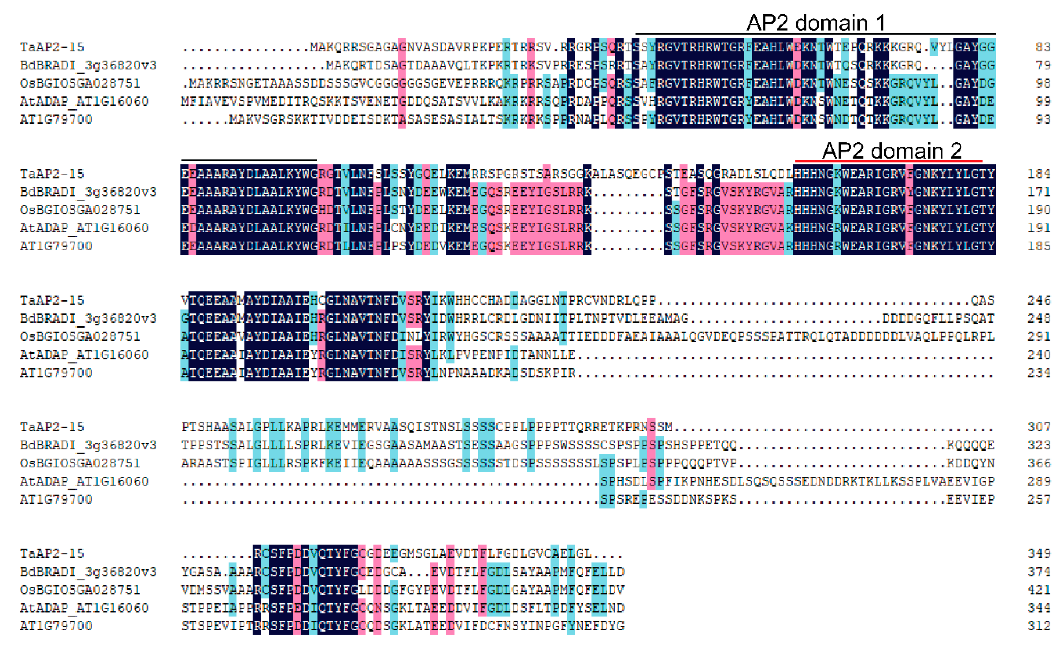
2.1. Sequence and Promoter Analysis

The full-length cDNA sequence of a previously designated AP2 gene, TaAP2-15 [19] was obtained from the cDNAs of wheat cv. Suwon 11 leaves infected with Pst. Blast analysis of TaAP2-15 nucleotide sequence in the Triticum aestivum genome sequence revealed two copies, localized on chromosome 6A and 6B (Figure S1). The predicted open reading frame (ORF) of TaAP2-15 contains 349 deduced amino acids with a molecular weight of 38.39 kDa and an isoelectric point (pI) of 9.47. Multisequence alignment indicated that TaAP2-15 is highly conserved with its orthologs from Brachypodium distachyon (BdBRADI_3g36820v3), Oryza sativum (OsBGIOSGA028751) and Arabidopsis thaliana (AtADAP_ AT1G16060 and AtAT1G79700) (Figure 1). Domain feature analysis indicated that TaAP2-15 contains two AP2 domains: AP2 domain 1 (amino acids 40-101) and AP2 domain 2 (amino acids 159–203) (Figure 1). Sequence analysis indicated that AP2 domain 1 of TaAP2-15 has high similarity with that of Brachypodium distachyon (87.3%), Oryza sativum (76.1%) and Arabidopsis thaliana (69.0% and 71.8%). In addition, AP2 domain 2 of TaAP2-15 shows high similarity with that of Brachypodium distachyon (94.6%), Oryza sativum (89.3%), and Arabidopsis thaliana (89.3% and 91.1%).
To gain further insight, an in silico analysis of possible cis-elements was performed. Cis-elements such as plant hormone-responsive motifs (TGACG-motif, P-box, CGTCA-motif) and low temperature stress responsiveness (LTR) were found within the promoter region of TaAP2-15. Interestingly, salicylic acid responsiveness (TCA-element) was also found in the promoter of TaAP2-15 (Table 1).

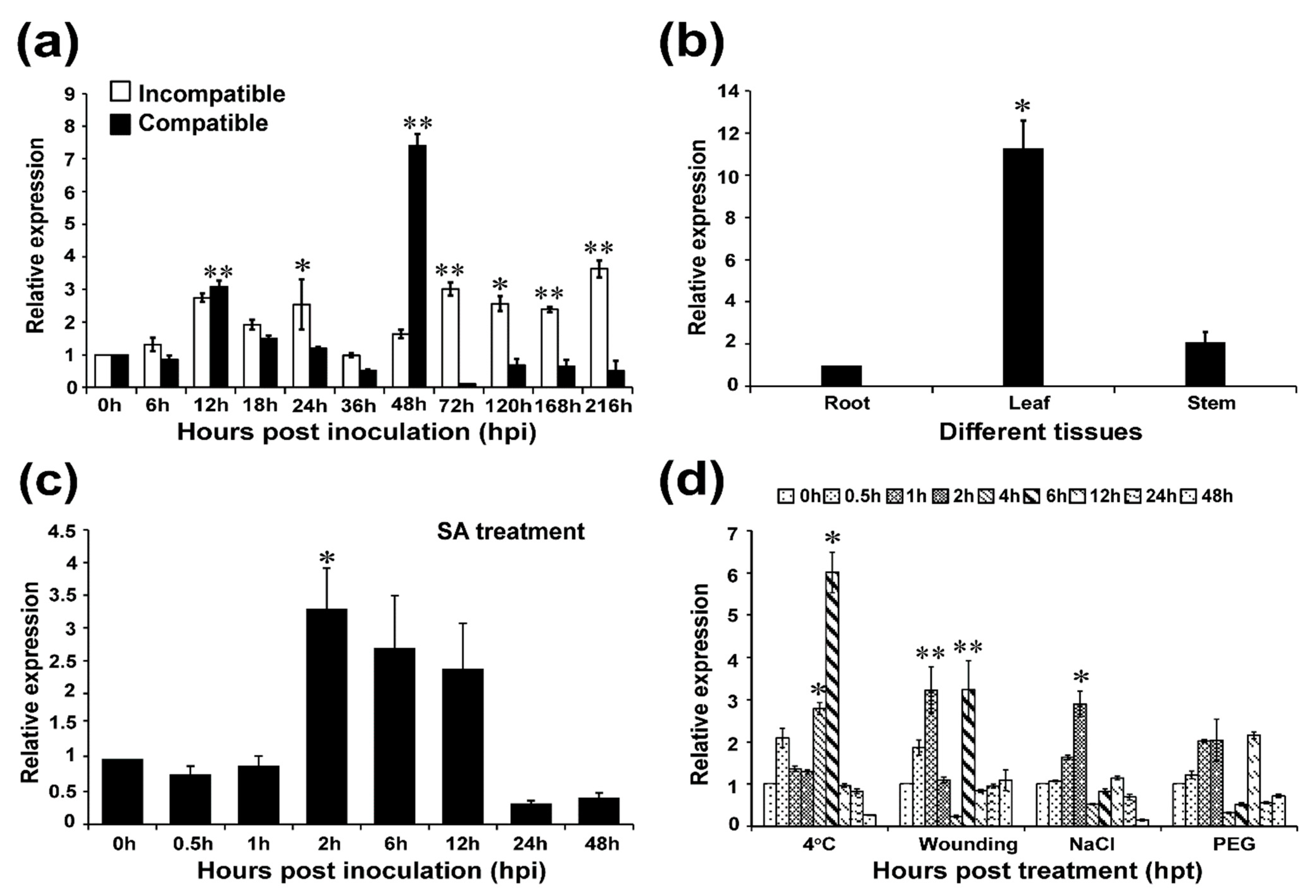
2.2. TaAP2-15 Is Significantly Induced When Challenged with Pst

To confirm whether TaAP2-15 participates in responses to Pst, transcript levels were determined by qRT-PCR. During the compatible interaction, the transcript profiles of TaAP2-15 were up-regulated more than 7-fold at 48 h post inoculation (hpi) relative to control (Figure 2a). During the incompatible interaction, the transcript level of TaAP2-15 was significantly induced at 12, 24, 72, 120, 168 hpi and reached a peak (more than 3.5-fold over that of the control) at 216 hpi. Therefore, we speculate that TaAP2-15 participates in resistance of wheat to Pst as the transcripts during the incompatible interaction remain up-regulated in most of the time points compared to controls.

2.3. Tissue-Specific Expression of TaAP2-15

AP2/ERF family genes are expressed in various organs of the plant [25]. We examined the expression profiles of TaAP2-15 in three different tissues (root, stem and leaf) of wheat plants. The results showed that the transcript levels of TaAP2-15 were most abundant in leaf tissue, as high as 10-fold over the level in roots (Figure 2b).

2.4. TaAP2-15 Is Induced under SA Treatment and Abiotic Stress

Transcript levels of TaAP2-15 in response to salicylic acid (SA), and abiotic stress were investigated via qRT-PCR. The transcript of TaAP2-15 in response to salicylic acid (SA), was induced more than 3-fold as early as 2 h post treatment (hpt) and subsequently decreased at 6 and 12 hpt but still remained up-regulated compared to the control (Figure 2c). Various treatments were applied to investigate the role of TaAP2-15 in response to abiotic stresses. As illustrated in Figure 2d, the transcripts of TaAP2-15 were significantly induced in all except drought treatments compared to the control. After incubation at low temperature, the transcript profiles of TaAP2-15 were up-regulated 6-fold over the control at 6 hpt. In response to wounding, the TaAP2-15 transcripts were induced 3-fold higher than the control at 1 and 6 hpt. Salt treatment also significantly increased the transcript levels of TaAP2-15 as high as 3-fold over the control at 2 hpt (Figure 2d). Taking these results together, we conclude that TaAP2-15 is induced in the response of wheat to multiple stresses.

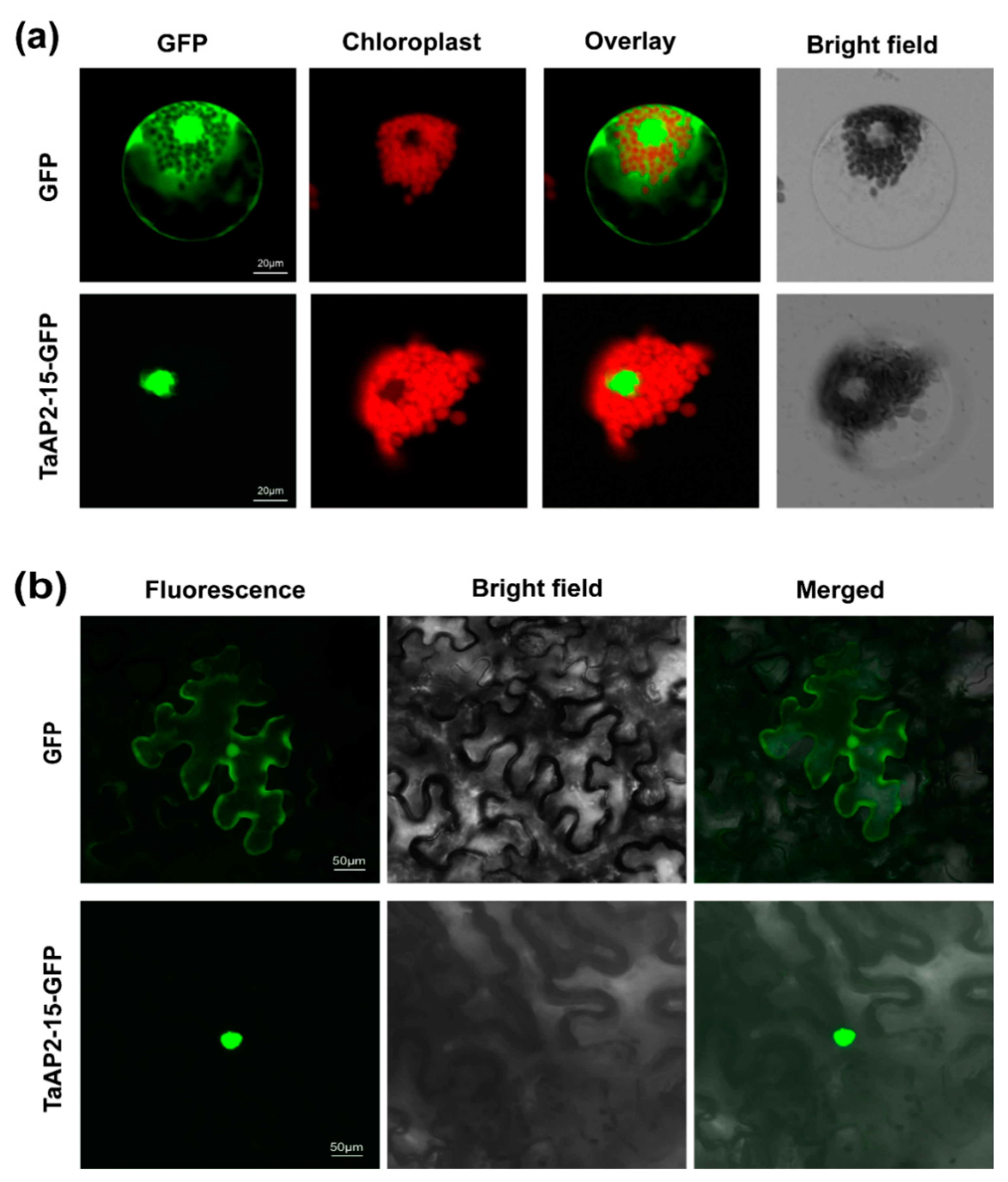
2.5. TaAP2-15 Is a Nuclear Localized Protein

To determine the subcellular localization of TaAP2-15, the generated constructs, p16318hGFP:TaAP2-15 and pCAMBIA1302:TaAP2-15, were transiently expressed in wheat protoplasts and N. benthamiana, respectively. The empty vector, p16318hGFP, was used as a control. GFP was ubiquitously distributed throughout the cell, including in the nucleus (Figure 3a,b). The cells expressing GFP tagged with TaAP2-15 were targeted in the nucleus of both wheat and N. benthamiana (Figure 3a,b). These results demonstrated that the TaAP2-15 was expressed in the nucleus.

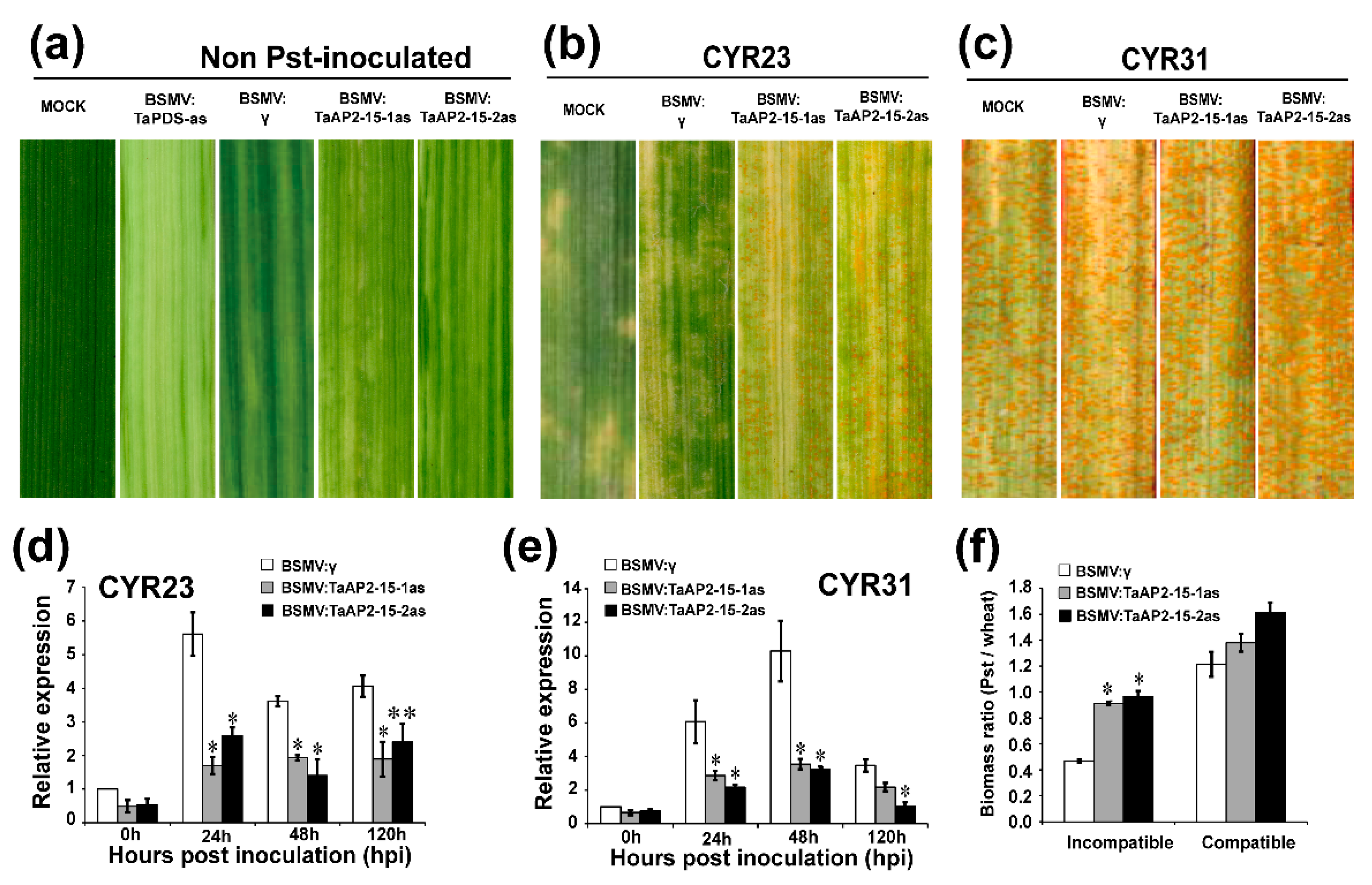
2.6. Silencing of TaAP2-15 Significantly Impaired Wheat Resistance to Pst

The functional role of TaAP2-15 during wheat-Pst interaction was analyzed by BSMV-mediated-VIGS (virus induced gene silencing) as employed previously [40]. Two fragments within the coding region that are specific to TaAP2-15 were designed for silencing purposes (Figure S1) and amplified with specific primers (Table S1). Two-weeks-old wheat seedlings were inoculated at the two-leaf stage with BSMV:TaPDS-as, BSMV:TaAP2-15-1as, BSMV:TaAP2-15-2as and BSMV: γ. Leaves inoculated with TaPDS showed strong photobleaching symptoms, whereas only mild symptoms of chlorotic mosaic were observed on BSMV:γ, TaAP2-15-1as and TaAP2-15-2as inoculated wheat leaves (Figure 4a), confirming that gene silencing was performed correctly. The fourth leaf of wheat plants that were pre-inoculated with BSMV:γ, TaAP2-15-1/2as were then inoculated with avirulent CYR23 or virulent CYR31 Pst isolates at 10 days post inoculation. At 14 days after inoculation with Pst CYR23, hypersensitive response (HR) symptoms were clearly observed on all leaves, mock or pre-inoculated with virus. Fungal uredia near the necrotic area were produced on leaves of TaAP2-15-silenced plants (Figure 4b). On the other hand, normal disease development was megascopically observed on CYR31 inoculated plants with slightly more spores on the silenced plants than the mock or BSMV:γ-inoculated plants (Figure 4c). To clarify whether TaAP2-15 was silenced successfully, transcript levels of TaAP2-15 were analyzed via qRT-PCR. The transcript levels in TaAP2-15-knockdown plants both in incompatible and compatible interactions were significantly reduced compared to control plants, indicating that TaAP2-15 was successfully silenced (Figure 4d,e). The fungal biomass was also significantly increased in silenced plants during incompatible interactions (Figure 4f). These results suggested that TaAP2-15 plays a positive role in wheat defense against Pst.

2.7. The Transcription of PR and ROS-Related Genes Is Influenced after Silencing of TaAP2-15

To investigate whether silencing of TaAP2-15 affects the expression of pathogenesis-related genes and ROS-scavenging genes, transcript levels of two PR-genes (TaPR1 and TaPR2) and two ROS-scavenging genes (TaCAT3, catalase, and TaFSOD3D, iron superoxide dismutase) were examined by qRT-PCR. The expression patterns of both PR-genes (TaPR1 and TaPR2) in the TaAP2-15-silenced plants were significantly down-regulated as compared to the control in both incompatible and compatible interactions (Figure 5a,b and Figure S2a,b). The transcripts of the ROS-scavenging genes, on the other hand, were significantly increased in TaAP2-15-silenced plants as compared to control plants (Figure 5c,d and Figure S2c,d). These results demonstrated that TaAP2-15 positively regulates the expression of certain defense-related genes during the wheat-Pst interaction.

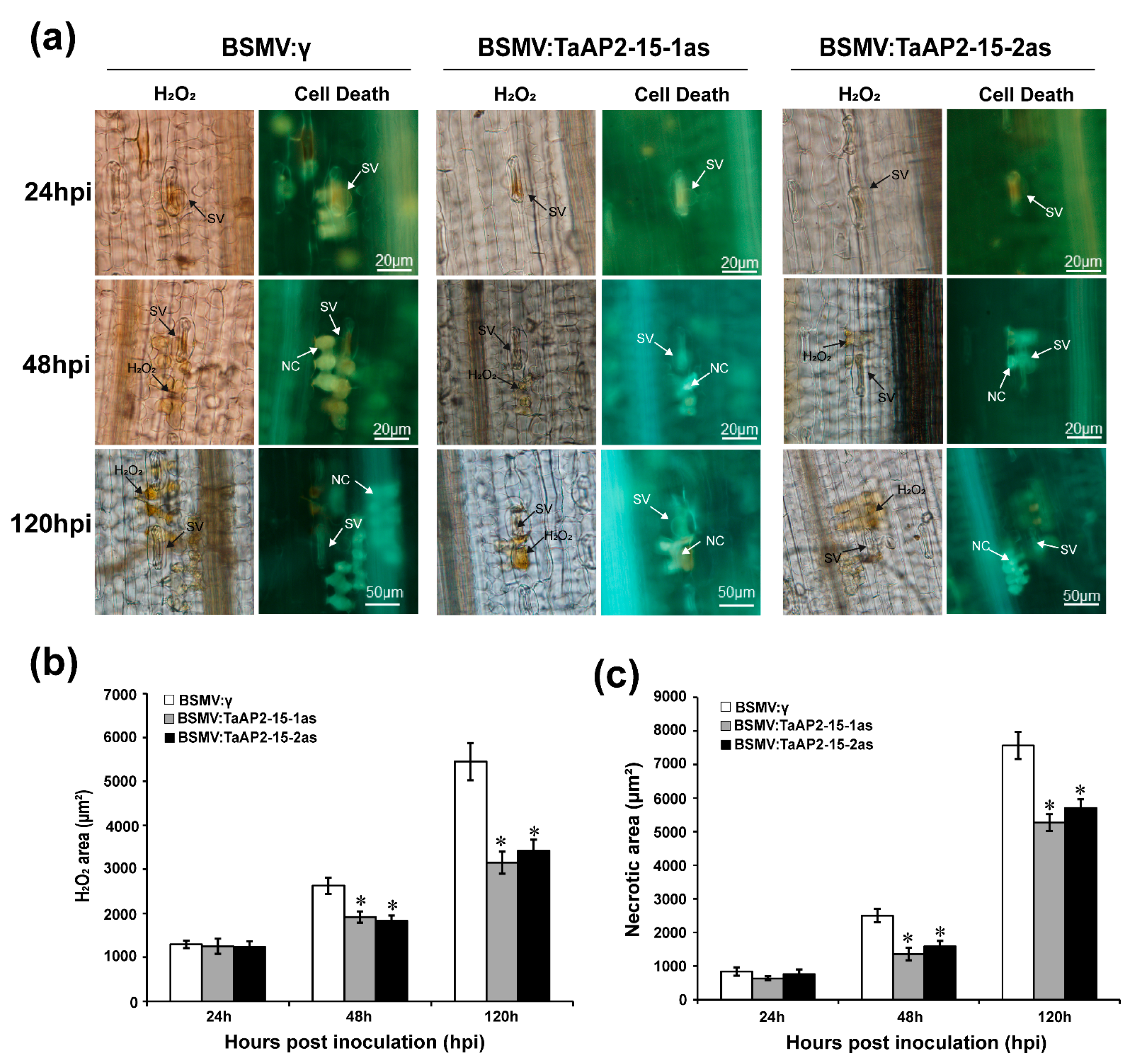
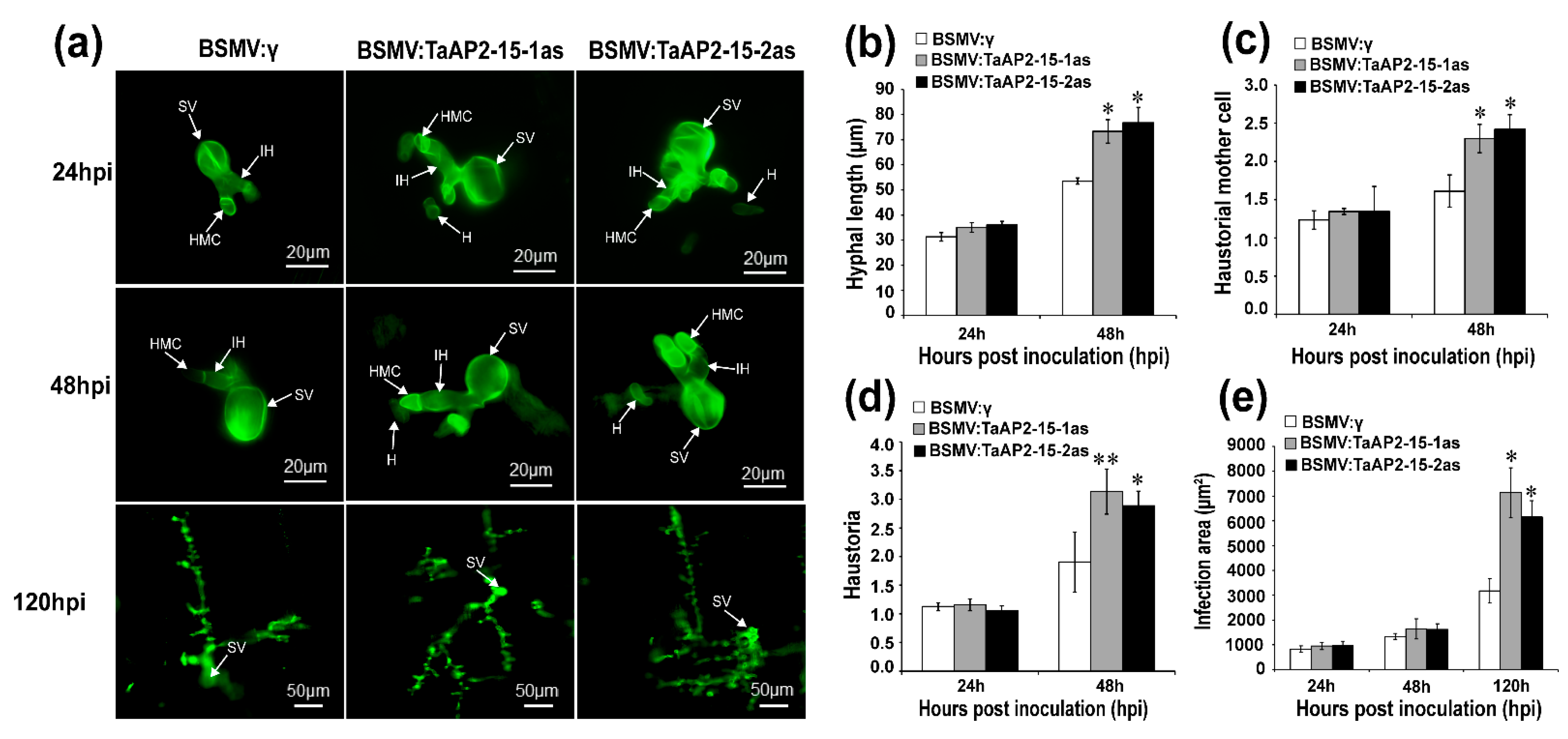
2.8. Silencing of TaAP2-15 Enhances Pst Growth and Decreases H2O2 Accumulation

On the basis of phenotypic variation between control (BSMV:γ) and BSMV: TaAP2-15-1/2as after Pst inoculation, we evaluated the host responses and the fungal development. Accumulation of H2O2, which is strongly correlated to host resistance response, was measured and quantified via DP-BSW software in each infection site. Production of H2O2 in TaAP2-15-silenced wheat plants showed a substantial decrease in comparison to BSMV:γ-treated plants after Pst inoculation during the incompatible interaction (Figure 6a,b). Similarly, H2O2 accumulation was significantly decreased in TaAP2-15-silenced plants at 48 and 120 hpi during the compatible interaction (Figure S3a,b). The necrotic area in the TaAP2-15-knockdown plants was significantly lower than that of control plants at 48 and 120 hpi during the incompatible wheat-Pst interaction (Figure 6c). Microscopic examination indicated that the number of haustoria and haustoria mother cells and hyphal length were significantly increased in the TaAP2-15-silenced plants relative to the control at 48 hpi during both compatible and incompatible interactions (Figure 7a–d and Figure S4a–d). The infected area in both incompatible (Figure 7a,e) and compatible (Figure S4a,e) wheat-Pst interactions was also significantly increased in TaAP2-15-silenced plants at 120 hpi. Therefore, silencing of TaAP2-15 enhances Pst growth and impairs plant defense to Pst.

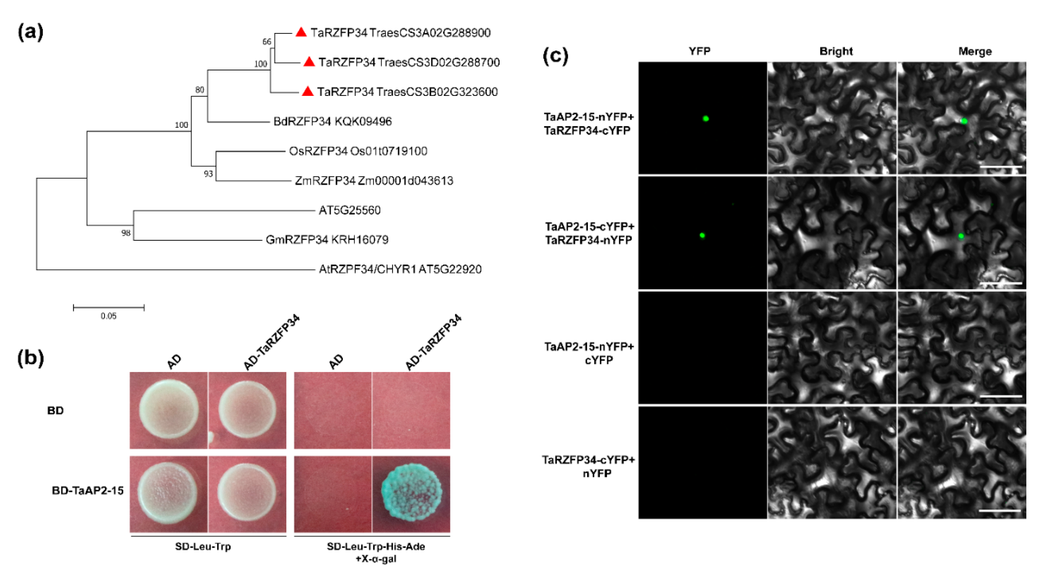
2.9. TaAP2-15 Physically Interacts with TaRZFP34

To identify the target protein, yeast-two hybrid (Y2H) screening was carried out from a cDNA library constructed from Pst infected wheat cv. Su11. TaAP2-15 was used as a bait for screening the cDNA library. A wheat zinc-finger protein, TaRZFP34 homolog of Arabidopsis AT5G25560 and rice OsRZFP34 (Os01t0719100) which encodes RING E3 ligase containing 299 amino acids, was identified (Figure 8a and Figure S5a,b, Table S2). To verify the interaction, the ORF sequence of TaAP2-15 was subcloned into the bait vector (pBD-TaAP2-15), whereas the TaRZFP34 was inserted into the prey vector (pAD-TaRZFP34) and co-expressed and grown in a selective medium lacking leucine, tryptophan, histidine and adenine but containing X-α-Gal. The interaction between TaAP2-15 and TaRZFP34 was confirmed by growth of yeast strain AH109 on selective medium staining with X-a-gal (Figure 8b). In vivo interaction between TaAP2-15 and TaRZFP34 was further verified by biomolecular fluorescence complementation (BiFC) assay. The following constructs including TaAP2-15-nYFP+TaRZFP34-cYFP, TaAP2-15-cYFP+TaRZFP34-nYFP, TaAP2-15-nYFP+cYFP and TaRZFP34-cYFP+nYFP, were generated for BiFC assay. The constructs were then transformed into A. tumefaciens, strain GV3101 and co-infiltrated into N. benthamiana leaves. Microscopic examination detected fluorescence only when the two constructs were co-expressed. No fluorescence was observed in leaves agroinfiltrated with either of the constructs mixed with the control (empty vector) (Figure 8c).

3. Discussion

The conserved AP2 DNA binding domains of amino acids 57-66 characterize the AP2/ERF superfamily [41]. In accordance with the double AP2 domain amino acid sequence and the nuclear location sequence, the AP2 family was subdivided into euAP2, eu-AINTEGUMENTA (euANT) and basalANT groups, respectively [42,43,44]. AP2 TFs play a number of roles in plant developmental processes and stress responses, such as vegetative and reproductive growth, cell proliferation, abiotic and biotic stress responses, and plant hormone responses [14,15,16]. ERFs often become up-regulated in hormonal and stress responses [45,46], and usually, these responses are immediate [27]. In order to understand plant adaptation to biotic stresses, research into the functions and mechanisms of wheat AP2/ERFs in the regulation of biotic stress responses is vital. Recent findings have shown that AP2 proteins perform a significant role in controlling responses to biotic and abiotic stress in plants.
In this study, qRT-PCR assays revealed that during the compatible interaction the transcript profiles of TaAP2-15 were highly up-regulated at one time point (48 hpi). However, during the incompatible interaction, the transcript levels were significantly induced at all the time points, and even reached to its peak at 216 hpi. During the incompatible interaction, induction of the TaAP2-15 transcript was generated faster and to a greater extent than those in the compatible interaction. In response to stress, gene expression triggered by environmental stresses not only protects cells from injury, but also controls the expression of genes involved in signal transduction processes [8]. Several studies have indicated that expression of ERFs was induced and up-regulated by abiotic stresses [47,48]. We found that expression of TaAP2-15 was significantly induced by abiotic stresses (cold, wound, salt and drought), implying that TaAP2-15 is induced in defense responses of wheat to multiple abiotic stresses. It was previously reported that AP2/ERF family members are expressed in various parts of the plant [25]. TaAP2-15 was most abundantly expressed in wheat leaves. Like other TFs, AP2s have also been reported to be localized in nucleus. He et al. (2019) reported that CsAP2-09 from citrus was localized to the nucleus of onion cells [27]. Similarly, in our study, TaAP2-15 was found to be localized within the nuclei of both wheat and N. benthamina.
Our current research shows that TaAP2-15 is required for resistance of wheat to infection by Pst. As a biotrophic parasite, Pst is dependent on haustoria to consume nutrients from the host [49]. By 48 hpi, the primary infection develops hypha, haustorial mother cell, and haustorium in both compatible and incompatible interactions, whereas the necrotic area in TaAP2-15-silenced plants was lower than the control at 48 and 120 hpi during the incompatible interaction. HR, or necrotic cell death, is the host defense reaction in response to invading pathogens. So, when resistant-related genes are silenced, it is obvious that necrotic cell death would be reduced in the silenced plants. In our findings, the reduced necrotic area in TaAP2-15-silenced plants indicated that TaAP2-15 contributes to resistance. Furthermore, the fungal biomass was also increased in TaAP2-15-silenced plants during the incompatible interaction. It has been documented that, during pathogen infection and environmental stress, the abiotic and biotic stresses that trigger an oxidative burst response act as a defense system [50,51].
One of the earliest signaling events in plants, the ROS burst, occurs in the early stages of plant-pathogen interactions [52]. Previous histological and cytological findings in wheat have shown that in incompatible interactions with Pst, the activation of response signaling is associated with bursts of ROS as early as 12 hpi [53]. In our study, silencing of TaAP2-15 decreased the content of H2O2 that is correlated with host resistance. The ROS burst is regulated by ROS-related genes. As demonstrated herein, the transcript levels of the ROS-related genes were significantly increased in TaAP2-15-silenced plants as compared to control plants. This finding agrees with earlier studies indicating that the induction of ROS accumulation in the signaling of biotic and abiotic stress is correlated with the ROS burst [54]. Transcription factors of ERF directly control the expression of pathogenesis-related (PR) genes [55,56,57]. The transcript levels of PR-genes, TaPR1, TaPR2, in the TaAP2-15-silenced plants were down-regulated compared to controls in both compatible and incompatible interactions, suggesting that TaAP2-15 regulates the expression of TaPR1 and TaPR2 directly or indirectly. Phytohormones like SA have been reported to play significant roles in abiotic stress responses in plants [58]. SA is a basal and SAR signaling molecule with HR activation during biotrophic invasion. Vlot et al. (2009) reported that SA-mediated signaling pathway is activated upon infection with biotrophic pathogens [59]. Furthermore, SA was induced upon infection with Pst [60]. Some transcription factors like AP2/ERFs have been implicated indirectly in regulating the SA response, perhaps through interaction with other TFs [28]. Here, we showed that the transcript abundance of TaAP2-15 was stimulated by application of exogenous SA. In Arabidopsis, accumulation of the plant hormone SA and transcriptional activation of PR genes is associated with SAR [61]. During host-pathogen interactions, PRs accumulate systemically [62], and in our study the accumulation of PR genes in silenced plants was diminished. Thus, we speculate that TaAP2-15 is required positively to acquire resistance against Pst infection.
To summarize, our data indicate that TaAP2-15 acts as positive regulator in resistance of wheat against Pst in a SA-induced pattern via ROS-mediated defense pathway. Analysis of the promoter region of TaAP2-15 further revealed the presence of a SA responsive TCA-element, a potential cis-acting element for SA responsiveness. Our findings also imply that TaAP2-15 physically interacts with TaRZFP34, a zinc finger protein homolog in rice that has been reported to play a role in stress conditions. Here, we hypothesize that TaAP2-15 and its co-protein are activated through induction by SA treatment as the result of downstream signaling pathways that confer ROS-mediated defense. More studies are required to define these phenomena. To the best of our knowledge, this is the first study indicating that the wheat AP2 is positively required for resistance against stripe rust fungus infection.

4. Materials and Methods

4.1. Plant Material, Fungal Pathogens and Inoculation/Treatments

Wheat (T. aestivum L.) cultivar Suwon11 (Su11) and Pst isolates, CYR23/avirulent and CYR31/virulent, were used. Su11 possesses a Pst resistance gene YrSu. This cultivar is reported to be resistant to CYR23 but susceptible to CYR31 [63]. For RNA isolation, wheat leaves challenged with these Pst isolates or water (distilled and sterile) were sampled at different time points [53].
The expression of TaAP2-15 in response to hormonal and environmental stress treatments was assessed. To investigate the transcripts of TaAP2-15 to hormone treatment, 2 nM salicylic acid (SA) was applied through spraying to leaves of wheat seedlings. Leaves subjected to SA or 0.1% (v/v) ethanol (mock) treatments were then sampled for RNA isolation. To verify the response to drought and salinity, roots of wheat seedlings were immersed in 200 mM NaCl or 20% PEG6000, respectively. To assess the reaction of TaAP2-15 in response to low temperature, wheat seedlings were maintained at 4 °C for 48 h. Wheat seedlings were also subjected to wounding by removing the tip of each leaf. Samples were collected at 0, 0.5, 1, 2, 4, 6, 12, 24 and 48 hpt from SA and different stress elicitors (NaCl, PEG 6000, low temperature and wound) treated seedlings. To determine tissues-specific expression of TaAP2-15, qRT-PCR analysis was carried out on different plant organs of two-weeks-old wheat seedlings. The experiment was repeated three times.

4.2. RNA Extraction, cDNA Synthesis and qRT-PCR Analysis

RNA was isolated with the Trizol reagent according to the manufacturer’s instructions (Invitrogen, Carlsbad, CA, USA) and treated with DNase I (Progema, Madison, WI, USA) to remove DNA contamination. cDNA was then synthesized using GoScript Reverse Transcription System (Progema) and an oligo (dT18) primer (Invitrogen). Primers specific to the gene of interest or reference gene were used to quantify the expression level by qRT-PCR using the synthesized cDNA [64] (Table S1). A 7500 Real-Time PCR System (Applied Biosystems, Foster City, CA, USA) was used to quantify the transcripts. TaEF-1α (GenBank accession Q03033) was used as the internal reference for normalizing the data. The relative expression of TaAP2-15 was determined by the comparative 2−ΔΔCt [65] method. All qRT-PCR experiments were carried out with three replications using RNA samples obtained from three independent replicates.

4.3. Identification, Sequence and Promoter Analysis of TaAP2-15

A 1050-bp nucleotide sequence (accession number TraesCS6A01G125700) with high homology to A. thaliana homolog, AT1G16060 ADAP, which is positively involved in hormone and stress response [26] was obtained from the cDNA library in our lab [54]. This gene was previously designated as TaAP2-15 [25]. TaAP2-15 was amplified from Pst inoculated cDNA template using specific primers (Table S1). The cDNA sequence of TaAP2-15 was further analyzed with the NCBI BLAST program (http://www.ncbi.nlm.nih.gov/blast/ (accessed on 17 February 2021)) and the ORF finder software in NCBI. The conserved domain was predicted with Pfam (http://pfam.sanger.ac.uk/ (accessed on 7 February 2021)), PROSITEScan (http://prosite.expasy.org/scanprosite/ (accessed on 7 February 2021)) and InterproScan (http://www.ebi.ac.uk/Tools/pfa/iprscan/ (accessed on 7 February 2021)). Multiple sequence alignments were performed using DNAMAN8.0 (Lynnon Biosoft).
To investigate the possible regulatory mechanisms of TaAP2-15 gene, a 1.5-kb promoter region upstream of the start codon of the gene was retrieved from the wheat Genome Database (http://plants.ensembl.org/ (accessed on 7 February 2021)) and analyzed through the PlantCARE (http://bioinformatics.psb.ugent.be/webtools/plantcare/html/ (accessed on 7 February 2021)), an online server.

4.4. Subcellular Localization of TaAP2-15 Protein

The subcellular localization of TaAP2-15 was determined in both wheat and tobacco (N. benthamiana). The ORF of TaAP2-15 was constructed into p16318hGFP or pCAMBIA1302 vectors. The constructs, p16318hGFP-TaAP2 and p16318hGFP were then introduced into the isolated protoplasts of wheat mesophyll tissue by the PEG-mediated transformation method [66]. To further confirm the localization of the TaAP2-15 protein, pCAMBIA1302:TaAP2-15 fusion was transformed into A. tumefaciens strain GV3101 by electroporation and infiltrated into tobacco leaves as described [67]. In a dark chamber, the transformed wheat protoplasts were incubated for 24–36 h at 24 °C. Infiltrated N. benthamiana leaves were also maintained in a growth chamber with a 16 h/8 h photoperiod at 25 °C for 2 to 3 days. Leaf tissue samples were then collected for detection of the autofluorescence signals with an Olympus FV1000 confocal laser microscope with a 480-nm filter [68].

4.5. BSMV-Mediated TaAP2-15 Gene Silencing

Gene silencing was performed through the BSMV-VIGS-mediated gene silencing method [69]. To silence TaAP2-15, two cDNA fragments of TaAP2-15 (Figure S1) were selected and constructed into BSMV-γ-vector, resulting in constructs BSMV-γ:TaAP2-15-1as/2as. Specificity of the fragments was confirmed through BLAST analysis (http://blast.ncbi.nlm.nih.gov/Blast/ (accessed on 7 February 2021)). Capped in vitro transcripts were obtained with the RiboMAXTM Large-Scale RNA Production System-T7 (Promega, Madison, WI, USA) and Ribom7G Cape Analog (Promega) based on the manufacturer’s protocol. Barley stripe mosaic virus constructs were inoculated onto the second leaf of wheat as described previously [40] and plants were maintained in darkness at 25 ± 2 °C for 24 h with sufficient humidity. Mock plants were inoculated with 1 × Fes. Ten days later, freshly collected urediniospores of Pst race CYR23 or CYR31 were inoculated onto the fourth leaf. Leaf samples inoculated with Pst were then collected at 0, 24, 48 and 120 hpi for histology and RNA extraction. Silencing efficiency of the TaAP2-15-silenced plants and relative expression of pathogenesis (PR) and ROS-related genes were quantified by qRT-PCR analysis compared to control plants (BSMV:γ). After 14 days, the level of infection of Pst-inoculated leaves was determined based on the McNeal measurement scale [70] and infection phenotypes were photographed. Genomic DNA was extracted by the CTAB method from Pst-inoculated leaves collected after 14 days. Fungal biomass was then quantified by qRT-PCR, and a standard curve was generated from the plasmid carrying the fragments of PsEF1 and TaEF1α [71]. The experiment was performed with three biological replications.

4.6. Histology of Fungal Growth and Host Response

Wheat leaves inoculated with Pst isolates were sampled at 24, 48 and 120 hpi and stained to detect the accumulation of H2O2 and fungal structures. H2O2 production was examined by staining samples with 3,3′-diamino benzidine stain (DAB) as described previously [53] and viewed under BX-51 microscope (Olympus). Wheat germ agglutinin stain (WGA) (Invitrogen) was used to visualize pathogen structures. During the wheat-Pst interaction, the formation of a substomatal vesicle is considered to be effective penetration [72]. At least 50 infection regions were examined for each treatment to assess the H2O2 accumulation, necrotic area and various fungal structures. Necrotic cells around the infected site, H2O2 accumulation and fungal structures, such as hypha, haustoria mother cell and haustoria, were observed with BX-51 microscope (Olympus) and their corresponding lengths were estimated using DP-BSW software. Student’s t-test was used to compute the statistical differences between treatments.

4.7. Yeast Two-Hybrid Assay

MatchMaker yeast two-hybrid assay (Clontech, Tokyo, Japan) was carried out to analyze the interaction between TaAP2-15 and TaRZFP34. The full-length sequence of TaAP2-15 was subcloned into pGBKT7-BD to generate TaAP2-15-BD, the DNA-binding domain bait protein fusion. A cDNA library generated from virulent Pst isolate-infected wheat leaves in the pGADT7 vector was used as a prey for screening the target gene. The bait (TaAP2-15-BD) and the prey (pGADT7-cDNA libraries) were co-transformed into a yeast strain AH109 and grown in a selective medium (SD/-Trp-Leu or SD/-Trp-Leu-His). The colonies grown on the SD/-Trp-Leu-His were then re-plated on SD medium lacking -Trp-Leu-His-Ade to check the interaction. The positive colonies were isolated, sequenced and analyzed by blasting on the NCBI database for the coding cDNA sequence (Table S1). Those candidate genes were then subcloned in to pGADT7 to generate the activation domain, AD prey protein fusion. To verify the interaction, TaAP2-15-BD and each candidate target gene in the AD vector were co-transformed into yeast and plated on SD/-Trp-Leu-His-Ade medium containing X-α-gal.

4.8. BiFC Assays

The ORF sequence of TaAP2-15 was cloned into a pUC-PSYNE vector and fused with the N-terminal fragment of the yellow fluorescent protein (YFP) to generate TaAP2-15-nYFP construct. The full-length coding sequence of TaRZFP34 was subcloned into a pUC-pSPYCE vector as a fusion with the C-terminal fragment of YFP to generate TaRZFP34-cYFP [73]. To verify if there is change in expression with exchange of the constructs in the vectors, we cloned TaAP2-15 into pUC-PSYCE to produce TaAP2-15-cYFP and TaRZFP34 into pUC-PSYNE to form TaRZFP34-nYFP. All the generated constructs TaAP2-15-nYFP, TaRZFP34-cYFP, TaAP2-15-cYFP, TaRZFP34-nYFP; TaAP2-15-nYFP+cYFP and TaRZFP34-cYFP+nYFP were then introduced into A. tumefaciens strain GV3101. The agrobacterium colonies containing the appropriate constructs were then infiltrated into 4-week-old N. benthamiana leaf tissue. In vivo interaction was then detected under a FV3000 confocal laser microscope 48 h after infiltration (Olympus).

4.9. Statistical Analysis

All data were subjected to analysis with Microsoft Excel. The statistical differences between treatments were computed by Student’s t-test.

Supplementary Materials

Supplementary materials are available at: https://www.mdpi.com/1422-0067/22/4/2080/s1, Figure S1: Multiple sequence alignment of the ORF sequences for the TaAP2-15 copies, Figure S2: Relative expression of pathogenesis related (PR) and ROS-scavenging genes in TaAP2-15 silenced and control plants challenged with virulent Pst race CYR31, Figure S3: Knockdown of TaAP2-15 enhanced wheat susceptibility to virulent Pst race CYR31 infection, Figure S4: Silencing of TaAP2-15 in wheat enhances growth of the Pst virulent race, CYR31, Figure S5: Multiple sequence alignment of TaRZFP34, Table S1: Primers used in this study, Table S2: Candidate interacting genes screened via Y2H.

Author Contributions

Data Curation, M.D.H., F.G., J.G. (Jia Guo), F.H. and X.B.; Funding Acquisition, J.G. (Jun Guo); Methodology, F.H., X.B. and P.Y.; Project Administration, Z.K. and J.G. (Jun Guo); Supervision, Z.K. and J.G. (Jun Guo); Writing—Original Draft Prepaaation, M.D.H., F.G. and M.A.I.; Writing—Review & Editing, Z.K. and J.G. (Jun Guo). All authors have read and agreed to the published version of the manuscript.

Funding

This study was financed by National Natural Science Foundation of China (31620103913 and 31972224), National Transgenic Key Project of the Ministry of Agriculture of China (2020ZX08009-15B), National Key R&D Program of China (2018YFD0200402), Natural Science Basic Research Program of Shaanxi (2020JZ-13) and the 111 Project from the Ministry of Education of China (B07049).

Institutional Review Board Statement

Not applicable.

Informed Consent Statement

Not applicable.

Data Availability Statement

Data is contained within the article and supplementary material.

Acknowledgments

We thank Larry Dunkle from the USDA-Agricultural Research Service at Purdue University, USA for critical reading of the manuscript.

Conflicts of Interest

The authors declare no conflict of interest.

References

  1. Wellings, C.R. Global status of stripe rust: A review of historical and current threats. Euphytica 2011, 179, 129–141. [Google Scholar] [CrossRef]
  2. Chen, W.; Wellings, C.; Chen, X.; Kang, Z.; Liu, T. Wheat stripe (yellow) rust caused by Puccinia striiformis f. sp. tritici. Mol. Plant. Pathol. 2014, 15, 433–446. [Google Scholar] [CrossRef]
  3. Staskawicz, B.J.; Ausubel, F.M.; Baker, B.J.; Ellis, J.G.; Jones, J. Molecular genetics of plant disease resistance. Science 1995, 268, 661–667. [Google Scholar] [CrossRef] [Green Version]
  4. Jones, J.D.; Dangl, J.L. The plant immune system. Nature 2006, 444, 323–329. [Google Scholar] [CrossRef] [PubMed] [Green Version]
  5. Thordal, H. A holistic view on plant effector-triggered immunity presented as an iceberg model. Cell Mol. Life Sci. 2020, 77, 3963–3976. [Google Scholar] [CrossRef] [PubMed] [Green Version]
  6. Wei, H.; Collmer, A. Defining essential processes in plant pathogenesis with Pseudomonas syringae pv. tomato DC3000 disarmed polymutants and a subset of key type III effectors. Mol. Plant Pathol. 2018, 19, 1779–1794. [Google Scholar] [CrossRef] [PubMed] [Green Version]
  7. Shinozaki, K.; Dennis, E.S. Cell signalling and gene regulation: Global analyses of signal transduction and gene expression profiles. Curr. Opin. Plant Biol. 2003, 6, 405–409. [Google Scholar] [CrossRef]
  8. Chen, W.J.; Zhu, T. Networks of transcription factors with roles in environmental stress response. Trends Plant Sci. 2004, 9, 591–596. [Google Scholar] [CrossRef]
  9. Yanagisawa, S. Dof domain proteins: Plant-specific transcription factors associated with diverse phenomena unique to plants. Plant Cell Physiol. 2004, 45, 386–391. [Google Scholar] [CrossRef] [PubMed] [Green Version]
  10. Rushton, D.L.; Tripathi, P.; Rabara, R.C.; Lin, J.; Ringler, P.; Boken, A.K.; Langum, T.J.; Smidt, L.; Boomsma, D.D.; Emme, N.J. WRKY transcription factors: Key components in abscisic acid signalling. Plant Biotechnol. J. 2012, 10, 2–11. [Google Scholar] [CrossRef]
  11. Agarwal, M.; Hao, Y.; Kapoor, A.; Dong, C.H.; Fujii, H.; Zheng, X.; Zhu, J.K. A R2R3 type MYB transcription factor is involved in the cold regulation of CBF genes and in acquired freezing tolerance. J. Biol. Chem. 2006, 281, 37636–37645. [Google Scholar] [CrossRef] [PubMed] [Green Version]
  12. Lata, C.; Prasad, M. Role of DREBs in regulation of abiotic stress responses in plants. J. Exp. Bot. 2011, 62, 4731–4748. [Google Scholar] [CrossRef] [Green Version]
  13. Mizoi, J.; Shinozaki, K.; Yamaguchi-Shinozaki, K. AP2/ERF family transcription factors in plant abiotic stress responses. Biochim. Biophys. Acta 2012, 1819, 86–96. [Google Scholar] [CrossRef] [PubMed]
  14. Nakano, T.; Suzuki, K.; Fujimura, T.; Shinshi, H. Genome-wide analysis of the ERF gene family in Arabidopsis and rice. Plant Physiol. 2006, 140, 411–432. [Google Scholar] [CrossRef] [PubMed] [Green Version]
  15. Licausi, F.; Giorgi, F.M.; Zenoni, S.; Osti, F.; Pezzotti, M.; Perata, P. Genomic and transcriptomic analysis of the AP2/ERF superfamily in Vitis vinifera. BMC Genom. 2010, 11, 719. [Google Scholar] [CrossRef] [PubMed] [Green Version]
  16. Sharoni, A.M.; Nuruzzaman, M.; Satoh, K.; Shimizu, T.; Kondoh, H.; Sasaya, T.; Choi, I.R.; Omura, T.; Kikuchi, S. Gene structures, classification and expression models of the AP2/EREBP transcription factor family in rice. Plant Cell Physiol. 2011, 52, 344–360. [Google Scholar] [CrossRef] [PubMed]
  17. Riechmann, J.L.; Heard, J.; Martin, G.; Reuber, L.; Jiang, C.Z.; Keddie, J.; Adam, L.; Pineda, O.; Ratcliffe, O.; Samaha, R. Arabidopsis transcription factors: Genome-wide comparative analysis among eukaryotes. Science 2000, 290, 2105–2110. [Google Scholar] [CrossRef]
  18. Sakuma, Y.; Liu, Q.; Dubouzet, J.G.; Abe, H.; Shinozaki, K.; Yamaguchi-Shinozaki, K. DNA-binding specificity of the ERF/AP2 domain of Arabidopsis DREBs, transcription factors involved in dehydration-and cold-inducible gene expression. Biochem. Biophys. Res. Commun. 2002, 290, 998–1009. [Google Scholar] [CrossRef] [PubMed]
  19. Licausi, F.; Masaru, O.T.; Pierdomenico, P. Apetala 2/Ethylene responsive factor (Ap2/ERF) transcription factors: Mediators of stress responses and developmental programs. New Phytol. 2013, 199, 639–649. [Google Scholar] [CrossRef]
  20. Li, M.Y.; Xu, Z.S.; Huang, Y.; Tian, C.; Wang, F.; Xiong, A.S. Genome-wide analysis of Ap2/ERF transcription factors in carrot (Daucus carota L.) reveals evolution and expression profiles under abiotic stress. Mol. Genet. Genom. 2015, 290, 2049–2061. [Google Scholar] [CrossRef] [PubMed]
  21. Dietz, K.J.; Marc, O.V.; Andrea, V. Ap2/Erebp transcription factors are part of gene regulatory networks and integrate metabolic, hormonal and environmental signals in stress acclimation and retrograde signalling. Protoplasma 2010, 245, 3–14. [Google Scholar] [CrossRef] [PubMed]
  22. Gibbs, D.J.; Jorge, V.C.; Sophie, B.; Geeta, P.; Guillermina, M.M.; Michael, J.H. Group Vii ethylene response factors coordinate oxygen and nitric oxide signal transduction and stress responses in plants. Plant Physiol. 2015, 169, 23–31. [Google Scholar] [CrossRef] [PubMed]
  23. Chandler, J.W. Class VIIIb APETALA2 ethylene response factors in plant development. Trends Plant Sci. 2018, 23, 151–162. [Google Scholar] [CrossRef] [PubMed]
  24. Phukan, U.J.; Gajendra, S.J.; Vineeta, T.; Rakesh, K.S. Regulation of apetala2/ethylene response factors in plants. Front. Plant Sci. 2017, 8, 150. [Google Scholar] [CrossRef] [PubMed] [Green Version]
  25. Zhao, Y.; Ma, R.; Xu, D.; Bi, H.; Xia, Z.; Peng, H. Genome-wide identification and analysis of the AP2 transcription factor gene family in wheat (Triticum aestivum L.). Front. Plant Sci. 2019, 10, 1286. [Google Scholar] [CrossRef] [Green Version]
  26. Lee, S.-J.; Kang, J.-Y.; Kim, S.Y. An ARIA-interacting AP2 domain protein is a novel component of ABA signaling. Mol. Cells 2009, 27, 409–416. [Google Scholar] [CrossRef]
  27. He, Y.; Jia, R.; Qi, J.; Chen, S.; Lei, T.; Xu, L.; Peng, A.; Yao, L.; Long, Q.; Li, Z. Functional analysis of citrus AP2 transcription factors identified CsAP2-09 involved in citrus canker disease response and tolerance. Gene 2019, 707, 178–188. [Google Scholar] [CrossRef]
  28. Gutterson, N.; Reuber, T.L. Regulation of disease resistance pathways by AP2/ERF transcription factors. Curr. Opin. Plant Biol. 2004, 7, 465–471. [Google Scholar] [CrossRef] [PubMed]
  29. Cui, H.; Tsuda, K.; Parker, J.E. Effector-triggered immunity: From pathogen perception to robust defense. Annu. Rev. Plant Biol. 2015, 66, 487–511. [Google Scholar] [CrossRef]
  30. Tsuda, K.; Katagiri, F. Comparing signaling mechanisms engaged in pattern-triggered and effector-triggered immunity. Curr. Opin. Plant Biol. 2010, 13, 459–465. [Google Scholar] [CrossRef]
  31. Mittler, R.; Vanderauwera, S.; Suzuki, N.; Miller, G.; Tognetti, V.B.; Vandepoele, K.; Gollery, M.; Shulaev, V.; Van Breusegem, F. ROS signaling: The new wave? Trends Plant Sci. 2011, 16, 300–309. [Google Scholar] [CrossRef] [PubMed]
  32. Zurbriggen, M.D.; Carrillo, N.; Hajirezaei, M.-R. ROS signaling in the hypersensitive response: When, where and what for? Plant Signal. Behav. 2010, 5, 393–396. [Google Scholar] [CrossRef] [Green Version]
  33. Danquah, A.; de Zelicourt, A.; Colcombet, J.; Hirt, H. The role of ABA and MAPK signaling pathways in plant abiotic stress responses. Biotechnol. Adv. 2014, 32, 40–52. [Google Scholar] [CrossRef] [PubMed]
  34. Hirayama, T.; Kazuo, S. Research on plant abiotic stress responses in the post-genome era: Past, present and future. Plant J. 2010, 6, 1041–1052. [Google Scholar] [CrossRef]
  35. Miura, K.; Jing, B.J.; Lee, J.; Yoo, C.Y.; Stirm, V.; Miura, T.; Ashworth, E.; Bressan, R.A.; Yun, D.J.; Hasegawa, P.M. Siz1-mediated dumoylation of Ice1 vontrols Cbf3/Dreb1a expression and freezing tolerance in Arabidopsis. Plant Cell 2007, 19, 1403–1414. [Google Scholar] [CrossRef] [Green Version]
  36. Dong, C.H.; Manu, A.; Zhang, Y.Y.; Xie, Q.; Zhu, J.K. The negative regulator of plant cold responses, Hos1, is a ring E3 ligase that mediates the ubiquitination and degradation of Ice1. Proc. Natl. Acad. Sci. USA 2006, 21, 8281–8286. [Google Scholar] [CrossRef] [PubMed] [Green Version]
  37. Laity, J.H.; Lee, B.M.; Wright, P.E. Zinc finger proteins: New insights into structural and functional diversity. Curr. Opin. Struct. Biol. 2001, 11, 39–46. [Google Scholar] [CrossRef]
  38. Hsu, K.H.; Liu, C.C.; Wu, S.J.; Kuo, Y.Y.; Lu, C.A.; Wu, C.R.; Lian, P.J.; Hong, C.Y.; Ke, Y.T.; Huang, J.H. Expression of a gene encoding a rice RING zinc-finger protein, OsRZFP34, enhances stomata opening. Plant Mol. Biol. 2014, 86, 125–137. [Google Scholar] [CrossRef]
  39. Qin, Q.; Wang, Y.; Huang, L.; Du, F.; Zhao, X.; Li, Z.; Wang, W.; Fu, B. A U-box E3 ubiquitin ligase OsPUB67 is positively involved in drought tolerance in rice. Plant Mol. Biol. 2020, 102, 89–107. [Google Scholar] [CrossRef] [PubMed]
  40. Scofield, S.R.; Huang, L.; Brandt, A.S.; Gill, B.S. Development of a virus-induced gene-silencing system for hexaploid wheat and its use in functional analysis of the Lr21-mediated leaf rust resistance pathway. Plant Physiol. 2005, 138, 2165–2173. [Google Scholar] [CrossRef] [PubMed] [Green Version]
  41. Okamuro, J.K.; Caster, B.; Villarroel, R.; Van Montagu, M.; Jofuku, K.D. The AP2 domain of APETALA2 defines a large new family of DNA binding proteins in Arabidopsis. Proc. Natl. Acad. Sci. USA 1997, 94, 7076–7081. [Google Scholar] [CrossRef] [Green Version]
  42. Shigyo, M.; Hasebe, M.; Ito, M. Molecular evolution of the AP2 subfamily. Gene 2006, 366, 256–265. [Google Scholar] [CrossRef]
  43. Wang, P.; Cheng, T.; Lu, M.; Liu, G.; Li, M.; Shi, J.; Lu, Y.; Laux, T.; Chen, J. Expansion and functional divergence of AP2 group genes in spermatophytes determined by molecular evolution and Arabidopsis mutant analysis. Front. Plant Sci. 2016, 7, 1383. [Google Scholar] [CrossRef] [PubMed] [Green Version]
  44. Dipp-Álvarez, M.; Cruz-Ramírez, A. A phylogenetic study of the ANT Family points to a preANT gene as the ancestor of basal and euANT transcription factors in land plants. Front. Plant Sci. 2019, 10, 17. [Google Scholar] [CrossRef] [Green Version]
  45. Yamaguchi-Shinozaki, K.; Shinozaki, K. Transcriptional regulatory networks in cellular responses and tolerance to dehydration and cold stresses. Annu. Rev. Plant Biol. 2006, 57, 781–803. [Google Scholar] [CrossRef] [Green Version]
  46. Zhang, Z.; Yao, W.; Dong, N.; Liang, H.; Liu, H.; Huang, R. A novel ERF transcription activator in wheat and its induction kinetics after pathogen and hormone treatments. J. Exp. Bot. 2007, 58, 2993–3003. [Google Scholar] [CrossRef] [PubMed]
  47. Cao, Y.; Song, F.; Goodman, R.M.; Zheng, Z. Molecular characterization of four rice genes encoding ethylene-responsive transcriptional factors and their expressions in response to biotic and abiotic stress. J. Plant Physiol. 2006, 163, 1167–1178. [Google Scholar] [CrossRef] [PubMed]
  48. Xu, Z.S.; Xia, L.Q.; Chen, M.; Cheng, X.G.; Zhang, R.Y.; Li, L.C.; Zhao, Y.X.; Lu, Y.; Ni, Z.Y.; Liu, L. Isolation and molecular characterization of the Triticum aestivum L. ethylene-responsive factor 1 (TaERF1) that increases multiple stress tolerance. Plant Mol. Biol. 2007, 65, 719–732. [Google Scholar] [CrossRef]
  49. Kang, Z.; Zhao, J.; Han, D.; Zhang, H.; Wang, X.; Wang, C.; Han, Q.; Guo, J.; Huang, L. Status of wheat rust research and control in China. In Proceedings of the BGRI 2010 Technical Workshop Oral Presentations, Saint Petersburg, Russia, 30–31 May 2010. [Google Scholar]
  50. Apel, K.; Hirt, H. Reactive oxygen species: Metabolism, oxidative stress, and signal transduction. Annu. Rev. Plant Biol. 2004, 55, 373–399. [Google Scholar] [CrossRef] [Green Version]
  51. Bhattacharjee, S. Reactive oxygen species and oxidative burst: Roles in stress, senescence and signal transduction in plants. Curr. Sci. India 2005, 89, 1113–1121. [Google Scholar]
  52. Garcia-Brugger, A.; Lamotte, O.; Vandelle, E.; Bourque, S.; Lecourieux, D.; Poinssot, B.; Wendehenne, D.; Pugin, A. Early signaling events induced by elicitors of plant defenses. Mol. Plant Microbe Interact. 2006, 19, 711–724. [Google Scholar] [CrossRef] [PubMed] [Green Version]
  53. Wang, C.F.; Huang, L.L.; Buchenauer, H.; Han, Q.M.; Zhang, H.C.; Kang, Z.S. Histochemical studies on the accumulation of reactive oxygen species (O2− and H2O2) in the incompatible and compatible interaction of wheat-Puccinia striiformis f. sp. tritici. Physiol. Mol. Plant Pathol. 2007, 71, 230–239. [Google Scholar] [CrossRef]
  54. Panday, A.; Sahoo, M.K.; Osorio, D.; Batra, S. NADPH oxidases: An overview from structure to innate immunity-associated pathologies. Cell Mol. Immunol. 2015, 12, 5–23. [Google Scholar] [CrossRef] [PubMed] [Green Version]
  55. Ohme-Takagi, M.; Shinshi, H. Ethylene-inducible DNA binding proteins that interact with an ethylene-responsive element. Plant Cell 1995, 7, 173–182. [Google Scholar]
  56. Büttner, M.; Singh, K.B. Arabidopsis thaliana ethylene-responsive element binding protein (AtEBP), an ethylene-inducible, GCC box DNA-binding protein interacts with an ocs element binding protein. Proc. Natl. Acad. Sci. USA 1997, 94, 5961–5966. [Google Scholar] [CrossRef] [Green Version]
  57. Zarei, A.; Körbes, A.P.; Younessi, P.; Montiel, G.; Champion, A.; Memelink, J. Two GCC boxes and AP2/ERF-domain transcription factor ORA59 in jasmonate/ethylene-mediated activation of the PDF1.2 promoter in Arabidopsis. Plant Mol. Biol. 2011, 75, 321–331. [Google Scholar] [CrossRef] [PubMed] [Green Version]
  58. Khan, M.I.R.; Fatma, M.; Per, T.S.; Anjum, N.A.; Khan, N.A. Salicylic acid-induced abiotic stress tolerance and underlying mechanisms in plants. Front. Plant Sci. 2015, 6, 462. [Google Scholar] [CrossRef] [PubMed] [Green Version]
  59. Vlot, A.C.; Dempsey, D.A.; Klessig, D.F. Salicylic acid, a multifaceted hormone to combat disease. Annu. Rev. Phytopathol. 2009, 47, 177–206. [Google Scholar] [CrossRef] [PubMed] [Green Version]
  60. Liu, P.; Duan, Y.; Liu, C.; Xue, Q.; Guo, J.; Qi, T.; Kang, Z.; Guo, J. The calcium sensor TaCBL4 and its interacting protein TaCIPK5 are required for wheat resistance to stripe rust fungus. J. Exp. Bot. 2018, 69, 4443–4457. [Google Scholar] [CrossRef]
  61. Fu, Z.Q.; Dong, X. Systemic acquired resistance: Turning local infection into global defense. Annu. Rev. Plant Biol. 2013, 64, 839–863. [Google Scholar] [CrossRef] [PubMed] [Green Version]
  62. Hamamouch, N.; Li, C.; Seo, P.J.; PARK, C.M.; Davis, E.L. Expression of Arabidopsis pathogenesis-related genes during nematode infection. Mol. Plant Pathol. 2011, 12, 355–364. [Google Scholar] [CrossRef] [PubMed]
  63. Cao, X.; Zhou, J.; Gong, X.; Zhao, G.; Jia, J.; Qi, X. Identification and validation of a major quantitative trait locus for slow-rusting resistance to stripe rust in wheat. J. Integr. Plant Biol. 2012, 54, 330–344. [Google Scholar] [CrossRef] [PubMed]
  64. Duan, Y.; Guo, J.; Shi, X.; Guan, X.; Liu, F.; Bai, P.; Huang, L.; Kang, Z. Wheat hypersensitive-induced reaction genes TaHIR1 and TaHIR3 are involved in response to stripe rust fungus infection and abiotic stresses. Plant Cell Rep. 2013, 32, 273–283. [Google Scholar] [CrossRef] [PubMed]
  65. Livak, K.J.; Schmittgen, T.D. Analysis of relative gene expression data using real-time quantitative PCR and the 2−ΔΔCT method. Methods 2001, 25, 402–408. [Google Scholar] [CrossRef] [PubMed]
  66. Yoo, S.D.; Cho, Y.H.; Sheen, J. Arabidopsis mesophyll protoplasts: A versatile cell system for transient gene expression analysis. Nat. Protoc. 2007, 2, 1565. [Google Scholar] [CrossRef] [PubMed] [Green Version]
  67. Cheng, Z.; Li, J.F.; Niu, Y.; Zhang, X.C.; Woody, O.Z.; Xiong, Y.; Djonović, S.; Millet, Y.; Bush, J.; McConkey, B.J. Pathogen-secreted proteases activate a novel plant immune pathway. Nature 2015, 521, 213–216. [Google Scholar] [CrossRef] [PubMed]
  68. Ito, T.; Shinozaki, K. The MALE STERILITY1 gene of Arabidopsis, encoding a nuclear protein with a PHD-finger motif, is expressed in tapetal cells and is required for pollen maturation. Plant Cell Physiol. 2002, 43, 1285–1292. [Google Scholar] [CrossRef] [Green Version]
  69. Islam, M.A.; Guo, J.; Peng, H.; Tian, S.; Bai, X.; Zhu, H.; Kang, Z.; Guo, J. TaYS1A, a yellow stripe-like transporter gene, is required for wheat resistance to Puccinia striiformis f. sp. tritici. Genes 2020, 11, 1452. [Google Scholar]
  70. McNeal, F.; Konzak, C.; Smith, E.; Tate, W.; Russell, T. A Uniform System for Recording and Processing Cereal Research Data; Agricultural Research Service: Beltsville, MD, USA, 1971; pp. 34–121. [Google Scholar]
  71. Liu, P.; Guo, J.; Zhang, R.; Zhao, J.; Liu, C.; Qi, T.; Duan, Y.; Kang, Z.; Guo, J. TaCIPK10 interacts with and phosphorylates TaNH2 to activate wheat defense responses to stripe rust. Plant Biotechnol. J. 2019, 17, 956–968. [Google Scholar] [CrossRef]
  72. Bozkurt, T.O.; McGrann, G.R.; MacCormack, R.; Boyd, L.A.; Akkaya, M.S. Cellular and transcriptional responses of wheat during compatible and incompatible race-specific interactions with Puccinia striiformis f. sp. tritici. Mol. Plant Pathol. 2010, 11, 625–640. [Google Scholar] [CrossRef] [PubMed]
  73. Kerppola, T.K. Bimolecular fluorescence complementation: Visualization of molecular interactions in living cells. Method Cell Biol. 2008, 85, 431–470. [Google Scholar]
Figure 1. Multisequence alignment of TaAP2-15 with its orthologs in other plant species. Comparison of amino acid sequences of Triticum aestivum (TaAP2-15), with its orthologs from Brachypodium distachyon (BRADI_3g36820v3), Oryza sativum (BGIOSGA028751) and Arabidopsis thaliana (AT1G16060 and AT1G79700). Conserved residues through all organisms are shown in black (100%), pink (75–100%) and light blue (50–75%), respectively. Sequences alignment was performed using DNAMAN8.0 (Lynnon Biosoft, San Ramon, QC, Canada).
Ijms 22 02080 g001
Figure 2. TaAP2-15 is induced by multiple stresses. (a) TaAP2-15 is induced in wheat leaves upon infection with Pst isolates, CYR23 (incompatible) and CYR31 (compatible) at diverse time points; (b) Expression of TaAP2-15 in three wheat organs (root, leaf and stem), (c) Exogenous application of 2 nM SA in wheat leaves induced the expression of TaAP2-15, (d) The expression of TaAP2-15 in wheat leaves is enhanced when treated with environmental stress (low temperature, wounding, salt and drought). Two-weeks-old wheat seedlings were used in all experiments. TaEF-1α was used as internal reference. Samples taken at each time point (uninoculated and untreated plants) were used as controls. Relative quantity of expression of TaAP2-15 was computed via the comparative threshold (2−ΔΔCt) method. The transcript levels were quantified by qRT-PCR and the values were standardized to those for TaEF-1α and presented as relative changes to untreated plants. The expression level of TaAP2-15 at time 0 h was normalized as 1. Statistical variations were analyzed using Student’s t-test. *, p < 0.05, **, p < 0.01. All data were obtained from three biological replicas.
Ijms 22 02080 g002
Figure 3. TaAP2-15 encodes a nuclear targeted protein. (a) TaAP2-15-GFP fused proteins were overexpressed in wheat protoplasts through PEG-mediated transfection method. GFP was used as a control. (b) TaAP2-15-GFP fused proteins were overexpressed in tobacco through agrobacterium-mediated transformation. The constructs, GFP or TaAP2-15-GFP, were first introduced into A. tumefaciens then agroinfiltrated into N. benthamiana leaves. GFP signals were observed with an Olympus FV1000 confocal microscope with 488 nm filter.
Ijms 22 02080 g003
Figure 4. Silencing of TaAP2-15 enhances wheat susceptibility to Pst. (a) Mild chlorotic mosaic virus symptoms on virus-inoculated plants. Mock: 1 x Fes buffer-treated leaves. (b,c) The reaction (infection type) of plants challenged with CYR23 (b) or CYR31(c) photographed 14 dpi. (d,e) Transcripts of TaAP2-15 in silenced or control plants (plants inoculated with BSMV:γ) challenged with the avirulent CYR23 (d) and the virulent CYR31 (e). Relative quantity of the expression of TaAP2-15 was computed by the comparative threshold (2−ΔΔCt) method. The transcript levels of this gene were quantified by qRT-PCR. The data were normalized with the transcripts of the reference gene, TaEF-1α and are expressed as fold changes relative to the control (BSMV:γ) at 0 h. Data obtained from control plants (BSMV:γ) after Pst inoculation at 0 h were normalized as 1. (f) Fungal and wheat biomass ratio quantified from total genomic DNA content at 14 dpi. PstEF and TaEF-1α were used as internal references. Significant differences between TaAP2-15- knockdown and control plants computed using Student’s t-test are indicated by asterisks. *, p < 0.05, **, p < 0.01. All data were obtained from three biological replicas.
Ijms 22 02080 g004
Figure 5. Relative expression of pathogenesis related (PR) and ROS-scavenging genes in TaAP2-15- silenced and control plants challenged with avirulent Pst race CYR23. The relative transcript levels of: (a) TaPR1, (b) TaPR2 (β-1,3-glucanase) (c) TaFSOD3D and; (d) TaCAT3 (catalase) were computed by qRT-PCR. Relative expression of these genes was computed by the comparative threshold (2−ΔΔCt) method. The transcript levels of these genes were quantified by qRT-PCR and the data were normalized with the transcripts of the reference gene, TaEF-1α and expressed as fold changes relative to the control (BSMV:γ) at 0 h. Data obtained from control plants (BSMV:γ) after Pst inoculation at 0 h were normalized as 1. Significant differences between TaAP2-15-knockdown and control plants determined by Student’s t-test are indicated by asterisks. *, p < 0.05, **, p < 0.01. All data were obtained from three biological replicas.
Ijms 22 02080 g005
Figure 6. Knockdown of TaAP2-15 enhanced wheat susceptibility to avirulent Pst race CYR23 infection. (a) Histological observations of wheat leaves treated with BSMV and infected with CYR23. Wheat leaves pre-infected with BSMV:γ, TaAP2-15-1/2as were subsequently inoculated with Pst race CYR23, and H2O2 and cell death were quantified. For H2O2 burst and detection of necrosis, Pst CYR23 inoculated leaves were sampled at 24, 48 and 120 hpi. These samples were then stained with DAB (3,3-diaminobenzidine). Microscopic examination (Olympus BX-51) was performed to assess H2O2 accumulation and necrosis around the infection sites. SV, substomatal vesicle; NC, necrotic cell. (b) H2O2 accumulation was quantified via DP-BSW software (Olympus, Tokyo, Japan) by measuring the area where DAB is visible at the infection site. (c) The area of cell death was determined by calculating the fluorescence area. All data are the means of ±SE of samples obtained from three independent biological replications. Data were computed from three biological replications and 50 infection sites. Significant differences between TaAP2-15-knockdown and control plants were estimated using Student’s t-test and are indicated by asterisks. *, p < 0.05.
Ijms 22 02080 g006
Figure 7. Silencing of TaAP2-15 enhances growth of Pst. (a) Fungal structures in knocked-down and control plants challenged with Pst isolate CYR23. Leaves inoculated with Pst were sampled at 24, 48 and 120 hpi. Samples were then stained with WGA (wheat germ agglutinin) for fungal growth detection. Microscopy detection of different structures of Pst was performed by using Olympus BX-51 microscope. SV, sub-stomatal vesicle; IH, infection hypha; HMC, haustorial mother cells; H, haustoria. (b) Hyphal length as measured from the juncture of the hypha and substomatal vesicle to the tip of the hypha. DP-BSW tool (Olympus) was used to compute the hyphal length. (c) The average number of haustoria mother cells per individual infection site. (d) The average number of haustoria per individual infection site. (e) The colony area per individual infection point. Data were computed from three biological replications and 50 infection sites. Significant differences between TaAP2-15-knockdown and control plants were estimated using Student’s t-test and indicated by asterisks. *, p < 0.05, **, p < 0.01.
Ijms 22 02080 g007
Figure 8. TaAP2-15 interacts with TaRZFP34. (a) Neighbor joining phylogenetic tree of TaRZFP34 with its orthologs. Bootstrap values shown at each node were estimated based on 1000 replications. Gene names and GenBank accession numbers are indicated. Ta, Triticum aestivum; Bd, Brachypodium distachyon; Os, Oryza sativa; Zm, Zea mays; Gm, Glycine max; At, Arabdopsis thaliana. The red triangle indicates three RZFP34 homologs in wheat. (b) TaAP2-15 interacts with TaRZFP34 verified by yeast two-hybrid. Constructs (pBD-TaAP2-15 and pAD-TaRZFP34) were plated on selective media SD-W/-L or SDW/-L/-H/-A (SD/-Trp/-Leu or SD/-Trp/- Leu/-His/-Ade containing 20 μg /mL X-α-gal). SD, synthetic dropout growth medium. (c) TaAP2-15 interacts with TaRZFP34 in planta. The constructs, TaAP2-15-nYFP+TaRZFP34-cYFP; TaAP2-15-cYFP+TaRZFP34-nYFP; TaAP2-15-nYFP+cYFP and TaRZFP34-cYFP+nYFP, were agroinfiltrated in to N. benthamiana leaves. After two or three days, samples from the infiltrated leaves were examined for fluorescent signals under a microscope. Bar, 100 μm.
Ijms 22 02080 g008
Table 1. Promoter analysis of TaAP2-15.
Site NamePositionStrandSequenceFunction
TCA-element 147CCATCTTTTTSalicylic acid response
TGACG-motif362+TGACGMeJA response
P-box1432+CCTTTTGGibberellin response
LTR315CCGAAALow temperature response
CGTCA-motif362CGTCAMeJA response
Publisher’s Note: MDPI stays neutral with regard to jurisdictional claims in published maps and institutional affiliations.

