Article

microRNA-mRNA Profile of Skeletal Muscle Differentiation and Relevance to Congenital Myotonic Dystrophy

by
Sarah U. Morton
1,2,*,†,
Christopher R. Sefton
3,†,
Huanqing Zhang
4,
Manhong Dai
4,
David L. Turner
4,5,
Michael D. Uhler
4,5 and
Pankaj B. Agrawal
1,2,6,7,*
1
Division of Newborn Medicine, Boston Children’s Hospital, Boston, MA 02115, USA
2
Department of Pediatrics, Harvard Medical School, Boston, MA 02115, USA
3
School of Medicine, University of North Carolina, Chapel Hill, NC 27516, USA
4
Michigan Neuroscience Institute, University of Michigan, Ann Arbor, MI 48109, USA
5
Department of Biological Chemistry, University of Michigan, Ann Arbor, MI 48109, USA
6
Division of Genetics and Genomics, Boston Children’s Hospital, Boston, MA 02115, USA
7
The Manton Center for Orphan Disease Research, Boston Children’s Hospital, Boston, MA 02115, USA
*
Authors to whom correspondence should be addressed.
These authors contributed equally to this work.
Int. J. Mol. Sci. 2021, 22(5), 2692; https://doi.org/10.3390/ijms22052692
Submission received: 6 February 2021 / Revised: 25 February 2021 / Accepted: 4 March 2021 / Published: 7 March 2021

Abstract

:
microRNAs (miRNAs) regulate messenger RNA (mRNA) abundance and translation during key developmental processes including muscle differentiation. Assessment of miRNA targets can provide insight into muscle biology and gene expression profiles altered by disease. mRNA and miRNA libraries were generated from C2C12 myoblasts during differentiation, and predicted miRNA targets were identified based on presence of miRNA binding sites and reciprocal expression. Seventeen miRNAs were differentially expressed at all time intervals (comparing days 0, 2, and 5) of differentiation. mRNA targets of differentially expressed miRNAs were enriched for functions related to calcium signaling and sarcomere formation. To evaluate this relationship in a disease state, we evaluated the miRNAs differentially expressed in human congenital myotonic dystrophy (CMD) myoblasts and compared with normal control. Seventy-four miRNAs were differentially expressed during healthy human myocyte maturation, of which only 12 were also up- or downregulated in CMD patient cells. The 62 miRNAs that were only differentially expressed in healthy cells were compared with differentiating C2C12 cells. Eighteen of the 62 were conserved in mouse and up- or down-regulated during mouse myoblast differentiation, and their C2C12 targets were enriched for functions related to muscle differentiation and contraction.

1. Introduction

MicroRNAs are 21- to 22-nucleotide RNAs that function in post-transcriptional regulation of gene expression [1]. miRNAs modulate gene expression by binding to 3′ untranslated regions (UTRs) of mRNA and recruiting protein complexes that modulate target mRNA abundance or translational efficiency [2]. Their effects on protein expression have been implicated in the regulation of a wide range of biological functions and diseases [3,4,5]. Several miRNAs have been demonstrated to regulate muscle differentiation [6,7]. MyomiRs, including, miR-1, miR-133a, miR-206, miR-208a/b, and miR-499, are important for muscle development [8]. For example, mice lacking miR-206 develop a muscular dystrophy phenotype with decreased bulk, muscle fibrosis, and fatty infiltrate [9]. miR-208a/b and miR-499 are necessary for muscle fiber type determination: overexpression of these miRNAs causes total conversion to slow twitch fibers while depletion leads to loss of slow twitch fibers [10]. Multiple miRNAs may function coordinately to target multiple mRNAs within a molecular pathway, or a single miRNA may target multiple mRNAs within a pathway: miR-1, miR-133a and miR-206 have been shown to regulate cell cycle progression and gene transcription through targeting of Hdac4, Pax7, Sp1 and Cyclin D [6,11,12,13]. Additionally, miR-133a has been shown to target Fgfr1 and Pp2ac, which both contribute to ERK1/2 signaling, downregulating that pathway to promote differentiation and inhibit proliferation [14]. Furthermore, miRNA dysregulation has been implicated in muscular disorders including Duchene muscular dystrophy [15,16], myotonic dystrophy (CMD) [17,18], and Becker muscular dystrophy [15].
Pairing of miRNA and mRNA expression data allows for prediction of miRNA targets based on reciprocal expression, strengthening predictions made from presence of a potential miRNA binding site alone. In this study, we analyze mRNA and miRNA expression data during C2C12 myoblast differentiation to identify candidate miRNA-mRNA regulatory pairs and assess for functional enrichment of predicted target mRNAs. Differential miRNA expression during muscle differentiation was also determined using published data from CMD and healthy myoblasts, and unique miRNA expression patterns were identified and compared with C2C12 cells. This analysis highlights importance of miRNAs during normal muscle development, and identifies potential regulatory functions during muscle differentiation.

2. Results

2.1. Differential Expression of mRNA and miRNA during Myoblast Differentiation

mRNA and miRNA levels were measured at three different time points: day 0 (D0, myoblast), day 2 (D2, early myocyte) and day 5 (D5, mature myocyte). Differential mRNA and miRNA expression were determined pairwise across these three timepoints: day 2 vs. day 0 (D2_D0), day 5 vs. day 0 (D5_D0) and day 5 vs. day 2 (D5_D2). On evaluating the above three time-interval comparisons, 112 miRNA and 693 mRNA were differentially expressed in at least one of them (Figure 1, Supplemental Table S1). D5_D0 had the largest number of differentially expressed miRNAs (99, 100%), and the majority (77, 77%) of those miRNAs were also differentially expressed in at least one other comparison. By contrast, there were a similar number of mRNAs differentially expressed during both the D2_D0 and D5_D0 comparisons (675 and 551), and the majority of those mRNAs were shared between those two comparisons (440, 80% of D5_D0). The number of miRNAs or mRNAs that were differentially expressed at both D2_D0 and D5_D2 (21/60 miRNAs and 31/575 mRNAs shared at D2_D0 compared to 21/56 miRNAs and 31/45 mRNAs shared at D5_D2) was not more than would be expected by chance (hypergeometric p-value 1.0 for both miRNA and mRNA).

2.2. Pathway Enrichment for Differentially Expressed mRNAs

The differentially expressed mRNAs were analyzed for functional enrichment in biological pathways (Figure 2). Overall, the pathway changes present on D2 vs. D0 were also seen on D5 vs. D0 comparison. Calcium signaling was upregulated with the greatest significance and positive z-score in both D2 and D5 compared to D0. Actin cytoskeleton signaling and cholesterol synthesis pathways also had positive z-scores in D2_D0 and D5_D0 comparisons. In contrast, the actin cytoskeleton pathway was downregulated on D5 vs. D2 comparison. Meanwhile, mitochondrial L-carnitine shuttle was uniquely enriched on D2 vs. D0 comparison while AMPK and G6P signaling pathways were upregulated on D5 vs. D0 comparison. Pathways significantly upregulated on D5_D2 comparison were: metabolism of thyroid hormone, nicotine, melatonin, and serotonin.

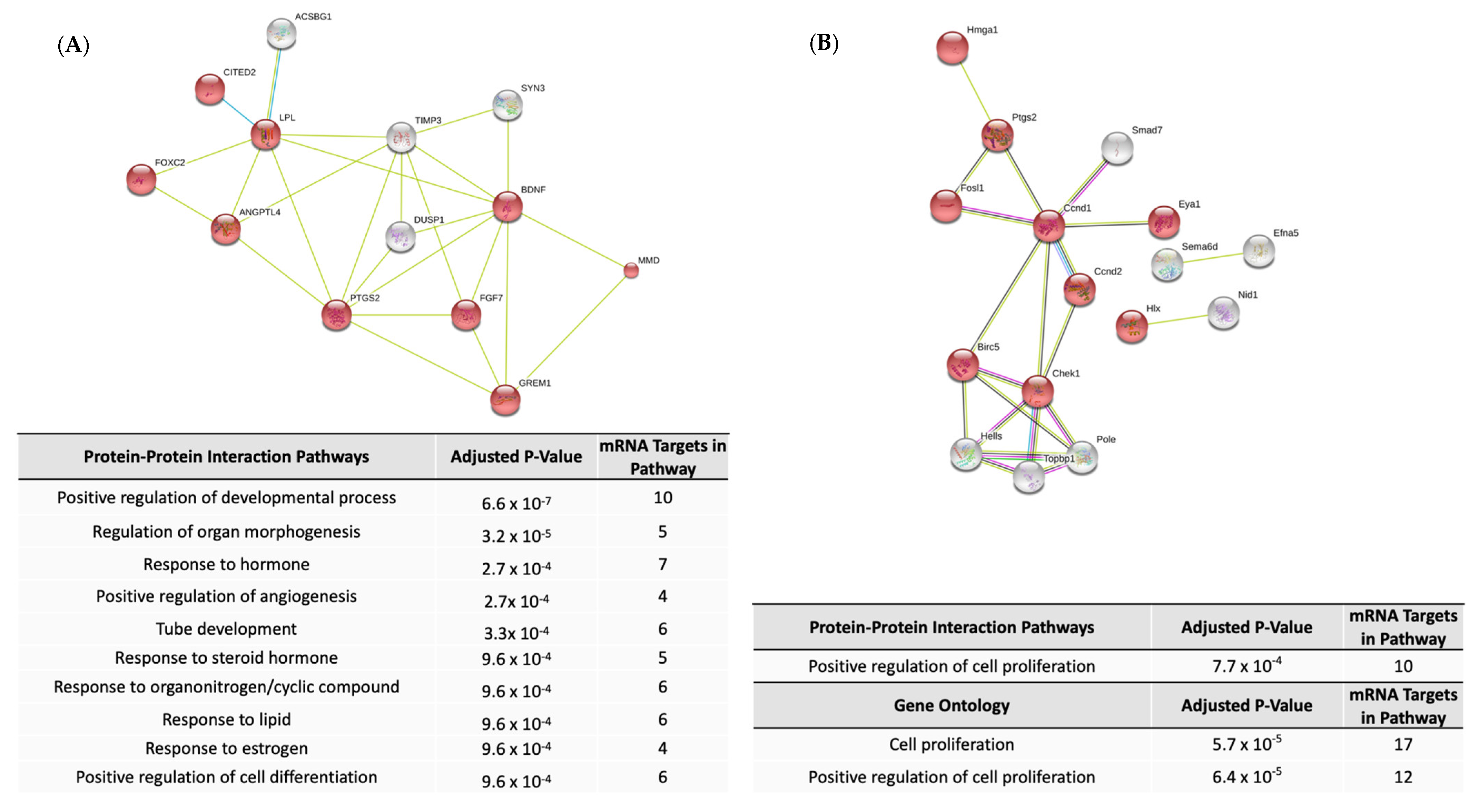
2.3. Pathway Enrichment for Predicted mRNA Targets of Differentially Expressed miRNAs

Predicted mRNA-miRNA interactions during muscle differentiation were identified (Supplemental Table S2) and then predicted target mRNAs were analyzed for enrichment in biological pathways (Table 1). Similar to enrichment among all differentially expressed mRNAs, calcium signaling was the most significantly upregulated pathway in the D2 vs. D0 and D5 vs. D0 comparisons while pathways related to degradation of nicotine, melatonin, and serotonin were all upregulated in D5 compared to D2. By contrast, there were several functional enrichments only observed among predicted mRNA targets including PPAR, protein kinase A, and RhoA signaling in the D5_D0 comparison. Further, D2_D0 targeted mRNAs were enriched for pathways predicted to be inhibited during this early interval: metabolism of thyroid hormone, nicotine, and melatonin. Those same pathways had been predicted to be activated among all differentially expressed mRNAs at D5_D2. Cyclins and cell regulation pathways were downregulated in both the D2_D0 and D5_D0 comparisons, while G1/S checkpoint and S-phase entry pathways were upregulated in D2_D0. In contrast to the total differential expression mRNA analysis, cholesterol synthesis pathways were not enriched among mRNAs targeted by miRNAs in the D2_D0 and D5_D0 comparisons.

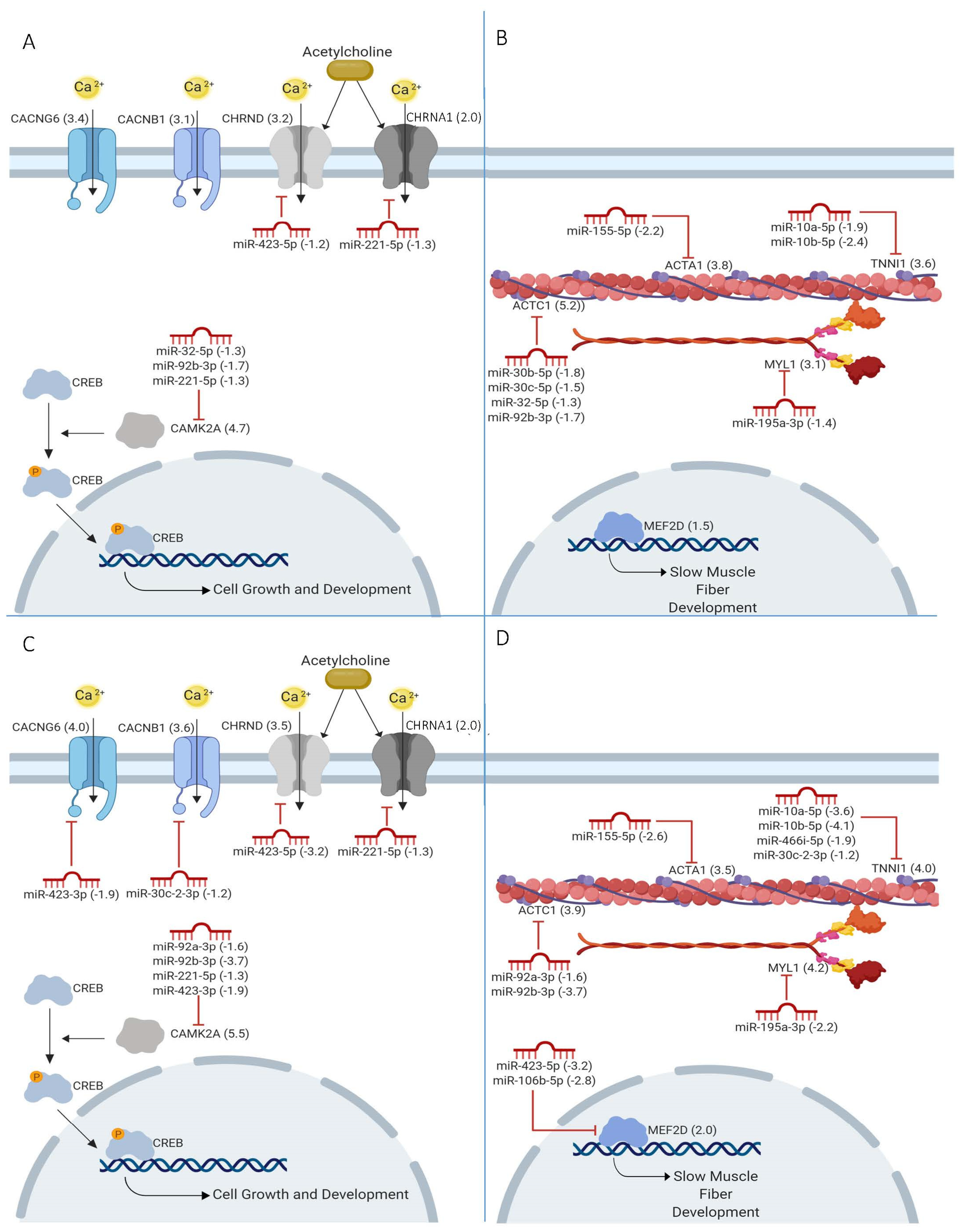
2.4. miRNAs in Calcium Signaling

Redundancy in miRNAs and targeted mRNAs was explored in the calcium signaling pathway at the D2_D0 and D5_D0 comparisons (Figure 3A,C). Of genes predicted to be targeted by miRNA, Camk2a was the only gene with multiple predicted miRNA interactions (miR-32-5p, miR-92b-3p, and miR-221-5p in D2_D0; miR-92a-3p, miR-92b-3p, miR-221-5p and miR-423-3p in D5_D0). Reciprocally, two miRNAs (miR-221-5p in D2_D0; miR-423-3p and miR-221-5p in D5_D0) have multiple mRNA targets within the calcium signaling pathway. In addition to the calcium pathway genes annotated by Ingenuity Pathway Analysis (IPA), 2 predicted targets of miR-423-5p, Lynx1 and Pvalb, have also been implicated in calcium signaling through stabilization of acetylcholine receptors and calcium binding, respectively [19,20]. Lynx1 was also a predicted target of miR-423-3p and miR-221-5p, and both of those miRNAs were predicted to target two subunits of the muscle acetylcholine receptor (Chrnd1 and Chrna1, respectively) [21,22].

2.5. Sarcomere Genes Targeted by miRNAs

Sarcomere structures, such as actin, troponin, and myosin, were also predicted to be targeted by several miRNAs during myoblast differentiation (Figure 3B,D). Both Actc1 and Tnni1 were the predicted targets of multiple miRNA (miR-30b/c-5p, miR-32-5p, and miR-92a/b-3p; and miR-10a-5p, miR-10b-5p, miR-466i-5p, and miR-30c-2-3p, respectively). Additionally, Mef2d, a member of the MEF2 family of transcription factors known to regulate the differentiation of myoblasts and maintenance of sarcomeres [23,24], was predicted to be targeted by miR-423-5p and miR-106b-5p. Several miRNAs were predicted to target genes related to both calcium signaling and sarcomeres structures including miR-30c-2-3p, miR-32-5p, miR-92a-3p, miR-92b-3p, and miR-423-5p.

2.6. myomiR Target Functions

Eight miRNAs (miR-1, miR-133a, miR-133b, miR-206, miR-208a, miR-208b, miR-499a, and miR-499b) are important for muscle development and are known as myoMirs. Of note, all myomiRs were significantly upregulated during muscle differentiation except miR-208a and miR-208b (Supplemental Table S1). Differentially expressed myoMirs were predicted to target mRNA with functions related to regulation of developmental processes, regulation of structural and organ morphogenesis, programmed cell death and apoptosis, and response to endogenous stimulus and hormones by gene ontology analysis and protein-protein interaction analysis (Figure 4A, Supplemental Table S3).

2.7. Loss of miRNA Differential Expression in CMD

Seventy-four miRNAs were significantly up- or downregulated during control human myoblast differentiation from myoblasts to mature myocytes (day 0 to day 3). Sixty-two of those 74 miRNAs were differentially expressed in myoblasts derived from patient with a moderate (DM13) CMD genotype. However, only 12 of the 74 were up- or downregulated in cells from the patient with severe CMD (DM15). All of those 12 were also differentially expressed in DM13 cells (Supplemental Table S4). Five of the 12 miRNAs with preserved differential expression in DM15 cells were known myomiRs (miR-1, miR-133a-3p, miR-133a-5p, miR-133b, miR-206). Among the 62 miRNAs with loss of significant up- or downregulation during DM15 myoblast differentiation, 25 miRNAs were conserved between human and mouse. Of those, 18 were also up- or downregulated during C2C12 differentiation (2 at D2_D0 only, 4 at D5_D0 only, and 12 at both D2_D0 and D5_D0). Those 18 miRNAs were predicted to target 166 mRNAs (Supplemental Table S5), which were enriched for functions related to muscle differentiation and formation (Table 2, Supplemental Table S6).

2.8. Chromosome X miRNAs in CMD

Two clusters of five miRNA on chromosome X (miR-450a-5p, miR-503-5p, and miR-542-3p; miR-221 and miR-222) were up- or down-regulated in control mouse and human myoblast differentiation, but not in the DM15 myoblasts (Supplemental Table S7). The mRNA targets of the miR-503 cluster in C2C12 differentiation were enriched for functions related to organismal development and cellular proliferation and differentiation (Figure 4B, Supplemental Table S8). Processes enriched in gene ontology analysis of the miR-221/222 cluster were numerous, including structural morphogenesis, regulation of cell development and differentiation, and regulation of muscle system processes, contraction, and structure development (Supplemental Table S9).

3. Discussion

miRNAs are essential regulators of muscle differentiation and understanding their mRNA targets can provide new insight into genetic regulation of development. In this study, we identified miRNAs and mRNAs that are up- or down-regulated during myoblast differentiation to myocytes. Further, we detected differential expression of miRNA that target sarcomere formation and calcium signaling genes during myoblast differentiation. mRNA targets predicted by our data had significant overlap with previously identified targets [25,26,27,28,29,30,31,32,33,34,35], providing support for the bioinformatic approach of requiring both reciprocal expression and miRNA binding sites to identify potential miRNA-mRNA interactions. Expression of genes that were upregulated and predicted to be targeted included the transcription factors Mef2a, Mef2c, and MefF2d, consistent with their role in the expression of sarcomere genes such as Actc1, Acta1, Myl1, and Tnni1 [24,36]. Known regulators of these transcription factors, miR-92, miR-30c, and miR-10 (Figure 2), were all correctly identified in our studies [25,26,28,29,30]. Our analyses also replicated previous findings of increased myomiR expression during muscle differentiation (miR-1, miR-133a, miR-133b, miR-206, and miR-499) [6,8,37], and correctly predicted their targeting of MEF2 transcription factors and myogenin [31]. Several additional miRNA-mRNA interactions predicted by our methods have also been experimentally validated. These include miR-106b targeting Mef2d [32] and Cdkn1a [33], miR-322 and miR-503 targeting Ccnd1 [34], miR-222 targeting Bmf [35], and miR-155 targeting Msi2 [27]. Novel miRNA-mRNA interacts worthy of further investigation include miR-423-5p and Mef2d, as miR-423-5p is already known to interact with Sufu, a myogenic regulator [26,38].

3.1. Calcium Signaling and Sarcomere Function

While miRNA regulation of calcium signaling has been previously described in cardiomyocytes and smooth muscle cells [39,40,41], this is the first identification of miRNAs targeting calcium signaling pathways during skeletal muscle differentiation. The miRNA and target mRNAs predicted by us are unique and different than those characterized in cardiac or smooth muscle [42], reflecting the specific calcium regulation pathways in each muscle type. The four miRNAs targeting calcium channels (miR-221-5p, miR-30c-2-3p, miR-423-3p and miR-423-5p) were downregulated during differentiation, which would allow for increased calcium channel abundance in mature muscle. The majority of the calcium channels predicted to be targeted by miRNA (Chrnd, Chrna1, Cacnb1, and Cacng6) have previously been shown to be upregulated during myoblast differentiation [43].
Downstream of calcium channels, calcium-dependent kinases contribute to regulation of sarcomere formation. Many miRNAs were predicted to regulate both calcium signaling and sarcomere gene expression including miR-30c-2-3p (targeting Cacnb1 in calcium signaling and Tnni1 in sarcomere formation), miR-92a/b (Camk2a and Actc1), and miR-423-5p (Chrnd and Actc1). This integration of calcium signaling and sarcomere function is well characterized, including regulation of muscle-specific MEF2 transcription factors by calcium/calmodulin dependent kinases such as Camk2 [44]. Camk2a, a subunit of CAMK2, is a known target of miR-289 in fruit fly [45]. While the miR-289 family is not present in humans or mice, we predicted several mammalian miRNAs to inhibit Camk2a expression in myoblasts including miR-32, miR-92a/b, miR-221, and miR-423-3p. In addition to regulation of calcium signaling, Camk2a contributes to the regulation of myosin heavy chain [46,47], slow muscle fiber [14,48,49], and mitochondrial gene [50,51,52] expression in skeletal muscle [53]. This highlights the potential role of these miRNAs to act as indirect regulators of myogenic gene expression through their regulation of Camk2a. CAMK2 has been implicated in disorders of both cardiac and skeletal muscle such as cardiac ischemia [54], arrhythmias [55], and limb girdle muscular dystrophy [56]. To date, treatment options have primarily focused on the development of small molecules to inhibit and modulate activity of CAMK2 [57]. Increasing evidence that Camk2a is regulated by miRNA suggests miRNAs as potential therapeutic targets in muscle disorders.

3.2. miRNA Dysregulation in CMD

The role of miRNAs in muscle disorders was supported by observation that global depletion of miRNAs through Dicer knockout in mice lead to impaired muscle development [58]. CMD patient muscle has lower expression of myomiRs such as miR-1 [59], possibly due to differences in miRNA processing [59,60]. In our analysis, we identify 62 miRNA that are differentially expressed during healthy human myoblast differentiation that are not up- or downregulated in differentiating CMD myoblasts. Using the more complete data from the mouse myoblast differentiation, we predicted potential functional consequences of this miRNA dysregulation in CMD. Three miRNAs, miR-155, miR-221, and miR-92b, failed to be downregulated during CMD muscle differentiation, which could lead to inappropriate suppression of the calcium signaling and sarcomere development. Calcium signaling has been shown to be altered in CMD cardiomyocytes, and several genes involved in calcium signaling are differentially expressed in CMD myoblasts [61]. Notably, upregulation of all myomiRs was conserved, supporting the essential role of these miRNAs in muscle development. Additionally, genes known to be implicated in CMD pathogenesis such as Dmpk [62], Rbfox1 [63], and Cacna1 [59] were differentially expressed during normal C2C12 myoblast differentiation in our data. Dmpk was not a predicted target of any miRNA. Rbfox1 and Cacna1 were predicted targets of let-7d-3p and miR-423-5p, respectively, but only with moderate confidence. Therefore, they were not considered for further analysis. Furthermore, neither of those two miRNAs were dysregulated during CMD myoblast differentiation. This suggests the study of miRNA could expand our knowledge of the molecular pathways contributing to CMD.
Several additional miRNAs dysregulated in the differentiation of CMD myoblasts are members of miRNA clusters on chromosome X: miR-221/222 and miR-450a/503/542. We observed loss of the expected downregulation of the miR-221/222 cluster and upregulation of the miR-503 cluster during differentiation of CMD myoblasts from a patient with severe disease. The miR-221/222 cluster is known to increase cell proliferation via targeting of p27 tumor suppressor [64] and is a negative regulator of muscle differentiation [65,66]. Over expression of the miR-221/222 cluster inhibits myoblast differentiation [64], so continued expression of this cluster in CMD myoblasts could contribute to abnormal muscle development. Conversely, the miR-503 cluster is a positive regulator of muscle differentiation through inhibition of Cdc25a, a kinase responsible for disinhibiting cyclin dependent kinase 2 [67]. During C2C12 myoblast differentiation, miR-503 was predicted to target both Ccnd1 and Ccnd2, which promote proliferation via the activity of cyclin dependent kinases similarly to Cdc25a. This is an example of potential de-repression due to lack of miRNA upregulation in CMD. The location of these miRNA clusters on the X chromosome provides a possible mechanism for some of the clinical differences seen between men and women with muscular dystrophies, particularly myotonic dystrophy [68]. More research is needed to determine if there are clinically relevant targets of these clusters.

3.3. MyomiRs and Glucuronyltransferases

Here we predict a novel role for miRNAs in regulation of glucuronyltransferase gene expression (including Ugt1a3, Ugt1a4, Ugt1a6, and Ugt1a8) during muscle development. In our data, the myomiR miR-486 is predicted to target several UDP-glucuronyltransferases, including Ugt1a, which were downregulated during the D2_D0 interval but then upregulated D5_D2. Ugt1a encodes UDP-glucuronosyltransferase, an enzyme implicated in skeletal and cardiac muscle differentiation [69], as well as in metabolism of unconjugated bilirubin, thyroxine (T4) and triiodothyronine (T3) [70]. UDP-glucuronosyltransferase is highly expressed in quiescent satellite cells, but is downregulated during differentiation [71]. Previous research has highlighted dysfunction of glucuronyltransferases in CMD. The LARGE gene modifies alpha-dystroglycan via glucuronyltransferase activity, depends on cations such as calcium, and is necessary for skeletal muscle sarcomere integrity [72]. Impairment of LARGE enzymatic activity has been demonstrated in the mouse model of CMD, and mutations in LARGE have been documented in CMD patients [73]. Additional research is needed to further elucidate the role of miRNA, glucuronyltransferases, and calcium signaling in pathogenesis of myotonic dystrophy.
In conclusion, many miRNAs are differentially expressed during muscle differentiation, and predict target mRNAs enrichment for functions related to calcium signaling and sarcomere formation. Limitations to this study include the use of a cell culture model of muscle differentiation, lack of availability of DM13 and DM15 cells to confirm the in silico findings including if C2C12 myotubes at day 5 and human DM13 at day 3 have same level of differentiation, and use of strict criteria for differential expression in prediction of targets. Cell culture models of development differ in many aspects from in vivo development, but nonetheless recapitulate many essential steps of muscle differentiation. It is possible that some miRNAs are targeting mRNAs that do not significantly change in expression and indirect targets would not be detected in our analysis because of the requirement for a miRNA binding site. Important future directions include biochemical confirmation of these predicted regulatory interactions. While some previous computational prediction of miRNA targets has demonstrated poor specificity, requiring anti-correlation of mRNA and miRNA expression increases specificity by providing orthogonal support for regulation [74]. However, the results included here generate new hypotheses about the role of miRNA regulation of calcium signaling and glucuronlytransferase activity in muscle development and disease. Future studies to confirm the many regulatory interactions will be an important next step towards identification of potential therapeutic targets.

4. Materials and Methods

4.1. Cell Culture

C2C12 mouse myoblasts were cultured and differentiated as per the ENCODE protocol (encodeproject.org, Accessed 26 February, 2015). In brief, myoblasts were grown in DMEM with 20% fetal bovine serum and differentiation initiated by transition to DMEM with 2% horse serum and 1 μM insulin (Gibco, Waltham, MA, USA). Cells were harvested at the specified timepoints and cell pellets were frozen at -80 until RNA extraction.

4.2. RNA Extraction and Sequencing

Total RNA was isolated from cell pellets using the RNAEasy kit (Qiagen, Hilden, Germany). RNA and miRNA sequencing libraries were prepared using NEBNext RNA library reagents. Libraries were sequenced on HiSeq 2500 (Illumina, San Diego, CA, USA), 50 bp reads).

4.3. mRNA Expression Analysis

mRNA sequencing data were aligned to the mm10 reference genome with TopHat (version 1.4) using the following parameters: ‘--splice-mismatches 1 --min-anchor-length 5 --segment-mismatches 3 --segment-length 25 --max-multihits 0 --no-novel-juncs’, and splice junctions defined by the Illumina iGenome genes.gtf reference. Mitochondrial and duplicate reads were discarded using Samtools and Picard’s MarkDuplicates, respectively. Genes with fewer than 5 counts at 2 or more time points were considered to have low counts and were excluded from the analysis. Differentially expressed genes were identified using the edgeR package for R (version 3.6.0, R Foundation for Statistical Computing, Vienna, Austria). First, effective library sizes were determined using trimmed mean of m values normalization. A common negative binomial dispersion parameter was estimated for the dataset since the samples were run without biological replicates. Gene-specific dispersions were then estimated with the quantile-adjusted conditional maximum likelihood method. Log fold change differential expression was calculated pairwise between each of the three time points (Day 2 vs. Day 0, Day 5 vs. Day 2, and Day 5 vs. Day 0) for both mRNA and miRNA expression using a negative-binomial exact gene-wise test. Significant differential expression was defined as an FDR q-value less than 0.05 and absolute log fold change greater than 1.2 for miRNA and 2.0 for mRNA. Overlap in differentially expressed miRNAs and mRNAs between the D2_D0 and D5_D2 periods were assessed using a hypergeometric distribution, for a total of 253 miRNA and 10287 mRNAs included in analyses after quality filtration.

4.4. miRNA-mRNA Correlation Analysis

Predicted miRNA targets were determined using the Ingenuity Pathway Analysis (IPA, Qiagen, Hilden, Germany) miRNA Target Filter feature. IPA predicts miRNA-mRNA interactions at 3 confidence levels: experimentally observed, high confidence, and moderate confidence. Pairings for DE miRNA and mRNAs were identified for each developmental interval (Day 2 vs. Day 0, Day 5 vs. Day 0, and Day 5 vs. Day 2) and filtered for inverse expression (miRNA upregulated when target mRNA downregulated or miRNA downregulated when mRNA target upregulated).

4.5. Functional Enrichment

mRNAs that were predicted targets of miRNAs based on experimental observation or a high confidence were assessed for gene ontology enrichment and protein-protein interactions using IPA and ShinyGO v0.60 (bioinformatics.sdstate.edu/go/, Accessed 19 July, 2019). mRNA targeted by myomiRs (miR-1, miR-133a, miR-133b, miR-206, miR-208a, miR-208b, miR-499a, and miR-499b) at all three confidence levels were also analyzed for functional enrichment. Enriched terms with FDR q-values below the pre-determined cutoff of 1 × 10⁻³ were considered significant. The method by which z-scores and p-values for functional enrichment are determined by IPA has been previously described [75]. In brief, a positive z-score indicates an increased number of genes that are upregulated and share a common pathway. The p-value represents the probability of these genes being present together by chance.

4.6. miRNA Expression during CMD Myoblast Differentiation

Differential miRNA expression was determined during differentiation of myoblasts from two CMD and one non-CMD participant myoblasts [76] as described above. The human CMD cell lines, DM13 and DM15, have 1800 and 3200 trinucleotide repeats at the 3′ UTR region of the Dystrophia Myotonic Protein Kinase (DMPK) gene, respectively, and are associated with intermediate and severe phenotypes, respectively. Data were downloaded under accession number GSE127296 (ncbi.nlm.nih.gov, accessed on 22 July 2019). The authors report that the DM13 cell line had differentiation comparable to that of the mature myocytes seen in the control cell line at day 3 of differentiation. However, the differentiation of the DM15 cell line was significantly impaired at day 3 [76]. These results are consistent with previous study of impaired differentiation in CMD myoblasts [77]. miRNAs that were differentially expressed in unaffected human myoblasts cells but not in CMD cells were identified, and those that were also found in the mouse genome and expressed in C2C12 cells were assessed for mRNA targets at all three confidence levels (experimentally observed, high confidence, and moderate confidence) using C2C12 paired mRNA-miRNA data. Functional enrichment among those mRNA targets was determined as above.

Supplementary Materials

Supplementary materials can be found at https://www.mdpi.com/1422-0067/22/5/2692/s1. Table S1: Differential expression of miRNA and mRNAs during C2C12 myoblast differentiation. Table S2: Predicted miRNA targets during C2C12 myoblast differentiation. Table S3: Predicted myomiR targets during C2C12 myoblast differentiation. Table S4: Differential expression of miRNA during human control and CMD myoblast differentiation. Table S5: Predicted targets of miRNA dysregulated in CMD. Table S6: Functional enrichment of predicted targets of miRNA dysregulated in CMD. Table S7: Chromosome X miRNA clusters dysregulated in CMD. Table S8: Predicted targets of the miR-503 cluster. Table S9: Predicted targets of the miR-221/222 cluster.

Author Contributions

Conceptualization, S.U.M., H.Z., D.L.T., M.D.U. and P.B.A.; methodology, S.U.M., C.R.S., M.D., D.L.T., M.D.U. and P.B.A.; formal analysis, S.U.M., C.R.S., M.D. and D.L.T.; investigation, S.U.M., H.Z., and M.D.U.; writing—original draft preparation, S.U.M. and C.R.S.; writing—review and editing, S.U.M., C.R.S., D.L.T., M.D.U., and P.B.A. All authors have read and agreed to the published version of the manuscript.

Funding

S.U.M. was supported by an American Academy of Pediatrics Marshall Klaus Perinatal Research Award. P.B.A. was supported by R01-AR068429-01 from National Institute of Arthritis and Musculoskeletal and Skin Diseases of National Institute of Health (NIH).

Institutional Review Board Statement

Not applicable.

Informed Consent Statement

Informed consent was previously obtained from all subjects involved in the study, and only deidentified data was accessed.

Data Availability Statement

Genome-wide differential expression values are contained within the supplementary material.

Acknowledgments

The authors would like to thank the Advanced Genomics Core at the University of Michigan for their sequencing services.

Conflicts of Interest

The authors declare no conflict of interest.

Abbreviations

CMDcongenital myotonic dystrophy
mRNAmessenger RNA
miRNAmicroRNA

References

  1. Bartel, D.P. Metazoan MicroRNAs. Cell 2018, 173, 20–51. [Google Scholar] [CrossRef] [Green Version]
  2. Pasquinelli, A.E. MicroRNAs and their targets: Recognition, regulation and an emerging reciprocal relationship. Nat. Rev. Genet. 2012, 13, 271–282. [Google Scholar] [CrossRef]
  3. Kim, J.; Kang, Y.; Kojima, Y.; Lighthouse, J.K.; Hu, X.; Aldred, M.A.; McLean, D.L.; Park, H.; Comhair, S.A.; Greif, D.M.; et al. An endothelial apelin-FGF link mediated by miR-424 and miR-503 is disrupted in pulmonary arterial hypertension. Nat. Med. 2013, 19, 74–82. [Google Scholar] [CrossRef] [PubMed] [Green Version]
  4. Kim, E.; Cook-Mills, J.; Morgan, G.; Sredni, S.T.; Pachman, L.M. Increased expression of vascular cell adhesion molecule 1 in muscle biopsy samples from juvenile dermatomyositis patients with short duration of untreated disease is regulated by miR-126. Arthritis Rheum. 2012, 64, 3809–3817. [Google Scholar] [CrossRef] [Green Version]
  5. Karolina, D.S.; Armugam, A.; Tavintharan, S.; Wong, M.T.K.; Lim, S.C.; Sum, C.F.; Jeyaseelan, K. MicroRNA 144 Impairs Insulin Signaling by Inhibiting the Expression of Insulin Receptor Substrate 1 in Type 2 Diabetes Mellitus. PLoS ONE 2011, 6, e22839. [Google Scholar] [CrossRef]
  6. Chen, J.-F.; Mandel, E.M.; Thomson, J.M.; Wu, Q.; Callis, T.E.; Hammond, S.M.; Conlon, F.L.; Wang, D.-Z. The Role of MicroRNA-1 and MicroRNA-133 in Skeletal Muscle Proliferation and Differentiation. Nat. Genet. 2005, 38, 228–233. [Google Scholar] [CrossRef] [PubMed]
  7. Zhao, Y.; Ransom, J.F.; Li, A.; Vedantham, V.; Von Drehle, M.; Muth, A.N.; Tsuchihashi, T.; McManus, M.T.; Schwartz, R.J.; Srivastava, D. Dysregulation of Cardiogenesis, Cardiac Conduction, and Cell Cycle in Mice Lacking miRNA-1-2. Cell 2007, 129, 303–317. [Google Scholar] [CrossRef] [PubMed] [Green Version]
  8. Horak, M.; Novak, J.; Bienertova-Vasku, J. Muscle-specific microRNAs in skeletal muscle development. Dev. Biol. 2016, 410, 1–13. [Google Scholar] [CrossRef]
  9. Liu, N.; Williams, A.H.; Maxeiner, J.M.; Bezprozvannaya, S.; Shelton, J.M.; Richardson, J.A.; Bassel-Duby, R.; Olson, E.N. microRNA-206 promotes skeletal muscle regeneration and delays progression of Duchenne muscular dystrophy in mice. J. Clin. Investig. 2012, 122, 2054–2065. [Google Scholar] [CrossRef] [PubMed] [Green Version]
  10. Van Rooij, E.; Quiat, D.; Johnson, B.A.; Sutherland, L.B.; Qi, X.; Richardson, J.A.; Kelm, R.J.; Olson, E.N. A Family of microRNAs Encoded by Myosin Genes Governs Myosin Expression and Muscle Performance. Dev. Cell 2009, 17, 662–673. [Google Scholar] [CrossRef] [PubMed] [Green Version]
  11. Dey, B.K.; Gagan, J.; Dutta, A. miR-206 and -486 Induce Myoblast Differentiation by Downregulating Pax7. Mol. Cell. Biol. 2010, 31, 203–214. [Google Scholar] [CrossRef] [Green Version]
  12. Zhang, D.; Li, X.; Chen, C.; Li, Y.; Zhao, L.; Jing, Y.; Liu, W.; Wang, X.; Zhang, Y.; Xia, H.; et al. Attenuation of p38-Mediated miR-1/133 Expression Facilitates Myoblast Proliferation during the Early Stage of Muscle Regeneration. PLoS ONE 2012, 7, e41478. [Google Scholar] [CrossRef] [Green Version]
  13. Chen, J.-F.; Tao, Y.; Li, J.; Deng, Z.; Yan, Z.; Xiao, X.; Wang, D.-Z. microRNA-1 and microRNA-206 regulate skeletal muscle satellite cell proliferation and differentiation by repressing Pax7. J. Cell Biol. 2010, 190, 867–879. [Google Scholar] [CrossRef] [PubMed] [Green Version]
  14. Feng, Y.; Niu, L.-L.; Wei, W.; Zhang, W.-Y.; Li, X.-Y.; Cao, J.-H.; Zhao, S.-H. A feedback circuit between miR-133 and the ERK1/2 pathway involving an exquisite mechanism for regulating myoblast proliferation and differentiation. Cell Death Dis. 2013, 4, e934. [Google Scholar] [CrossRef] [Green Version]
  15. Eisenberg, I.; Eran, A.; Nishino, I.; Moggio, M.; Lamperti, C.; Amato, A.A.; Lidov, H.G.; Kang, P.B.; North, K.N.; Mitrani-Rosenbaum, S.; et al. Distinctive patterns of microRNA expression in primary muscular disorders. Proc. Natl. Acad. Sci. USA 2007, 104, 17016–17021. [Google Scholar] [CrossRef] [Green Version]
  16. Alexander, M.S.; Kawahara, G.; Motohashi, N.; Casar, J.C.; Eisenberg, I.; Myers, J.A.; Gasperini, M.J.; Estrella, E.A.; Kho, A.T.; Mitsuhashi, S.; et al. MicroRNA-199a is induced in dystrophic muscle and affects WNT signaling, cell proliferation, and myogenic differentiation. Cell Death Differ. 2013, 20, 1194–1208. [Google Scholar] [CrossRef]
  17. Perbellini, R.; Greco, S.; Sarra-Ferraris, G.; Cardani, R.; Capogrossi, M.C.; Meola, G.; Martelli, F. Dysregulation and cellular mislocalization of specific miRNAs in myotonic dystrophy type 1. Neuromuscul. Disord. 2011, 21, 81–88. [Google Scholar] [CrossRef] [PubMed]
  18. Gambardella, S.; Rinaldi, F.; Lepore, S.M.; Viola, A.; Loro, E.; Angelini, C.; Vergani, L.; Novelli, G.; Botta, A. Overexpression of microRNA-206 in the skeletal muscle from myotonic dystrophy type 1 patients. J. Transl. Med. 2010, 8, 48. [Google Scholar] [CrossRef]
  19. Gaudet, P.; Livstone, M.S.; Lewis, S.E.; Thomas, P.D. Phylogenetic-based propagation of functional annotations within the Gene Ontology consortium. Briefings Bioinform. 2011, 12, 449–462. [Google Scholar] [CrossRef] [Green Version]
  20. Lyukmanova, E.N.; Shenkarev, Z.O.; Shulepko, M.A.; Mineev, K.S.; D’Hoedt, D.; Kasheverov, I.E.; Filkin, S.Y.; Krivolapova, A.P.; Janickova, H.; Dolezal, V.; et al. NMR Structure and Action on Nicotinic Acetylcholine Receptors of Water-soluble Domain of Human LYNX1. J. Biol. Chem. 2011, 286, 10618–10627. [Google Scholar] [CrossRef] [PubMed] [Green Version]
  21. Todd, E.J.; Yau, K.S.; Ong, R.; Slee, J.; McGillivray, G.; Barnett, C.P.; Haliloglu, G.; Talim, B.; Akcoren, Z.; Kariminejad, A.; et al. Next generation sequencing in a large cohort of patients presenting with neuromuscular disease before or at birth. Orphanet J. Rare Dis. 2015, 10, 1–14. [Google Scholar] [CrossRef] [PubMed] [Green Version]
  22. Cetin, H.; Epstein, M.; Liu, W.W.; Maxwell, S.; Cruz, P.M.R.; Cossins, J.; Vincent, A.; Webster, R.; Biggin, P.C.; Beeson, D. Muscle acetylcholine receptor conversion into chloride conductance at positive potentials by a single mutation. Proc. Natl. Acad. Sci. USA 2019, 116, 21228–21235. [Google Scholar] [CrossRef] [PubMed] [Green Version]
  23. Ow, J.R.; Palanichamy, M.K.; Rao, V.K.; Choi, M.H.; Bharathy, N.; Taneja, R. G9a inhibits {MEF2C} activity to control sarcomere assembly. Sci. Rep. 2016, 6, 34163. [Google Scholar] [CrossRef] [Green Version]
  24. Potthoff, M.J.; Olson, E.N. MEF2: A central regulator of diverse developmental programs. Development 2007, 134, 4131–4140. [Google Scholar] [CrossRef] [Green Version]
  25. Liang, T.; Zhou, B.; Shi, L.; Wang, H.; Chu, Q.; Xu, F.; Li, Y.; Chen, R.; Shen, C.; Schinckel, A.P. IncRNA AK017368 promotes proliferation and suppresses differentiation of myoblasts in skeletal muscle development by attenuating the function of miR-30c. FASEB J. 2018, 32, 377–389. [Google Scholar] [CrossRef] [Green Version]
  26. Luo, W.; Li, G.; Yi, Z.; Nie, Q.; Zhang, X. E2F1-miR-20a-5p/20b-5p auto-regulatory feedback loop involved in myoblast proliferation and differentiation. Sci. Rep. 2016, 6, 27904. [Google Scholar] [CrossRef] [Green Version]
  27. Selbach, M.; Schwanhäusser, B.; Thierfelder, N.; Fang, Z.; Khanin, R.; Rajewsky, N. Widespread changes in protein synthesis induced by microRNAs. Nat. Cell Biol. 2008, 455, 58–63. [Google Scholar] [CrossRef] [PubMed]
  28. Penzkofer, D.; Bonauer, A.; Fischer, A.; Tups, A.; Brandes, R.P.; Zeiher, A.M.; Dimmeler, S. Phenotypic Characterization of miR-92a−/− Mice Reveals an Important Function of miR-92a in Skeletal Development. PLoS ONE 2014, 9, e101153. [Google Scholar] [CrossRef]
  29. Seok, H.Y.; Tatsuguchi, M.; Callis, T.E.; He, A.; Pu, W.T.; Wang, D.-Z. miR-155 Inhibits Expression of the MEF2A Protein to Repress Skeletal Muscle Differentiation. J. Biol. Chem. 2011, 286, 35339–35346. [Google Scholar] [CrossRef] [Green Version]
  30. Ge, G.; Yang, D.; Tan, Y.; Chen, Y.; Jiang, D.; Jiang, A.; Li, Q.; Liu, Y.; Zhong, Z.; Li, X.; et al. miR-10b-5p Regulates C2C12 Myoblasts Proliferation and Differentiation. Biosci. Biotechnol. Biochem. 2019, 83, 291–299. [Google Scholar] [CrossRef]
  31. Buckingham, M.; Bajard, L.; Chang, T.; Daubas, P.; Hadchouel, J.; Meilhac, S.; Montarras, D.; Rocancourt, D.; Relaix, F. The formation of skeletal muscle: From somite to limb. J. Anat. 2003, 202, 59–68. [Google Scholar] [CrossRef] [PubMed]
  32. Beveridge, N.J.; Tooney, P.A.; Carroll, A.P.; Tran, N.; Cairns, M.J. Down-regulation of miR-17 family expression in response to retinoic acid induced neuronal differentiation. Cell. Signal. 2009, 21, 1837–1845. [Google Scholar] [CrossRef] [PubMed]
  33. Ivanovska, I.; Ball, A.S.; Diaz, R.L.; Magnus, J.F.; Kibukawa, M.; Schelter, J.M.; Kobayashi, S.V.; Lim, L.; Burchard, J.; Jackson, A.L.; et al. MicroRNAs in the miR-106b Family Regulate p21/CDKN1A and Promote Cell Cycle Progression. Mol. Cell. Biol. 2008, 28, 2167–2174. [Google Scholar] [CrossRef] [PubMed] [Green Version]
  34. Forrest, A.R.R.; Kanamori-Katayama, M.; Tomaru, Y.; Lassmann, T.; Ninomiya, N.; Takahashi, Y.; De Hoon, M.J.L.; Kubosaki, A.; Kaiho, A.; Suzuki, M.; et al. Induction of microRNAs, mir-155, mir-222, mir-424 and mir-503, promotes monocytic differentiation through combinatorial regulation. Leukemia 2009, 24, 460–466. [Google Scholar] [CrossRef] [PubMed] [Green Version]
  35. Gramantieri, L.; Fornari, F.; Ferracin, M.; Veronese, A.; Sabbioni, S.; Calin, G.A.; Grazi, G.L.; Croce, C.M.; Bolondi, L.; Negrini, M. MicroRNA-221 Targets Bmf in Hepatocellular Carcinoma and Correlates with Tumor Multifocality. Clin. Cancer Res. 2009, 15, 5073–5081. [Google Scholar] [CrossRef] [Green Version]
  36. Potthoff, M.J.; Arnold, M.A.; McAnally, J.; Richardson, J.A.; Bassel-Duby, R.; Olson, E.N. Regulation of Skeletal Muscle Sarcomere Integrity and Postnatal Muscle Function by Mef2c. Mol. Cell. Biol. 2007, 27, 8143–8151. [Google Scholar] [CrossRef] [Green Version]
  37. Kim, H.K.; Lee, Y.S.; Sivaprasad, U.; Malhotra, A.; Dutta, A. Muscle-specific microRNA miR-206 promotes muscle differentiation. J. Cell Biol. 2006, 174, 677–687. [Google Scholar] [CrossRef] [PubMed]
  38. Ge, J.; Zhu, J.; Xia, B.; Cao, H.; Peng, Y.; Li, X.; Yu, T.; Chu, G.; Yang, G.; Shi, X. miR-423-5p inhibits myoblast proliferation and differentiation by targeting Sufu. J. Cell. Biochem. 2018, 119, 7610–7620. [Google Scholar] [CrossRef] [PubMed]
  39. Poon, E.N.-Y.; Hao, B.; Guan, D.; Li, M.J.; Lu, J.; Yang, Y.; Wu, B.; Wu, S.C.-M.; Webb, S.E.; Liang, Y.; et al. Integrated transcriptomic and regulatory network analyses identify microRNA-200c as a novel repressor of human pluripotent stem cell-derived cardiomyocyte differentiation and maturation. Cardiovasc. Res. 2018, 114, 894–906. [Google Scholar] [CrossRef] [PubMed]
  40. Xu, T.; Liu, N.; Shao, Y.; Huang, Y.; Zhu, D. MiR-218 regulated cardiomyocyte differentiation and migration in mouse embryonic stem cells by targeting PDGFRα. J. Cell. Biochem. 2019, 120, 4355–4365. [Google Scholar] [CrossRef] [PubMed]
  41. Turczyńska, K.M.; Sadegh, M.K.; Hellstrand, P.; Swärd, K.; Albinsson, S. MicroRNAs Are Essential for Stretch-induced Vascular Smooth Muscle Contractile Differentiation via MicroRNA (miR)-145-dependent Expression of L-type Calcium Channels. J. Biol. Chem. 2012, 287, 19199–19206. [Google Scholar] [CrossRef] [Green Version]
  42. Harada, M.; Luo, X.; Murohara, T.; Yang, B.; Dobrev, D.; Nattel, S. MicroRNA Regulation and Cardiac Calcium Signaling. Circ. Res. 2014, 114, 689–705. [Google Scholar] [CrossRef] [Green Version]
  43. Tomczak, K.K.; Marinescu, V.D.; Ramoni, M.F.; Sanoudou, D.; Montanaro, F.; Han, M.; Kunkel, L.M.; Kohane, I.S.; Beggs, A.H. Expression profiling and identification of novel genes involved in myogenic differentiation. FASEB J. 2003, 18, 1–23. [Google Scholar] [CrossRef] [Green Version]
  44. McKinsey, T.A.; Zhang, C.-L.; Lu, J.; Olson, E.N. Signal-dependent nuclear export of a histone deacetylase regulates muscle differentiation. Nat. Cell Biol. 2000, 408, 106–111. [Google Scholar] [CrossRef]
  45. Kuklin, E.A.; Alkins, S.; Bakthavachalu, B.; Genco, M.C.; Sudhakaran, I.; Raghavan, K.V.; Ramaswami, M.; Griffith, L.C. The Long 3’UTR mRNA of CaMKII Is Essential for Translation-Dependent Plasticity of Spontaneous Release in Drosophila melanogaster. J. Neurosci. 2017, 37, 10554–10566. [Google Scholar] [CrossRef] [Green Version]
  46. Allen, D.L.; Leinwand, L.A. Intracellular Calcium and Myosin Isoform Transitions. J. Biol. Chem. 2002, 277, 45323–45330. [Google Scholar] [CrossRef] [PubMed] [Green Version]
  47. Mu, X.; Brown, L.D.; Liu, Y.; Schneider, M.F. Roles of the calcineurin and CaMK signaling pathways in fast-to-slow fiber type transformation of cultured adult mouse skeletal muscle fibers. Physiol. Genom. 2007, 30, 300–312. [Google Scholar] [CrossRef] [Green Version]
  48. Chin, E.R.; Olson, E.N.; Richardson, J.A.; Yang, Q.; Humphries, C.; Shelton, J.M.; Wu, H.; Zhu, W.; Bassel-Duby, R.; Williams, R.S. A calcineurin-dependent transcriptional pathway controls skeletal muscle fiber type. Genes Dev. 1998, 12, 2499–2509. [Google Scholar] [CrossRef] [PubMed] [Green Version]
  49. Serrano, A.L.; Murgia, M.; Pallafacchina, G.; Calabria, E.; Coniglio, P.; Lømo, T.; Schiaffino, S. Calcineurin controls nerve activity-dependent specification of slow skeletal muscle fibers but not muscle growth. Proc. Natl. Acad. Sci. USA 2001, 98, 13108–13113. [Google Scholar] [CrossRef] [PubMed] [Green Version]
  50. Garcia-Roves, P.M.; Huss, J.; Holloszy, J.O. Role of calcineurin in exercise-induced mitochondrial biogenesis. Am. J. Physiol. Metab. 2006, 290, E1172–E1179. [Google Scholar] [CrossRef] [PubMed] [Green Version]
  51. Jiang, L.Q.; Garcia-Roves, P.M.; Barbosa, T.D.C.; Zierath, J.R. Constitutively active calcineurin in skeletal muscle increases endurance performance and mitochondrial respiratory capacity. Am. J. Physiol. Metab. 2010, 298, E8–E16. [Google Scholar] [CrossRef] [Green Version]
  52. Long, Y.C.; Glund, S.; Garcia-Roves, P.M.; Zierath, J.R. Calcineurin Regulates Skeletal Muscle Metabolism via Coordinated Changes in Gene Expression. J. Biol. Chem. 2007, 282, 1607–1614. [Google Scholar] [CrossRef] [Green Version]
  53. Hawkins, C.; Xu, A.; Narayanan, N. Comparison of the effects of the membraneassociated Ca2+/calmodulin-dependent protein kinase on Ca2+-ATPase function in cardiac and slow-twitch skeletal muscle sarcoplasmic reticulum. Mol. Cell. Biochem. 1995, 142, 131–138. [Google Scholar] [CrossRef] [PubMed]
  54. Aurora, A.B.; Mahmoud, A.I.; Luo, X.; Johnson, B.A.; Van Rooij, E.; Matsuzaki, S.; Humphries, K.M.; Hill, J.A.; Bassel-Duby, R.; Sadek, H.A.; et al. MicroRNA-214 protects the mouse heart from ischemic injury by controlling Ca2+ overload and cell death. J. Clin. Investig. 2012, 122, 1222–1232. [Google Scholar] [CrossRef]
  55. Wang, Q.; Quick, A.P.; Cao, S.; Reynolds, J.; Chiang, D.Y.; Beavers, D.; Li, N.; Wang, G.; Rodney, G.G.; Anderson, M.E.; et al. Oxidized CaMKII (Ca 2+ /Calmodulin-Dependent Protein Kinase II) Is Essential for Ventricular Arrhythmia in a Mouse Model of Duchenne Muscular Dystrophy. Circ. Arrhythm. Electrophysiol. 2018, 11, e005682. [Google Scholar] [CrossRef] [PubMed]
  56. Kramerova, I.; Ermolova, N.; Eskin, A.; Hevener, A.; Quehenberger, O.; Armando, A.M.; Haller, R.; Romain, N.; Nelson, S.F.; Spencer, M.J. Failure to up-regulate transcription of genes necessary for muscle adaptation underlies limb girdle muscular dystrophy 2A (calpainopathy). Hum. Mol. Genet. 2016, 25, 2194–2207. [Google Scholar] [CrossRef] [PubMed] [Green Version]
  57. Epellicena, P.; Eschulman, H. CaMKII inhibitors: From research tools to therapeutic agents. Front. Pharmacol. 2014, 5, 21. [Google Scholar] [CrossRef] [Green Version]
  58. Oikawa, S.; Lee, M.; Akimoto, T. Conditional Deletion of Dicer in Adult Mice Impairs Skeletal Muscle Regeneration. Int. J. Mol. Sci. 2019, 20, 5686. [Google Scholar] [CrossRef] [Green Version]
  59. Rau, F.; Freyermuth, F.; Fugier, C.; Villemin, J.-P.; Fischer, M.-C.; Jost, B.; Dembele, D.; Gourdon, G.; Nicole, A.; Duboc, D.; et al. Misregulation of miR-1 processing is associated with heart defects in myotonic dystrophy. Nat. Struct. Mol. Biol. 2011, 18, 840–845. [Google Scholar] [CrossRef]
  60. Laurent, F.-X.; Sureau, A.; Klein, A.F.; Trouslard, F.; Gasnier, E.; Furling, D.; Marie, J. New function for the RNA helicase p68/DDX5 as a modifier of MBNL1 activity on expanded CUG repeats. Nucleic Acids Res. 2011, 40, 3159–3171. [Google Scholar] [CrossRef] [Green Version]
  61. Kim, E.Y.; Barefield, D.Y.; Vo, A.H.; Gacita, A.M.; Schuster, E.J.; Wyatt, E.J.; Davis, J.L.; Dong, B.; Sun, C.; Page, P.; et al. Distinct pathological signatures in human cellular models of myotonic dystrophy subtypes. JCI Insight 2019, 4. [Google Scholar] [CrossRef] [Green Version]
  62. Kaliman, P.; Llagostera, E. Myotonic dystrophy protein kinase (DMPK) and its role in the pathogenesis of myotonic dystrophy 1. Cell. Signal. 2008, 20, 1935–1941. [Google Scholar] [CrossRef]
  63. Klinck, R.; Fourrier, A.; Thibault, P.; Toutant, J.; Durand, M.; Lapointe, E.; Caillet-Boudin, M.-L.; Sergeant, N.; Gourdon, G.; Meola, G.; et al. RBFOX1 Cooperates with MBNL1 to Control Splicing in Muscle, Including Events Altered in Myotonic Dystrophy Type 1. PLoS ONE 2014, 9, e107324. [Google Scholar] [CrossRef] [Green Version]
  64. Kedde, M.; Van Kouwenhove, M.; Zwart, W.; Vrielink, J.A.F.O.; Elkon, R.; Agami, R. A Pumilio-induced RNA structure switch in p27-3′ UTR controls miR-221 and miR-222 accessibility. Nat. Cell Biol. 2010, 12, 1014–1020. [Google Scholar] [CrossRef]
  65. Cardinali, B.; Castellani, L.; Fasanaro, P.; Basso, A.; Alemà, S.; Martelli, F.; Falcone, G. Microrna-221 and Microrna-222 Modulate Differentiation and Maturation of Skeletal Muscle Cells. PLoS ONE 2009, 4, e7607. [Google Scholar] [CrossRef] [Green Version]
  66. Liu, B.; Shi, Y.; He, H.; Cai, M.; Xiao, W.; Yang, X.; Chen, S.; Jia, X.; Wang, J.; Lai, S. miR-221 modulates skeletal muscle satellite cells proliferation and differentiation. Vitr. Cell. Dev. Biol. Anim. 2017, 54, 147–155. [Google Scholar] [CrossRef] [PubMed]
  67. Sarkar, S.; Dey, B.K.; Dutta, A. MiR-322/424 and -503 Are Induced during Muscle Differentiation and Promote Cell Cycle Quiescence and Differentiation by Down-Regulation of Cdc25A. Mol. Biol. Cell 2010, 21, 2138–2149. [Google Scholar] [CrossRef] [PubMed]
  68. Dogan, C.; De Antonio, M.; Hamroun, D.; Varet, H.; Fabbro, M.; Rougier, F.; Amarof, K.; Bes, M.-C.A.; Bedat-Millet, A.-L.; Behin, A.; et al. Gender as a Modifying Factor Influencing Myotonic Dystrophy Type 1 Phenotype Severity and Mortality: A Nationwide Multiple Databases Cross-Sectional Observational Study. PLoS ONE 2016, 11, e0148264. [Google Scholar] [CrossRef] [PubMed]
  69. Siissalo, S.; Zhang, H.; Stilgenbauer, E.; Kaukonen, A.M.; Hirvonen, J.; Finel, M. The Expression of Most UDP-Glucuronosyltransferases (UGTs) Is Increased Significantly during Caco-2 Cell Differentiation, whereas UGT1A6 Is Highly Expressed Also in Undifferentiated Cells. Drug Metab. Dispos. 2008, 36, 2331–2336. [Google Scholar] [CrossRef] [PubMed] [Green Version]
  70. Tukey, R.H.; Strassburg, C.P. Human UDP-Glucuronosyltransferases: Metabolism, Expression, and Disease. Annu. Rev. Pharmacol. Toxicol. 2000, 40, 581–616. [Google Scholar] [CrossRef] [PubMed]
  71. Pallafacchina, G.; François, S.; Regnault, B.; Czarny, B.; Dive, V.; Cumano, A.; Montarras, D.; Buckingham, M. An adult tissue-specific stem cell in its niche: A gene profiling analysis of in vivo quiescent and activated muscle satellite cells. Stem Cell Res. 2010, 4, 77–91. [Google Scholar] [CrossRef]
  72. Barresi, R. Dystroglycan: From biosynthesis to pathogenesis of human disease. J. Cell Sci. 2006, 119, 199–207. [Google Scholar] [CrossRef] [Green Version]
  73. Inamori, K.-I.; Willer, T.; Hara, Y.; Venzke, D.; Anderson, M.E.; Clarke, N.F.; Guicheney, P.; Bönnemann, C.G.; Moore, S.A.; Campbell, K.P. Endogenous Glucuronyltransferase Activity of LARGE or LARGE2 Required for Functional Modification of α-Dystroglycan in Cells and Tissues. J. Biol. Chem. 2014, 289, 28138–28148. [Google Scholar] [CrossRef] [PubMed] [Green Version]
  74. McGeary, S.E.; Lin, K.S.; Shi, C.Y.; Pham, T.M.; Bisaria, N.; Kelley, G.M.; Bartel, D.P. The biochemical basis of microRNA targeting efficacy. Science 2019, 366, eaav1741. [Google Scholar] [CrossRef]
  75. Krämer, A.; Green, J.; Pollard, J.; Tugendreich, S. Causal analysis approaches in Ingenuity Pathway Analysis. Bioinform. 2014, 30, 523–530. [Google Scholar] [CrossRef] [PubMed]
  76. Rizzo, M.; Beffy, P.; Del Carratore, R.; Falleni, A.; Pretini, V.; D’Aurizio, R.; Botta, A.; Evangelista, M.; Stoccoro, A.; Coppedè, F.; et al. Activation of the interferon type I response rather than autophagy contributes to myogenesis inhibition in congenital DM1 myoblasts. Cell Death Dis. 2018, 9, 1071. [Google Scholar] [CrossRef] [PubMed]
  77. Beffy, P.; Del Carratore, R.; Masini, M.; Furling, D.; Puymirat, J.; Masiello, P.; Simili, M. Altered signal transduction pathways and induction of autophagy in human myotonic dystrophy type 1 myoblasts. Int. J. Biochem. Cell Biol. 2010, 42, 1973–1983. [Google Scholar] [CrossRef]
Figure 1. Differentially expressed (A) miRNA and (B) mRNA at day 2 vs. day 0 (D2_D0, myoblast to early myocyte), day 5 vs. day 0 (D5_D0, myoblast to mature myocyte), and day 5 vs. day 2 (D5_D2, early myocyte to mature myocyte).
Ijms 22 02692 g001
Figure 2. Top functional enrichments among differentially expressed mRNAs during muscle differentiation. Only the most significantly enriched term among closely related terms was included. Z-score for pathway activity is written on the relevant bar where applicable.
Ijms 22 02692 g002
Figure 3. miRNAs are predicted to target multiple mRNAs at D2_D0 (A,B) and D5_D0 (C,D) that encode proteins related to calcium signaling (A,C) and sarcomeres (B,D).
Ijms 22 02692 g003
Figure 4. Functional enrichment among predicted mRNA targets of myomiRs (A) and miR-503 cluster miRNAs (B) based on protein-protein interaction and gene ontology analysis. In A, red denotes proteins that contribute to positive regulation of developmental process. In B, red spheres denotes proteins that are involved in proliferation. White spheres denote proteins that lack a directional pattern of effect. Only the most significantly enriched term among closely related terms was included.
Ijms 22 02692 g004
Table 1. Top functional enrichments among differentially expressed mRNAs that are predicted targets of miRNAs. Only the most significantly enriched term among closely related terms was included.
PathwayD2_D0D5_D0D5_D2
p-Valuez-Scorep-Valuez-Scorep-Valuez-Score
Calcium Signaling5.6 × 10−82.651.9 × 10−62.236--
Estrogen-mediated S-phase Entry1.3 × 10−5−1.003.7 × 10−4---
Nicotine Degradation2.3 × 10−5−1.34--2.6 × 10−82.00
Melatonin Degradation3.2 × 10−5−1.34--3.4 × 10−82.00
Cell Cycle: G1/S Checkpoint Regulation5.6 × 10−51.004.8 × 10−4---
Thyroid Hormone Metabolism8.3 × 10−5−2.00--5.2 × 10−92.00
nNOS Signaling in Skeletal Muscle Cells1.1 × 10−4-----
Cyclins and Cell Cycle Regulation1.4 × 10−4−1.349.8 × 10−4-1--
Clathrin-mediated Endocytosis Signaling--9.1 × 10⁻⁵---
Actin Cytoskeleton Signaling--2.0 × 10⁻⁴---
PPAR Signaling--2.6 × 10⁻⁴−0.45--
Protein Kinase A Signaling--3.2 × 10−41.63--
RhoA Signaling--5.6 × 10−41.34 -
Serotonin Degradation----5.4 × 10−82.00
Xenobiotic Metabolism Signaling----1.8 × 10⁻⁵-
Table 2. Top functional enrichments among predicted targets of miRNAs differentially expressed during control human myoblast differentiation but not in CMD myoblasts.
Functional Categoryp-ValuemRNAs Targets
Muscle structure development 7.4 × 10−1330
Muscle system process 1.3 × 10−1225
Circulatory system development 3.2 × 10−1135
Muscle contraction 6.7 × 10−1121
Anatomical structure formation involved in morphogenesis 1.9 × 10−1035
Muscle organ development 5.1 × 10−920
Heart development 5.8 × 10−923
Striated muscle tissue development 1.4 × 10−819
Response to nitrogen compound 1.9 × 10−831
Cell differentiation 1.9 × 10−868
Cellular developmental process 1.9 × 10−870
Muscle tissue development 1.9 × 10−819
Publisher’s Note: MDPI stays neutral with regard to jurisdictional claims in published maps and institutional affiliations.

