Article

Broad-Spectrum Bactericidal Activity of a Synthetic Random Copolymer Based on 2-Methoxy-6-(4-Vinylbenzyloxy)-Benzylammonium Hydrochloride

1
Department of Surgical Sciences and Integrated Diagnostics (DISC), University of Genoa, Viale Benedetto XV, 6, I-16132 Genova, Italy
2
Department of Pharmacy, University of Genoa, Viale Cembrano, 16148 Genoa, Italy
*
Author to whom correspondence should be addressed.
Int. J. Mol. Sci. 2021, 22(9), 5021; https://doi.org/10.3390/ijms22095021
Submission received: 17 April 2021 / Revised: 4 May 2021 / Accepted: 7 May 2021 / Published: 9 May 2021
(This article belongs to the Special Issue Bio-Nano Interactions)

Abstract

:
Low-molecular-weight organic ammonium salts exert excellent antimicrobial effects by interacting lethally with bacterial membranes. Unfortunately, short-term functionality and high toxicity limit their clinical application. On the contrary, the equivalent macromolecular ammonium salts, derived from the polymerization of monomeric ammonium salts, have demonstrated improved antibacterial potency, a lower tendency to develop resistance, higher stability, long-term activity, and reduced toxicity. A water-soluble non-quaternary copolymeric ammonium salt (P7) was herein synthetized by copolymerizing 2-methoxy-6-(4-vinylbenzyloxy)-benzylammonium hydrochloride monomer with N, N-di-methyl-acrylamide. The antibacterial activity of P7 was assessed against several multidrug-resistant (MDR) clinical isolates of both Gram-positive and Gram-negative species. Except for colistin-resistant Pseudomonas aeruginosa, most isolates were susceptible to P7, also including some Gram-negative bacteria with a modified charge in the external membrane. P7 showed remarkable antibacterial activity against isolates of Enterococcus, Staphylococcus, Acinetobacter, and Pseudomonas, and on different strains of Escherichia coli and Stenotrophomonas maltophylia, regardless of their antibiotic resistance. The lowest minimal inhibitory concentrations (MICs) observed were 0.6–1.2 µM and the minimal bactericidal concentrations (MBC) were frequently overlapping with the MICs. In 24-h time–kill and turbidimetric studies, P7 displayed a rapid non-lytic bactericidal activity. P7 could therefore represent a novel and potent tool capable of counteracting infections sustained by several bacteria that are resistant to the presently available antibiotics.

Graphical Abstract

1. Introduction

Small-molecular-weight quaternary ammonium salts (QASs), such as the well-known benzalkonium hydrochloride, commercially available as CITROSIL® [1], and several of its derivatives, have been broadly studied and intensively used as antimicrobial agents, endowed with considerable broad-spectrum potency against Gram-positive and Gram-negative bacteria and some fungi [2]. In general, quaternary ammonium derivatives act as ionic detergents, capable of rapidly binding to the negatively charged bacterial wall and membranes [2]. After absorption on the bacterial surface, intercalating with the membranes and altering their normal structure, QASs cause the formation of pores, modifying membranes’ permeability, thus causing the loss of enzymes, coenzymes, and ions. Due to these events, QASs compromise the biosynthetic activities of bacteria, triggering their death [2]. Unfortunately, QASs’ non-specific antimicrobial mechanism translates into low selectivity for pathogens, significant toxicity toward eukaryotic cells, and hemolytic toxicity [2,3,4]. These drawbacks significantly hamper their administration in vivo by the oral route and/or intravenous injection, limiting their use as surface disinfectants and in epidermal treatment [2]. However, although the side effects caused by the topical application of QASs are relatively moderate in comparison to those derived from systemic administrations [5], a number of limitations concerning the topical use of low-molecular-weight QASs have also become noticeable.
The strongly cationic and highly water-soluble structure of QASs is subjected to uncontrollable diffusion, sustained by QASs’ low molecular weight, which results in short-term functionality on target surfaces [6] and the need to increase the dosage [7].
On the other hand, when less cationic small antimicrobial agents with amphiphilic structures ere topically administered, undesired permeation and penetration can occur, causing transdermal delivery. Consequently, unnecessarily burdening of the systemic circulation [5] and damage to organs, causing local allergic contact dermatitis (ACD) on the skin [8] or drug-induced liver injury (DILI) [9], can develop.
A recognized and effective strategy used to overcome these issues consists in providing the small QASs with polymerizing groups, obtaining active cationic monomers and in transforming them, through polymerization reactions, into macromolecular structures, in which the cationic groups are incorporated into the polymer backbones [10]. The polymeric quaternary ammonium salts (PQASs) thus obtained have been demonstrated to possess improved antibacterial potency, less of a tendency to develop resistance, higher stability, long-term activity, and reduced toxicity [11]. Collectively, the antimicrobial effects can be maintained and even improved due to the increased local density of cationic groups allowed by the multivalence of the polymer [11]. Simultaneously, the polymer architecture attenuates the excessive cationic and polar character of the small molecules, which could also be detrimental for mammalian cell membranes, thus reducing the hemolytic toxicity and cytotoxicity [11,12].
The possible excessive residual cationic character present in homo-polymers, causing outstanding hemolytic toxicity, could be further toned down by diluting the active QAS monomers with uncharged comonomers. Through this approach, random or block copolymers with tunable selectivity and modular antimicrobial effects based on the ratio of cationic to uncharged residues can be obtained [11,12,13]. In this regard, several types of monomers containing permanently cationic tetra alkyl ammonium groups have been developed and widely employed to prepare homopolymers and copolymers that are able to inhibit bacteria growth and/or to kill them simply on contact [11,14,15].
A new trend in the design of antimicrobial polymers involves employing monomers containing primary ammonium groups in the form of acidic salts to mimic the amphiphilic properties and cationic functionalities of antimicrobial poly-lysine [11,14,15,16].
Comparisons between macromolecules containing primary ammonium groups and those possessing quaternary ones revealed that the polymers bearing primary ammonium groups outperformed their tertiary and quaternary analogues, in terms of having more potent antibacterial activity and lower toxicity [11,13,16]. Furthermore, the preparation of random copolymers, merging such monomers with uncharged acrylates, methacrylates, acrylamides and methacrylamides, allowed researchers to modulate the balance between hydrophobic and cationic properties (HLB), and to control the polymer length [16].
Fast inhibitory effects, high selectivity for pathogens, and a limited tendency to develop resistance are significant advantages associated with the use of cationic polymers and copolymers [11,13,17].
Generally, the selectivity of cationic macromolecules towards bacterial membranes depends on fundamental differences in the cell membrane lipid composition and surface components [13,17,18,19,20]. Since similar differences have also been observed in the membrane of cancer cells, cationic peptides [21], polymers, and copolymers [20] have been reported to exert significant cytotoxic activity against several type of tumors, regardless of the resistance of cells to conventional therapies such as etoposide (ETO). Moreover, it is generally accepted that the very low propensity to induce resistance displayed by these cationic macromolecules is derived from their non-specific mechanism of action. In virtue of this, the cationic agents do not necessarily need to enter the cell and target enzymatic processes, which can mutate and develop resistance, but rather, they are capable of killing the target rapidly, simply on contact [11,13,22,23,24]. Consequently, widespread and rapid resistance development towards membrane-active and cell lysing antimicrobial macromolecules is unlikely compared to conventional antimicrobials.
Specifically, random copolymers have positively charged regions and neutral fractions randomly isolated along the polymer backbone.
In this context, we recently reported the preparation, physicochemical characterization, and antibacterial properties of a cationic water-soluble copolymer (P5) (Figure 1), obtained by polymerizing the 4-ammoniumbuthylstyrene hydrochloride monomer (M5) with di-methyl acrylamide (DMAA) used as a comonomer [13].
P5 proved to be much more potent than the low-molecular-weight monomer M5 and displayed remarkable antibacterial activity against most of the 61 isolates tested, belonging both to Gram-positive and Gram-negative species [13]. Since it is known that the cytoplasmic membrane of cancer cells, including neuroblastoma (NB) cell lines, is more like that of prokaryotes than that of eukaryotes, and cationic material active on bacteria are selectively cytotoxic also on tumor cells, including multidrug-resistant ones [20,21,25], we recently observed the cytotoxic effects of P5 on two cell lines that were differently sensitive to ETO [20]. In that study, we also reported the preparation, physicochemical characterization, and the significant cytotoxic effects of a new water-soluble cationic copolymer (P7) (Figure 2), obtained by polymerizing 2-methoxy-6-(4-vinylbenzyloxy)-benzylammonium hydrochloride monomer (M7) with di-methyl acrylamide (DMAA) comonomer [20].
Based on the results obtained with P7, which showed strong reactive oxygen species (ROS)-related cytotoxic effects on NB cells and particularly on those resistant to ETO (HTLA-ER cells), in the present study we aimed to evaluate its cytotoxic effects on bacterial cells. In this work, the antibacterial activity of P7 was first assessed against many Gram-positive and Gram-negative MDR nosocomial isolates by determining MIC and MBC values. Subsequently, P7′s biocidal properties and the mechanism of action by which P7 kills bacterial cells were investigated on opportunely selected strains of both species by performing time–kill and turbidimetric experiments.

2. Results and Discussion

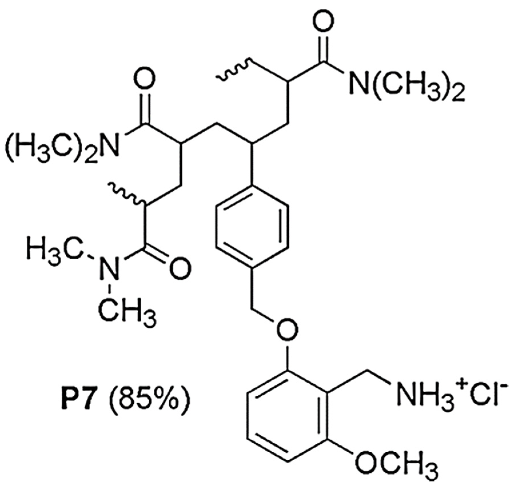
2.1. Synthesis and Spectrophotometric Characterization of 2-Methoxy-6-[(4-Vinyl)Benzyloxy]Benzylammonium Hydrochloride M7 (7)

M7 was prepared by performing the synthetic procedure reported and extensively discussed in a recent study [20]. The experimental details are also included in Section S1 of the Supplementary Information (SI) (Scheme S1 and Sections S1.1–S1.7) associated with this article. The synthetic intermediates and the final compound M7 were spectrophotometrically characterized by means of FTIR and NMR analyses, as shown in the SI, which confirmed their structures, according to what has been reported in the literature [20]. In addition, other analysis, including HPLC, GC-MS, elemental analysis, as well as melting and boiling point determinations, were carried out when useful or necessary and the results, which are also available in Section S1 (SI), were successfully compared with those that have previously been reported [20].

2.2. Preparation of Copolymer P7 by Radical Copolymerizations in Solution and Spectroscopic characterizations of P7

Copolymer P7 was obtained according to a recently reported and discussed procedure [20], which was also included in Section S2 of SI (Section S2.1, Table S1, and Scheme S2). Particularly, M7 was copolymerized with DMAA in the presence of azobisisobutyronitrile (AIBN) as a radical initiator, in MeOH at 60 °C, for 72 h, achieving a conversion of 85%. P7 was characterized by several techniques, as reported in Section S1 of SI (Figure S1) and Section S2 of SI (Sections S2.2–S2.5, Tables S2–S5, and Figure S2). However, for the sake of clarity, the main physicochemical properties of P7 have been included in Table 1 below.
A detailed discussion of the results reported in Table 1 is available in Alfei et al. (2021) [20].

2.3. Antibacterial Properties

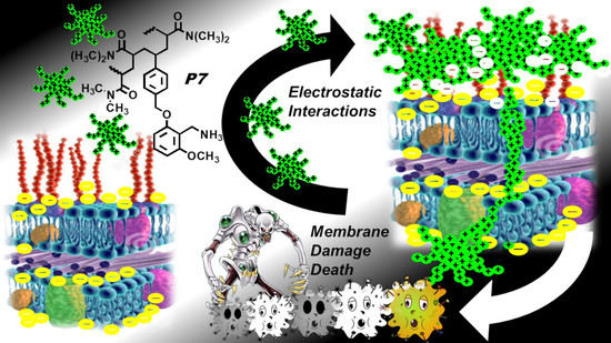
2.3.1. The Reasons for Our Interest in Monomer M7 and Copolymer P7

Having shown remarkable cytotoxicity on tumor cells with membranes resembling those of bacteria in terms of charge [20], and differing from those of eukaryotic cells, towards which cationic materials such as P7 have been reported to be non-cytotoxic [21,25], the P7 copolymer appeared to us as an excellent candidate for developing a new antibacterial tool for several reasons. Due to the structure of M7, used as a cationic monomer, P7 possessed primary benzylammonium hydrochloride residues as cationic moieties, which have been reported to confer interesting antimicrobial properties on several homopolymers and copolymers [11,26,27,28,29]. In addition, the primary amino group, in the form of hydrochloride salt with cationic functionality, was considered preferable to the permanently protonated quaternary ammonium groups because, among the various studies published, the macromolecules containing the quaternary benzylammonium group were less effective than those not containing quaternary ammonium residues, providing reversible cationic groups [12]. Accordingly, we recently reported the broad-spectrum bactericidal effects of a polystyrene-based copolymer containing a primary ammonium group as a cationic function, characterized by a C4 carbon chain as a spacer between the cationic groups and the phenyl rings [13]. Moreover, several copolymers were synthetized containing primary amine groups in the form of acid salts, which proved to possess considerable broad-spectrum antimicrobial properties, often superior to those of the quaternary ammonium analogues [11]. P7 attracted our attention as a probable antibacterial cationic copolymer because, although antibacterial polymers obtained by polymerizing or copolymerizing 4-aminomethylstyrene monomers are well documented, to our knowledge no studies have investigated the use of benzylamine monomers containing the polymerizable styrene moiety as a substituent on the aromatic ring containing the active benzylamine cationic group. In addition, we noted that P7, due to the structure of M7, that contain di-alkyl ether groups in meta-positions respectively, possessed residues like those present in the aromatic ring of vancomycin. Indeed, vancomycin owns di-phenyl ether groups in meta-position each other, and is indicated for the treatment of serious, life-threatening infections by Gram-positive bacteria unresponsive to other antibiotics (MIC < 4 µg/mL) [30]. Moreover, P7 possessed 1,2,3-tri-substituted phenyl residues, having ether groups in positions 2 and 6 and containing the CH3O ether group. In this regard, P7 reproduces, in its structure, that of natural substances made of aromatic rings 1,2,3-tri-substituted with two CH3O groups in positions 2 and 6, such as combretastatin E, which has displayed MIC values lower or comparable to those of ampicillin and chloramphenicol against S. aureus, P. aeruginosa, and E. faecalis [31]. Note that ampicillin is classified by the World Health Organization (WHO) as critically important for human medicine [32], whereas chloramphenicol is on the WHO’s List of essential medicines [33]. Finally, in view of a possible clinical application of P7, its high water-solubility would assure easy routes of administration and high bioavailability.

2.3.2. Antimicrobial Activity of P7 by Determination of MIC and MBC Values

MIC and MBC values for P7 were obtained by analyzing a total of 61 strains of clinical origin, including MDR isolates of both Gram-positive and Gram-negative species and Gram-negative strains with modifications in the outer membrane, therefore having a reduced total negative surface charge.
For comparison, monomer M7 (7) was also analyzed under the same conditions. Although against various Gram-positive and Gram-negative strains M7 showed significantly lower MICs than those of the recently published cationic styrene monomer [13], accordingly to several articles [11,13,27,28,29,34,35] M7 was considered ineffective against all isolates evaluated in this study. On the contrary, the macromolecular compound P7 demonstrated very interesting results both on Gram-positive (Table 2) and Gram-negative species (Table 3), inhibiting the growth of bacteria regardless of their multidrug resistance pattern. In this regard, it has generally been accepted that cationic macromolecules, possessing a non-specific mechanism of action, do not need necessarily to enter the cell and target enzymatic processes to which bacteria have mutated, developing resistance. Therefore, they are also capable of killing MDR isolates rapidly, simply on contact, thus overcoming their acquired ability to resist traditional antibiotics [11,13,22,23,24].
Interestingly, the MIC values of P7 against Gram-positive isolates were very similar to those obtained by the recently published P5 copolymer [13]. However, differently from what was observed when testing its anti-tumor activity on NB cells, which was lower than that of P5 [20], when tested on bacteria P7 was found to be more cytotoxic than P5 [13], showing both MIC and MBC values very much lower than those of P5. The lowest MIC values were observed against the genus Enterococcus, including some vancomycin-resistant strains (VRE) (MICs = 0.6–1.15 µM on E. faecium and 2.3 µM on E. faecalis) and against the sporogenic Bacillus subtilis (MIC = 1.15 µM), which reported double or overlapping MBC values compared to the MICs. Concerning the Staphylococcus genus, in which methicillin-resistant strains (MRS) were also included, the range of MIC values was slightly wider (MICs = 0.6–4.6 µM). P7 proved to be far more potent than three random cationic copolymers (namely PAI1–PAI3) containing alkyl ammonium hydrochloride moieties and aromatic rings like P7, against methicillin-resistant S. aureus (MRSA) (MIC = 14.9 µM (PAI2), 17.7 µM (PAI3) and 267.8 µM (PAI1)) [36]. Against S. epidermidis, P7 was slightly less effective than PAI2, equally active to PAI3 and more potent than PAI1. With regard to bacterial strains with modified membrane charges, which are usually non-susceptible to antibacterial cationic peptides and cationic macromolecules [37,38,39,40,41] and against which the previously reported copolymer P5 was totally ineffective, P7 displayed significant activity, particularly against Yersinia enterocolitica (strain 342) and Providencia stuartii (strain 374) (MIC = 9.3 µM).
Curiously, P7 was slightly less active than P5 against Klebsiella pneumoniae 259 but showed lower MIC values against all Klebsiella (4.6–9.3 µM) and Pseudomonas (2.3–9.3 µM), and against all the strains of Escherichia coli (2.3–4.6 µM), all isolates of Acinetobacter baumannii (2.3–9.3 µM), and Stenotrophomonas maltophylia (2.3–9.3 µM), as well as against the Salmonella gr. B strain (4.6 µM).
The MICs observed for P7 on E. coli (2.3–4.6 µM) were in a narrower and lower range than those of the self-degradable antimicrobial copolymers (P4, P6, P7, and P9) prepared by Mizutani and colleagues (2012) [42], bearing similar cationic groups in the form of primary ammonium salts.
In a recent study by Wen and colleagues, it was reported the synthesis, characterization, and antibacterial properties of four cationic nanosized copolymers (CNPS 1–4), prepared by copolymerizing different amounts of the quaternary cationic monomer N-[2-(methacryloyloxy)ethyl]-N,N-dimethyltetradecane-1-ammonium bromide (MDTP) and styrene as a comonomer [26]. The study showed that the antibacterial activity of CNPS increased with the increase in the content of MDTB in the formulae. Furthermore, regarding the best-performing copolymer, CNPS-4, containing 80% MDTP and with ζ-p of +58 mV, the MIC values reported on E. coli and S. aureus (both ATCC reference strains) were 48.8 and 25.0 µM, respectively.
These results confirmed that the increase in cationic groups in macromolecular formulas, and consequently the increase in the density of positive charges on the surface of polymers, improves the interaction between the cationic (co)polymers and the negatively charged bacteria, leading to serious consequences for bacteria, such as growth inhibition and death [26].
Based on our results, P7 was shown to be 5–10 times more active than CNPS-4 against clinical E. coli isolates and more than 10 times active against S. aureus (MIC (P7) = 4.6 µM vs 50 µM of CNPS-4), although P7 contained a smaller fraction of cationic monomer (32 mol%) and had a lower ζ-p and charge density than CNPS-4. To obtain greater antibacterial activity, the winning strategy of adopting primary amino groups in the form of hydrochloride salts, instead of the widely used quaternary ammonium group, therefore appeared to be confirmed.
In addition, when compared to another of the copolymers published by Wen (i.e., CNPS-3), which possesses the same surface electrical charge as that of P7 (ζ-p = + 50 mV) P7 was found to be 44–88 times more active against E. coli and 35 times more active on S. aureus.
Regarding Gram-positive species, P7 was much more active against MRSA isolates (4.6 µM vs > 7.4 µM) and up to 12 times more active against E. faecium than the ACP1Gly molecule, which belongs to a family of cationic amino-acid-modified polymers recently prepared by Barman et al. (2019) [43].
As for its antibacterial effects against B. subtilis, S. aureus, and E. faecium, P7 proved to be more potent than the homopolymer (Poly1), containing benzylamine residues, reported by Gelman et al. (2004) [12]. P7 displayed MIC values (µM) that were 4.3 times lower against B. subtilis, 4.3–8.3 times lower against E. faecium, and values that were two times lower against S. aureus. Against E. coli, P7 displayed MIC values (µM) that were 2–4 times lower than those of Poly1. Like the cationic antibacterial copolymer, we recently reported [13], P7 was ineffective against a colistin-resistant strain of P. aeruginosa, due to a reduced possibility of electrostatic interactions with the outer membrane of the isolate, caused by an adaptive modification of lipid A [13,37]. Additionally, P7 was found to be more active than a derivative of the potent natural cationic peptide magainin II, known as Ala8,13,18-magainin 2 amide, and which has been reported to exhibit potent antimicrobial activity.
Regarding the Enterobacteriaceae family, although P7 was less active than the cationic polymer ACP1Gly [43] against E. coli, it was more active against some isolates of KPC-producing K. pneumonia. Furthermore, Weiyang et al. (2018) [44] reported the remarkable antibacterial activity of two types of polyionenes, namely 2a (Mn = 5362) and 2b (Mn = 5281), against 20 clinical strains of K. pneumoniae which are responsible for lung infections, and also associated with faster killing kinetics than imipenem and other commonly used antibiotics. Although the MICs displayed by the two polymers were in the range of 1.5–6.0 µM(2a) and in the range of 6.0–48.5 µM (2b) on K. pneumoniae, P7, which displayed MICs values in the range of 4.6–9.3, proved to be less active than 2a, but far more active than 2b, with an activity that was independent of the diversity between the strains. In the same study, the MIC values of 2a and 2b were also determined against one strain of E. coli (MICs = 3.0 µM (2a) and 24.2 µM (2b)), one MRSA isolate (1.5 µM (2a) and 24.2 µM (2b)) and on representative strains of non-fermenting Gram-negative bacterial species, such as A. baumannii (6.0 µM (2a) and 24.2 µM (2b)) and P. aeruginosa (6.0 µM (2a) and 48.4 µM (2b)). Based on these results, P7 was slightly less active than 2a on E. coli and significantly less active on MRSA, but more active against both A. baumannii and P. aeruginosa, also considering the greater number of isolates tested by us. If compared to 2b, P7 was much more powerful against all these species. The scope of this study was to develop a new antibacterial molecule that was more effective than available antibiotics against MDR bacteria. Consequently, in Table 4, a comparison is reported between the MIC values observed for P7 on the MDR isolates of the different species tested here and the MIC values obtained by the antibiograms of the same strains against commonly used antibiotics.
The data reported in Table 4 demonstrate that P7 can be considered an efficient antibacterial agent, capable of inhibiting numerous MDR pathogens, towards which many of the current antibiotics are no longer active. Furthermore, it can be observed that, although the MIC values of P7 against different MDR isolates of the same species have very homogeneous values or fall within a narrow range of concentrations, those relating to antibiotics are less homogeneous and denote strain-dependent variability.

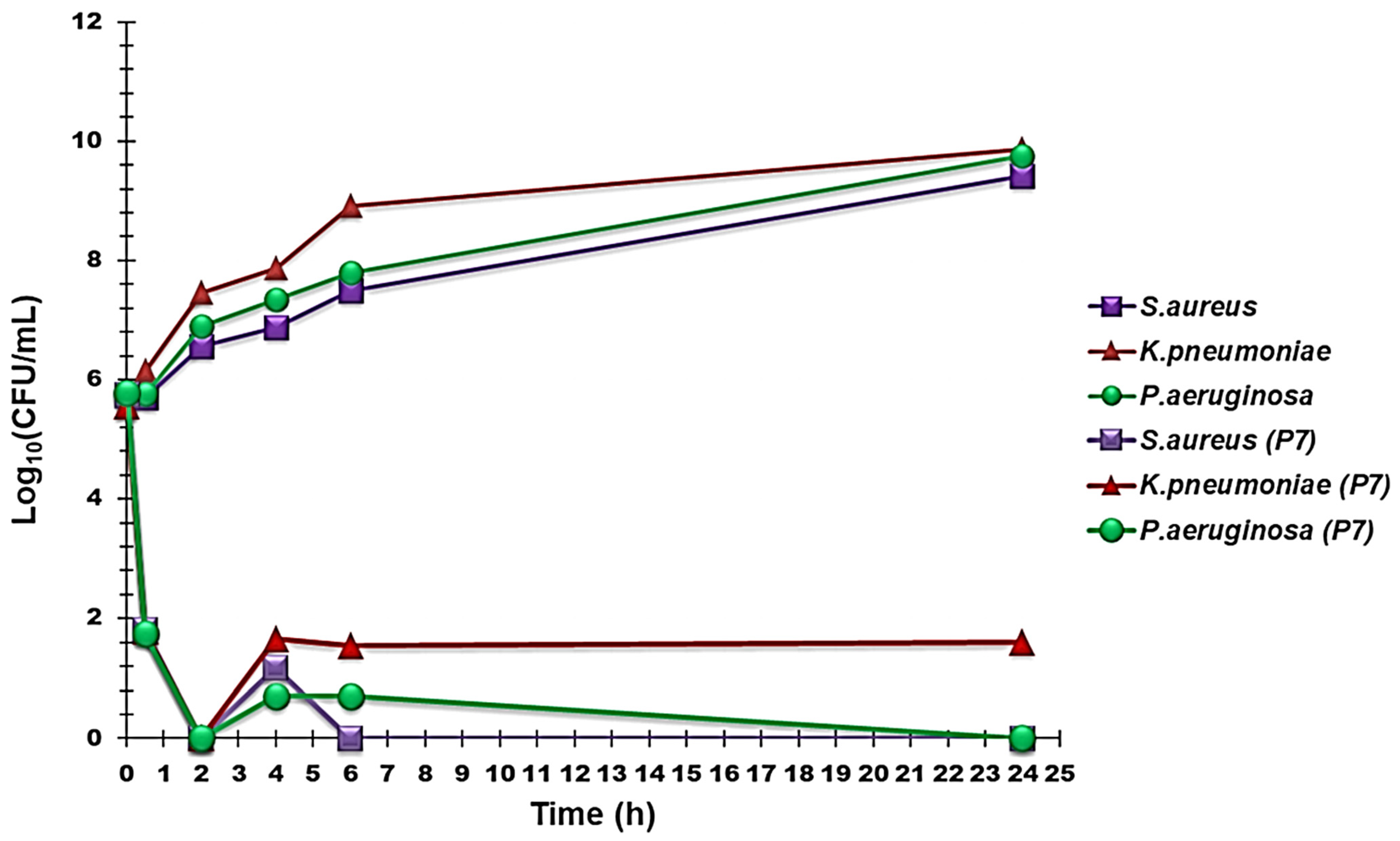
2.3.3. Time–Kill Curves

Time–kill experiments were carried out using P7 at concentrations four times the MIC values on three isolates of P. aeruginosa, two of K. pneumoniae, and three of S. aureus. Figure 3 shows the most representative curves obtained for each species, which established that P7 possessed a very strong bactericidal effect on all the assayed pathogens. Indeed, a rapid decrease of 3.8–4.1 logs in the original cell number was evident already after 30 min of exposure to P7, with P. aeruginosa being the most susceptible species. After two hours of exposure, a total decrease in the original cell number occurred, regardless of the bacterial species tested. During the next two hours, a slightly and not significant regrowth for all the species was observed, particularly for K. pneumoniae. In the subsequent period up to 24 h, the number of K. pneumoniae colonies remained practically unchanged, and no further regrowth was observed; those of S. aureus cleared after 6 h and did not undergo further regrowth, whereas the colonies of P. aeruginosa underwent a progressive decrease until complete elimination after 24 h.
Overall, no significant regrowth was observed after 24 h of incubation with P7 for all strains of the three species tested. Interestingly, this mechanism of action overlapped that of the P5 copolymer that we have recently reported [13], even though it is structurally different (P7 was in fact prepared from a monomer (M5) having styrene group units as polymerizing units and through copolymerization with DMAA). To the best of our knowledge, cases in which the bactericidal behaviors of cationic materials last up to 24 h are rarely reported in the literature. Commonly, for cationic antibacterial molecules, time–kill experiments are often not reported for more than 2, 4, 6, or 8 h [43,44,45]. In fact, for cationic bactericidal peptides, such as colistin [46] and dendrimers [24], for which data have been reported at 24 h, although rapid killing occurred even after just 5 min [46] and 1 h [24] after contact with the antibacterial device, an abundant bacterial regrowth arose after 24 h of action. We conclude that, since both P5 and P7 were obtained by polymerizing structurally very different cationic monomers using the same fraction of the same comonomer (DMAA), the above behavior, which is rarely observed for cationic macromolecules, could be related to the presence of DMAA moieties in the P5 and P7 backbone.

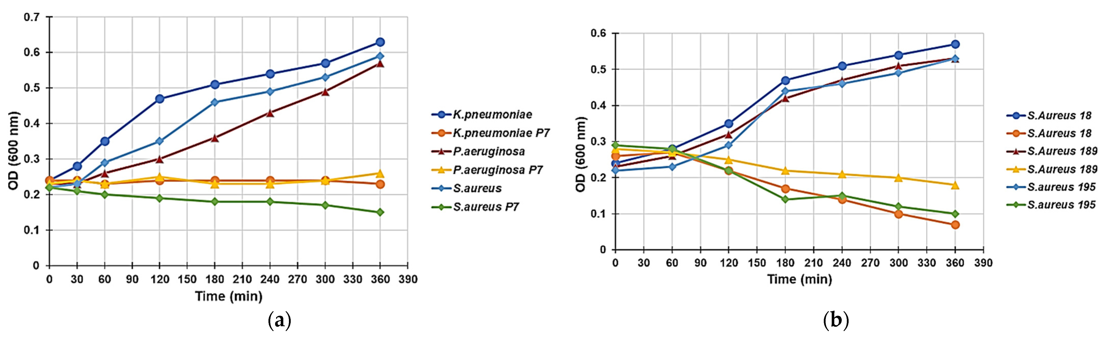
2.3.4. Effect of P5 on the Growth Curve of P. Aeruginosa, K. Pneumoniae, and S. Aureus

The kinetics of growth in MH broth of the selected strains of P. aeruginosa, K. pneumoniae, and S. aureus used in time–kill studies were monitored at 600 nm for 6 h in the absence and presence of P7, which was used at a concentration of 4 × MIC. Figure 4 illustrates the results obtained for one representative strain of P. aeruginosa, K. pneumoniae, and S. aureus.
Although, as expected, the control cultures showed an exponential turbidimetric increase, the presence of P7 resulted in complete inhibition of growth. In a recent study on the antibacterial cationic copolymer P5 [13], the turbidimetric experiments did not show any decrease in optical density for all the isolates examined, thus suggesting a non-lytic bactericidal mechanism, regardless of the different species. On the contrary, in this study, the exposure of Gram-positive S. aureus (strains 18, 189, and 195) to P7 (Figure 4b) highlighted a different trend and a progressive reduction in the OD 600 values during the 6 h of the experiment. Interestingly, the decrease in optical density for the tested isolates showed a strain-dependent intensity.
Since bacterial lysis should have led to an immediate and much faster reduction in absorption at 600 nm, hypothesizing a lytic-type mechanism would be unfounded. Therefore, we speculated that these phenomena could be due to a tendency of the Gram-positive bacteria to form aggregates.
In this regard, Mukherjee and colleagues assessed the effects of exposing E. coli and B. subtilis to cationic antibacterial polymers based on side-chain amino acids. E. coli, when exposed to the cationic homopolymer based on leucine, has undergone a notable change in the morphology of its cells, which have changed during the treatment from a rod shape to a spherical shape. However, B. subtilis cells did not show any evident morphological changes, although in both cases an aggregation of cells was observed [47].
Collectively, the experiments by Mukherjee confirm that the optical density decrement observed by us during the treatment of Gram-positive S. aureus with P7 could be due to the tendency of this species to form aggregates.
Furthermore, it is rational to advance the hypothesis that the cationic P7 could behave like other CAPs and the recently reported P5 copolymer, i.e., causing damage to the bacterial membrane due to its electrostatic interaction with the negatively charged bacterial surface [11,13,22,48]. The inactivity of P7 on the colistin-resistant strain of P. aeruginosa included in our study and its reduced activity against isolates with reduced superficial negative charge, such as Y. enterocolitica (strain 342), P. stuartii (strain 374), M. morganii (strain 372), S. marcescens (strain 228), and P. mirabilis (strain 254) would confirm these hypotheses.
Regarding the non-lytic bactericidal activity of P7 on Gram-negative species, as with that of P5 and other previously reported cationic dendrimers [13,24], this fact could depend on the absence of the N-terminated hydrophobic fatty acid side chain, which is present in colistin and reported to be the main cause of its lytic behavior. However, the absence of a similar fatty acid side chain in P7, as in P5 [13], which enhances their hydrophilic character, could limit their diffusion towards the cytoplasmic membrane (CM) and consequently prevent cell lysis. Therefore, despite being bactericidal (inducing irreparable and lethal alterations of the membrane), they lack lytic properties.

3. Materials and Methods

3.1. Chemicals and Instruments

Monomer M7 and copolymer P7 were prepared according to recently reported procedures [20], which have been described in detail in the SI (Section S1 and Section S2, respectively, including Sections S1.1–S1.7, Scheme S1, Sections S.2.1–S2.5, Scheme S2, Tables S1–S5, and Figure S2. The physicochemical characterization of M7 and P7 was carried out by performing the same analysis previously described and using the same instruments reported in recent studies [13,20] and the details of this process are available in the SI.

3.2. Microbiology

3.2.1. Microorganisms

A total of 61 isolates, belonging to several Gram-positive and Gram-negative species, were used in this study. All were clinical strains from a collection isolated by the clinical laboratories of the University of Genoa (Italy), and identified by VITEK® 2 (Biomerieux, Firenze, Italy) or the matrix-assisted laser desorption/ionization time-of-flight (MALDI-TOF) mass spectrometric technique (Biomerieux, Firenze, Italy). Of the 23 Gram-positive organisms tested, ten strains belonged to the Enterococcus genus, (four Enterococcus faecalis (resistant to vancomycin (VRE)), three E. faecium VRE, one E. casseliflavus (intrinsically resistant to vancomycin), and one E.durans and one E. gallinarum (intrinsecally resistant to vancomycin)); 12 strains pertained to the Staphylococcus genus, including two methicillin-resistant S. auresus (MRSA) and one susceptible, three methicillin-resistant S. epidermidis (MRSE), two of which were also resistant to linezolid, one methicillin-resistant (MR) S. haemolyticus, one S. hominis MR, one S. lugdunensis, one S. sapropyticus, one S. simulans MR, and one S. warneri. A strain of the sporogenic Bacillus subtilis was also added. Regarding the thirty-eight Gram-negative isolates, 18 strains were Enterobacteriaceae: three Escherichia coli (one was susceptibile to all antibiotics tested and one was a O157:H7 strain), one Proteus mirabilis, one Morganella morganii, one Providencia stuartii, one group B Salmonella, one Serratia marcescens, one Yersinia enterocolitica, six group A carbapenemase-producing Klebsiella pneumoniae, two non-carbapenemase-producing K. pneumoniae, and one K. oxytoca. Twenty strains belonged to the non-fermenting group: six Pseudomonas aeruginosa, one P. fluorescens, one P. putida, six Stenotrophomonas maltophylia, five Acinetobacter baumannii, and one A. pittii.

3.2.2. Determination of MIC and MBC

To investigate the antimicrobial activity of M7 and P7 on the 61 pathogens, their minimal inhibitory concentrations (MICs) were determined by following the microdilution procedures detailed by the European Committee on Antimicrobial Susceptibility Testing (EUCAST) [49].
Briefly, after overnight incubation, cultures of bacteria were diluted to yield a standardized inoculum of 1.5 × 10⁸ CFU/mL. Appropriate aliquots of each suspension were added to 96-well microplates containing the same volumes of serial 2-fold dilutions (ranging from 1 to 512 μg/mL) of M5 or P5 to yield a final concentration of approximately 5 × 10⁵ cells/mL. The plates were then incubated at 37 °C. After 24 h of incubation at 37 °C, the lowest concentration of M7 or P7 that prevented a visible growth was recorded as the MIC. All MICs were obtained in triplicate, the degree of concordance in all the experiments was 3/3, and the standard deviation (±SD) was zero. The minimal bactericidal concentration (MBC) has been defined as the lowest concentration of a drug that results in killing 99.9% of the bacteria being tested [50].
The MBCs of M5 and P5 on the 61 pathogens were determined by subculturing the broths used for MIC determination. Ten microliters of the culture broths of the wells corresponding to the MIC and to higher MIC concentrations where plated onto fresh MH agar plates, and further incubated at 37 °C overnight.
The highest dilution that yielded no bacterial growth on the agar plates was taken as the MBC. All tests were performed in triplicate and the results were expressed as the mode.

3.2.3. Time–Kill Curves

Time–kill curve assays for P7 were performed on three representative isolates of P. aeruginosa (strains 247, 256, and 259), two representative strains of K. pneumoniae (strains 366 and 369, both producing class A carbapenemases), and two representative isolates of S. aureus (strains 18 and 195, both MRSA), as previously reported [51]. All were clinical strains from a collection or isolated from volunteers. Experiments were performed over 24 h at P7 concentrations of four times the MIC for all strains.
A mid-logarithmic phase culture was diluted in Mueller–Hinton (MH) broth (Merck, Darmstadt, Germany) (10 mL) containing 4 x MIC of the selected compound in order to give a final inoculum of 1.0 × 10⁵ CFU/mL. The same inoculum was added to cation-supplemented Mueller–Hinton broth (CSMHB) (Merck, Darmstadt, Germany) as a growth control. Tubes were incubated at 37 °C with constant shaking for 24 h. Samples of 0.20 mL from each tube were removed at 0, 30 min, 2, 4, 6, and 24 h, diluted appropriately with a 0.9% sodium chloride solution to avoid carryover of P7 being tested, plated onto MH plates, and incubated for 24 h at 37 °C. Growth controls were run in parallel. The percentage of surviving bacterial cells was determined for each sampling time by comparing colony counts with those of standard dilutions of the growth control. Results have been expressed as log10 values of viable cell numbers (CFU/mL) of surviving bacterial cells over a 24 h period. A bactericidal effect was defined as a 3 log10 decrease of CFU/mL (99.9% killing) of the initial inoculum. All time–kill curve experiments were performed in triplicate.

3.2.4. Evaluation of the Antimicrobial Effect of P7 through Turbidimetric Studies

The study of the antimicrobial activity of P7 was carried out by measuring the optical density variations (OD) as a function of time in cultures of the same strains employed for the time–kill experiments (three strains of P. aeruginosa, two of K. pneumoniae, and two of S. aureus) at a wavelength of 600 nm in a Thermospectronic spectrophotometer (Ultrospec 2100pro, Amersham Biosciences, Little Chalfont, UK) [52].
Bacterial cells were harvested from 10 mL of bacterial cultures in MH broth, and the cell number was adjusted to produce a heavy inoculum (OD adjusted to 0.2) corresponding to 108 cells/mL. Cell suspensions were treated with or without P5 at concentrations equal to 4 MIC and incubated at 37 °C. After 30 min and 1, 2, 3, 4, 5, and 6 h of incubation, aliquots were taken from the cultures, and absorbance values were recorded at 600 nm. Measurements were blanked with MH broth containing an equivalent amount of P7 as that being tested. Experiments were performed in triplicate. The number of CFU was determined in parallel with the method described in the time–kill section and compared with the untreated sample.

4. Conclusions

In this study, it has been established that the hydrophilic cationic random copolymer P7, which has already been shown to possess potent ROS-related cytotoxicity against NB cells, and particularly against those resistant to etoposide, is also an excellent antibacterial compound, with bactericidal activity against various strains of different species belonging to both Gram-positive and Gram-negative bacteria. In time–kill experiments and turbidimetric studies, P7 very quickly killed MDR isolates that were representative of threatening pathogens, such as S. aureus, K. pneumoniae, and P. aeruginosa, with no regrowth observed even after 24 h of exposure. P7 was far more active than the recently reported copolymer P5 and susceptible bacteria were eradicated regardless of their antibiotic resistance at MIC values even lower than 1 µM. If, as reported in previous studies concerning similar cationic materials, P7 proves not to express toxicity for normal mammalian cells [21,25], and if it manifests favorable pharmacokinetic properties, thanks to its remarkable bactericidal action, to its favorable physicochemical characteristics, and its water solubility, this new copolymer could represent an innovative agent capable of successfully treating infections caused by various pathogens that are resistant to antibiotics currently available.

Supplementary Materials

The following are available online at https://www.mdpi.com/article/10.3390/ijms22095021/s1. Section S1. Synthesis of 2-methoxy-6-(4-vinyl benzyloxy)-benzylammonium hydrochloride M7 (7), including Sections S1.1–S1.7; Scheme S1. Synthetic path to prepare M7 (7); Section S2. Preparation and Characterization of copolymer P7 through radical copolymerization in solution, including Sections S.2.1–S2.5. Scheme S2. Reaction scheme of copolymerization of M7 with DMAA to afford P7; Table S1. Experimental data on copolymerization of M7 with DMAA; Figure S1. Significant part of FTIR spectrum of P7 (bands over 3000 cm−1 omitted); Table S2. Calibration data, results of measurements, and estimation of Mn of P7 according to Eq. (2); Table S3. Results of volumetric titration of an exactly weighted amount of P7; Table S4. Data on potentiometric titration of P7 used to construct the titration curve (Figure S2, red line) and computed values of dpH/dV used to construct the relative first derivative curve (Figure S2, purple line). The last three rows report the max values of dpH/dV computed for P7 and the corresponding values of HCL 0.1 N volumes and of pH; Figure S2. Titration curve of P7 (red line); first derivative lines of the titration curve (purple line).; Table S5. Hydrodynamic size (nm), Z-potential, and PDI at 37 °C of P7, determined by DLS analysis (degree of freedom N = 3); References.

Author Contributions

Conceptualization, S.A. and A.M.S.; methodology, software, validation, formal analysis, investigation, resources, data curation, writing—original draft preparation, visualization, supervision, and project administration, S.A. (for part of the synthesis and characterization of all chemicals present in the study), A.M.S., G.P. and D.C. (for all the microbiology). G.Z. provided the discussion on the DLS analyses. Writing—review and editing, S.A. and A.M.S. All authors have read and agreed to the published version of the manuscript.

Funding

This research received no external funding.

Institutional Review Board Statement

Not applicable.

Informed Consent Statement

Not applicable, because in this study humans were not involved. We have performed only in vitro essay by using bacterial cells from a collection obtained by University of Genoa (Italy).

Data Availability Statement

All data concerning this study are contained in the present manuscript or in previous articles whose references have been provided.

Acknowledgments

The authors thank Gian Carlo Schito, who contributed to the phase of reviewing and editing.

Conflicts of Interest

The authors declare no conflict of interest.

References

  1. My Personal Trailer. Available online: https://www.my-personaltrainer.it/farmaci/citrosil.html (accessed on 15 April 2021).
  2. Lei, Y.; Zhou, S.; Dong, C.; Zhang, A.; Lin, Y. PDMS tri-block copolymers bearing quaternary ammonium salts for epidermal antimicrobial agents: Synthesis, surface adsorption and non-skin-penetration. React. Funct. Polym. 2018, 124, 20–28. [Google Scholar] [CrossRef]
  3. Jiao, Y.; Niu, L.; Ma, S.; Li, J.; Tay, F.R.; Chen, J. Quaternary ammonium-based biomedical materials: State-of-the-art, toxicological aspects and antimicrobial resistance. Prog. Polym. Sci. 2017, 71, 53–90. [Google Scholar] [CrossRef]
  4. Zhang, C.; Cui, F.; Zeng, G.; Jiang, M.; Yang, Z.; Yu, Z.; Zhu, M.; Shen, L. Quaternary ammonium compounds (QACs): A review on occurrence, fate and toxicity in the environment. Sci. Total Environ. 2015, 518, 352–362. [Google Scholar] [CrossRef] [PubMed]
  5. Lam, P.L.; Lee, K.K.H.; Wong, R.S.M.; Cheng, G.Y.M.; Bian, Z.X.; Chui, C.H.; Gambari, R. Recent advances on topical antimicrobials for skin and soft tissue infections and their safety concerns. Crit. Rev. Microbiol. 2017, 44, 40–78. [Google Scholar] [CrossRef] [PubMed]
  6. Kenawy, E.-R.; Abdel-Hay, F.I.; El-Magd, A.A.; Mahmoud, Y. Biologically active polymers: VII. Synthesis and antimicrobial activity of some crosslinked copolymers with quaternary ammonium and phosphonium groups. React. Funct. Polym. 2006, 66, 419–429. [Google Scholar] [CrossRef]
  7. Llor, C.; Bjerrum, L. Antimicrobial resistance: Risk associated with antibiotic overuse and initiatives to reduce the problem. Ther. Adv. Drug Saf. 2014, 229–241. [Google Scholar] [CrossRef] [Green Version]
  8. Alavi, A.; Sibbald, R.G.; Ladizinski, B.; Saraiya, A.; Lee, K.C.; Skotnicki-Grant, S.; Maibach, H. Wound-Related Allergic/Irritant Contact Dermatitis (Article). Adv. Ski. Wound Care 2016, 29, 278–286. [Google Scholar] [CrossRef]
  9. Licata, A. Adverse drug reactions and organ damage: The liver. Eur. J. Intern. Med. 2016, 28, 9–16. [Google Scholar] [CrossRef] [PubMed] [Green Version]
  10. Siedenbiedel, F.; Tiller, J.C. Antimicrobial Polymers in Solution and on Surfaces: Overview and Functional Principles. Polymers 2012, 4, 46–71. [Google Scholar] [CrossRef] [Green Version]
  11. Alfei, S.; Schito, A.M. Positively Charged Polymers as Promising Devices against Multidrug Resistant Gram-Negative Bacteria: A Review. Polymers 2020, 12, 1195. [Google Scholar]
  12. Gelman, M.A.; Weisblum, B.; Lynn, D.M.; Gellman, S.H. Biocidal activity of polystyrenes that are cationic by virtue of protonation. Org. Lett. 2004, 6, 557–560. [Google Scholar] [CrossRef]
  13. Alfei, S.; Piatti, G.; Caviglia, D.; Schito, A.M. Synthesis, Characterization and Bactericidal Activity of a 4-Ammoniumbuthylstyrene-Based Random Copolymer. Polymers 2021, 13, 1140. [Google Scholar] [CrossRef] [PubMed]
  14. Palermo, E.; Kuroda, K. Chemical structure of cationic groups in amphiphilic polymethacrylates modulates the antimicrobial and hemolytic activities. Biomacromolecules 2009, 10, 1416–1428. [Google Scholar] [CrossRef] [PubMed]
  15. Ergene, C.; Yasuharab, K.; Palermo, E.F. Biomimetic antimicrobial polymers: Recent advances in molecular design. Polym. Chem. 2018, 9, 2047. [Google Scholar] [CrossRef] [Green Version]
  16. Ganewatta, M.S.; Tang, C. Controlling macromolecular structures towards effective antimicrobial polymers. Polymer 2015, 63, A1–A29. [Google Scholar] [CrossRef]
  17. Gabriel, G.J.; Som, A.; Madkour, A.E.; Eren, T.; Tew, G.N. Infectious disease: Connecting innate immunity to biocidal polymers. Mater. Sci. Eng. R Rep. 2007, 57, 28–64. [Google Scholar] [CrossRef] [Green Version]
  18. Matsuzaki, K. Control of cell selectivity of antimicrobial peptides. Biochim. Et Biophys. Acta 2009, 1687–1692. [Google Scholar] [CrossRef] [PubMed] [Green Version]
  19. Matsuzaki, K.; Sugishita, K.; Fujii, N.; Miyajima, K. Molecular Basis for Membrane Selectivity of an Antimicrobial Peptide, Magainin 2. Biochemistry 1995, 34, 3423–3429. [Google Scholar] [CrossRef]
  20. Alfei, S.; Marengo, B.; Valenti, G.E.; Domenicotti, C. Synthesis of Polystyrene-Based Cationic Nanomaterials with Pro-Oxidant Cytotoxic Activity on Etoposide-Resistant Neuroblastoma Cells. Nanomaterials 2021, 11, 977. [Google Scholar] [CrossRef] [PubMed]
  21. Eliassen, L.T.; Berge, G.; Leknessund, A.; Wikman, M.; Lindin, I.; Løkke, C.; Ponthan, F.; Johnsen, J.I.; Sveinbjørnsson, B.; Kogner, P.; et al. The antimicrobial peptide, Lactoferricin B, is cytotoxic to neuroblastoma cells in vitro and inhibits xenograft growth in vivo. Int. J. Cancer 2006, 119, 493–500. [Google Scholar] [CrossRef] [Green Version]
  22. Alfei, S.; Schito, A.M. From Nanobiotechnology, Positively Charged Biomimetic Dendrimers as Novel Antibacterial Agents: A Review. Nanomaterials 2020, 10, 2022. [Google Scholar] [CrossRef]
  23. Schito, A.M.; Schito, G.C.; Alfei, S. Synthesis and Antibacterial Activity of Cationic Amino Acid-Conjugated Dendrimers Loaded with a Mixture of Two Triterpenoid Acids. Polymers 2021, 13, 521. [Google Scholar] [CrossRef]
  24. Schito, A.M.; Alfei, S. Antibacterial Activity of Non-Cytotoxic, Amino Acid-Modified Polycationic Dendrimers against Pseudomonas aeruginosa and Other Non-Fermenting Gram-Negative Bacteria. Polymers 2020, 12, 1818. [Google Scholar] [CrossRef]
  25. Tan, J.; Tay, J.; Hedrick, J.; Yang, Y.Y. Synthetic macromolecules as therapeutics that overcome resistance in cancer and microbial infection. Biomaterials 2020, 252, 120078. [Google Scholar] [CrossRef] [PubMed]
  26. Wen, Q.; Xu, L.; Xiao, X.; Wang, Z. Preparation, characterization, and antibacterial activity of cationic nanopolystyrenes. J. Appl. Polym. Sci. 2019, 137, 48405. [Google Scholar] [CrossRef]
  27. Ikeda, T.; Tazuke, S.; Suzuki, Y. Biologically active polycations 4: Synthesis and antimicrobial activity of poly(trialkylvinylbenzylammonium chloride)s. Makromol. Chem. 1984, 185, 869–876. [Google Scholar] [CrossRef]
  28. Timofeeva, L.; Kleshcheva, N. Antimicrobial polymers: Mechanism of action, factors of activity, and applications. Appl. Microbiol. Biotechnol. 2011, 89, 475–492. [Google Scholar] [CrossRef]
  29. Kougia, E.; Tselepi, M.; Vasilopoulos, G.; Lainioti, G.C.; Koromilas, N.D.; Druvari, D.; Bokias, G.; Vantarakis, A.; Kallitsis, J.K. Evaluation of Antimicrobial E_ciency of New Polymers Comprised by Covalently Attached and/or Electrostatically Bound Bacteriostatic Species, Based on Quaternary Ammonium Compounds. Molecules 2015, 20, 21313–21327. [Google Scholar] [CrossRef] [PubMed]
  30. Vancomycin. Available online: https://en.wikipedia.org/wiki/Vancomycin (accessed on 15 April 2021).
  31. Eloff, J.N.; Famakin, J.O.; Katerere, D.P.R. Isolation of an antibacterial stilbene from Combretum woodii (Combretaceae) leaves. Afr. J. Biotechnol. 2005, 4, 1167–1171. [Google Scholar]
  32. World Health Organization. Critically Important Antimicrobials for Human Medicine, 6th ed.; World Health Organ: Geneva, Switzerland, 2019; hdl: 10665/312266. [Google Scholar]
  33. World Health Organization. World Health Organization Model List of Essential Medicines: 21st List 2019; World Health Organ: Geneva, Switzerland, 2019; hdl: 10665/325771. [Google Scholar]
  34. Kanazawa, A.; Ikeda, T.; Endo, T. Novel polycationic biocides: Synthesis and antibacterial activity of polymeric phosphonium salts. J. Polym. Sci. A Polym. Chem. 1993, 31, 335–343. [Google Scholar] [CrossRef]
  35. Santos, M.R.E.; Mendonça, P.V.; Almeida, M.C.; Branco, R.; Serra, A.C.; Morais, P.V.; Coelho, J.F.J. Increasing the Antimicrobial Activity of Amphiphilic Cationic Copolymers by the Facile Synthesis of High Molecular Weight Stars by Supplemental Activator and Reducing Agent Atom Transfer Radical Polymerization. Biomacromolecules 2019, 20, 1146–1156. [Google Scholar] [CrossRef]
  36. Locock, K.E.S.; Michl, T.D.; Stevens, N.; Hayball, J.D.; Vasilev, K.; Postma, A.; Griesser, H.J.; Meagher, L.; Haeussler, M. Antimicrobial Polymethacrylates Synthesized as Mimics of Tryptophan-Rich Cationic Peptides. ACS Macro Lett. 2014, 3, 319–323. [Google Scholar] [CrossRef]
  37. Lee, J.-Y.; Park, Y.K.; Chung, E.S.; Na, I.Y.; Ko, K.S. Evolved Resistance to Colistin and Its Loss Due to Genetic Reversion in Pseudomonas Aeruginosa. Sci. Rep. 2016, 6, 25543. [Google Scholar] [CrossRef] [PubMed]
  38. Pacheco, T.; Bustos, R.-H.; González, D.; Garzón, V.; García, J.-C.; Ramírez, D. An Approach to Measuring Colistin Plasma Levels Regarding the Treatment of Multidrug-Resistant Bacterial Infection. Antibiotics 2019, 8, 100. [Google Scholar] [CrossRef] [PubMed] [Green Version]
  39. Liu, H.; Zhu, J.; Hu, Q.; Rao, X. Morganella morganii, a non-negligent opportunistic pathogen. Int. J. Infect. Dis. 2016, 50, 10–17. [Google Scholar] [CrossRef] [PubMed] [Green Version]
  40. Band, V.I.; Weiss, D.S. Mechanisms of Antimicrobial Peptide Resistance in Gram-Negative Bacteria. Antibiotics 2015, 4, 18–41. [Google Scholar] [CrossRef] [PubMed] [Green Version]
  41. Ostrowska, K.; Kamysz, W.; Dawgul, M.; Różalski, A. Synthetic amphibian peptides and short amino-acids derivatives against planktonic cells and mature biofilm of Providencia stuartii clinical strains. Pol. J. Microbiol. 2014, 63, 423–431. [Google Scholar] [CrossRef]
  42. Mizutani, M.; Palermo, E.F.; Thoma, L.M.; Satoh, K.; Kamigaito, M.; Kuroda, K. Design and Synthesis of Self-Degradable Antibacterial Polymers by Simultaneous Chain- and Step-Growth Radical Copolymerization. Biomacromolecules 2012, 13, 1554–1563. [Google Scholar] [CrossRef] [PubMed]
  43. Barman, S.; Konai, M.M.; Samaddar, S.; Haldar, J. Amino Acid Conjugated Polymers: Antibacterial Agents Effective against Drug-Resistant Acinetobacter baumannii with No Detectable Resistance. Acs Appl. Mater. Interfaces 2019, 11, 33559–33572. [Google Scholar] [CrossRef]
  44. Weiyang, L.; Shrinivas, V.; Guansheng, Z.; Bisha, D.; Tan, P.K.J.; Xu, L.; Weimin, F.; Yi, Y.Y. Antimicrobial polymers as therapeutics for treatment of multidrug-resistant Klebsiella pneumoniae lung infection. Acta Biomater. 2018, 78, 78–88. [Google Scholar] [CrossRef]
  45. Sudip, M.; Swagatam, B.; Riya, M.; Jayanta, H. Amphiphilic Cationic Macromolecules Highly Effective Against Multi-Drug Resistant Gram-Positive Bacteria and Fungi With No Detectable Resistance. Front. Bioeng. Biotechnol. 2020, 8, e55. [Google Scholar] [CrossRef]
  46. Landman, D.; Georgescu, C.; Martin, D.A.; Quale, J. Polymyxins revisited. Clin. Microbiol. Rev. 2008, 21, 449–465. [Google Scholar] [CrossRef] [Green Version]
  47. Mukherjee, I.; Ghosh, A.; Bhadury, P.; De, P. Side-Chain Amino Acid-Based Cationic Antibacterial Polymers: Investigating the Morphological Switching of a Polymer-Treated Bacterial Cell. ACS Omega 2017, 2, 1633–1644. [Google Scholar] [CrossRef] [PubMed]
  48. Gough, N.R. Stressing bacteria to death. Sci. Signal. 2011, 4, e164. [Google Scholar] [CrossRef]
  49. EUCAST. European Committee on Antimicrobial Susceptibility Testing. Available online: https://www.eucast.org/ast_of_bacteria/ (accessed on 15 April 2021).
  50. Pearson, R.D.; Steigbigel, R.T.; Davis, H.T.; Chapman, S.W. Method for reliable determination of minimal lethal antibiotic concentrations. Antimicrob. Agents Chemother. 1980, 18, 699–708. [Google Scholar] [CrossRef] [PubMed] [Green Version]
  51. Schito, A.M.; Piatti, G.; Stauder, M.; Bisio, A.; Giacomelli, E.; Romussi, G.; Pruzzo, C. Effects of demethylfruticuline A and fruticuline A from Salvia corrugata Vahl. on biofilm production in vitro by multiresistant strains of Staphylococcus aureus, Staphylococcus epidermidis and Enterococcus Faecalis. Int. J. Antimicrob. Agents 2011, 37, 129–134. [Google Scholar] [CrossRef] [Green Version]
  52. Dalgaard, P.; Ross, T.; Kamperman, L.; Neumeyer, K.; McMeekin, T.A. Estimation of bacterial growth rates from turbidimetric and viable count data. Int. J. Food Microbiol. 1994, 23, 391–404. [Google Scholar] [CrossRef]
Figure 1. Structure of copolymer P5.
Ijms 22 05021 g001
Figure 2. Structure of copolymer P7.
Ijms 22 05021 g002
Figure 3. Time–kill curves performed with P7 (at concentrations equal to 4 × MIC) on P. aeruginosa 247, K. pneumoniae 366, and S. aureus 18.
Ijms 22 05021 g003
Figure 4. Effect of P7 (at 4 × MIC) on the growth curve of K. pneumoniae (strain 466), S. aureus (strain 18), and P. aeruginosa followed at 600 nm for a period of 6 h (a); effect of P7 (at 4 × MIC) on the growth curve of all S. aureus strains used in the experiments (strains 18, 189, and 195) followed at 600 nm for a period of 6 h (b).
Ijms 22 05021 g004
Table 1. Main physicochemical features of P7 [20].
AnalysisFeatureDeterminations
FTIRNH3+3500 cm−1
C-H alkyl2800–2900 cm−1
Overtones *2000–1700 cm−1
C=ONH1649 cm−1
-C=C- *1575, 1510 cm−1
o-disubstituted §754 cm−1
VPO 1 analysis (MeOH, 45 °C)Mn13719
Volumetric TitrationµequivNH2/gP7305
DLS 2 AnalysisZ-Ave 3 (nm)220 ± 18
PDI 40.809 ± 0.004
Z-potential 5 (ζ-p)+49.8 ± 5.8
Potentiometric TitrationMax dpH/dV 610.754
HCl 0.1N (mL) 70.61.2
pH 86.854.80
1 Vapor-pressure osmometry; 2 dynamic light scattering; 3 hydrodynamic diameters of P7 particles; 4 polydispersity index; 5 measure of the electrical charge of P7 particles suspended in the liquid of acquisition (water); 6 max values of first derivative curve of titration curve, indicating the existence of a two-step protonation process; 7 volumes of HCl 0.1N needed to protonate P7; 8 pH values at which protonations occur; * refers to phenyl rings; § refers to phenyl rings derived from styrene.
Table 2. MIC and MBC values of P7 and of the monomer M7 against bacteria of Gram-positive species, obtained from experiments carried out in triplicate 1, expressed as µM and as µg/mL.
P7 (13719) 2M7 (274) 2
StrainsMIC
µM (µg/mL)
MBC
µM (µg/mL)
MIC
µM (µg/mL)
MBC
µM (µg/mL)
Enterococcus Genus
E. faecalis 1 *2.3 (32)2.3 (32)233.6 (64)467.1 (128)
E. faecalis 18 *2.3 (32)2.3 (32)233.6 (64)467.1 (128)
E. faecalis 51 *2.3 (32)4.6 (64)233.6 (64)467.1 (128)
E. faecalis 365 *2.3 (32)4.6 (64)467.1 (128)467.1 (128)
E. faecium 325 *1.15 (16)2.3 (32)116.8 (32)233.6 (64)
E. faecium 341 *1.15 (16)2.3 (32)233.6 (64)467.1 (128)
E. faecium 364 *0.6 (8)1.15 (16)233.6 (64)467.1 (128)
Minor Strains
E. casseliflavus 184 °1.15 (16)2.3 (32)233.6 (64)467.1 (128)
E. durans 103 °1.15 (16)2.3 (32)467.1 (128)934.3 (256)
E. gallinarum 150 *1.15 (16)2.3 (32)467.1 (128)467.1 (128)
Staphylococcus Genus
S. aureus 18 **4.6 (64)4.6 (64)233.6 (64)467.1 (128)
S. aureus 195 **4.6 (64)4.6 (64)233.6 (64)467.1 (128)
S. aureus 1894.6 (64)4.6 (64)233.6 (64)467.1 (128)
S. epidermidis 22 **1.15 (16)2.3 (32)233.6 (64)467.1 (128)
S. epidermidis 180 ***1.15 (16)2.3 (32)116.8 (32)233.6 (64)
S. epidermidis 181 ***1.15 (16)2.3 (32)233.6 (64)467.1 (128)
S. haemolyticus 193 **3.15 (16)3.15 (16)233.6 (64)233.6 (64)
S. hominis 125 **0.6 (8)0.6 (8)116.8 (32)116.8 (32)
S. lugdunensis 1291.15 (16)1.15 (16)116.8 (32)233.6 (64)
S. warneri 741.15 (16)1.15 (16)233.6 (64)233.6 (64)
S. saprophyticus 411.15 (16)2.3 (32)116.8 (32)233.6 (64)
S. simulans 163 **1.15 (16)1.15 (16)467.1 (128)467.1 (128)
Sporogenic Isolate
B. subtilis1.15 (16)1.15 (16)233.6 (64)467.1 (128)
1 the degree of concordance was 3/3 in all the experiments, and standard deviation (±SD) was zero; 2 Mn of copolymer P7 and MW of monomer M7; * denotes vancomycin resistance (VRE); ° denotes intrinsic resistance to vancomycin; ** denotes methicillin resistance; *** denotes resistance toward methicillin and linezolid.
Table 3. MIC and MBC values of P7 and of the monomer M7 against bacteria of Gram-negative species, obtained from experiments carried out in triplicate 1, expressed as µM and as µg/mL.
P7 (13719) 2M7 (274) 2
StrainsMIC
µM (µg/mL)
MBC
µM (µg/mL)
MIC
µM (µg/mL)
MBC
µM (µg/mL)
Enterobacteriaceae Family
E. coli 224 S2.3 (32)2.3 (32)467.1 (128)467.1 (128)
E. coli 238 #4.6 (64)4.6 (64)934.3 (256)934.3 (256)
E. coli 246 §2.3 (32)2.3 (32)467.1 (128)467.1 (128)
Y. enterocolitica 3429.3 (128)18.6 (256)467.1 (128)934.3 (256)
S. marcescens 228>18.6 (>256)nt 31868.6 (512)1868.6 (512)
P. mirabilis 25418.6 (256)18.6 (256)1868.6 (512)1868.6 (512)
M. morganii 37218.6 (256)>18.6 (>256)1868.6 (512)1868.6 (512)
K. oxytoca 2529.3 (128)9.3 (128)467.1 (128)467.1 (128)
K. pneumoniae 231 #4.6 (64)4.6 (64)467.1 (128)467.1 (128)
K. pneumoniae 232 #9.3 (128)18.6 (256)467.1 (128)467.1 (128)
K. pneumoniae 233 #4.6 (64)4.6 (64)467.1 (128)467.1 (128)
K. pneumoniae 260 #9.3 (128)9.3 (128)467.1 (128)467.1 (128)
K. pneumoniae 366 #4.6 (64)4.6 (64)467.1 (128)467.1 (128)
K. pneumoniae 367 #9.3 (128)9.3 (128)467.1 (128)467.1 (128)
K. pneumoniae 3699.3 (128)18.6 (256)467.1 (128)467.1 (128)
K. pneumoniae 377 S4.6 (64)4.6 (64)467.1 (128)467.1 (128)
Salmonella gr. B 2274.6 (64)4.6 (64)467.1 (128)467.1 (128)
P. stuartii 3749.3 (128)9.3 (128)1868.6 (512)1868.6 (512)
Non-Fermenting Species
A. baumannii 2572.3 (32)4.6 (64)467.1 (128)934.3 (256)
A. baumannii 2792.3 (32)2.3 (32)467.1 (128)467.1 (128)
A. baumannii 2369.3 (128)9.3 (128)233.6 (64)467.1 (128)
A. baumannii 2459.3 (128)4.6 (64)467.1 (128)934.3 (256)
A. baumannii 3834.6 (64)4.6 (64)467.1 (128)467.1 (128)
A. pittii 2722.3 (32)2.3 (32)233.6 (64)467.1 (128)
P. aeruginosa 2294.6 (64)4.6 (64)>1868.6 (>512)nt 3
P. aeruginosa 2474.6 (64)9.3 (128)>1868.6 (>512)nt 3
P. aeruginosa 2564.6 (64)4.6 (64)>1868.6 (>512)nt 3
P. aeruginosa 2599.3 (128)18.6 (256)>1868.6 (>512)nt 3
P. aeruginosa 2684.6 (64)4.6 (64)>1868.6 (>512)nt 3
P. aeruginosa 2692.3 (32)2.3 (32)>1868.6 (>512)nt 3
P. fluorescens 2782.3 (32)2.3 (32)934.3 (256)934.3 (256)
P. putida 2624.6 (64)4.6 (64)934.3 (256)934.3 (256)
S. maltophylia 2559.3 (128)18.6 (256)467.1 (128)934.3 (256)
S. maltophylia 2804.6 (64)9.3 (128)467.1 (128)934.3 (256)
S. maltophylia 22.3 (32)2.3 (32)467.1 (128)467.1 (128)
S. maltophylia 34.6 (64)9.3 (128)467.1 (128)467.1 (128)
S. maltophylia 42.3 (32)4.6 (64)233.6 (64)233.6 (64)
S. maltophylia 59.3 (128)18.6 (256)467.1 (128)467.1 (128)
1 the degree of concordance was 3/3 in all the experiments, and standard deviation (±SD) was zero; 2 Mn of copolymer P7 and MW of monomer M7; 3 means not tested; S denotes susceptibility to all antibiotics; # denotes carbapenemase (KPC)-producing. § denotes O157:H7; P. aeruginosa, S. maltophylia and A. baumannii are all MDR bacteria.
Table 4. MIC values of P7 against the most relevant MDR isolates tested in this study compared to the MIC values of antibiotics commonly used against the same species. MIC values were obtained from experiments carried out in triplicate1 and are expressed as µM concentrations.
P7 (13719) 2Commercial Antibiotics
StrainsMIC (µM)MIC (µM)
Enterococcus Genus
E. faecalis *2.322.1–88.3 3
E. faecium *1.1588.3–176.6 3
Staphylococcus Genus
S. aureus **4.6 637.7–1275 4
S. epidermidis ***1.15637.7 4
Sporogenic Isolate
B. subtilis1.15212.4 5
Enterobacteriaceae Family
E. coli #4.616.8 6
96.6 7
13.3–26.5 5
K. pneumoniae #4.6–9.34.4–16.8 6
96.6–193.2 7
6.7–13.3 5
Salmonella gr. B4.6 235.5 8
M. morganii18.6212.4 5
Non-Fermenting Species
A. baumannii2.3–9.396.6–193.2 7
P. aeruginosa2.3–9.319.6–156.5 9
S. maltophylia2.3–9.358.8–117.7 8
1 The degree of concordance was 3/3 in all the experiments, and standard deviation (±SD) was zero; 2 Mn of copolymer P7; 3 vancomycin; 4 oxacillin; 5 amoxy-clavulanate; 6 ertapenem; 7 ciprofloxacin; 8 trimethoprim-sulfamethoxazole; 9 piperacillin tazobactam; * denotes vancomycin resistance (VRE); ** denotes methicillin resistance; *** denotes resistance toward methicillin and linezolid; # denotes carbapenemase (KPC)-producing; P. aeruginosa, S. maltophylia, and A. baumannii are all MDR bacteria.
Publisher’s Note: MDPI stays neutral with regard to jurisdictional claims in published maps and institutional affiliations.

