Article

Lb1G04202, an Uncharacterized Protein from Recretohalophyte Limonium bicolor, Is Important in Salt Tolerance

Shandong Provincial Key Laboratory of Plant Stress, College of Life Sciences, Shandong Normal University, Ji’nan 250014, China
*
Authors to whom correspondence should be addressed.
Int. J. Mol. Sci. 2022, 23(10), 5401; https://doi.org/10.3390/ijms23105401
Submission received: 22 April 2022 / Revised: 8 May 2022 / Accepted: 10 May 2022 / Published: 12 May 2022
(This article belongs to the Special Issue Mechanisms of Plant Salt Resistance)

Abstract

:
With global increases in saline soil, it has become increasingly important to decipher salt-tolerance mechanisms and identify strategies to improve salt tolerance in crops. Halophytes complete their life cycles in environments containing ≥200 mM NaCl; these remarkable plants provide a potential source of genes for improving crop salt tolerance. Recretohalophytes such as Limonium bicolor have salt glands that secrete Na+ on their leaf epidermis. Here, we identified Lb1G04202, an uncharacterized gene with no conserved domains, from L. bicolor, which was highly expressed after NaCl treatment. We confirmed its expression in the salt gland by in situ hybridization, and then heterologously expressed Lb1G04202 in Arabidopsis thaliana. The transgenic lines had a higher germination rate, greater cotyledon growth percentage, and longer roots than the wild type (WT) under NaCl treatments (50, 100 and 150 mM). At the seedling stage, the transgenic lines grew better than the WT and had lower Na+ and malonyldialdehyde accumulation, and higher K+ and proline contents. This corresponded with the high expression of the key proline biosynthesis genes AtP5CS1 and AtP5CS2 under NaCl treatment. Isotonic mannitol treatment showed that Lb1G04202 overexpression significantly relieved osmotic stress. Therefore, this novel gene provides a potential target for improving salt tolerance.

1. Introduction

Irrigated soils are becoming increasingly salinized worldwide [1]. According to the Food and Agriculture Organization Harmonized World Soil Database, soil salinization affects one-third of river basins to some extent [2]. In 2021, more than 20% of global irrigated farmland was affected by soil salinization [3]. It is estimated that 12 million hectares of irrigated agricultural land may no longer be usable in 2050 due to soil salinization [4]. Salinization decreases the area of cultivated land, which seriously affects food production and food security [5,6,7]. Therefore, it is extremely urgent to develop, utilize, and improve saline land [8]. However, physical methods to improve saline soil are costly and unsustainable; therefore, it is particularly important to use biological methods to improve saline soil [9].
Most plants are sensitive to salinity, but some plants, called halophytes, grow normally and complete their life cycles in naturally occurring saline soil (≥200 mM NaCl), and there are three different types of halophytes according to the various salt resistance mechanisms [6]. Pseudohalophytes (salt excluders), such as Phragmites australis, prevent salt from entering the root stele with an apoplast barrier in the roots to maintain low levels of salt in the aboveground [6,10]. Euhalophytes, such as Suaeda salsa, are typical halophytes that compartmentalize salt in the central vacuole to avoid damage to other organelles [6,11]. Finally, recretohalophytes excrete salt from unique epidermal structures at organ level such as salt glands (such as Limonium bicolor [12]) or salt bladders (such as Chenopodium quinoa [13]). The salt secretory structures can help to secrete excess Na+ out of the plants to avoid salt damage. These specific structures or regulation mechanisms enable halophytes to grow normally on saline land and complete their life cycle [8].
Generally, halophytes develop common mechanisms that increase salt tolerance, such as increased cellular organic osmolyte contents and ion compartmentation for osmotic adjustment. Under salt stress, plants decompose starch to produce soluble sugar, increase cell fluid concentration and reduce osmotic potential [14]. Halophytes can also increase proline and K+ content (some halophytes can accumulate amounts of Na+ instead of K+) to reduce osmotic potential and prevent plant water loss [15]. In addition to these characteristics, recretohalphytes have evolved specific salt secretory epidermal structures to secrete excess salt out of the plant.
The roles of salt glands in salt resistance are of particular interest due to their presence in the leaf epidermis [16]. Unlike other halophytes, salt glands endow recretohalophytes with particular morphological and physiological mechanisms to resist salt damage [17]. Excess Na+ can be directly secreted from plants via the secretory pores of the salt glands. NaCl crystals form on the leaf surface of L. bicolor [18] and Distichlis spicata [19]. Three hypotheses have been proposed to explain the salt secretion mechanism: the osmotic hypothesis [20], the animal fluid transport similarity hypothesis [21], and vesicle-mediated exocytosis [22,23]. There is increasing evidence that vesicle-mediated transport plays an essential role in salt secretion, in which vesicles collect the excess ions and secrete them out of the cells [24,25,26].
Limonium bicolor could play an important role in improving saline environments by secreting excess salt from glands on stems and leaves [27,28]. It has a potential role for practical use in environment restoration through desalinization in practice. Additionally, it can also be used as a model halophyte to study salt resistance mechanisms related to salt gland development and salt secretion. The salt glands of L. bicolor have four spontaneous blue fluorescent foci, which can be clearly seen under a fluorescence microscope [29]. Moreover, a stable transformation system [29] and a virus-induced gene silencing (VIGS) system [26] are available for functional validation at the molecular level. Therefore, it is possible to identify genes involved in salt gland development and salt resistance in L. bicolor. Using the leaf development transcriptome of L. bicolor, many genes have been proposed to participate in salt resistance, including LbTTG1 (TRANSPARENT TESTA GLABRA1), LbSAD2 (SUPER SENSITIVE TO ABA AND DROUGHT2) and LbTRY (TRIPTYCHON) [30,31,32]. In addition, on the basis of the analysis of transcriptome data, we found many genes highly expressed during the development of salt glands; however, these genes have very low or no sequence identity with published plant genome sequences, such as LbHLH, which promotes salt resistance [33].
At present, most studies on the regulation of plant development and growth report known genes or proteins. For example, TLR1 (TrichomeLess Regulator1) and TLR2 (MYB transcription factor) negatively regulate trichome density and artemisinin levels in Artemisia annua [34]. Strigolactone (SLS) can regulate plant branching and other development processes [35], as can other genes in this species [30,31,32]. However, genes with undefined functions could also play important roles in plant vegetative growth, developmental regulation, and reproductive metabolism [33,36,37,38,39]. For example, in Arabidopsis, the newly identified protein IbSUT4 (sucrose transporters4) from Ipomoea batatas regulates the expression of AtFT (FLOWERING LOCUS T) and promotes growth, prolonging flowering time of plants. The promoter region of IbSUT4 contains an ABRE motif, which induces the expression of ABA signaling pathway genes and inhibits the expression of ABI1 (ABA insensitive1) [38].
By analyzing the leaf developmental transcriptome [40] and NaCl-treated salt secretion transcriptome [18] of L. bicolor, we found that several uncharacterized genes were highly expressed in early leaf development and under high-NaCl conditions. Given that no reference genome has been reported in species with salt glands, these novel genes with unknown functions have been proposed to participate in salt gland development or salt resistance in recretohalophytes. Among these unknown candidate genes, Lb1G04202 showed high expression under NaCl treatment and was chosen for further analysis. Therefore, heterologous expression was carried out in Arabidopsis to investigate the potential role of the novel gene Lb1G04202 from L. bicolor. Given that Lb1G04202 was selected based on salt gland development, it is expected that this gene may play an important role in salt tolerance. The possible mechanism of enhancement in salt tolerance was also discussed, which will be further applied in saline transformation in the future.

2. Results

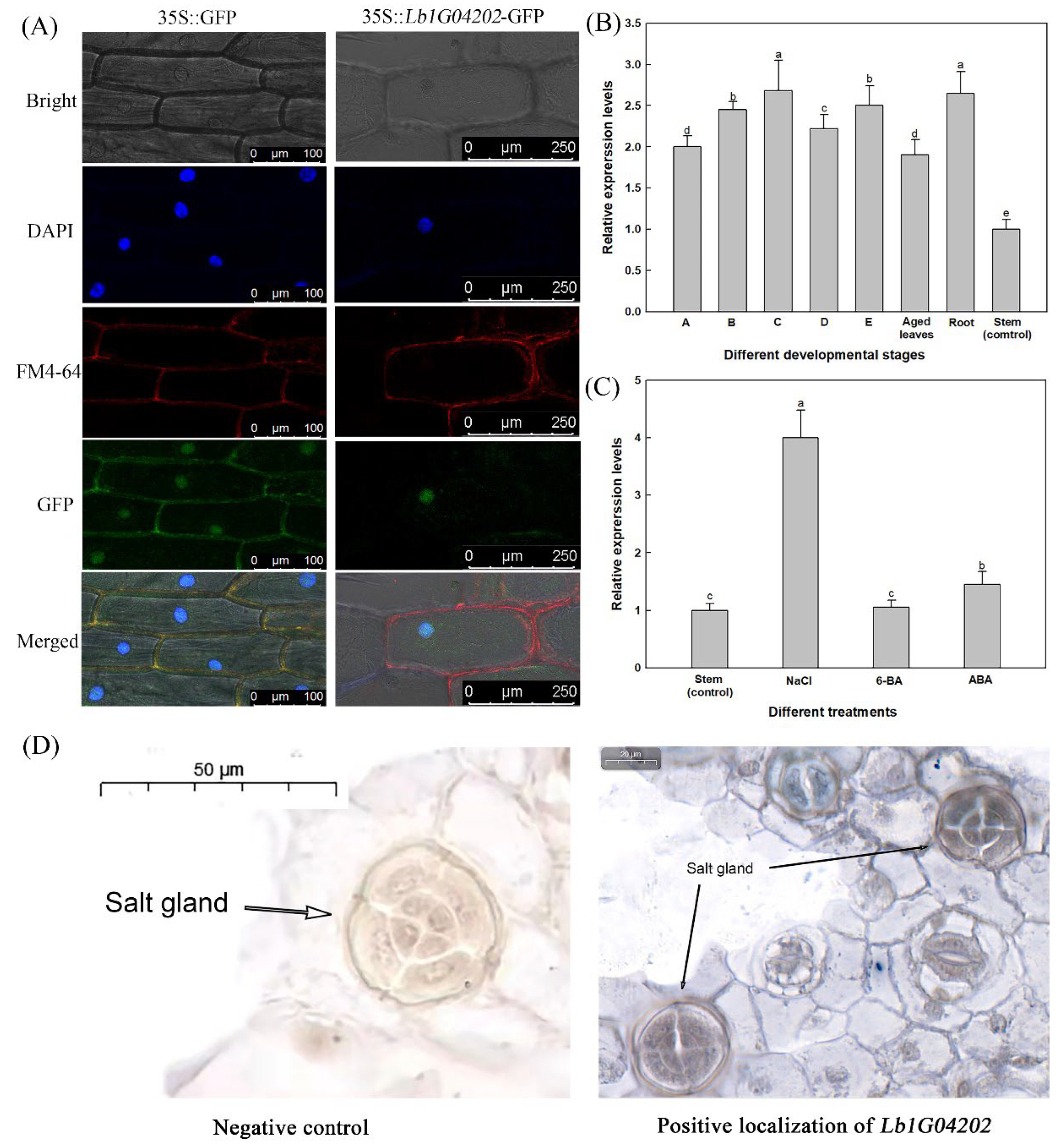
2.1. Lb1G04202 Encodes an Uncharacterized Protein, Is Expressed in Salt Glands and Is Highly Expressed after Salt Treatment

The full-length coding sequence of Lb1G04202 from L. bicolor. Lb1G04202 contains 522 base pairs and encodes 173 amino acids was cloned (Figure 1A). Lb1G04202 has no conserved domain or low-complexity region (Figure 1B). There were no genes from other species with more than 35% similarity with Lb1G04202 (Figure 1C) and the genes with limited similarity were all uncharacterized or hypothetical. This indicates that Lb1G04202 from L. bicolor is a unique and novel gene.
Subcellular localization analysis and in situ hybridization for Lb1G04202 in L. bicolor was conducted (Figure 2). DAPI (stains the nucleus) and FM4-64 (stains the membrane) were applied to determine the subcellular localization of Lb1G04202 after transformation with the p35S::Lb1G04202-GFP expression vector in onion epidermal cells. As shown in Figure 2A, Lb1G04202-GFP was only located in the nucleus, while GFP was located in both the nucleus and plasma membrane.
Furthermore, the expression position of Lb1G04202 was determined using in situ hybridization to confirm its relationship with salt glands. As shown in Figure 2D, the hybridization signal was detected in the salt gland. We next determined the expression pattern of Lb1G04202 in different plant tissue at various developmental stages (Figure 2B) and under different salt treatments (100 mM NaCl, 0.04 mg/L 6-BA and 0.1 mg/L ABA; Figure 2C). Lb1G04202 expression was highest (relative to stem tissue as the control) under 100 mM NaCl treatment (Figure 2C), indicating that Lb1G04202 is related to salt stress.

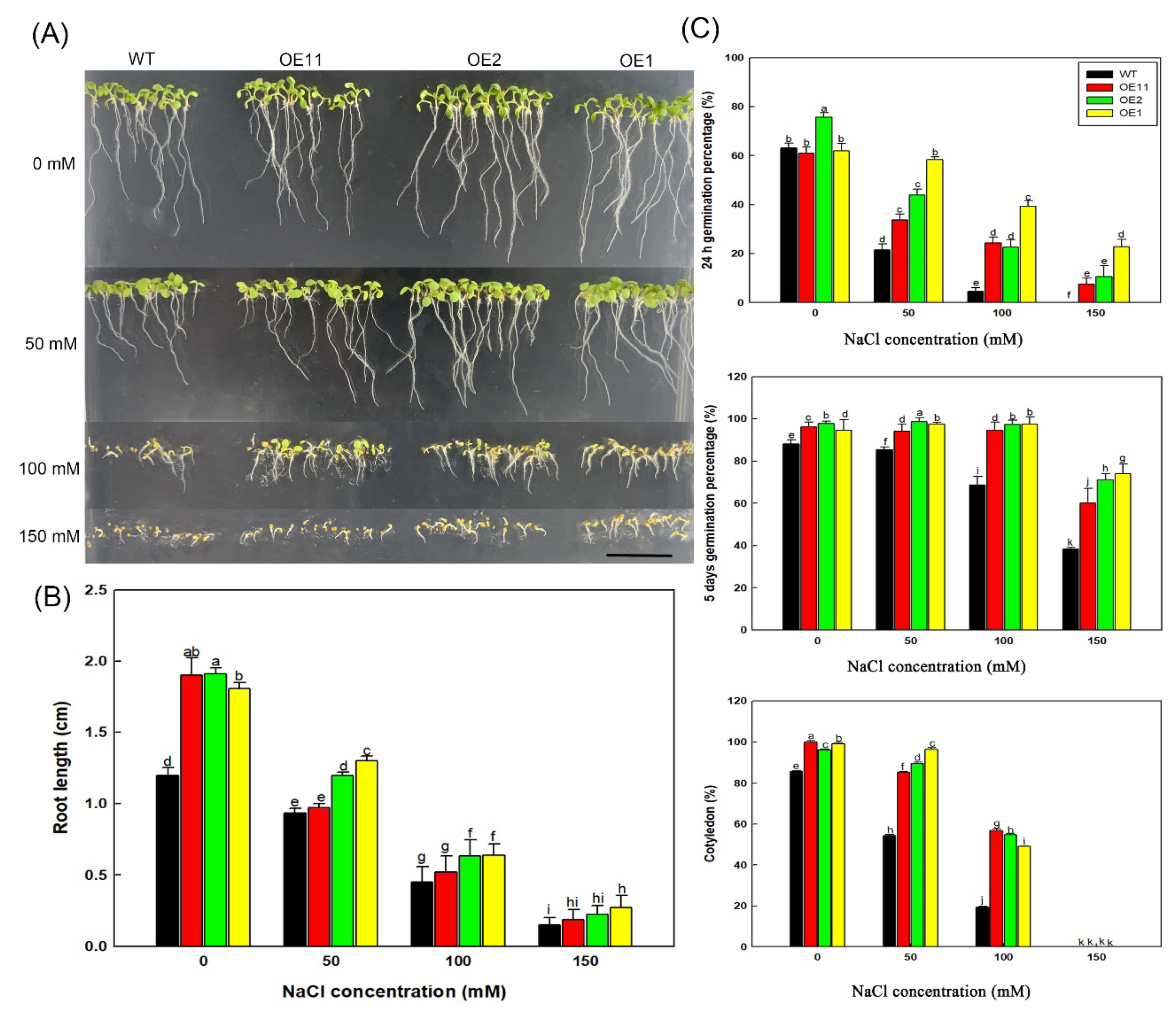
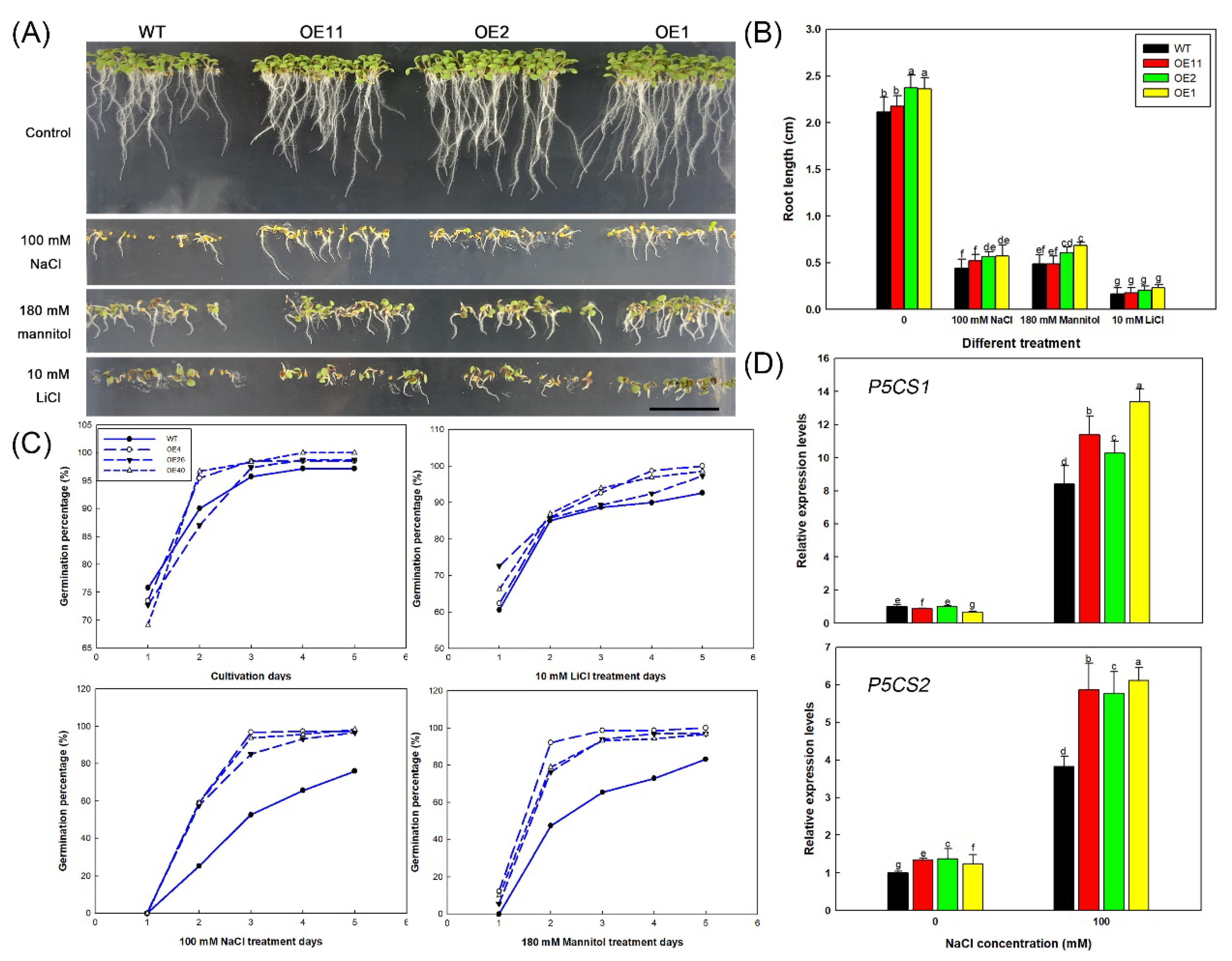
2.2. Heterologous Expression of Lb1G04202 Increases Arabidopsis Salt Resistance during Germination

After transformation of Arabidopsis using the pCAMBIA3301 vector, 14 lines were obtained by Basta selection. Eleven positive Col-35S::Lb1G04202 lines were identified at the DNA level (Figure 3A). After analyzing the expression level of Lb1G04202 in the 11 transgenic lines (Figure 3B), OE1, OE2, and OE11 were selected as high, medium, and low expression level lines of Col-35S::Lb1G04202, respectively, for further experiments.
We investigated the effect of Lb1G04202 on salt tolerance in Arabidopsis grown in three NaCl concentrations (50, 100 and 150 mM) and a control (0 mM). As shown in Figure 4A, the transgenic lines, especially OE1 and OE2, had higher growth under the NaCl treatments than the WT. The three Col-35S::Lb1G04202 lines had a higher germination rate compared with the WT (Figure 4C) at both 24 h and 5 d after sowing under the 50, 100, and 150 mM NaCl treatments, particularly under the 150 mM NaCl treatment. None of the WT seeds had germinated by 24 h under the 150 mM NaCl treatment. There were minimal differences in germination rate among the different lines under the 0, 50, and 100 mM NaCl treatments. However, under the 150 mM NaCl treatment, the germination rates of the three Col-35S::Lb1G04202 lines were significantly (almost two-fold) higher than that of the WT.
The differences among lines in cotyledon growth percentage were more obvious than the differences in germination rate (Figure 4C). In the absence of NaCl, the cotyledon growth rate was similar among the four lines. However, under the 50 and 100 mM NaCl treatments, the cotyledon growth of Col-35S::Lb1G04202 lines was much higher than that of the WT. Under the 150 mM NaCl treatment, none of the four lines had cotyledons. Root growth was inhibited by NaCl. However, under each NaCl treatment, the root length of Col-35S::Lb1G04202 lines was longer than that of the WT (Figure 4B).

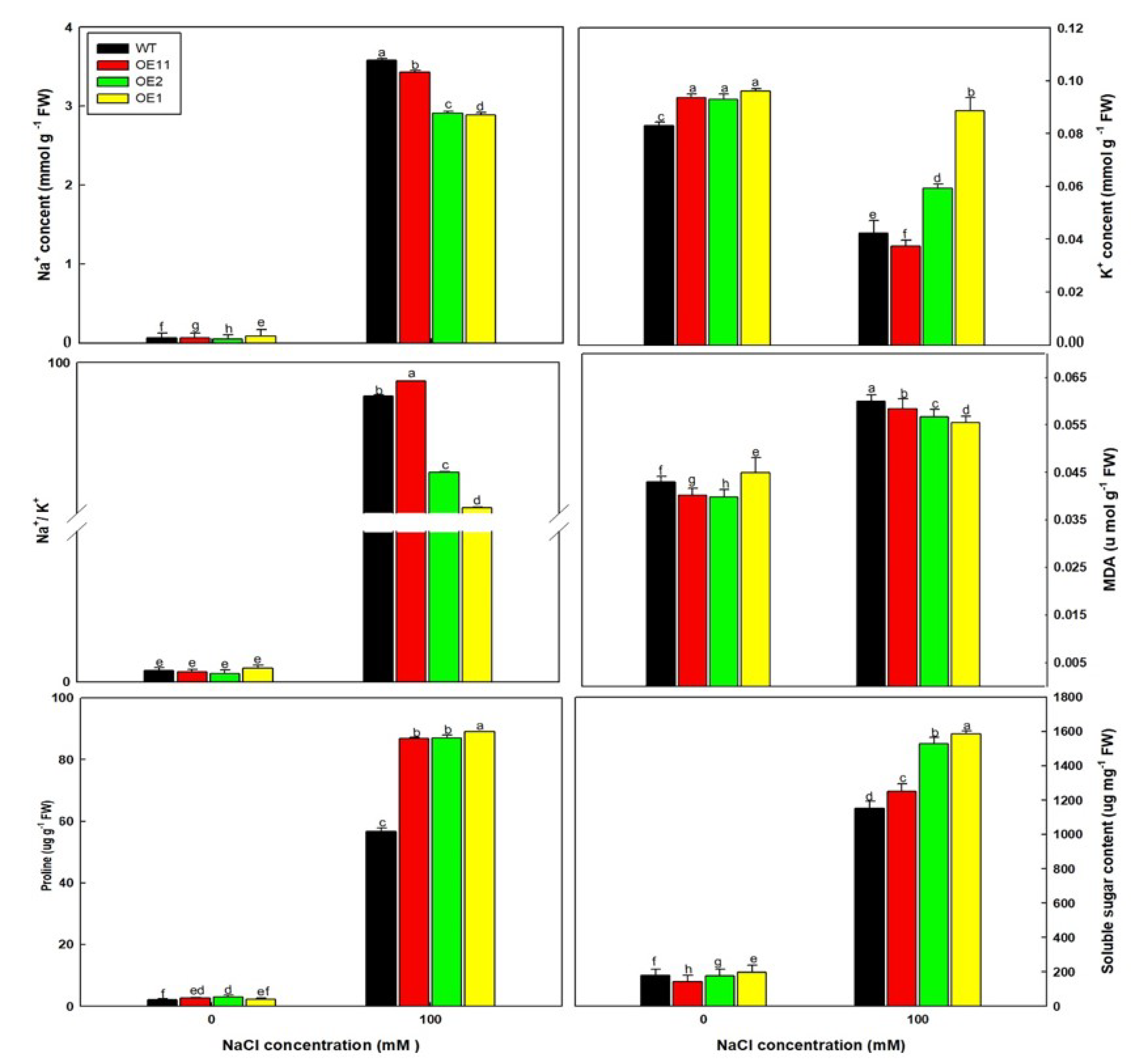
2.3. Lb1G04202 Reduces NaCl Damage at the Seedling Stage

Given that Lb1G04202 can improve the salt tolerance of Arabidopsis during seed germination, its effect on salt tolerance at the seedling stage was then explored. We transplanted seedlings into nutrient soil and, after 10 d, watered the seedlings with Hoagland solution with or without 100 mM NaCl. After one week, the leaves of each line grown under the NaCl treatment yellowed, deformed, and wilted to varying degrees (Figure 5A). The NaCl treatment had the strongest effect on the WT; WT plants were smaller and had more serious yellowing and wilting than Col-35S::Lb1G04202 plants. The biomass data confirmed this trend; transgenic lines had significantly higher fresh and dry weights than the WT under the 100 mM NaCl treatment (Figure 5B).
Subsequently, various physiological properties of plants under the 0 and 100 mM NaCl treatments were measured (Figure 6). Under 100 mM NaCl, OE lines accumulated more K+, proline, and soluble sugar but less Na+ and MDA than WT plants. MDA is the product of membrane lipid peroxidation, and therefore it represents the degree of tissue damage in plants [41]. The trend in MDA contents was consistent with that of Na+. Therefore, these results suggest that Lb1G04202 overexpression can reduce the damage caused by NaCl.

2.4. Lb1G04202 Enhances Salt Tolerance by Alleviating Osmotic Stress

NaCl generates osmotic stress and ionic stress [42]. Therefore, to further explore the mechanism by which Lb1G04202 improves salt tolerance, 180 mM mannitol was used to generate the same osmotic potential as 100 mM NaCl and 10 mM LiCl to generate the same ionic stress as 100 mM NaCl (Figure 7A). Under the isotonic mannitol treatment, the overexpression lines showed the same growth pattern as under the 100 mM NaCl treatment, that is, OE plants had higher germination rates (Figure 7C) and longer roots (Figure 7B) than the WT. Under the 10 mM LiCl treatment, the growth of all lines was severely inhibited, and no growth advantage was detected in the transgenic lines. These results showed that under NaCl treatment, WT plants were subjected to both osmotic and ionic stress, while the transgenic lines of Lb1G04202 showed higher salt tolerance during germination due to higher resistance to osmotic stress.
Next, the expression of osmotic response-related genes in the OE lines under 0 and 100 mM NaCl was determined to explore the mechanisms by which Lb1G04202 improves Arabidopsis salt tolerance. Specifically, we investigated the expression of P5CS1 and P5CS2, which participate in proline synthesis. The expression levels of AtP5CS1 (DELTA1-PYRROLINE-5-CARBOXYLATE SYNTHASE1) and AtP5CS2 were much higher in Col-35S::Lb1G04202 lines than those in WT under 100 mM NaCl (Figure 7D). Expression level patterns were consistent with the osmotic stress resistance level and proline accumulation level of these plants. In summary, Lb1G04202 does enhance genes involved in proline production.

3. Discussion

The uncharacterized gene reported here, Lb1G04202 from recretohalophyte L. bicolor, had no conserved domain and no homologous genes among reported species. Due to its position in the salt gland and high expression under salt conditions, we investigated its salt resistance function by heterologously expressing it in Arabidopsis, where it enhanced the salt tolerance of Arabidopsis at the germination and seedling stages by alleviating osmotic stress.
Salt tolerance is a complex trait controlled by multiple mechanisms at the morphological, physiological and molecular levels [43]. For decades, scientists have been devoted to unraveling potential salt tolerance mechanisms, such as the SOS pathway [44,45] and NHX1 unloading transporter [46]. This research has provided the foundation for in-depth investigations of salt resistance. Halophytes have abundant salt-resistance genes and therefore make ideal research material. In particular, recretohalophytes have attracted the attention of researchers due to their specific salt secretory structures and salt excretion function. Among them, L. bicolor has a distinct genetic background with a transformation system [29] and mutant library [47]. No reference genome of a species with salt glands has been reported; however, many novel genes with unknown functions have been proposed to participate in salt resistance.
Limonium bicolor is a recretohalophyte with typical salt glands. It can grow normally and complete its life cycle under high levels of NaCl [6,8]. Salt glands are typical epidermal structures that secrete salt. Many genes have been proposed to participate in salt resistance and salt gland development [26,32,33] in L. bicolor, such as LbTTG1, LbTRY and LbSAD2 [30,31,32]. However, these genes are all homologs of known functional genes. In other halophytes, functional verification is always carried out in genes with clear domains, for instance, the transformation of Glycerol-3-Phosphate Acyltransferase from Suaeda salsa improves salt tolerance in Arabidopsis [48] and the overexpression of genes related to glycine betaine biosynthesis from Salicornia europaea improves salt-tolerance of tobacco [49]. For L. bicolor, which has salt glands, the unknown functional genes, such as LbHLH, may play a significant role in salt resistance [33]. We believe that the unknown functional genes in L. bicolor may be unique to salt gland development. In fact, uncharacterized functional genes have rarely been studied due to unclear domains [50]. Recent studies have identified many novel genes involved in plant growth and morphological construction, for example, PebHLH35 from Populus euphratica improves drought resistance when overexpressed in Arabidopsis [36] and a WD40-repeat protein from the halophyte C. quinoa is related to the formation of epidermal bladder cells [37]. Therefore, identifying the function of unknown genes expressed in L. bicolor may be important for understanding salt gland development and salt resistance.
In the current transformation of Lb1G04202 in Arabidopsis, the transgenic lines showed enhanced salt tolerance in biomass and germination with increasing exogenous expression, and a typical dose effect could be observed. There may be two main reasons for the salt tolerance. On the one hand, with respect to the physiological mechanism, transgenic lines overexpressing Lb1G04202 accumulated less Na+ than the WT, which may be related to the increased absorption of K+, proline and soluble sugar. This is consistent with previous reports that salt-tolerant lines often have low Na+ content to avoid salt damage and a high accumulation of K+ instead [51,52]. In particular, the high expression level line was the most tolerant to salt, indicating that salt tolerance was proportional to gene expression level.
On the other hand, in general, NaCl results in both osmotic stress and ionic stress [42]. Therefore, to untangle the effect of Lb1G04202 on NaCl tolerance, we simulated osmotic stress and ion stress, separately. Iso-osmotic mannitol was applied to generate similar osmotic stress of NaCl. The transgenic lines showed high resistance to osmotic stress, similar to their response to NaCl treatment. This indicates that expression of Lb1G04202 in Arabidopsis can directly resolve the osmotic stress generated by NaCl. Moreover, the expression of P5CS1 and P5CS2, which encode key rate-limiting enzymes in proline synthesis and thus affect osmoregulation [53], were higher in OE lines that in the WT. This corresponded with the high proline contents in the Col-35S::Lb1G04202 lines. Proline is an important organic osmotic substance [54,55,56] that can reduce osmotic potential and prevent plant water loss, alleviating osmotic stress. While transgenic plants showed tolerance to osmotic stress, they were not tolerant to LiCl (Figure 7). This indicates that the enhanced tolerance of transgenic lines to NaCl stress was due to the enhanced tolerance to osmotic stress and not ion stress.
In our study, Lb1G04202 was localized to the nucleus. Therefore, it may regulate gene expression via direct or indirect binding to promoters of target genes, which will be further investigated in future studies. Moreover, many novel genes have been reported halophytes such as L. bicolor. These genes could be further explored to develop salt-resistant crops and improve saline land.

4. Materials and Methods

4.1. Plant Materials and Growth Conditions

The seeds of wild Limonium bicolor were obtained from a site with saline soil (N37°40′; E118°55′) in the Yellow River Delta (Shandong Province, China). The completely dried seeds were stored at 4 °C for at least six months before use. The dried seeds were sterilized with 70% ethanol for 5 min and shaken with 6% (v/v) NaClO (sigma, City of Saint Louis, MO, USA) for 18 min. Then, the seeds were washed with sterile distilled water 5–7 times and soaked for another 20 min. After disinfection, the seeds were germinated on Murashige and Skoog (MS) (Zkorigin Bioscience Co., Ltd., Beijing, China) basic medium with 0.9% agar in Sterile Petri dish to obtain sterile seedlings. Seedlings were then cultured in the flowerpots (10 cm × 10 cm) with nutrient soil (soil: vermiculite: perlite, 3:1:1) at 28 ± 3 °C/23 ± 3 °C (day/night), 600 µmol/m2/s (15 h photoperiod) photon flux density and 70% relative humidity.
Arabidopsis thaliana ecotype Col-0 (Columbia-0) seeds were disinfected by shaking with 70% ethanol twice (5 min each time) and 95% ethanol three times (4 min each time). After 2 d of vernalization at 4 °C, the seedlings were cultivated at 22 °C/18 °C (day/night) under a 16 h/8 h photoperiod with a photon flux density of 150 µmol/m2/s and 70% relative humidity. The seedlings were cultured on 1/2 MS medium for one week and transplanted into pots (10 cm height × 10 cm diameter) filled with nutrient soil (soil: vermiculite: perlite, 3:1:1). After the inflorescence had grown, plants were infected and transformed using Agrobacterium tumefaciens.

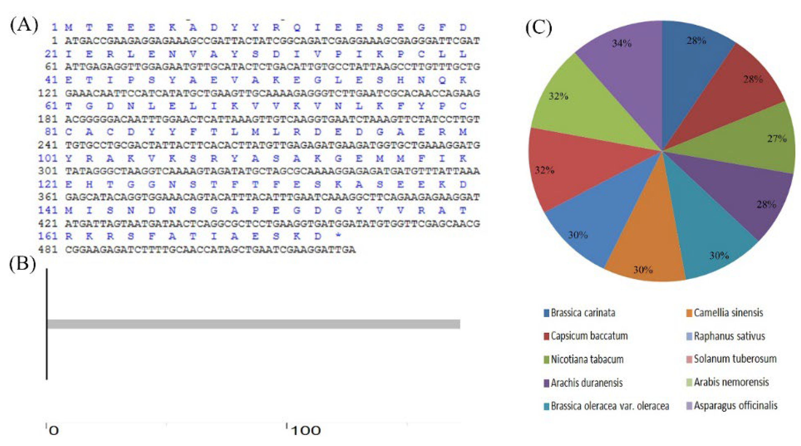
4.2. Cloning and Bioinformatic Analysis of Lb1G04202

To extract the total RNA of L. bicolor, the first true leaves of L. bicolor were collected at the following developmental stages: the undifferentiated stage (stage A; ~5000 leaves), salt gland differentiation stage (stage B; ~4000 leaves), stomata differentiation stage (stage C; ~3000 leaves), epidermis differentiation stage (stage D; ~1000 leaves) and mature stage (stage E; ~1000 leaves) [40]. Total RNA was extracted from leaves using a FastPure Plant Total RNA Isolation kit (RC401-01; Vazyme Biotech Co., Ltd., Nanjing, China). cDNA was reverse transcribed from the RNA with 2× T5 Fast qPCR Mix (SYBR Green I) (Tsingke Biological Technology, Beijing, China) according to the manufacturer’s instructions.
The full-length sequence of Lb1G04202 was cloned with primers Lb1G04202-S and Lb1G04202-A (Table S1), which were designed with Primer Premier 5.0. The cDNA of L. bicolor was used as the template. The online tool EXPASY (http://www.bio-soft.net/sms/index.html, accessed on 8 April 2022) was used to translate the protein encoded by the gene. The structure and conserved domain of Lb1G04202 were predicted using the online tool SMART (http://smart.embl-heidelberg.de/, accessed on 8 April 2022). The coding protein sequences were applied in NCBI-BLAST (https://blast.ncbi.nlm.nih.gov/Blast.cgi, accessed on 8 April 2022) to analyze sequence similarity.

4.3. Localization of Lb1G04202

The circular pCAMBIA1300 plasmid was digested by SalI to obtain a linear pCAMBIA1300 vector. The Lb1G04202 fragment carrying the SalI digestion site was amplified with the primers Lb1G04202-OE1-S and Lb1G04202-OE1-A (Table S1). Homologous recombination was used to connect the Lb1G04202 fragment with the SalI digestion site to the linear pCAMBIA1300 vector to form a recombinant circular expression vector pCAMBIA1300-Lb1G04202. This was done with a ClonExpress II One Step Cloning Kit (Vazyme Biotech Co., Ltd., China) according to the manufacturer’s instructions. After transforming Agrobacterium tumefaciens GV3101, the recombinant expression vector pCAMBIA1300-Lb1G04202 was transformed into onion epidermal cells to explore the subcellular localization of Lb1G04202. After 2 d of cultivation in the light, fluorescent signals of GFP-labeled Lb1G04202 were detected under a TCS S8 MP two-photon laser-scanning confocal microscope (Leica, Heidelberg, Baden-Württemberg, Germany). DAPI was used to locate the nucleus specifically and was observed under excitation at 358 nm. FM4−64 (N-(3-triethylammoniumpropyl)-4-(6-(4-(diethylamino)phenyl) hexatrienyl) pyridinium dibromide)(Invitrogen, Carlsbad, CA, USA) was used to locate the plasma membrane and the signal was observed under excitation at 559 nm.
To determine the expression position of Lb1G04202 in L. bicolor, developing leaves (the first true leaf at 6–8 d after germination) were isolated from L. bicolor for in situ hybridization. Briefly, the leaves were fixed in 4% paraformaldehyde, embedded in paraffin, and dehydrated through an alcohol series. Thin sections (8 µm) of tissue were treated with proteinase K and hybridized in 6 ng/µL hybridization solution at 37 °C overnight. The digoxin-labeled Lb1G04202 probe (5′-DIG-GCCCUAUACAUCCUUUCAGCACCAUCUUCAU-3′, purified by HPLC) appeared blue-violet. The negative control used the sense strand labeled with digoxin.

4.4. Analysis of the Expression Pattern of Lb1G04202 during the Development of L. bicolor and under Different Treatments

Limonium bicolor was cultivated on MS medium containing different additives (100 mM NaCl, 0.04 mg/L 6-BA and 0.1 mg/L abscisic acid) and the seedlings were collected for RNA extraction. In addition, the A–E stage leaves, stems, roots and mature leaves of L. bicolor grown on MS medium were harvested; these materials were used for RNA extraction individually.
Plant total RNA was reverse transcribed to obtain cDNA, and quantitative RT-PCR was performed in a 20-μL reaction system containing 10 μL SYBR qPCR Master Mix (Vazyme Biotech Co., Ltd.), 0.2 µM primers (Table S1), and 300 ng cDNA. The PCR was conducted in a fluorometric thermal cycler (Bio-Rad CFX96 Realtime PCR System) with the following conditions: pre-incubation at 94 °C for 30 s; 40 cycles of denaturation and annealing at 94 °C for 5 s and 60 °C for 30 s; and melting at 95 °C for 15 s, 60 °C for 60 s, and 95 °C for 1 s. Lbtubulin was used as an internal control [30,31,32,33,57]. The Lbtubulin-RT-S, Lbtubulin-RT-A, Lb1G04202-RT-S and Lb1G04202-RT-A primers (Table S1) were designed using Beacon Designer software (version 7.8). The expression level of Lb1G04202 in different tissues was calculated relative to the expression level in the stem (which was set to 1). Three biological replicates (separate experiments) were performed. Relative expression levels were calculated using 2–ΔΔC(T).

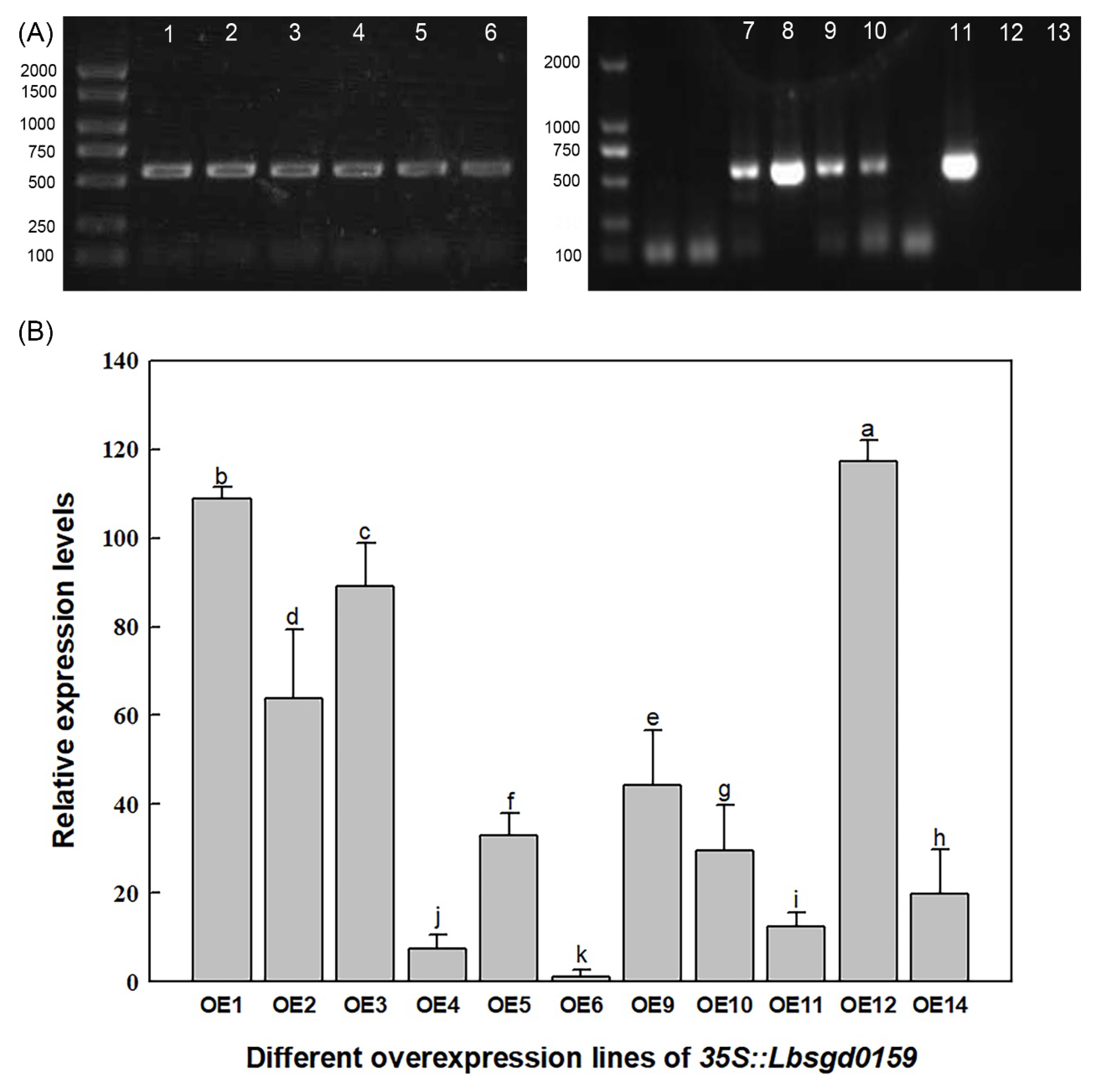
4.5. Heterologous Expression in Arabidopsis Lines

The circular pCAMBIA3301 vector was digested by NcoI to obtain a linear pCAMBIA3301 vector. To connect the Lb1G04202 fragment with the linear pCAMBIA3301 vector, the full-length coding sequence (CDS) of Lb1G04202 carrying NcoI digestion sites was amplified with the primers Lb1G04202-OE2-S and Lb1G04202-OE2-A (Table S1). Plasmid p35S::Lb1G04202 was generated by homologous recombination using a ClonExpress II One Step Cloning Kit (Vazyme Biotech Co., Ltd., Nanjing, China) to express Lb1G04202 under the control of the CaMV 35S promoter. p35S::Lb1G04202 was transformed into Agrobacterium tumefaciens GV3101 cells for heterologous overexpression before transforming the Arabidopsis by floral dip. Harvested Arabidopsis seeds were cultured in nutrient soil. After three generations of screening with Basta (0.1%, v/v), the Arabidopsis seeds homozygous for the p35S::Lb1G04202 transgene were obtained.
The DNA extracted from the leaves of transgenic lines was used as a template, and the primers pCAMBIA3301-S and pCAMBIA3301-A (Table S1) were used to amplify Lb1G04202. Agarose gel electrophoresis was used to identify the positive transgenic plants. Then, mRNA was extracted from different Col-35S::Lb1G04202 lines using a FastPure Plant Total RNA Isolation kit (Vazyme Biotech Co., Ltd.) according to the manufacturer’s instructions. Lb1G04202 expression levels in different Col-35S::Lb1G04202 lines were analyzed by qRT-PCR using the primers Lb1G04202-RT-S and Lb1G04202-RT-A (Table S1). AtACTIN (primers Atactin- RT-S and AtACTIN- RT-A, Table S1) of Arabidopsis was used as the internal control. Since the Lb1G04202 gene of L. bicolor has no homologous gene in Arabidopsis, the line with the lowest Lb1G04202 expression level (OE11) was used as the control (the relative expression level was set to 1) to calculate the expression level of Lb1G04202 in other Col-35S::Lb1G04202 lines. Three biological replicates were performed for each group. Three Col-35S::Lb1G04202 lines were selected for physiological characterization based on Lb1G04202 expression: lines with high (OE1), medium (OE2), and low expression (OE11) level.

4.6. Determination of Salt Tolerance Index during Germination

OE11, OE2, OE1 and wild-type (WT) Arabidopsis seeds were cultured on 1/2 MS medium (1% agar) containing different concentrations of NaCl (0, 50, 100, and 150 mM) to analyze the salt tolerance of Col-35S::Lb1G04202 overexpression lines. We conducted three repeated experiments. Each repeated test contained 40 seeds per line. The number of germinated seeds was counted every day for the first 1–5 d. Specifically, seeds with a radicle >1 mm long that had emerged from the seed coat were considered to be germinated. The emergence of green cotyledons was used as an indicator of cotyledon growth. The cotyledon growth percentage of each line was measured after 3 d of germination. Cotyledon growth percentage (%) = (number of seeds with cotyledons/number of all tested seeds) × 100%. After continuous cultivation for 5 d on different media, the root lengths of different lines were measured using ImageJ software. At least 30 seeds were used for repeated experiments for each index measurement.

4.7. Measurement of Physiological Indexes under Salt Treatment at Seedling Stages

The seedlings (OE11, OE2, OE1, and WT) grown on 1/2 MS basic medium for 5 d were separately transplanted into nutrient soil. After two weeks of adaptation to growth, they were irrigated with different concentrations of NaCl (0, 50, 100, and 150 mM NaCl dissolved in Hoagland solution, pH 6.2). Leaf tissue (0.5 g fresh weight per replicate) was harvested from seedlings under the 0 or 100 mM NaCl condition to measure physiological indicators. The Na+, K+, proline, malonyldialdehyde (MDA), and soluble sugar contents were measured as described previously [30,31,32,33,57]. Ion concentrations were measured with a flame photometer (Cole-Parmer, Chicago, IL, USA). Four replicates per measurement were performed for each line.

4.8. Performance of Transgenic Lines under Single Ion and Osmotic Stress

Four Arabidopsis lines (OE11, OE2, OE1, and WT) were cultured in 1/2 MS medium containing 10 mM LiCl (the same ionic effect as 100 mM NaCl) and 1/2 MS medium containing 180 mM mannitol (the same osmotic pressure as 100 mM NaCl) for 5 d. To determine salt tolerance, the germination percentage (%), cotyledon growth percentage (%) and root length were measured with the same methods as those mentioned above. The growth of seedlings under either ion or osmotic stress was compared among different lines to determine why Lb1G04202 alleviates salt stress and improves salt tolerance.

4.9. Expression Analysis of Osmotic Stress-Related Marker Genes in Transgenic Arabidopsis

Four Arabidopsis lines (OE11, OE2, OE1, and WT) were cultured in 1/2 MS medium containing 0 or 100 mM NaCl for 10 d. The RNA of eight samples was extracted with a FastPure Plant Total RNA Isolation kit (RC401-01; Vazyme Biotech Co., Ltd., Nanjing, China). Products obtained after RNA reverse transcription were used for qRT-PCR to analyze the expression of related marker genes under salt stress, including DELTA1-PYRROLINE-5-CARBOXYLATE SYNTHASE 1 (AtP5CS1) and AtP5CS2 (Table S1). Three biological replicates were performed. AtACTIN was used as the internal control.

4.10. Statistical Analysis

Statistical significance at p = 0.05 (Duncan’s multiple range tests) was determined using SPSS. ANOVA with orthogonal contrasts and mean comparison procedures was used to detect significant differences among treatments.

5. Conclusions

We determined that the uncharacterized Lb1G04202 of L. bicolor is localized in salt glands and expressed in the nucleus. Overexpression of Lb1G04202 in Arabidopsis alleviated osmotic stress by enhancing the expression of AtP5CS1 and AtP5CS2 to produce more proline under salt stress. These results indicate that Lb1G04202 confers salt tolerance by directly affecting the osmotic stress tolerance of plants and could therefore be useful for breeding salt-tolerant crops.

Supplementary Materials

The following supporting information can be downloaded at: https://www.mdpi.com/article/10.3390/ijms23105401/s1.

Author Contributions

F.Y. designed the research; X.W. performed the research; X.W. and F.Y. analyzed the data; X.W. wrote the paper; F.Y. and B.W. revised the paper. All authors have read and agreed to the published version of the manuscript.

Funding

This work was supported by the National Natural Science Research Foundation of China (NSFC, project nos. 32170301, 31770288 and 31600200).

Institutional Review Board Statement

Not applicable.

Informed Consent Statement

Not applicable. All experiments in the manuscript were performed at Shandong Provincial Key Laboratory of Plant Stress and are in compliance with relevant laws in China.

Data Availability Statement

The datasets used and/or analyzed during the current study are available from the corresponding author on reasonable request.

Conflicts of Interest

The authors declare no conflict of interest.

Abbreviations

L. bicolorLimonium bicolor
TTG1TRANSPARENT TESTA GLABRA1
SAD2SUPER SENSITIVE TO ABA AND DROUGHT2
TRYTRIPTYCHON
CPCCAPRICE
AtSOS1SALT OVERLY SENSITIVE 1
AtSOS3SALT OVERLY SENSITIVE 3
AtP5CS1DELTA1-PYRROLINE-5-CARBOXYLATE SYNTHASE 1
AtP5CS2DELTA1-PYRROLINE-5-CARBOXYLATE SYNTHASE 2
Col-0Columbia-0
qPCRquantitative PCR
CDScoding sequence
WTwild-type

References

  1. Hou, D.; Bolan, N.S.; Tsang, D.C.W.; Kirkham, M.B.; O’Connor, D. Sustainable soil use and management: An interdisciplinary and systematic approach. Sci. Total Environ. 2020, 729, 138961. [Google Scholar] [CrossRef] [PubMed]
  2. Perri, S.; Suweis, S.; Holmes, A.; Marpu, P.R.; Entekhabi, D.; Molini, A. River basin salinization as a form of aridity. Proc. Natl. Acad. Sci. USA 2020, 117, 17635–17642. [Google Scholar] [CrossRef] [PubMed]
  3. Singh, A. Soil salinization management for sustainable development: A review. J. Environ. Manag. 2021, 277, 111383. [Google Scholar] [CrossRef] [PubMed]
  4. Litalien, A.; Zeeb, B. Curing the earth: A review of anthropogenic soil salinization and plant-based strategies for sustainable mitigation. Sci. Total Environ. 2020, 698, 134235. [Google Scholar] [CrossRef] [PubMed]
  5. Munns, R.; Tester, M. Mechanisms of salinity tolerance. Annu. Rev. Plant Biol. 2008, 59, 651–681. [Google Scholar] [CrossRef] [PubMed] [Green Version]
  6. Flowers, T.J.; Colmer, T.D. Salinity tolerance in halophytes. New Phytol. 2008, 179, 945–963. [Google Scholar] [CrossRef] [PubMed]
  7. Asano, T.; Hayashi, N.; Kobayashi, M.; Aoki, N.; Miyao, A.; Mitsuhara, I.; Ichikawa, H.; Komatsu, S.; Hirochika, H.; Kikuchi, S.; et al. A rice calcium-dependent protein kinase OsCPK12 oppositely modulates salt-stress tolerance and blast disease resistance. Plant J. 2012, 69, 26–36. [Google Scholar] [CrossRef]
  8. Song, J.; Wang, B. Using euhalophytes to understand salt tolerance and to develop saline agriculture: Suaeda salsa as a promising model. Ann. Bot. 2015, 115, 541–553. [Google Scholar] [CrossRef] [Green Version]
  9. Ma, X.; Zhang, J.J.S.; Review, T. Research Advances in Physiological Ecology Adaptation of Plant Salt-tolerance. Sci. Technol. Rev. 2011, 29, 76–79. [Google Scholar]
  10. Yuan, F.; Guo, J.; Shabala, S.; Wang, B. Reproductive Physiology of Halophytes: Current Standing. Front. Plant Sci. 2018, 9, 1954. [Google Scholar] [CrossRef]
  11. Guo, J.; Du, M.; Lu, C.; Wang, B. NaCl improves reproduction by enhancing starch accumulation in the ovules of the euhalophyte Suaeda salsa. BMC Plant Biol. 2020, 20, 262. [Google Scholar] [CrossRef] [PubMed]
  12. Leng, B.Y.; Yuan, F.; Dong, X.X.; Wang, B.S. Salt gland distribution in Limonium bicolor at the individual level. Earth Environ. Sci. 2018, 113, 012202. [Google Scholar] [CrossRef]
  13. Böhm, J.; Messerer, M.; Müller, H.; Scholz-Starke, J.; Gradogna, A.; Scherzer, S.; Maierhofer, T.; Bazihizina, N.; Zhang, H.; Stigloher, C.J.C.B. Understanding the Molecular Basis of Salt Sequestration in Epidermal Bladder Cells of Chenopodium quinoa. Curr. Biol. 2018, 28, 3075–3085.e7. [Google Scholar] [CrossRef] [PubMed] [Green Version]
  14. Bai, J.; Liu, J.; Na, Z.; Sa, R.; Jiang, L. Technology, Effect of Salt Stress on Antioxidant Enzymes, Soluble Sugar and Yield of Oat. J. Integr. Agric. 2013, 12, 1441–1449. [Google Scholar] [CrossRef]
  15. Luo, D.; Zhou, Q.; Wu, Y.; Chai, X.; Liu, W.; Wang, Y.; Yang, Q.; Wang, Z.; Liu, Z. Full-length transcript sequencing and comparative transcriptomic analysis to evaluate the contribution of osmotic and ionic stress components towards salinity tolerance in the roots of cultivated alfalfa (Medicago sativa L.). BMC Plant Biol. 2019, 19, 32. [Google Scholar] [CrossRef] [PubMed] [Green Version]
  16. Liang, P.; Wang, Z. Effects of different types of halophytes on the concentration of cadmium in coastal saline soil. Acta Ecol. Sin. 2017, 37, 4656–4662. [Google Scholar]
  17. Yuan, F.; Leng, B.Y.; Wang, B.S. Research Progress in Salt Secretion of Salt Glands in Plants. Plant Physiol. J. 2015, 7, 1531–1537. [Google Scholar]
  18. Yuan, F.; Lyu, M.J.; Leng, B.Y.; Zhu, X.G.; Wang, B.S. The transcriptome of NaCl-treated Limonium bicolor leaves reveals the genes controlling salt secretion of salt gland. Plant Mol. Biol. 2016, 91, 241–256. [Google Scholar] [CrossRef]
  19. Semenova, G.A.; Fomina, I.R.; Biel, K.Y. Structural features of the salt glands of the leaf of Distichlis spicata ‘Yensen 4a’ (Poaceae). Protoplasma 2010, 240, 75–82. [Google Scholar] [CrossRef]
  20. Arisz, W.; Camphuis, I.; Heikens, H.; Tooren, V. The secretion of the salt glands of Limonium latifolium Ktze. Acta Bot. Neerl. 1955, 4, 322–338. [Google Scholar] [CrossRef]
  21. Levering, C.A.; Thomson, W.W. The ultrastructure of the salt gland of Spartina foliosa. Planta 1971, 97, 183–196. [Google Scholar] [CrossRef] [PubMed]
  22. Ziegler, H.; Lüttge, U. Die Salzdrüsen von Limonium vulgare. Planta 1967, 70, 193–206. [Google Scholar] [CrossRef] [PubMed]
  23. Jelte, R.; Ingrid, R.; Taede, S. A light and electron-microscopical study on the structure and function of the salt gland of Glaux maritima L. New Phytol. 1977, 79, 665–671. [Google Scholar]
  24. Morris, L.; Yun, K.; Rutter, A.; Zeeb, B.A. Characterization of Excreted Salt from the Recretohalophytes Distichlis spicata and Spartina pectinata. J. Environ. Qual. 2019, 48, 1775–1780. [Google Scholar] [CrossRef]
  25. Feng, Z.; Sun, Q.; Deng, Y. Study on pathway and characteristics of ion secretion of salt glands of Limonium bicolor. Acta Physiol. Plant. 2014, 36, 2729–2741. [Google Scholar] [CrossRef]
  26. Lu, C.; Feng, Z.; Yuan, F. The SNARE protein LbSYP61 participates in salt secretion in Limonium bicolor. Environ. Exp. Bot. 2020, 176, 104076. [Google Scholar] [CrossRef]
  27. Deng, Y.; Feng, Z.; Yuan, F.; Guo, J.; Suo, S.; Wang, B. Identification and functional analysis of the autofluorescent substance in Limonium bicolor salt glands. Plant Physiol. Biochem. 2015, 97, 20–27. [Google Scholar] [CrossRef]
  28. Ding, F.; Chen, M.; Sui, N. Ca2+ significantly enhanced development and salt-secretion rate of salt glands of Limonium bicolor under NaCl treatment. S. Afr. J. Bot. 2010, 76, 95–101. [Google Scholar] [CrossRef] [Green Version]
  29. Yuan, F.; Chen, M.; Yang, J. A system for the transformation and regeneration of the recretohalophyte Limonium bicolor. Vitr. Cell. Dev. Biol.-Plant 2014, 50, 610–617. [Google Scholar] [CrossRef]
  30. Xu, Y.; Jiao, X.; Wang, X.; Zhang, H.; Wang, B.; Yuan, F. Importin-beta From the Recretohalophyte Limonium bicolor Enhances Salt Tolerance in Arabidopsis thaliana by Reducing Root Hair Development and Abscisic Acid Sensitivity. Front. Plant Sci. 2020, 11, 582459. [Google Scholar] [CrossRef]
  31. Yuan, F.; Leng, B.; Zhang, H.; Wang, X.; Han, G.; Wang, B. A WD40-Repeat Protein from the Recretohalophyte Limonium bicolor Enhances Trichome Formation and Salt Tolerance in Arabidopsis. Front. Plant Sci. 2019, 10, 1456. [Google Scholar] [CrossRef]
  32. Leng, B.; Wang, X.; Yuan, F.; Zhang, H.; Lu, C.; Chen, M.; Wang, B. Heterologous expression of the Limonium bicolor MYB transcription factor LbTRY in Arabidopsis thaliana increases salt sensitivity by modifying root hair development and osmotic homeostasis. Plant Sci. 2021, 302, 110704. [Google Scholar] [CrossRef] [PubMed]
  33. Wang, X.; Zhou, Y.; Xu, Y.; Wang, B.; Yuan, F. A novel gene LbHLH from the halophyte Limonium bicolor enhances salt tolerance via reducing root hair development and enhancing osmotic resistance. BMC Plant Biol. 2021, 21, 284. [Google Scholar] [CrossRef] [PubMed]
  34. Lv, Z.; Li, J.; Qiu, S.; Qi, F.; Su, H.; Bu, Q.; Jiang, R.; Tang, K.; Zhang, L.; Chen, W. The transcription factors TLR1 and TLR2 negatively regulate trichome density and artemisinin levels in Artemisia annua. J. Integr. Plant Biol. 2022, 35355415. [Google Scholar] [CrossRef] [PubMed]
  35. Abea, S.; Sadob, A.; Tanakac, K.; Kisugic, T. Carlactone is converted to carlactonoic acid by MAX1 in Arabidopsis and its methyl ester can directly interact with AtD14 in vitro. Proc. Natl. Acad. Sci. USA 2014, 111, 18084–18089. [Google Scholar] [CrossRef] [PubMed] [Green Version]
  36. Dong, Y.; Wang, C.; Han, X.; Tang, S.; Liu, S.; Xia, X.; Yin, W. A novel bHLH transcription factor PebHLH35 from Populus euphratica confers drought tolerance through regulating stomatal development, photosynthesis and growth in Arabidopsis. Biochem. Biophys. Res. Commun. 2014, 450, 453–458. [Google Scholar] [CrossRef]
  37. Imamura, T.; Yasui, Y.; Koga, H.; Takagi, H.; Abe, A.; Nishizawa, K.; Mizuno, N.; Ohki, S.; Mizukoshi, H.; Mori, M. A novel WD40-repeat protein involved in formation of epidermal bladder cells in the halophyte quinoa. Commun. Biol. 2020, 3, 513. [Google Scholar] [CrossRef]
  38. Wang, D.; Liu, H.; Wang, H.; Zhang, P.; Shi, C. A novel sucrose transporter gene IbSUT4 involves in plant growth and response to abiotic stress through the ABF-dependent ABA signaling pathway in Sweetpotato. BMC Plant Biol. 2020, 20, 157. [Google Scholar] [CrossRef] [Green Version]
  39. Wang, L.; Zhu, C.; Jin, L.; Xiao, A.; Duan, J.; Ma, L. A novel gene of Kalanchoe daigremontiana confers plant drought resistance. Sci. Rep. 2018, 8, 2547. [Google Scholar] [CrossRef] [Green Version]
  40. Yuan, F.; Lyu, M.J.; Leng, B.Y.; Zheng, G.Y.; Feng, Z.T.; Li, P.H.; Zhu, X.G.; Wang, B.S. Comparative transcriptome analysis of developmental stages of the Limonium bicolor leaf generates insights into salt gland differentiation. Plant Cell Environ. 2015, 38, 1637–1657. [Google Scholar] [CrossRef]
  41. Liu, Y.; Wang, Q.; Zhang, Y.; Cui, J.; Chen, G.; Xie, B.; Wu, C.; Liu, H. Synergistic and antagonistic effects of salinity and pH on germination in switchgrass (Panicum virgatum L.). PLoS ONE 2014, 9, e85282. [Google Scholar] [CrossRef] [PubMed]
  42. Zhao, K.F.; Song, J.; Fan, H.; Zhou, S.; Zhao, M. Growth response to ionic and osmotic stress of NaCl in salt-tolerant and salt-sensitive maize. J. Integr. Plant Biol. 2010, 52, 468–475. [Google Scholar] [CrossRef] [PubMed]
  43. Dix, P.J.; Mclysaght, U.A.; Plunkett, A. Salt stress: Resistance mechanisms and in vitro selection procedures. Plant Tissue Cult. Its Agric. Appl. 1986, 469–478. [Google Scholar]
  44. Ji, H.; Pardo, J.M.; Batelli, G.; Van Oosten, M.J.; Bressan, R.A.; Li, X. The Salt Overly Sensitive (SOS) pathway: Established and emerging roles. Mol. Plant 2013, 6, 275–286. [Google Scholar] [CrossRef] [Green Version]
  45. Qiu, Q.; Guo, Y.; Dietrich, M.A.; Schumaker, K.S.; Zhu, J.-K. Regulation of SOS1, a plasma membrane Na+/H+ exchanger in Arabidopsis thaliana, by SOS2 and SOS3. Proc. Natl. Acad. Sci. USA 2002, 99, 8436–8441. [Google Scholar] [CrossRef] [Green Version]
  46. Zhang, W.-D.; Wang, P.; Bao, Z.; Ma, Q.; Duan, L.-J.; Bao, A.-K.; Zhang, J.-L.; Wang, S.-M. SOS1, HKT1;5, and NHX1 Synergistically Modulate Na+ Homeostasis in the Halophytic Grass Puccinellia tenuiflora. Front. Plant Sci. 2017, 8, 576. [Google Scholar] [CrossRef] [Green Version]
  47. Yuan, F.; Chen, M.; Leng, B.Y.; Wang, B.S. An efficient autofluorescence method for screening Limonium bicolor mutants for abnormal salt gland density and salt secretion. S. Afr. J. Bot Afr. J. Bot. 2013, 88, 110–117. [Google Scholar] [CrossRef]
  48. Sui, N.; Tian, S.; Wang, W.; Wang, M.; Fan, H. Overexpression of Glycerol-3-Phosphate Acyltransferase from Suaeda salsa Improves Salt Tolerance in Arabidopsis. Front. Plant Sci. 2017, 8, 1337. [Google Scholar] [CrossRef]
  49. Hu, Y.M.; Su, Q.; Zu, Y.; Liu, J.W. Coexpression of Genes Related to Glycine Betaine Biosynthesis from Salicornia europaea Increase Salt Tolerance of Transgenic Tobacco. Chin. Agric. Sci. Bull. 2010, 26, 55–59. [Google Scholar]
  50. Mach, J. Fear Not the Unknown: OPENER as a Study in Shedding Light on Genes with Unknown Function. Plant Cell 2019, 31, 1420. [Google Scholar] [CrossRef] [Green Version]
  51. Liu, J.; Zhu, J. A calcium sensor homolog required for plant salt tolerance. Science 1998, 280, 1943–1945. [Google Scholar] [CrossRef] [Green Version]
  52. Sanchez-Barrena, M.J.; Fujii, H.; Angulo, I.; Martinez-Ripoll, M.; Zhu, J.K.; Albert, A. The structure of the C-terminal domain of the protein kinase AtSOS2 bound to the calcium sensor AtSOS3. Mol. Cell 2007, 26, 427–435. [Google Scholar] [CrossRef] [PubMed] [Green Version]
  53. Fabro, G.; Kovács, I.; Pavet, V.; Szabados, L.; Alvarez, M.E. Proline Accumulation and AtP5CS2 Gene Activation Are Induced by Plant-Pathogen Incompatible Interactions in Arabidopsis. Mol. Plant Microbe Interact. 2004, 17, 343–350. [Google Scholar] [CrossRef] [PubMed] [Green Version]
  54. Nounjan, N.; Theerakulpisut, P. Effects of exogenous proline and trehalose on physiological responses in rice seedlings during salt-stress and after recovery. Plant Soil Environ. 2012, 58, 309–315. [Google Scholar] [CrossRef] [Green Version]
  55. Liang, W.J.; Ma, X.L.; Wan, P.; Liu, L.Y. Plant salt-tolerance mechanism: A review. Biochem. Biophys. Res. Commun. 2018, 495, 286–291. [Google Scholar] [CrossRef]
  56. Muchate, N.S.; Nikalje, G.C.; Rajurkar, N.S.; Suprasanna, P.; Nikam, T.D. Plant Salt Stress: Adaptive Responses, Tolerance Mechanism and Bioengineering for Salt Tolerance. Bot. Rev. 2016, 82, 371–406. [Google Scholar] [CrossRef]
  57. Han, G.; Yuan, F.; Guo, J.; Zhang, Y.; Sui, N.; Wang, B. AtSIZ1 improves salt tolerance by maintaining ionic homeostasis and osmotic balance in Arabidopsis. Plant Sci. 2019, 285, 55–67. [Google Scholar] [CrossRef]
Figure 1. Bioinformatic analysis of Lb1G04202. (A) Protein translated by Lb1G04202. Lb1G04202 contains 522 base pairs and encodes 173 amino acids. (B) Structural prediction of Lb1G04202. Lb1G04202 has no conserved domain or low complexity region. (C) Homology analysis between Lb1G04202 of Limonium bicolor and other species. The genetic similarity between Lb1G04202 of L. bicolor and other species was no more than 35%.
Ijms 23 05401 g001
Figure 2. Localization and expression level analysis of Lb1G04202. (A) Subcellular localization analysis of 35S::Lb1G04202-GFP in onion epidermal cells. The GFP-Lb1G04202 fusion protein was expressed in the nucleus only. 35S::GFP was used as the empty vector control. Bar = 50 µm. DAPI appears as blue fluorescence specifically in the nucleus and FM4-64 appears as red fluorescence at the plasma membrane. (B) Lb1G04202 expression levels in Limonium bicolor at developmental stages (A–E) and in different tissue parts. A: undifferentiated stage, 4–5 d after sowing; B: salt gland differentiation stage, 6–7 d after sowing; C: stomatal differentiation stage, 8–10 d after sowing; D: epidermal differentiation stage, 11–16 d after sowing; E: mature stage, more than 17 d after sowing; Aged leaves: fully expanded leaves. Roots and stems were collected from mature plants. Data are the means ± SD of three replicates. Different letters indicate significant differences at p = 0.05 according to Duncan’s multiple range test. (C) Lb1G04202 expression levels in L. bicolor under different treatments. NaCl: mature leaves from stage E seedlings after 24 h of 200 mM NaCl treatment; 6-BA: mature leaves from stage E seedlings grown on 0.04 mg/L 6-BA; ABA: mature leaves from stage E seedlings grown on 0.1 mg/L ABA. (D) In situ hybridization of Lb1G04202 using stage B–D leaves from L. bicolor. Negative control: the probe did not connect any nucleic acid sequence and could not detect any transcripts. Positive localization of Lb1G04202: Lb1G04202 transcripts were detected using a digoxin-labeled anti-sense probe, which produces a blue-violet color.
Ijms 23 05401 g002
Figure 3. Molecular identification and expression analysis of Arabidopsis Col-35S::Lb1G04202 lines. (A) PCR of genomic DNA from Col-35S::Lb1G04202 lines. Lanes 1–6, 7–10 and 11, different transgenic lines; lane 12, blank control with ddH2O used as a template; lane 13, negative control with wild-type DNA used as a template. (B) Expression levels of Lb1G04202 in Col-35S::Lb1G04202 lines examined by qRT-PCR. Data are means ± SD of three replicates; different letters indicate significant differences at p = 0.05 according to Duncan’s multiple range test.
Ijms 23 05401 g003
Figure 4. Lb1G04202 increased the salt resistance of Arabidopsis during germination. (A) Col-35S::Lb1G04202 seeds grown on medium with different concentrations of NaCl (0, 50, 100, and 150 mM) for 5 d. Bar = 1 cm. (B) Root lengths of 5-d-old seedlings were determined using ImageJ software. Thirty seedlings were analyzed per line. Root length data are the mean ± SD of 30 plants. (C) Analysis of germination levels and cotyledon growth under different NaCl treatments. Germination percentage was measured at 48 h and 5 d after sowing. Forty seeds per line were sown per treatment, and three replicates were performed. Germination percentage data are the mean ± SD of 40 plants. The cotyledon growth rate (expressed as the percentage of plants with emerged cotyledons) was calculated 3 d after sowing on different media. Forty seeds per line were sown per treatment, and three replicates were performed. Cotyledon growth rate data are mean ± SD of 40 plants. Different letters indicate significant differences at p = 0.05 according to Duncan’s multiple range test.
Ijms 23 05401 g004
Figure 5. Lb1G04202 increased the salt resistance of Arabidopsis at the seeding stage. (A) Growth of transgenic seedlings treated with Hoagland solution containing different concentrations of NaCl (0 and 100 mM). Bar = 2 cm. (B) Fresh weight and dry weight of complete plant seedlings under different NaCl treatments. Data are means ± SD of four replicates; different letters indicate significant differences at p = 0.05 according to Duncan’s multiple range test.
Ijms 23 05401 g005
Figure 6. Na+, K+, MDA, proline and soluble sugar contents of Arabidopsis Col-35S::Lb1G04202 lines under control and 100 mM NaCl growth conditions. Measurements were performed on 20-d-old seedlings, and four replicates were performed per line. Data are the mean ± SD of four plants; different letters indicate significant differences at p = 0.05 according to Duncan’s multiple range test.
Ijms 23 05401 g006
Figure 7. Lb1G04202 enhances salt tolerance by alleviating osmotic stress in Arabidopsis. (A) Col-35S::Lb1G04202 seeds grown on medium containing 100 mM NaCl, 10 mM LiCl, and 180 mM mannitol for 5 d. Bar = 1 cm. (B) Root lengths of 5-d-old seedlings determined using ImageJ software. Thirty seedlings were analyzed per line. Data are the mean ± SD of 30 plants. (C) Germination percentage calculated each day after sowing on different media. Forty seeds per line were sown per treatment, and three replicates were performed. Data are the mean ± SD of 40 plants; different letters indicate significant differences at p = 0.05 according to Duncan’s multiple range test. (D) Relative expression levels of AtP5CS1 and AtP5CS2 in Arabidopsis Col-35S::Lb1G04202 lines. The expression level of each gene was measured by qRT-PCR with three biological replicates (separate experiments); different letters indicate significant differences at p = 0.05 according to Duncan’s multiple range test.
Ijms 23 05401 g007
Publisher’s Note: MDPI stays neutral with regard to jurisdictional claims in published maps and institutional affiliations.

