Article

Lactobacillus brevis BGZLS10-17 and Lb. plantarum BGPKM22 Exhibit Anti-Inflammatory Effect by Attenuation of NF-κB and MAPK Signaling in Human Bronchial Epithelial Cells

1
Laboratory for Molecular Biology, Institute of Molecular Genetics and Genetic Engineering, University of Belgrade, 11042 Belgrade, Serbia
2
Laboratory for Molecular Microbiology, Institute of Molecular Genetics and Genetic Engineering, University of Belgrade, 11042 Belgrade, Serbia
*
Author to whom correspondence should be addressed.
Int. J. Mol. Sci. 2022, 23(10), 5547; https://doi.org/10.3390/ijms23105547
Submission received: 23 April 2022 / Revised: 12 May 2022 / Accepted: 12 May 2022 / Published: 16 May 2022
(This article belongs to the Section Molecular Immunology)

Abstract

:
Bronchial epithelial cells are exposed to environmental influences, microbiota, and pathogens and also serve as a powerful effector that initiate and propagate inflammation by the release of pro-inflammatory mediators. Recent studies suggested that lung microbiota differ between inflammatory lung diseases and healthy lungs implicating their contribution in the modulation of lung immunity. Lactic acid bacteria (LAB) are natural inhabitants of healthy human lungs and also possess immunomodulatory effects, but so far, there are no studies investigating their anti-inflammatory potential in respiratory cells. In this study, we investigated immunomodulatory features of 21 natural LAB strains in lipopolysaccharide (LPS)-stimulated human bronchial epithelial cells (BEAS-2B). Our results show that several LAB strains reduced the expression of pro-inflammatory cytokine and chemokine genes. We also demonstrated that two LAB strains, Lactobacillus brevis BGZLS10-17 and Lb. plantarum BGPKM22, effectively attenuated LPS-induced nuclear factor-κB (NF-κB) nuclear translocation. Moreover, BGZLS10-17 and BGPKM22 reduced the activation of p38, extracellular signal-related kinase (ERK), and c-Jun amino-terminal kinase (JNK) signaling cascade resulting in a reduction of pro-inflammatory mediator expressions in BEAS-2B cells. Collectively, the LAB strains BGZLS10-17 and BGPKM22 exhibited anti-inflammatory effects in BEAS-2B cells and could be employed to balance immune response in lungs and replenish diminished lung microbiota in chronic lung diseases.

1. Introduction

The interior of the airways is lined with epithelial cells acting as a defensive barrier [1]. Bronchial epithelial cells are the site of contact with environmental factors, such as microorganisms, gases, and allergens, and have a crucial role in maintaining normal airway structure and function [1,2]. Although long considered sterile, airways are characterized by a relatively low density of commensal bacteria that constitute lung microbiota. The microbiota of healthy human lungs are dominated by Firmicutes, Bacteroidetes, Proteobacteria, Fusobacteria, and Actinobacteria phyla showing substantial interindividual and intraindividual variability [3,4]. Exposure of the airways to the commensal bacteria in early life has a crucial role in priming and shaping the immune system and prevents the development of early life asthma [4,5,6].
The epithelial barrier also mediates the clearance of microbial pathogens and the adaptation to the commensal bacteria by immune responses that minimize inflammation and maintain tissue homeostasis [2]. Bronchial epithelial cells recognize molecules that are specific to microbes, namely pathogen-associated molecular patterns (PAMPs), via the expression of pattern recognition receptors (PRRs), such as Toll-like receptors (TLRs), and release cytokines and chemokines that are important in innate and adaptive immune processes [1]. The activation of TLR4, which recognizes lipopolysaccharides (LPSs) from Gram-negative bacteria, leads to the activation of nuclear factor-κB (NF-κB) and mitogen-activated protein kinases (MAPKs), including extracellular signal-related kinases (ERKs), c-Jun amino-terminal kinases (JNKs), and p38 as the main kinases, subsequently leading to the transcription of the pro-inflammatory genes interleukin (IL)-, IL-6, and IL-8 (CXCL-8) and the tumor necrosis factor (TNF) [7,8]. Inflammation plays an important role in destroying the invading microbes and protecting the organisms, but unregulated inflammation is an underlying cause of many chronic lung diseases.
Chronic inflammatory lung diseases such as chronic obstructive pulmonary disease (COPD), neutrophilic asthma, cystic fibrosis (CF), bronchiectasis, and pulmonary fibrosis are characterized by activated immune and structural lung cells producing a variety of pro-inflammatory mediators, leading to excessive and self-perpetuating airway inflammation and tissue destruction [9,10,11,12,13]. Lung inflammation is increased with the progress of disease, and it is even further amplified during acute exacerbations, which are usually triggered by pathogen expansion [9,10,11,12,13]. Exacerbations are associated with significant morbidity and mortality and are followed by the activation of NF-κB and MAPK signaling cascades and the expression of multiple inflammatory genes [7,14,15]. Moreover, changes in the structure, abundance, and diversity of lung microbiota are associated with the chronic inflammatory lung diseases [4,16,17,18,19,20,21]. Lung microbiota dysbiosis is also correlated with disease severity and exacerbations [3,21].
Hence, it has been presumed that the alteration of lung microbiota contributes to the impairment in lung immunity and pulmonary inflammation and promotes infections, susceptibility, and exacerbation of chronic lung diseases leading to the destruction of the lungs [4,17,18,19,20]. However, it is still unclear whether certain members of the lung microbiota lead to the progress of pulmonary diseases or protect the organism [4,21].
Lactic acid bacteria (LAB) are well known for their immunomodulatory effects, and particular strains belonging to the genus Lactobacillus may have health-promoting effects and are denoted as probiotics [22]. A vast number of studies showed that LAB can have a wide range of immunomodulatory activities from immunostimulatory to anti-inflammatory activity [23]. Western Balkan countries (WBCs) have a long and rich experience in the production of artisanal dairy products, known for their diversity of LAB species and strains, with highly diverse health-promoting properties related to certain pathological conditions, e.g., diabetes, inflammatory pain, gut–brain axis-related diseases, aging, etc. [24,25,26,27,28]. However, up to now, the molecular mechanisms implicated in the effects of probiotics in respiratory epithelial cells have not been investigated though these species represent common members of healthy human lungs.
For this study, we selected 21 LAB strains from WBC artisanal dairy products together with human intestinal isolates from the collection at the Institute of Molecular Genetics and Genetic Engineering (IMGGE). In particular, the strains selected for this study were chosen based on preliminary results pointing to their antioxidant and anti-inflammatory activity, with the aim to select the best probiotic candidates that could be used in the prevention or treatment of inflammatory lung diseases. To the best of our knowledge, the present study, for the first time, investigates the immunomodulatory abilities of 21 natural LAB strains in LPS-stimulated human bronchial epithelial cells. We showed that LAB strains have an immunostimulatory effect, as well as an anti-inflammatory effect reducing the expression of pro-inflammatory mediators such as IL-1β, IL-6, IL-8, TNF, and monocyte chemoattractant protein-1 (MCP-1). Additionally, we demonstrated that two LAB strains are able to attenuate the LPS-induced activity of NF-κB and MAPK signaling cascade resulting in the reduction of pro-inflammatory cytokine and chemokine expressions in bronchial epithelial cells.

2. Results

2.1. Safety Assessment of LAB Strains

In this study, 21 LAB strains were chosen from the IMGGE collection in order to evaluate their immunomodulatory properties in LPS-induced BEAS-2B cells. The strains belong to the: Lactobacillus plantarum (6), Lb. paraplantarum (1), Lb. paracasei subsp. paracasei (3), Lb. helveticus (1), Lb. brevis (2), Lb. rhamnosus (2), Lactococcus lactis subsp. lactis (4), Lc. lactis subsp. cremoris (1), and Streptococcus thermophilus (1) species. LAB strains and their sources are listed in Table 1.
A safety evaluation of the LAB strains was performed by minimal inhibitory concentration (MIC) determination of bacterial antimicrobial susceptibility and an analysis of hemolytic and gelatinase activities. The results reveal that none of the strains exhibited hemolytic and/or gelatinase activity (data not shown). Moreover, in line with the European Food Safety Authority (EFSA) recommendations, the susceptibility of the strains to nine antibiotic groups was evaluated [40]. All of the LAB strains were susceptible to the tested antibiotics, and thus all of them have a qualified presumption of safety (QPS) status as shown in Table 2.

2.2. The Cytotoxicity

The cytotoxicity was tested by measuring the level of released lactate dehydrogenase (LDH) from the BEAS-2B cells exposed to the LAB strains at a 1:10 ratio or LPS (100 ng/mL). Our results show that there was no cytotoxicity (≤5%) of the chosen LAB strains or LPS used in the treatments of BEAS-2B cells, as shown in Figure 1A.

2.3. Immunomodulatory Effects of 21 LAB Strains on the Expression of Pro-Inflammatory Genes

The ability of the chosen LAB strains to modulate immune responses was primarily analyzed in the BEAS-2B cells exposed to the LAB strains and stimulated with LPS using quantitative real-time polymerase chain reaction (qRT-PCR). Treatment with LPS significantly induced the expression of IL-1β, IL-6, IL-8, MCP-1, and TNF genes (Figure 1B,C). A total of 8 out of the chosen 21 LAB strains showed the ability to reduce the expression of at least one of the analyzed pro-inflammatory genes. Significant decrease in the LPS-induced inflammatory response was observed when BEAS-2B cells were treated with the following LAB strains: Lb. brevis BGZLS10-17 for IL-8 (0.82 ± 0.03; p < 0.01) and MCP-1 (0.78 ± 0.04; p < 0.001); Lb. plantarum BGPKM22 for IL-1β (0.82 ± 0.06; p < 0.05), IL-8 (0.83 ± 0.01; p = 0.0001), and TNF (0.87 ± 0.02; p < 0.005); Lb. plantarum BGGO7-29 for IL-6 (0.80 ± 0.04; p < 0.01); Lc. lactis BGAR8 for TNF (0.73 ± 0.05; p < 0.01); Lb. rhamnosus BGHI22 for IL-6 (0.76 ± 0.07; p < 0.05) (Figure 1B); Lb. paracasei BGAR88-2 for IL-1β (0.79 ± 0.08; p < 0.05), IL-8 (0.61 ± 0.09; p < 0.05), MCP-1 (0.70 ± 0.08; p < 0.05), and TNF (0.56 ± 0.03; p = 0.0005); Lc. cremoris BGTRM1-22 for IL-8 (0.69 ± 0.07; p < 0.01) and MCP-1 (0.75 ± 0.01; p < 0.0001); and St. thermophilus BGKMJ1-36 for IL-6 (0.79 ± 0.05; p < 0.01), IL-8 (0.69 ± 0.08; p < 0.05), and TNF (0.65 ± 0.08; p < 0.01) gene expression in comparison to treatment with LPS (Figure 1B,C). On the contrary, a significant increase in the LPS-induced inflammatory response was detected when the cells were treated with the strains: Lb. plantarum BGAN8 for IL-6 (1.55 ± 0.25; p < 0.05); Lc. lactis subsp. lactis BGTRK4-21 for MCP-1 (1.12 ± 0.05; p < 0.05); and Lb. helveticus BGRA43 for IL-1β (1.72 ± 0.18; p < 0.001) and IL-6 (1.93 ± 0.40; p < 0.05) gene expression compared to the LPS treatment (Figure 1B). Accordingly, the LAB strains BGZLS10-17, BGPKM22, BGGO7-29, BGAR88-2, and BGKMJ1-36 that exhibited the highest anti-inflammatory effects were selected for further characterization and analysis of molecular mechanisms.

2.4. Adhesion of Selected LAB Strains to BEAS-2B Cells

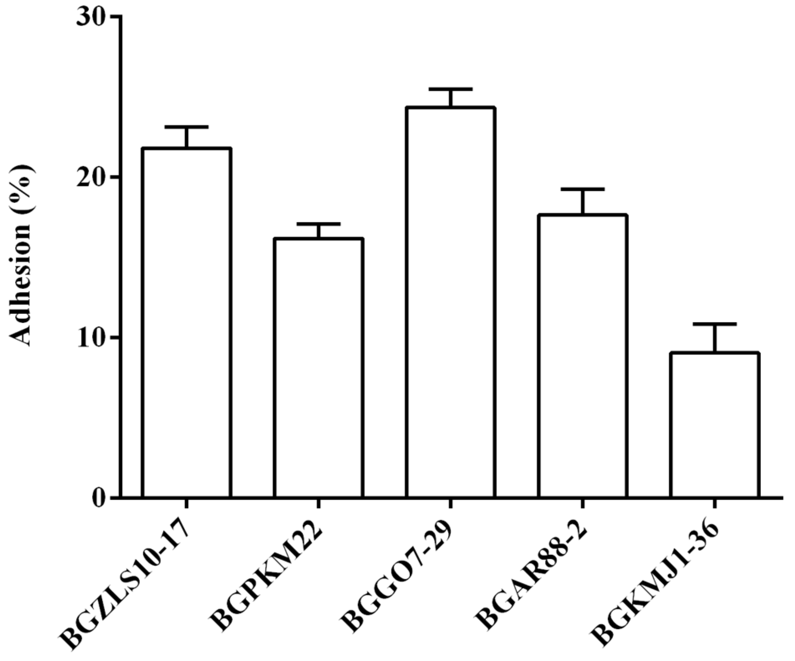
The ability of the five selected LAB strains to adhere to the BEAS-2B cells was tested by the adhesion assay. According to our results, all selected LAB strains were able to adhere to the BEAS-2B cells though the levels of adhesion varied among the strains, as shown in Figure 2. The strain BGGO7-29 showed the highest adhesion (24.3 ± 1.1%), while BGKMJ1-36 showed the lowest adhesion (9.04 ± 1.8%) to BEAS-2B cells.

2.5. LAB Strains BGZLS10-17 and BGPKM22 Attenuated LPS-Induced Nuclear Translocation of NF-κB

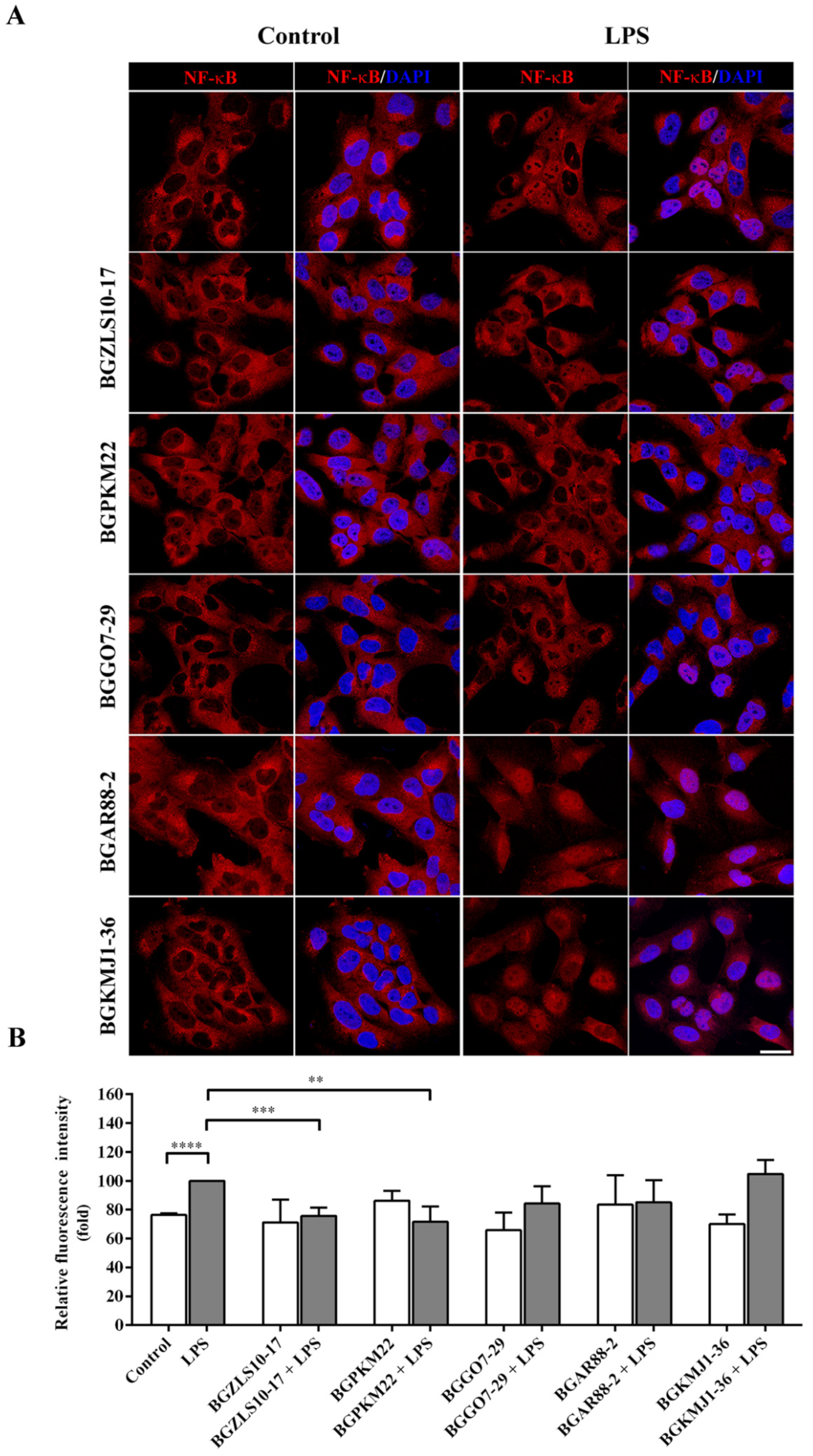
In order to scrutinize the molecular mechanisms implicated in the anti-inflammatory effects of selected LAB, the ability of selected strains to affect the NF-κB nuclear translocation was tested. The activation of TLR4 by LPS leads to the translocation of the phosphorylated NF-κB transcription factor from the cytosol to the nucleus, consequently inducing the expression of pro-inflammatory genes. The BEAS-2B cells treated with LPS showed a significant increase in nuclear NF-κB compared to the control cells. The effects of the LAB strains on the LPS-induced nuclear translocation of NF-κB are shown in Figure 3A,B.
The results of immunofluorescent staining and subsequent confocal microscopy show that the exposure of the BEAS-2B cells to selected LAB strains did not have a significant effect on the level of nuclear NF-κB (Figure 3A,B). However, Lb. brevis BGZLS10-17 and Lb. plantarum BGPKM22 showed the ability to attenuate the LPS-induced nuclear translocation of NF-κB. The level of nuclear NF-κB was significantly lower in cells treated with BGZLS10-17 (75.7 ± 5.8%, p = 0.0001) or BGPKM22 (71.6 ± 10.7%, p < 0.005) and LPS compared to cells treated with LPS only (Figure 3B). However, exposure of the BEAS-2B cells to the LAB strains BGGO7-29, BGAR88-2, and BGKMJ1-36 after LPS induction did not have a significant effect on the levels of nuclear NF-κB (Figure 3A,B).
Our results show that the LAB strains BGZLS10-17 and BGPKM22 block LPS-induced expression of pro-inflammatory genes by preventing NF-κB nuclear translocation in human bronchial epithelial cells. These two LAB strains able to block NF-κB signaling were further tested for the ability to interfere with the MAPK signaling cascade.

2.6. LAB Strains BGZLS10-17 and BGPKM22 Attenuate LPS-Induced Activation of p38, ERK1/2, and JNK MAPK Signaling

Apart from activating the NF-κB transcription factor, TLR4 signaling also leads to the phosphorylation and activation of p38, ERK1/2, and JNK kinases [8]. The ability of the LAB strains BGZLS10-17 and BGPKM22, which showed a significant decrease in the nuclear translocation of NF-κB, to influence the activation of p38, ERK1/2, and JNK MAPK was tested in the BEAS-2B cells stimulated with LPS.
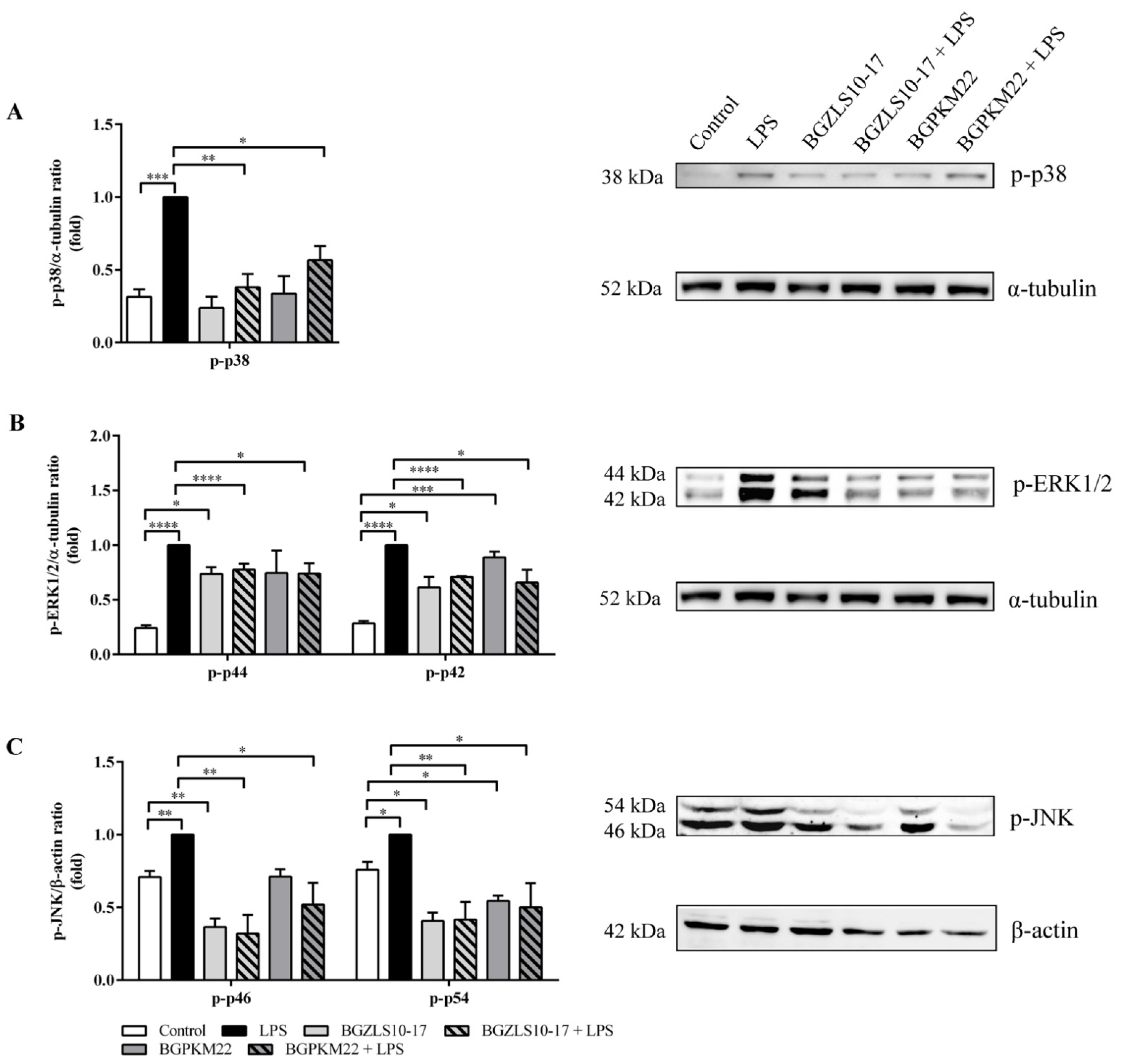
After exposure to LPS, the levels of phosphorylated p38, ERK1/2, and JNK noticeably increased in BEAS-2B cells compared with non-treated cells, as shown in Figure 4A–C. The levels of phosphorylated p38 were decreased in cells treated with the BGZLS10-17 (0.38 ± 0.09, p = 0.001) or BGPKM22 (0.57 ± 0.10, p < 0.05) and LPS in comparison to LPS (Figure 4A).
When the cells were exposed to the BGZLS10-17 or BGPKM22 strain, there was an increase in activated ERK1/2 (phospho-p44 and phospho-p42), suggesting immunostimulatory effects of both strains. However, the levels of phosphorylated ERK1/2 were decreased if the cells were treated with BGZLS10-17 (phospho-p44/p42 0.78 ± 0.05/0.71 ± 0.01, p < 0.0001) or BGPKM22 (phospho-p44/p42 0.74 ± 0.09/0.66 ± 0.12, p < 0.05) and LPS (Figure 4B) indicating the anti-inflammatory effects of the selected LAB strains.
Our results show a decrease in activated JNK when the cells were treated with BGZLS10-17 or BGPKM22 strains for one or both isoforms of JNK, p46, and p54 (Figure 4C). The levels of phosphorylated JNK were also decreased in cells treated with the strain BGZLS10-17 (phospho-p46/p54 0.32 ± 0.13/0.42 ± 0.12, p < 0.01) or BGPKM22 (phospho-p46/p54 0.52 ± 0.15/0.50 ± 0.17, p < 0.05) and LPS, as shown in Figure 4C.
The obtained results show that both LAB strains, BGZLS10-17 and BGPKM22, are able to attenuate LPS-induced MAPK signaling that is responsible for pro-inflammatory gene expression via suppressing the activation of p38, ERK1/2, and JNK kinases.

3. Discussion

Chronic inflammatory lung diseases, such as COPD and asthma, are among the leading causes of severe illness and death, affecting more than 540 million people worldwide [41]. Smoking and respiratory infections are a major cause of the burden in chronic lung diseases, which are potentially preventable. Currently available anti-inflammatory pharmacological treatments have been ineffective in the suppression of lung inflammation, particularly in severe cases of lung diseases, which largely affect society and healthcare systems [41,42,43,44,45,46]. Thus, there is an urgent need for the application of novel approaches with alleviating effects on inflammation in chronic lung diseases. As the recruitment of inflammatory cells in the lungs is orchestrated via bronchial epithelial cells, modifying their inflammatory response could be an effective approach in the therapy of lung diseases [1]. As a target of environmental influences, microbiota, and bacterial pathogens, bronchial epithelial cells are also potent effectors that initiate and propagate inflammatory processes via the release of pro-inflammatory mediators, which further recruit and activate immune cells [1,2].
Although long considered as a sterile organ, lungs are characterized by a relatively low density of a complex microbial community of commensal bacteria. In contrast to the gastrointestinal tract, as an organ with the most studied bacterial community until now, the role of lung microbiota is relatively poorly investigated, and a comprehensive meta-analysis is still missing [47]. Recent studies demonstrated that lung microbiota change with the occurrence of pulmonary pathologies, such as inflammatory lung diseases, and also correlate with the severity and exacerbation of diseases implicating a direct contribution of the microbiota composition in the modulation of lung immunity [3,17,20]. The lung microbiota are characterized by a low density and a small number of bacteria, 2.2 × 10³ per cm2, due to the generally inhospitable lung environment for bacterial growth, and are constantly renewed and replaced by immigration, microaspiration, and elimination [4,18]. Gut microbiota are mainly involved in digestion, energy provision, synthesis of vitamins and other essential bioactive molecules, and maturation of the immune system. On the other hand, much less is known about the role of the lung microbiota. The growing body of evidence points to the gut–lung axis, suggesting the communication and interaction between gut and lung mucosal sites through the mucosal immune system as a wide organ [18,48,49]. It has been reported that oral probiotics reduce the risk of exacerbations and infections in chronic lung diseases [42,50]. For example, an intestinal commensal Parabacteroides goldsteinii has the ability to block NF-κB activation and inhibit lung inflammation by acting through the gut–lung axis [51]. Moreover, the lungs are exposed to multiple microbial challenges through different mechanisms, e.g., aerodigestive reflux and microaspiration, which may play an important role in the host immune response and immunological priming or contribute to a pathogenic immunological process relevant for disease development [52]. It has been demonstrated that the lung microbiome has a functional role in lung immunity and inflammation, but the components of this effects are still not clearly elucidated [16,53]. Moreover, there is a lack of data about the mechanisms of activity of potential probiotics that could be directly applied for the prevention or treatment of many lung disorders.
As natural inhabitants of healthy human lungs, LAB are also well-known for a variety of health-promoting effects including anti-inflammatory abilities, which are of particular interest in this study as they may express alleviating effects related to lung inflammation. Moreover, it has been reported that decreased bacterial diversity related to Firmicutes phyla is associated with the pathogenesis of emphysema and airway remodeling [21,50]. Due to the complexity of the lung microbiome, it is likely that various microbial products play an important role in lung immunity [52]. Moreover, it has been suggested that particular Lactobacillus spp. strains with probiotic properties could maintain physiological homeostasis of the lung and may provide a novel perspective for treating pulmonary diseases [16]. Probiotics can accomplish immunomodulatory effects by the inhibition of LPS binding to the CD14 receptor, but they also produce bioactive molecules or activate the expression of endogenous inhibitors of TLR4 signaling resulting in a reduction of pro-inflammatory cytokine production [8,51,54,55]. Thus, LAB strains capable of reducing the inflammatory response may be employed in the prevention or treatment of lung diseases characterized by diminished lung microbiota.
However, so far, immunomodulatory effects of LAB (Firmicutes) have not been investigated in the respiratory epithelial cells. Hence, the main objective of this study was to investigate the ability of natural LAB strains to modulate the inflammatory response in human bronchial epithelial cells used as an in vitro model system. Here, we showed that two LAB strains, Lb. brevis BGZLS10-17 and Lb. plantarum BGPKM22, possess anti-inflammatory properties by attenuating the activity of the NF-κB transcription factor and MAPK signaling cascade, consequently leading to lower pro-inflammatory gene expression.
In this study, the effect of 21 natural LAB strains on the expression of LPS-induced pro-inflammatory genes in normal human bronchial epithelial cells was tested. The obtained results reveal that the strains exhibited a strain-specific effect, which is in line with previously published data [56].
In particular, the strains Lb. plantarum BGAN8, Lc. lactis subsp. lactis BGTRK4-21, and Lb. helveticus BGRA43 showed an immunostimulatory response increasing the expression of the IL-1β, IL-6, and MCP-1 genes. On the contrary, several LAB strains exhibited an anti-inflammatory effect by reducing the expression of pro-inflammatory genes. In particular, the strains Lb. brevis BGZLS10-17, Lb. plantarum BGPKM22, BGGO7-29, Lb. paracasei BGAR88-2, and St. thermophilus BGKMJ1-36 showed significant anti-inflammatory abilities in LPS-stimulated BEAS-2B cells. Since LAB are lung commensals, the immune response of the lung may vary depending on the strains that come into the composition of lung microbiota. This opens up the possibility for the manipulation of lung microbiota, and LAB strains capable of reducing the inflammatory response, may be employed in the prevention or treatment of lung diseases characterized by diminished lung microbiota.
It is well-known that probiotics, such as Lactobacillus spp. and Bifidobacterium spp., can suppress inflammation by interfering with NF-κB and MAPK signaling checkpoints, which was mostly studied in intestinal epithelial or immune cells [8,51,54,55]. For the first time here, we show that LAB strains are capable to suppress inflammation by interfering with NF-κB and MAPK signaling in respiratory epithelial cells. We showed that two out of five LAB strains with anti-inflammatory abilities were capable to affect the activity of the main components of the TLR4 signaling cascade. We identified that two LAB strains, Lb. brevis BGZLS10-17 and Lb. plantarum BGPKM22, possess the capability to attenuate the LPS-induced nuclear translocation of NF-κB in bronchial epithelial cells resulting in a decrease in inflammatory gene expression. Additionally, we showed that these strains, BGZLS10-17 and BGPKM22, inhibit the activity of MAPK signaling components including p38, ERK1/2, and JNK, contributing to anti-inflammatory effects, too. The pro-inflammatory transcription factor NF-κB and MAPK kinases are activated in chronic lung diseases and implicated in the amplification of the inflammatory response in lungs [7,14,15]. They also have a prominent role in corticosteroid insensitivity and acute exacerbations of chronic lung diseases associated with significant morbidity and mortality [7]. Huge attempts have been made to develop drugs that can control the activity of NF-κB in chronic inflammatory lung diseases, such as the inhibitor of κB kinase (IKK) β inhibitors, but due to the critical role of NF-κB in the host’s defense against pathogens, there are concerns that inhibitors may increase the risk of infection [7]. Moreover, the application of inhibitors is usually followed by adverse side effects. In addition, there is a strong need for the development of MAPK inhibitors in order to reduce chronic lung inflammation and restore corticosteroid sensitivity. However, clinical trials have been largely disappointing [7]. Most anti-inflammatory drugs target turning off the pro-inflammatory arm of the inflammatory response, while approaches that turn on the body’s own anti-inflammatory mechanisms may be more precious. From that view, LAB would be ideal candidates. Probiotics can suppress inflammation by inhibiting various signaling pathways, and the use of probiotics targeting the NF-κB and MAPK pathways may have a profound anti-inflammatory effect [8]. Hence, the autochthonous strains, Lb. brevis BGZLS10-17 and Lb. plantarum BGPKM22, isolated from semi-hard cheese and artisanal sour milk that attenuate NF-κB and MAPK signaling may be employed for the alleviation or prevention of signs of chronic lung diseases.
The ability of the strains BGZLS10-17 and BGPKM22 to colonize human bronchial epithelial cells and to persist in the lungs, which is the prerequisite for their potential application as a probiotic for lungs, was tested in BEAS-2B cells [57]. The obtained results reveal that both strains are able to adhere to bronchial epithelial cells. In addition, antibiotic sensitivity and the absence of hemolytic and gelatinase activity were confirmed for these strains, indicating their QPS status and enabling their safety for application in humans as potential probiotics.
In conclusion, this is the first study providing insight into the immunomodulatory effects of autochthonous natural LAB strains from Western Balkan countries in LPS-stimulated human bronchial epithelial cells. Lb. brevis BGZLS10-17 and Lb. plantarum BGPKM22, capable to inhibit prominent mediators of lung inflammation NF-κB and MAPK, could balance immune response and replenish diminished lung microbiota. These strains fulfil the demands for preclinical testing in an animal model in order to advance their application for the treatment of chronic lung diseases. Furthermore, a precise characterization of the LAB components responsible for anti-inflammatory effects will enable us to gain deeper insight into potential health benefit effects of Lactobacillus spp. strains.

4. Materials and Methods

4.1. Bacterial Strains, Media, and Growth Conditions

For the purpose of this study, 21 natural LAB strains were chosen from the IMGGE collection of microorganisms. The Lactobacillus strains were grown in De Man-Rogosa-Sharpe (MRS) medium (Merck GmbH, Darmstadt, Germany), while Lactococcus and Streptococcus strains were grown in M17 medium (Merck GmbH, Darmstadt, Germany) supplemented with 0.5% (w/v) glucose (GM17). Bacteria were grown at 30 or 37 °C under aerobic or anaerobic conditions depending on the strain. Solid medium was prepared by adding agar (1.7%, w/v) (Torlak, Belgrade, Serbia) to the broth medium. The strains were stored at −80 °C in an appropriate medium (GM17 or MRS) supplemented with 15% (v/v) glycerol. For the in vitro immunomodulatory assays, overnight cultures were harvested by centrifugation, washed with sterile phosphate-buffered saline (PBS), counted in a Petroff–Hausser counting chamber, and resuspended in Roswell Park Memorial Institute (RMPI) medium until use.

4.2. Antibiotic Susceptibility

Minimal inhibitory concentrations (MICs) were determined by microdilution testing according to the European Food Safety Authority (EFSA, Panel, 2018) guidance for bacterial antimicrobial susceptibility. Microbiological cut-off values (mg/L) of 9 antibiotics (ampicillin, vancomycin, gentamicin, kanamycin, streptomycin, erythromycin, clindamycin, tetracycline, and chloramphenicol) for each bacterial species were determined in accordance to the EFSA [40]. Determination of the MICs was performed by microdilution tests in Iso-Sensitest Broth (Oxoid, Hampshire, United Kingdom). The final coloni formin unit (CFU) per well was 5 × 10⁶. Cell density was monitored by OD600 measurements after 24 h of incubation at appropriate temperature in a microtiter plate reader (Tecan Austria GmbH, Grödig, Austria). The lowest concentration of antibiotic at which no growth of bacteria was detected was taken as MIC. Experiments were performed in triplicate.

4.3. Hemolytic and Gelatinase Activity Assays

Hemolytic activity was determined on Columbia Blood Agar (Oxoid Limited, Hampshire, United Kingdom) containing 5% (v/v) defibrinated horse blood after 48 hours of incubation at 37 °C [58]. The phenotypic assay of gelatinase activity was determined on agar plates containing 3% (w/v) gelatin (Difco, Becton Dickinson, NJ, USA) [59].

4.4. Cell Culture

The normal human bronchial epithelial cell line BEAS-2B (ATCC CRL-9609) was cultured in RPMI medium (Gibco, ThermoFisher Scientific, Waltham, MA, USA) supplemented with 10% fetal bovine serum (FBS) (Gibco), 100 µg/mL streptomycin, and 100 U/mL penicillin (Sigma-Aldrich, St. Louis, MO, USA) at 37 °C and 5% CO2. For immunomodulatory assay, BEAS-2B cells with 80% confluence were pre-treated with natural LAB strains (Table 1) using 1:10 ratio in RPMI medium supplemented with 10% FBS without antibiotics for up to two hours. Subsequently, medium with LAB was removed, and cells were treated with 100 ng/mL of LPS (Sigma-Aldrich, St. Louis, MO, USA) for up to 4 h. All experiments were performed at least three times, independently.

4.5. Lactate Dehydrogenase (LDH) Release Assay

The cellular cytotoxicity was determined by measuring the activity of LDH from the culture medium. The activity of LDH was measured spectrophotometrically by CyQUANT LDH Cytotoxicity Assay Kit (ThermoFisher Scientific, Waltham, MA, USA), as per the manufacturer’s instructions. Briefly, 1.7 × 10⁴ cells per well in 96-well plate were seeded and treated with LAB, using a 1:10 ratio for two hours and LPS (Sigma-Aldrich, St. Louis, MO, USA), 100 ng/mL for 4 h. The activity of LDH was determined by measuring the absorbance at 490 nm and 680 nm, using a microtiter plate reader (Tecan Austria GmbH, Grödig, Austria). The cytotoxicity was calculated as the percentage relative to unstimulated cells.

4.6. Quantitative Real Time-PCR (qRT-PCR) Assay

A total of 2.5 × 10⁵ BEAS-2B cells per well were seeded, and the next day the cells were pre-treated with LAB in a 1:10 ratio for two hours. Afterwards, the medium was removed and replaced with fresh medium with LPS 100 ng/mL for 4 h. Total RNA was isolated from BEAS-2B cells using the RNeasy Plus Mini Kit (Qiagen, Hilden, Germany) according to the manufacturer’s instructions. RNA purity and concentration were determined by BioSpec-nano (Shimadzu Corporation, Kyoto, Japan).
cDNA was synthesized from 1 μg of total RNA using the High Capacity cDNA Reverse Transcription Kit (ThermoFisher Scientific, Waltham, MA, USA) as per the manufacturer’s instructions. TaqMan Gene Expression Assays (ThermoFisher Scientific, Waltham, MA, USA) for the IL-1β, IL-6, IL-8, MCP-1, TNF, and endogenous control, glyceraldehyde 3-phosphate dehydrogenase (GAPDH) gene, were used for RT-PCR on 7500 Real-Time PCR system (Applied Biosystems, Waltham, MA, USA) following manufacturer’s protocols. Relative quantification by ΔΔCT method was used to calculate gene expression relative to positive control (LPS), which was used as a calibrator. Results were analyzed using Applied Biosystems 7500 System Software.

4.7. Adhesion Assay

The ability of selected LAB to adhere to BEAS-2B cells was analyzed by adhesion assay according to Mushtaq et al. with minor modifications [60]. Overnight bacterial culture was washed twice with PBS solution, and the pellets were resuspended in RPMI media without antibiotics. A total of 2.5 × 10⁵ BEAS-2B cells, seeded the day before, were washed twice with PBS and treated with bacterial suspensions using a ratio of 1:10 (BEAS-2B cells: LAB). Following co-incubation for two hours at 37 °C and 5% CO2, the cells were gently washed with PBS and lysed with 0.25% Trypsin–EDTA solution (Sigma-Aldrich, St. Louis, MO, USA). Serial dilutions of samples, before and after adhesion, were plated on appropriate agar plates. The adhesion was calculated as (%) = (CFU/mL adhered bacteria/CFU/mL added bacteria) × 100.

4.8. Immunocytochemistry

BEAS-2B cells were seeded on coverslips and pre-treated the next day with LAB in 1:10 ratio for one hour and treated with LPS 100 ng/mL for 30 min. The cells were fixed using 3% paraformaldehyde and 2% sucrose for 20 min at room temperature (RT) and permeabilized in 0.2% Triton X-100 for 10 min. Non-specific binding was blocked in 10% normal goat serum (Sigma-Aldrich, St. Louis, MO, USA) and 1% bovine serum albumin (BSA) (Serva, Heidelberg, Germany) in PBS for one hour at RT. An anti-NF-kB p65 antibody (Alexa Fluor® 594, Abcam) was diluted 1:400 in 1% BSA in PBS and applied for one hour at RT. Coverslips were washed 3 times for 10 min in 0.2% Tween 20 in PBS. Nuclei were stained with 0.1 mg/mL diamino phenylindole (DAPI, Sigma-Aldrich, St. Louis, MO, USA). Coverslips were mounted on to the glass slides using Fluorescent Mounting Medium (Dako, Via Real Carpinteria, CA, USA).

4.9. Image Acquisition and Analysis

Fluorescence images were captured on a Leica SP8 confocal laser scanning system (Leica Microsystems, Wetzlar, Germany). The AF594 labelled antibody was excited with an OPSL 552 laser, while DAPI was excited with 405 diode laser. DAPI (excitation 405 nm, emission 400–450 nm) and AF594 (excitation 590 nm, emission 618 nm) fluorescence were captured using sequential acquisition to give separate image files for each. A pin hole of 1 Airy (95.5 μm), scan speed of 200 Hz and 4 frame averaging, was used. Photomultiplier tube gain and offset were adjusted to give sub-saturating fluorescence intensity with optimal signal to noise ratio. Image acquisition settings were the same for all examined samples in order to compare fluorescence intensities. Six Z stacks in the range of 1.47 µm were acquired.
Quantification of NF-κB nuclear translocation was performed by image analysis using LAS X software [61]. Mean fluorescence intensity was measured on the maximal projections. Fluorescence was quantified in a minimum of 100 nuclei per sample, in 3–5 randomly chosen fields. Quantification was performed with the histogram tool and polygonal region of interest (ROI), by two independent measurements. The nuclear ROIs were defined on DAPI channel. Quantitative fluorescence data were exported from LAS X-generated data into Microsoft Excel software for further analysis and presentation. Nuclear NF-κB was expressed as % of increase in nuclear fluorescent signal compared to 100% set for the cells treated with LPS.

4.10. Western Blotting

A total of 105 BEAS-2B cells were seeded, and the next day the medium was changed to serum-free RPMI for 24 hours. The cells were pre-treated with LAB in a 1:10 ratio for one hour and treated with LPS 100 ng/mL for 20 min. The cells were washed with ice-cold PBS and lysed with Radio Immuno Precipitation Assay buffer (50 mM Tris-HCl pH 7.4; 150 mM NaCl; 1% NP-40; 0.25% sodium deoxycholate) containing protease inhibitor cocktail tablets (Roche, Basel, Switzerland) and 1 mM phenylmethylsulphonyl fluoride (Sigma-Aldrich, St. Louis, MO, USA), for 30 min on ice. The total protein concentrations were determined using the BCA protein assay kit (ThermoFisher Scientific, Waltham, MA, USA). Equal amounts of proteins (15 μg) were separated by 12.5% sodium dodecyl-sulfate polyacrylamide gel electrophoresis (SDS-PAGE). Electrophoresed proteins were transferred from the gel to a 0.2 μm nitrocellulose membrane (GE Healthcare) using a Bio-Rad Mini trans-blot system (Bio-Rad, Hercules, CA, USA). Immunoblots were blocked in a 5% non-fat dry milk in PBS-Tween (50 mM Tris-HCl, pH 7.4; 150 mM NaCl, and 0.05% Tween-20) overnight at 4 °C. After that, the immunoblots were incubated with primary antibodies anti-phospho-p38 MAPK (Thr180/Tyr182), anti-phospho-p44/42 MAPK (Erk1/2) (Thr202/Tyr204), and anti-phospho-JNK (Thr183/Tyr185) (Cell Signaling Technology, Danvers, MA, USA) and anti-α-tubulin and anti-β-actin as a loading controls (Cell Signaling Technology, Danvers, MA, USA) for two hours at RT. The membranes were subsequently washed and incubated with appropriate horseradish peroxidase (HRP) conjugated secondary antibody (ThermoFisher Scientific, Waltham, MA, USA) for one hour at RT. Proteins were detected by enhanced chemiluminescence Immobilon Western (Merck Millipore, Burlington, MA, USA).

4.11. Statistical Analysis

The data are expressed as mean ± standard effort of mean (SEM). The statistical significance of the differences between treatment and control was tested by Student’s t-test. A p-value of less than 0.05 was considered significant. Statistical analysis was carried out, and graphs were prepared by using the GraphPad Prism 6 software.

Author Contributions

M.S.: conceptualization, investigation, analysis, project administration, and writing original draft; K.V.: contribution to conceptualization, investigation, analysis, writing sections, and reviewing; N.P.: investigation, analysis, writing section, and reviewing; S.K.: investigation, analysis, writing section, and reviewing; S.D.M.: investigation, analysis, and reviewing; D.R.: contribution to conceptualization, analysis and reviewing; N.G.: contribution to conceptualization and reviewing. All authors have read and agreed to the published version of the manuscript.

Funding

This research was funded by the Science Fund of the Republic of Serbia, grant PROMIS, #6066974 LABLUNG. The APC was funded by grant PROMIS, #6066974 LABLUNG.

Institutional Review Board Statement

Not applicable.

Informed Consent Statement

Not applicable.

Data Availability Statement

The data presented in this study are available on request from the corresponding author.

Conflicts of Interest

The authors declare no conflict of interest.

Abbreviations

BEAS-2B: Normal human bronchial epithelial cell line; BSA: Bovine serum albumin; CF: Cystic fibrosis; CFU: Colony forming units; COPD: Chronic obstructive pulmonary disease; DAPI: 4′,6-Diamidino-2-phenylindole dihydrochloride; EFSA: European Food Safety Authority; ERK: Extracellular signal-regulated kinase; FBS: Fetal bovine serum; HRP: Horseradish peroxidase; IKK: Inhibitor of κB kinase; IL: Interleukin; IMGGE: Institute of Molecular Genetics and Genetic Engineering; JNK: c-Jun NH 2-terminal kinase; LAB: Lactic acid bacteria; Lb. brevis: Lactobacillus brevis; Lb. helveticus: Lactobacillus helveticus; Lb. paracasei: Lactobacillus paracasei; Lb. paraplantarum: Lactobacillus paraplantarum; Lb. plantarum: Lactobacillus plantarum; Lb. rhamnosus: Lactobacillus rhamnosus; Lc. Lactis: Lactococcus lactis; LDH: Lactate dehydrogenase; LPS: Lipopolysaccharides; MAPK: Mitogen-activated protein kinase; MCP: Monocyte chemoattractant protein; MIC: Minimal inhibitory concentration; MRS: De Man Rogosa Sharpe; NF-κB: Nuclear factor-κB; PAMP: Pathogen-associated molecular pattern; PBS: Phosphate-buffered saline; PRR: Pattern recognition receptor; QPS: Qualified presumption of safety; qRT-PCR: quantitative Real time-polymerase chain reaction; ROI: Region of interest; RPMI: Roswell Park Memorial Institute; RT: Room temperature; SDS-PAGE: Sodium dodecyl-sulfate polyacrylamide gel electrophoresis; SEM: Standard effort of mean; St. thermophilus: Streptococcus thermophilus; TLR: Toll-like receptor; and TNF: Tumor necrosis factor; WBC: Western Balkan countries.

References

  1. Gao, W.; Li, L.; Wang, Y.; Zhang, S.; Adcock, I.M.; Barnes, P.J.; Mao, H.; Xin, Y. Bronchial epithelial cells: The key effector cells in the pathogenesis of chronic obstructive pulmonary disease? Respirology 2015, 20, 722–729. [Google Scholar] [CrossRef] [PubMed]
  2. Whitsett, J.A.; Alenghat, T. Respiratory epithelial cells orchestrate pulmonary innate immunity. Nat. Immunol. 2015, 16, 27–35. [Google Scholar] [CrossRef] [PubMed] [Green Version]
  3. Fabbrizzi, A.; Amedei, A.; Lavorini, F.; Renda, T.; Fontana, G. The lung microbiome: Clinical and therapeutic implications. Inter. Emerg. Med. 2019, 14, 1241–1250. [Google Scholar] [CrossRef] [PubMed] [Green Version]
  4. Mathieu, E.; Escribano-Vazquez, U.; Descamps, D.; Cherbuy, C.; Langella, P.; Riffault, S.; Aude, R.; Muriel, T. Paradigms of lung microbiota functions in health and disease, particularly, in asthma. Front. Physiol. 2018, 9, 1168. [Google Scholar] [CrossRef] [PubMed]
  5. Pattaroni, C.; Watzenboeck, M.L.; Schneidegger, S.; Kieser, S.; Wong, N.C.; Bernasconi, E.; Pernot, J.; Mercier, L.; Knapp, S.; Nicod, L.P.; et al. Early-life formation of the microbial and immunological environment of the human airways. Cell Host Microbe 2018, 24, 857–865.e4. [Google Scholar] [CrossRef] [PubMed] [Green Version]
  6. Frati, F.; Salvatori, C.; Incorvaia, C.; Bellucci, A.; Di-Cara, G.; Marcucci, F.; Esposito, S. The role of the microbiome in asthma: The Gut-Lung Axis. Int. J. Mol. Sci. 2018, 20, 123. [Google Scholar] [CrossRef] [PubMed] [Green Version]
  7. Barnes, P.J. Kinases as novel therapeutic targets in asthma and chronic obstructive pulmonary disease. Pharmacol. Rev. 2016, 68, 788–815. [Google Scholar] [CrossRef] [Green Version]
  8. Llewellyn, A.; Foey, A. Probiotic modulation of innate cell pathogen sensing and signaling events. Nutrients 2017, 9, 1156. [Google Scholar] [CrossRef] [Green Version]
  9. Barnes, P.J. Inflammatory mechanisms in patients with chronic obstructive pulmonary disease. J. Allergy Clin. Immunol. 2016, 138, 16–27. [Google Scholar] [CrossRef] [Green Version]
  10. Yang, X.; Li, H.; Ma, Q.; Zhang, Q.; Wang, C. Neutrophilic asthma is associated with increased airway bacterial burden and disordered community composition. Biomed. Res. Int. 2018, 2018, 9230234. [Google Scholar] [CrossRef] [Green Version]
  11. Roesch, E.A.; Nichols, D.P.; Chmiel, J.F. Inflammation in cystic fibrosis: An update. Pediatr. Pulmonol. 2018, 53, S30–S50. [Google Scholar] [CrossRef] [PubMed] [Green Version]
  12. Smith, D.J.; Chang, A.B.; Bell, S.C. Anti-inflammatory therapies in bronchiectasis. Eur. Respir. Monogr. 2011, 52, 223–238. [Google Scholar] [CrossRef] [Green Version]
  13. Desai, O.; Winkler, J.; Minasyan, M.; Herzog, E.L. The role of immune and inflammatory cells in idiopathic pulmonary fibrosis. Front. Med. (Lausanne) 2018, 5, 43. [Google Scholar] [CrossRef] [PubMed]
  14. Dong, Z.W.; Chen, J.; Ruan, Y.C.; Zhou, T.; Chen, Y.; Chen, Y.; Tsang, L.L.; Chan, H.C.; Peng, Y.Z. CFTR-regulated MAPK/NF-κB signaling in pulmonary inflammation in thermal inhalation injury. Sci. Rep. 2015, 5, 15946. [Google Scholar] [CrossRef] [Green Version]
  15. Dong, J.; Ma, Q. In Vivo activation and pro-fibrotic function of NF-κB in fibroblastic cells during pulmonary inflammation and fibrosis induced by carbon nanotubes. Front. Pharmacol. 2019, 10, 1140. [Google Scholar] [CrossRef]
  16. Li, K.; Chen, Z.; Huang, Y.; Zhang, R.; Luan, X.; Lei, T.; Chen, L. Dysbiosis of lower respiratory tract microbiome are associated with inflammation and microbial function variety. Respir. Res. 2019, 20, 272. [Google Scholar] [CrossRef]
  17. Ubags, N.D.J.; Marsland, B.J. Mechanistic insight into the function of the microbiome in lung diseases. Eur. Respir. J. 2017, 11, 50. [Google Scholar] [CrossRef]
  18. O’Dwyer, D.N.; Dickson, R.P.; Moore, B.B. The lung microbiome, immunity, and the pathogenesis of chronic lung disease. J. Immunol. 2016, 196, 4839–4847. [Google Scholar] [CrossRef] [Green Version]
  19. Mammen, M.J.; Sethi, S. COPD and the microbiome. Respirology 2016, 21, 590–599. [Google Scholar] [CrossRef] [Green Version]
  20. Dicker, A.J.; Chalmers, J.D. Microbial dysbiosis in bronchiectasis and cystic fibrosis. Arch. Bronconeumol. 2017, 53, 471–472. [Google Scholar] [CrossRef] [Green Version]
  21. Toraldo, D.M.; Conte, L. Influence of the lung microbiota dysbiosis in chronic obstructive pulmonary disease exacerbations: The controversial use of corticosteroid and antibiotic treatments and the role of eosinophils as a disease marker. J. Clin. Med. Res. 2019, 11, 667–675. [Google Scholar] [CrossRef] [PubMed] [Green Version]
  22. Oh, N.S.; Joung, J.Y.; Lee, J.Y.; Kim, Y. Probiotic and anti-inflammatory potential of Lactobacillus rhamnosus 4B15 and Lactobacillus gasseri 4M13 isolated from infant feces. PLoS ONE 2018, 13, e0192021. [Google Scholar] [CrossRef] [PubMed]
  23. Suzuki, H.; Yamazaki, T.; Ohshio, K.; Sugamata, M.; Yoshikawa, M.; Kanauchi, O.; Morita, Y. A specific strain of lactic acid bacteria, Lactobacillus paracasei, inhibits inflammasome activation in vitro and prevents inflammation-related disorders. J. Immunol. 2020, 205, 811–821. [Google Scholar] [CrossRef] [PubMed]
  24. Rajilić-Stojanović, M.; Dimitrijević, S.; and Golić, N. Lactic acid bacteria in the gut. In Lactic acid Bacteria, 5th ed.; Chapter, 24, Vinderola, G., Ouwehand, A., Salminen, S., von Wright, A., Eds.; CRC Press: Boca Raton, FL, USA, 2019; p. 383. [Google Scholar]
  25. Terzić-Vidojević, A.; Veljović, K.; Tolinački, M.; Živković, M.; Lukić, J.; Lozo, J.; Fira, Đ.; Jovčić, B.; Strahinić, I.; Begović, J.; et al. Diversity of non-starter lactic acid bacteria in autochthonous dairy products from Western Balkan Countries Technological and probiotic properties. Food Res. Int. 2020, 136, 109494. [Google Scholar] [CrossRef] [PubMed]
  26. Popović, N.; Brdarić, E.; Ðokić, J.; Dinić, M.; Veljović, K.; Golić, N.; Terzić-Vidojević, A. Yogurt produced by novel natural starter cultures improves gut epithelial barrier in vitro. Microorganisms 2020, 8, 1586. [Google Scholar] [CrossRef] [PubMed]
  27. Dinić, M.; Herholz, M.; Kačarević, U.; Radojević, D.; Novović, K.; Đokić, J.; Trifunović, A.; Golić, N. Host-commensal interaction promotes health and lifespan in Caenorhabditis elegans through the activation of HLH-30/TFEB-mediated autophagy. Aging 2021, 13, 8040–8054. [Google Scholar] [CrossRef]
  28. Dinić, M.; Jakovljević, S.; Đokić, J.; Popović, N.; Radojević, D.; Strahinić, I.; Golić, N. Probiotic-mediated p38 MAPK immune signaling prolongs thesurvival of Caenorhabditis elegans exposed to pathogenic bacteria. Sci. Rep. 2021, 11, 21258. [Google Scholar] [CrossRef]
  29. Veljović, K.; Terzić-Vidojević, A.; Vukasinović, M.; Strahinić, I.; Begović, J.; Lozo, J.; Ostojic, M.; Topisirovic, L. Preliminary characterization of lactic acid bacteria isolated from Zlatar cheese. J. Appl. Microbiol. 2007, 103, 2142–2152. [Google Scholar] [CrossRef]
  30. Sokovic-Bajić, S.; Đokić, J.; Dinić, M.; Veljović, K.; Golić, N.; Mihajlović, S.; Tolinacki, M. GABA-producing natural dairy isolate from artisanal Zlatar cheese attenuates gut inflammation and strengthens gut epithelial barrier in vitro. Front. Microbiol. 2019, 10, 527. [Google Scholar] [CrossRef]
  31. Terzic-Vidojevic, A.; Tolinacki, M.; Nikolic, M.; Veljovic, K.; Jovanovic, S.; Macej, O.; Topisirovic, L. Vlasina raw goat’s milk cheese: Evaluation and selection of autochthonous lactic acid bacteria as starter cultures. Food Technol. Biotechnol. 2013, 51, 253–264. [Google Scholar]
  32. Golić, N.; Čadež, N.; Terzić-Vidojević, A.; Šuranská, H.; Beganović, J.; Lozo, J.; Kos, B.; Sušković, J.; Raspor, P.; Topisirović, L. Evaluation of lactic acid bacteria and yeast diversity in traditional white pickled and fresh soft cheeses from the mountain regions of Serbia and lowland regions of Croatia. Int. J. Food Microbiol. 2013, 166, 294–300. [Google Scholar] [CrossRef] [PubMed]
  33. Terzić-Vidojević, A.; Mihajlović, S.; Uzelac, G.; Veljović, K.; Tolinački, M.; Nikolić, M.; Topisirovic, L.; Kojic, M. Characterization of lactic acid bacteria isolated from artisanal Travnik young cheeses, sweet creams and sweet kajmaks over four seasons. Food Microbiol. 2014, 39, 27–38. [Google Scholar] [CrossRef] [PubMed]
  34. Nikolic, M.; Terzic-Vidojevic, A.; Jovcic, B.; Begovic, J.; Golic, N.; Topisirovic, L. Characterization of lactic acid bacteria isolated from Bukuljac, a homemade goat’s milk cheese. Int. J. Food Microbiol. 2008, 122, 162–170. [Google Scholar] [CrossRef] [PubMed]
  35. Strahinic, I.; Lozo, J.; Terzic-Vidojevic, A.; Fira, Đ.; Kojic, M.; Golic, N.; Begovic, J. Technological and probiotic potential of BGRA43 a natural isolate of Lactobacillus helveticus. Front. Microbiol. 2013, 4, 2. [Google Scholar] [CrossRef] [PubMed] [Green Version]
  36. Veljović, K.; Dinić, M.; Lukić, J.; Mihajlović, S.; Tolinački, M.; Živković, M.; Begović, J.; Mrvaljević, I.; Golić, N.; Terzić-Vidojević, A. Promotion of early gut colonization by probiotic intervention on microbiota diversity in pregnant sows. Front. Microbiol. 2017, 8, 2028. [Google Scholar] [CrossRef] [PubMed]
  37. Golić, N.; Veljović, K.; Popović, N.; Djokić, J.; Strahinić, I.; Mrvaljević, I.; Terzić-Vidojević, A. In vitro and in vivo antagonistic activity of new probiotic culture against Clostridium difficile and Clostridium perfringens. BMC Microbiol. 2017, 17, 108. [Google Scholar] [CrossRef] [Green Version]
  38. Lozo, J.; Jovcic, B.; Kojic, M.; Dalgalarrondo, M.; Chobert, J.M.; Haertlé, T.; Topisirovic, L. Molecular characterization of a novel bacteriocin and an unusually large aggregation factor of Lactobacillus paracasei subsp. paracasei BGSJ2-8, a natural isolate from home-made cheese. Curr. Microbiol. 2007, 55, 266–271. [Google Scholar] [CrossRef]
  39. Strahinic, I.; Kojic, M.; Tolinacki, M.; Fira, D.; Topisirovic, L. The presence of prtP proteinase gene in natural isolate Lactobacillus plantarum BGSJ3-18. Lett. Appl. Microbiol. 2010, 50, 43–49. [Google Scholar] [CrossRef]
  40. EFSA Biohaz Panel (EFSA Panel on Biological Hazards); Ricci, A.; Allende, A.; Bolton, D.; Chemaly, M.; Davies, R.; Escámez, P.S.F.; Girones, R.; Herman, L.; Koutsoumanis, K.; et al. Update of the list of QPS-recommended biological agents intentionally added to food or feed as notified to EFSA 8: Suitability of taxonomic units notified to EFSA until March 2018. EFSA J. 2018, 16, e05315. [Google Scholar] [CrossRef]
  41. GBD Chronic Respiratory Disease Collaborators. Prevalence and attributable health burden of chronic respiratory diseases, 1990–2017: A systematic analysis for the Global Burden of Disease Study 2017. Lancet Respir. Med. 2020, 8, 585–596. [Google Scholar] [CrossRef]
  42. Barnes, P.J. New anti-inflammatory targets for chronic obstructive pulmonary disease. Nat. Rev. Drug Discov. 2013, 12, 543–559. [Google Scholar] [CrossRef]
  43. Cantin, A.M.; Hartl, D.; Konstan, M.W.; Chmiel, J.F. Inflammation in cystic fibrosis lung disease: Pathogenesis and therapy. J. Cyst. Fibros. 2015, 14, 419–430. [Google Scholar] [CrossRef] [PubMed] [Green Version]
  44. Funke, M.; Geiser, T. Idiopathic pulmonary fibrosis: The turning point is now! Swiss Med. Wkly. 2015, 145, w14139. [Google Scholar] [CrossRef] [Green Version]
  45. Gross, N.J.; Barnes, P.J. New therapies for asthma and chronic obstructive pulmonary disease. Am. J. Respir. Crit. Care Med. 2017, 195, 159–166. [Google Scholar] [CrossRef] [PubMed]
  46. Mitri, C.; Xu, Z.; Bardin, P.; Corvol, H.; Touqui, L.; Tabary, O. Novel anti-inflammatory approaches for cystic fibrosis lung disease: Identification of molecular targets and design of innovative therapies. Front. Pharmacol. 2020, 11, 1096. [Google Scholar] [CrossRef] [PubMed]
  47. Mancabelli, L.; Milani, C.; Fontana, F.; Lugli, G.A.; Tarracchini, C.; Turroni, F.; van Sinderen, D.; Ventura, M. Mapping bacterial diversity and metabolic functionality of the human respiratory tract microbiome. J. Oral Microbiol. 2022, 14, 2051336. [Google Scholar] [CrossRef]
  48. Gill, N.; Wlodarska, M.; Finlay, B.B. The future of mucosal immunology: Studying an integrated system-wide organ. Nat. Immunol. 2010, 11, 558–560. [Google Scholar] [CrossRef]
  49. Kulas, J.; Mirkov, I.; Tucovic, D.; Zolotarevski, L.; Glamoclija, J.; Veljovic, K.; Tolinacki, M.; Golic, N.; Kataranovski, M. Pulmonary Aspergillus fumigatus infection in rats affects gastrointestinal homeostasis. Immunobiology 2019, 224, 116–123. [Google Scholar] [CrossRef]
  50. Pulvirenti, G.; Parisi, G.F.; Giallongo, A.; Papale, M.; Manti, S.; Savasta, S.; Licari, A.; Marseglia, G.L.; Leonardi, S. Lower airway microbiota. Front. Pediatr. 2019, 7, 393. [Google Scholar] [CrossRef] [Green Version]
  51. Villena, J.; Kitazawa, H. Modulation of intestinal TLR4-inflammatory signaling pathways by probiotic microorganisms: Lessons learned from Lactobacillus jensenii TL2937. Front. Immunol. 2014, 4, 512. [Google Scholar] [CrossRef] [Green Version]
  52. Wu, B.G.; Segal, L.N. Chapter 7: Lung Microbiota and its Impact on the Mucosal Immune Phenotype. Microbiol. Spectr. 2017, 5, 1. [Google Scholar] [CrossRef] [PubMed] [Green Version]
  53. Yadava, K.; Pattaroni, C.; Sichelstiel, A.K.; Trompette, A.; Gollwitzer, E.S.; Salami, O.; von Garnier, C.; Nicod, L.P.; Marsland, B.J. Microbiota Promotes Chronic Pulmonary Inflammation by Enhancing IL-17A and Autoantibodies. Am. J. Respir. Crit. Care Med. 2016, 193, 975–987. [Google Scholar] [CrossRef] [PubMed] [Green Version]
  54. Oh, Y.C.; Cho, W.K.; Oh, J.H.; Im, G.Y.; Jeong, Y.H.; Yang, M.C.; Ma, J.Y. Fermentation by Lactobacillus enhances anti-inflammatory effect of Oyaksungisan on LPS- stimulated RAW 264.7 mouse macrophage cells. BMC Complement. Altern. Med. 2012, 12, 17. [Google Scholar] [CrossRef] [PubMed] [Green Version]
  55. Indira, M.; Venkateswarulu, T.C.; Peele, A.K.; Bobby, N.; Krupanidhi, S. Bioactive molecules of probiotic bacteria and their mechanism of action: A review. Biotech 2019, 9, 306. [Google Scholar] [CrossRef] [PubMed]
  56. Hemarajata, P.; Versalovic, J. Effects of probiotics on gut microbiota: Mechanisms of intestinal immunomodulation and neuromodulation. Ther. Adv. Gastroenterol. 2013, 6, 39–51. [Google Scholar] [CrossRef] [Green Version]
  57. Muñoz-Provencio, D.; Llopis, M.; Antolín, M.; de Torres, I.; Guarner, F.; Pérez-Martínez, G.; Monedero, V. Adhesion properties of Lactobacillus casei strains to resected intestinal fragments and components of the extracellular matrix. Arch. Microbiol. 2009, 191, 153–161. [Google Scholar] [CrossRef]
  58. Valenzuela, A.S.; Omar, N.B.; Abriouel, H.; López, R.L.; Veljovic, K.; Canamero, M.M.; Kojic, M.; Topisirovic, L.; Gálvez, A. Virulence factors, antibiotic resistance, and bacteriocins in enterococci from artisan foods of animal origin. Food Control 2009, 20, 381–385. [Google Scholar] [CrossRef]
  59. Lopes, M.F.; Simões, A.P.; Tenreiro, R.; Marques, J.J.F.; Crespo, M.T.B. Activity and expression of a virulence factor, gelatinase, in dairy enterococci. Int. J. Food Microbiol. 2006, 112, 208–214. [Google Scholar] [CrossRef]
  60. Mushtaq, N.; Ezzati, M.; Hall, L.; Dickson, I.; Kirwan, M.; Ken, M.Y.; Mudway, I.S.; Grigg, J. Adhesion of Streptococcus pneumoniae to human airway epithelial cells exposed to urban particulate matter. J. Allergy Clin. Immunol. 2011, 127, 1236–1242. [Google Scholar] [CrossRef]
  61. Available online: www.leica-microsystems.com/products/microscope-software/p/leica-las-x-ls/ (accessed on 22 April 2022).
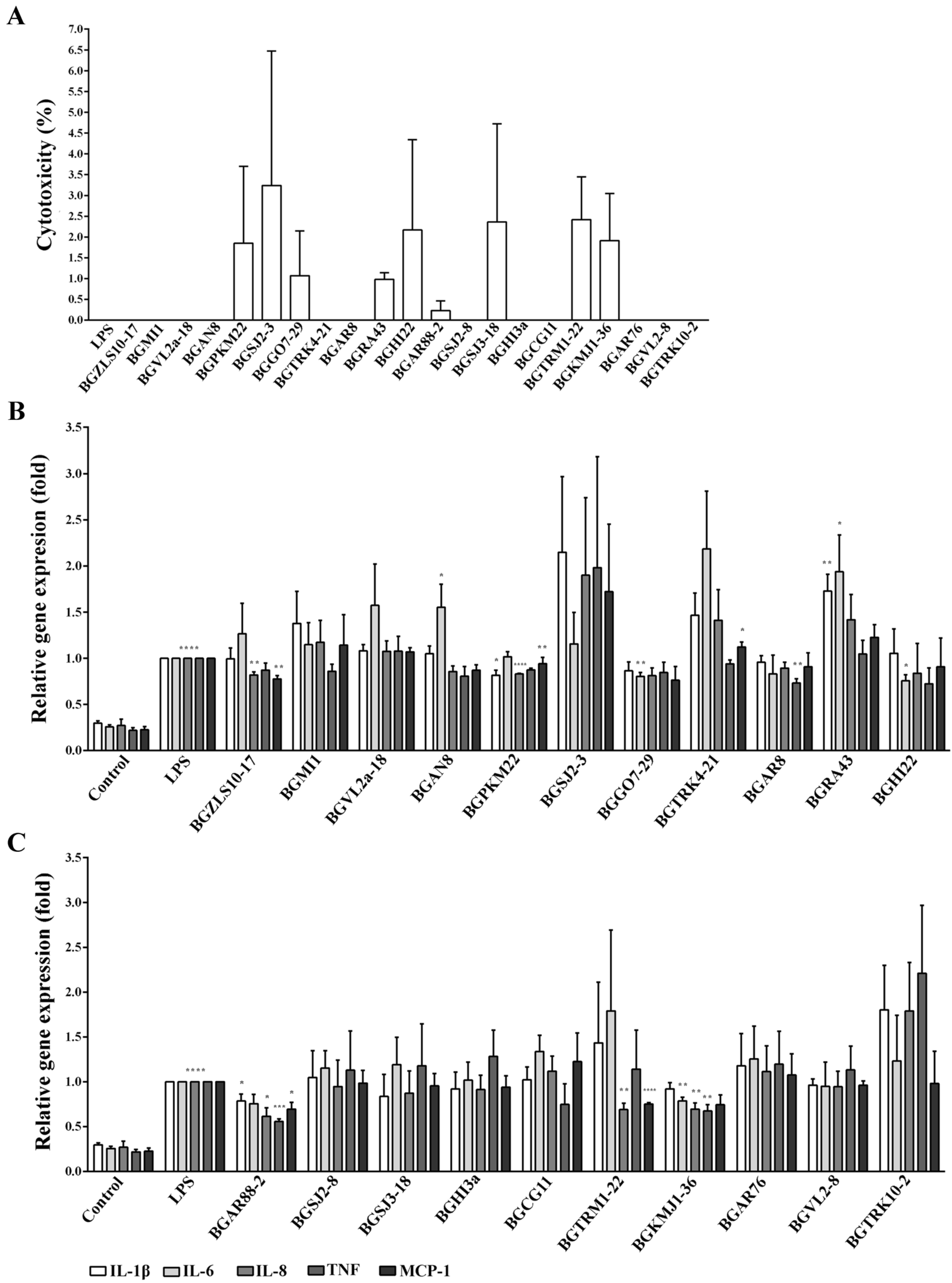
Figure 1. Cytotoxicity and the effect of 21 lactic acid bacteria (LAB) strains on the expression of pro-inflammatory genes in BEAS-2B cells. (A) Cells were treated with 21 LAB strains for two hours or lipopolysaccharide (LPS) (100 ng/mL) for 4 h, and the cytotoxicity was measured by lactate dehydrogenase (LDH) assay. (B) and (C) BEAS-2B cells without treatment (control) or treated with LPS served as experimental controls. Cells were pre-treated with 21 LAB strains for two hours and then treated with LPS for 4 h. The expression of interleukin (IL)-1β, IL-6, IL-8, tumor necrosis factor (TNF), and monocyte chemoattractant protein 1 (MCP-1) was determined by qRT-PCR. Glyceraldehyde 3-phosphate dehydrogenase (GAPDH) was used as endogenous control for normalization. The gene expression was calculated by ΔΔCT method relative to LPS. The results represent data from three independent experiments. Values are expressed as mean ± standard error of mean (SEM). Treatment with LPS is compared to control and treatment with LAB and LPS to LPS. Asterisks indicate significant differences when compared to LPS * p < 0.05, ** p < 0.01, *** p < 0.001, and **** p < 0.0001.
Ijms 23 05547 g001
Figure 2. Adhesion of selected LAB strains on BEAS-2B cells. Cells were treated with LAB strains (1:10) for two hours. The levels of adhesion are expressed as % of LAB used for treatment. The results represent data from three independent experiments. Values are expressed as mean ± SEM.
Ijms 23 05547 g002
Figure 3. Nuclear translocation of nuclear factor (NF)-κB in BEAS-2B cells treated with selected LAB and LPS. BEAS-2B cells without treatment (control) or treated with LPS served as experimental controls. Cells were pre-treated with LAB strains for one hour and then treated with LPS for 30 min. (A) Immunostaining of NF-kB p65 nuclear translocation (red), nuclei were stained with diamino phenylindole (DAPI) (blue). Scale bar is 25 μm. (B) Mean fluorescence intensity of nuclear NF-kB is normalized to LPS. The results represent data from three independent experiments. Values are expressed as mean ± SEM. Treatment with LPS is compared to control, LAB to control, and LAB and LPS to LPS. Asterisks indicate significant differences ** p < 0.01, *** p < 0.001, and **** p < 0.0001.
Ijms 23 05547 g003
Figure 4. The activity of p38, extracellular signal-related kinases (ERK) 1/2, and c-Jun amino-terminal kinases (JNK) in BEAS-2B cells. BEAS-2B cells without treatment (control) or treated with LPS served as experimental controls. Cells were pre-treated with selected LAB for one hour and then treated with LPS for 20 min. Detection of (A) phospho-p38 (p-p38), (B) phospho-ERK1/2 (p-p44/p-p42), and (C) phospho-JNK (p-p46 and p-p54) by Western blot. Anti-α-tubulin and anti-β-actin were used as loading controls. Representative Western blots are shown on the right. The results represent data from three independent experiments. Values are expressed as mean ± SEM. Treatment with LPS is compared to control, LAB to control, and LAB and LPS to LPS. Asterisks indicate significant differences * p < 0.05, ** p < 0.01 *** p < 0.001, and **** p < 0.0001.
Ijms 23 05547 g004
Table 1. The list of bacterial strains and their sources used in this study.
Bacterial StrainSourceReferences
Lactobacillus brevis BGZLS10-1710-day-old semi-hard cheese[29,30]
Lactobacillus plantarum BGMI1White cheeseIMGGE collection
Lactobacillus plantarum BGVL2a-1815-day-old raw goat milk cheese[31]
Lactobacillus plantarum BGAN8Artisanal soft cheeseIMGGE collection
Lactobacillus plantarum BGPKM22Artisanal sour milkIMGGE collection
Lactobacillus plantarum BGSJ2-3White cow cheeseIMGGE collection
Lactobacillus plantarum BGGO7-2960-day-old white pickled cheese[32]
Lactococcus lactis subsp. lactis BGTRK4-21Sweet kajmak[33]
Lactococcus lactis subsp. lactis BGAR85-day-old goat cheese[34]
Lactobacillus helveticus BGRA43Human intestinum[35,36,37]
Lactobacillus rhamnosus BGHI22Human intestinum, neonatusIMGGE collection
Lactobacillus paracasei BGAR88-25-day-old goat cheese[34]
Lactobacillus paracasei BGSJ2-8White cow cheese[38]
Lactobacillus rhamnosus BGSJ3-18White cow cheese[39]
Lactobacillus brevis BGHI3aHuman intestinum, neonatusIMGGE collection
Lactobacillus paraplantarum BGCG11Old full-fat cheeseIMGGE collection
Lactococcus lactis subsp. cremoris BGTRM1-22Sweet cream[33]
Streptococcus thermophilus BGKMJ1-36Artisanal sour milk[26]
Lactobacillus paracasei BGAR765-day-old goat cheese[34]
Lactococcus lactis subsp. lactis BGVL2-85-day-old raw goat milk cheese[31]
Lactococcus lactis subsp. lactis biovar. diacetylactis BGTRK10-2Sweet kajmak[33]
IMGGE—Institute of Molecular Genetics and Genetic Engineering.
Table 2. Minimal inhibitory concentrations (MICs) of nine antibiotics according to EFSA (2018).
Antibiotic with the MIC (ug/mL)
StrainsAmpVanGenKanStrEryClnTetChl
Lb. plantarum
BGMI1<1n.r.832n.r.<12164
BGVL2a-181n.r.416n.r.<12164
BGAN81n.r.816n.r.<12164
BGPKM22<1n.r.416n.r.<12164
BGSJ2-31n.r.432n.r.<12164
BGGO7-291n.r.816n.r.<12164
BGCG11<1n.r.48n.r.<1181
Lb. brevis
BGZLS10-171n.r.43232<1242
BGHI3a1n.r.43216<1242
Lb. helveticus
BGRA43<11444<1121
Lb. rhamnosus
BGHI222n.r.83216<1222
BGSJ3-182n.r.81616<1222
Lb. casei
BGSJ2-82n.r.161632<1222
BGAR761n.r.83232<1222
BG88-21n.r.83232<1222
St. thermophilus
BGKMJ1-36<1<0.516n.r.3210.521
Lc. lactis
BGTRK4-21<12163216<1<124
BGAR8<12163216<1<124
BGAR7612163216<1<122
BGVL2-8<128328<1<122
BGTRK10-2<12163216<1<122
n.r.—not required; Amp—ampicillin; Van—vancomycin; Gen—gentamicin; Kan—kanamycin; Str—streptomycin; Ery—erythromycin; Cln—clindamycin; Tet—tetracycline; Chl—chloramphenicol.
Publisher’s Note: MDPI stays neutral with regard to jurisdictional claims in published maps and institutional affiliations.