Article

Silicon Controls Bacterial Wilt Disease in Tomato Plants and Inhibits the Virulence-Related Gene Expression of Ralstonia solanacearum

1
Guangdong Provincial Key Laboratory of Eco-Circular Agriculture, Guangzhou 510642, China
2
Tea Research Institute, Guangdong Academy of Agricultural Sciences/Guangdong Key Laboratory of Tea Plant Resources Innovation & Utilization, Guangzhou 510640, China
3
College of Natural Resources and Environment, South China Agricultural University, Guangzhou 510642, China
*
Authors to whom correspondence should be addressed.
Int. J. Mol. Sci. 2022, 23(13), 6965; https://doi.org/10.3390/ijms23136965
Submission received: 25 April 2022 / Revised: 26 May 2022 / Accepted: 21 June 2022 / Published: 23 June 2022
(This article belongs to the Special Issue Role of Silicon and Biochar in Plant Stress Tolerance)

Abstract

:
Silicon (Si) has a multifunctional role in improving plant growth and enhancing plant disease resistance, but its mechanisms are not fully understood. In this study, we investigated the impacts of silicon application on the control of bacterial wilt and elucidated the molecular mechanisms using transcriptome sequencing. Compared to non-Si treatment, Si application (0.5–2 mM) significantly reduces tomato bacterial wilt index by 46.31–72.23%. However, Si does not influence the growth of R. solanacearum. Si application negatively influences R. solanacearum exopolysaccharide (EPS) synthesis and biofilm formation. Transcriptome analysis showed that Si treatment significantly downregulates the expression of virulence genes’ transcriptional regulator (xpsR), EPS synthesis-related genes (epsD and tek), and type III effectors (HrpB2, SpaO, and EscR) in R. solanacearum. In addition, Si remarkably upregulates the expression of twitch motor-related genes (pilE2, pilE, fimT, and PilX). These findings suggest that silicon-suppressed tomato wilt incidence may be due to the regulation of the virulence-related genes of R. solanacearum by Si. Our research adds new knowledge to the application of Si in the field of disease control.

1. Introduction

Silicon (Si) accounts for about 28% of the Earth’s crust. Many studies have shown that Si could enhance plant resistance to disease stresses [1,2,3,4]. The mechanisms of Si control plant disease by increasing cell wall strength, activating host defense response through increasing antioxidant enzyme activities or antifungal compounds, and regulating the signal transduction network [5,6,7,8,9]. Si can also directly inhibit the growth of many pathogens, such as Magnaporthe grisea [10], Fusarium solani [11], Alternaria solani [12], and Fusarium sulphureum [13] in vitro. Together, Si inhibits pathogen growth by reducing conidia germination and appressorium formation [1,14].
Ralstonia solanacearum (R. solanacearum) is a soilborne plant pathogen that invades the plant roots from the soil and aggressively colonizes the xylem vessels, which causes wilt symptoms. R. solanacearum infection leads to lethal wilting disease in more than 200 plant species and is particularly harmful to Solanaceae plants [15,16]. Many studies report that Si significantly suppresses bacterial wilt in hydroponic, soil, and substrate culture conditions [17,18,19]. However, most of these studies only focus on the interaction between Si and plants, ignoring the effect of silicon on R. solanacearum. This pathogen is extremely vigorous and can survive for a long time in soil and water; it can infect different plant tissues through a series of virulence determinants [20]. The virulence factors include plant cell-wall-degrading enzymes, bacterial extracellular polysaccharide (EPS), type III secretion system (T3SS), and motility activity, contributing to the development of wilt disease [21,22]. The expression of virulence factors in R. solanacearum is controlled by a complex and precise regulatory network that responds to environmental conditions, host cells, and bacterial density [23]. EPS and T3SS are controlled by a global regulator (PhcA). PhcA is activated by quorum sensing (QS), and it can induce the expression of xpsR and influence the biosynthesis of EPS [24].
Some chemical compounds, such as volatile organic compounds and R-methyl 3-hydroxymyristate, can inhibit biofilm formation, EPS production, and R. solanacearum colonization in plant roots [25,26]. Tahir et al. (2017) found that Bacillus volatiles could reduce bacterial wilt incidence by changing the expression of the virulence factors (PhcA, T3SS, ESP, and chemotaxis) [27]. On the contrary, swimming motility is also a critical factor in bacterial wilt virulence. The virulence of R. solanacearum nonmotile mutant (lack of fliC or fliM gene) was significantly decreased [28]. Yang et al. (2016) found that hydroxycoumarins weakened bacterial wilt virulence by reducing biofilm formation and downregulating flagellar genes such as fliA and flhC [29]. Several studies have shown that exogenous substances (umbelliferone, hydroxycoumarins, and oleanolic acid) could reduce the infectivity of R. solanacearum to host plants by downregulating the expression of R. solanacearum virulence factors, which could be applied for the integrated control of bacterial wilt [30,31].
Although some studies have noted that silicon can reduce the infection rate of plants by inhibiting pathogens, the effect of silicon on the virulence of pathogenic bacteria is still unclear. In the present study, our scientific hypothesis is as follows: (1) Si treatment significantly inhibits the expression of R. solanacearum virulence genes; (2) Si treatment can reduce R. solanacearum EPS synthesis; and (3) Si inhibits biofilm formation of R. solanacearum. Therefore, we used transcriptome sequencing to analyze the expression of virulence-related genes of R. solanacearum influenced by Si in vitro. In addition, the EPS synthesis and biofilm formation of R. solanacearum were analyzed under Si treatment.

2. Results

2.1. Silicon Application Reduced Disease Index of Bacterial Wilt in Tomato

The low Si concentrations (0.05 and 0.1 mM) did not significantly alleviate bacterial wilt in tomato plants (Figure 1a). However, when the added Si concentration was over 0.5 mM, the disease index of bacterial wilt significantly decreased. Compared to the control (0 mM Si), applying 0.5, 1.0, and 2.0 mM of Si reduced the disease index by 46.31%, 66.67%, and 72.23%, respectively (Figure 1b). Regression analysis showed that the disease index was significantly and negatively correlated with Si concentration (R2 = 0.92, p < 0.01; Figure 1c).

2.2. Silicon Did Not Affect the Growth of R. solanacearum

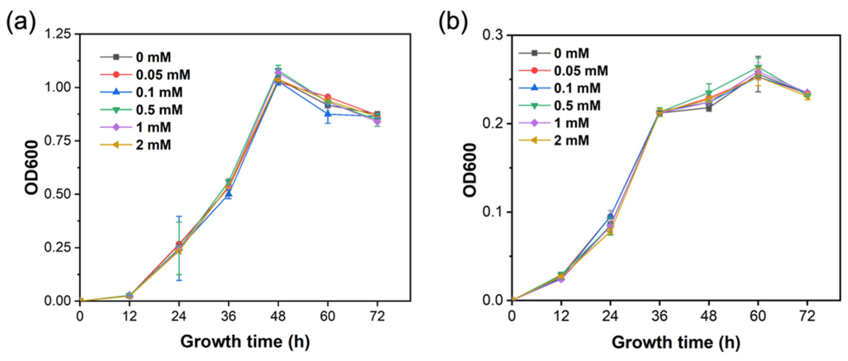
The results show there was no significant difference in the growth of R. solanacearum among all treatments (Figure 2). The growth curve of R. solanacearum in the liquid Luria–Bertani (LB) and minimal mineral (MM) medium was not significantly influenced by Si treatment with different concentrations, indicating that Si did not have a direct inhibition on R. solanacearum.

2.3. Silicon Regulated the Expression of 119 Genes of R. solanacearum In Vitro

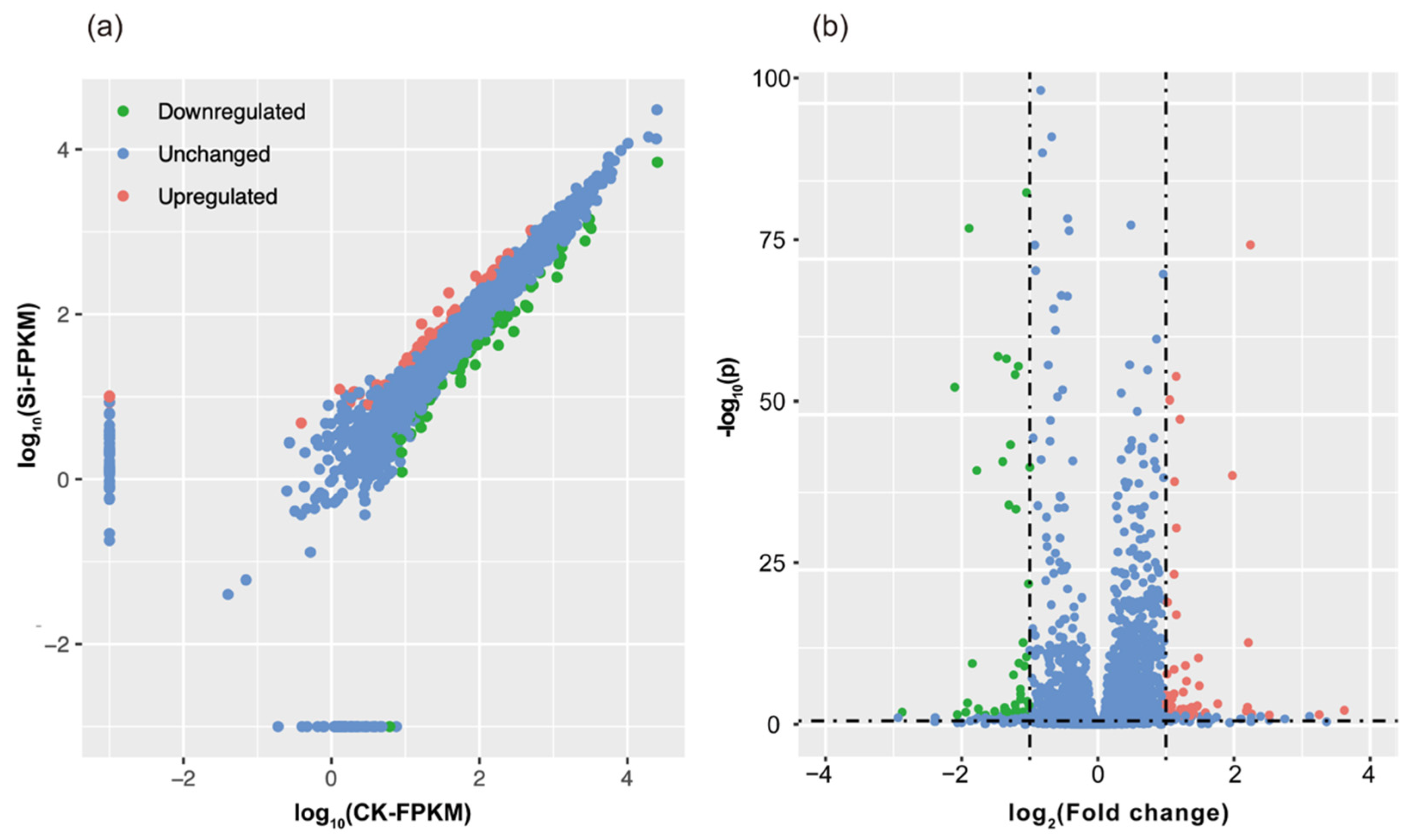
The sequencing data were mapped onto the R. solanacearum strain GMI1000 reference genome [32], and 4,496 genes were detected (Figure 3). In this study, 119 differentially-expressed genes (DEG) were found to be significantly influenced by Si application, accounting for 2.6% of all known genes. Among these 119 genes, 57 were upregulated, and 62 were downregulated (Tables S1 and S2).
The expression of 15 genes (gene primer was shown in Table S3) related to the virulence of R. solanacearum was analyzed by qRT-PCR, including four biological replicates under two housekeeping genes (GAPDH and thyA). The qRT-PCR data for these genes were significantly correlated with the RNA-Seq results in two housekeeping genes, namely GAPDH (Figure S1a) and thyA (Figure S1b), which suggested that the RNA-Seq results are reliable. DEGs were compared with the KEGG Orthology database, and the corresponding pathways were established (Table S4). A total of 37 genes (72.5%) in the KEGG pathway database were related to basal metabolism, indicating that the effect of Si on R. solanacearum was primarily focused on the basal metabolic process (Figure S2). In addition, 15.68% of the genes were related to the development of environmental information. Si significantly upregulated the expression of the R. solanacearum DNA repair gene (ogt) and protein repair gene (degP, Table S4). Meanwhile, the KEGG BRITE functional annotation showed that 41.81% of genes were involved in enzyme functions, and 23.63% of genes were transporters (Table S5).

2.4. Silicon Altered the Transcriptional Expression of xpsR, EPS, and T3SS in R. solanacearum

DEGs involved in metabolic pathways were analyzed to decipher the molecular mechanism of silicon in influencing R. solanacearum virulence. The ModA, AraH, XylF, and LivK genes involved in the ABC transporter pathway and EPS synthesis were significantly downregulated by Si (Figure 4a). These genes encoded proteins that mediated the transportation of bacterial molybdate, L-arabinose, D-xylose, and branched-chain amino acid. Moreover, IbpA, which was involved in Myo-Inositol transport, was significantly upregulated by Si. In the two-component system pathway, four DEGs (degP, DctA, xpsR, and epsD) were enriched (Figure 4b). degP was remarkably upregulated by Si treatment. However, DctA, xpsR, and epsD were significantly downregulated by Si application. The pathway analysis results show that the FliN gene was related to bacterial chemotaxis and flagellar assembly (Figure 4c).
The flagellar assembly pathway is a key pathway downstream of the bacterial chemotaxis. Four genes (pilE2, pilE, PilX, and fimT) encoding type-4 fimbriae were downregulated by Si (Table S6). Twelve virulence-related genes were screened, among which four were upregulated, and eight were downregulated (Table S6). Some genes encoding known virulence traits were screened under Si treatment, such as transcription regulator (xpsR), EPS-related genes (epsD and tek), and type III effectors (HrpB2, SpaO, and EscR). The qPCR results show that the expression of the vital virulence genes, namely xpsR (2.86 folds) and epsD (3.33 folds), were significantly downregulated by Si application (Figure 5a,b). The qPCR results also show that the twitching motility-related gene (pilE) was remarkably upregulated by Si treatment (Figure 5c). Furthermore, Si application significantly reduced the expression of LivK (Figure 4d).

2.5. Silicon Disrupted EPS and Biofilm Formation

The epsD gene, which was involved in the synthesis of EPS (Table S6), was significantly downregulated by Si application. By contrast, EPS concentration was markedly reduced in 2 mM Si treatment, which was consistent with the transcriptome results (Figure 6a). The biofilm formation was an essential mechanism for bacteria to resist external environmental stress. Our results show that bacterial biofilm synthesis in Si treatment was significantly lower than that of non-Si treatment (Figure 6b).

3. Discussion

Multiple studies have investigated the role of Si in controlling bacterial wilt [18,33,34]. Our results show that 0.5–2.0 mM of Si significantly reduced the disease index of bacterial wilt by 46.31%–72.23% (Figure 1a,b), which is consistent with previous studies. However, our study found that Si did not significantly inhibit the growth of R. solanacearum (Figure 2a,b). Previous studies demonstrated that exogenous additives (such as benzimidazole and vitamin C) could prevent pathogens from infecting the host not by directly suppressing pathogens, but by reducing the bacterial virulence, which weakens the ability of bacteria to infect the host [35,36].
Our study showed that Si could significantly downregulate EPS synthesis-related genes, namely, epsD and tek (Table S6, Figure 5b). EPS is also a key virulence factor of R. solanacearum and part of the exopolysaccharide operon, which is the primary substance used by R. solanacearum to block the xylem of plants, and the mutants of the EPS gene lose their virulence ability [37,38]. Minic et al. (2007) reported that epsD affected EPS biosynthesis in Streptococcus thermophilus, and epsD mutant did not produce EPS [39]. tek is an extracellular protein associated with EPS1 and regulated by PhcA [40].
Our studies showed that EPS production decreased in Si treatment (Figure 6a). Therefore, we suggest that Si inhibited the expression of espD and tek, thereby reducing R. solanacearum EPS synthesis. Notably, other genes, except for epsD in the eps gene cluster of R. solanacearum, are not significantly expressed in our experiment. Different genes in the same gene cluster have a different substrate specificity, so there will be differences in the expression levels in the same environmental conditions. In order to uncover the exact reason why silicon inhibits EPS synthesis of R. solanacearum, more molecular experiments need to be carried out in future research.
Biofilms are microbial defense and communication systems [41]. The results show that the formation of R. solanacearum biofilm was significantly inhibited by Si treatment (Figure 6b). Kong et al. (2018) demonstrated that a benzimidazole derivative (UM-C162) prevented the formation of Staphylococcus aureus biofilm, but this derivative had no effect on bacterial viability, and the transcriptome analysis showed that UM-C162 treatment inhibited the expression of bacterial biofilm formation and bacterial attachment-associated genes [42]. Hence, Si inhibited R. solanacearum biofilm synthesis, which might weaken bacterial virulence.
In our study, the transcriptome results show that Si treatment significantly downregulated the expression of eight genes (HrpB2, SpaO, EscR, xpsR, tek, epsD, RSc2755, and RSp1004), which were primarily related to bacterial virulence regulation (Table S6). HrpB2, SpaO, and EscR were type III effectors’ proteins. T3SS were used to inject effectors proteins into plant cells and delivered collections of type III effectors proteins to weaken host defenses [43]. The HrpB2 gene belongs to the hrp gene cluster, and it is an essential component of T3SS [44]. The expression of HrpB2, SpaO, and EscR genes were downregulated by Si treatment, indicating that T3SS of R. solanacearum was inhibited, which may lead to a reduction in bacterial virulence. The xpsR gene is controlled by PhcA, which regulates EPS production [45].
Considerable evidence suggested that the LysR-type regulatory factor PhcA gene was the core of the virulence regulatory network of R. solanacearum [33,46,47]. PhcA is a LysR-type transcriptional regulator, which can regulate virulence factors, activate EPSs and cellulase, and inhibit the mobility of R. solanacearum [48]. Chen et al. (2015) found that the expression of R. solanacearum virulence-related genes (xpsR, tek, and epsE) was significantly inhibited in the PhcA mutant. Collectively, Si suppressed the expression of virulence-related genes, which might decrease the infection rate of R. solanacearum in tomato [45].
Twitching motility is an important bacterial behavior that allows pathogens to efficiently migrate and colonize host plants. Our study also found that the type-4 fimbriae genes, pilE2, pilE, PilX, and fimT, were significantly upregulated by Si (Table S6). The swimming motility of R. solanacearum can be affected by flagella, which are related to bacterial virulence [49]. The flagellar motor switch is composed of three proteins: FliG, FliM, and FliN, which can control flagellum’s rotation [50]. Our study found that Si inhibited the expression of CheZ and FliN (Figure 4c), indicating that Si enhanced the movement of fueling flagella and promoted the twitching motility of bacteria. Twitching motility is important to pathogens in avoiding stressful environments.

4. Materials and Methods

4.1. Experimental Materials

The R. solanacearum strains (GMI1000) and tomato seeds (genotype HYT, bacterial wilt-susceptible tomato) were provided by Professor Guoping Wang (College of Horticulture, South China Agricultural University, Guangzhou, China). Si was applied in the form of anhydrous potassium silicate (K2SiO3, Thermo Fisher Scientific, mean weight ratio: SiO2:K2O = 2.5).

4.2. Effects of Different Si Concentrations on Wilt Incidence

Tomato seeds were surface sterilized with 10% H2O2 for 10 min, followed by rinsing three times with ultrapure water. Sterilized seeds were germinated in petri dishes containing two layers of filter paper for 48 h at 30 °C. The germinated tomato seeds were transferred to plug trays containing sterilized peat soil (Klasmann, Germany), grown in an incubator (30 °C, RH 80%, 12 day/12 night, MGC-400B, Yiheng-Shanghai, China), and watered daily with 1/2 concentration tomato special nutrient solution (each liter of nutrient solution contains 5 mM Ca(NO3)2, 1.88 mM K2SO4, 1.63 mM MgSO4, 0.5 mM KH2PO4, 0.04 mM H3BO3, 0.001 mM ZnSO4, 0.001 mM CuSO4, 0.01 mM MnSO4, 0.00025 mM Na2MoO4, 0.05 mM NaCl and 0.1 mM Fe-EDTA).
Tomato seedlings at the third-leaf stage were transplanted into the pot (5 cm × 5 cm × 8 cm) containing 1 kg of sterilized peat soil. There are six treatments, including the 0, 0.05, 0.1, 0.5, 1.0, and 2.0 mM Si, with twelve replications in this experiment. Tomato seedlings were irrigated daily with 50 mL of 1/2 concentration tomato special nutrient solution (containing 0, 0.05, 0.1, 0.5, 1.0, and 2.0 mM Si) in each pot. The non-silicon treatment was supplemented with a corresponding amount of KCl to balance the increased K element in the Si nutrient solution.
Tomato plants at the sixth-leaf stage were inoculated with pathogens. R. solanacearum strains were grown on B medium (Difco Peptone 10 g·L−1, yeast extract 1 g·L−1, and casamino acids 1 g·L−1) at 28 °C and 150 r·min−1 [51]. After 48 h, bacterial cells were collected and centrifuged three times with sterilized water. R. solanacearum suspension was resuspended in deionized water and adjusted to OD600 = 0.1 (108 cfu·mL−1) with a spectrophotometer (Shimadzu UV-2600, Kyoto, Japan). Then, 50 mL of the bacterial suspension was poured into each pot. The disease index of tomato plants was recorded from the wilting of the first leaf and continuously recorded for 20 d.

4.3. Effects of Different Silicon Concentrations on the Growth of R. solanacearum

The growth curves of R. solanacearum in LB and MM medium were investigated, according to a modification of the method described by Lowe-Power et al. (2018) [52]. There were six treatments: 0, 0.05, 0.1, 0.5, 1.0, and 2.0 mM Si. R. solanacearum strains were grown on liquid LB medium at 28 °C and 150 r·min−1 for 24 h. The cultured cells were adjusted to OD600 = 0.1 using sterile water, and 200 μL of bacterial suspension was added to 30 mL of liquid LB and MM medium. The medium was incubated at 28 °C with shaking at 150 rpm for 72 h in an incubator-shaker (HZQ-X300, Yiheng-Shanghai, China). Growth rate was assessed by measuring OD600 values every 12 h. All experiments have four replicates.

4.4. Pathogen Symptom Evaluation

The disease index was determined according to the methods described by Chen et al. (2015) [33]. A disease index was recorded from the first wilting leaf of tomato plants. Grade 0: asymptomatic; Grade 1: 1 leaf half wilting; Grade 3: 2 to 3 leaves wilting; Grade 5: all leaves are wilted except for the top 1 to 2 leaves; Grade 7: all leaves are wilting; Grade 9: the leaves and plants die.

4.5. EPS Assay

EPS was extracted from R. solanacearum strain [53]. R. solanacearum strain was grown at 30 °C for 48 h in LB medium. Five milliliters of overnight cultures of R. solanacearum were adjusted to OD600 = 0.1, then R. solanacearum was added to 95 mL of sterile liquid MM medium (with and without Si medium) and cultured with shaking for 48 h at 28 °C. R. solanacearum cell suspension (108 CFU·mL−1) was centrifuged at 12,000 rpm for 10 min. The supernatant was filtered by a 0.22 μL filter membrane. The filtrate was placed in a lyophilizer overnight to freeze dry, then 1 mL of 95% ethanol was added, and the mixture was kept at 4 °C for 24 h. Then, the mixture was centrifuged at 5000 r·min−1 for 10 min to collect the precipitate, and 1 mL of water was heated to dissolve the residue and obtain a crude EPS solution. The purified EPS was diluted to 25 mL with distilled water and frozen to store for later use. EPS was determined using the Elson–Morgan method [54].

4.6. Biofilm Assay

The biofilms of R. solanacearum were measured in vitro with a minor modification of the polyvinyl chloride microtiter plate assay [55]. Briefly, R. solanacearum strain was grown at 30 °C for 48 h in LB medium. Then, 5 mL of R. solanacearum suspension (OD600 = 1.0) was added to 45 mL of MM liquid culture broth (with and without Si treatment). Two hundred microliters of culture solution were added to 96-well polystyrene microtiter plates. After the 96-well plates with culture solution were incubated at 28 °C for 48 h, the culture medium was carefully removed, and the biofilm was washed twice with 200 μL ultrapure water. The 96-well plates with bacterial membrane were dried at 60 °C for 30 min to fix the bacterial membrane. Then, 200 μL of 0.1% crystal violet was added to stain the biofilm for 30 min, and the culture solution was washed two times with 200 μL of distilled water to move the crystal violet. Then, 95% ethanol was used to adsorb the crystal violet from the biofilm. The solution was measured for absorbance at 530 nm.

4.7. Transcriptome Analysis

R. solanacearum strain was grown at 30 °C for 48 h in LB medium. Five milliliters of R. solanacearum suspension were adjusted to OD600 = 0.1 and were added into an Erlenmeyer flask containing 50 mL of MM medium with or without 2 mM of Si. Potassium chloride was used to adjust potassium differences between controls and treatments. There were six samples for each treatment. The medium was incubated at 28 °C with shaking at 150 rpm for 48 h, and bacterial cells were collected for transcriptome analysis.
RNA was extracted using the Bacterial RNA Kit (Tiangen Biotechnology, Beijing, China) according to the manufacturer’s recommendations. Preliminary quantification and accurate quantification were detected using a NanoDrop 2000 spectrophotometer (Thermo Fisher Scientific, Inc., Wilmington, DE, USA) and electrophoresis concentration Agilent 2100 RNA 6000 Nano Kit (Figure S3), respectively. Twenty microliters of RNA per treatment were taken for cDNA library construction. The construction of cDNA libraries and RNA-Seq was performed by Genedenovo Bio-Tech Co., Ltd. (Guangzhou, China).

4.8. Data Quality Check and Analysis

The high-throughput sequencing and preliminary analysis of the data were completed by Guangzhou Gideo Biotechnology Co., Ltd. The raw data obtained from Illumina HiSeqTM 2500 sequencing were converted into sequenced reads by CASAVA base calling and stored in FASTQ file format. Raw sequences with adaptors and unknown nucleotides above 5% or those that were of low quality were removed to obtain clean reads. The filtered reads of ribosomes were compared with the reference genome by TopHat2 version 2.0.14 [56]. Cufflinks version 2.2.1 [57] was used to assemble transcripts based on reference annotation-based transcripts.
Fragments per kilobase million (FPKM) value estimation was used to measure gene expression. RSEM version 1.3.3 [58] was used to count the bowtie’s comparison results. The DEGseq version 1.18.0 [57] was applied to normalize the FPKM values among samples for statistical analysis. DEGs were screened based on the criteria of ∣log2 Ratio∣ ≥ 1 and Q ≤ 0.05. p-value was determined by controlling the false discovery rate. DEGs were also submitted to the GO program (http://www.blast2go.org (accessed on 22 June 2022)) for functional annotation [59]. We used KEGG (http://www.kegg.jp/ (accessed on 22 June 2022) pathway analysis to compare and enrich differential genes and determine the main biochemical metabolic pathways and signal transduction processes.

4.9. qRT-PCR

qRT-PCR was performed to validate the RNA-Seq results for 15 gene transcripts. The qPCR experiment used the SYBR Premix Ex Taq Kit (Takara, Japan), and qPCR was measured by the ABI Step One Plus Real-Time PCR System (Applied Biosystems, Foster City, CA, USA). The reaction system (total volume, 20 μL) included 10 μL of SYBR Premix Ex Taq II, 0.6 μL of PCR Forward Primer (10 μM), 0.6 μL of PCR Reversed Primer (10 μM), 2.0 μL of cDNA, and 2.8 μL of ddH2O. The amplification cycling program was as follows: 90 s at 95 °C, followed by 40 cycles at 95 °C for 5 s, 60 °C for 15 s, and 72 °C for 20 s. Relative quantification was used for the conversion of gene expression. Data were analyzed using the 2ΔΔCt method [60]. Two internal reference genes (GAPDH and thyA) were selected, and each sample was subjected to four technical replicates. The primers used for qPCR are listed in Table S3.

4.10. Statistical Analysis

All data were statistically analyzed by t-test and Duncan’s method for multiple comparisons at a 5% significance level. All the statistical analyses were performed with SPSS 20.0 (IBM, Chicago, IL, USA).

5. Conclusions

In this study, we found that Si significantly suppressed bacterial wilt not by directly inhibiting the growth of R. solanacearum, but by influencing the expression of virulence-related genes. Si also reduced the synthesis of R. solanacearum EPS and biofilm. On the contrary, Si upregulated R. solanacearum flagellar genes that promoted bacterial twitching motility. Hence, we hypothesized that Si had a negative influence on R. solanacearum. In the presence of Si treatment, Si downregulated virulence-related genes (HrpB2, SpaO, EscR, xpsR, tek, and epsD) of R. solanacearum and inhibited R. solanacearum EPS synthesis. Meanwhile, Si promoted R. solanacearum movement-related genes (pilE2, pilE, PilX, and fimT). However, the specific mechanism by which silicon inhibits the virulence factors of R. solanacearum remains unclear. Further research needs to be conducted with R. solanacearum mutants or plant infection experiments. This study provides a new perspective to decipher the role of silicon in controlling plant diseases.

Supplementary Materials

The following supporting information can be downloaded at: https://www.mdpi.com/article/10.3390/ijms23136965/s1.

Author Contributions

W.L. designed and performed the experiments, analyzed the data. L.W. analyzed the data and wrote the paper. Y.G., N.J. and J.Y. revised the paper. K.C. obtained funding, designed the experiments, and revised the paper. All authors have read and agreed to the published version of the manuscript.

Funding

This study was financially supported by grants from the National Natural Science Foundation of China (31870420, 31370456), Doctoral Foundation of the Ministry of Education of China (20124404110007), the Natural Science Foundation of Guangdong Province (S2012010010331, 2017A030313177) and Science and Technology Program of Guangdong Province (2121A0505030057).

Institutional Review Board Statement

Not applicable.

Informed Consent Statement

Not applicable.

Data Availability Statement

The data presented in this study are available on request from the corresponding author.

Conflicts of Interest

The authors declare no conflict of interest.

References

  1. Kanto, T.; Maekawa, K.; Aino, M. Suppression of conidial germination and appressorial formation by silicate treatment in powdery mildew of strawberry. J. Gen. Plant Pathol. 2007, 73, 1–7. [Google Scholar] [CrossRef]
  2. Kvedaras, O.L.; Keeping, M.G. Silicon impedes stalk penetration by the borer Eldana saccharina in sugarcane. Entomol. Exp. Appl. 2007, 125, 103–110. [Google Scholar] [CrossRef]
  3. Wang, L.; Chen, Y.; Wang, G.P.; Cai, K.Z. Silicon-mediated tomato resistance against Ralstonia solanacearum is associated with modification of soil microbial community structure and activity. Biol. Trace Elem. Res. 2013, 152, 275–283. [Google Scholar] [CrossRef] [PubMed]
  4. Song, A.; Xue, G.; Cui, P.; Fan, F.; Liu, H.; Yin, C.; Sun, W.; Liang, Y. The role of silicon in enhancing resistance to bacterial blight of hydroponic-and soil-cultured rice. Sci. Rep. 2016, 6, 24640. [Google Scholar] [CrossRef] [Green Version]
  5. Fauteux, F.; Rémus-Borel, W.; Menzies, J.G.; Bélanger, R.R. Silicon and plant disease resistance against virulence fungi. FEMS Microbiol. Lett. 2005, 249, 1–6. [Google Scholar] [CrossRef] [Green Version]
  6. Rémus-Borel, W.; Menzies, J.G.; Bélanger, R.R. Aconitate and methyl aconitate are modulated by silicon in powdery mildew-infected wheat plants. J. Plant Physiol. 2009, 166, 1413–1422. [Google Scholar] [CrossRef]
  7. Rahman, A.; Wallis, C.M.; Uddin, W. Silicon-induced systemic defense responses in perennial ryegrass against infection by Magnaporthe oryzae. Phytopathology 2015, 105, 748–757. [Google Scholar] [CrossRef] [Green Version]
  8. Ghareeb, H.; Bozsó, Z.; Ott, P.G.; Repenning, C.; Stahl, F.; Wydra, K. Transcriptome of silicon-induced resistance against Ralstonia solanacearum in the silicon non-accumulator tomato implicates priming effect. Physiol. Mol. Plant Pathol. 2011, 75, 83–89. [Google Scholar] [CrossRef]
  9. Wang, M.; Gao, L.; Dong, S.; Sun, Y.; Shen, Q.; Guo, S. Role of silicon on plant-pathogen interactions. Front. Plant Sci. 2017, 8, 701. [Google Scholar] [CrossRef] [Green Version]
  10. Maekawa, K.; Watanabe, K.; Kanto, T.; Aino, M.; Saigusa, M. Effect of soluble silicic acid on suppression of rice leaf blast. Int. J. Mol. Sci. 2003, 74, 293–299. [Google Scholar]
  11. Bekker, T.F.; Kaiser, C.; Merwe, R.; Labuschagne, N. In-vitro inhibition of mycelial growth of several phytovirulence fungi by soluble potassium silicate. S. Afr. J. Plant Soil. 2006, 23, 169–172. [Google Scholar] [CrossRef] [Green Version]
  12. Bekker, T.F.; Kaiser, C.; Labuschagne, N. The antifungal activity of potassium silicate and the role of pH against selected plant virulence fungi in vitro. S. Afr. J. Plant Soil. 2009, 26, 55–57. [Google Scholar] [CrossRef] [Green Version]
  13. Li, Y.; Bi, Y.; Ge, Y.; Sun, X.J.; Wan, Y. Antifungal activity of sodium silicate on Fusarium sulphureum and its effect on dry rot of potato tubers. J. Food Sci. 2009, 74, M213–M218. [Google Scholar] [CrossRef] [PubMed]
  14. Qin, G.Z.; Tian, S.P. Enhancement of biocontrol activity of Cryptococcus laurentii by silicon and the possible mechanisms involved. Phytopathology 2005, 95, 69–75. [Google Scholar] [CrossRef] [Green Version]
  15. Denny, T.P. Ralstonia solanacearum—A plant pathogen in touch with its host. Trends Microbiol. 2000, 8, 486–489. [Google Scholar] [CrossRef]
  16. Peeters, N.; Guidot, A.; Vailleau, F.; Valls, M. Ralstonia solanacearum, a widespread bacterial plant pathogen in the post-genomic era. Mol. Plant Pathol. 2013, 14, 651–662. [Google Scholar] [CrossRef] [Green Version]
  17. Dannon, E.A.; Wydra, K. Interaction between silicon amendment, bacterial wilt development and phenotype of Ralstonia solanacearum in tomato genotypes. Physiol. Mol. Plant Pathol. 2004, 64, 233–243. [Google Scholar] [CrossRef]
  18. Diogo, R.V.C.; Wydra, K. Silicon-induced basal resistance in tomato against Ralstonia solanacearum is related to modification of pectic cell wall polysaccharide structure. Physiol. Mol. Plant Pathol. 2007, 70, 120–129. [Google Scholar] [CrossRef]
  19. Kiirika, L.M.; Stahl, F.; Wydra, K. Phenotypic and molecular characterization of resistance induction by single and combined application of chitosan and silicon in tomato against Ralstonia solanacearum. Physiol. Mol. Plant Pathol. 2013, 81, 1–12. [Google Scholar] [CrossRef]
  20. Coll, N.S.; Valls, M. Current knowledge on the Ralstonia solanacearum type III secretion system. Microb. Miotechnol. 2013, 6, 614–620. [Google Scholar]
  21. Yao, J.; Allen, C. The plant pathogen Ralstonia solanacearum needs aerotaxis for normal biofilm formation and interactions with its tomato host. J. Bacteriol. 2007, 189, 6415–6424. [Google Scholar] [CrossRef] [PubMed] [Green Version]
  22. Genin, S. Molecular traits controlling host range and adaptation to plants in Ralstonia solanacearum. New Phytol. 2010, 187, 920–928. [Google Scholar] [CrossRef] [PubMed]
  23. Schell, M.A. Control of virulence and virulence genes of Ralstonia solanacearum by an elaborate sensory network. Annu. Rev. Phytopathol. 2000, 38, 263–292. [Google Scholar] [CrossRef] [PubMed]
  24. Hikichi, Y.; Yoshimochi, T.; Tsujimoto, S.; Shinohara, R.; Nakaho, K.; Kanda, A.; Kiba1, A.; Ohnishi, K. Global regulation of virulence mechanism of Ralstonia solanacearum. Curr. Top. Microbiol. Immunol. 2007, 24, 149–154. [Google Scholar]
  25. Raza, W.; Ling, N.; Yang, L.; Huang, Q.; Shen, Q. Response of tomato wilt pathogen Ralstonia solanacearum to the volatile organic compounds produced by a biocontrol strain Bacillus amyloliquefaciens SQR-9. Sci. Rep. 2016, 6, 24856. [Google Scholar] [CrossRef] [Green Version]
  26. Yoshihara, A.; Shimatani, M.; Sakata, M.; Takemura, C.; Kai, K. Quorum Sensing Inhibition Attenuates the Virulence of the Plant Pathogen Ralstonia solanacearum Species Complex. ACS Chem. Biol. 2020, 15, 3050–3059. [Google Scholar] [CrossRef]
  27. Tahir, H.A.S.; Gu, Q.; Wu, H.; Wu, H.J.; Niu, Y.D.; Huo, R.; Gao, X.W. Bacillus volatiles adversely affect the physiology and ultra-structure of Ralstonia solanacearum and induce systemic resistance in tobacco against bacterial wilt. Sci. Rep. 2017, 7, 40481. [Google Scholar] [CrossRef]
  28. Tans-Kersten, J.; Huang, H.; Allen, C. Ralstonia solanacearum needs motility for invasive virulence on tomato. J. Bacteriol. 2011, 183, 3597–3605. [Google Scholar] [CrossRef] [Green Version]
  29. Liang, Y.; Wei, D.; Yu, X.; Wu, D.; Li, S.; Chen, J.; Bing, G. New Insights into the Antibacterial Activity of Hydroxycoumarins against Ralstonia solanacearum. Molecules 2016, 21, 468. [Google Scholar]
  30. Wu, D.; Ding, W.; Zhang, Y.; Liu, X.; Liang, Y. Oleanolic acid induces the type III secretion system of Ralstonia solanacearum. Front. Microbiol. 2015, 6, 1466. [Google Scholar] [CrossRef] [Green Version]
  31. Yang, L.; Li, S.; Qin, X.; Chen, J.; Li, B.; Yao, X.; Liang, P.; Zhang, Y.; Ding, W. Exposure to umbelliferone reduces Ralstonia solanacearum biofilm formation, transcription of type III secretion system regulators and effectors and virulence on tobacco. Front. Microbiol. 2017, 8, 1234. [Google Scholar] [CrossRef] [PubMed]
  32. Salanoubat, M.; Genin, S.; Artiguenave, F.; Gouzy, J.; Mangenot, S.; Arlat, M.; Billault, A.; Brottier, P.; Camus, J.C.; Cattolico, L. Genome sequence of the plant pathogen Ralstonia solanacearum. Nature 2002, 415, 497–502. [Google Scholar] [CrossRef] [PubMed] [Green Version]
  33. Chen, Y.T.; Liu, M.; Wang, L.; Lin, W.P.; Fan, X.Y.; Cai, K.Z. Proteomic characterization of silicon-mediated resistance against R. solanacearum in tomato. Plant Soil 2015, 387, 425–440. [Google Scholar] [CrossRef]
  34. Jiang, N.H.; Fan, X.Y.; Lin, W.P.; Wang, G.P.; Cai, K.Z. Transcriptome analysis reveals new insights into the bacterial wilt resistance mechanism mediated by silicon in tomato. Int. J. Mol. Sci. 2019, 20, 761. [Google Scholar] [CrossRef] [Green Version]
  35. Sambanthamoorthy, K.; Gokhale, A.A.; Lao, W.W.; Parashar, V.; Neiditch, M.B.; Semmelhack, M.F. Identification of a novel benzimidazole that inhibits bacterial biofilm formation in a broad-spectrum manner. Antimicrob. Agents Chemother. 2011, 55, 4369–4378. [Google Scholar] [CrossRef] [Green Version]
  36. Pandit, S.; Ravikumar, V.; Abdel-Haleem, A.M.; Abderahmane, D.; Mokkapati, V.; Carina, S.; Katsuhiko, M.; Takashi, G.; Gao, X.; Fredrik, W. Low concentrations of vitamin C reduce the synthesis of extracellular polymers and destabilize bacterial biofilms. Front. Microbiol. 2017, 8, 2599. [Google Scholar] [CrossRef] [Green Version]
  37. Saile, E.; McGarvey, J.A.; Schell, M.A.; Denny, T.P. Role of extracellular polysaccharide and endoglucanase in root invasion and colonization of tomato plants by Ralstonia solanacearum. Phytopathology 1997, 87, 1264–1271. [Google Scholar] [CrossRef] [Green Version]
  38. Bocsanczy, A.M.; Achenbach, U.C.M.; Mangravita-Novo, A.; Chow, M.; Norman, D.J. Proteomic comparison of Ralstonia solanacearum strains reveals temperature dependent virulence factors. BMC Genom. 2014, 15, 280. [Google Scholar] [CrossRef] [Green Version]
  39. Minic, Z.; Marie, C.; Delorme, C.; Faurie, J.M.; Mercier, G.; Ehrlich, D.; Renault, P. Control of epsE, the phosphoglycosyltransferase initiating exopolysaccharide synthesis in Streptococcus thermophilus, by EpsD tyrosine kinase. J. Bacteriol. 2007, 189, 1351–1357. [Google Scholar] [CrossRef] [Green Version]
  40. Denny, T.P.; Ganova-Raeva, L.M.; Huang, J.; Schell, M.A. Cloning and characterization of tek, the gene encoding the major extracellular protein of Pseudomonas solanacearum. Mol. Plant-Microbe Interact. 1996, 9, 272–281. [Google Scholar] [CrossRef]
  41. Kumar, J.S.; Umesha, S.; Prasad, K.S.; Niranjana, P. Detection of quorum sensing molecules and biofilm formation in Ralstonia solanacearum. Curr. Microbiol. 2016, 72, 297–305. [Google Scholar] [CrossRef]
  42. Kong, C.; Chee, C.F.; Richter, K.; Thomas, N.; Rahman, N.A.; Nathan, S. Suppression of Staphylococcus aureus biofilm formation and virulence by a benzimidazole derivative, UM-C162. Sci. Rep. 2018, 8, 2758. [Google Scholar] [CrossRef] [PubMed]
  43. Kimbrel, J.A.; Thomas, W.J.; Jiang, Y.; Creason, A.L.; Chang, J.H. Mutualistic co-evolution of type III effector genes in Sinorhizobium fredii and Bradyrhizobium japonicum. PLoS Pathog. 2013, 9, e1003204. [Google Scholar] [CrossRef] [PubMed] [Green Version]
  44. Scheibner, F.; Schulz, S.; Hausner, J.; Marillonnet, S.; Büttner, D. Type III-dependent translocation of HrpB2 by a nonvirulence hpaABC mutant of the plant-virulence bacterium Xanthomonas campestris pv. vesicatoria. Appl. Environ. Microbiol. 2016, 82, 3331–3347. [Google Scholar] [CrossRef] [PubMed] [Green Version]
  45. Clough, S.J.; Lee, K.E.; Schell, M.A.; Denny, T.P. A two-component system in Ralstonia solanacearum modulates production of PhcA-regulated virulence factors in response to 3-hydroxypalmitic acid methyl ester. J. Bacteriol. 1997, 179, 3639–3648. [Google Scholar] [CrossRef] [Green Version]
  46. Genin, S.; Denny, T.P. Pathogenomics of the Ralstonia solanacearum species complex. Annu. Rev. Phytopathol. 2012, 50, 67–89. [Google Scholar] [CrossRef]
  47. Chen, D.; Li, C.; Wu, K.; Xun, G.H.; Yuan, S.F.; Shen, Q.R.; Shen, B. A PhcA-marker-free mutant of Ralstonia solanacearum as potential biocontrol agent of tomato bacterial wilt. Biol. Control. 2015, 80, 96–102. [Google Scholar] [CrossRef]
  48. Yoshimochi, T.; Hikichi, Y.; Kiba, A.; Ohnishi, K. The global virulence regulator PhcA negatively controls the Ralstonia solanacearum hrp regulatory cascade by repressing expression of the PrhIR signaling proteins. J. Bacteriol. 2009, 191, 3424–3428. [Google Scholar] [CrossRef] [Green Version]
  49. Corral, J.; Sebastià, P.; Coll, N.S.; Barbé, J.; Valls, M. Twitching and swimming motility play a role in Ralstonia solanacearum virulence. mSphere 2020, 5, e00740-19. [Google Scholar] [CrossRef] [Green Version]
  50. Bremer, H.; Dennis, P.P. Modulation of chemical composition and other parameters of the cell by growth rate. In Escherichia coli and Salmonella typhimurium: Cellular and Molecular Biology; Neidhardt, F.C., Ed.; American Society for Microbiology: Washington, DC, USA, 1996; pp. 1553–1569. [Google Scholar]
  51. Boucher, C.A.; Barberis, P.A.; Trigalet, A.P.H.; Demery, D.A. Transposon mutagenesis of Pseudomonas solanacearum: Isolation of Tn5-induced avirulent mutants. Microbiology 1985, 131, 2449–2457. [Google Scholar] [CrossRef] [Green Version]
  52. Lowe-Power, T.M.; Hendrich, C.G.; von Roepenack-Lahaye, E.; Li, B.; Wu, D.; Mitra, R.; Dalsing, B.L.; Patrizia Ricca, P.; Naidoo, J.; David Cook, D.; et al. Metabolomics of tomato xylem sap during bacterial wilt reveals Ralstonia solanacearum produces abundant putrescine, a metabolite that accelerates wilt disease. Environ. Microbiol. 2018, 20, 1330–1349. [Google Scholar] [CrossRef] [PubMed]
  53. Milling, A.; Babujee, L.; Allen, C. Ralstonia solanacearum extracellular polysaccharide is a specific elicitor of defense responses in wilt-resistant tomato plants. PLoS ONE 2011, 6, e15853. [Google Scholar] [CrossRef] [PubMed] [Green Version]
  54. Elson, L.A.; Morgan, W.T. A colorimetric method for the determination of glucosamine and chondrosamine. Biochem. J. 1933, 27, 1824–1828. [Google Scholar] [CrossRef] [PubMed] [Green Version]
  55. O’Toole, G.A.; Kolter, R. Initiation of biofilm formation in Pseudomonas fluorescens WCS365 proceeds via multiple, convergent signalling pathways: A genetic analysis. Mol. Microbiol. 1998, 28, 449–461. [Google Scholar] [CrossRef] [PubMed]
  56. Kim, D.; Pertea, G.; Trapnell, C.; Pimentel, H.; Salzberg, S.L. TopHat2: Accurate alignment of transcriptomes in the presence of insertions, deletions and gene fusions. Genome Biol. 2013, 14, R36. [Google Scholar] [CrossRef] [Green Version]
  57. Trapnell, C.; Williams, B.A.; Pertea, G.; Mortazavi, A.; Kwan, G.; Baren, M.J.; Salzberg, S.L.; Wold, B.J.; Pachter, L. Transcript assembly and quantification by RNA-Seq reveals unannotated transcripts and isoform switching during cell differentiation. Nat. Biotechnol. 2010, 28, 511–515. [Google Scholar] [CrossRef] [Green Version]
  58. Li, B.; Dewey, C.N. RSEM: Accurate transcript quantification from RNA-Seq data with or without a reference genome. BMC Bioinform. 2011, 12, 323. [Google Scholar] [CrossRef] [Green Version]
  59. Götz, S.; García-Gómez, J.M.; Terol, J.; Williams, T.D.; Nagaraj, S.H.; Nueda, M.J.; Robles, M.; Talón, M.; Dopazo, J.; Conesa, A. High-throughput functional annotation and data mining with the Blast2GO suite. Nucleic Acids Res. 2008, 36, 3420–3435. [Google Scholar] [CrossRef]
  60. Livak, K.J.; Schmittgen, T.D. Analysis of relative gene expression data using real-time quantitative PCR and the 2−ΔΔct method. Methods 2012, 25, 402–408. [Google Scholar] [CrossRef]
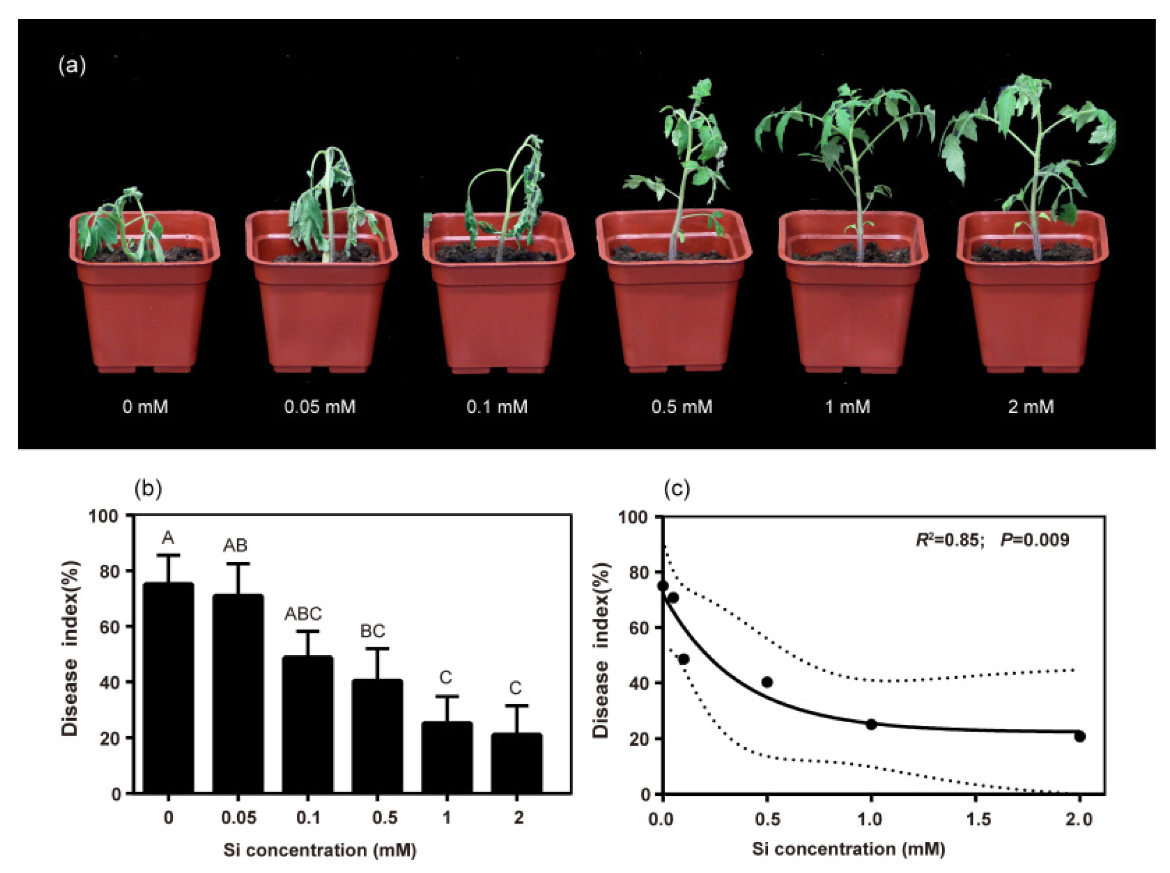
Figure 1. Effects of Si concentration on the disease index of bacterial wilt in tomato. (a) Effects of different Si concentrations on tomato plants under R. solanacearum inoculation conditions. (b) Disease index at 20 days post inoculation (dpi). (c) The simple linear regression (solid line) and 95% confidence interval of the regression (dashed line) for the disease index at 20 dpi and Si concentration in peat soil. Different letters among treatments denote statistical difference at p < 0.05 according to Duncan’s new multiple range tests.
Ijms 23 06965 g001
Figure 2. Effects of different Si concentrations on the growth curve of R. solanacearum. (a) The growth curve of R. solanacearum in liquid LB medium. (b) The growth curve of R. solanacearum in liquid MM medium.
Ijms 23 06965 g002
Figure 3. Gene expression changes and associated significant values across different treatments. Scatter plot (a) and volcano plot (b). Green dots represent significantly downregulated genes {Log2FPKM(Si)/FPKM(CK) > 2 and Log10FDR [FPMK (Si)/FPKM (CK)]) < 0.05}; red dots represent significantly upregulated genes {Log2FPKM(Si)/FPKM(CK) < −2 and Log10FDR [FPMK (Si)/FPKM (CK)]) < 0.05}; blue dots represent no DEGs.
Ijms 23 06965 g003
Figure 4. KEGG pathway of DEGs. (a) DEGs in the ABC transporter pathway. (b) DEGs in the two-component system. (c) DEGs in the bacterial chemotaxis pathway. (d) DEGs in the quorum sensing pathway. Red: upregulated by Si treatment; Green: downregulated by Si treatment.
Ijms 23 06965 g004
Figure 5. Effects of exogenous silicon on the relative expression of virulence-related genes of R. solanacearum. (a) xpsR (transcriptional regulator). (b) epsD (the genes coding for EPS). (c) pilE (the genes coding for twitching motility). (d) fimT (the genes coding for swimming motility). CK: non-Si treatment; Si: Si treatment. “ns” indicates not significant (p > 0.05), * and ** indicate significant difference among treatments at p ≤ 0.05 and p ≤ 0.01 in t-test.
Ijms 23 06965 g005
Figure 6. Effects of Si application on EPS concentration and biofilm formation of R. solanacearum. EPS concentration (a) and biofilm formation (b); CK: non-Si treatment; Si: Si treatment. ** indicates significant difference among treatments at p ≤ 0.01 in t-test.
Ijms 23 06965 g006
Publisher’s Note: MDPI stays neutral with regard to jurisdictional claims in published maps and institutional affiliations.