Review

Bioenergetics and Reactive Nitrogen Species in Bacteria

1
Belozersky Institute of Physico-Chemical Biology, Lomonosov Moscow State University, Leninskie Gory, 119991 Moscow, Russia
2
Department of Biochemical Sciences, Sapienza University of Rome, 00185 Rome, Italy
*
Author to whom correspondence should be addressed.
Int. J. Mol. Sci. 2022, 23(13), 7321; https://doi.org/10.3390/ijms23137321
Submission received: 8 June 2022 / Revised: 27 June 2022 / Accepted: 28 June 2022 / Published: 30 June 2022
(This article belongs to the Special Issue Molecular Mechanisms of Infection for Pathogenic Bacteria)

Abstract

:
The production of reactive nitrogen species (RNS) by the innate immune system is part of the host’s defense against invading pathogenic bacteria. In this review, we summarize recent studies on the molecular basis of the effects of nitric oxide and peroxynitrite on microbial respiration and energy conservation. We discuss possible molecular mechanisms underlying RNS resistance in bacteria mediated by unique respiratory oxygen reductases, the mycobacterial bcc-aa3 supercomplex, and bd-type cytochromes. A complete picture of the impact of RNS on microbial bioenergetics is not yet available. However, this research area is developing very rapidly, and the knowledge gained should help us develop new methods of treating infectious diseases.

1. Introduction

Primary bacterial pathogens are infectious agents responsible for severe and often deadly diseases in humans. In addition, commensal bacteria can produce opportunistic infections in immunosuppressed patients. Disease-causing bacteria are becoming resistant to most commonly available antibiotics, which poses a threat to global public health. The production of reactive nitrogen species (RNS) by the innate immune system is part of the host’s defense against invading microbes. RNS refers to various nitrogenous products including nitric oxide (NO), peroxynitrite anion (ONOO), nitroxyl (HNO), dinitrogen trioxide (N2O3), nitrite (NO2), nitrogen dioxide (NO2), nitronium cation (NO2+), nitrosonium cation (NO+), nitrosoperoxycarbonate anion (ONOOCO2), nitryl chloride (Cl-NO2), S-nitrosothiols (RSNOs) [1]. NO, along with carbon monoxide and hydrogen sulfide, is considered an endogenous gaseous signaling molecule [2,3,4,5]. NO is the main RNS produced by the host and the main source for the generation of the other RNS. This small diatomic molecule is a free radical, i.e., with one unpaired electron, and can diffuse easily through biological membranes. The enzymes that produce NO are NO synthases (NOS). They convert l-arginine and O2 into l-citrulline and NO using NADPH as the electron donor. There are three NOS isoforms: neuronal (nNOS), endothelial (eNOS), and inducible (iNOS). nNOS and eNOS are constitutively expressed whereas iNOS expression is induced by immunological stimuli. The latter occurs predominantly in macrophages and plays an essential role in immune defense. NO can combine with superoxide radical (O2•−) produced by the NADPH oxidase at diffusion-controlled rates yielding another RNS, ONOO. Under physiological conditions, ONOO is in equilibrium with peroxynitrous acid, ONOOH (pKa = 6.8), and local pH affects peroxynitrite reactivity. Both ONOO and ONOOH are able to cross biological membranes. Peroxynitrite is a potent oxidant and nitrating agent, with a very important role in the destruction of invading pathogens by macrophages, as ONOOH spontaneously homolyzes to hydroxyl radical (OH) and NO2 [6,7]. As they are within bacteria-containing phagolysosomes in macrophages, RNS creates a hostile environment that impairs microbial growth. RNS inhibit DNA replication and bacterial respiration [8]. NO and ONOO were reported to damage metalloproteins containing heme cofactors and/or iron-sulfur clusters [9]. Additionally, NO mediates post-translational modifications through S-nitrosylation of protein thiol groups, and peroxynitrite promotes the nitration of protein tyrosine residues [6,10]. This review focuses on the effects of NO and ONOO on bacterial aerobic (O2-dependent) respiratory (electron transport) chains, namely on their last component represented by a heme-containing terminal oxidase, in light of recent findings.
We have chosen to focus only on aerobic bacteria because data on the RNS stress response of pathogenic anaerobes remain scarce. For instance, in the case of multidrug-resistant Klebsiella pneumoniae, a common cause of hospital-acquired pneumonia, some data on its adaptive response toward oxidative stress are available [11,12] but none addressed the bacterial response to NO. A search of the genome of K. pneumoniae points out the presence of NO-detoxifying enzymes Hmp and Hcp nonetheless [9]. The intracellular pathogen Shigella flexneri, which is the causative agent of bacillary dysentery, was reported to be sensitive to NO produced in vitro; on the contrary, NO is not required for clearance of the microbes in infected mice or macrophages [13]. A search of the genome of S. flexneri, however, indicates putative flavorubredoxin, Hmp and Hcp enzymes are involved in nitrosative detoxification [9]. Clostridioides difficile is the cause most implicated in antibiotic-associated diarrhea and severe inflammation of the bowel. This anaerobic enteropathogen encodes a few putative NO-consuming enzymes, such as two flavodiiron proteins FdpA and FdpF, and Hcp [14,15]. FdpA and FdpF, however, show negligible NO reductase activity but instead significant O2 reductase activity [15,16]. Although there is no data on the specific NO activity of Hcp, the physiological nitrosating agent S-nitrosoglutathione (GSNO) was reported to induce the expression of hcp [17]. This finding indicates that Hcp is involved in NO resistance.

2. Bacterial Aerobic Respiratory Chains

Similar to mitochondria of eukaryotic cells, bacteria contain aerobic respiratory chains. The main function of the chains is to create a proton motive force (PMF), a central energy currency. The well-known mammalian mitochondrial chain is linear [18,19]. It consists of the enzyme complexes I, II, III, and IV (Table 1). The complexes I, III, and IV catalyze the oxidation of NADH by ubiquinone, oxidation of ubiquinol by ferricytochrome c, and oxidation of ferrocytochrome c by molecular oxygen, respectively. Each redox reaction is coupled to the generation of PMF that can be used further for the production of one more central energy currency, ATP, by ATP synthase (also termed complex V) or for active transport of solutes across the membrane. Complex II (succinate dehydrogenase) belongs to both the respiratory chain and the Krebs cycle. Complex II catalyzes the electron transfer from succinate to ubiquinone but unlike complexes I, III, and IV, the transfer is not coupled to the formation of PMF [20,21]. The bacterial respiratory chains, in contrast to the mitochondrial one, are branched, with different routes of electron transfer depending on the growth conditions [22,23]. As a quinone, bacteria can use not only ubiquinone but also menaquinone, plastoquinone, or caldariellaquinone.
In order to transfer electrons from NADH to quinone, bacteria use three different families of NADH:quinone reductases (dehydrogenases)—NDH-1, NDH-2, and NQR (Table 2). NDH-1 reductases are closely related to the mitochondrial complex I and function as redox-driven proton pumps [24,25]. Both NDH-2 and NQR are unrelated to the canonical complex I. NDH-2 enzymes are non-electrogenic and therefore unable to support PMF [26,27]. NQR reductases operate as redox-driven sodium pumps, i.e., they generate a sodium ion motive force rather than PMF [28,29,30]. The sodium ion motive force, along with PMF and ATP, is the third energy currency used by a few bacteria [31]. Bacteria with more than one NADH:quinone reductase show a preference for one or another enzyme depending on the growth conditions.
Bacterial complex III, also termed cytochrome bc1 complex, transfers electrons from quinol to ferricytochrome c. This redox reaction is coupled with the production of PMF via the Q-cycle (Mitchellian redox-loop) mechanism [32,33]. The presence of complex III in bacterial respiratory chains is optional. Some bacteria, e.g., Escherichia coli, have no cytochrome c at all, and hence no cytochrome bc1 [34]. Cytochrome c of other bacteria is not water-soluble but fused either to complex III or complex IV. This leads to the formation of a supercomplex between complex III and complex IV (Table 2). Accordingly, the cytochrome bcc-aa3 (III2–IV2) supercomplex was discovered in Mycobacterium smegmatis and Corynebacterium glutamicum [35,36,37]. A supercomplex composed of cytochrome bc1 and aa3-type cytochrome c oxidase was also identified in Rhodobacter sphaeroides [38]. Figure 1 shows examples of three different types of branched bacterial respiratory chains in which the complex III is absent (E. coli [34]), present as a separate enzyme (Pseudomonas aeruginosa [29]), or forms a tight supercomplex with the aa3-type cytochrome c oxidase (M. tuberculosis [39,40]).
The membrane-bound terminal oxidases are divided into two superfamilies: heme–copper oxidases and bd-type cytochromes [41,42,43]. The active site of a heme–copper oxidase termed the binuclear center (BNC) is composed of a high-spin heme (a3, o3, or b3) and a copper ion (CuB). The enzyme catalyzes the transfer of electrons from cytochrome c or quinol to O2 with the production of 2H2O. The reaction is coupled to the generation of PMF using the mechanism of redox-coupled proton pumping across the membrane [21,22,44,45,46,47,48,49,50,51,52,53,54,55,56,57,58,59]. A heme–copper oxidase that uses cytochrome c as an electron donor (cytochrome c oxidase) has the second copper site, CuA. CuA directly accepts electrons from cytochrome c. If the enzyme uses quinol as an electron donor (quinol oxidase), CuA is absent. Heme–copper oxidases also contain a low-spin heme (a or b) that accepts electrons from CuA (cytochrome c oxidase) or directly from an electron donor (quinol oxidase) and donates them to the BNC. In caa3 and cbb3 oxidases, the reduction of CuA by water-soluble cytochrome c is followed by an intermediate reduction of additional heme(s) c. The classification of the heme–copper oxidases is based on the organization of the intraprotein proton transfer pathways. Accordingly, the enzymes are divided into three main families: A, B, and C [60,61,62].
The active site of cytochrome bd contains a high-spin heme d but not a copper ion [39,63,64,65,66,67,68,69]. There are data that one more high-spin heme, b595, could perform some of the functions of CuB [70,71,72,73,74,75,76,77,78,79,80,81,82,83,84,85,86]. Similar to heme–copper oxidases, bd-type cytochromes couple the reduction of O2 to 2H2O to the formation of PMF [44,87,88]. However, in contrast to the heme–copper enzymes, cytochromes bd do so without being a proton pump [89,90,91]. The lack of proton-pumping machinery decreases the energetic efficiency of the bd-type oxidases. Until now, all biochemically characterized cytochromes bd turned out to be quinol oxidases [49,92,93,94]. Accordingly, the third heme in cytochrome bd, a low-spin b558, mediates electron transfer from quinol to hemes b595 and d. The bd-oxidases typically have a very high affinity for O2 and CO due to specific features of heme d, which is an iron-chlorin [77,95,96,97,98,99]. In some cases, heme d can be replaced with heme b [42,100]. Intriguingly, phylogenomic analyses performed by Murali et al. suggest that there are bd-type cytochromes that use cytochrome c as an electron donor [42]. Phylogenomics by Murali et al. identified three families and several subfamilies within the cytochrome bd superfamily. At the same time, earlier classification of the bd-type oxidases based on the size of the hydrophilic region between transmembrane helices 6 and 7 in subunit I (a binding domain for quinol oxidation termed the Q-loop) is still commonly used. According to this classification, cytochromes bd are divided into two subfamilies: L (long Q-loop) and S (short Q-loop) [101,102].
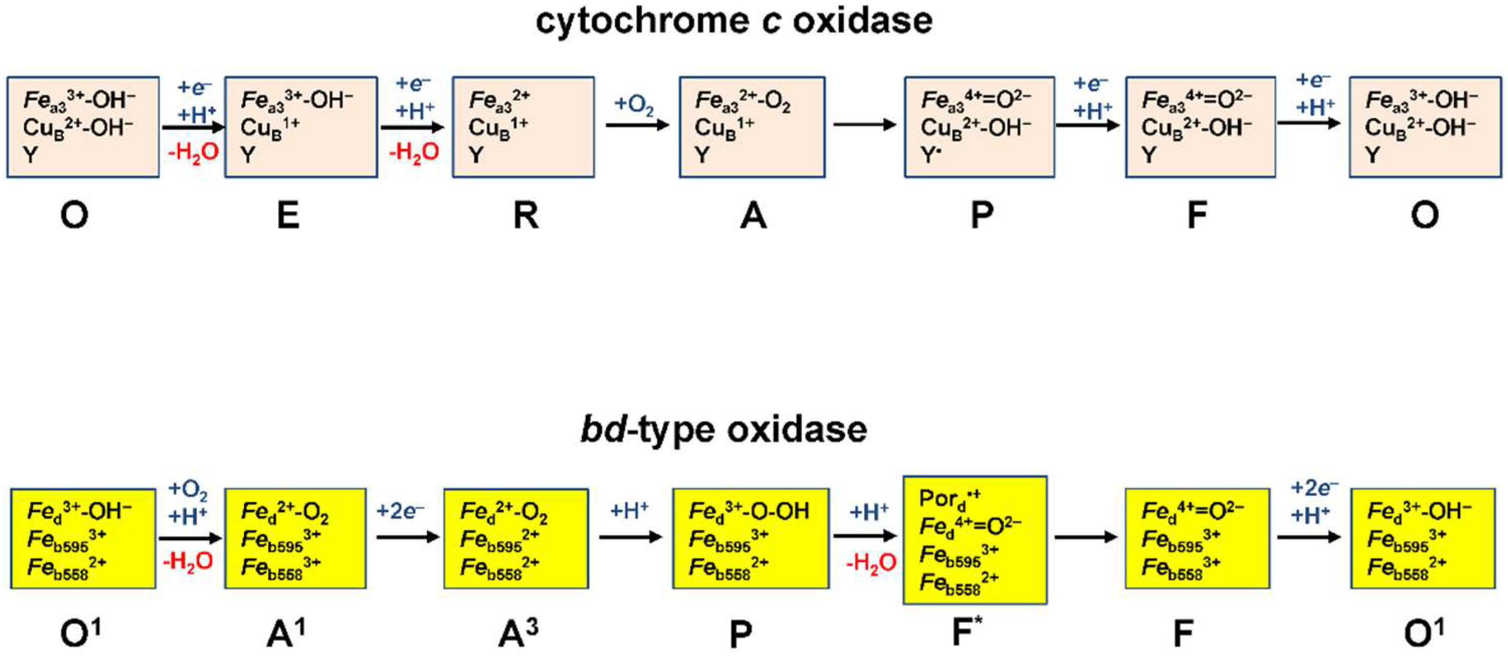
The catalytic cycle of heme–copper oxidases is best studied for the aa3-type cytochrome c oxidases (Figure 2). It includes the intermediates termed O, E, R, A, P, F (see [41] and references therein). The sequential transfer of two electrons to O (the fully oxidized state of the BNC) results in the sequential formation of E and R, one-electron reduced and fully reduced states of the BNC, respectively. R binds O2 to produce the A state. Then, the O–O bond is cleaved, and the P state is formed in which heme a3 is ferryl, CuB is oxidized, and a conserved tyrosine residue in the BNC is oxidized to a radical, Y. The transfer of the third electron to the BNC re-reduces Y to Y bringing about the F state. The transfer of the fourth electron to the BNC leads to the reduction of ferryl heme a3 to ferric form that regenerates the O state and completes the cycle. The O→ E, E→ A, P→ F, and F→ O transitions are electrogenic and coupled to the transfer of a pumped proton (not shown in Figure 2).
The catalytic cycle of bd-type oxidases is deduced from the studies on the E. coli cytochrome bd-I [90,103,104,105,106] (Figure 2). It includes the intermediates termed O1, A1, A3, P, F*, F, and takes into account that the quinol substrate is a two-electron donor. In the O1→ A1 transition, an electron transfers from heme b558 to heme d and the latter binds O2. In the next A1→ A3 transition, two electrons from a quinol reduce heme b558 and heme b595. In the A3→ P transition, a true transient peroxy complex of ferric heme d is formed concomitant with oxidation of heme b595. The O–O bond cleavage occurs in the next, P→ F* transition in which the ferric heme d is further oxidized to the ferryl form with a porphyrin π-cation radical (Por•+). Then in the F*→ F transition, the radical is quenched by an electron from the ferrous heme b558. The F→ O1 transition, in which two electrons from a second quinol reduce the ferryl heme d (to the ferric form) and heme b558, completes the cycle. The P/F*→ F and F→ O1 transitions were reported to be electrogenic [88,89,90,91,107].
The key role of most heme–copper oxidases in bacterial metabolism is to create PMF. In the case of cytochromes bd, the bioenergetic function is not the only. The bd enzymes play other critical roles in microbes [94,108,109,110,111]. They contribute significantly to the ability of bacteria to resist stresses induced by peroxide [49,112,113,114,115,116], sulfide [5,117,118,119,120], ammonia [121], chromate [122], cyanide [117,123]. Due to the fact that the bd oxidases are often found in pathogenic bacteria but absent in humans, they can be used as protein targets for next-generation antimicrobials [43,64,68,124,125,126,127,128,129,130,131,132,133,134].

3. NO and Bacterial Terminal Oxidases

3.1. NO and Bacterial Heme–Copper Terminal Oxidases

With the exception of the mycobacterial aa3-type oxidase (see Section 3.1.1), the bacterial heme–copper oxidases tested to date, such as the cbb3-type oxidases from Vibrio cholerae and Rhodobacter sphaeroides, and the aa3-type oxidase from R. sphaeroides, are rapidly and strongly inhibited by NO [135], similar to their mitochondrial homolog, cytochrome c oxidase [136]. The reaction of the mitochondrial enzyme with NO was studied in more detail. It was shown that low, nanomolar levels of NO reversibly inhibit the enzyme activity [136] whereas high, micromolar levels of NO cause irreversible damage to the enzyme [137]. The reversible inhibition occurs via two pathways. At high reductive pressure (high turnover conditions) and low O2 tensions, the O2-competitive inhibition pathway prevails. It occurs through the reaction of NO with the two-electron reduced (and possibly one-electron reduced) BNC leading to the production of the nitrosyl derivative of the enzyme. At low reductive pressure (low turnover conditions) and high O2 tensions, the noncompetitive pathway prevails. The latter proceeds via reaction of NO with the catalytic intermediates that have CuB oxidized, resulting in the generation of the nitrite-bound enzyme [138,139,140,141]. It is reasonable to assume that the bacterial heme–copper oxidases studied [135] are inhibited by NO through similar mechanisms.

3.1.1. NO-Metabolizing Activity of the Mycobacterial bcc-aa3 Supercomplex in Turnover

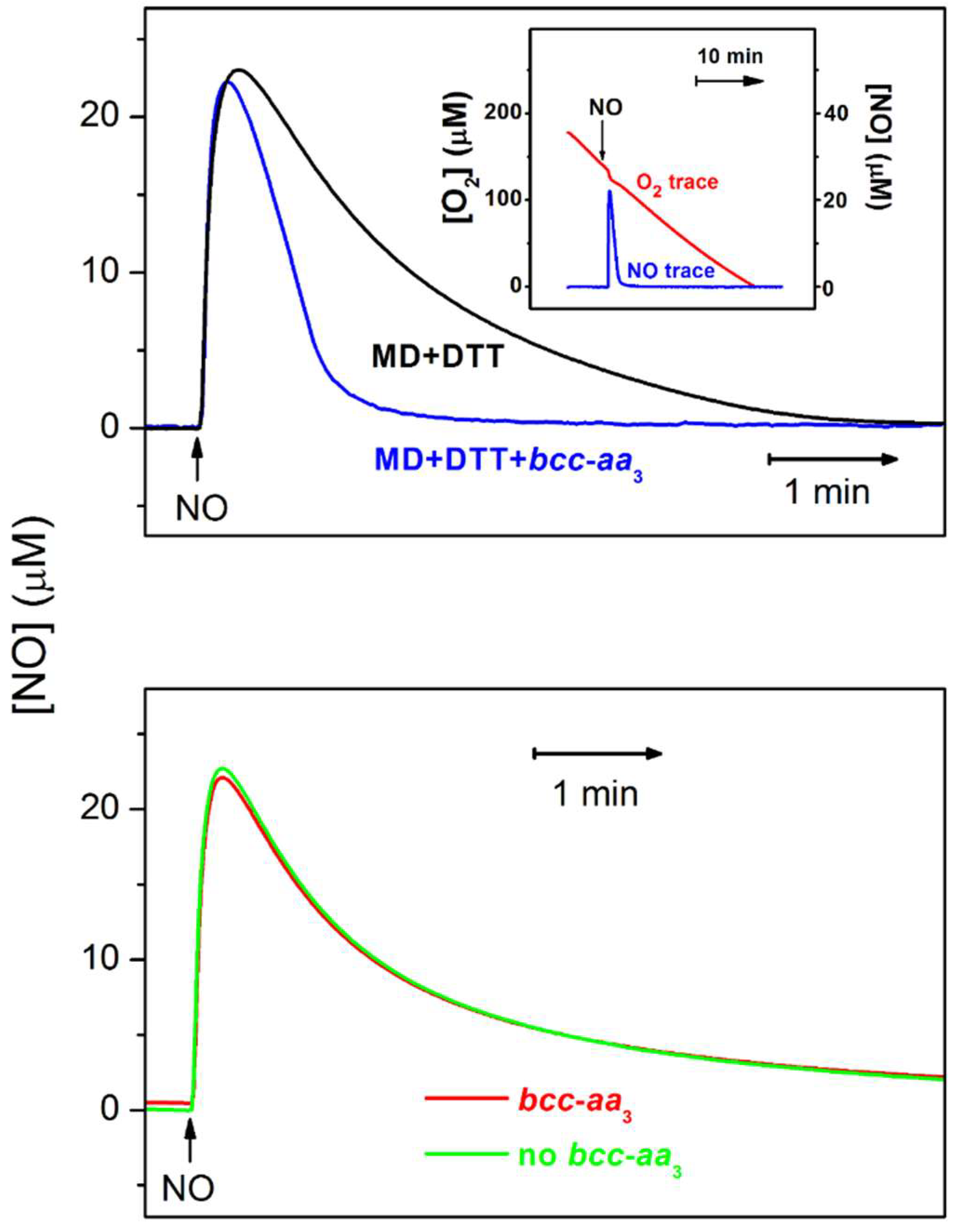
Mycobacteria contain no water-soluble cytochrome c. Probably for this reason their aa3-type cytochrome oxidase needs to be in a tight supercomplex with cytochrome bcc, a homolog of the mitochondrial cytochrome bc1 [35,36]. Forte et al. reported that a purified chimeric supercomplex composed of M. tuberculosis cytochrome bcc and M. smegmatis aa3-type oxidase resists inhibition by NO [57]. The effect of NO on the O2 consumption by the bcc-aa3 supercomplex in the presence of excess dithiothreitol (DTT) and menadione (MD) was evaluated amperometrically. A very small, short-term decrease in the O2 consumption induced by NO is followed by quick and complete restoration of the initial enzyme’s activity (Figure 3, inset). Surprisingly, the NO decay allowing for the activity recovery occurs much faster than one would expect. The reason for this turned out to be the ability of the bcc-aa3 supercomplex to degrade NO under turnover conditions. The rate of NO decay in the presence of the enzyme and reductants is significantly higher than in the presence of the reductants only (Figure 3, top panel). Furthermore, in the absence of DTT and MD, the kinetic profiles of NO decay in aerobic solution with and without the bcc-aa3 are identical (Figure 3, bottom panel). The latter two observations support the conclusion that the NO decomposition is indeed catalyzed by the purified bcc-aa3 supercomplex in turnover with O2 and the electron donors. The maximum NO-consuming activity of the enzyme measured following the addition of 30 µM NO appeared to be about 300 mol NO × (mol bcc-aa3)−1 × min−1 [57] (Table 3).
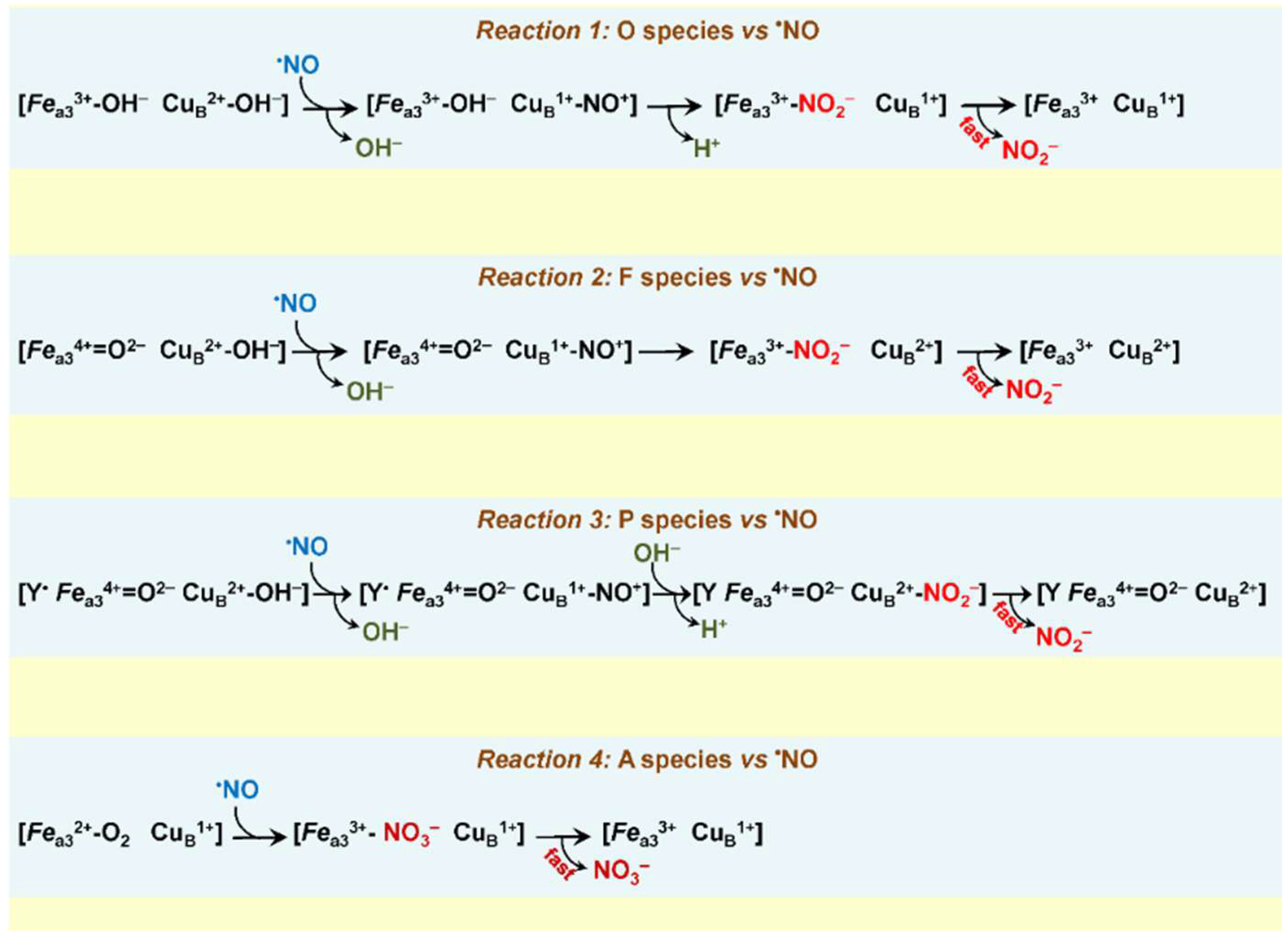
Possible mechanisms for this reaction catalyzed by the bcc-aa3 are worth discussing. Earlier, it was reported that in the mitochondrial cytochrome oxidase, NO can react with the catalytic intermediates O, P, and F, each according to a 1:1 stoichiometry [138,140]. One could suggest that in the bcc-aa3 NO also reacts with these species populated at a steady-state. In view of the fact that in the bcc-aa3 the NO/O2 stoichiometry was estimated to be 2.65 [57] i.e., >1, we assume that in this enzyme NO can react with more than one intermediate during the catalytic cycle. Figure 4 shows possible reaction pathways for the bcc-aa3 taking into account modern views on the structures of intermediates O, F, and P. As in the mitochondrial enzyme [138,140], in the reactions with O, F, and P, NO is thought to donate one electron to CuB2+ yielding nitrosonium ion (NO+) and CuB1+. This results in the oxidation of NO to NO2 and the conversion of a corresponding intermediate into the succeeding one along the catalytic cycle of the bcc-aa3 (Figure 4, reactions 1, 2, 3, see also Figure 2). In other words, following the reaction with one molecule of NO, O is converted into E, F—into O, and P—into F. In the mitochondrial cytochrome oxidase, NO2 produced from NO binds with a relatively high affinity to the oxidized heme a3 (or CuB) in the BNC [140]. This impedes the complete reduction of the BNC and, hence, its ability to bind and further reduce O2. As a result, O2 consumption is halted. We hypothesize that in the case of the bcc-aa3 NO2 generated from NO does not bind to the BNC with high affinity. Instead, NO2 is quickly ejected into the bulk phase from the supercomplex without affecting the catalytic O2 consumption.
Since the bcc-aa3 is an O2-binding heme protein, it cannot be ruled out that the enzyme is also capable of acting as a NO dioxygenase. A possible mechanism of such reaction similar to that reported for the truncated hemoglobin N of M. tuberculosis [144] is shown in Figure 4 (reaction 4). According to the proposed pathway, the reaction of the catalytic intermediate A with NO yields nitrate (NO3) that should leave the BNC rapidly in order to avoid inhibition of the main O2 reductase activity. All proposed reaction mechanisms (Figure 4, reactions 1–4) await experimental confirmation.

3.1.2. NO Reductase Activity of Heme–Copper Oxidases

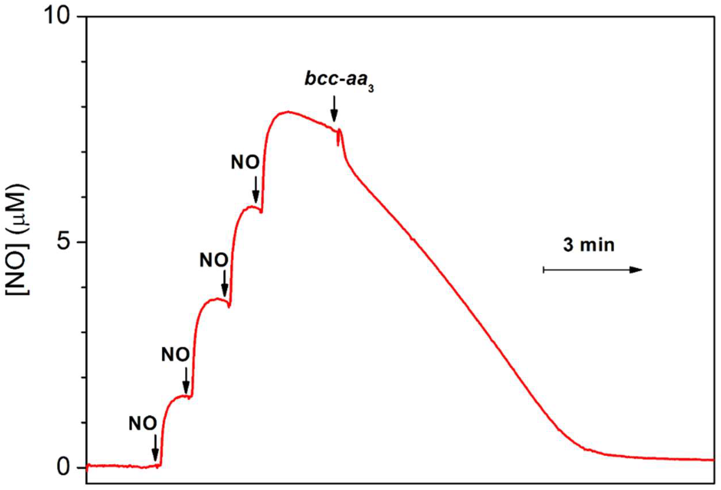
The amperometric studies showed that a few bacterial heme–copper oxidases are able to decompose NO under reducing anaerobic conditions at NO concentrations in the solution in the range of 5 to 10 µM. Figure 5 demonstrates such activity of the purified mycobacterial bcc-aa3 supercomplex [57]. The pre-reduced enzyme was anaerobically added to an O2-free solution of NO in the presence of excess DTT and MD. The addition of the enzyme was shown to increase the rate of the decomposition of NO. It has to be noted that the slow NO decay observed before the addition of the bcc-aa3 is due to the non-enzymatic reaction of NO with the reductants. Additionally, the initial fast drop in the NO concentration detected immediately after the addition of the enzyme is probably due to NO binding to the bcc-aa3. The NO-consuming activity of the bcc-aa3 under anaerobic conditions at ~8 µM NO added appeared to be about 3 mol NO × (mol bcc-aa3)−1 × min−1 [57] (Table 3). As one can see, this is ~100 times lower than that observed under aerobic turnover conditions. A similar activity was also reported previously for such heme–copper oxidases as the ba3 and caa3 from Thermus thermophilus [145], the bo3 from E. coli [146], the cbb3 from Pseudomonas stutzeri [147] and R. sphaeroides [148]. Notably, the mitochondrial beef heart aa3-type oxidase does not catalyze the anaerobic degradation of NO [149].
For the ba3 oxidase from T. thermophilus it was directly shown by gas chromatography that the end product of the catalytic NO decay under reducing anaerobic conditions is nitrous oxide (N2O), i.e., the NO reductase activity takes place [145]. It is reasonable to suggest that this is also the case for the other bacterial oxidases, which were reported to degrade NO under the same conditions [57,146,148]. The reaction mechanism could resemble that used by native bacterial NO reductases. Both mechanisms, however, are still under debate [150,151]. In general, two NO molecules react with the fully reduced BNC of the oxidase yielding one molecule of N2O as the end product, with the formation of the hyponitrite species as a transient intermediate. For more details, see Figure 23 in [151].
Since the NO reductase activity measured in some bacterial oxidases is not too high and the conditions requested hardly often occurs in vivo, we do not expect that this contributes significantly to microbial defense mechanisms against NO-induced stress.

3.2. bd-Type Oxidases Confer Bacterial Resistance to NO

Evidence is accumulating that in at least some pathogenic bacteria, cytochrome bd is involved in their defense against NO-induced stress. Jones-Carson et al. examined the role of the two major terminal oxidases of Salmonella Typhimurium, the heme–copper cytochrome bo3 (encoded by the cyoABCD operon) and cytochrome bd (encoded by the cydAB operon), in its antinitrosative defensive system [152]. The authors compared growth rates of the wild-type strain, ΔcyoABCD, and ΔcydAB mutants in LB broth supplemented with 5 mM DETA NONOate. The latter is the NO donor that at the added concentration produced a stable flux of 5 μM NO during the experiment. In contrast to the wild-type and ΔcyoABCD strains, the ΔcydAB mutant appeared to be hypersusceptible to NO as manifested by the extended lag phase following the DETA NONOate addition. Jones-Carson et al. also compared the rates of respiration in the wild-type, ΔcyoABCD, and ΔcydAB bacterial cultures treated with 50 μM spermine NONOate. The O2 consumption activity of the ΔcydAB mutant was much more sensitive to spermine NONOate as compared to that of the wild-type bacteria. Additionally, unlike the wild-type and ΔcyoABCD cells, the O2 consuming activity of the ΔcydAB cells did not improve over time following the addition of spermine NONOate. Cytochrome bd was reported to add to the NO-detoxifying activity of the flavohemoglobin Hmp that converts NO into NO3. Both Hmp and the bd oxidase contribute to similar extents to S. Typhimurium pathogenesis. Furthermore, there is a substantial degree of independence between these two proteins in S. Typhimurium pathogenesis. It is suggested that low O2 levels in mice favor NO detoxification by cytochrome bd whereas high O2 tension favor Hmp as the NO-detoxifier. Bacteria may experience different O2 and NO levels as the inflammatory response evolves over time during the infection. Therefore, S. Typhimurium may preferentially use Hmp or the bd oxidase according to the availability of O2 and NO. Thus, cytochrome bd, along with Hmp, is an important component of the antinitrosative defensive system of S. Typhimurium [152].
Shepherd et al. examined the relative contribution of cytochrome bd-I (CydAB), Hmp, the flavorubredoxin NorVW, the nitrite reductase NrfA, and the iron–sulfur cluster repair protein YtfE to the NO-tolerance mechanisms in a multidrug-resistant uropathogenic E. coli (UPEC), strain EC958 [153]. For this purpose, the authors mutated the cydAB, hmp, norVW, nrfA and ytfE genes in EC958. Growth rates of wild-type EC958, and cydAB, hmp, norVW, nrfA and ytfE mutants were measured following the addition of the NO-releaser NOC-12 under microaerobic conditions. It turned out that mutation of cydAB and hmp confers the highest sensitivity to NO. Furthermore, the ΔcydAB mutant displayed increased sensitivity to neutrophil killing, reduced survival within primed macrophages, and an attenuated colonization phenotype in the mouse bladder. The fact that deletion of cydAB impairs survival in a mouse model suggests that the bd oxidase-dependent respiration under nitrosative stress conditions is a key factor for host colonization. Thus, the UPEC cytochrome bd-I provides the greatest contribution to NO tolerance and host colonization at low O2 tensions and is of major importance for the accumulation of high microbial loads in the course of infection of the urinary tract [153].
Beebout et al. reported that cytochrome bd of UPEC (E. coli cystitis isolate UTI89) is highly expressed in biofilms and that loss of the bd-oxidase-expressing subpopulation impairs barrier function and reduces the abundance of extracellular matrix [154]. The authors hypothesized that cytochrome bd is preferentially expressed in the UPEC biofilm because the enzyme provides protection against nitrosative stress. The addition of the NO donor NOC-12 to planktonic cultures was found to significantly reduce the growth rate of the ΔcydAB mutant: the doubling time increased from 37 to 106 min after the treatment. This finding suggests that during aerobic growth the bd oxidase serves as an NO sink that reversibly sequesters NO. This protects respiration mediated by cytochrome bo3 which is a proton pump that is more efficient at transducing energy but susceptible to irreversible inhibition by NO. Beebout et al. proposed that cytochrome bd-expressing subpopulations in UPEC are critical for withstanding such harmful metabolic by-products as NO while in the biofilm state [154].
Consistently, NO caused more significant growth inhibition in non-pathogenic E. coli strains lacking cytochrome bd as compared to cytochrome bo3-deficient ones [155]. In Shewanella oneidensis, the bd oxidase provides tolerance to nitrite rather than NO, but this is an exceptional case [156]. A protective role of cytochrome bd against NO stress also agrees with the expression of this enzyme in E. coli [154,157,158], S. Typhimurium [152], Staphylococcus aureus [159], Bacillus subtilis [160], and M. tuberculosis [161] in response to NO. Interestingly, in M. tuberculosis, the bd oxidase was reported to be necessary for optimal respiration at acidic pH as the bcc-aa3 supercomplex is markedly inhibited under these conditions [162].
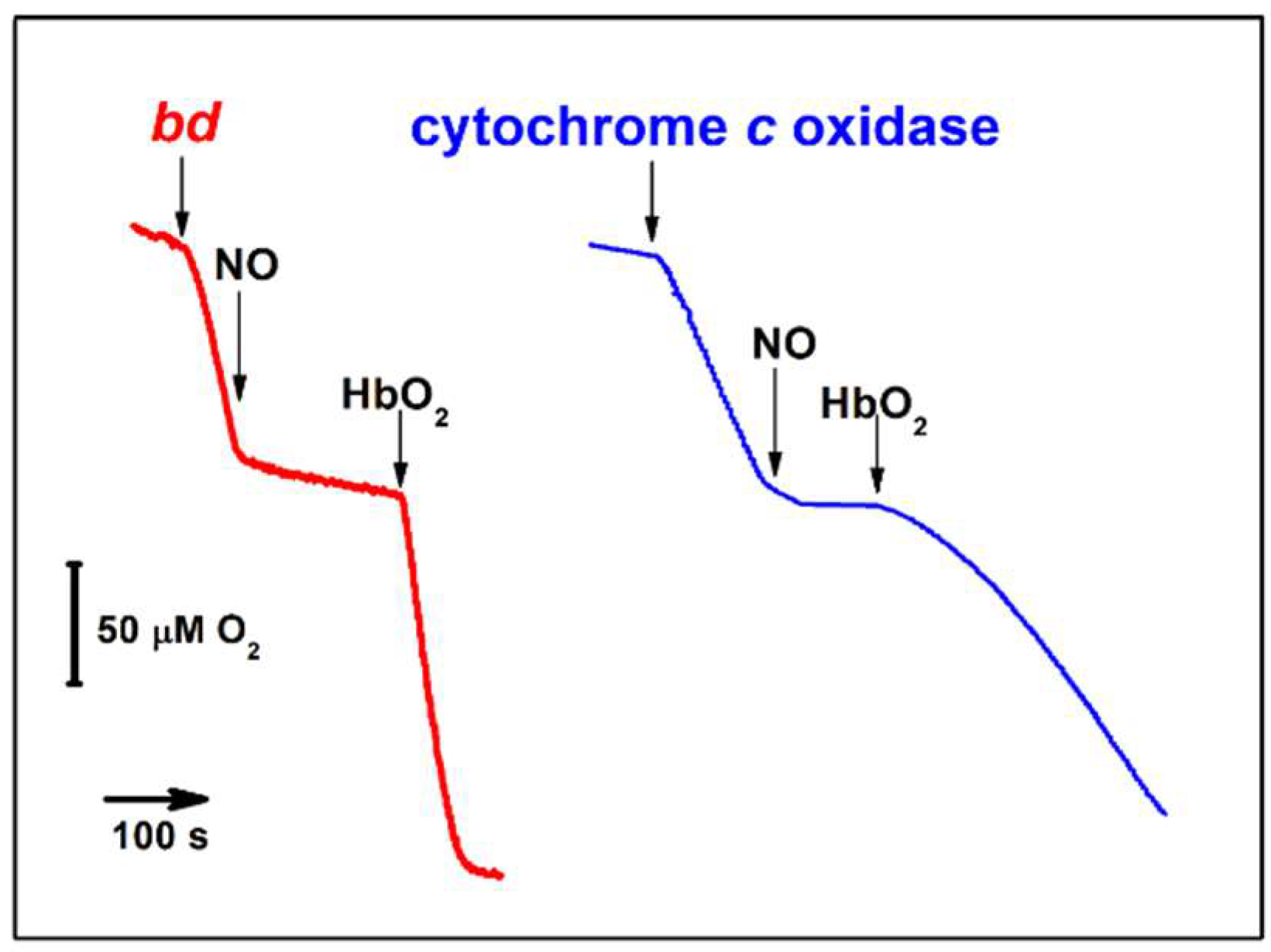
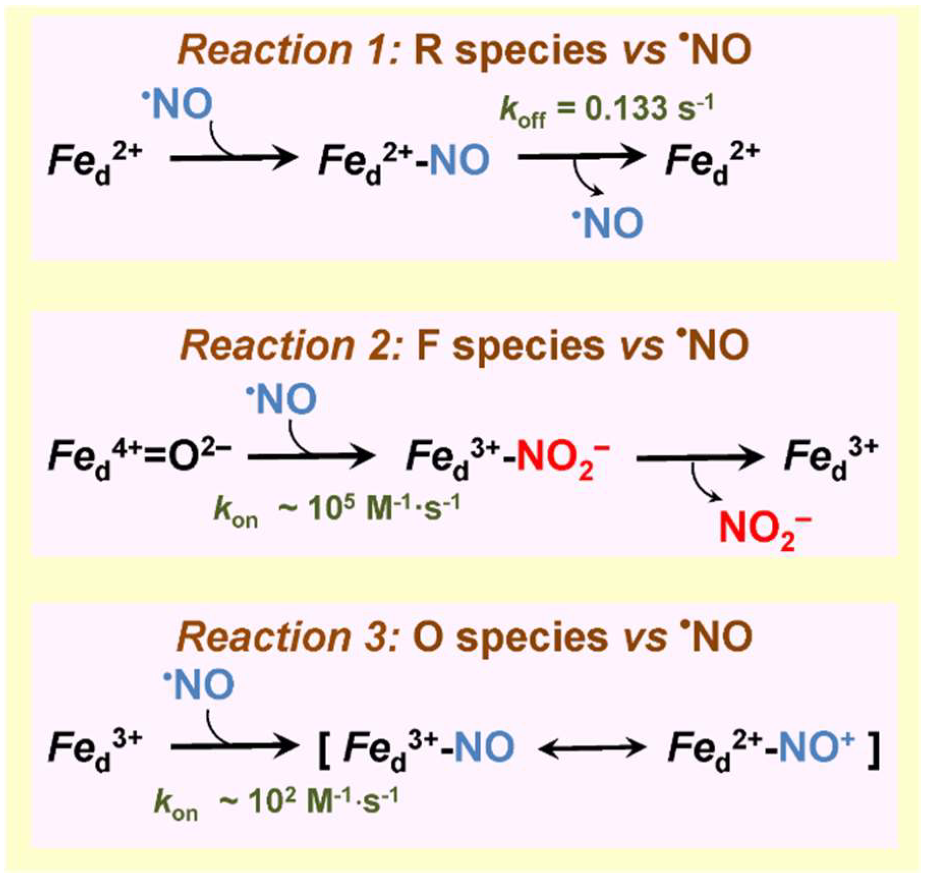
Like most heme–copper oxidases tested (see Section 3.1), the bd-type oxidases from non-pathogenic E. coli and A. vinelandii are rapidly inhibited by NO [142]. This was demonstrated on the level of both the purified enzymes from these bacteria [142] and the E. coli cells lacking cytochrome bo3 [155,163]. The inhibition is reversible with the IC50 value of 100 nM NO for the purified bd oxidases from E. coli and A. vinelandii at 70 μM O2 in the assay medium [142] (Table 3). Unlike some heme–copper oxidases (see Section 3.1.2), cytochrome bd does not exhibit a measurable NO reductase activity under anaerobic conditions. The question arises as to if cytochrome bd is quickly inhibited by submicromolar concentrations of NO and unable even scavenge this RNS via NO reductase-like reaction, how can it serve as one of the key mechanisms for protecting bacteria against nitrosative stress? Phenomenologically, the answer to this question can be obtained by comparing the kinetic profiles of activity recovery from NO inhibition following the addition of the NO scavenger oxyhemoglobin (HbO2) for the bd oxidase and the mitochondrial cytochrome c oxidase (Figure 6). Upon NO depletion in solution by HbO2, the recovery is significantly faster in cytochrome bd than in the mitochondrial oxidase under similar experimental conditions [142,164]. However, what molecular mechanisms underlie such a rapid recovery of activity in the case of the bd oxidase? Studies of the interaction of NO with different cytochrome bd species made it possible to shed light on the molecular mechanisms [142,143,165,166]. NO binds at the level of the heme d active site. The reaction occurs if heme d is in the ferrous, ferryl, or ferric state. The rate of NO binding to the ferrous uncomplexed heme d (R species) has never been measured. One may expect that its value (kon) is comparable with those for the binding of CO and O2 to the fully reduced enzyme, i.e., in the range of 108 to 109 M−1·s−1 [101]. The reaction yields the nitrosyl ferrous heme d adduct (Figure 7, reaction 1) [72]. It turned out that the rate of NO dissociation from heme d2+ (koff) in the purified fully reduced cytochrome bd-I of E. coli is unusually high, 0.133 s−1 [143] (Table 3). A similar value (0.163 s−1) was later reported for membrane preparations of E. coli mutant strain RKP4544 devoid of cytochrome bo3 [155]. This koff value is about 30 times higher than that for NO dissociation from ferrous heme a3 in the mitochondrial cytochrome c oxidase [164]. Furthermore, the NO off-rate for cytochrome bd is faster than that detected for almost all heme proteins. Such a high NO dissociation rate obviously explains why after NO-inhibition the activity of cytochrome bd is restored much faster than that of the mitochondrial oxidase (Figure 6). The reaction of NO with the A. vinelandii cytochrome bd in the ferryl state (F species) is fast (~105 M−1·s−1) and likely produces the oxidized enzyme with nitrite bound at ferric heme d (Figure 7, reaction 2) [165]. This is about 10 times faster than the same reaction for the mitochondrial cytochrome c oxidase (~104 M−1·s−1) [138,167]. Then, NO2 likely escapes from heme d3+ to the bulk phase, but the off rate for nitrite has to be determined. Since intermediate F is highly populated in turnover [105], we think that the rapid oxidation of NO into NO2 by cytochrome bd also contributes to the mechanisms of bacterial resistance to NO. The reaction of NO with ferric heme d in the purified fully oxidized cytochrome bd-I of E. coli (O species) proceeds with kon of ~ 102 M−1·s−1 yielding a nitrosyl adduct, d3+–NO or d2+–NO+ (Figure 7, reaction 3) [166]. The reaction is rather slow and the O species is not a catalytic intermediate of cytochrome bd [168] therefore it barely contributes to mechanisms of NO-inhibition or NO tolerance. Thus, we can conclude that the bd oxidase confers NO resistance to bacteria due to (i) extraordinary high NO off-rate and (ii) the ability to rapidly convert NO into NO2 in turnover.

4. Peroxynitrite and Bacterial Terminal Oxidases

The study of the interaction of peroxynitrite with bacterial terminal oxidases is at the very initial stage. To date, the only bacterial oxidase that has been studied for the reaction with this highly reactive toxic compound is cytochrome bd-I from E. coli [109,169]. Earlier, the interaction of the eukaryotic heme–copper oxidase, the aa3-type cytochrome c oxidase isolated from bovine heart mitochondria, with ONOO was investigated [170]. It was shown that the mitochondrial enzyme when solubilized or in proteoliposomes is irreversibly damaged by ONOO (Table 4). At concentrations of less than 20 µM ONOO significantly raises the enzyme’s Km for O2. This effect was tentatively explained by the nitration of some tyrosine residues [137]. At higher concentrations ONOO was reported to decrease the Vmax. The ONOO-induced lowering of the Vmax could be due to both the destruction of the CuA site in cytochrome c oxidase, and the irreversible loss of the 830-nm absorption band characteristic of the oxidized CuA was observed [170], and the degradation of hemes a and a3.
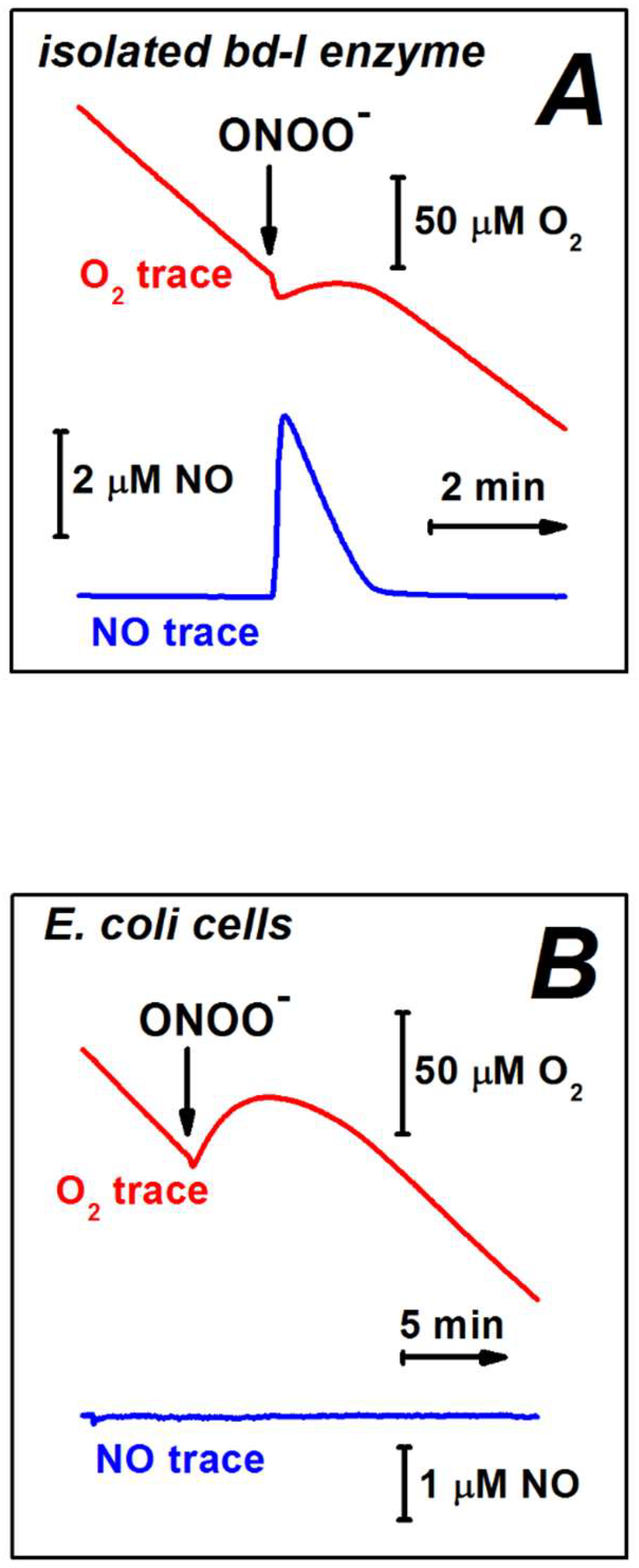
Borisov et al. studied amperometrically the effect of ONOO on the O2 consumption by the E. coli cytochrome bd-I at the level of the isolated detergent-solubilized enzyme and the bd-I overexpressing bacterial cells [169]. It turned out that in both cases, the O2 consumption by the bd-I oxidase is not inhibited by up to 0.1 mM ONOO (Figure 8, Table 4). The effect of higher ONOO concentrations was not tested. After the addition of ONOO a slight short-term generation of O2 was observed (Figure 8). This is likely due to the catalase-like activity of cytochrome bd-I that scavenges H2O2, a contaminant in the commercial ONOO or a product of the peroxynitrite degradation [109,113,114]. Furthermore, using the stopped-flow rapid mixing technique it was shown that the bd-I oxidase is able to catalyze scavenging of ONOO. The kinetics of this reaction was measured [169]. In these experiments, the enzyme pre-reduced anaerobically with excess reducing agents, N,N,N’,N’-tetramethyl-p-phenylenediamine (TMPD), and ascorbate, was mixed with an air-equilibrated solution of ONOO. The ONOO decomposition rate was determined at 310 nm. It was found that ONOO disappears with an observed rate constant that is proportional to the cytochrome bd-I concentration and increases with the TMPD concentration. Importantly, in control experiments, neither the protein nor the reductants tested independently reveal the decay of ONOO to a significant extent. The apparent turnover rate at which the bd-I oxidase, in turnover with O2 and excess TMPD and ascorbate, decomposes ONOO, was estimated to be ~600 mol ONOO × (mol enzyme)−1 × min−1 [169] (Table 4). Since the rate constant was found to increase with the enzyme activity (the electron flux), in the bacterial cell in which cytochrome bd-I utilizes ubiquinol as the substrate, the peroxynitrite-decomposing activity may be even higher. For instance, a turnover number of cytochrome bd-I is about seven times higher when the reducing system is ubiquinone-1 plus DTT as compared to that for TMPD plus ascorbate [168]. If the peroxynitrite-neutralizing activity of the bd-I oxidase is proportional to the electron flux, its apparent turnover rate in the E. coli cell could be as high as ~4200 mol ONOO × (mol enzyme)−1 × min−1. To summarize, the E. coli cytochrome bd-I in the catalytic steady state is not only resistant not ONOO, but also capable of decomposing this highly reactive cytotoxic effector, thus serving as an important detoxifier of ONOO in vivo.
A possible mechanism of the peroxynitrite decomposition catalyzed by the bd-I enzyme has never been proposed. We assume that the most likely site for the reaction is the high-spin heme d. We may suggest at least four possible reaction mechanisms. The fact that the addition of ONOO to the isolated bd-I protein in turnover with O2 and reductants resulted in the production of NO [169] (Table 4) points out that NO could be the main product. If this is the case, a one-electron reduction of ONOO to NO and H2O2 by the ferrous heme d may occur (Figure 9, reaction 1). If so, at least part of the H2O2 transiently generated following the addition of ONOO to the enzyme is also the main reaction product. There are two observations that are not consistent with the mechanism proposed. According to the reaction scheme (Figure 9, reaction 1), the decay of one molecule of ONOO added should generate one molecule of NO. In the experiments, however, the amount of NO produced was approximately 12 times less than the amount of ONOO added. In addition, no NO production was detected with the ONOO–treated cells while the short-term generation of H2O2 is in place (Figure 8). The latter two findings indicate that the NO produced in the case of the isolated enzyme might be a secondary product, possibly non-enzymatic because the formation of NO was also observed in the absence of the protein, albeit to a lesser extent [169].
It was reported that ONOO generates Compound II (Fe4+ = O2−) in myeloperoxidase, lactoperoxidase, and catalase, and Compound I (Fe4+ = O2− Por•+, where Por•+ is a porphyrin radical) in horseradish peroxidase [171,172]. Since these are ferriheme (Fe3+) enzymes, in these reactions ONOO serves as a one-electron and two-electron oxidant, respectively. We, therefore, suggest that in cytochrome bd-I ONOO also could react with the ferric heme d, (e.g., to the O1 catalytic intermediate, see Figure 2). In the case of one-electron oxidation heme d3+ is converted to Compound F (analog of Compound II, see Figure 2) with the concomitant release of NO2 from ONOO (Figure 9, reaction 2).
It is also possible that the ferric heme d catalyzes the isomerization of peroxynitrite to nitrate (NO3). If so, Compound F and NO2 are transient reaction intermediates, not the final products (Figure 9, reaction 3). The fact that certain iron (III) porphyrins are capable of catalyzing the isomerization of ONOO to NO3 [173] is in agreement with this hypothesis.
In the case of two-electron oxidation heme d3+ is converted to Compound F* (analog of Compound I, see Figure 2) with the co-production of NO2 from ONOO (Figure 9, reaction 4). It is worth noting that microbial and mammalian peroxiredoxins catalyze detoxification of peroxynitrite via its two-electron reduction to nitrite [174,175].

5. Concluding Remarks

Usually, terminal oxygen reductases of bacterial respiratory chains are strongly inhibited by nitric oxide and peroxynitrite. However, some of the respiratory enzymes, such as the mycobacterial bcc-aa3 supercomplex and bd-type oxidases, confer resistance to RNS, thereby contributing to microbial pathogenicity. An understanding of the molecular mechanisms of bacterial pathogenicity is essential for the development of new strategies to combat infectious diseases. In this regard, it would be interesting to figure out the reaction mechanisms underlying bcc-aa3 supercomplex-mediated NO detoxification and importantly, whether this unique property of the mycobacterial enzyme is shared by other aa3-type oxidases, eventually complexed with the bc1. The interest in bd-type oxidases is increasing due to their peculiar enzymatic abilities, stress tolerance, and importance to pathogens—features that merit more in-depth functional and structural studies. Determination of cytochrome bd structure from different microorganisms would help in the characterization and rational design of selective inhibitors of these oxidases. Based on already published 3D structures of bd-type oxidases, one of the main challenges in the structure-driven design of quinone substrate-like inhibitors is expected to be the high flexibility of the N-terminal part of the quinol binding site called the Q-loop. Another promising direction for future research is the study of the effect of RNS on the anaerobic terminal reductases and other bioenergetic enzymes in anaerobic pathogenic bacteria. All in all, the development of next-generation antibiotics selectively targeting the RNS-insensitive respiratory complexes in pathogens may reduce their impact on human health and social development.

Author Contributions

V.B.B. and E.F. performed the literature review and wrote the paper. All authors have read and agreed to the published version of the manuscript.

Funding

This work was funded by the Russian Science Foundation (project № 22-24-00045, https://rscf.ru/en/project/22-24-00045/ (accessed on 4 June 2022).

Institutional Review Board Statement

Not applicable.

Informed Consent Statement

Not applicable.

Data Availability Statement

Not applicable.

Conflicts of Interest

The authors declare no conflict of interest. The funders had no role in the design of the study; in the collection, analyses, or interpretation of data; in the writing of the manuscript, or in the decision to publish the results.

References

  1. Martinez, M.C.; Andriantsitohaina, R. Reactive nitrogen species: Molecular mechanisms and potential significance in health and disease. Antioxid. Redox Signal. 2009, 11, 669–702. [Google Scholar] [CrossRef] [PubMed]
  2. Wareham, L.K.; Southam, H.M.; Poole, R.K. Do nitric oxide, carbon monoxide and hydrogen sulfide really qualify as ‘gasotransmitters’ in bacteria? Biochem. Soc. Trans. 2018, 46, 1107–1118. [Google Scholar] [CrossRef] [Green Version]
  3. Bryan, N.S.; Lefer, D.J. Update on gaseous signaling molecules nitric oxide and hydrogen sulfide: Strategies to capture their functional activity for human therapeutics. Mol. Pharmacol. 2019, 96, 109–114. [Google Scholar] [CrossRef] [PubMed]
  4. Mendes, S.S.; Miranda, V.; Saraiva, L.M. Hydrogen sulfide and carbon monoxide tolerance in bacteria. Antioxidants 2021, 10, 729. [Google Scholar] [CrossRef] [PubMed]
  5. Borisov, V.B.; Forte, E. Impact of hydrogen sulfide on mitochondrial and bacterial bioenergetics. Int. J. Mol. Sci. 2021, 22, 12688. [Google Scholar] [CrossRef] [PubMed]
  6. Radi, R. Oxygen radicals, nitric oxide, and peroxynitrite: Redox pathways in molecular medicine. Proc. Natl. Acad. Sci. USA 2018, 115, 5839–5848. [Google Scholar] [CrossRef] [PubMed] [Green Version]
  7. Perez de la Lastra, J.M.; Juan, C.A.; Plou, F.J.; Perez-Lebena, E. The nitration of proteins, lipids and DNA by peroxynitrite derivatives-chemistry involved and biological relevance. Stresses 2022, 2, 3. [Google Scholar] [CrossRef]
  8. Fang, F.C.; Vazquez-Torres, A. Reactive nitrogen species in host-bacterial interactions. Curr. Opin. Immunol. 2019, 60, 96–102. [Google Scholar] [CrossRef]
  9. Carvalho, S.M.; Beas, J.Z.; Videira, M.A.M.; Saraiva, L.M. Defenses of multidrug resistant pathogens against reactive nitrogen species produced in infected hosts. Adv. Microb. Physiol. 2022, 80, 85–155. [Google Scholar] [CrossRef]
  10. Gusarov, I.; Nudler, E. Protein S-nitrosylation: Enzymatically controlled, but intrinsically unstable, post-translational modification. Mol. Cell 2018, 69, 351–353. [Google Scholar] [CrossRef] [Green Version]
  11. Srinivasan, V.B.; Mondal, A.; Venkataramaiah, M.; Chauhan, N.K.; Rajamohan, G. Role of oxyRKP, a novel LysR-family transcriptional regulator, in antimicrobial resistance and virulence in Klebsiella pneumoniae. Microbiology (Reading) 2013, 159, 1301–1314. [Google Scholar] [CrossRef] [PubMed] [Green Version]
  12. Anes, J.; Dever, K.; Eshwar, A.; Nguyen, S.; Cao, Y.; Sivasankaran, S.K.; Sakalauskaite, S.; Lehner, A.; Devineau, S.; Daugelavicius, R.; et al. Analysis of the oxidative stress regulon identifies soxS as a genetic target for resistance reversal in multidrug-resistant Klebsiella pneumoniae. mBio 2021, 12, e0086721. [Google Scholar] [CrossRef] [PubMed]
  13. Way, S.S.; Goldberg, M.B. Clearance of Shigella flexneri infection occurs through a nitric oxide-independent mechanism. Infect. Immun. 1998, 66, 3012–3016. [Google Scholar] [CrossRef] [PubMed] [Green Version]
  14. Rodionov, D.A.; Dubchak, I.L.; Arkin, A.P.; Alm, E.J.; Gelfand, M.S. Dissimilatory metabolism of nitrogen oxides in bacteria: Comparative reconstruction of transcriptional networks. PLoS Comput. Biol. 2005, 1, e55. [Google Scholar] [CrossRef] [PubMed]
  15. Kint, N.; Alves Feliciano, C.; Martins, M.C.; Morvan, C.; Fernandes, S.F.; Folgosa, F.; Dupuy, B.; Texeira, M.; Martin-Verstraete, I. How the anaerobic enteropathogen Clostridioides difficile tolerates low O2 tensions. mBio 2020, 11, e01559-01520. [Google Scholar] [CrossRef]
  16. Folgosa, F.; Martins, M.C.; Teixeira, M. The multidomain flavodiiron protein from Clostridium difficile 630 is an NADH:oxygen oxidoreductase. Sci. Rep. 2018, 8, 10164. [Google Scholar] [CrossRef]
  17. Kumar, M.; Adhikari, S.; Hurdle, J.G. Action of nitroheterocyclic drugs against Clostridium difficile. Int. J. Antimicrob. Agents 2014, 44, 314–319. [Google Scholar] [CrossRef] [Green Version]
  18. Caruana, N.J.; Stroud, D.A. The road to the structure of the mitochondrial respiratory chain supercomplex. Biochem. Soc. Trans. 2020, 48, 621–629. [Google Scholar] [CrossRef] [Green Version]
  19. Cogliati, S.; Cabrera-Alarcon, J.L.; Enriquez, J.A. Regulation and functional role of the electron transport chain supercomplexes. Biochem. Soc. Trans. 2021, 49, 2655–2668. [Google Scholar] [CrossRef]
  20. Sharma, P.; Maklashina, E.; Cecchini, G.; Iverson, T.M. Crystal structure of an assembly intermediate of respiratory Complex II. Nat. Commun. 2018, 9, 274. [Google Scholar] [CrossRef]
  21. Hederstedt, L. Molecular biology of Bacillus subtilis cytochromes anno 2020. Biochemistry 2021, 86, 8–21. [Google Scholar] [CrossRef] [PubMed]
  22. Melo, A.M.; Teixeira, M. Supramolecular organization of bacterial aerobic respiratory chains: From cells and back. Biochim. Biophys. Acta 2016, 1857, 190–197. [Google Scholar] [CrossRef] [PubMed]
  23. Kaila, V.R.I.; Wikstrom, M. Architecture of bacterial respiratory chains. Nat. Rev. Microbiol. 2021, 19, 319–330. [Google Scholar] [CrossRef] [PubMed]
  24. Nuber, F.; Merono, L.; Oppermann, S.; Schimpf, J.; Wohlwend, D.; Friedrich, T. A quinol anion as catalytic intermediate coupling proton translocation with electron transfer in E. coli respiratory complex I. Front. Chem. 2021, 9, 672969. [Google Scholar] [CrossRef]
  25. Grba, D.N.; Blaza, J.N.; Bridges, H.R.; Agip, A.A.; Yin, Z.; Murai, M.; Miyoshi, H.; Hirst, J. Cryo-electron microscopy reveals how acetogenins inhibit mitochondrial respiratory complex I. J. Biol. Chem. 2022, 298, 101602. [Google Scholar] [CrossRef]
  26. Marreiros, B.C.; Sena, F.V.; Sousa, F.M.; Batista, A.P.; Pereira, M.M. Type II NADH:quinone oxidoreductase family: Phylogenetic distribution, structural diversity and evolutionary divergences. Environ. Microbiol. 2016, 18, 4697–4709. [Google Scholar] [CrossRef]
  27. Blaza, J.N.; Bridges, H.R.; Aragao, D.; Dunn, E.A.; Heikal, A.; Cook, G.M.; Nakatani, Y.; Hirst, J. The mechanism of catalysis by type-II NADH:quinone oxidoreductases. Sci. Rep. 2017, 7, 40165. [Google Scholar] [CrossRef] [Green Version]
  28. Bertsova, Y.V.; Baykov, A.A.; Bogachev, A.V. A simple strategy to differentiate between H+- and Na+-transporting NADH:quinone oxidoreductases. Arch. Biochem. Biophys. 2020, 681, 108266. [Google Scholar] [CrossRef]
  29. Liang, P.; Fang, X.; Hu, Y.; Yuan, M.; Raba, D.A.; Ding, J.; Bunn, D.C.; Sanjana, K.; Yang, J.; Rosas-Lemus, M.; et al. The aerobic respiratory chain of Pseudomonas aeruginosa cultured in artificial urine media: Role of NQR and terminal oxidases. PLoS ONE 2020, 15, e0231965. [Google Scholar] [CrossRef]
  30. Hreha, T.N.; Foreman, S.; Duran-Pinedo, A.; Morris, A.R.; Diaz-Rodriguez, P.; Jones, J.A.; Ferrara, K.; Bourges, A.; Rodriguez, L.; Koffas, M.A.G.; et al. The three NADH dehydrogenases of Pseudomonas aeruginosa: Their roles in energy metabolism and links to virulence. PLoS ONE 2021, 16, e0244142. [Google Scholar] [CrossRef]
  31. Kozlova, M.I.; Bushmakin, I.M.; Belyaeva, J.D.; Shalaeva, D.N.; Dibrova, D.V.; Cherepanov, D.A.; Mulkidjanian, A.Y. Expansion of the "Sodium World" through evolutionary time and taxonomic space. Biochemistry 2020, 85, 1518–1542. [Google Scholar] [CrossRef] [PubMed]
  32. Wilson, C.A.; Crofts, A.R. Dissecting the pattern of proton release from partial process involved in ubihydroquinone oxidation in the Q-cycle. Biochim. Biophys. Acta Bioenerg. 2018, 1859, 531–543. [Google Scholar] [CrossRef] [PubMed]
  33. Francia, F.; Khalfaoui-Hassani, B.; Lanciano, P.; Musiani, F.; Noodleman, L.; Venturoli, G.; Daldal, F. The cytochrome b lysine 329 residue is critical for ubihydroquinone oxidation and proton release at the Qo site of bacterial cytochrome bc1. Biochim. Biophys. Acta Bioenerg. 2019, 1860, 167–179. [Google Scholar] [CrossRef] [PubMed]
  34. Borisov, V.B.; Verkhovsky, M.I. Oxygen as Acceptor. EcoSal Plus 2015, 6. [Google Scholar] [CrossRef] [PubMed]
  35. Wiseman, B.; Nitharwal, R.G.; Fedotovskaya, O.; Schafer, J.; Guo, H.; Kuang, Q.; Benlekbir, S.; Sjostrand, D.; Adelroth, P.; Rubinstein, J.L.; et al. Structure of a functional obligate complex III2IV2 respiratory supercomplex from Mycobacterium smegmatis. Nat. Struct. Mol. Biol. 2018, 25, 1128–1136. [Google Scholar] [CrossRef] [PubMed]
  36. Gong, H.; Li, J.; Xu, A.; Tang, Y.; Ji, W.; Gao, R.; Wang, S.; Yu, L.; Tian, C.; Li, J.; et al. An electron transfer path connects subunits of a mycobacterial respiratory supercomplex. Science 2018, 362, eaat8923. [Google Scholar] [CrossRef] [Green Version]
  37. Kao, W.C.; Ortmann de Percin Northumberland, C.; Cheng, T.C.; Ortiz, J.; Durand, A.; von Loeffelholz, O.; Schilling, O.; Biniossek, M.L.; Klaholz, B.P.; Hunte, C. Structural basis for safe and efficient energy conversion in a respiratory supercomplex. Nat. Commun. 2022, 13, 545. [Google Scholar] [CrossRef]
  38. Fedotovskaya, O.; Albertsson, I.; Nordlund, G.; Hong, S.; Gennis, R.B.; Brzezinski, P.; Adelroth, P. Identification of a cytochrome bc1-aa3 supercomplex in Rhodobacter sphaeroides. Biochim. Biophys. Acta Bioenerg. 2021, 1862, 148433. [Google Scholar] [CrossRef]
  39. Friedrich, T.; Wohlwend, D.; Borisov, V.B. Recent advances in structural studies of cytochrome bd and its potential application as a drug target. Int. J. Mol. Sci. 2022, 23, 3166. [Google Scholar] [CrossRef]
  40. Vilcheze, C.; Weinrick, B.; Leung, L.W.; Jacobs, W.R., Jr. Plasticity of Mycobacterium tuberculosis NADH dehydrogenases and their role in virulence. Proc. Natl. Acad. Sci. USA 2018, 115, 1599–1604. [Google Scholar] [CrossRef] [Green Version]
  41. Siletsky, S.A.; Borisov, V.B. Proton pumping and non-pumping terminal respiratory oxidases: Active sites intermediates of these molecular machines and their derivatives. Int. J. Mol. Sci. 2021, 22, 10852. [Google Scholar] [CrossRef] [PubMed]
  42. Murali, R.; Gennis, R.B.; Hemp, J. Evolution of the cytochrome bd oxygen reductase superfamily and the function of CydAA’ in Archaea. ISME J. 2021, 15, 3534–3548. [Google Scholar] [CrossRef] [PubMed]
  43. Borisov, V.B.; Siletsky, S.A.; Paiardini, A.; Hoogewijs, D.; Forte, E.; Giuffre, A.; Poole, R.K. Bacterial oxidases of the cytochrome bd family: Redox enzymes of unique structure, function and utility as drug targets. Antioxid. Redox Signal. 2021, 34, 1280–1318. [Google Scholar] [CrossRef] [PubMed]
  44. Siletsky, S.A.; Borisov, V.B.; Mamedov, M.D. Photosystem II and terminal respiratory oxidases: Molecular machines operating in opposite directions. Front. Biosci. (Landmark Ed.) 2017, 22, 1379–1426. [Google Scholar] [CrossRef] [PubMed] [Green Version]
  45. Malatesta, F.; Antonini, G.; Sarti, P.; Brunori, M. Structure and function of a molecular machine: Cytochrome c oxidase. Biophys. Chem. 1995, 54, 1–33. [Google Scholar] [CrossRef]
  46. Pereira, M.M.; Sousa, F.L.; Verissimo, A.F.; Teixeira, M. Looking for the minimum common denominator in haem-copper oxygen reductases: Towards a unified catalytic mechanism. Biochim. Biophys. Acta 2008, 1777, 929–934. [Google Scholar] [CrossRef] [Green Version]
  47. Papa, S.; Capitanio, N.; Capitanio, G.; Palese, L.L. Protonmotive cooperativity in cytochrome c oxidase. Biochim. Biophys. Acta 2004, 1658, 95–105. [Google Scholar] [CrossRef] [Green Version]
  48. Borisov, V.B.; Siletsky, S.A. Features of organization and mechanism of catalysis of two families of terminal oxidases: Heme-copper and bd-type. Biochemistry 2019, 84, 1390–1402. [Google Scholar] [CrossRef]
  49. Borisov, V.B.; Siletsky, S.A.; Nastasi, M.R.; Forte, E. ROS defense systems and terminal oxidases in bacteria. Antioxidants 2021, 10, 839. [Google Scholar] [CrossRef]
  50. Sousa, F.L.; Alves, R.J.; Ribeiro, M.A.; Pereira-Leal, J.B.; Teixeira, M.; Pereira, M.M. The superfamily of heme-copper oxygen reductases: Types and evolutionary considerations. Biochim. Biophys. Acta 2012, 1817, 629–637. [Google Scholar] [CrossRef] [Green Version]
  51. Wikstrom, M.; Krab, K.; Sharma, V. Oxygen activation and energy conservation by cytochrome c oxidase. Chem. Rev. 2018, 118, 2469–2490. [Google Scholar] [CrossRef] [PubMed] [Green Version]
  52. Capitanio, N.; Palese, L.L.; Capitanio, G.; Martino, P.L.; Richter, O.M.; Ludwig, B.; Papa, S. Allosteric interactions and proton conducting pathways in proton pumping aa3 oxidases: Heme a as a key coupling element. Biochim. Biophys. Acta 2012, 1817, 558–566. [Google Scholar] [CrossRef] [PubMed] [Green Version]
  53. Maneg, O.; Malatesta, F.; Ludwig, B.; Drosou, V. Interaction of cytochrome c with cytochrome oxidase: Two different docking scenarios. Biochim. Biophys. Acta 2004, 1655, 274–281. [Google Scholar] [CrossRef] [PubMed] [Green Version]
  54. von Ballmoos, C.; Adelroth, P.; Gennis, R.B.; Brzezinski, P. Proton transfer in ba3 cytochrome c oxidase from Thermus thermophilus. Biochim. Biophys. Acta 2012, 1817, 650–657. [Google Scholar] [CrossRef] [PubMed] [Green Version]
  55. Rich, P.R. Mitochondrial cytochrome c oxidase: Catalysis, coupling and controversies. Biochem. Soc. Trans. 2017, 45, 813–829. [Google Scholar] [CrossRef]
  56. Yoshikawa, S.; Shimada, A. Reaction mechanism of cytochrome c oxidase. Chem. Rev. 2015, 115, 1936–1989. [Google Scholar] [CrossRef]
  57. Forte, E.; Giuffre, A.; Huang, L.S.; Berry, E.A.; Borisov, V.B. Nitric oxide does not inhibit but is metabolized by the cytochrome bcc-aa3 supercomplex. Int. J. Mol. Sci. 2020, 21, 8521. [Google Scholar] [CrossRef]
  58. Borisov, V.B. Defects in mitochondrial respiratory complexes III and IV, and human pathologies. Mol. Aspects Med. 2002, 23, 385–412. [Google Scholar] [CrossRef]
  59. Borisov, V.B. Mutations in respiratory chain complexes and human diseases. Ital. J. Biochem. 2004, 53, 34–40. [Google Scholar]
  60. Pereira, M.M.; Santana, M.; Teixeira, M. A novel scenario for the evolution of haem-copper oxygen reductases. Biochim. Biophys. Acta 2001, 1505, 185–208. [Google Scholar] [CrossRef] [Green Version]
  61. Pereira, M.M.; Gomes, C.M.; Teixeira, M. Plasticity of proton pathways in haem-copper oxygen reductases. FEBS Lett. 2002, 522, 14–18. [Google Scholar] [CrossRef] [Green Version]
  62. Pereira, M.M.; Teixeira, M. Proton pathways, ligand binding and dynamics of the catalytic site in haem-copper oxygen reductases: A comparison between the three families. Biochim. Biophys. Acta 2004, 1655, 340–346. [Google Scholar] [CrossRef] [PubMed] [Green Version]
  63. Safarian, S.; Rajendran, C.; Muller, H.; Preu, J.; Langer, J.D.; Ovchinnikov, S.; Hirose, T.; Kusumoto, T.; Sakamoto, J.; Michel, H. Structure of a bd oxidase indicates similar mechanisms for membrane-integrated oxygen reductases. Science 2016, 352, 583–586. [Google Scholar] [CrossRef] [Green Version]
  64. Thesseling, A.; Rasmussen, T.; Burschel, S.; Wohlwend, D.; Kagi, J.; Muller, R.; Bottcher, B.; Friedrich, T. Homologous bd oxidases share the same architecture but differ in mechanism. Nat. Commun. 2019, 10, 5138. [Google Scholar] [CrossRef] [PubMed] [Green Version]
  65. Safarian, S.; Hahn, A.; Mills, D.J.; Radloff, M.; Eisinger, M.L.; Nikolaev, A.; Meier-Credo, J.; Melin, F.; Miyoshi, H.; Gennis, R.B.; et al. Active site rearrangement and structural divergence in prokaryotic respiratory oxidases. Science 2019, 366, 100–104. [Google Scholar] [CrossRef]
  66. Wang, W.; Gao, Y.; Tang, Y.; Zhou, X.; Lai, Y.; Zhou, S.; Zhang, Y.; Yang, X.; Liu, F.; Guddat, L.W.; et al. Cryo-EM structure of mycobacterial cytochrome bd reveals two oxygen access channels. Nat. Commun. 2021, 12, 4621. [Google Scholar] [CrossRef]
  67. Safarian, S.; Opel-Reading, H.K.; Wu, D.; Mehdipour, A.R.; Hards, K.; Harold, L.K.; Radloff, M.; Stewart, I.; Welsch, S.; Hummer, G.; et al. The cryo-EM structure of the bd oxidase from M. tuberculosis reveals a unique structural framework and enables rational drug design to combat TB. Nat. Commun. 2021, 12, 5236. [Google Scholar] [CrossRef]
  68. Grauel, A.; Kagi, J.; Rasmussen, T.; Makarchuk, I.; Oppermann, S.; Moumbock, A.F.A.; Wohlwend, D.; Muller, R.; Melin, F.; Gunther, S.; et al. Structure of Escherichia coli cytochrome bd-II type oxidase with bound aurachin D. Nat. Commun. 2021, 12, 6498. [Google Scholar] [CrossRef]
  69. Grund, T.N.; Radloff, M.; Wu, D.; Goojani, H.G.; Witte, L.F.; Josting, W.; Buschmann, S.; Muller, H.; Elamri, I.; Welsch, S.; et al. Mechanistic and structural diversity between cytochrome bd isoforms of Escherichia coli. Proc. Natl. Acad. Sci. USA 2021, 118, e2114013118. [Google Scholar] [CrossRef]
  70. Hill, J.J.; Alben, J.O.; Gennis, R.B. Spectroscopic evidence for a heme-heme binuclear center in the cytochrome bd ubiquinol oxidase from Escherichia coli. Proc. Natl. Acad. Sci. USA 1993, 90, 5863–5867. [Google Scholar] [CrossRef] [Green Version]
  71. Tsubaki, M.; Hori, H.; Mogi, T.; Anraku, Y. Cyanide-binding site of bd-type ubiquinol oxidase from Escherichia coli. J. Biol. Chem. 1995, 270, 28565–28569. [Google Scholar] [CrossRef] [PubMed] [Green Version]
  72. Borisov, V.; Arutyunyan, A.M.; Osborne, J.P.; Gennis, R.B.; Konstantinov, A.A. Magnetic circular dichroism used to examine the interaction of Escherichia coli cytochrome bd with ligands. Biochemistry 1999, 38, 740–750. [Google Scholar] [CrossRef] [PubMed]
  73. Vos, M.H.; Borisov, V.B.; Liebl, U.; Martin, J.L.; Konstantinov, A.A. Femtosecond resolution of ligand-heme interactions in the high-affinity quinol oxidase bd: A di-heme active site? Proc. Natl. Acad. Sci. USA 2000, 97, 1554–1559. [Google Scholar] [CrossRef] [PubMed] [Green Version]
  74. Borisov, V.B.; Sedelnikova, S.E.; Poole, R.K.; Konstantinov, A.A. Interaction of cytochrome bd with carbon monoxide at low and room temperatures: Evidence that only a small fraction of heme b595 reacts with CO. J. Biol. Chem. 2001, 276, 22095–22099. [Google Scholar] [CrossRef] [PubMed] [Green Version]
  75. Borisov, V.B.; Liebl, U.; Rappaport, F.; Martin, J.L.; Zhang, J.; Gennis, R.B.; Konstantinov, A.A.; Vos, M.H. Interactions between heme d and heme b595 in quinol oxidase bd from Escherichia coli: A photoselection study using femtosecond spectroscopy. Biochemistry 2002, 41, 1654–1662. [Google Scholar] [CrossRef] [Green Version]
  76. Arutyunyan, A.M.; Borisov, V.B.; Novoderezhkin, V.I.; Ghaim, J.; Zhang, J.; Gennis, R.B.; Konstantinov, A.A. Strong excitonic interactions in the oxygen-reducing site of bd-type oxidase: The Fe-to-Fe distance between hemes d and b595 is 10 A. Biochemistry 2008, 47, 1752–1759. [Google Scholar] [CrossRef]
  77. Borisov, V.B. Interaction of bd-type quinol oxidase from Escherichia coli and carbon monoxide: Heme d binds CO with high affinity. Biochemistry 2008, 73, 14–22. [Google Scholar] [CrossRef]
  78. Bloch, D.A.; Borisov, V.B.; Mogi, T.; Verkhovsky, M.I. Heme/heme redox interaction and resolution of individual optical absorption spectra of the hemes in cytochrome bd from Escherichia coli. Biochim. Biophys. Acta 2009, 1787, 1246–1253. [Google Scholar] [CrossRef] [Green Version]
  79. Rappaport, F.; Zhang, J.; Vos, M.H.; Gennis, R.B.; Borisov, V.B. Heme-heme and heme-ligand interactions in the di-heme oxygen-reducing site of cytochrome bd from Escherichia coli revealed by nanosecond absorption spectroscopy. Biochim. Biophys. Acta 2010, 1797, 1657–1664. [Google Scholar] [CrossRef]
  80. Borisov, V.B.; Verkhovsky, M.I. Accommodation of CO in the di-heme active site of cytochrome bd terminal oxidase from Escherichia coli. J. Inorg. Biochem. 2013, 118, 65–67. [Google Scholar] [CrossRef]
  81. Siletsky, S.A.; Zaspa, A.A.; Poole, R.K.; Borisov, V.B. Microsecond time-resolved absorption spectroscopy used to study CO compounds of cytochrome bd from Escherichia coli. PLoS ONE 2014, 9, e95617. [Google Scholar] [CrossRef] [PubMed]
  82. Siletsky, S.A.; Rappaport, F.; Poole, R.K.; Borisov, V.B. Evidence for fast electron transfer between the high-spin haems in cytochrome bd-I from Escherichia coli. PLoS ONE 2016, 11, e0155186. [Google Scholar] [CrossRef]
  83. Siletsky, S.A.; Dyuba, A.V.; Elkina, D.A.; Monakhova, M.V.; Borisov, V.B. Spectral-kinetic analysis of recombination reaction of heme centers of bd-type quinol oxidase from Escherichia coli with carbon monoxide. Biochemistry 2017, 82, 1354–1366. [Google Scholar] [CrossRef]
  84. Borisov, V.B. Effect of membrane environment on ligand-binding properties of the terminal oxidase cytochrome bd-I from Escherichia coli. Biochemistry 2020, 85, 1603–1612. [Google Scholar] [CrossRef] [PubMed]
  85. Gavrikova, E.V.; Grivennikova, V.G.; Borisov, V.B.; Cecchini, G.; Vinogradov, A.D. Assembly of a chimeric respiratory chain from bovine heart submitochondrial particles and cytochrome bd terminal oxidase of Escherichia coli. FEBS Lett. 2009, 583, 1287–1291. [Google Scholar] [CrossRef] [PubMed] [Green Version]
  86. Azarkina, N.; Borisov, V.; Konstantinov, A.A. Spontaneous spectral changes of the reduced cytochrome bd. FEBS Lett. 1997, 416, 171–174. [Google Scholar] [CrossRef] [Green Version]
  87. Puustinen, A.; Finel, M.; Haltia, T.; Gennis, R.B.; Wikstrom, M. Properties of the two terminal oxidases of Escherichia coli. Biochemistry 1991, 30, 3936–3942. [Google Scholar] [CrossRef]
  88. Jasaitis, A.; Borisov, V.B.; Belevich, N.P.; Morgan, J.E.; Konstantinov, A.A.; Verkhovsky, M.I. Electrogenic reactions of cytochrome bd. Biochemistry 2000, 39, 13800–13809. [Google Scholar] [CrossRef]
  89. Belevich, I.; Borisov, V.B.; Zhang, J.; Yang, K.; Konstantinov, A.A.; Gennis, R.B.; Verkhovsky, M.I. Time-resolved electrometric and optical studies on cytochrome bd suggest a mechanism of electron-proton coupling in the di-heme active site. Proc. Natl. Acad. Sci. USA 2005, 102, 3657–3662. [Google Scholar] [CrossRef] [Green Version]
  90. Belevich, I.; Borisov, V.B.; Verkhovsky, M.I. Discovery of the true peroxy intermediate in the catalytic cycle of terminal oxidases by real-time measurement. J. Biol. Chem. 2007, 282, 28514–28519. [Google Scholar] [CrossRef] [Green Version]
  91. Borisov, V.B.; Belevich, I.; Bloch, D.A.; Mogi, T.; Verkhovsky, M.I. Glutamate 107 in subunit I of cytochrome bd from Escherichia coli is part of a transmembrane intraprotein pathway conducting protons from the cytoplasm to the heme b595/heme d active site. Biochemistry 2008, 47, 7907–7914. [Google Scholar] [CrossRef] [PubMed]
  92. Borisov, V.B. Cytochrome bd: Structure and properties. Biochemistry 1996, 61, 565–574. [Google Scholar]
  93. Junemann, S. Cytochrome bd terminal oxidase. Biochim. Biophys. Acta 1997, 1321, 107–127. [Google Scholar] [CrossRef] [PubMed] [Green Version]
  94. Forte, E.; Borisov, V.B.; Vicente, J.B.; Giuffre, A. Cytochrome bd and gaseous ligands in bacterial physiology. Adv. Microb. Physiol. 2017, 71, 171–234. [Google Scholar] [CrossRef] [PubMed]
  95. Borisov, V.B.; Smirnova, I.A.; Krasnosel’skaya, I.A.; Konstantinov, A.A. Oxygenated cytochrome bd from Escherichia coli can be converted into the oxidized form by lipophilic electron acceptors. Biochemistry 1994, 59, 437–443. [Google Scholar]
  96. D’mello, R.; Hill, S.; Poole, R.K. The cytochrome bd quinol oxidase in Escherichia coli has an extremely high oxygen affinity and two-oxygen-binding haems: Implicaitons for regulation of activity in vivo by oxygen inihibition. Microbiology 1996, 142, 755–763. [Google Scholar] [CrossRef] [Green Version]
  97. Belevich, I.; Borisov, V.B.; Konstantinov, A.A.; Verkhovsky, M.I. Oxygenated complex of cytochrome bd from Escherichia coli: Stability and photolability. FEBS Lett. 2005, 579, 4567–4570. [Google Scholar] [CrossRef] [Green Version]
  98. Belevich, I.; Borisov, V.B.; Bloch, D.A.; Konstantinov, A.A.; Verkhovsky, M.I. Cytochrome bd from Azotobacter vinelandii: Evidence for high-affinity oxygen binding. Biochemistry 2007, 46, 11177–11184. [Google Scholar] [CrossRef]
  99. Forte, E.; Borisov, V.B.; Siletsky, S.A.; Petrosino, M.; Giuffre, A. In the respiratory chain of Escherichia coli cytochromes bd-I and bd-II are more sensitive to carbon monoxide inhibition than cytochrome bo3. Biochim. Biophys. Acta Bioenerg. 2019, 1860, 148088. [Google Scholar] [CrossRef]
  100. Azarkina, N.; Siletsky, S.; Borisov, V.; von Wachenfeldt, C.; Hederstedt, L.; Konstantinov, A.A. A cytochrome bb’-type quinol oxidase in Bacillus subtilis strain 168. J. Biol. Chem. 1999, 274, 32810–32817. [Google Scholar] [CrossRef] [Green Version]
  101. Borisov, V.B.; Gennis, R.B.; Hemp, J.; Verkhovsky, M.I. The cytochrome bd respiratory oxygen reductases. Biochim. Biophys. Acta 2011, 1807, 1398–1413. [Google Scholar] [CrossRef] [PubMed] [Green Version]
  102. Arutyunyan, A.M.; Sakamoto, J.; Inadome, M.; Kabashima, Y.; Borisov, V.B. Optical and magneto-optical activity of cytochrome bd from Geobacillus thermodenitrificans. Biochim. Biophys. Acta 2012, 1817, 2087–2094. [Google Scholar] [CrossRef] [PubMed] [Green Version]
  103. Borisov, V.B.; Gennis, R.B.; Konstantinov, A.A. Interaction of cytochrome bd from Escherichia coli with hydrogen peroxide. Biochemistry 1995, 60, 231–239. [Google Scholar]
  104. Borisov, V.; Gennis, R.; Konstantinov, A.A. Peroxide complex of cytochrome bd: Kinetics of generation and stability. Biochem. Mol. Biol. Int. 1995, 37, 975–982. [Google Scholar]
  105. Borisov, V.B.; Forte, E.; Sarti, P.; Giuffre, A. Catalytic intermediates of cytochrome bd terminal oxidase at steady-state: Ferryl and oxy-ferrous species dominate. Biochim. Biophys. Acta 2011, 1807, 503–509. [Google Scholar] [CrossRef]
  106. Paulus, A.; Rossius, S.G.; Dijk, M.; de Vries, S. Oxoferryl-porphyrin radical catalytic intermediate in cytochrome bd oxidases protects cells from formation of reactive oxygen species. J. Biol. Chem. 2012, 287, 8830–8838. [Google Scholar] [CrossRef] [Green Version]
  107. Borisov, V.B.; Murali, R.; Verkhovskaya, M.L.; Bloch, D.A.; Han, H.; Gennis, R.B.; Verkhovsky, M.I. Aerobic respiratory chain of Escherichia coli is not allowed to work in fully uncoupled mode. Proc. Natl. Acad. Sci. USA 2011, 108, 17320–17324. [Google Scholar] [CrossRef] [Green Version]
  108. Forte, E.; Borisov, V.B.; Konstantinov, A.A.; Brunori, M.; Giuffre, A.; Sarti, P. Cytochrome bd, a key oxidase in bacterial survival and tolerance to nitrosative stress. Ital. J. Biochem. 2007, 56, 265–269. [Google Scholar]
  109. Borisov, V.B.; Forte, E.; Siletsky, S.A.; Arese, M.; Davletshin, A.I.; Sarti, P.; Giuffre, A. Cytochrome bd protects bacteria against oxidative and nitrosative stress: A potential target for next-generation antimicrobial agents. Biochemistry 2015, 80, 565–575. [Google Scholar] [CrossRef]
  110. Giuffre, A.; Borisov, V.B.; Mastronicola, D.; Sarti, P.; Forte, E. Cytochrome bd oxidase and nitric oxide: From reaction mechanisms to bacterial physiology. FEBS Lett. 2012, 586, 622–629. [Google Scholar] [CrossRef]
  111. Giuffre, A.; Borisov, V.B.; Arese, M.; Sarti, P.; Forte, E. Cytochrome bd oxidase and bacterial tolerance to oxidative and nitrosative stress. Biochim. Biophys. Acta 2014, 1837, 1178–1187. [Google Scholar] [CrossRef] [PubMed] [Green Version]
  112. Borisov, V.B.; Davletshin, A.I.; Konstantinov, A.A. Peroxidase activity of cytochrome bd from Escherichia coli. Biochemistry 2010, 75, 428–436. [Google Scholar] [CrossRef] [PubMed]
  113. Borisov, V.B.; Forte, E.; Davletshin, A.; Mastronicola, D.; Sarti, P.; Giuffre, A. Cytochrome bd oxidase from Escherichia coli displays high catalase activity: An additional defense against oxidative stress. FEBS Lett. 2013, 587, 2214–2218. [Google Scholar] [CrossRef] [PubMed]
  114. Forte, E.; Borisov, V.B.; Davletshin, A.; Mastronicola, D.; Sarti, P.; Giuffre, A. Cytochrome bd oxidase and hydrogen peroxide resistance in Mycobacterium tuberculosis. mBio 2013, 4, e01006-01013. [Google Scholar] [CrossRef] [Green Version]
  115. Al-Attar, S.; Yu, Y.; Pinkse, M.; Hoeser, J.; Friedrich, T.; Bald, D.; de Vries, S. Cytochrome bd displays significant quinol peroxidase activity. Sci. Rep. 2016, 6, 27631. [Google Scholar] [CrossRef] [PubMed] [Green Version]
  116. Forte, E.; Nastasi, M.R.; Borisov, V.B. Preparations of terminal oxidase cytochrome bd-II isolated from Escherichia coli reveal significant hydrogen peroxide scavenging activity. Biochemistry, 2022; in press. [Google Scholar]
  117. Forte, E.; Borisov, V.B.; Falabella, M.; Colaco, H.G.; Tinajero-Trejo, M.; Poole, R.K.; Vicente, J.B.; Sarti, P.; Giuffre, A. The terminal oxidase cytochrome bd promotes sulfide-resistant bacterial respiration and growth. Sci. Rep. 2016, 6, 23788. [Google Scholar] [CrossRef] [Green Version]
  118. Korshunov, S.; Imlay, K.R.; Imlay, J.A. The cytochrome bd oxidase of Escherichia coli prevents respiratory inhibition by endogenous and exogenous hydrogen sulfide. Mol. Microbiol. 2016, 101, 62–77. [Google Scholar] [CrossRef] [Green Version]
  119. Forte, E.; Giuffre, A. How bacteria breathe in hydrogen sulphide-rich environments. Biochemist 2016, 38, 8–11. [Google Scholar] [CrossRef]
  120. Borisov, V.B.; Forte, E. Terminal oxidase cytochrome bd protects bacteria against hydrogen sulfide toxicity. Biochemistry 2021, 86, 22–32. [Google Scholar] [CrossRef]
  121. Forte, E.; Siletsky, S.A.; Borisov, V.B. In Escherichia coli ammonia inhibits cytochrome bo3 but activates cytochrome bd-I. Antioxidants 2021, 10, 13. [Google Scholar] [CrossRef]
  122. Xia, X.; Wu, S.; Li, L.; Xu, B.; Wang, G. The cytochrome bd complex is essential for chromate and sulfide resistance and is regulated by a GbsR-type regulator, CydE, in Alishewanella sp. WH16-1. Front. Microbiol. 2018, 9, 1849. [Google Scholar] [CrossRef] [PubMed]
  123. Sakamoto, J.; Koga, E.; Mizuta, T.; Sato, C.; Noguchi, S.; Sone, N. Gene structure and quinol oxidase activity of a cytochrome bd-type oxidase from Bacillus stearothermophilus. Biochim. Biophys. Acta 1999, 1411, 147–158. [Google Scholar] [CrossRef] [Green Version]
  124. Kalia, N.P.; Hasenoehrl, E.J.; Ab Rahman, N.B.; Koh, V.H.; Ang, M.L.T.; Sajorda, D.R.; Hards, K.; Gruber, G.; Alonso, S.; Cook, G.M.; et al. Exploiting the synthetic lethality between terminal respiratory oxidases to kill Mycobacterium tuberculosis and clear host infection. Proc. Natl. Acad. Sci. USA 2017, 114, 7426–7431. [Google Scholar] [CrossRef] [PubMed] [Green Version]
  125. Lee, B.S.; Hards, K.; Engelhart, C.A.; Hasenoehrl, E.J.; Kalia, N.P.; Mackenzie, J.S.; Sviriaeva, E.; Chong, S.M.S.; Manimekalai, M.S.S.; Koh, V.H.; et al. Dual inhibition of the terminal oxidases eradicates antibiotic-tolerant Mycobacterium tuberculosis. EMBO Mol. Med. 2021, 13, e13207. [Google Scholar] [CrossRef] [PubMed]
  126. Meunier, B.; Madgwick, S.A.; Reil, E.; Oettmeier, W.; Rich, P.R. New inhibitors of the quinol oxidation sites of bacterial cytochromes bo and bd. Biochemistry 1995, 34, 1076–1083. [Google Scholar] [CrossRef]
  127. Radloff, M.; Elamri, I.; Grund, T.N.; Witte, L.F.; Hohmann, K.F.; Nakagaki, S.; Goojani, H.G.; Nasiri, H.; Hideto, M.; Bald, D.; et al. Short-chain aurachin D derivatives are selective inhibitors of E. coli cytochrome bd-I and bd-II oxidases. Sci. Rep. 2021, 11, 23852. [Google Scholar] [CrossRef]
  128. Miyoshi, H.; Takegami, K.; Sakamoto, K.; Mogi, T.; Iwamura, H. Characterization of the ubiquinol oxidation sites in cytochromes bo and bd from Escherichia coli using aurachin C analogues. J. Biochem. 1999, 125, 138–142. [Google Scholar] [CrossRef]
  129. Makarchuk, I.; Nikolaev, A.; Thesseling, A.; Dejon, L.; Lamberty, D.; Stief, L.; Speicher, A.; Friedrich, T.; Hellwig, P.; Nasiri, H.R.; et al. Identification and optimization of quinolone-based inhibitors against cytochrome bd oxidase using an electrochemical assay. Electrochim. Acta 2021, 381, 138293. [Google Scholar] [CrossRef]
  130. Lu, P.; Heineke, M.H.; Koul, A.; Andries, K.; Cook, G.M.; Lill, H.; van Spanning, R.; Bald, D. The cytochrome bd-type quinol oxidase is important for survival of Mycobacterium smegmatis under peroxide and antibiotic-induced stress. Sci. Rep. 2015, 5, 10333. [Google Scholar] [CrossRef] [Green Version]
  131. Harikishore, A.; Chong, S.S.M.; Ragunathan, P.; Bates, R.W.; Gruber, G. Targeting the menaquinol binding loop of mycobacterial cytochrome bd oxidase. Mol. Divers. 2021, 25, 517–524. [Google Scholar] [CrossRef]
  132. Hopfner, S.M.; Lee, B.S.; Kalia, N.P.; Miller, M.J.; Pethe, K.; Moraski, G.C. Structure guided generation of thieno[3,2-d]pyrimidin-4-amine Mycobacterium tuberculosis bd oxidase inhibitors. RSC Med. Chem. 2021, 12, 73–77. [Google Scholar] [CrossRef] [PubMed]
  133. Anand, P.; Akhter, Y. A review on enzyme complexes of electron transport chain from Mycobacterium tuberculosis as promising drug targets. Int. J. Biol. Macromol. 2022, 212, 474–494. [Google Scholar] [CrossRef] [PubMed]
  134. Hards, K.; Cheung, C.Y.; Waller, N.; Adolph, C.; Keighley, L.; Tee, Z.S.; Harold, L.K.; Menorca, A.; Bujaroski, R.S.; Buckley, B.J.; et al. An amiloride derivative is active against the F1Fo-ATP synthase and cytochrome bd oxidase of Mycobacterium tuberculosis. Commun. Biol. 2022, 5, 166. [Google Scholar] [CrossRef]
  135. Arjona, D.; Wikstrom, M.; Adelroth, P. Nitric oxide is a potent inhibitor of the cbb3-type heme-copper oxidases. FEBS Lett. 2015, 589, 1214–1218. [Google Scholar] [CrossRef] [PubMed] [Green Version]
  136. Brown, G.C.; Cooper, C.E. Nanomolar concentrations of nitric oxide reversibly inhibit synaptosomal respiration by competing with oxygen at cytochrome oxidase. FEBS Lett. 1994, 356, 295–298. [Google Scholar] [CrossRef] [Green Version]
  137. Cooper, C.E.; Davies, N.A.; Psychoulis, M.; Canevari, L.; Bates, T.E.; Dobbie, M.S.; Casley, C.S.; Sharpe, M.A. Nitric oxide and peroxynitrite cause irreversible increases in the Km for oxygen of mitochondrial cytochrome oxidase: In Vitro and In Vivo studies. Biochim. Biophys. Acta 2003, 1607, 27–34. [Google Scholar] [CrossRef] [Green Version]
  138. Torres, J.; Cooper, C.E.; Wilson, M.T. A common mechanism for the interaction of nitric oxide with the oxidized binuclear centre and oxygen intermediates of cytochrome c oxidase. J. Biol. Chem. 1998, 273, 8756–8766. [Google Scholar] [CrossRef] [Green Version]
  139. Mason, M.G.; Nicholls, P.; Wilson, M.T.; Cooper, C.E. Nitric oxide inhibition of respiration involves both competitive (heme) and noncompetitive (copper) binding to cytochrome c oxidase. Proc. Natl. Acad. Sci. USA 2006, 103, 708–713. [Google Scholar] [CrossRef] [Green Version]
  140. Sarti, P.; Forte, E.; Mastronicola, D.; Giuffre, A.; Arese, M. Cytochrome c oxidase and nitric oxide in action: Molecular mechanisms and pathophysiological implications. Biochim. Biophys. Acta 2012, 1817, 610–619. [Google Scholar] [CrossRef]
  141. Chen, J.; Xie, P.; Huang, Y.; Gao, H. Complex interplay of heme-copper oxidases with nitrite and nitric oxide. Int. J. Mol. Sci. 2022, 23, 979. [Google Scholar] [CrossRef]
  142. Borisov, V.B.; Forte, E.; Konstantinov, A.A.; Poole, R.K.; Sarti, P.; Giuffre, A. Interaction of the bacterial terminal oxidase cytochrome bd with nitric oxide. FEBS Lett. 2004, 576, 201–204. [Google Scholar] [CrossRef] [PubMed] [Green Version]
  143. Borisov, V.B.; Forte, E.; Sarti, P.; Brunori, M.; Konstantinov, A.A.; Giuffre, A. Redox control of fast ligand dissociation from Escherichia coli cytochrome bd. Biochem. Biophys. Res. Commun. 2007, 355, 97–102. [Google Scholar] [CrossRef] [PubMed]
  144. Carabet, L.A.; Guertin, M.; Lague, P.; Lamoureux, G. Mechanism of the nitric oxide dioxygenase reaction of Mycobacterium tuberculosis hemoglobin N. J. Phys. Chem. B 2017, 121, 8706–8718. [Google Scholar] [CrossRef] [PubMed]
  145. Giuffre, A.; Stubauer, G.; Sarti, P.; Brunori, M.; Zumft, W.G.; Buse, G.; Soulimane, T. The heme-copper oxidases of Thermus thermophilus catalyze the reduction of nitric oxide: Evolutionary implications. Proc. Natl. Acad. Sci. USA 1999, 96, 14718–14723. [Google Scholar] [CrossRef] [Green Version]
  146. Butler, C.; Forte, E.; Maria Scandurra, F.; Arese, M.; Giuffre, A.; Greenwood, C.; Sarti, P. Cytochrome bo3 from Escherichia coli: The binding and turnover of nitric oxide. Biochem. Biophys. Res. Commun. 2002, 296, 1272–1278. [Google Scholar] [CrossRef]
  147. Forte, E.; Urbani, A.; Saraste, M.; Sarti, P.; Brunori, M.; Giuffre, A. The cytochrome cbb3 from Pseudomonas stutzeri displays nitric oxide reductase activity. Eur. J. Biochem. 2001, 268, 6486–6491. [Google Scholar] [CrossRef]
  148. Huang, Y.; Reimann, J.; Lepp, H.; Drici, N.; Adelroth, P. Vectorial proton transfer coupled to reduction of O2 and NO by a heme-copper oxidase. Proc. Natl. Acad. Sci. USA 2008, 105, 20257–20262. [Google Scholar] [CrossRef] [Green Version]
  149. Stubauer, G.; Giuffre, A.; Brunori, M.; Sarti, P. Cytochrome c oxidase does not catalyze the anaerobic reduction of NO. Biochem. Biophys. Res. Commun. 1998, 245, 459–465. [Google Scholar] [CrossRef]
  150. Ohta, T.; Soulimane, T.; Kitagawa, T.; Varotsis, C. Nitric oxide activation by caa3 oxidoreductase from Thermus thermophilus. Phys. Chem. Chem. Phys. 2015, 17, 10894–10898. [Google Scholar] [CrossRef] [Green Version]
  151. Blomberg, M.R.A. Activation of O2 and NO in heme-copper oxidases-mechanistic insights from computational modelling. Chem. Soc. Rev. 2020, 49, 7301–7330. [Google Scholar] [CrossRef]
  152. Jones-Carson, J.; Husain, M.; Liu, L.; Orlicky, D.J.; Vazquez-Torres, A. Cytochrome bd-dependent bioenergetics and antinitrosative defenses in Salmonella pathogenesis. mBio 2016, 7, e02052-02016. [Google Scholar] [CrossRef] [PubMed] [Green Version]
  153. Shepherd, M.; Achard, M.E.; Idris, A.; Totsika, M.; Phan, M.D.; Peters, K.M.; Sarkar, S.; Ribeiro, C.A.; Holyoake, L.V.; Ladakis, D.; et al. The cytochrome bd-I respiratory oxidase augments survival of multidrug-resistant Escherichia coli during infection. Sci. Rep. 2016, 6, 35285. [Google Scholar] [CrossRef] [PubMed]
  154. Beebout, C.J.; Eberly, A.R.; Werby, S.H.; Reasoner, S.A.; Brannon, J.R.; De, S.; Fitzgerald, M.J.; Huggins, M.M.; Clayton, D.B.; Cegelski, L.; et al. Respiratory heterogeneity shapes biofilm formation and host colonization in uropathogenic Escherichia coli. mBio 2019, 10, e02400-18. [Google Scholar] [CrossRef] [PubMed] [Green Version]
  155. Mason, M.G.; Shepherd, M.; Nicholls, P.; Dobbin, P.S.; Dodsworth, K.S.; Poole, R.K.; Cooper, C.E. Cytochrome bd confers nitric oxide resistance to Escherichia coli. Nat. Chem. Biol. 2009, 5, 94–96. [Google Scholar] [CrossRef] [PubMed]
  156. Meng, Q.; Yin, J.; Jin, M.; Gao, H. Distinct nitrite and nitric oxide physiologies in Escherichia coli and Shewanella oneidensis. Appl. Environ. Microbiol. 2018, 84, e00559-00518. [Google Scholar] [CrossRef] [PubMed] [Green Version]
  157. Pullan, S.T.; Gidley, M.D.; Jones, R.A.; Barrett, J.; Stevanin, T.M.; Read, R.C.; Green, J.; Poole, R.K. Nitric oxide in chemostat-cultured Escherichia coli is sensed by Fnr and other global regulators: Unaltered methionine biosynthesis indicates lack of S nitrosation. J. Bacteriol. 2007, 189, 1845–1855. [Google Scholar] [CrossRef] [Green Version]
  158. Hyduke, D.R.; Jarboe, L.R.; Tran, L.M.; Chou, K.J.; Liao, J.C. Integrated network analysis identifies nitric oxide response networks and dihydroxyacid dehydratase as a crucial target in Escherichia coli. Proc. Natl. Acad. Sci. USA 2007, 104, 8484–8489. [Google Scholar] [CrossRef] [Green Version]
  159. Richardson, A.R.; Dunman, P.M.; Fang, F.C. The nitrosative stress response of Staphylococcus aureus is required for resistance to innate immunity. Mol. Microbiol. 2006, 61, 927–939. [Google Scholar] [CrossRef]
  160. Moore, C.M.; Nakano, M.M.; Wang, T.; Ye, R.W.; Helmann, J.D. Response of Bacillus subtilis to nitric oxide and the nitrosating agent sodium nitroprusside. J. Bacteriol. 2004, 186, 4655–4664. [Google Scholar] [CrossRef] [Green Version]
  161. Shi, L.; Sohaskey, C.D.; Kana, B.D.; Dawes, S.; North, R.J.; Mizrahi, V.; Gennaro, M.L. Changes in energy metabolism of Mycobacterium tuberculosis in mouse lung and under In Vitro conditions affecting aerobic respiration. Proc. Natl. Acad. Sci. USA 2005, 102, 15629–15634. [Google Scholar] [CrossRef] [Green Version]
  162. Cai, Y.; Jaecklein, E.; Mackenzie, J.S.; Papavinasasundaram, K.; Olive, A.J.; Chen, X.; Steyn, A.J.C.; Sassetti, C.M. Host immunity increases Mycobacterium tuberculosis reliance on cytochrome bd oxidase. PLoS Pathog. 2021, 17, e1008911. [Google Scholar] [CrossRef] [PubMed]
  163. Stevanin, T.M.; Ioannidis, N.; Mills, C.E.; Kim, S.O.; Hughes, M.N.; Poole, R.K. Flavohemoglobin Hmp affords inducible protection for Escherichia coli respiration, catalyzed by cytochromes bo’ or bd, from nitric oxide. J. Biol. Chem. 2000, 275, 35868–35875. [Google Scholar] [CrossRef] [PubMed] [Green Version]
  164. Sarti, P.; Giuffre, A.; Forte, E.; Mastronicola, D.; Barone, M.C.; Brunori, M. Nitric oxide and cytochrome c oxidase: Mechanisms of inhibition and NO degradation. Biochem. Biophys. Res. Commun. 2000, 274, 183–187. [Google Scholar] [CrossRef] [PubMed]
  165. Borisov, V.B.; Forte, E.; Sarti, P.; Brunori, M.; Konstantinov, A.A.; Giuffre, A. Nitric oxide reacts with the ferryl-oxo catalytic intermediate of the CuB-lacking cytochrome bd terminal oxidase. FEBS Lett. 2006, 580, 4823–4826. [Google Scholar] [CrossRef] [PubMed] [Green Version]
  166. Borisov, V.B.; Forte, E.; Giuffre, A.; Konstantinov, A.; Sarti, P. Reaction of nitric oxide with the oxidized di-heme and heme-copper oxygen-reducing centers of terminal oxidases: Different reaction pathways and end-products. J. Inorg. Biochem. 2009, 103, 1185–1187. [Google Scholar] [CrossRef]
  167. Giuffre, A.; Barone, M.C.; Mastronicola, D.; D’Itri, E.; Sarti, P.; Brunori, M. Reaction of nitric oxide with the turnover intermediates of cytochrome c oxidase: Reaction pathway and functional effects. Biochemistry 2000, 39, 15446–15453. [Google Scholar] [CrossRef]
  168. Yang, K.; Borisov, V.B.; Konstantinov, A.A.; Gennis, R.B. The fully oxidized form of the cytochrome bd quinol oxidase from E. coli does not participate in the catalytic cycle: Direct evidence from rapid kinetics studies. FEBS Lett. 2008, 582, 3705–3709. [Google Scholar] [CrossRef] [Green Version]
  169. Borisov, V.B.; Forte, E.; Siletsky, S.A.; Sarti, P.; Giuffre, A. Cytochrome bd from Escherichia coli catalyzes peroxynitrite decomposition. Biochim. Biophys. Acta 2015, 1847, 182–188. [Google Scholar] [CrossRef] [Green Version]
  170. Sharpe, M.A.; Cooper, C.E. Interaction of peroxynitrite with mitochondrial cytochrome oxidase. Catalytic production of nitric oxide and irreversible inhibition of enzyme activity. J. Biol. Chem. 1998, 273, 30961–30972. [Google Scholar] [CrossRef] [Green Version]
  171. Floris, R.; Piersma, S.R.; Yang, G.; Jones, P.; Wever, R. Interaction of myeloperoxidase with peroxynitrite. A comparison with lactoperoxidase, horseradish peroxidase and catalase. Eur. J. Biochem. 1993, 215, 767–775. [Google Scholar] [CrossRef]
  172. Van Zyl, J.M.; Van der Walt, B.J. Apparent hydroxyl radical generation without transition metal catalysis and tyrosine nitration during oxidation of the anti-tubercular drug, isonicotinic acid hydrazide. Biochem. Pharmacol. 1994, 48, 2033–2042. [Google Scholar] [CrossRef]
  173. Stern, M.K.; Jensen, M.P.; Kramer, K. Peroxynitrite decomposition catalysts. J. Am. Chem. Soc. 1996, 118, 8735–8736. [Google Scholar] [CrossRef]
  174. Bryk, R.; Griffin, P.; Nathan, C. Peroxynitrite reductase activity of bacterial peroxiredoxins. Nature 2000, 407, 211–215. [Google Scholar] [CrossRef] [PubMed]
  175. Ferrer-Sueta, G.; Campolo, N.; Trujillo, M.; Bartesaghi, S.; Carballal, S.; Romero, N.; Alvarez, B.; Radi, R. Biochemistry of peroxynitrite and protein tyrosine nitration. Chem. Rev. 2018, 118, 1338–1408. [Google Scholar] [CrossRef] [PubMed]
Figure 1. Aerobic respiratory chains of Escherichia coli, Pseudomonas aeruginosa, and Mycobacterium tuberculosis. In E. coli, two NADH dehydrogenases, NDH-1 and NDH-2, and succinate dehydrogenase (SDH) transfer electrons to ubiquinone (UQ)/menaquinone (MQ) pool. Three quinol oxidases, cytochromes bo3, bd-I, and bd-II, oxidize ubiquinol/menaquinol with the concomitant reduction of O2 to 2H2O. P. aeruginosa has three NADH dehydrogenases, NDH-1, NDH-2, NQR, and SDH. The electrons from ubiquinol are further transferred to O2 either directly via two quinol oxidases, cytochrome bo3 and bd-type cyanide insensitive oxidase (CIO), or via the bc1 complex to three cytochrome c oxidases, caa3, cbb3-1, and cbb3-2. M. tuberculosis possesses three NADH dehydrogenases, one NDH-1, two NDH-2, and two succinate dehydrogenases, SDH-1 and SDH-2. The electrons from menaquinol are then transferred to O2 via cytochrome bd or cytochrome bcc-aa3 supercomplex.
Ijms 23 07321 g001
Figure 2. Proposed catalytic cycles of heme–copper cytochrome c oxidase and bd-type oxidase.
Ijms 23 07321 g002
Figure 3. Purified mycobacterial cytochrome bcc-aa3 supercomplex scavenges NO under turnover conditions. Top panel: the bcc-aa3 in turnover with 5 mM DTT and 0.26 mM MD accelerates the decomposition of 30 µM NO added. (Bottom panel) in the absence of DTT and MD, i.e., under non-turnover conditions, the bcc-aa3 does not accelerate the decomposition of 30 µM NO added. (Inset) the effect of 30 µM NO on the O2 consumption by the bcc-aa3. The Figure was modified from Forte et al. [57] under the terms of the Creative Commons Attribution 4.0 International License.
Ijms 23 07321 g003
Figure 4. Possible mechanisms of the NO detoxification catalyzed by the mycobacterial cytochrome bcc-aa3 supercomplex under turnover conditions. Y in Reaction 3—a conserved tyrosine residue in the BNC.
Ijms 23 07321 g004
Figure 5. NO reductase activity of the mycobacterial cytochrome bcc-aa3 supercomplex. Four aliquots of 2.1 µM NO were sequentially added to degassed buffer containing 5 mM DTT, 0.26 mM MD, 5 mM glucose, and 16 units/mL glucose oxidase. Then, the pre-reduced cytochrome bcc-aa3 (200 nM) was added. The Figure was modified from Forte et al. [57] under the terms of the Creative Commons Attribution 4.0 International License.
Ijms 23 07321 g005
Figure 6. Activity recovery from NO inhibition of E. coli cytochrome bd-I and beef heart cytochrome c oxidase. Shown are time courses of O2 consumption by the enzymes. NO inhibits the enzymatic O2 consumption. Oxyhemoglobin (HbO2) scavenges rapidly all free NO that leads to reversal of NO inhibition. Modified from [111] with permission.
Ijms 23 07321 g006
Figure 7. Reactions of NO with different cytochrome bd species.
Ijms 23 07321 g007
Figure 8. The effect of peroxynitrite on the O2 consumption by cytochrome bd-I from E. coli. (A) 50 µM ONOO was added to the isolated enzyme in the presence of 10 mM ascorbate and 0.5 mM TMPD. (B) 80 µM ONOO was added to the cell suspension of the E. coli strain GO105/pTK1 overexpressing cytochrome bd-I. The NO concentration was measured in parallel. Modified from [169] with permission.
Ijms 23 07321 g008
Figure 9. Possible mechanisms of the peroxynitrite decomposition catalyzed by cytochrome bd-I from E. coli.
Ijms 23 07321 g009
Table 1. Major enzyme complexes of the mammalian mitochondrial electron transport chain.
Enzyme ComplexElectron DonorElectron AcceptorEnergy Currency Produced
Complex INADHubiquinoneproton motive force (PMF)
Complex IIsuccinateubiquinonenone
Complex IIIubiquinolferricytochrome cPMF
Complex IVferrocytochrome cO2PMF
Table 2. Major enzyme complexes found in aerobic bacterial electron transport chains.
Enzyme ComplexElectron DonorElectron AcceptorEnergy Currency Produced
NDH-1NADHquinonePMF
NDH-2NADHquinonenone
NQRNADHquinoneNa+ motive force
Complex IIsuccinatequinonenone
Complex IIIquinolferricytochrome cPMF
Heme–copper oxidases (aa3, caa3, bo3, cbb3, ba3)ferrocytochrome c or quinolO2PMF
Cytochrome bcc-aa3 supercomplexquinolO2PMF
Cytochrome bd (bd-I, bd-II)quinolO2PMF
Cyanide insensitive bd-type oxidase (CIO)quinolO2n.d.
Table 3. Overview of NO interactions with mycobacterial cytochrome bcc-aa3 supercomplex and E. coli cytochrome bd-I, respiratory enzyme complexes which contribute to mechanisms of bacterial resistance to NO.
Enzyme ComplexInhibition by NONO Degradation in TurnoverAnaerobic NO DegradationNO off-RateNO2 off-RateReference
Mycobacterial cytochrome bcc-aa3 supercomplexNoYes (~300 mol NO × (mol bcc-aa3)−1 × min−1)Yes (~3 mol NO × (mol bcc-aa3)−1 × min−1)n.d.n.d.[57]
E. coli cytochrome bd-IYes (IC50 = 100 nM NO at 70 μM O2)NoNo0.133 s−1 n.d.[142,143]
Table 4. Overview of ONOO interactions with bovine heart aa3-type cytochrome c oxidase and E. coli cytochrome bd-I.
Enzyme ComplexInhibition by ONOONO Production after ONOO AdditionShort-Term Generation of O2 just after ONOO AdditionDirect Observation of ONOO Degradation in TurnoverReference
Purified bovine heart aa3-type cytochrome c oxidaseYes (irreversible damage to enzyme complex)YesNoNo[170]
Purified E. coli cytochrome bd-INo (up to 0.1 mM ONOO)YesYesYes (~600 mol ONOO × (mol bd-I)−1 × min−1)[169]
Publisher’s Note: MDPI stays neutral with regard to jurisdictional claims in published maps and institutional affiliations.