Article

Identification of Multiple Domains of Entamoeba histolytica Intermediate Subunit Lectin-1 with Hemolytic and Cytotoxic Activities

1
Department of Eco-Epidemiology, Institute of Tropical Medicine (NEKKEN), Nagasaki University, 1-12-4 Sakamoto, Nagasaki 852-8523, Nagasaki, Japan
2
School of Tropical Medicine and Global Health, Nagasaki University, 1-12-4 Sakamoto, Nagasaki 852-8523, Nagasaki, Japan
3
Department of Parasitology, Tokai University School of Medicine, 143 Shimokasuya, Isehara 259-1193, Kanagawa, Japan
*
Author to whom correspondence should be addressed.
Int. J. Mol. Sci. 2022, 23(14), 7700; https://doi.org/10.3390/ijms23147700
Submission received: 22 June 2022 / Revised: 8 July 2022 / Accepted: 9 July 2022 / Published: 12 July 2022
(This article belongs to the Special Issue Glycoconjugates and Glycomimetics for Targeting Lectins)

Abstract

:
Galactose and N-acetyl-D-galactosamine-inhibitable lectin of Entamoeba histolytica have roles in the pathogenicity of intestinal amoebiasis. Igl1, the intermediate subunit lectin-1 of E. histolytica, has been shown to have both hemolytic and cytotoxic activities that reside in the C-terminus of the protein. To identify the amino acid regions responsible for these activities, recombinant proteins were prepared and used in hemolytic and cytotoxic assays. The results revealed that Igl1 has multiple domains with hemolytic and cytotoxic activities and that amino acids 787-846, 968-1028 and 1029-1088 are involved in these activities. The hemolytic activities of the fragments were partly inhibited by mannose, galactose and N-acetylgalactosamine, and glucose showed lower or negligible inhibitory effects for the activities. This is the first report of a protozoan protein with hemolytic and cytotoxic activities in multiple domains.

1. Introduction

Entamoeba histolytica (E. histolytica) causes amoebiasis and an estimated 50 million cases of dysentery, colitis and extraintestinal abscesses, resulting in 40,000 to 100,000 deaths annually [1]. Galactose (Gal)- and N-acetyl-D-galactosamine (GalNAc)-inhibitable lectins are required for adherence of E. histolytica trophozoites to colonic mucins and host cells [2,3]. These lectins consist of a 260 kDa heterodimer of transmembrane heavy subunit (Hgl) and glycosylphosphatidylinositol (GPI)-anchored light subunit (Lgl), and intermediate subunit (Igl) glycoproteins. Igl is non-covalently associated with the Hgl/Lgl dimer in lipid raft-like domains and contributes to adherence of the parasite to target host cells [4,5,6,7].
There are two isoforms of Igl, referred to as Igl1 and Igl2, and both contain multiple CXXC motifs with different localization in E. histolytica trophozoites [8,9]. Previously, we reported the hemolytic activity of both Igl proteins and the cytotoxic activity of Igl1, which reside in the C-terminus of the protein (Igl1603-1088) [10,11]. The hemolytic activity of Igl1 gene-silenced E. histolytica strains was lower than that of a control strain, indicating that the protein has roles in both the adherence and virulence of E. histolytica [11].
Development of inhibitors of these activities requires more detailed identification on the responsible amino acid regions of Igl. To achieve this goal, we prepared recombinant fragment proteins based on the Igl1 protein sequence and used these proteins in hemolytic and cytotoxic assays. Following identification of fragments with hemolytic activities, we evaluated inhibition of these fragment activities by monosaccharides in vitro.

2. Results

2.1. E. histolytica Igl1 Has Hemolytic Activity between Amino Acids 787 and 846

We have previously shown that Igl1 has hemolytic activity at its C-terminus (Igl1603-1088) and that three fragments (Igl1603-846, Igl1726-967 and Igl1847-1088) showed this activity [10]. In the present study, a further six fragments (Igl1726-846, Igl1847-967, Igl1787-906, Igl1726-786, Igl1757-817 and Igl1787-846) were generated based on the amino acid sequence of Igl1726-967 to narrow down the region with activity. We did not include fragments from Igl1603-726 in the study because Igl1294-753 does not have activity [10]. After purifying the recombinant proteins using a Ni-NTA column (Figure 1A), the proteins (1 μM) were incubated with horse red blood cells (HoRBCs) for several hours (Figure 1B). Among the fragments, Igl1726-846, Igl1787-906 and Igl1787-846 showed hemolytic activity visually, and this was confirmed by the concentration of hemoglobin (Hb) released in the supernatant of incubated samples (Figure 1B,C). These results suggest that the minimum fragment with activity in this region is Igl1787-846 (60 amino acids) (Figure 1D).

2.2. E. histolytica Igl1 Has Other Regions with Hemolytic Activity

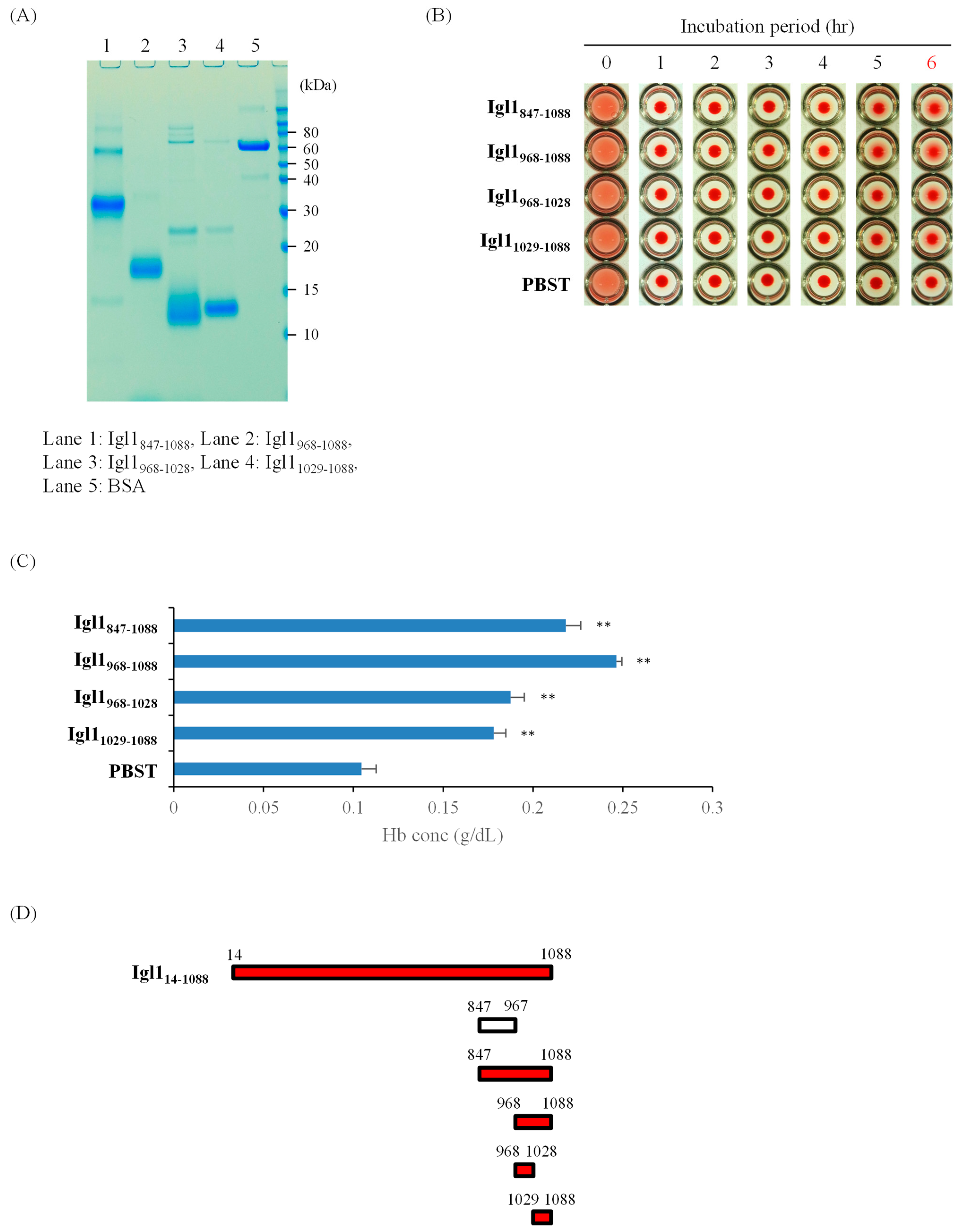
The results in Figure 1 indicate that Igl1787-846 has hemolytic activity, but this does not explain the similar activity of Igl1847-1088 [10]. To confirm that Igl1 has multiple regions with this activity, we generated three Igl1 fragments (Igl1968-1088, Igl1968-1028 and Igl11029-1088) based on the sequence of Igl1847-1088. Fragments of Igl1847-967 were not included because this region has no activity (Figure 1). As shown in Figure 2, all fragments studied had hemolytic activity, indicating that multiple regions in Igl1968-1028 and Igl11029-1088 have this activity (Figure 2D). Taken together, the results in Figure 1 and Figure 2 suggest that Igl1 has at least three regions with hemolytic activity.

2.3. Partial Inhibition of Hemolytic Activity of Igl1 Fragment Proteins by Monosaccharides

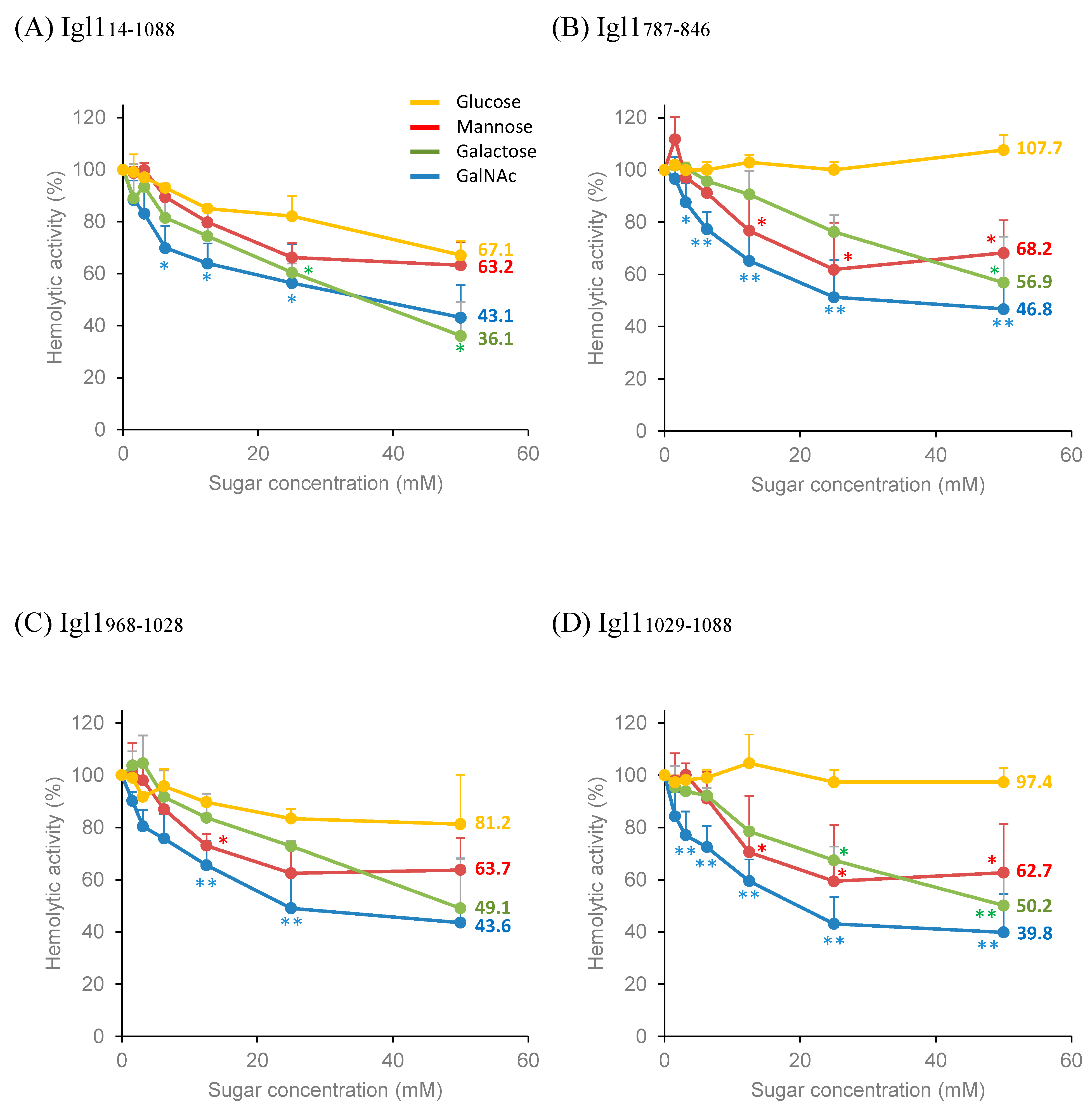
The hemolytic activity of Igl114-1088 (1 μM) is not inhibited by mixing with ten-fold higher concentrations (10 μM) of galactose or mannose [10]. In a study of the effects of glucose, lactose and GalNAc on hemolysis by a lectin-hemolytic peptide conjugate, 43-CEL-I, the range of carbohydrate concentrations was 1.56–50 mM, and 25 mM GalNAc and 50 mM lactose inhibited the hemolytic activity of the conjugate by about 90% [12]. Therefore, we examined whether higher concentrations of monosaccharides could inhibit the hemolytic activities of Igl1 fragment proteins (Figure 3). The maximal inhibition of the activity of Igl114-1088 was 57% with 50 mM GalNAc and 64% with 50 mM galactose, but only 37% with 50 mM mannose and 33% with 50 mM glucose (Figure 3A). The hemolytic activities of Igl1787-846 and Igl1968-1028 were inhibited by about 55% by 50 mM GalNAc, 45-50% by 50 mM galactose, and about 38% by 25 mM mannose (Figure 3B,C), and that of Igl11029-1088 was inhibited by about 60% by 50 mM GalNAc, 50% by 50 mM galactose and 40% by 25 mM mannose (Figure 3D). Incubation with 50 mM glucose inhibited about 20% of the activity of Igl1968-1028 but did not inhibit Igl1787-846 and Igl11029-1088 (Figure 3B–D). GalNAc treatment showed the steepest inhibition against the hemolytic activity of all Igl1 fragments at a concentration < 25 mM, but this inhibition was still only about 60% at maximum.

2.4. Cytotoxicity of Recombinant Fragment Proteins of Igl1 against Caco-2 Cells

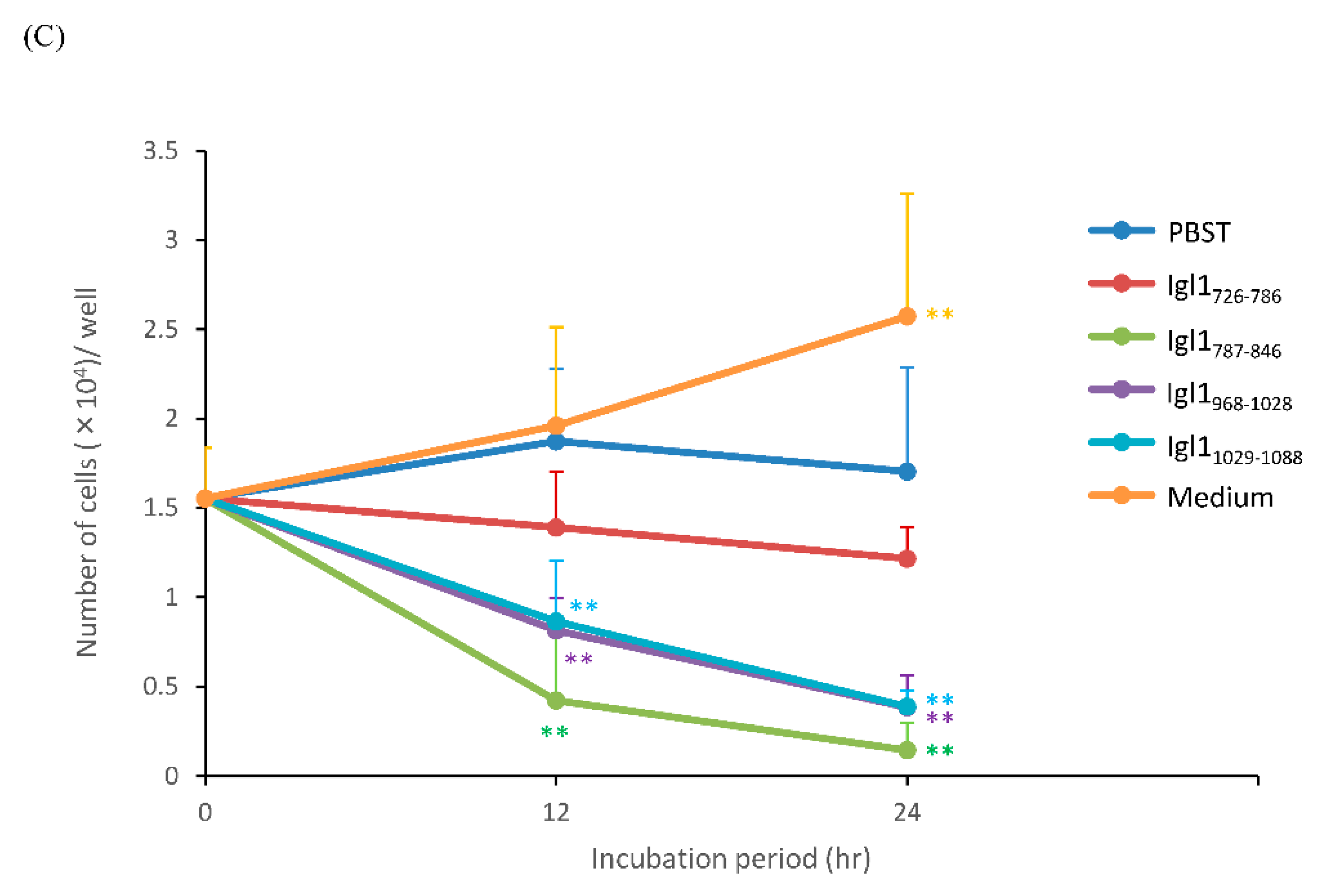
We have previously shown that Igl1 has both hemolytic and cytotoxic activities [10]. To examine whether the three Igl1 fragments (Igl1787-846, Igl1968-1028 and Igl11029-1088) with hemolytic activity also have cytotoxicity, Caco-2 cells were incubated with these fragments for 12 or 24 h (Figure 4), and with Igl1726-786, a fragment of 61 amino acids that did not show hemolytic activity (Figure 1). A cobblestone appearance of Caco-2 cells was observed after 12 or 24 h of incubation with PBST, Igl1726-786 or medium, but this appearance was destroyed when the cells were incubated with Igl1787-846, Igl1968-1028 or Igl11029-1088 (Figure 4B). There was a significant decrease in the number of cells remaining on the plate in incubation with Igl1787-846, Igl1968-1028 or Igl11029-1088, compared to that with PBST (Figure 4C). In contrast, Igl1726-786 showed no cytotoxicity. These results indicate that Igl1 fragments with hemolytic activity also have cytotoxicity.

3. Discussion

We have shown previously that the Igl subunit of E. histolytica lectin has hemolytic activity [10,11]. However, all the tested C-terminus fragment Igl proteins had this activity, and we were unable to identify the specific region(s) responsible for the activity. In the current study, we prepared smaller recombinant proteins and used these fragments in a hemolytic assay to clarify the regions with activity. Three fragments (Igl1787-846, Igl1968-1028 and Igl11029-1088) showed activity, indicating that Igl1 has multiple regions (at least three) that contribute to hemolytic activity. This explains why a monoclonal antibody recognizing the C-terminus of Igl1 could not completely inhibit the hemolytic activity of the protein [10].
Monosaccharides only partially inhibited the hemolytic activities of the three Igl1 fragment proteins, even at millimolar concentrations of mannose, galactose and GalNAc, and glucose showed lower or negligible inhibitory effects. These results agree with previous findings showing that the hemolytic activity of Igl114-1088 could not be inhibited by 10 μM galactose or mannose [10] and that 250 mM glucose could not inhibit Igl1 binding to Chinese hamster ovary (CHO) cells [7]. In contrast, intact Igl protein can be purified from E. histolytica using a Gal-affinity column [7], and the protein binds to GalNAc-BSA neoglycoprotein-coated beads [13]. These results indicate that Igl has a strong affinity for galactose and GalNAc, and it has also been shown that Igl binding to CHO cells is inhibited by 250 mM galactose or GalNAc [7]. Thus, the lectin domain(s) of Igl1 may not have a major role in hemolytic activity. However, Igl lacks the carbohydrate recognition domain (CRD) found in other lectins, and identification of this domain is required for proving or disproving this possibility. Using glycoproteins with more complex carbohydrates in an inhibition assay is also required for further studies.
Cytotoxic activities were also observed for Igl1787-846, Igl1968-1028 and Igl11029-1088, but not for Igl1726-786, which has a similar molecular size to those of the three other fragments. Igl1818-846 would be a fragment with activity, and we tried to obtain recombinant proteins with 30 amino acids, but it was difficult to generate these proteins in E. coli culture. Recombinant proteins with hemolytic activity also had cytotoxicity, suggesting that there are common molecular mechanisms underlying these activities. These mechanisms were not examined in this study, but this is the first report to show that a protozoan protein can have both hemolytic and cytotoxic activities in multiple regions.
The results in the previous [10] and the present studies suggest that Igl1787-846, Igl1968-1028 and Igl11029-1088 have fewer hemolytic and cytotoxic activities than Igl114-1088. This indicates that the three regions are required for the full activities of Igl1, but we need further studies to prove this speculation.
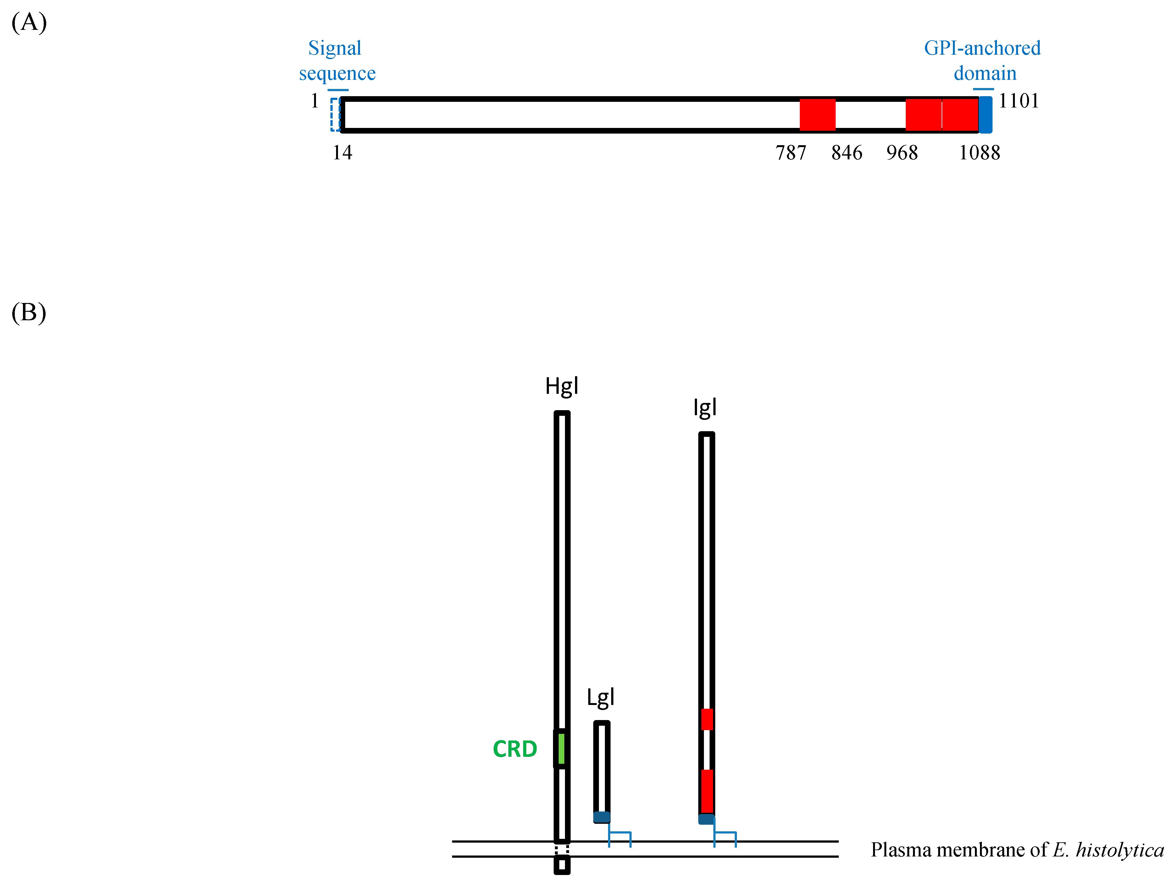
Two of the three regions in Igl1 with these activities (Igl1968-1028 and Igl11029-1088) are close to a GPI-anchored domain of the protein, as illustrated in Figure 5A. Therefore, on the plasma membrane of E. histolytica, the regions might be hindered intramolecularly or by associated subunits of the lectin, such as Hgl and Lgl (Figure 5B) [5]. However, gene suppression of Igl1 in E. histolytica resulted in less hemolytic activity of the gene-suppressed strain, indicating that the protein is exposed and has a role in the activity on site [11]. The reactivity of sera from patients with amoebiasis, including asymptomatic cyst passers, is highest against Igl1603-1088 [14], and anti-Igl1603-1088 antibodies in sera have effects against amoebic liver abscess formation in hamsters [15]. These findings also indicate that this region of Igl1 is exposed on the surface of E. histolytica. However, it is unclear whether Igl1 anchored to the cell membrane has hemolytic and cytotoxic activities or if Igl1 needs to be cleaved or shed from the membrane to permit these activities.
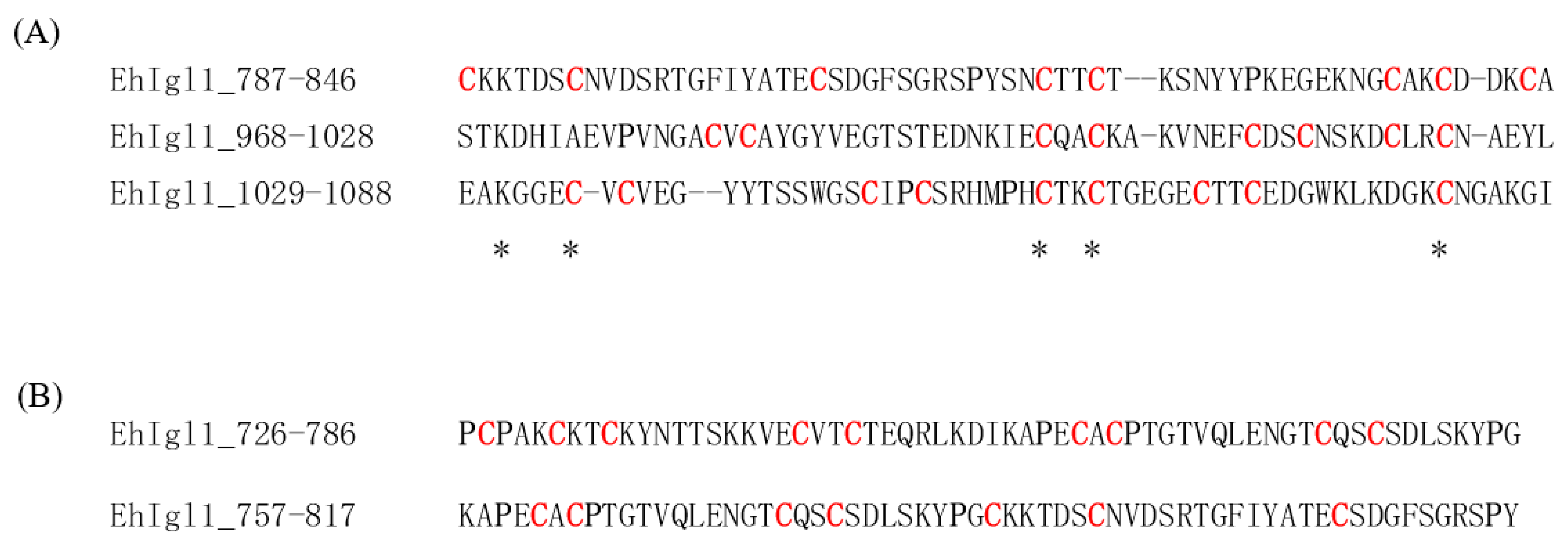
There is no common amino acid sequence among Igl1787-846, Igl1968-1028 and Igl11029-1088, as shown in Figure 6A. Comparison with the sequences of Igl1726-786 or Igl1757-817, which did not have hemolytic activity, also failed to answer why some Igl1 fragments have hemolytic and cytotoxic activities (Figure 6B). The top 20 amino-acid motifs in hemolytic peptides have recently been predicted using neural networks [16], but none of these motifs were found in the hemolytic regions of Igl1. Single proline substitutions in the Bordetella pertussis CyaA pore-forming fragment reduced its hemolytic activity [17]. In contrast, a proline residue at position 14 of Trichoderma alamethicin is essential for the hemolytic activity [18]. It is possible that the number of prolines in the fragments affects the structure and activity of Igl1, since there are one or two prolines in all Igl1 fragments with activity, but four or five in the fragments without activity.
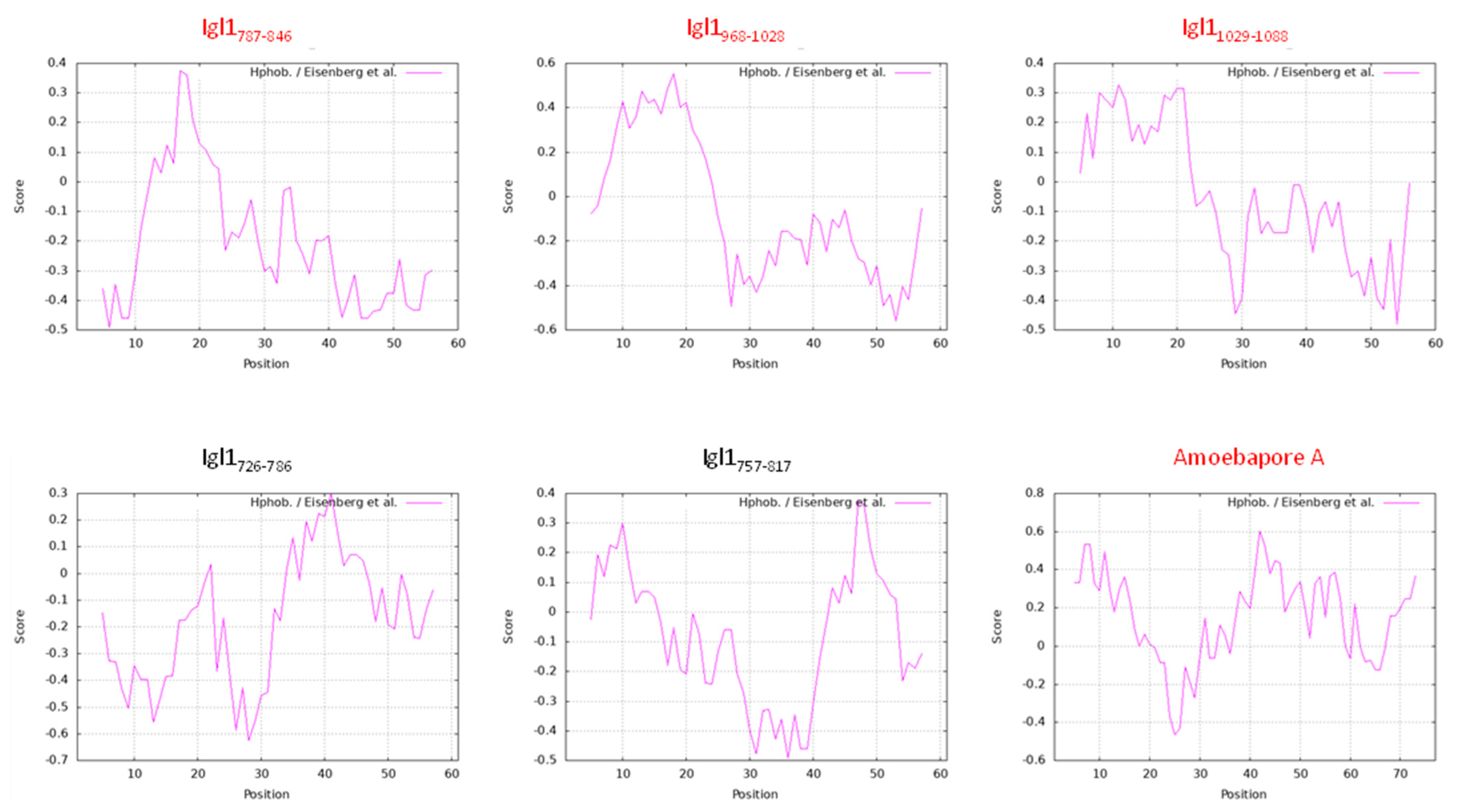
Hydrophobicity correlates to the hemolytic activity of antimicrobial peptides [19]. Igl1 fragments with hemolytic activity have hydrophobic amino acids in the N-terminus around amino acid position 18 and hydrophilic regions in the C-terminus (Figure 7). This characteristic may confer hemolytic and cytotoxic activities to the fragments. However, a pore-forming E. histolytica amoebapore-A which has hemolytic activity does not have the characteristic. In any case, structure analyses of Igl1 protein are required to obtain a clearer answer. Collectively, the results of this study suggest the need to identify inhibitors of all regions of Igl1 with hemolytic activity to reduce the virulence of E. histolytica related to the protein.

4. Materials and Methods

4.1. Expression and Refolding of Recombinant Igl1 Proteins and Ni Column Purification

Recombinant proteins with a His-tag at the N-terminus were expressed in Escherichia coli ECOSTM competent BL21(DE3) cells (Nippon Gene Co., LTD., Toyama, Japan) using pET19b expression vector (69677-3, Novagen, MA, USA) and the primers shown in Table 1. The proteins were further purified using a Ni column, as described in detail elsewhere [10,11,14].

4.2. SDS-PAGE and Coomassie Brilliant Blue Staining of Purified Recombinant Proteins

Recombinant proteins or bovine serum albumin (1 µg each) were mixed with SDS sample buffer (Invitrogen, CA, USA) and subjected to SDS-PAGE. The gel was treated with SimplyBlue Safe stain solution (Invitrogen, CA, USA) and incubated until blue bands appeared on the gel [10,11].

4.3. Hemolytic Assays Using Recombinant Lectins and Measurement of Released Hemoglobin

Hemolytic assays and quantification of hemolytic activity were conducted as previously described [10]. Briefly, recombinant Igl1 proteins (2 µM each, 50 µL) in PBST were mixed with 50 µL 2% (v/v) horse red blood cells (HoRBCs) (Japan Bio Serum, Tokyo, Japan) in PBS at room temperature, and images were taken at several time points. A Hemoglobin B Test Kit (Wako, Osaka, Japan) was used to measure the concentration of hemoglobin (Hb) in supernatants of RBCs incubated with recombinant proteins for 6 h. The results are expressed as the mean of 5 experiments with a standard deviation (SD). For inhibition assays using D(+)-galactose (Cat. No. 071–00032), D(+)-mannose (Cat. No. 130–00872), N-acetyl-D-galactosamine (Cat. No. 013–12821) and D(+)-glucose (Cat. No. 049–31165), monosaccharides were included in the incubations at final concentrations of 50 mM to 1.56 mM using 2-times serial dilutions. All monosaccharides were purchased from Fujifilm Wako Pure Chemical Corporation, Japan. The Hb concentration in the supernatant of the samples without monosaccharides was defined as 100% hemolytic activity for data analysis. The results are shown as the mean of 3 independent experiments with SD.

4.4. Cytotoxicity Assay

Caco-2 cells (ATCC, HTB-37) were cultured in MEM basic medium (Gibco, Beijing, China) supplemented with Earle’s salts, L-glutamine and 20% fetal bovine serum. After detachment with 0.25% Trypsin-EDTA (Gibco, NY, USA), the cells were cultured in a 96-well plate at approximately 2 × 10⁴ cells/100 µL/well at 37 °C under 5% CO2 for 24 h. Volumes (100 µL) of 2 µM recombinant Igl1 proteins, PBST or medium containing 200 units/mL penicillin G and 200 µg/mL streptomycin (Wako, Osaka, Japan) were added (0 h), and the cells were incubated for an additional 12 or 24 h under the same conditions. Images of Caco-2 cells in a 96-well plate were taken with an EVOS-XL microscope. The cells were trypsinized and harvested after 0, 12 or 24 h of incubation, and the number of cells per well was counted. The results are shown as the mean of 5 experiments with SD.

4.5. Statistical Analysis

Multiple comparisons were performed by ANOVA with a Dunn test or Dunnett test, with p < 0.05 considered to be significant.

Author Contributions

Conceptualization, K.K. and H.T.; Methodology, K.K. and H.T.; Validation, K.K. and H.T.; Formal Analysis, K.K.; Investigation, K.K.; Resources, K.K. and H.T.; Data Curation, K.K.; Writing—Original Draft, K.K.; Writing—Review and Editing, K.K. and H.T.; Visualization, K.K.; Project Administration, K.K.; Funding Acquisition, K.K. and H.T. All authors have read and agreed to the published version of the manuscript.

Funding

This work was supported by the Joint Usage/Research Center on Tropical Disease, Institute of Tropical Medicine, Nagasaki University (to H.T., 27-Ippan-7 and 28-Ippan-5), Grants-in-Aid for Scientific Research (C) (to K.K., JP16K08761 and JP22K05331) and Fostering Joint International Research (A) (to K.K., JP18KK0451) from the Japan Society for the Promotion of Science (JSPS) and JSPS KAKENHI (to H.T., grant nos. JP16H05819, JP17K08811 and JP20H03482).

Institutional Review Board Statement

Not applicable.

Informed Consent Statement

Not applicable.

Data Availability Statement

The data presented in this study are available in the article.

Conflicts of Interest

The authors declare no conflict of interest.

References

  1. Stanley, S.L., Jr. Amoebiasis. Lancet 2003, 361, 1025–1034. [Google Scholar] [CrossRef]
  2. Petri, W.A., Jr.; Haque, R.; Mann, B.J. The bittersweet interface of parasite and host: Lectin-carbohydrate interactions during human invasion by the parasite Entamoeba histolytica. Annu. Rev. Microbiol. 2002, 56, 39–64. [Google Scholar] [CrossRef] [PubMed]
  3. Haque, R.; Huston, C.D.; Hughes, M.; Houpt, E.; Petri, W.A., Jr. Amebiasis. N. Engl. J. Med. 2003, 348, 1565–1573. [Google Scholar] [CrossRef]
  4. Laughlin, R.C.; McGugan, G.C.; Powell, R.R.; Welter, B.H.; Temesvari, L.A. Involvement of Raft-Like Plasma Membrane Domains of Entamoeba histolytica in Pinocytosis and Adhesion. Infect. Immun. 2004, 72, 5349–5357. [Google Scholar] [CrossRef] [PubMed] [Green Version]
  5. Welter, B.H.; Goldston, A.M.; Temesvari, L.A. Localisation to lipid rafts correlates with increased function of the Gal/GalNAc lectin in the human protozoan parasite, Entamoeba histolytica. Int. J. Parasitol. 2011, 41, 1409–1419. [Google Scholar] [CrossRef] [PubMed] [Green Version]
  6. Tachibana, H.; Takekoshi, M.; Cheng, X.J.; Maeda, F.; Aotsuka, S.; Ihara, S. Bacterial expression of a neutralizing mouse monoclonal antibody Fab fragment to a 150-kilodalton surface antigen of Entamoeba histolytica. Am. J. Trop. Med. Hyg. 1999, 60, 35–40. [Google Scholar] [CrossRef] [PubMed]
  7. Cheng, X.J.; Tsukamoto, H.; Kaneda, Y.; Tachibana, H. Identification of the 150-kDa surface antigen of Entamoeba histolytica as a galactose- and N-acetyl-D-galactosamine-inhibitable lectin. Parasitol. Res. 1998, 84, 632–639. [Google Scholar] [CrossRef] [PubMed]
  8. Cheng, X.J.; Hughes, M.A.; Huston, C.D.; Loftus, B.; Gilchrist, C.A.; Lockhart, L.A.; Ghosh, S.; Miller-Sims, V.; Mann, B.J.; Petri, W.A., Jr.; et al. Intermediate subunit of the Gal/GalNAc lectin of Entamoeba histolytica is a member of a gene family containing multiple CXXC sequence motifs. Infect. Immun. 2001, 69, 5892–5898. [Google Scholar] [CrossRef] [PubMed] [Green Version]
  9. Tachibana, H.; Cheng, X.J.; Tsukamoto, H.; Itoh, J. Characterization of Entamoeba histolytica intermediate subunit lectin-specific human monoclonal antibodies generated in transgenic mice expressing human immunoglobulin loci. Infect. Immun. 2009, 77, 549–556. [Google Scholar] [CrossRef] [PubMed] [Green Version]
  10. Kato, K.; Yahata, K.; Dhoubhadel, B.G.; Fujii, Y.; Tachibana, H. Novel hemagglutinating, hemolytic and cytotoxic activities of the intermediate subunit of Entamoeba histolytica lectin. Sci. Rep. 2015, 5, e13901. [Google Scholar] [CrossRef] [PubMed] [Green Version]
  11. Kato, K.; Makiuchi, T.; Cheng, X.; Tachibana, H. Comparison of hemolytic activity of the intermediate subunit of Entamoeba histolytica and Entamoeba dispar lectins. PLoS ONE 2017, 12, e0181864. [Google Scholar] [CrossRef] [PubMed] [Green Version]
  12. Hatakeyama, T.; Kamine, T.; Konishi, Y.; Kuwahara, H.; Niidome, T.; Aoyagi, H. Carbohydrate-dependent hemolytic activity of the conjugate composed of a C-type lectin, CEL-I, and an amphiphilic alpha-helical peptide, 4(3)-beta Ala2. Biosci. Biotechnol. Biochem. 1999, 63, 1312–1314. [Google Scholar] [CrossRef] [PubMed]
  13. McCoy, J.J.; Mann, B.J. Proteomic analysis of Gal/GalNAc lectin-associated proteins in Entamoeba histolytica. Exp. Parasitol. 2005, 110, 220–225. [Google Scholar] [CrossRef] [PubMed]
  14. Tachibana, H.; Cheng, X.J.; Masuda, G.; Horiki, N.; Takeuchi, T. Evaluation of recombinant fragments of Entamoeba histolytica Gal/GalNAc lectin intermediate subunit for serodiagnosis of amebiasis. J. Clin. Microbiol. 2004, 42, 1069–1074. [Google Scholar] [CrossRef] [PubMed] [Green Version]
  15. Min, X.; Feng, M.; Guan, Y.; Man, S.; Fu, Y.; Cheng, X.; Tachibana, H. Evaluation of the C-terminal fragment of Entamoeba histolytica Gal/GalNAc lectin intermediate subunit as a vaccine candidate against amebic liver abscess. PLoS. Negl. Trop. Dis. 2016, 10, e0004419. [Google Scholar] [CrossRef] [PubMed] [Green Version]
  16. Timmons, P.B.; Hewage, C.M. HAPPENN is a novel tool for hemolytic activity prediction for therapeutic peptides which employs neural networks. Sci. Rep. 2020, 10, 10869. [Google Scholar] [CrossRef] [PubMed]
  17. Powthongchin, B.; Angsuthanasombat, C. Effects on haemolytic activity of single proline substitutions in the Bordetella pertussis CyaA pore-forming fragment. Arch. Microbiol. 2009, 191, 1–9. [Google Scholar] [CrossRef] [PubMed]
  18. Dathe, M.; Kaduk, C.; Tachikawa, E.; Melzig, M.F.; Wenschuh, H.; Bienert, M. Proline at position 14 of alamethicin is essential for hemolytic activity, catecholamine secretion from chromaffin cells and enhanced metabolic activity in endothelial cells. Biochim. Biophys. Acta. 1998, 1370, 175–183. [Google Scholar] [CrossRef] [Green Version]
  19. Greco, I.; Molchanova, N.; Holmedal, E.; Jenssen, H.; Hummel, B.D.; Watts, J.L.; Håkansson, J.; Hansen, P.R.; Svenson, J. Correlation between hemolytic activity, cytotoxicity and systemic in vivo toxicity of synthetic antimicrobial peptides. Sci. Rep. 2020, 10, 13206. [Google Scholar] [CrossRef] [PubMed]
  20. Eisenberg, D.; Schwarz, E.; Komaromy, M.; Wall, R. Analysis of membrane and surface protein sequences with the hydrophobic moment plot. J. Mol. Biol. 1984, 179, 125–142. [Google Scholar] [CrossRef]
Figure 1. Hemolytic activity of recombinant Igl1 fragment proteins based on Igl1726-967. (A) Purities and amounts of recombinant proteins were confirmed by SDS-PAGE, with 1 µg of each protein run on the gels. (B) HoRBCs were incubated in a 96-well plate with Igl1 fragment proteins for the indicated periods. Representative images of 5 independent studies are shown. (C) Concentrations of hemoglobin (Hb) released in the supernatant of samples incubated for 6 h. Data are the mean + SD from 5 independent experiments. Hemolytic activities of Igl1 fragments shown in bars with asterisks were significantly higher than those of phosphate-buffered saline with 0.05% Tween 20 (PBST) (* p < 0.05, ** p < 0.01 by ANOVA with a Dunn test). (D) Summary of data for fragment proteins with hemolytic activities. Recombinant Igl1 fragments with hemolytic activity are shown in red bars. Open bars indicate fragment proteins without this activity.
Ijms 23 07700 g001
Figure 2. Hemolytic activity of recombinant Igl1 fragment proteins based on Igl1847-1088. (A) Purities and amounts of recombinant proteins were confirmed by SDS-PAGE, with 1 µg of each protein run on the gels. (B) HoRBCs were incubated with Igl1 fragment proteins in a 96-well plate for the indicated periods. Representative images of 5 independent studies are shown. (C) Concentrations of hemoglobin (Hb) released in the supernatant of samples incubated for 6 h. Data are the mean + SD from 5 independent experiments. Hemolytic activities of Igl1 fragments shown in bars with asterisks were significantly higher than that of PBST (** p < 0.01 by ANOVA with a Dunn test). (D) Summary of data for fragment proteins with hemolytic activities. Recombinant Igl1 fragments with hemolytic activity are shown in red bars. The open bar (Igl1847-967) indicates a fragment without activity (Figure 1).
Ijms 23 07700 g002
Figure 3. Effects of monosaccharides on hemolytic activities of Igl1 fragment proteins. Igl1 fragments and 2% (v/v) horse red blood cells were incubated with or without monosaccharides for 6 h at room temperature. Hb concentrations in the supernatant were measured and those in samples without monosaccharides were defined as 100% hemolytic activity. Numbers in the line graphs indicate the hemolytic activity (%) of Igl1 fragments treated with 50 mM monosaccharides. (A) Igl114-1088. (B) Igl1787-846. (C) Igl1968-1028. (D) Igl11029-1088. Data are the mean + SD from 3 independent studies. Asterisks indicate hemolytic activities of Igl1 fragments with mannose, galactose or GalNAc that are significantly lower than that with glucose (* p < 0.05, ** p < 0.01 by Dunnett test).
Ijms 23 07700 g003
Figure 4. Cytotoxicity of Igl1 fragment proteins against Caco-2 cells. Caco-2 cells were cultured with 1 μM Igl1 fragments and incubated at 37 °C for 12 or 24 h. (A) Purities and amounts of Igl1 fragments were confirmed by SDS-PAGE using a NuPAGE gel, with 1 µg of each protein run in the gel. Lane 1: Igl1726-786, Lane 2: Igl1787-846, Lane 3: Igl1968-1028, Lane 4: Igl11029-1088, Lane 5: BSA. (B) Images of Caco-2 cells incubated with Igl1 fragments in a 96-well plate for the indicated periods. Images were taken with an EVOS-XL microscope. Bars indicate 100 μm. (C) Number of Caco-2 cells remaining in the well after incubations for 0, 12 and 24 h. Data are the mean + SD from 5 independent studies. ** p < 0.01 vs. PBST (Dunnett test).
Ijms 23 07700 g004aIjms 23 07700 g004b
Figure 5. Hemolytic and cytotoxic regions of Igl1. (A) The Igl114-1088 recombinant fragment lacks the signal sequence (aa 1–13) and GPI-anchored domain (aa 1089–1101). Regions in red are those with both hemolytic and cytotoxic activities. (B) Schematic of Hgl, Lgl and Igl on the cell membrane of E. histolytica. Igl is associated with other lectin subunits: Hgl and Lgl. CRD: carbohydrate recognition domain.
Ijms 23 07700 g005
Figure 6. Amino acid sequences of Igl1 fragments. (A) Sequence alignment of Igl1 fragments with hemolytic and cytotoxic activities. Asterisks indicate common amino acids. (B) Sequences of Igl1 fragments without these activities. The Igl1 sequence refers to protein ID AAK92361. Cysteine residues are shown in red and proline residues are shown in bold.
Ijms 23 07700 g006
Figure 7. Hydrophobicity of Igl1 fragments and amoebapore-A. Hydrophobicity of Igl1 fragments and amoebapore-A (PDB: 1OF9_A) was calculated by ProtScale program in Expasy (https://web.expasy.org/protscale/) (accessed on 6 July 2022) with an amino acid scale published by Eisenberg et al. [20]. Igl1 fragments in red or black indicate the fragments with or without hemolytic activity, respectively.
Ijms 23 07700 g007
Table 1. Oligonucleotide primers used in the study.
PrimerPosition aSequence (5’ to 3’) b
EhIgl-S1440–59CCCTCGAGGATTATACTGCTGATAAGCT
EhIgl-S7262176–2195CCCTCGAGCCATGTCCTGCAAAATGTAA
EhIgl-S7572269–2289CCCTCGAGAAAGCACCAGAATGTGCTTGT
EhIgl-S7872359–2378CCCTCGAGTGTAAAAAAACTGATTCATG
EhIgl-S8472539–2558CCCTCGAGACATGTTCAGATAAAGACAC
EhIgl-S9682902–2921CCCTCGAGTCAACAAAAGATCATATTGC
EhIgl-S10293085–3105CCCTCGAGGAAGCAAAAGGAGGAGAATGT
EhIgl-AS7862341–2358CCCTCGAGTTATCCTGGATATTTTGAAAG
EhIgl-AS8172434–2451CCCTCGAGTTAATAAGGACTACGTCCACT
EhIgl-AS8462521–2538CCCTCGAGTTATGCACATTTATCATCACA
EhIgl-AS9062701–2718CCCTCGAGTTACTTGTATGTATCACTCTC
EhIgl-AS9672884–2901CCCTCGAGTTAACATTTAGTACATTCTTC
EhIgl-AS10283066–3084CCCTCGAGTTATAAATATTCAGCATTGCAT
EhIgl-AS10883247–3264CCCTCGAGTTAAATGCCTTTAGCTCCATT
a Nucleic acid numbering is based on the E. histolytica Igl1 gene sequence (AF337950). b Nucleotides added for cloning and translation termination are underlined.
Publisher’s Note: MDPI stays neutral with regard to jurisdictional claims in published maps and institutional affiliations.