Review

The Role of Mitochondrial Abnormalities in Diabetic Cardiomyopathy

by
Siarhei A. Dabravolski
1,*,
Nikolay K. Sadykhov
2,
Andrey G. Kartuesov
2,
Evgeny E. Borisov
3,
Vasily N. Sukhorukov
3,4 and
Alexander N. Orekhov
4
1
Department of Clinical Diagnostics, Vitebsk State Academy of Veterinary Medicine [UO VGAVM], 7/11 Dovatora Str., 210026 Vitebsk, Belarus
2
Laboratory of Angiopathology, Institute of General Pathology and Pathophysiology, Russian Academy of Medical Sciences, 125315 Moscow, Russia
3
Petrovsky National Research Centre of Surgery, 2, Abrikosovsky Lane, 119991 Moscow, Russia
4
Institute for Atherosclerosis Research, Osennyaya 4-1-207, 121609 Moscow, Russia
*
Author to whom correspondence should be addressed.
Int. J. Mol. Sci. 2022, 23(14), 7863; https://doi.org/10.3390/ijms23147863
Submission received: 30 May 2022 / Revised: 14 July 2022 / Accepted: 15 July 2022 / Published: 16 July 2022
(This article belongs to the Special Issue Molecular Pathways in Diabetic Cardiomyopathy)

Abstract

:
Diabetic cardiomyopathy (DCM) is defined as the presence in diabetic patients of abnormal cardiac structure and performance (such as left ventricular hypertrophy, fibrosis, and arrhythmia) in the absence of other cardiac risk factors (such as hypertension or coronary artery disease). Although the pathogenesis of DCM remains unclear currently, mitochondrial structural and functional dysfunctions are recognised as a central player in the DCM development. In this review, we focus on the role of mitochondrial dynamics, biogenesis and mitophagy, Ca2+ metabolism and bioenergetics in the DCM development and progression. Based on the crucial role of mitochondria in DCM, application of mitochondria-targeting therapies could be effective strategies to slow down the progression of the disease.

1. Introduction

Cardiomyopathy is a heterogeneous group of diseases of the myocardium associated with diverse conditions. It essential to distinguish primary (e.g., hypertrophic, dilated, restrictive, arrhythmogenic right ventricular, left ventricular non-compaction, ion channelopathies, Takotsubo cardiomyopathy, tachycardia-induced cardiomyopathy, and myocarditis) and secondary (induced by complicating conditions such as hypertension, CAD and valvular heart diseases) cardiomyopathies. However, in many cases, it is not possible to define the clear reason for the development of cardiomyopathy [1]. DCM is usually described by structural and functional abnormalities in patients with DM (diabetes mellitus) without other heart disease risk factors (such as hypertension, coronary artery, and valvular diseases) [2]. Patients with obesity, insulin resistance, and dyslipidaemia are likely to develop a similar metabolism-related cardiomyopathy even without diabetes, which could be referred to as metabolic cardiomyopathy, insulin resistance-induced cardiomyopathy, obesity-related cardiomyopathy, or lipotoxic cardiomyopathy. These patients may not be diagnosed with diabetic cardiomyopathy in the clinical setting because of the absence of diabetes [3]. Number of bio-markers (such as levels of glycated haemoglobin, glucose and low-density lipoprotein, IGFBP7 (Insulin Like Growth Factor Binding Protein 7), TGF-β1 (Transforming growth factor beta 1) and others) could help in DCM diagnosis [4,5]. On the functional level, DCM is associated with diastolic and further systolic dysfunction, cardiac remodelling and myocardial fibrosis, arrhythmia, hypertrophy, and ultimately with clinical heart failure [6].
The development and progression of DCM were associated with inflammation; mitochondrial dysfunction; oxidative stress; impaired cardiac insulin signalling and calcium metabolism; microvascular dysfunction; endoplasmic reticulum stress; endothelial dysfunction; cell death and number of other pathologic conditions [7,8,9,10]. Comparing all DM complications, DM patients have a two-fold greater risk of developing heart failure with DCM accounting for nearly 80% of mortality cases [11].

2. Structural and Functional Alterations in DCM

Structural changes in diabetic myocardium are usually assessed by non-invasive reliable and reproducible techniques (such as magnetic resonance imaging and echocardiography), with myocardial fibrosis and hypertrophy as the main hallmarks of DCM [12,13]. While MRI has better sensitivity and capability to provide a more detailed image, it is also more expensive, thus, not so widely used as echocardiography––the main diagnostic tool for the early DCM detection [14,15].
Myocardial fibrosis plays a crucial role in structural reconstruction of the heart in DCM, contributing to the altered LV (left ventricular) function and diastolic dysfunction [16]. Usually, it is described by lower early diastolic filling and increased atrial filling, prolonged isovolumetric relaxation time and supraventricular premature beats, increased LV end-diastolic pressure and diminished LV end-diastolic volume, resulted in the lowered LV ejection fraction and cardiac output [17,18]. On the molecular level, structural and functional changes in the diabetic heart are caused by increased expression of TGF-β1, TSP-1 (thrombospondin-1), MMPs (matrix metalloproteinases) and other related genes, promoting collagen production and increasing content of connective tissue and interrupting normal cardiac function [19,20,21]. Eventually, those changes lead to the loss of cardiomyocytes via the terminal cell death pathway, which could be classified into several forms: autophagy, necrosis, apoptosis, entosis, ferroptosis, oxeiptosis and others [22,23].
Myocardial hypertrophy is the next hallmark of DCM, resulting from the cell-death-mediated loss of cardiomyocytes and subsequent compensative enlargement of the remaining viable cardiomyocytes. On the cellular level, hypertrophy is described with disorganised cardiomyocyte myofiber; on the molecular level, with the over-expression of the key regulatory genes: β-MHC (β-myosin heavy chain), FoxO1 (forkhead box protein O 1), BNP (brain natriuretic peptide), ANP (atrial natriuretic peptide), adiponectin signalling pathway, and others [24,25]. Despite extensive and meticulous research, the exact molecular mechanism of cardiac hypertrophy pathogenesis is not fully understood.
In the further steps of the DCM development, myocardial fibrosis and hypertrophy promote progression of cardiomyocyte stiffness and subsequent diastolic dysfunction. Mechanically, such an effect could be explained by impaired insulin signalling, which affects GLUT4 (glucose transporter type 4) and Ca2+ pumps, and resulted in reduced glucose intake and increased intracellular level of Ca2+ in cardiomyocytes [26]. Ca2+ overload triggers mitochondrial permeability transition pore opening and leads to cardiomyocytes death, while the restoration of Ca2+ levels with MCU (mitochondrial calcium uniporter) expression reduces the infarcted area in diabetic heart and increases glucose utilization (which manifested by improved mitochondrial respiratory function and ATP production) [27]. Simultaneously, disturbed insulin signalling leads to reduced activity of eNOS (endothelial nitric oxide synthase), which also affects Ca2+ metabolism and reduces Ca2+ intake by sarcoplasm [28,29]. Altogether, such pathophysiological abnormalities lead to cardiac stiffness, impaired cardiac relaxation and diastolic dysfunction as the early DCM manifestation.

3. Mitochondrial Dynamics in DCM

Mitochondria capacity and functions are regulated via their biogenesis, dynamics and recycling. PGC1α (peroxisome proliferator-activated receptor gamma coactivator 1-alpha) is the major regulator of mitochondrial biogenesis, which could be triggered by development signals, internal and environmental stimuli [30]. Damaged and dysfunctional parts of mitochondria are separated from healthy parts during mitochondrial turnover—cycles of fission (split) and fusion (merge). Mitochondrial fission and fusion are regulated by DNM1L (dynamin 1 like)/FIS1 (fission, mitochondrial 1) and MFN1, MFN2 (mitofusin 1 and 2)/OPA1 (optic atrophy protein 1) genes, respectively [31]. Furthermore, damaged parts of mitochondria are fused with lysosome and digested during the process of a specialised form of mitochondrial autophagy––mitophagy. In general, mitophagy regulation could be classified as conventional PINK1/Parkin (PTEN Induced Kinase 1/Parkin RBR E3 Ubiquitin Protein Ligase)-mediated and alternative Parkin-independent mitophagy [32]. Healthy parts of mitochondria are returned to the mitochondrial network to continue normal functioning [33,34]. Mitophagy is crucial for many cellular processes, normal organism functioning, and development; its dysregulation is associated with neurodegenerative diseases, cancer, inflammation and other pathological conditions [35,36]. The heart relies on the ATP generated by mitochondria via aerobic metabolism (from the oxidation of glucose and FAs (fatty acids)). Therefore, any dysregulation of mitochondrial cellular functions eventually leads to a decrease in ATP and increase of ROS (reactive oxygen species) production, which contributes to the development of different cardiovascular diseases [31,37]. In normal physiological conditions, the balance between mitochondrial fusion and fission keeps mitochondrial functions at the level required by the current cellular needs. However, fission/fusion imbalance leads to a significant rise in ROS release and reduction of ATP production, stimulating cell death and further disease progression [38,39].
Recently, it was shown that hearts from diabetic mice have a surplus rate of mitochondrial fission, resulting in a decrease of mean mitochondrial size and number of mitochondria per μm2. In addition, the expression level of MFN2 was significantly decreased, while levels of other dynamics-related proteins (Opa1, Mfn1, Fis1 and Drp1) were not changed (Figure 1). Furthermore, over-expression of Mfn2 in diabetic hearts reduces ROS production, restores mitochondrial membrane potential, and normalises fission. Mfn2 expression could be regulated via its positive regulator PPARα, which directly binds to an Mfn2 promoter [40]. Similarly to Mfn2 over-expression, treatment of HFD-induced prediabetic mice with necrostatin-1, an inhibitor of cell death and inflammation receptor RIPK1, with known cardioprotective properties [41], effectively enhanced Mfn2 protein levels, with no alterations in Mfn1 and Opa1 protein levels, thus increasing mitochondrial fusion and alleviating mitochondrial dysfunction. In addition, necrostatin-1 effectively reduced cardiac dysfunction, cardiac autonomic imbalance, and blood pressure in prediabetic rats [42].
High glucose levels could induce mitochondrial dysfunction and cell death in rats’ atrial cardiomyocytes. In in vitro conditions, hyperglycaemia increases ER (endoplasmic reticulum) stress, mitochondrial ROS levels, and Mfn2 levels. On the contrary, for the results from the hearts of diabetic mice, Mfn2 silencing decreases Ca2+ transfer from ER to mitochondria under ER stress conditions, ameliorating mitochondrial ROS production and oxygen consumption, thus preventing cells from mitochondrial dysfunction and protecting from ER-mediated cell death [43,44]. Similarly, hyperglycaemia increases mitochondria–ER contact and Ca2+ levels in the ER and mitochondria of cardiomyocytes. However, Mfn2 silencing reduces mitochondria-ER cooperation, Ca2+ concentrations in both ER and mitochondria, mitochondrial ROS production, ER stress and mitochondria-mediated apoptosis, preserving mitochondrial function and cardiomyocyte viability under hyperglycaemic conditions [45].
Drp1 (Dynamic relative protein 1) regulates mitochondrial fission by changing its level of phosphorylation. As such, mitochondrial fission is driven via S616 phosphorylation by ERK (extracellular signal-regulated kinase), while calcineurin induces S637 dephosphorylation and mediates Ca2+-induced mitochondrial fission [46]. Recent research suggests that, besides Drp1, the Orai1 (Ca2+ release-activated calcium channel protein 1) calcium channel plays an important role in cardiomyocytes Ca2+ metabolism and DCM development. Cardiac hypertrophy and impaired cardiac function, accompanied by increased mitochondrial fission and intracellular Ca2+ overload, were described in diabetic rats on HGD (high glucose diet). However, application of Drp1 inhibitor prevents HGD-induced cardiomyocyte hypertrophy by reducing Drp1 S616 and increasing S637 phosphorylation. Likewise, Drp1 activity could be suppressed with Orai1 inhibition, which also alleviates HGD-induced mitochondrial dysfunction and cardiomyocyte hypertrophy, suggesting Drp1 as a downstream target of Orai1-mediated Ca2+ influx [47].
Recently, the role of Mst1 (Mammalian sterile 20-like kinase 1), one of the crucial components of the Hippo signalling pathway involved in regulating cell apoptosis and cell proliferation, in the development of cardiovascular disorders was shown [48]. Mst1 regulates mitochondrial fission via upregulating Drp1 expression and its recruitment to the mitochondria, stimulating S616 phosphorylation and D637 dephosphorylation. Furthermore, Mst1 knockdown in diabetic mice inhibits mitochondrial fission and mitigates LV remodelling and cardiac dysfunction, thus preventing the DCM development. In addition, Drp1 knockdown prevents the effects of Mst1 on mitochondrial fission and DCM development, thus supporting the proposed Drp1-mediated mechanism [49].
Cardiomyocyte-specific over-expression of LACS1 (long chain acyl-CoA synthetase 1), used as a mice model system with increased cardiac FA uptake, resulted in dramatically remodelled mitochondrial network and abnormal mitochondrial dynamics. Long-term exposure of transgenic mice to FA leads to increased mitochondrial ROS production, fission rate, and loss of the mitochondrial reticulum. On the molecular level, high FA induces surplus ubiquitination of AKAP121 (A-kinase anchor protein), a crucial regulator of mitochondria physiology [50], which further reduces S637 phosphorylation of Drp1 and enhances Opa1 proteolysis. Therefore, cardiac lipid overload induces increased mitochondrial ROS generation and causes alteration in post-translational modifications of Drp1 and Opa1, which leads to dysregulation of mitochondria fusion and fission, thus contributing to cardiac dysfunction in diabetes and obesity [51].
In total, DM-associated pathological conditions (such as hyperglycaemia and lipid overload) cause increased mitochondrial ROS generation and post-translational modifications of mitochondrial proteins that regulate mitochondrial dynamics, which further lead to imbalanced mitochondrial dynamics and contribute to DCM development (Figure 1). Therefore, further research is required to investigate the role of mitochondrial dynamics in DCM development and to comprehend its potential for DCM treatment.

4. Mitophagy

Mitophagy is a specialised form of autophagy, crucial for normal organism functioning and development. Normally, nutritional deficiency and cell senescence stimulate mitophagy, targeting damaged and malfunction mitochondria to fuse with lysosomes to degrade. Mice in prediabetic conditions were characterised with a mild diastolic dysfunction, increased SR mitoROS production, increased number of myocardial lipid droplets, elevated LV mass and wall thickness, increased expression of the MFN2 and decreased expression of a mitophagy marker BNIP3 (Bcl-2/adenovirus E1B 19-kDa protein-interacting protein 3). On the contrary, no sign of cardiac hypertrophy or fibrosis was observed. Those results suggest that abnormal mitochondrial dynamics and mitophagy are the primary metabolic derangements responsible for initial cardiac pathological changes [52]. Additionally, the crucial role of mitophagy was confirmed in experiments on mice with HFD-induced diabetes, where the mitophagy rate increases after three weeks of HFD feeding. Deletion of atg7 or Parkin resulted in impaired mitophagy, increased lipid accumulation, exacerbated diastolic dysfunction and induced systolic dysfunction. On the contrary, mitophagy activation with injection of Tat-Beclin1 attenuated mitochondrial dysfunction, decreased lipid accumulation, and protected against cardiac diastolic dysfunction during HFD feeding, suggesting its protective role against HFD-induced diabetic cardiomyopathy [53].
Besides the role in regulating mitochondrial dynamics, Mst1 was shown to inhibit mitophagy in a SIRT3-Parkin-dependent way. Sirt3 (NAD-Dependent Deacetylase Sirtuin-3, Mitochondrial) is a vital member of the class III histone deacetylases, known to eliminate mitoROS, inhibit apoptosis, and prevents the formation of cancer cells. Mst1 deficiency stimulated mitophagy in DCM and alleviated DCM-associated phenotype in an HGD-induced diabetic mice model. However, positive effects of Mst1 knockdown were abolished in Parkin−/− and Sirt3−/− mice [54]. Another research supports the role of Sirt3 in DCM via the Foxo3A-Parkin signalling pathway. Sirt3 knockout aggravated effects of streptozotocin-induced diabetes in mice: suppressed autophagy and mitophagy, cardiomyocyte apoptosis and mitochondrial injury, cardiac dysfunction, and interstitial fibrosis. At the same time, the deacetylation of Foxo3A and expression of Parkin were decreased. In vitro Sirt3 overexpression activated autophagy and mitophagy, inhibited mitochondrial injury and cardiomyocyte apoptosis, normalised Foxo3A deacetylation and Parkin expression [55]. Because of the crucial role of Sirt3 and Mst1 regulation of mitochondrial dynamics and mitophagy, further studies required to better understand their involvement in the development of diabetic cardiomyopathy and potential use as therapeutic targets for DCM treatment [56].
Recent research proved that HFD itself suppressed mitophagy and the accumulation of damaged mitochondria in the heart of obese/diabetic mice. Experiments with cardiac-specific deletion of ACC2 (acetyl-CoA carboxylase 2), a vital enzyme, catalysing the carboxylation of acetyl-CoA to malonyl-CoA, which could inhibit carnitine-palmitoyl-CoA transferase I, the rate-limiting step in FA uptake and oxidation by mitochondria, suggested that enhanced cardiac FAO does not cause cardiomyopathy in non-obese mice. On the contrary, ACC2 knockout in mice on HFD increased cardiac FAO, preventing HFD-induced downregulation of Parkin and subsequent mitophagy suppression, mitochondrial dysfunction and pathological heart remodelling, thus protecting from DCM [57].
The epigenetic mechanism is also involved in regulation of mitochondrial energy production and mitophagy, and could be used for DCM treatment. BRD4 (Bromodomain Containing 4) is an ubiquitously expressed chromatin regulator protein, which plays a pivotal role in cardiomyocyte specific mitochondrial energy production and homeostasis [58]. Application of selective BRD4 inhibitor JQ1 mitigates DCM, heart failure, and cardiac hypertrophy [59,60]. Interestingly, diabetic mice hearts have increased the level of BRD4, which inhibits PINK1/Parkin-mediated mitophagy and causes further accumulation of damaged mitochondria, impairment of cardiac function and heart remodelling. Treatment with a JQ1 inhibitor improves mitochondrial and heart function in a Pink1-dependent way and prevents HFD-induced DCM, supporting BRD4 inhibition as a valuable strategy for DCM treatment [61].
Interestingly, chronic HFD consumption inactivates conventional mitophagy (mediated by the Atg7 and LC3), whereas stimulates alternative (Ulk1 (unc51 like kinase 1)- and Rab9 (Ras-related protein Rab-9)-dependent mechanism of mitophagy. Thus, 24 weeks of HFD consumption upregulated both Ulk1 and Rab9 in the mitochondrial fraction, while cardiac-specific ulk1 knockout and Rab9 knock-in mice showed impaired mitophagy and both diastolic and systolic dysfunction (Figure 1). On the contrary, heart-specific Rab9 over-expression increased mitophagy and protected against cardiac dysfunction during HFD consumption. Apparently, alternative mitophagy mechanisms do not sustain mitochondrial quality under prolonged HFD burden; however, they could be therapeutically enhanced to obstruct DCM progression in patients with obesity and diabetes [62].
In summary, mitophagy plays an important role in the prevention of cardiac mitochondria dysfunction, cardiac hypertrophy, diastolic dysfunction, and lipid accumulation in the hearts of diabetic mice (Figure 1). Although the exact molecular mechanism of the cardiac mitophagy regulation is still unknown, stimulation of conventional and alternative mitophagy effectively protects the heart against DCM and provides a rationale to develop new mitophagy-targeted therapeutic approaches.

5. Mitochondrial Unbalanced Calcium Homeostasis

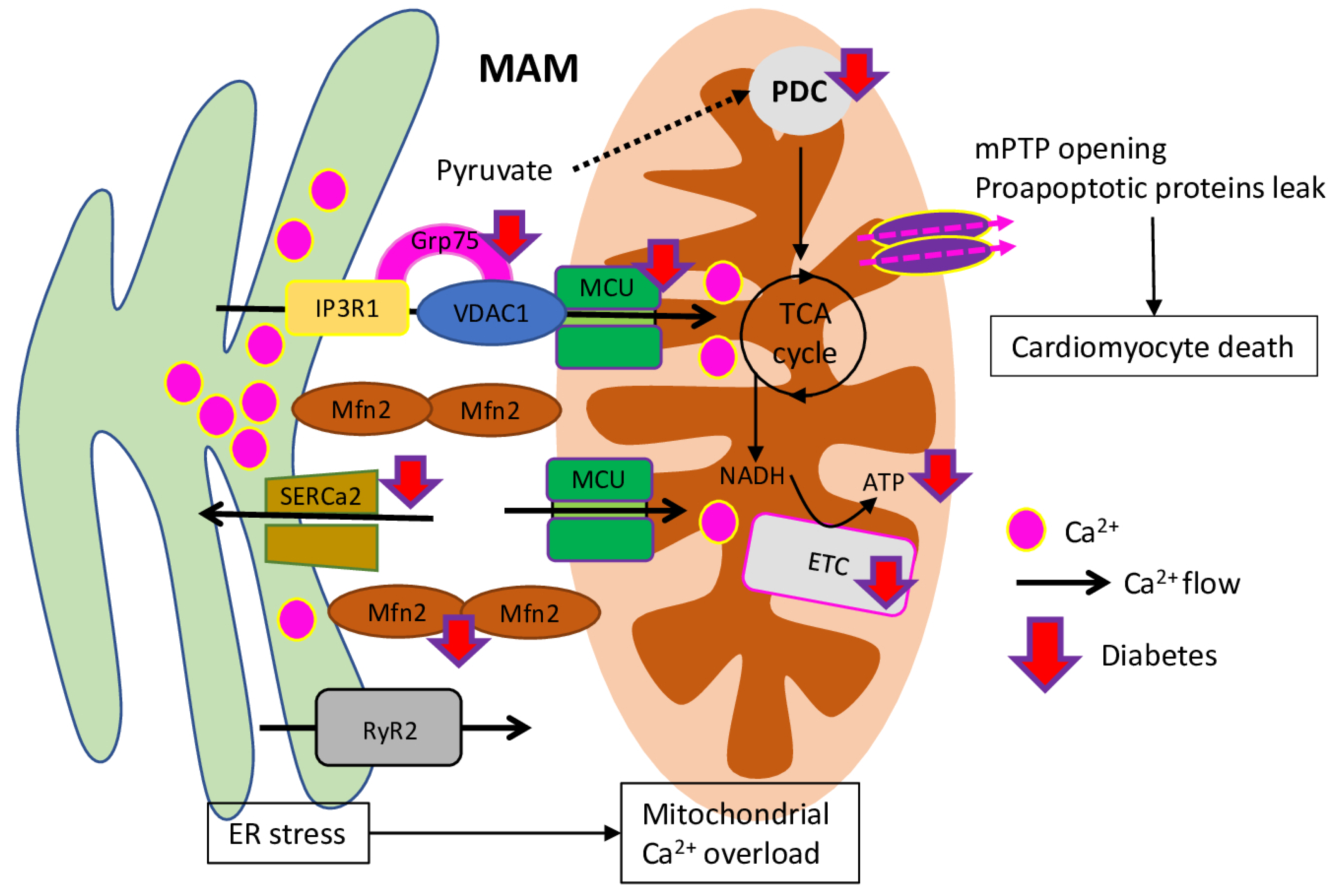
The mitochondrial calcium concentrations play a critical role in cardiac excitation-contraction coupling. In particular, buffering capacity is facilitating signalling crosstalk between mitochondria and ER/SR (sarcoplasmic reticulum), mitochondria, and plasma membrane. Mitochondrial Ca2+ regulates glucose oxidation (via activation of PDC (pyruvate dehydrogenase complex)) and enhances ATP formation (via activation of mitochondrial complexes I, III, IV, and V) (Figure 2). It is known that free mitochondrial Ca2+ ([Ca2+]m) concentration, as a key signalling molecule for mitochondrial energetic metabolism, is decreased in cardiomyocytes from diabetic hearts [63]. During myocardium excitation contraction coupling, Ca2+ gets into the cytoplasm via voltage sensitive LTCCs (L-type calcium channels) after sarcolemma depolarisation, triggering Ca2+ release from the SR. During the diastolic process, Ca2+ is pumped back to SR, accompanied by the excess Ca2+ pumped out through Ca2+ pump on the plasma membrane and sarcolemma Na+/Ca2+ exchanger [64,65]. However, in DCM, the Ca2+ transporters and Ca2+ transfer between mitochondria, ER and SR calcium homeostasis is disrupted, which resulted in increased duration of action potential and prolonged diastolic relaxation time [10].
As it was shown, mouse cardiac myocytes under hyperglycaemic conditions have reduced [Ca2+]m and protein level of MCU (mitochondrial Ca2+ uniporter)––the channel responsible for mitochondrial Ca2+ uptake in mitochondria and regulation of Ca2+-dependent mitochondrial metabolism. However, MCU over-expression normalises the [Ca2+]m level, which is responsible for subsequent reduction of oxidative stress, apoptosis and FAO (fatty acid β-oxidation), increase of glucose oxidation, PDH (pyruvate dehydrogenase) activity, and mitochondrial membrane potential, thus reversing hyperglycaemia-induced metabolic alterations [66]. In addition, those results were confirmed in vivo in streptozotocin (STZ)-induced diabetic mice, where MCU normalisation besides improved mitochondrial function and Ca2+ handling, cardiac energetic metabolism and both cardiac myocyte and heart function [26]. Similar results were obtained on db/db diabetic mice, where over-expression of MICU1 (mitochondrial calcium uptake 1), the regulatory subunit of MCU, inhibits the development of DCM (through the enhanced cardiac function, reduced myocardial fibrosis and cardiac hypertrophy). In vitro data suggest that hyperglycaemia and hyperlipidaemia inhibited the expression of MICU1-regulating transcription factor Sp1, while MICU1 over-expression increases mitochondrial Ca2+ uptake and inhibits ROS production through enhancing the NADPH-dependent antioxidant system [67].
Recent research has showed that, besides mitochondrial Ca2+ entry through the MCU uniporter, the dysregulation of reticulum–mitochondrial Ca2+ coupling is also involved in the DCM development. Such hotspot for Ca2+ fluxes from ER to mitochondria are called MAM (mitochondria-associated membranes) and formed primarily by IP3R/Grp75/VDAC (Inositol 1,4,5-Trisphosphate Receptor Type 3/Stress-70 Protein, Mitochondrial/Voltage-dependent anion-selective channel protein 1) Ca2+ channelling and tethering complex [68]. Diabetic mice on a high-fat high-sucrose diet with cardiac insulin resistance, hypertrophy, diastolic dysfunction, fibrosis and lipid accumulation, on the molecular level, were represented with a decreased IP3R–VDAC interaction and a reduced IP3-stimulated Ca2+ transfer to mitochondria, while the MCU function, cytosolic Ca2+ transients and reticular Ca2+ level were not changed. In total, described alterations lead to a decreased mitochondrial bioenergetics and reduced cell contraction. However, switching back to a standard diet could reverse such Ca2+ miscoupling, restore normal cardiac and mitochondrial functions, and prevent DCM development [69].
Additionally, CaMKII (Ca2+/calmodulin kinase II), the key mediator of many of the second messenger effects of Ca2+, and RyR2 (ryanodine receptors), which mediates the release of Ca2+ from the cardiac muscle SR into the cytoplasm and, therefore, plays a crucial role in triggering cardiac muscle contraction, are involved in diabetic impaired Ca2+ handling, cardiac arrhythmias and apoptosis [70]. Mice with fructose-rich diet-induced prediabetic conditions have increased CaMKII activity, which resulted in SR Ca2+ leak by RyR2 activation, enhanced SR-mitochondria tethering, and increased mitochondrial fission and H2O2 production, arrhythmia. However, such changes were prevented in AC3-I mice with myocardial-targeted CaMKII inhibition [71]. Recent research has established the connection between diabetes, inflammation, and arrhythmias. As it was shown in the HFD-induced DM mice model, increased mitochondrial ROS is accompanied with elevated levels of cardiac IL-1β (interleukin), increased oxidation of SR Ca2+ channel RyR2, and subsequent Ca2+ leakage through it and risk of arrhythmia. However, application of IL-1β and mitoROS inhibitors stabilises RyR2 oxidation, reducing Ca2+ leak and diabetic arrhythmia risk, thus suggesting involvement of the innate immune mechanisms in the DCM development [72]. Similar results were obtained on the PPARγ mice with cardiac lipid overload, where the level of mitochondrial ROS was connected with increased RyR2 oxidation. Furthermore, surplus RyR2 oxidation resulted in increased calcium transient amplitude and SR calcium, and, subsequently, in increased spontaneous contractions and spark frequency. However, application of mitochondrial-targeted anti-oxidants normalises Ca2+ handling and heart rhythm, thus suggesting their high potential as a therapy to prevent arrhythmia and sudden cardiac death in obese and diabetic patients [73].
Interestingly, trace elements, such as Zn and Cu, also involved in cell signalling, glucose homeostasis, mitochondrial function, and other processes, related to DM progression and DCM. For example, application of zinc sulphate effectively ameliorates cardiac hypertrophy, inflammation, oxidative damage and fibrosis of transgenic OVE mice T1DM model [74]. Treatment of T2DM model ZDF (Zucker diabetic fatty) rats with zinc and acetylsalicylic acid (as bis(aspirinato)zinc(II)-complex Zn(ASA)2) resulted in normalisation of left-ventricular diastolic stiffness, collagen content, reduction of cardiomyocyte DNA-fragmentation, and nitro-oxidative stress, presumably, acting via upregulation of AKT (Protein Kinase B), a crucial regulator of many cellular functions (such as cell proliferation, metabolism, survival, angiogenesis, and others) [75,76]. Furthermore, gestational diabetes-associated foetal myocardial damage could be effectively restored with Zn sulfate, which normalises myofibrils architecture, decreases apoptosis and the levels of ROS, replenishing the antioxidant pool [77]. Similarly, Cu(II)-selective chelator, triethylenetetramine, improves cardiac pump function, levels of the copper chaperones (Cox17 and Cox11), enzymatic activity of mitochondria-resident copper-enzymes (cytochrome c oxidase and superoxide dismutase 1), and normalises PGC1α expression in LV tissues of STZ-induced diabetic rats [78]. However, while many studies have proved an important role of several trace elements in the DM and DCM pathogenesis, those elements are toxic in excessive amounts [79].
In total, impaired mitochondrial Ca2+ handling plays the key role in DCM. However, many other factors and trace elements contribute to the development of diabetes-associated pathological conductions. Impaired mitochondrial Ca2+ metabolism affects other signalling pathways and interactions with other organelles. Therefore, more profound investigation is required to define the exact molecular mechanisms and signalling pathways involved in Ca2+ and trace elements regulation and DCM development.

6. Mitochondrial Energy Metabolism in DCM

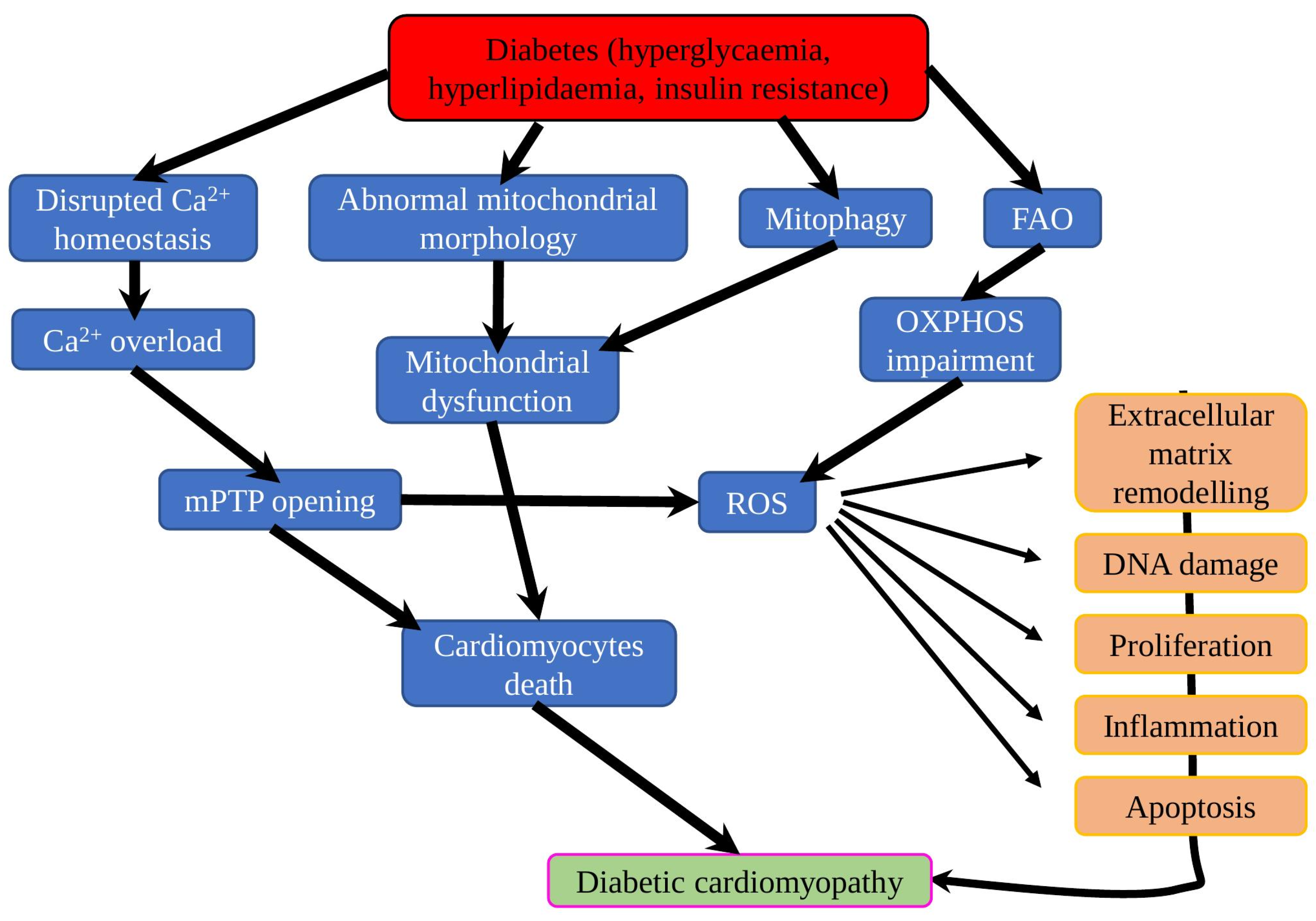
Under physiological conditions, cardiomyocytes require a high amount of energy, which is mostly produced by mitochondria via the FAO (constitutes about 70%) and the remaining part originating from other substrates (ketone bodies, glucose, amino acid and lactate). However, compared to glucose, energy production from FAs requires 12% more oxygen to earn the same amount of ATP [80]. The diabetic heart is characterised by decreased glucose oxidation and increased FAO, which resulted in increased oxygen consumption, respiratory dysfunction in mitochondria and hypoxia aggravation in myocardium with microangiopathy (Figure 3) [81]. As it was shown in the mice fructose-induced T2DM model, early diabetic hearts have evidence of oxidative stress, accompanied with increased cellular and mitochondrial FA uptake, and increased FAO, but also reduced LCAD (Acyl-CoA Dehydrogenase Long Chain) activity and mitochondrial mass. In addition, the activity of SIRT1, a crucial regulator of FA metabolism, was decreased, confirming its important role in cardiac metabolic adaptations via FAO modulation [82]. In the later stages, more severe effects of DMC appeared, for instance, STZ-induced DM mice on HFD for 26 weeks had increased myocardial fibrosis and LV diastolic dysfunction, elevated LV superoxide levels, decreased mitochondria area, increased levels of mitochondrial complex III, and complex V protein abundance, while reducing complex II oxygen consumption [83].
Recent research revealed that reduced cardiac glucose uptake and oxidation is a protective metabolic adaptation, protecting mitochondria from glucotoxicity. Transgenic non-diabetic mice with inducible cardiomyocyte-specific expression of the GLUT4 had decreased mitochondrial ATP generation and evidence of diastolic dysfunction. On the contrary, diabetic mice had increased O-GlcNAcylation of the transcription factor Sp1, many electron transport chain subunits and other mitochondrial proteins, which resulted in aggravated mitochondrial oxidative dysfunction, suggesting mitochondria as a main target of glucotoxicity [84].
Results from the T1DM-induced rat model suggested increased heart mitochondrial mass, volume and number per unit area, whereas the mean area of each mitochondrion was decreased compared to control rats. In addition, diabetic rats’ heart mitochondria had higher production of H2O2 and NO while a lower ATP production rate. Apparently, increased H2O2 and decreased energy production triggered a compensatory mechanism to stimulate the de novo mitochondria biogenesis via upregulation of PGC1α, the major mitochondria biogenesis regulator, which aimed to restore normal energy supply in diabetic pathological conditions [85]. Further research shows that complex I dysfunction reduces ATP production, and the increase in H2O2 and NO production could be considered as early subcellular signals of cardiac mitochondrial dysfunction, while PGC1α-mediated de novo synthesis of mitochondria is activated on the later stage of DCM [86,87]. Interestingly, other researchers have proved that PGC1α expression is reduced because of endogenous accumulation of nitric oxide synthase inhibitor ADMA (asymmetric dimethylarginine) in both T1DM and T2DM rat models, thus suggesting it as a novel therapeutic target to prevent or treat DCM [88].
Interestingly, the presence of DM-related diseases (such as CVDs and obesity) could also affect cardiac mitochondrial energetics and ETC (electron transport chain) efficacy. As it was recently shown, atrial appendages from patients with diabetes and AF (atrial fibrillation) had impaired function of complex I and II, aggravated ETC supercomplex assembly and increased oxidative damage. On the contrary, such abnormalities were not observed in patients with diabetes but without AF [89]. Similarly, mitochondria isolated from right atrial appendage tissues patients with coronary heart disease with/without diabetes had depressed complex respiration CHD patients regardless of the presence of diabetes, while complex II was repressed only in CHD patients with diabetes. Those results are suggesting a more significant mitochondrial dysfunction in the presence of diabetes [90]. In another study with similar set-up, mitochondria isolated from right atrial appendage tissues of DM patients with CHD had decreased oxygen consumption rate for fatty acid substrate and impaired complex I activity. Moreover, mtDNA copy number, levels of ETC complex components, FA metabolism and regulation proteins were unchanged between diabetic and non-diabetic patients [91].
Surprisingly, maternal HFD and/or diabetes could cause cardiac dysfunction in offspring through metabolic stress and impairment of mitochondrial function. Glycolytic and respiratory capacity was impaired in DM-exposed offspring, while HFD-exposed offspring had evidence of mitochondrial dysfunction, increased mitochondrial copy number and rate of lipid peroxidation. Offspring exposed to both DM and HFD had severe symptoms similar to adult DCM, such as accumulation of cardiac lipid droplet and diastolic/systolic cardiac dysfunction [92]. Interestingly, further research suggested different effect of maternal diabetes on male and female offspring of weaning and adult age. As such, only adult male offspring had an elevated level of autophagy and decreased respiration of mitochondrial complex I and II. However, all groups (age and gender) had normal expression levels of antioxidative enzymes (glutathione peroxidise and mitochondrial superoxide dismutase), suggesting absence of oxidative stress [93].
In total, the presence of DM in combination with obesity, CVD and other co-morbidities gradually decreasing efficacy of mitochondria energy production, affecting mostly complexes I and II, finally resulted in mitochondrial dysfunction and DCM. However, mitochondria have several compensatory mechanisms, aiming to restore mitochondrial functions and normal energy supply, suggesting that those mechanisms could be targeted to improve mitochondrial function in DCM treatment or prevention.

7. Mitochondria-Targeting Approach in DCM Treatment

The primary role of mitochondria in DCM pathogenesis was recently established in experiments on PGDM (pre-gestational diabetes mellitus)-exposed rats. PGDM-exposed offspring had cardiac dysfunction at birth, accompanied by low ATP generation, high lipid peroxidation, and high apoptosis rate under metabolic stress. However, transfer of mitochondria isolated from normal rat myocardium to PGDM-exposed rat cardiomyocytes resulted in a sex-specific stimulation of oxygen consumption, ATP production, mitophagy stimulation and reduction of stress-induced apoptosis [94].
Because of the crucial role of mitochondria in DCM pathogenesis and development, several specific mitochondria-targeting therapeutics for DCM treatment and prevention have been invented and successfully tested. For example, mitochondrial ROS inhibitor mito-TEMPO reduced adverse cardiac changes and mitigated myocardial dysfunction in both T1DM and T2DM mice models [95]. Similarly, MitoGamide, the scavenger of methylglyoxal, a mitochondria-damaging by-product of glycolysis and a reactive carbonyl species, showed cardioprotective properties in tests on diabetic Akita mice [96], where it significantly improved the E/A ratio—the key indicator of diastolic dysfunction in the development of diabetes-induced HFpEF (heart failure with preserved ejection fraction) [97]. Application of alisporivir, a mitochondria-targeted non-immunosuppressive analogue of cyclosporin A, which acts as a selective inhibitor of the MPT pore opening, was investigated in the heart of DM-induced mice. Alisporivir effectively restored the blood glucose level and the indicator of heart rate, preventing mitochondrial swelling and ultrastructural alterations in DM mice cardiomyocytes. Simultaneously, alisporivir stimulated the mitophagy rate in the heart tissue, thus activating removal of damaged mitochondria [98]. Similarly, hydrogen sulfide (H2S) was shown to provide mitochondria-mediated cardioprotective effects in STZ-induced T1DM rats. Particularly, exogenous H2S treatment leads to decreased expression of mitochondrial apoptotic proteins, cyt C, mPTP opening, normalised MFN2 expression and cardiac function [99]. FGFs (fibroblast growth factors) are involved in the regulation of metabolism on many levels. Primarily, FGF21 was used to correct metabolic dysfunction associated with diabetes and obesity, while cardioprotective properties were discovered and explored later (reviewed in [100,101]). FGF21 long-term supplementation improved heart rate variability and left ventricular function, attenuated insulin resistance and activated anti-apoptotic and cardiac mitochondrial FAO signalling pathways in obese, insulin-resistant rats [102]. Mechanically, FGF21 prevented T2DM lipotoxicity-induced cardiomyopathy through antioxidative (AMPK–AKT2–NRF2-mediated pathway) and lipid-lowering (AMPK–ACC–CPT-1-mediated pathway) effects in the diabetic heart [103]. Similarly, FGF1 treatment reduced mitochondrial fragmentation and ROS generation, cytochrome c leakage and enhanced mitochondrial respiration rate and β-oxidation, which resulted in restored cardiac function [104].
To sum up, application of mitochondria-targeted treatments provides wide physiological effects and stimulates favourable metabolic processes, confirming its high potential in the treatment of DCM and other metabolic disorders [105].

8. Zinc Supplementation in DM Treatment and Cardioprotection

It is known that Zn2+-associated signalling pathways are involved in the development of diabetic cardiomyopathy and other heart diseases. Mitochondria coordinate [Ca2+]i and [Zn2+]i homeostasis, which affect cellular ROS production, mitochondria ultrastructure and efficiency [106,107]. The role of Zn2+ homeostasis in DM and obesity-induced cardiac remodelling, inflammation, dysfunction and other cardiovascular diseases and complications was covered in several recent reviews [108,109,110]. Here, we wish to describe several recent randomised trials designed to investigate the effect of zinc supplementation on DM progression.
Zn supplementation (20 mg daily) of subjects with pre-diabetes in a randomized, double-blind, placebo-controlled Phase II clinical trial in Sri Lanka for 12 months resulted in a reduction of blood glucose and insulin resistance, improvement of β-cell function, total and LDL cholesterol levels. On the contrary, 25% of participants in the Placebo group have developed T2DM [111]. However, a randomised trial, designed in a similar condition in Australia (except for the zinc dose—30 mg of zinc gluconate daily) [112], did not support the beneficial effects of zinc supplemental in populations with a Western diet [113].
In total, zinc could be considered as an important marker of a healthier diet and/or lifestyle, associated with reduced risk of DM development. However, it is definitively not the key element, responsible for DM development and progression, and other factors, such as initial status of the target population (zinc replete or zinc deficient), prevalent diet and lifestyle, should be considered.

9. Conclusions

Mitochondria are the central player in the DCM research and development of the novel therapeutics for DCM treatment, as their dysfunction appears in the early stages of the DCM development. Maintenance of proper mitochondrial integrity, adequate balance between mitochondrial biogenesis, dynamics and mitophagy are crucial to maintain Ca2+ metabolism and a sufficient level of ATP production during DM-mediated metabolic alterations. Although in the past decade the certain advancement has been made in the understanding of the molecular mechanisms of DCM development, many questions remained unanswered and required more systematic biomedical and pharmaceutical research, preclinical and clinical investigations to develop the better treatments to prevent and treat the cardiovascular complications in diabetic patients.

Author Contributions

S.A.D. and A.N.O. conceptualised the manuscript; S.A.D. wrote the manuscript text; N.K.S. and A.G.K. reviewed the text; E.E.B. and V.N.S., methodology; N.K.S., E.E.B. and A.G.K. formal analysis; V.N.S. and A.N.O. obtained funding and supervised. All authors have read and agreed to the published version of the manuscript.

Funding

This work was supported by the Russian Science Foundation (22-15-00064).

Institutional Review Board Statement

Not applicable.

Informed Consent Statement

Not applicable.

Data Availability Statement

Not applicable.

Conflicts of Interest

The authors declare no conflict of interest. The funder had no role in the design of the study; in the collection, analyses, or interpretation of data; in the writing of the manuscript, or in the decision to publish the results.

Abbreviations

ACC2acetyl-CoA carboxylase 2
AFatrial fibrillation
AKAP121A-kinase anchor protein
ANPatrial natriuretic peptide
Atg7Autophagy-Related Protein 7
BNIP3Bcl-2/adenovirus E1B 19-kDa protein-interacting protein 3
BNPbrain natriuretic peptide
BRD4Bromodomain Containing 4
DCMDiabetic cardiomyopathy
DMdiabetes mellitus
DNM1LDynamin 1 like
Drp1Dynamic relative protein 1
eNOSendothelial nitric oxide synthase
ETCelectron transport chain
FAsfatty acids
FIS1fission, mitochondrial 1
FoxO1forkhead box protein O 1
GLUT4glucose transporter type 4
Grp75Stress-70 Protein, Mitochondrial
HFhigh fat
HGhigh glucose
IGFBP7Insulin Like Growth Factor Binding Protein 7
IP3RInositol 1,4,5-Trisphosphate Receptor Type 3
LACS1long chain acyl-CoA synthetase 1
LC3Autophagy-Related Ubiquitin-Like Modifier LC3 A
LCADAcyl-CoA Dehydrogenase Long Chain
LTCCsL-type calcium channels
LVleft ventricular
MAMsmitochondrial associated membranes
MCUmitochondrial Ca2+ uniporter
MFN1, MFN2mitofusin 1 and 2
MICU1mitochondrial calcium uptake 1
MMPsmatrix metalloproteinases
Mst1Mammalian sterile 20-like kinase 1
OPA1optic atrophy protein 1
Orai1Ca2+ release-activated calcium channel protein 1
ParkinParkin RBR E3 Ubiquitin Protein Ligase
PDCpyruvate dehydrogenase complex
PGC1αPeroxisome proliferator-activated receptor gamma coactivator 1-alpha
PINK1PTEN Induced Kinase 1
Rab9Ras-related protein Rab-9
ROSreactive oxygen species
SRsarcoplasmic reticulum
STZstreptozotocin
TGF-β1Transforming growth factor beta 1
TSP-1thrombospondin-1
Ulk1unc51 like kinase 1
VDACVoltage-dependent anion-selective channel protein 1
ZDFZucker diabetic fatty
β-MHCβ-myosin heavy chain

References

  1. Cardiomyopathy. National Center for Chronic Disease Prevention and Health Promotion. Available online: https://www.cdc.gov/heartdisease/cardiomyopathy.htm (accessed on 20 February 2022).
  2. Paolillo, S.; Marsico, F.; Prastaro, M.; Renga, F.; Esposito, L.; De Martino, F.; Di Napoli, P.; Esposito, I.; Ambrosio, A.; Ianniruberto, M.; et al. Diabetic cardiomyopathy: Definition, diagnosis, and therapeutic implications. Heart Fail. Clin. 2019, 15, 341–347. [Google Scholar] [CrossRef] [PubMed]
  3. Grubić Rotkvić, P.; Planinić, Z.; Liberati Pršo, A.-M.; Šikić, J.; Galić, E.; Rotkvić, L. The mystery of diabetic cardiomyopathy: From early concepts and underlying mechanisms to novel therapeutic possibilities. IJMS 2021, 22, 5973. [Google Scholar] [CrossRef] [PubMed]
  4. Kumric, M.; Ticinovic Kurir, T.; Borovac, J.A.; Bozic, J. Role of novel biomarkers in diabetic cardiomyopathy. World J. Diabetes 2021, 12, 685–705. [Google Scholar] [CrossRef] [PubMed]
  5. Chistiakov, D.A.; Sobenin, I.A.; Orekhov, A.N. Strategies to deliver microRNAs as potential therapeutics in the treatment of cardiovascular pathology. Drug Deliv. 2012, 19, 392–405. [Google Scholar] [CrossRef] [PubMed] [Green Version]
  6. Avagimyan, A. The pathophysiological basis of diabetic cardiomyopathy development. Curr. Probl. Cardiol. 2022, 101156. [Google Scholar] [CrossRef]
  7. Varma, U.; Koutsifeli, P.; Benson, V.L.; Mellor, K.M.; Delbridge, L.M.D. Molecular mechanisms of cardiac pathology in diabetes—Experimental Insights. Biochim. Biophys. Acta Mol. Basis Dis. 2018, 1864, 1949–1959. [Google Scholar] [CrossRef]
  8. Dhalla, N.S.; Shah, A.K.; Tappia, P.S. Role of oxidative stress in metabolic and subcellular abnormalities in diabetic cardiomyopathy. Int. J. Mol. Sci. 2020, 21, E2413. [Google Scholar] [CrossRef] [Green Version]
  9. Makrecka-Kuka, M.; Liepinsh, E.; Murray, A.J.; Lemieux, H.; Dambrova, M.; Tepp, K.; Puurand, M.; Käämbre, T.; Han, W.H.; de Goede, P.; et al. Altered mitochondrial metabolism in the insulin-resistant heart. Acta Physiol. 2020, 228, e13430. [Google Scholar] [CrossRef] [Green Version]
  10. Smani, T.; Gallardo-Castillo, I.; Ávila-Médina, J.; Jimenez-Navarro, M.F.; Ordoñez, A.; Hmadcha, A. Impact of diabetes on cardiac and vascular disease: Role of calcium signaling. Curr. Med. Chem. 2019, 26, 4166–4177. [Google Scholar] [CrossRef]
  11. Kim, A.H.; Jang, J.E.; Han, J. Current status on the therapeutic strategies for heart failure and diabetic cardiomyopathy. Biomed. Pharm. 2022, 145, 112463. [Google Scholar] [CrossRef]
  12. Katogiannis, K.; Vlastos, D.; Kousathana, F.; Thymis, J.; Kountouri, A.; Korakas, E.; Plotas, P.; Papadopoulos, K.; Ikonomidis, I.; Lambadiari, V. Echocardiography, an indispensable tool for the management of diabetics, with or without coronary artery disease, in clinical practice. Medicina 2020, 56, E709. [Google Scholar] [CrossRef] [PubMed]
  13. Tadic, M.; Cuspidi, C.; Calicchio, F.; Grassi, G.; Mancia, G. Diabetic cardiomyopathy: How can cardiac magnetic resonance help? Acta Diabetol. 2020, 57, 1027–1034. [Google Scholar] [CrossRef] [PubMed]
  14. Khalil, H.; Alzahrani, T. Cardiomyopathy imaging. In StatPearls; StatPearls Publishing: Treasure Island, FL, USA, 2022. [Google Scholar]
  15. Soldatov, V.O.; Malorodova, T.N.; Balamutova, T.I.; Ksenofontov, A.O.; Dovgan, A.P.; Urozhevskaya, Z.S. Endothelial dysfunction: Comparative evaluation of ultrasound dopplerography, laser dopplerflowmetry and direct monitoring of arterial pressure for conducting pharmacological tests in rats. RRP 2018, 4, 73–80. [Google Scholar] [CrossRef]
  16. Tuleta, I.; Frangogiannis, N.G. Fibrosis of the diabetic heart: Clinical significance, molecular mechanisms, and therapeutic opportunities. Adv. Drug Deliv. Rev. 2021, 176, 113904. [Google Scholar] [CrossRef]
  17. Ibrahim, E.-S.H.; Dennison, J.; Frank, L.; Stojanovska, J. Diastolic cardiac function by MRI-imaging capabilities and clinical applications. Tomography 2021, 7, 75. [Google Scholar] [CrossRef] [PubMed]
  18. Tadic, M.; Cuspidi, C. Left atrial function in diabetes: Does it help? Acta Diabetol. 2021, 58, 131–137. [Google Scholar] [CrossRef] [PubMed]
  19. Yue, Y.; Meng, K.; Pu, Y.; Zhang, X. Transforming growth factor beta (TGF-β) mediates cardiac fibrosis and induces diabetic cardiomyopathy. Diabetes Res. Clin. Pract. 2017, 133, 124–130. [Google Scholar] [CrossRef] [PubMed]
  20. Xu, L.; Zhang, Y.; Chen, J.; Xu, Y. Thrombospondin-1: A key protein that induces fibrosis in diabetic complications. J. Diabetes Res. 2020, 2020, 8043135. [Google Scholar] [CrossRef]
  21. Li, L.; Zhao, Q.; Kong, W. Extracellular matrix remodeling and cardiac fibrosis. Matrix Biol. 2018, 68–69, 490–506. [Google Scholar] [CrossRef]
  22. Chen, Y.; Hua, Y.; Li, X.; Arslan, I.M.; Zhang, W.; Meng, G. Distinct types of cell death and the implication in diabetic cardiomyopathy. Front. Pharmacol. 2020, 11, 42. [Google Scholar] [CrossRef]
  23. Chistiakov, D.; Revin, V.; Sobenin, I.; Orekhov, A.; Bobryshev, Y. Vascular endothelium: Functioning in norm, changes in atherosclerosis and current dietary approaches to improve endothelial function. MRMC 2015, 15, 338–350. [Google Scholar] [CrossRef] [PubMed]
  24. Sharma, A.; Mah, M.; Ritchie, R.H.; De Blasio, M.J. The adiponectin signalling pathway—A therapeutic target for the cardiac complications of type 2 diabetes? Pharmacol. Ther. 2022, 232, 108008. [Google Scholar] [CrossRef] [PubMed]
  25. Yu, W.; Chen, C.; Cheng, J. The role and molecular mechanism of FoxO1 in mediating cardiac hypertrophy. ESC Heart Fail. 2020, 7, 3497–3504. [Google Scholar] [CrossRef] [PubMed]
  26. Suarez, J.; Cividini, F.; Scott, B.T.; Lehmann, K.; Diaz-Juarez, J.; Diemer, T.; Dai, A.; Suarez, J.A.; Jain, M.; Dillmann, W.H. Restoring mitochondrial calcium uniporter expression in diabetic mouse heart improves mitochondrial calcium handling and cardiac function. J. Biol. Chem. 2018, 293, 8182–8195. [Google Scholar] [CrossRef] [Green Version]
  27. Ong, S.-B.; Samangouei, P.; Kalkhoran, S.B.; Hausenloy, D.J. The mitochondrial permeability transition pore and its role in myocardial ischemia reperfusion injury. J. Mol. Cell. Cardiol. 2015, 78, 23–34. [Google Scholar] [CrossRef]
  28. Chengji, W.; Xianjin, F. Treadmill exercise alleviates diabetic cardiomyopathy by suppressing plasminogen activator inhibitor expression and enhancing ENOS in streptozotocin-induced male diabetic rats. Endocr. Connect. 2018, 7, 553–559. [Google Scholar] [CrossRef]
  29. Sobenin, I.A.; Salonen, J.T.; Zhelankin, A.V.; Melnichenko, A.A.; Kaikkonen, J.; Bobryshev, Y.V.; Orekhov, A.N. Low density lipoprotein-containing circulating immune complexes: Role in atherosclerosis and diagnostic value. BioMed Res. Int. 2014, 2014, 205697. [Google Scholar] [CrossRef]
  30. Popov, L.-D. Mitochondrial biogenesis: An update. J. Cell. Mol. Med. 2020, 24, 4892–4899. [Google Scholar] [CrossRef] [Green Version]
  31. Forte, M.; Schirone, L.; Ameri, P.; Basso, C.; Catalucci, D.; Modica, J.; Chimenti, C.; Crotti, L.; Frati, G.; Rubattu, S.; et al. The role of mitochondrial dynamics in cardiovascular diseases. Br. J. Pharm. 2021, 178, 2060–2076. [Google Scholar] [CrossRef]
  32. Onishi, M.; Okamoto, K. Mitochondrial clearance: Mechanisms and roles in cellular fitness. FEBS Lett. 2021, 595, 1239–1263. [Google Scholar] [CrossRef]
  33. Tilokani, L.; Nagashima, S.; Paupe, V.; Prudent, J. Mitochondrial dynamics: Overview of molecular mechanisms. Essays Biochem. 2018, 62, 341–360. [Google Scholar] [CrossRef] [PubMed] [Green Version]
  34. Sobenin, I.A.; Sazonova, M.A.; Postnov, A.Y.; Bobryshev, Y.V.; Orekhov, A.N. Changes of mitochondria in atherosclerosis: Possible determinant in the pathogenesis of the disease. Atherosclerosis 2013, 227, 283–288. [Google Scholar] [CrossRef] [PubMed]
  35. Onishi, M.; Yamano, K.; Sato, M.; Matsuda, N.; Okamoto, K. Molecular mechanisms and physiological functions of mitophagy. EMBO J. 2021, 40, e104705. [Google Scholar] [CrossRef] [PubMed]
  36. Sobenin, I.A.; Sazonova, M.A.; Postnov, A.Y.; Bobryshev, Y.V.; Orekhov, A.N. Mitochondrial mutations are associated with atherosclerotic lesions in the human aorta. Clin. Dev. Immunol. 2012, 2012, 832464. [Google Scholar] [CrossRef]
  37. Song, Y.; Xu, Y.; Liu, Y.; Gao, J.; Feng, L.; Zhang, Y.; Shi, L.; Zhang, M.; Guo, D.; Qi, B.; et al. Mitochondrial quality control in the maintenance of cardiovascular homeostasis: The roles and interregulation of UPS, mitochondrial dynamics and mitophagy. Oxid. Med. Cell. Longev. 2021, 2021, 3960773. [Google Scholar] [CrossRef]
  38. Kaludercic, N.; Di Lisa, F. Mitochondrial ROS formation in the pathogenesis of diabetic cardiomyopathy. Front. Cardiovasc. Med. 2020, 7, 12. [Google Scholar] [CrossRef] [Green Version]
  39. Sobenin, I.A.; Sazonova, M.A.; Postnov, A.Y.; Salonen, J.T.; Bobryshev, Y.V.; Orekhov, A.N. Association of mitochondrial genetic variation with carotid atherosclerosis. PLoS ONE 2013, 8, e68070. [Google Scholar] [CrossRef] [Green Version]
  40. Hu, L.; Ding, M.; Tang, D.; Gao, E.; Li, C.; Wang, K.; Qi, B.; Qiu, J.; Zhao, H.; Chang, P.; et al. Targeting mitochondrial dynamics by regulating Mfn2 for therapeutic intervention in diabetic cardiomyopathy. Theranostics 2019, 9, 3687–3706. [Google Scholar] [CrossRef]
  41. Garvin, A.M.; Jackson, M.A.; Korzick, D.H. Inhibition of programmed necrosis limits infarct size through altered mitochondrial and immune responses in the aged female rat heart. Am. J. Physiol. Heart Circ. Physiol. 2018, 315, H1434–H1442. [Google Scholar] [CrossRef]
  42. Apaijai, N.; Jinawong, K.; Singhanat, K.; Jaiwongkam, T.; Kerdphoo, S.; Chattipakorn, S.C.; Chattipakorn, N. Necrostatin-1 reduces cardiac and mitochondrial dysfunction in prediabetic rats. J. Endocrinol. 2021, 251, 27–39. [Google Scholar] [CrossRef]
  43. Yuan, M.; Gong, M.; Zhang, Z.; Meng, L.; Tse, G.; Zhao, Y.; Bao, Q.; Zhang, Y.; Yuan, M.; Liu, X.; et al. Hyperglycemia induces endoplasmic reticulum stress in atrial cardiomyocytes, and mitofusin-2 downregulation prevents mitochondrial dysfunction and subsequent cell death. Oxid. Med. Cell. Longev. 2020, 2020, 6569728. [Google Scholar] [CrossRef] [PubMed]
  44. Sobenin, I.A.; Mitrofanov, K.Y.; Zhelankin, A.V.; Sazonova, M.A.; Postnov, A.Y.; Revin, V.V.; Bobryshev, Y.V.; Orekhov, A.N. Quantitative assessment of heteroplasmy of mitochondrial genome: Perspectives in diagnostics and methodological pitfalls. BioMed Res. Int. 2014, 2014, 292017. [Google Scholar] [CrossRef] [PubMed]
  45. Zhang, J.; Zhang, F.; Wang, Y. Mitofusin-2 enhances mitochondrial contact with the endoplasmic reticulum and promotes diabetic cardiomyopathy. Front. Physiol. 2021, 12, 707634. [Google Scholar] [CrossRef] [PubMed]
  46. Roe, A.J.; Qi, X. Drp1 phosphorylation by MAPK1 causes mitochondrial dysfunction in cell culture model of Huntington’s disease. Biochem. Biophys. Res. Commun. 2018, 496, 706–711. [Google Scholar] [CrossRef] [PubMed]
  47. Wu, Q.-R.; Zheng, D.-L.; Liu, P.-M.; Yang, H.; Li, L.-A.; Kuang, S.-J.; Lai, Y.-Y.; Rao, F.; Xue, Y.-M.; Lin, J.-J.; et al. High glucose induces Drp1-mediated mitochondrial fission via the orai1 calcium channel to participate in diabetic cardiomyocyte hypertrophy. Cell Death Dis. 2021, 12, 216. [Google Scholar] [CrossRef]
  48. Yang, Y.; Wang, H.; Ma, Z.; Hu, W.; Sun, D. Understanding the role of mammalian sterile 20-like kinase 1 (MST1) in cardiovascular disorders. J. Mol. Cell. Cardiol. 2018, 114, 141–149. [Google Scholar] [CrossRef]
  49. Feng, X.; Wang, S.; Yang, X.; Lin, J.; Man, W.; Dong, Y.; Zhang, Y.; Zhao, Z.; Wang, H.; Sun, D. Mst1 knockout alleviates mitochondrial fission and mitigates left ventricular remodeling in the development of diabetic cardiomyopathy. Front. Cell Dev. Biol. 2021, 8, 628842. [Google Scholar] [CrossRef]
  50. Czachor, A.; Failla, A.; Lockey, R.; Kolliputi, N. Pivotal role of AKAP121 in mitochondrial physiology. Am. J. Physiol. Cell Physiol. 2016, 310, C625–C628. [Google Scholar] [CrossRef] [Green Version]
  51. Tsushima, K.; Bugger, H.; Wende, A.R.; Soto, J.; Jenson, G.A.; Tor, A.R.; McGlauflin, R.; Kenny, H.C.; Zhang, Y.; Souvenir, R.; et al. Mitochondrial reactive oxygen species in lipotoxic hearts induce post-translational modifications of AKAP121, DRP1, and OPA1 that promote mitochondrial fission. Circ. Res. 2018, 122, 58–73. [Google Scholar] [CrossRef]
  52. Koncsos, G.; Varga, Z.V.; Baranyai, T.; Boengler, K.; Rohrbach, S.; Li, L.; Schlüter, K.-D.; Schreckenberg, R.; Radovits, T.; Oláh, A.; et al. Diastolic dysfunction in prediabetic male rats: Role of mitochondrial oxidative stress. Am. J. Physiol. Heart Circ. Physiol. 2016, 311, H927–H943. [Google Scholar] [CrossRef] [Green Version]
  53. Tong, M.; Saito, T.; Zhai, P.; Oka, S.-I.; Mizushima, W.; Nakamura, M.; Ikeda, S.; Shirakabe, A.; Sadoshima, J. Mitophagy is essential for maintaining cardiac function during high fat diet-induced diabetic cardiomyopathy. Circ. Res. 2019, 124, 1360–1371. [Google Scholar] [CrossRef] [PubMed]
  54. Wang, S.; Zhao, Z.; Fan, Y.; Zhang, M.; Feng, X.; Lin, J.; Hu, J.; Cheng, Z.; Sun, C.; Liu, T.; et al. Mst1 inhibits Sirt3 expression and contributes to diabetic cardiomyopathy through inhibiting parkin-dependent mitophagy. Biochim. Biophys. Acta Mol. Basis Dis. 2019, 1865, 1905–1914. [Google Scholar] [CrossRef] [PubMed]
  55. Yu, W.; Gao, B.; Li, N.; Wang, J.; Qiu, C.; Zhang, G.; Liu, M.; Zhang, R.; Li, C.; Ji, G.; et al. Sirt3 deficiency exacerbates diabetic cardiac dysfunction: Role of Foxo3A-parkin-mediated mitophagy. Biochim. Biophys. Acta Mol. Basis Dis. 2017, 1863, 1973–1983. [Google Scholar] [CrossRef]
  56. Kitada, M.; Ogura, Y.; Monno, I.; Koya, D. Sirtuins and type 2 diabetes: Role in inflammation, oxidative stress, and mitochondrial function. Front. Endocrinol. 2019, 10, 187. [Google Scholar] [CrossRef] [PubMed] [Green Version]
  57. Shao, D.; Kolwicz, S.C.; Wang, P.; Roe, N.D.; Villet, O.; Nishi, K.; Hsu, Y.-W.A.; Flint, G.V.; Caudal, A.; Wang, W.; et al. Increasing fatty acid oxidation prevents high-fat diet-induced cardiomyopathy through regulating parkin-mediated mitophagy. Circulation 2020, 142, 983–997. [Google Scholar] [CrossRef] [PubMed]
  58. Padmanabhan, A.; Alexanian, M.; Linares-Saldana, R.; González-Terán, B.; Andreoletti, G.; Huang, Y.; Connolly, A.J.; Kim, W.; Hsu, A.; Duan, Q.; et al. BRD4 (bromodomain-containing protein 4) interacts with GATA4 (GATA binding protein 4) to govern mitochondrial homeostasis in adult cardiomyocytes. Circulation 2020, 142, 2338–2355. [Google Scholar] [CrossRef]
  59. Duan, Q.; McMahon, S.; Anand, P.; Shah, H.; Thomas, S.; Salunga, H.T.; Huang, Y.; Zhang, R.; Sahadevan, A.; Lemieux, M.E.; et al. BET bromodomain inhibition suppresses innate inflammatory and profibrotic transcriptional networks in heart failure. Sci. Transl. Med. 2017, 9, eaah5084. [Google Scholar] [CrossRef] [Green Version]
  60. Guo, M.; Wang, H.-X.; Chen, W.-J. BET-inhibition by JQ1 alleviates streptozotocin-induced diabetic cardiomyopathy. Toxicol. Appl. Pharmacol. 2018, 352, 9–18. [Google Scholar] [CrossRef]
  61. Mu, J.; Zhang, D.; Tian, Y.; Xie, Z.; Zou, M.-H. BRD4 inhibition by JQ1 prevents high-fat diet-induced diabetic cardiomyopathy by activating PINK1/parkin-mediated mitophagy in vivo. J. Mol. Cell. Cardiol. 2020, 149, 1–14. [Google Scholar] [CrossRef]
  62. Tong, M.; Saito, T.; Zhai, P.; Oka, S.-I.; Mizushima, W.; Nakamura, M.; Ikeda, S.; Shirakabe, A.; Sadoshima, J. Alternative mitophagy protects the heart against obesity-associated cardiomyopathy. Circ. Res. 2021, 129, 1105–1121. [Google Scholar] [CrossRef]
  63. Diaz-Juarez, J.; Suarez, J.A.; Dillmann, W.H. Mitochondrial calcium handling and heart disease in diabetes mellitus. Biochim. Biophys. Acta BBA—Mol. Basis Dis. 2021, 1867, 165984. [Google Scholar] [CrossRef] [PubMed]
  64. Hoang-Trong, M.T.; Ullah, A.; Lederer, W.J.; Jafri, M.S. Cardiac alternans occurs through the synergy of voltage- and calcium-dependent mechanisms. Membranes 2021, 11, 794. [Google Scholar] [CrossRef] [PubMed]
  65. Kettlewell, S.; Saxena, P.; Dempster, J.; Colman, M.A.; Myles, R.C.; Smith, G.L.; Workman, A.J. Dynamic clamping human and rabbit atrial calcium current: Narrowing ICaL window abolishes early afterdepolarizations. J. Physiol. 2019, 597, 3619–3638. [Google Scholar] [CrossRef] [PubMed] [Green Version]
  66. Diaz-Juarez, J.; Suarez, J.; Cividini, F.; Scott, B.T.; Diemer, T.; Dai, A.; Dillmann, W.H. Expression of the mitochondrial calcium uniporter in cardiac myocytes improves impaired mitochondrial calcium handling and metabolism in simulated hyperglycemia. Am. J. Physiol. Cell Physiol. 2016, 311, C1005–C1013. [Google Scholar] [CrossRef] [PubMed] [Green Version]
  67. Ji, L.; Liu, F.; Jing, Z.; Huang, Q.; Zhao, Y.; Cao, H.; Li, J.; Yin, C.; Xing, J.; Li, F. MICU1 alleviates diabetic cardiomyopathy through mitochondrial Ca2+-dependent antioxidant response. Diabetes 2017, 66, 1586–1600. [Google Scholar] [CrossRef] [PubMed] [Green Version]
  68. Lopez-Crisosto, C.; Pennanen, C.; Vasquez-Trincado, C.; Morales, P.E.; Bravo-Sagua, R.; Quest, A.F.G.; Chiong, M.; Lavandero, S. Sarcoplasmic reticulum–mitochondria communication in cardiovascular pathophysiology. Nat. Rev. Cardiol. 2017, 14, 342–360. [Google Scholar] [CrossRef]
  69. Dia, M.; Gomez, L.; Thibault, H.; Tessier, N.; Leon, C.; Chouabe, C.; Ducreux, S.; Gallo-Bona, N.; Tubbs, E.; Bendridi, N.; et al. Reduced reticulum–mitochondria Ca2+ transfer is an early and reversible trigger of mitochondrial dysfunctions in diabetic cardiomyopathy. Basic Res. Cardiol. 2020, 115, 74. [Google Scholar] [CrossRef]
  70. Federico, M.; Portiansky, E.L.; Sommese, L.; Alvarado, F.J.; Blanco, P.G.; Zanuzzi, C.N.; Dedman, J.; Kaetzel, M.; Wehrens, X.H.T.; Mattiazzi, A.; et al. Calcium-calmodulin-dependent protein kinase mediates the intracellular signalling pathways of cardiac apoptosis in mice with impaired glucose tolerance. J. Physiol. 2017, 595, 4089–4108. [Google Scholar] [CrossRef] [Green Version]
  71. Federico, M.; Zavala, M.; Vico, T.; López, S.; Portiansky, E.; Alvarez, S.; Abrille, M.C.V.; Palomeque, J. CaMKII activation in early diabetic hearts induces altered sarcoplasmic reticulum-mitochondria signaling. Sci. Rep. 2021, 11, 20025. [Google Scholar] [CrossRef]
  72. Liu, H.; Zhao, Y.; Xie, A.; Kim, T.-Y.; Terentyeva, R.; Liu, M.; Shi, G.; Feng, F.; Choi, B.-R.; Terentyev, D.; et al. Interleukin-1β, oxidative stress, and abnormal calcium handling mediate diabetic arrhythmic risk. JACC Basic Transl. Sci. 2021, 6, 42–52. [Google Scholar] [CrossRef]
  73. Joseph, L.C.; Subramanyam, P.; Radlicz, C.; Trent, C.M.; Iyer, V.; Colecraft, H.M.; Morrow, J.P. Mitochondrial oxidative stress during cardiac lipid overload causes intracellular calcium leak and arrhythmia. Heart Rhythm 2016, 13, 1699–1706. [Google Scholar] [CrossRef] [PubMed] [Green Version]
  74. Wang, J.; Wang, S.; Wang, W.; Chen, J.; Zhang, Z.; Zheng, Q.; Liu, Q.; Cai, L. Protection against diabetic cardiomyopathy is achieved using a combination of sulforaphane and zinc in type 1 diabetic OVE26 mice. J. Cell. Mol. Med. 2019, 23, 6319–6330. [Google Scholar] [CrossRef] [PubMed] [Green Version]
  75. Korkmaz-Icöz, S.; Al Said, S.; Radovits, T.; Li, S.; Brune, M.; Hegedűs, P.; Atmanli, A.; Ruppert, M.; Brlecic, P.; Lehmann, L.H.; et al. Oral treatment with a zinc complex of acetylsalicylic acid prevents diabetic cardiomyopathy in a rat model of type-2 diabetes: Activation of the akt pathway. Cardiovasc. Diabetol. 2016, 15, 75. [Google Scholar] [CrossRef] [Green Version]
  76. Puchenkova, O.A.; Nadezhdin, S.V.; Soldatov, V.O.; Zhuchenko, M.A.; Korshunova, D.S.; Kubekina, M.V.; Korshunov, E.N.; Korokina, L.V.; Golubinskaya, P.A.; Kulikov, A.L.; et al. Study of antiatherosclerotic and endothelioprotective activity of peptide agonists of EPOR/CD131 heteroreceptor. Pharm. Pharmacol. 2020, 8, 100–111. [Google Scholar] [CrossRef]
  77. Shams, A.S.; Mohammed, M.H.; Loka, M.M.; Abdel Rahman, G.M. Assessment of the protective role of prenatal zinc versus insulin supplementation on fetal cardiac damage induced by maternal diabetes in rat using caspase-3 and KI67 immunohistochemical stains. Cardiol. Res. Pract. 2016, 2016, 7469549. [Google Scholar] [CrossRef] [PubMed] [Green Version]
  78. Zhang, S.; Liu, H.; Amarsingh, G.V.; Cheung, C.C.H.; Wu, D.; Narayanan, U.; Zhang, L.; Cooper, G.J.S. Restoration of myocellular copper-trafficking proteins and mitochondrial copper enzymes repairs cardiac function in rats with diabetes-evoked heart failure. Metallomics 2020, 12, 259–272. [Google Scholar] [CrossRef] [PubMed]
  79. Bjørklund, G.; Dadar, M.; Pivina, L.; Doşa, M.D.; Semenova, Y.; Aaseth, J. The role of zinc and copper in insulin resistance and diabetes mellitus. Curr. Med. Chem. 2020, 27, 6643–6657. [Google Scholar] [CrossRef]
  80. Honka, H.; Solis-Herrera, C.; Triplitt, C.; Norton, L.; Butler, J.; DeFronzo, R.A. Therapeutic manipulation of myocardial metabolism: JACC state-of-the-art review. J. Am. Coll. Cardiol. 2021, 77, 2022–2039. [Google Scholar] [CrossRef]
  81. Mereweather, L.J.; Montes Aparicio, C.N.; Heather, L.C. Positioning metabolism as a central player in the diabetic heart. J. Lipid Atheroscler. 2020, 9, 92–109. [Google Scholar] [CrossRef]
  82. Lou, P.-H.; Lucchinetti, E.; Scott, K.Y.; Huang, Y.; Gandhi, M.; Hersberger, M.; Clanachan, A.S.; Lemieux, H.; Zaugg, M. Alterations in fatty acid metabolism and sirtuin signaling characterize early type-2 diabetic hearts of fructose-fed rats. Physiol. Rep. 2017, 5, e13388. [Google Scholar] [CrossRef]
  83. Parker, A.M.; Tate, M.; Prakoso, D.; Deo, M.; Willis, A.M.; Nash, D.M.; Donner, D.G.; Crawford, S.; Kiriazis, H.; Granata, C.; et al. Characterisation of the myocardial mitochondria structural and functional phenotype in a murine model of diabetic cardiomyopathy. Front. Physiol. 2021, 12, 672252. [Google Scholar] [CrossRef] [PubMed]
  84. Wende, A.R.; Schell, J.C.; Ha, C.-M.; Pepin, M.E.; Khalimonchuk, O.; Schwertz, H.; Pereira, R.O.; Brahma, M.K.; Tuinei, J.; Contreras-Ferrat, A.; et al. Maintaining myocardial glucose utilization in diabetic cardiomyopathy accelerates mitochondrial dysfunction. Diabetes 2020, 69, 2094–2111. [Google Scholar] [CrossRef] [PubMed]
  85. Bombicino, S.S.; Iglesias, D.E.; Rukavina-Mikusic, I.A.; Buchholz, B.; Gelpi, R.J.; Boveris, A.; Valdez, L.B. Hydrogen peroxide, nitric oxide and ATP are molecules involved in cardiac mitochondrial biogenesis in diabetes. Free Radic. Biol. Med. 2017, 112, 267–276. [Google Scholar] [CrossRef]
  86. Rukavina-Mikusic, I.A.; Rey, M.; Martinefski, M.; Tripodi, V.; Valdez, L.B. Temporal evolution of cardiac mitochondrial dysfunction in a type 1 diabetes model. mitochondrial complex i impairment, and H2O2 and NO productions as early subcellular events. Free Radic. Biol. Med. 2021, 162, 129–140. [Google Scholar] [CrossRef] [PubMed]
  87. Bombicino, S.S.; Iglesias, D.E.; Mikusic, I.A.R.; D’Annunzio, V.; Gelpi, R.J.; Boveris, A.; Valdez, L.B. Diabetes impairs heart mitochondrial function without changes in resting cardiac performance. Int. J. Biochem. Cell Biol. 2016, 81, 335–345. [Google Scholar] [CrossRef] [PubMed]
  88. Xiong, Y.; He, Y.-L.; Li, X.-M.; Nie, F.; Zhou, X.-K. Endogenous asymmetric dimethylarginine accumulation precipitates the cardiac and mitochondrial dysfunctions in type 1 diabetic rats. Eur. J. Pharmacol. 2021, 902, 174081. [Google Scholar] [CrossRef]
  89. Kanaan, G.N.; Patten, D.A.; Redpath, C.J.; Harper, M.-E. Atrial fibrillation is associated with impaired atrial mitochondrial energetics and supercomplex formation in adults with type 2 diabetes. Can. J. Diabetes 2019, 43, 67–75.e1. [Google Scholar] [CrossRef] [Green Version]
  90. Duicu, O.M.; Lighezan, R.; Sturza, A.; Balica, R.; Vaduva, A.; Feier, H.; Gaspar, M.; Ionac, A.; Noveanu, L.; Borza, C.; et al. Assessment of mitochondrial dysfunction and monoamine oxidase contribution to oxidative stress in human diabetic hearts. Oxid. Med. Cell. Longev. 2016, 2016, 8470394. [Google Scholar] [CrossRef] [Green Version]
  91. Jayakumari, N.R.; Rajendran, R.S.; Sivasailam, A.; Vimala, S.S.; Nanda, S.; Manjunatha, S.; Pillai, V.V.; Karunakaran, J.; Gopala, S. Impaired substrate-mediated cardiac mitochondrial complex i respiration with unaltered regulation of fatty acid metabolism and oxidative stress status in type 2 diabetic Asian Indians. J. Diabetes 2020, 12, 542–555. [Google Scholar] [CrossRef]
  92. Mdaki, K.S.; Larsen, T.D.; Wachal, A.L.; Schimelpfenig, M.D.; Weaver, L.J.; Dooyema, S.D.R.; Louwagie, E.J.; Baack, M.L. Maternal high-fat diet impairs cardiac function in offspring of diabetic pregnancy through metabolic stress and mitochondrial dysfunction. Am. J. Physiol. Heart Circ. Physiol. 2016, 310, H681–H692. [Google Scholar] [CrossRef] [Green Version]
  93. Raji, S.R.; Nandini, R.J.; Ashok, S.; Anand, R.C.; Vivek, P.V.; Karunakaran, J.; Sreelatha, H.V.; Manjunatha, S.; Gopala, S. Diminished substrate-mediated cardiac mitochondrial respiration and elevated autophagy in adult male offspring of gestational diabetic rats. IUBMB Life 2021, 73, 676–689. [Google Scholar] [CrossRef] [PubMed]
  94. Louwagie, E.J.; Larsen, T.D.; Wachal, A.L.; Gandy, T.C.T.; Baack, M.L. Mitochondrial transfer improves cardiomyocyte bioenergetics and viability in male rats exposed to pregestational diabetes. IJMS 2021, 22, 2382. [Google Scholar] [CrossRef] [PubMed]
  95. Ni, R.; Cao, T.; Xiong, S.; Ma, J.; Fan, G.-C.; Lacefield, J.C.; Lu, Y.; Tissier, S.L.; Peng, T. Therapeutic inhibition of mitochondrial reactive oxygen species with mito-TEMPO reduces diabetic cardiomyopathy. Free Radic. Biol. Med. 2016, 90, 12–23. [Google Scholar] [CrossRef] [PubMed] [Green Version]
  96. Tate, M.; Higgins, G.C.; De Blasio, M.J.; Lindblom, R.; Prakoso, D.; Deo, M.; Kiriazis, H.; Park, M.; Baeza-Garza, C.D.; Caldwell, S.T.; et al. The mitochondria-targeted methylglyoxal sequestering compound, mitogamide, is cardioprotective in the diabetic heart. Cardiovasc. Drugs 2019, 33, 669–674. [Google Scholar] [CrossRef] [Green Version]
  97. Park, M.; Nishimura, T.; Baeza-Garza, C.D.; Caldwell, S.T.; Pun, P.B.L.; Prag, H.A.; Young, T.; Sauchanka, O.; Logan, A.; Forkink, M.; et al. Confirmation of the cardioprotective effect of mitogamide in the diabetic heart. Cardiovasc. Drugs 2020, 34, 823–834. [Google Scholar] [CrossRef]
  98. Belosludtseva, N.V.; Starinets, V.S.; Mikheeva, I.B.; Serov, D.A.; Astashev, M.E.; Belosludtsev, M.N.; Dubinin, M.V.; Belosludtsev, K.N. Effect of the MPT pore inhibitor alisporivir on the development of mitochondrial dysfunction in the heart tissue of diabetic mice. Biology 2021, 10, 839. [Google Scholar] [CrossRef]
  99. Yang, F.; Yu, X.; Li, T.; Wu, J.; Zhao, Y.; Liu, J.; Sun, A.; Dong, S.; Wu, J.; Zhong, X.; et al. Exogenous H 2 S regulates endoplasmic reticulum-mitochondria cross-talk to inhibit apoptotic pathways in stz-induced type i diabetes. Am. J. Physiol. Endocrinol. Metab. 2017, 312, E190–E203. [Google Scholar] [CrossRef] [Green Version]
  100. Gasser, E.; Moutos, C.P.; Downes, M.; Evans, R.M. FGF1—A new weapon to control type 2 diabetes mellitus. Nat. Rev. Endocrinol. 2017, 13, 599–609. [Google Scholar] [CrossRef]
  101. Flippo, K.H.; Potthoff, M.J. Metabolic messengers: FGF21. Nat. Metab. 2021, 3, 309–317. [Google Scholar] [CrossRef]
  102. Tanajak, P.; Sa-Nguanmoo, P.; Wang, X.; Liang, G.; Li, X.; Jiang, C.; Chattipakorn, S.C.; Chattipakorn, N. Fibroblast growth factor 21 (FGF21) therapy attenuates left ventricular dysfunction and metabolic disturbance by improving FGF21 sensitivity, cardiac mitochondrial redox homoeostasis and structural changes in pre-diabetic rats. Acta Physiol. 2016, 217, 287–299. [Google Scholar] [CrossRef]
  103. Yang, H.; Feng, A.; Lin, S.; Yu, L.; Lin, X.; Yan, X.; Lu, X.; Zhang, C. Fibroblast growth factor-21 prevents diabetic cardiomyopathy via AMPK-mediated antioxidation and lipid-lowering effects in the heart. Cell Death Dis. 2018, 9, 227. [Google Scholar] [CrossRef] [PubMed] [Green Version]
  104. Wang, D.; Yin, Y.; Wang, S.; Zhao, T.; Gong, F.; Zhao, Y.; Wang, B.; Huang, Y.; Cheng, Z.; Zhu, G.; et al. FGF1ΔHBS prevents diabetic cardiomyopathy by maintaining mitochondrial homeostasis and reducing oxidative stress via AMPK/Nur77 suppression. Signal Transduct. Target. Ther. 2021, 6, 133. [Google Scholar] [CrossRef] [PubMed]
  105. Hassany, M.A.; El-Ebidi, A.M.; Hafez, M.; Tawfik, N.M.; Wang, P.H. Role of mitochondria in diabetic cardiomyopathy and treatment challenges for mitochondrial dysfunction. IOSR J. Dent. Med. Sci. 2022, 21, 7–19. [Google Scholar] [CrossRef]
  106. Asghari, K.; Shargh, Z.; Fatehfar, S.; Chodari, L.; Sameei, P. The impact of zinc on the molecular signaling pathways in the diabetes disease. J. Trace Elem. Med. Biol. 2022, 72, 126985. [Google Scholar] [CrossRef] [PubMed]
  107. Di Filippo, E.S.; Checcaglini, F.; Fanò-Illic, G.; Fulle, S. H2O2/Ca2+/Zn2+ complex can be considered a “collaborative sensor” of the mitochondrial capacity? Antioxidants 2022, 11, 342. [Google Scholar] [CrossRef]
  108. Zhang, H.; Cai, L. Zinc homeostasis plays an important role in the prevention of obesity-induced cardiac inflammation, remodeling and dysfunction. J. Trace Elem. Med. Biol. 2020, 62, 126615. [Google Scholar] [CrossRef]
  109. Tamura, Y. The role of zinc homeostasis in the prevention of diabetes mellitus and cardiovascular diseases. JAT 2021, 28, 1109–1122. [Google Scholar] [CrossRef]
  110. MacKenzie, S.; Bergdahl, A. Zinc homeostasis in diabetes mellitus and vascular complications. Biomedicines 2022, 10, 139. [Google Scholar] [CrossRef]
  111. Ranasinghe, P.; Wathurapatha, W.S.; Galappatthy, P.; Katulanda, P.; Jayawardena, R.; Constantine, G.R. Zinc supplementation in prediabetes: A randomized double-blind placebo-controlled clinical trial. J. Diabetes 2018, 10, 386–397. [Google Scholar] [CrossRef]
  112. Peel, R.; Hure, A.; Wiggers, J.; McEvoy, M.; Holliday, E.; Searles, A.; Reeves, P.; Ranasinghe, P.; Jayawardena, R.; Samman, S.; et al. Zinc in preventing the progression of pre-diabetes (ZIPPeD Study)—Study protocol for a randomised placebo-controlled trial in Australia. Trials 2019, 20, 219. [Google Scholar] [CrossRef]
  113. Attia, J.R.; Holliday, E.; Weaver, N.; Peel, R.; Fleming, K.C.; Hure, A.; Wiggers, J.; McEvoy, M.; Searles, A.; Reeves, P.; et al. The effect of zinc supplementation on glucose homeostasis: A randomised double-blind placebo-controlled trial. Acta Diabetol. 2022, 59, 965–975. [Google Scholar] [CrossRef] [PubMed]
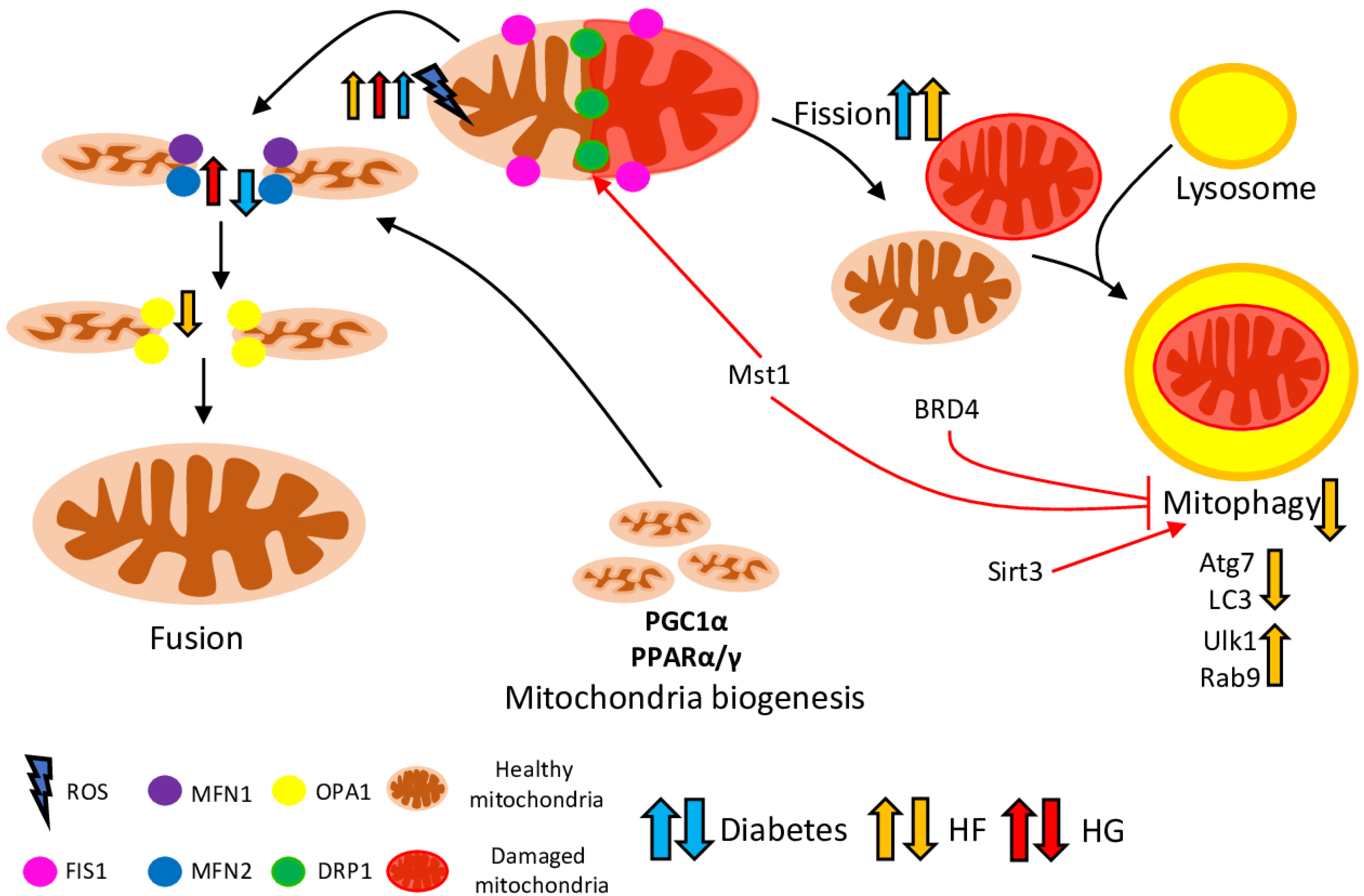
Figure 1. Interconnections between mitophagy, mitochondrial dynamics, and mitochondrial biogenesis in DCM. Damaged and malfunctional mitochondria are isolated via mitochondrial fission, merged with lysosome, and digested through mitophagy. Healthy parts of mitochondria are returned to the mitochondrial network through mitochondrial fusion. Mitochondrial fission is mediated by Drp1 and Fis1, fusion––Mfn1 and 2, OPA1, while conventional mitophagy is guided by Atg7 (Autophagy-Related Protein 7) and LC3 (Autophagy-Related Ubiquitin-Like Modifier LC3 A), and alternative––by Ulk1 (unc51 like kinase 1)- and Rab9 (Ras-related protein Rab-9)-dependent mechanism. The adequate mitochondrial pool is supported via generation of new mitochondria, which is regulated by transcription factors (such as PPARα/γ) and cofactors (such as PGC1α), and sustained the supply of energy to the myocardium. The imbalance between mitochondrial dynamics, mitophagy, and mitochondrial biogenesis leads to metabolic dysregulation and cardiomyocytes’ damage. The effects of the diabetes, high fat (HF), and high glucose (HG) conditions on the key players are depicted with blue, yellow and red arrows, respectively.
Ijms 23 07863 g001
Figure 2. Influence of diabetes on mitochondrial Ca2+ homeostasis. Diabetes inhibits key players, indicated by red arrows. Mitochondria and ER/SR are linked by several proteins resulting in optimised Ca2+ flows between these two organelles. GRP75 and Mfn2 serve as anchoring proteins, narrowing the MAMs (mitochondrial associated membranes) space between IP3R2/VDAC and RyR2. The mPTP opening can be induced by increased ROS, Ca2+ or decreased ΔΨm, which is resulting in the release of the mitochondrial content including pro-apoptotic proteins. Abnormal expression of proteins of the MCU results in diminished Ca2+ uptake with reduced [Ca2+]m. This [Ca2+]m reduction contributes to the occurrence of mitochondrial dysfunctions, in particular, a decrease in the PDC activity, followed by decreased mitochondrial bioenergetics. Consequently, the insufficient ATP content cannot match the energy demand for the cardiomyocyte contraction required for the normal functioning of the heart.
Ijms 23 07863 g002
Figure 3. Involvement of mitochondrial dysfunction in diabetic cardiomyopathy. In the diabetic condition, ATP is mostly generated from FAO instead of glucose, which leads to OXPHOS impairment and generates more ROS. Disrupted Ca2+ handling leads to mitochondrial Ca2+ overload and mPTP opening, which causes mitochondrial respiratory dysfunction, increased ROS production and a number of consequential effects (such as increased DNA damage, apoptosis rate, inflammation and others). Mitophagy plays a protective role, removing damaged mitochondria and attenuating mitochondrial dysfunction.
Ijms 23 07863 g003
Publisher’s Note: MDPI stays neutral with regard to jurisdictional claims in published maps and institutional affiliations.