Article

Prenatal Exposure to Delta-9-tetrahydrocannabinol (THC) Alters the Expression of miR-122-5p and Its Target Igf1r in the Adult Rat Ovary

1
Department of Obstetrics and Gynecology, McMaster University, Hamilton, ON L8S 4K1, Canada
2
The Children’s Health Research Institute, The Lawson Health Research Institute, Departments of Obstetrics and Gynecology and Physiology and Pharmacology, Western University, London, ON N6A 3K7, Canada
3
Department of Biomedical Sciences, University of Guelph, Guelph, ON N1G 2W1, Canada
*
Author to whom correspondence should be addressed.
Int. J. Mol. Sci. 2022, 23(14), 8000; https://doi.org/10.3390/ijms23148000
Submission received: 23 June 2022 / Revised: 11 July 2022 / Accepted: 15 July 2022 / Published: 20 July 2022
(This article belongs to the Special Issue New Insight into Cannabinoid Effects)

Abstract

:
As cannabis use during pregnancy increases, it is important to understand its effects on the developing fetus. Particularly, the long-term effects of its psychoactive component, delta-9-tetrahydrocannabinol (THC), on the offspring’s reproductive health are not fully understood. This study examined the impact of gestational THC exposure on the miRNA profile in adult rat ovaries and the possible consequences on ovarian health. Prenatal THC exposure resulted in the differential expression of 12 out of 420 evaluated miRNAs. From the differentially expressed miRNAs, miR-122-5p, which is highly conserved among species, was the only upregulated target and had the greatest fold change. The upregulation of miR-122-5p and the downregulation of its target insulin-like growth factor 1 receptor (Igf1r) were confirmed by RT-qPCR. Prenatally THC-exposed ovaries had decreased IGF-1R-positive follicular cells and increased follicular apoptosis. Furthermore, THC decreased Igf1r expression in ovarian explants and granulosa cells after 48 h. As decreased IGF-1R has been associated with diminished ovarian health and fertility, we propose that these THC-induced changes may partially explain the altered ovarian follicle dynamics observed in THC-exposed offspring. Taken together, our data suggests that prenatal THC exposure may impact key pathways in the developing ovary, which could lead to subfertility or premature reproductive senescence.

1. Introduction

The use of cannabis, which is the generic term that denotes several preparations of the plant Cannabis sativa, has increased considerably in recent decades [1]. The increase in cannabis use has been more prominent in developed countries, where there is a growing tendency towards legalization, and specifically among the younger population, whose perception of cannabis as a harmful drug has decreased over the past decade [1]. Regarding cannabis use during pregnancy, several studies have confirmed that consumption during gestation has also increased during the last decades [2,3,4]. Ko and colleagues reported that 70% of pregnant and nonpregnant women in the US believe that there is little to no harm in using cannabis once or twice per week [5], and a longitudinal prospective study discovered that 48% of women in the UK who used cannabis in the year prior to their pregnancy continued to smoke throughout gestation [6]. Despite advice against its use during pregnancy [7], cannabis is often used to mitigate pregnancy-related symptoms such as pain, nausea and vomiting [8,9,10]. Studies that have investigated the prevalence of cannabis use in developed countries revealed that approximately 4–7% of women reported using cannabis during pregnancy [3,4,11,12]. However, given that most studies rely solely on self-reporting, it is likely that these data underestimate the actual prevalence of cannabis use during gestation [13,14].
Many researchers have demonstrated that prenatal cannabis use is associated with an increased risk of stillbirth [15,16], preterm birth [4,15,17,18], being small for gestational age [17,18,19,20], low birthweight [17,21,22,23], increased admission to neonatal intensive care units [18,20] and death within one year of birth [17]. In addition, prenatal exposure to cannabis may also result in long-term alterations in the offspring’s health [24]. Considering that delta-9-tetrahydrocannabinol (THC), the psychoactive component of cannabis [25], efficiently crosses the placental barrier and can be detected in cord blood and fetal tissue [26], this compound could have direct effects on the developing organs, which are particularly sensitive to exogenous chemicals [27,28]. It is known that adverse prenatal conditions are associated with an increased risk of disease later in life, and it has been proposed that while the fetus adapts to these adverse conditions by favoring the development of organs that ensure survival, this reprogramming has harmful long-term effects on other organs, including the ovaries [29]. Indeed, it has been shown that prenatal conditions such as maternal undernutrition, hormonal imbalance or exposure to exogenous chemicals have the potential to alter ovarian development, with major implications for reproductive health and fertility later in life [30].
Studies have suggested that prenatal cannabinoid exposure results in long-lasting neurobehavioral, metabolic, cardiovascular and reproductive abnormalities in offspring [31,32,33,34,35,36,37,38]. Given that these effects are observed long after exposure, one of the proposed mechanisms is that cannabis may cause epigenetic changes in the developing fetus [24]. Alterations in epigenetic regulation can be a consequence of exposomes, including environmental factors (e.g., nutrition and oxygen levels) and/or exogenous compounds (e.g., drugs), which results in heritable phenotypic changes without affecting the DNA sequence [39]. Indeed, several studies have shown that cannabis constituents (i.e., THC and cannabidiol) have the ability to induce epigenetic changes such as altered DNA methylation, histone modifications and microRNA (miRNA) expression in multiple tissues, resulting in long-lasting effects [40,41,42,43,44,45,46,47,48].
MiRNAs are small, endogenous, noncoding single-stranded RNA molecules with a length of approximately 22 to 24 nucleotides, which act as posttranscriptional regulators of gene expression [49,50]. Mature miRNAs can regulate gene expression through different mechanisms, including mRNA cleavage and translational inactivation [51]. Given that miRNAs can bind to the 3′-UTR region with partial sequence homology, a single miRNA may have multiple mRNA targets, and a single mRNA transcript may be targeted by several different miRNAs [52]. MiRNAs are involved in the regulation of several cellular processes, including differentiation, proliferation, apoptosis and hormone biosynthesis and secretion [53,54]. In the ovary, miRNAs play an important role in steroidogenesis, oocyte maturation, ovulation, luteinization, follicular development and atresia, and their dysregulation has been associated with disorders such as polycystic ovary syndrome (PCOS), premature ovarian failure (POF) and ovarian cancer [55,56].
A previous study from our research group revealed that prenatal exposure of rats to THC resulted in altered ovarian follicle dynamics and vascularization in the adult offspring [57]. Considering that miRNAs are involved in the regulation of key ovarian processes, and that cannabis constituents have been shown to affect miRNA expression, the aims of this study were to assess the effect of prenatal exposure to THC on the ovarian miRNA profile and to determine the possible impact of these changes on ovarian health and function.

2. Results

2.1. Gestational Exposure to THC Alters miRNA Profile in Adult Rat Ovaries

The differential expression of miRNA targets in adult rat ovaries as a result of gestational exposure to THC is shown in Figure 1. Of the 420 miRNAs evaluated by the Nanostring assay, prenatal exposure to THC altered the expression of 12 miRNA targets (fold change ≥ 1.5, p < 0.05).
From the 12 differentially expressed miRNAs, 11 were downregulated (miR-154-5p, miR-214-5p, miR-3552, miR-18a-5p, miR-296-3p, miR-874-3p, miR-211-5p, miR-20b-3p, miR-598-3p, miR-330-5p and miR-190b-5p) and only miR-122-5p was upregulated (Table 1). For this reason, along with the fact that it exhibited the greatest fold change in expression, miR-122-5p (miR-122) was chosen for further evaluation.

2.2. Prenatal THC-Exposure Increases Ovarian miR-122-5p Expression in Adult Offspring

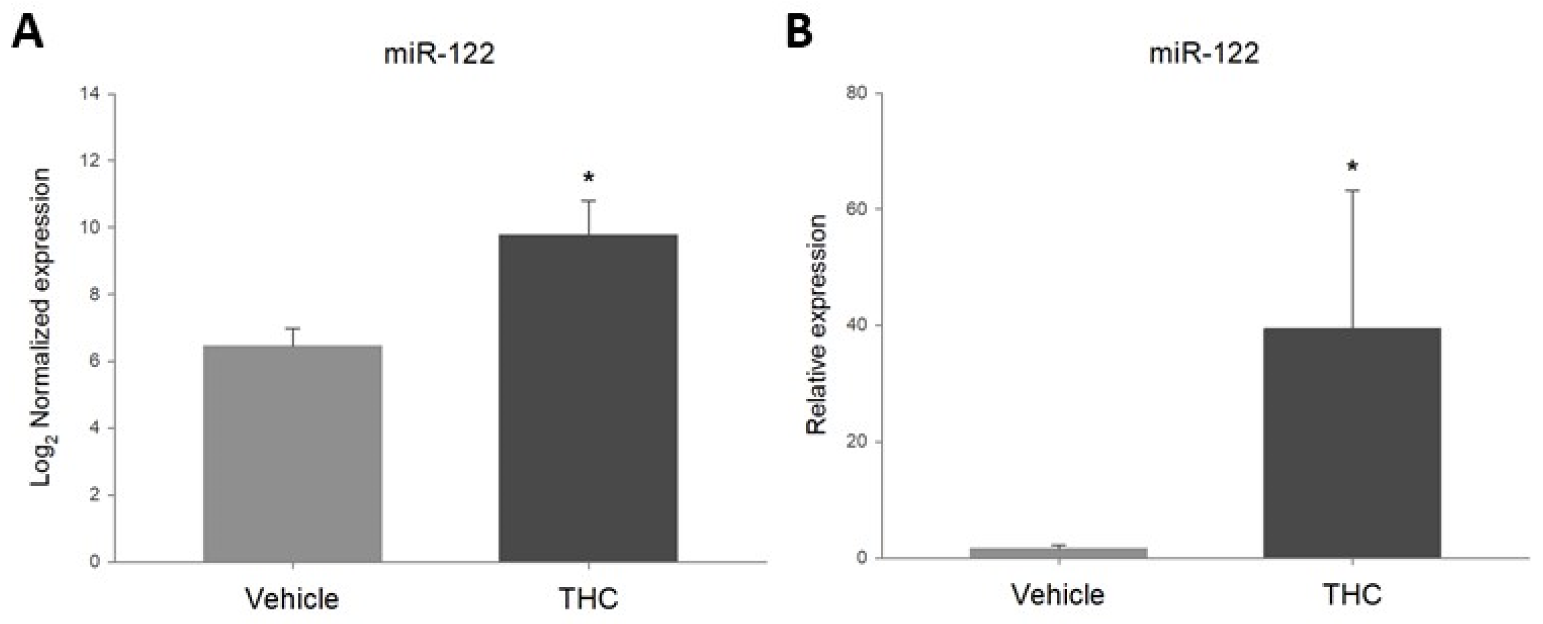
In order to confirm the increase in miR-122 in the prenatally THC-exposed ovaries, the expression of this target was assessed by RT-qPCR. As shown in Figure 2B, RT-qPCR results revealed that miR-122 was significantly upregulated in the ovaries from THC-exposed offspring, similar to the results obtained with the Nanostring assay (Figure 2A).

2.3. Gestational Exposure to THC Decreases Igf1r Expression in the Adult Ovary

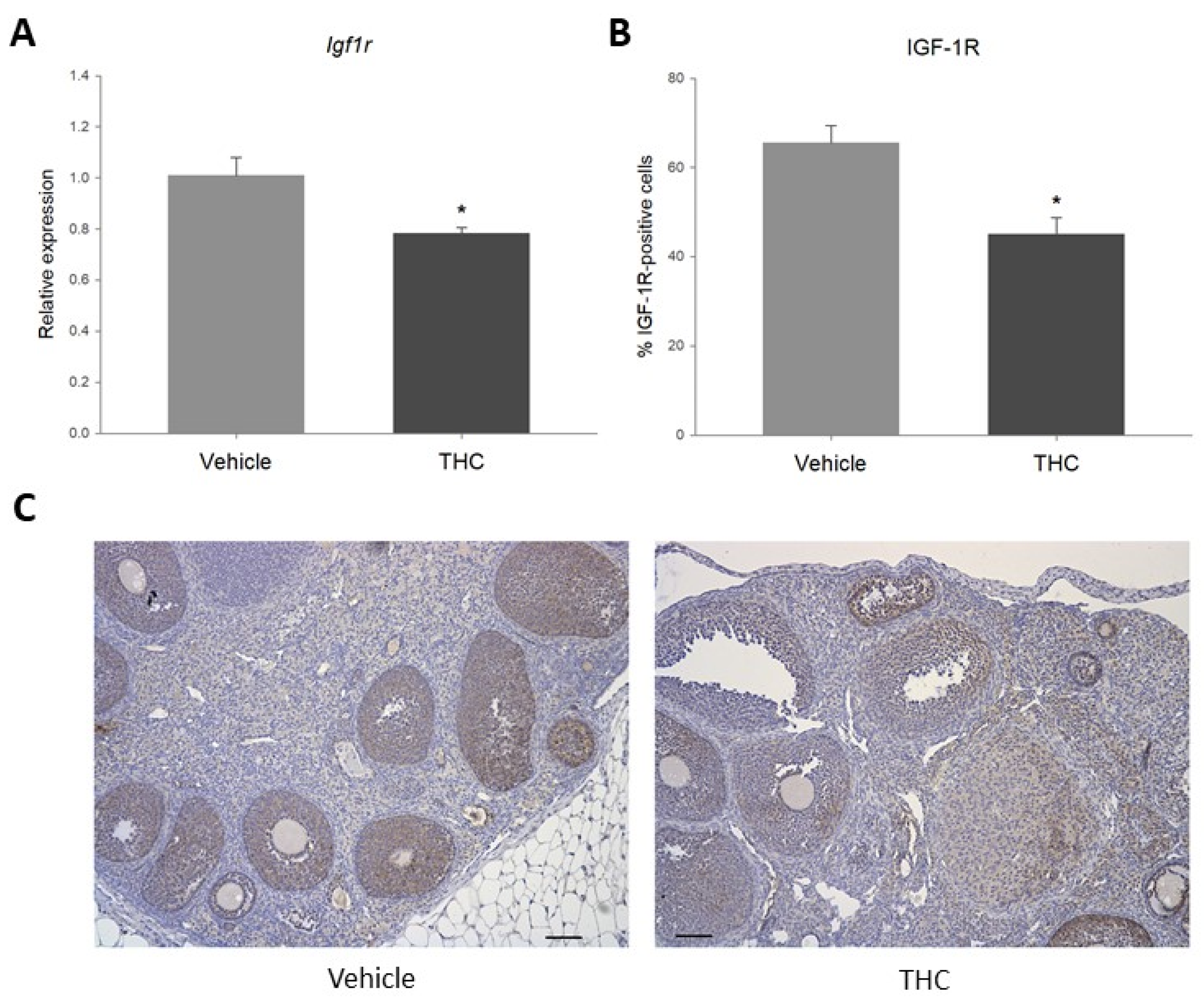
Changes in miRNA expression may affect the expression of their target genes [51]. Given that gestational THC-exposure resulted in a significant increase in miR-122, the online databases TargetScan and miRTarBase were used to determine validated miR-122 target genes involved in the regulation of ovarian processes. Insulin-like growth factor 1 receptor (Igf1r) was identified as a key ovarian miR-122 target based on its role in steroidogenesis, folliculogenesis, angiogenesis, cell proliferation and apoptosis [58,59]. Real-time qPCR revealed that prenatal exposure to THC significantly decreased the relative expression of Igf1r in the ovary, as well as the percentage of IGF-1R-positive follicular cells, as determined by immunohistochemistry (IHC) (Figure 3).

2.4. Prenatal Exposure to THC Increases Follicular Apoptosis in the Adult Ovary

It has previously been established that IGF-1/IGF-1R signaling plays an essential role in follicular growth and atresia, mostly by regulating granulosa cell proliferation and apoptosis [58]. Considering that gestational exposure to THC resulted in a significant decrease in IGF-1R, the expression of the apoptotic marker caspase-3 was evaluated. As shown in Figure 4, while there were no changes in the steady-state mRNA expression of Casp3, gestational exposure to THC significantly increased the percentage of cleaved caspase-3-positive ovarian follicular cells.

2.5. Acute Exposure to THC Decreases Expression of Igf1r in the Ovary

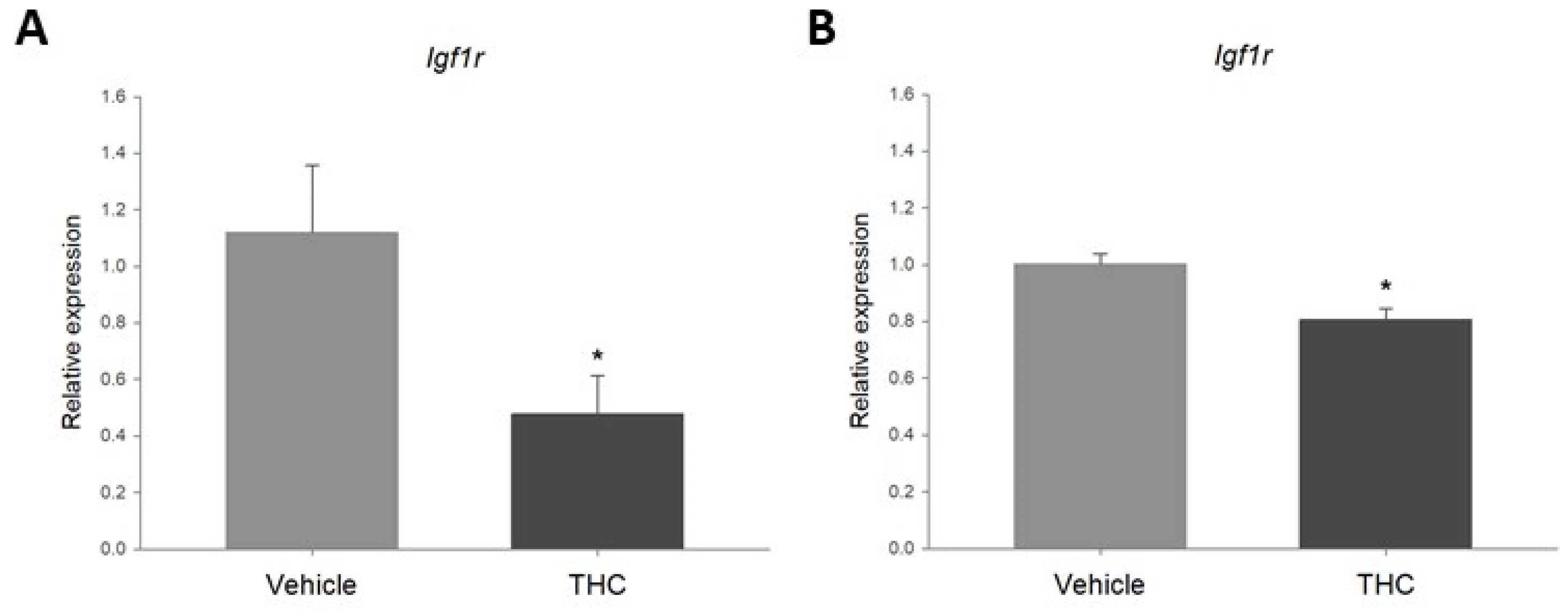
In order to determine if THC had a direct effect on the expression of Igf1r in the ovaries, ex vivo and in vitro models were used. While it is possible to observe short-term effects of direct THC exposure on gene expression in both models, the use of ovarian explants represents a microenvironment and cell diversity that more closely resemble in vivo conditions. On the other hand, the use of an immortalized cell line provides information on the effect of the compound on that specific cell type. Ovarian explants and spontaneously immortalized rat granulosa cells were cultured and exposed to 3 μM THC for 48 h. As observed in Figure 5, exposure to 3 μM THC significantly decreased the relative expression of Igf1r in the ovary explants (Figure 5A) and the granulosa cells (Figure 5B).

3. Discussion

As the use of cannabis during pregnancy increases, it is important to understand the possible effects of its constituents on the developing fetus. The present study examined the effect of gestational exposure to THC on the miRNA profile in adult rat ovaries, as a proposed mechanism through which THC-induced changes in miRNA expression could influence the altered ovarian follicle dynamics and vascularization previously observed in these offspring [57]. Prenatal exposure to THC resulted in the differential expression of 12 miRNA targets, as determined by a Nanostring assay that evaluated the expression of 420 biologically relevant rat miRNAs. While there are several studies that report in vivo alterations of miRNA expression as a consequence of direct exposure of primates and rodents to THC [60,61,62], work from our group has also shown that gestational exposure to THC resulted in altered miRNA profiles in the liver of adult rat offspring [37]. Similarly, in this study we report an altered miRNA profile in the ovaries of THC-exposed female offspring.
Of the 420 miRNAs included in the Nanostring assay, miR-122-5p (miR-122) was the only upregulated target, as well as having the largest fold-change compared to the controls. As miR-122 is a highly conserved miRNA among multiple species, including rat and human [63], this target was chosen for further evaluation. RT-qPCR confirmed the upregulation of miR-122 in the prenatally THC-exposed ovaries. There is growing evidence that suggests miR-122 is involved in the regulation of cell proliferation, differentiation, migration and apoptosis [64,65]. A recent study revealed increased expression of miR-122 in atrophic chicken ovaries compared to healthy ones [63]. Flow cytometry revealed that granulosa cell apoptosis was significantly decreased by a miR-122 inhibitor, while treatment with a miR-122 mimic increased apoptosis and caspase-3 protein levels in vitro [63]. Similarly, Zhang and colleagues recently reported increased granulosa cell apoptosis in a rodent model of primary ovarian insufficiency following treatment with a miR-122 mimic, while a miR-122 inhibitor reduced granulosa cell apoptosis [66]. Furthermore, Menon and colleagues observed that miR-122 plays an important role in the regulation of the luteinizing hormone receptor mRNA binding protein (LRBP) during FSH-induced follicular growth, and therefore may impact folliculogenesis and ovulation [67,68].
To evaluate the effects of the upregulation of miR-122 in the ovaries, the online databases TargetScan and miRTarBase were used to identify validated miR-122 targets involved in the regulation of ovarian processes. The validated miR-122 target insulin-like growth factor 1 receptor (Igf1r) was selected for further evaluation. Studies have reported that miR-122 can directly bind to and decrease Igf1r transcript in vitro [69,70,71]. Moreover, IGF-1R has been shown to play a role in the regulation of folliculogenesis, ovulation, angiogenesis and granulosa cell proliferation and apoptosis [58,72]. IGF-1R is a receptor tyrosine kinase that, among other functions, activates the AKT pathway, linked to prevention of apoptosis, and the ERK pathway, which is associated with growth and proliferation [73]. IGF-1R expression has been detected in most structures of the rodent ovary, including the stroma, oocytes, corpus luteum and theca cells, with the strongest staining found in granulosa cells [58]. Gestational exposure to THC significantly decreased mRNA expression of Igf1r and the percentage of IGF-1R-positive follicular cells in the ovaries. These results are in accordance with the observed increase in miR-122 and with studies that have reported the downregulation of Igf1r as a result of miR-122 overexpression [69,70,71]. Furthermore, the decrease in Igf1r expression appears to be directly attributable to the effect of THC on the ovary, as this compound significantly decreased the relative expression of Igf1r in ovary explants and spontaneously immortalized rat granulosa cells (SIGCs) exposed to 3 μM for 48 h. Considering the key role of IGF-1R in the ovary [58,59], it is possible that a similar mechanism may partially explain the adverse reproductive outcomes such as an increased risk of infertility due to ovulatory abnormalities [74], fewer and poorer quality oocytes and lower pregnancy rates by in vitro fertilization (IVF) [75] that have been reported in adult cannabis users.
Protein levels of IGF-1R have been shown to be reduced in granulosa cells from patients with PCOS and ovarian tissue of PCOS rat models [76]. This reduction was linked to decreased AKT phosphorylation and increased caspase-3 activity, suggesting that increased granulosa cell apoptosis plays a role in the abnormal folliculogenesis and anovulation in PCOS [76]. It has also been shown that knockdown of Igf1r in granulosa cells results in a lack of response to FSH in vitro, and that inhibiting IGF-1R activity in vivo prevents eCG-induced follicular growth [72]. Moreover, Baumgarten and colleagues demonstrated that female mice with a conditional Igf1r knockdown in granulosa cells were sterile, with small ovaries lacking antral follicles, even after stimulation with gonadotropin [58]. Accordingly, these animals failed to ovulate after a superovulation protocol, and their granulosa cells expressed significantly lower levels of preovulatory markers [58]. Similar to what has been observed in PCOS models, ovaries with reduced IGF-1R had impaired AKT activation, as well as increased levels of caspase-3-dependent apoptosis in follicles transitioning from the primary to the large secondary stages [58]. The authors concluded that the lack of IGF-1R signaling in the granulosa cells resulted in increased apoptosis and failure to respond to FSH, which in turn led to the complete arrest of folliculogenesis and the subsequent loss of fertility. In the present study, in addition to reduced Igf1r expression, THC-exposed offspring had an increased percentage of cleaved caspase-3-positive follicular cells in the ovaries, suggesting an increase in apoptosis. In addition, IHC revealed that cleaved caspase-3 was predominantly expressed in granulosa cells, which could suggest increased granulosa cell apoptosis, an important factor of follicular atresia [77].
The reduction in Igf1r expression may also help explain the altered follicle dynamics in the ovaries of THC exposed offspring previously reported by our group [57]. In the same cohort of animals, we observed that ovaries from THC-exposed offspring had a significant increase in transitioning follicles without any differences in later stages of follicular development, suggesting a portion of the follicles did not continue to develop. While this was partially attributed to the reduced blood vessel formation in these ovaries, it is possible that the increased apoptosis revealed in the present study also contributed to the impaired follicular development. The reduced vascularization of these ovaries was associated with a decrease in VEGF and VEGFR2 expression and an increase in TSP-1. This is interesting considering it has been shown that activation of IGF-1R results in increased VEGF expression and secretion in bovine luteal cells [78]. Additionally, it has been demonstrated that in vitro treatment of immortalized rat granulosa cells with TSP-1 increases the expression of pro-apoptotic factors [79], while VEGF-VEGFR2 signaling appears to have the opposite effect [80]. It was therefore proposed that the dysregulation of these factors could manifest as increased apoptosis (atresia) and ultimately a loss of developing follicles [57]. Although there were no statistically significant differences in the number of atretic follicles in the THC-exposed ovaries (p = 0.197), this could be a reflection of the small sample size. It is also possible that the increase in apoptosis may manifest more clearly as follicle loss as the animal ages. Indeed, a rodent model with granulosa cell-specific silencing of Vegf revealed a decrease in Igf1r expression and increased granulosa cell apoptosis [81]. According to the authors, these animals were subfertile, and the effect seemed to be greater as the mice aged [81]. It would therefore be of great interest to continue monitoring the ovarian follicle reserve as the animals approach reproductive senescence and to assess the THC-exposed offspring’s fertility, since the observed effects may have an impact on other reproductive aspects such as oocyte quality.

4. Materials and Methods

4.1. Animals

Pregnant Wistar rats were purchased from Charles River (La Salle, St. Constant, QC, Canada) and maintained at 22 °C at Western University Animal House Facility on a 12:12 h light:dark cycle with access to food and water ad libitum throughout the experiment. All animal experiments were done based on the approved animal use protocol by the subcommittee of the Canadian Council of Animal Care, Western University (AUP# 2019-126) in accordance with the ARRIVE guidelines (https://arriveguidelines.org, accessed on 16 July 2022). Dams were randomly assigned to receive a daily intraperitoneal (IP) injection of either vehicle (1:18 cremophor:saline) or 3 mg/kg THC (Sigma-Aldrich, St Louis, MO, USA) from gestation day (GD) 6 to GD22. This dose has been shown to result in maternal blood concentrations (8.6–12.4 ng/mL) comparable to those detected after moderate cannabis smoking in adults (13–63 ng/mL), as well as in aborted fetal tissue (4–287 ng/mL) after maternal cannabis use [82,83,84]. We have previously demonstrated that 3 mg/kg THC per day does not cause fetal demise, alterations in litter size or gestational length, or maternal weight gain [85]. Dams were allowed to deliver normally, and litters were randomly culled to four female pups and four male pups each. At 6 months of age, prenatal THC-exposed offspring were euthanized by IP pentobarbital overdose. Per female, one ovary was fixed in 10% formalin and embedded in paraffin for histological analysis, while the other ovary was flash frozen in liquid nitrogen to evaluate miRNA and mRNA expression.

4.2. Nanostring Analysis

To determine the effect of gestational THC exposure on the miRNA profile in adult rat ovaries, RNA was extracted from the ovaries of THC-exposed offspring with the use of a mirVana™ miRNA isolation kit (Thermo Fisher Scientific, Waltham, MA, USA). RNA quality was assessed using a 2100 Bioanalyzer (Agilent, Santa Clara, CA, USA), obtaining RNA integrity numbers that ranged from 7.4 to 9.3. The Rat v1.5 miRNA Assay (Nanostring Technologies, Seattle, WA, USA) was used to determine the expression levels of 420 biologically relevant rat miRNA targets according to the manufacturer’s instructions. Results were analyzed with the use of nSolver® analysis software and ROSALIND®. A statistically significant effect was considered to be a fold change of ≥1.5 or ≤−1.5 and a p value < 0.05.

4.3. miRNA Real-Time Quantitative PCR

In order to confirm the results obtained with the Nanostring assay, the expression of the miRNA target with the greatest fold change (miR-122-5p) was evaluated by real-time quantitative PCR (RT-qPCR). For this, the miRCURY™ LNA miRNA PCR Assay system (Qiagen N.V., Hilden, Germany) was used. Complementary DNA (cDNA) was synthesized from the extracted RNA with the use of a miRCURY™ LNA RT kit (Qiagen). The cDNA was then amplified and detected with the use of a miRCURY™ LNA SYBR® Green PCR kit (Qiagen) and a CFX384 Touch™ Real-Time PCR Detection System (Bio-Rad Laboratories, Hercules, CA, USA). RT-qPCR results were analyzed with the 2∆∆CT method [86] using RNU5G snRNA and miR-191-5p as internal references (Table 2). These reference genes were chosen based on previous studies and showed the lowest variation between vehicle and THC-exposed samples when analyzed using RefFinder [87,88,89].

4.4. Immunohistochemistry

Paraffin-embedded ovaries were sectioned (8 μm) and deparaffinized using reagent-grade xylene, and subjected to a series of decreasing ethanol concentrations for rehydration. Endogenous peroxidase activity was quenched through a 10-min incubation period in 3% hydrogen peroxide, followed by antigen retrieval using 10 mM sodium citrate buffer with Tween 20 (0.05%) for 12 min. To reduce nonspecific binding of antibodies, samples were blocked with 5% bovine serum albumin (with 0.02% sodium azide) for 10 min at room temperature. Sections were exposed to either rabbit polyclonal anti-IGF-1R (Abcam, Cambridge, UK; 1:400) or anti-cleaved caspase-3 (Cell Signaling Technology, Danvers, MA, USA; 1:300) overnight at 4 °C. Slides were then incubated with biotinylated anti-rabbit secondary antibody (Thermo Fisher Scientific; 1:100) for 2 h at room temperature followed by Extravidin (Sigma-Aldrich; 1:50) for 1 h at room temperature. Antigens were visualized using 3,3′-diaminobenzidine tetrahydrochloride (DAB; Sigma-Aldrich), and tissues were counterstained with hematoxylin. Slides were imaged using bright-field microscopy at 200x magnification. The percentage of immunopositive follicular cells was quantified by the same individual, who was blinded to the treatment group until all the data had been collected, with the use of an integrated morphometry software (MetaMorph Inc., Nashville, TN, USA). The average of 5 fields of view/ovary was used to calculate the percentage of immunopositive cells.

4.5. Ovarian Explant Culture

For ex vivo experiments, ovaries were collected from nulliparous Wistar rats (268.09 ± 2.96 g) and transferred to sterile D-PBS (Corning Inc., New York, NY, USA). In a biosafety cabinet, ovaries were trimmed of fat, washed with D-PBS and cut into four equal pieces using a sterile blade. Each piece was transferred to a single well in a 24-well plate containing DMEM/F12 media with L-glutamine (Corning) supplemented with 10% fetal bovine serum (FBS) and 2% penicillin/streptomycin (Thermo Fisher Scientific). After 24 h of culture, explants were exposed to vehicle or 3 μM THC for 48 h, changing the media daily. This concentration of THC was based on a pharmacokinetic study that reported similar levels in the serum of cannabis users [90].

4.6. Cell Culture

For in vitro experiments, spontaneously immortalized rat granulosa cells (SIGCs) were cultured in DMEM/F12 media with L-glutamine (Corning Inc.) supplemented with 10% fetal bovine serum (FBS) and 2% penicillin/streptomycin. As with ex vivo experiments, cells were cultured with either vehicle or 3 μM THC for 48 h after confirming this concentration had no effect on cell viability (data not shown).

4.7. RNA Isolation and RT-qPCR

To evaluate gene expression, total RNA was extracted from THC-exposed ovary explants and SIGCs. Briefly, samples were homogenized in TRIzol™ reagent (Thermo Fisher Scientific) by sonication, and RNA was extracted by precipitation with isopropanol and subsequent ethanol washes. RNA concentration and purity were assessed using a NanoDrop™ One micro-UV/vis spectrophotometer (Thermo Fisher Scientific), and cDNA was synthesized with the use of a high capacity cDNA reverse transcription kit (Thermo Fisher Scientific).
In order to confirm the effects of miRNA dysregulation, the online databases miRTarBase (https://mirtarbase.cuhk.edu.cn/~miRTarBase/miRTarBase_2022/php/index.php, accessed on 16 July 2022) and TargetScan (https://www.targetscan.org/vert_80/, accessed on 16 July 2022) were used to select validated target genes involved in essential ovarian processes. With this in mind, the expression of the validated miR-122-5p target insulin-like growth factor 1 receptor (Igf1r) was determined. In addition, given that both miR-122-5p and IGF-1R are involved in the regulation of apoptosis [58,66], and that alterations in follicle dynamics were previously observed in the prenatally THC-exposed ovaries [57], the expression of the apoptotic marker caspase-3 was evaluated.
For the ovaries from prenatally THC-exposed offspring, the ovary explants and the SIGCs, RT-qPCR was performed using PerfeCTa SYBR® Green FastMix (Quantabio, Beverly, MA, USA) and the CFX384 Touch™ real-time PCR detection system (Bio-Rad). RT-qPCR results were analyzed with the 2−∆∆CT method using beta-2-microglobulin (B2m) and hypoxanthine phosphoribosyltransferase 1 (Hprt1) as internal references (Table 3).

4.8. Statistical Analysis

For in vivo assessments, one female offspring per dam was included in the statistical analysis. For ex vivo and in vitro assessments, results from 5 independent experiments are presented. The Nanostring assay results were analyzed with the use of nSolver® and ROSALIND® analysis software, considering a fold change ≥ 1.5 and a p-value < 0.05 as statistically different. For miRNA and mRNA RT-qPCR, as well as for immunohistochemical evaluation, statistical analyses were performed using SigmaPlot®. A Student’s t-test was performed to determine statistically significant differences (p < 0.05) between vehicle- and THC-exposed samples.

5. Conclusions

We have previously shown that prenatal exposure to delta-9-tetrahydrocannabinol (THC), the psychoactive component of cannabis, resulted in altered follicle dynamics and vascularization in the adult rat ovary. In this study, we propose that gestational exposure to THC altered miRNA expression in the ovary, which in turn affected ovarian health in adulthood. Particularly, prenatal THC exposure resulted in a significant increase in the expression of miR-122-5p and a significant decrease in the expression of its validated target gene insulin-like growth factor 1 receptor (Igf1r). Importantly, decreased IGF-1R expression has been linked to increased apoptosis and abnormal folliculogenesis, both of which have been observed in the THC-exposed offspring. Taken together, these data suggest that prenatal THC-exposure may impact key pathways in the developing ovary that could lead to subfertility or premature reproductive senescence. As the use of cannabis during pregnancy increases, it is important to understand the safety of this drug and its constituents, as well as the possible long-term effects on the offspring’s endocrine and reproductive health.

Author Contributions

Conceptualization, A.A.M.-P., J.J.P., D.B.H. and A.C.H.; methodology, A.A.M.-P., K.L. and M.P.; formal analysis, A.A.M.-P.; resources, J.J.P., D.B.H. and A.C.H.; writing—original draft preparation, A.A.M.-P.; writing—review and editing, A.A.M.-P., A.A., J.J.P., D.B.H. and A.C.H.; supervision, A.C.H.; funding acquisition, D.B.H. and A.C.H. All authors have read and agreed to the published version of the manuscript.

Funding

This research was funded by a Canadian Institutes of Health Research (CIHR) (Canada)) Project Grant to ACH (PJT-155981) and an HSFC (Heart and Stroke Foundation of Canada (Canada)) Grant-in-Aid to DBH (G-19-0026343). AAMP was sponsored by CONACYT (National Council of Science and Technology (Mexico)).

Institutional Review Board Statement

All procedures were performed according to guidelines set by the Canadian Council of Animal Care, and Animal Use Protocol (AUP #2019-126) was approved by the Animal Care Committee at The University of Western Ontario. All investigators understood and followed the ethical principles outlined by Grundy, and study design was informed by ARRIVE guidelines.

Informed Consent Statement

Not applicable.

Data Availability Statement

Not applicable.

Conflicts of Interest

The authors declare no conflict of interest.

References

  1. Labor Unooda. World Drug Report 2021 (Set of 5 Booklets); S.l.: United Nations: 2022. Available online: https://www.unodc.org/unodc/en/data-and-analysis/wdr2021.html (accessed on 16 July 2022).
  2. Corsi, D.J.; Hsu, H.; Weiss, D.; Fell, D.B.; Walker, M. Trends and correlates of cannabis use in pregnancy: A population-based study in Ontario, Canada from 2012 to 2017. Can. J. Public Health. 2018, 110, 76–84. [Google Scholar] [CrossRef]
  3. 2019 National Survey on Drug Use and Health: Women|CBHSQ Data n.d. Available online: https://www.samhsa.gov/data/report/2019-nsduh-women (accessed on 4 June 2022).
  4. Leemaqz, S.Y.; Dekker, G.A.; McCowan, L.M.; Kenny, L.C.; Myers, J.E.; Simpson, N.A.B.; Poston, L.; Roberts, C.T.; SCOPE Consortium. Maternal marijuana use has independent effects on risk for spontaneous preterm birth but not other common late pregnancy complications. Reprod. Toxicol. 2016, 62, 77–86. [Google Scholar] [CrossRef]
  5. Ko, J.Y.; Farr, S.L.; Tong, V.T.; Creanga, A.A.; Callaghan, W.M. Prevalence and patterns of marijuana use among pregnant and nonpregnant women of reproductive age. Am. J. Obstet. Gynecol. 2015, 213, 201.e1–201.e10. [Google Scholar] [CrossRef]
  6. Moore, D.G.; Turner, J.D.; Parrott, A.C.; Goodwin, J.E.; Fulton, S.E.; Min, M.O.; Fox, H.C.; Braddick, F.M.B.; Axelsson, E.L.; Lynch, S.; et al. During pregnancy, recreational drug-using women stop taking ecstasy (3,4-methylenedioxy-N-methylamphetamine) and reduce alcohol consumption, but continue to smoke tobacco and cannabis: Initial findings from the Development and Infancy Study. J. Psychopharmacol. 2010, 24, 1403–1410. [Google Scholar] [CrossRef] [Green Version]
  7. PregnancyInfo. Available online: https://www.pregnancyinfo.ca/learn-more/ (accessed on 16 July 2022).
  8. Volkow, N.D.; Han, B.; Compton, W.M.; McCance-Katz, E.F. Self-reported Medical and Nonmedical Cannabis Use among Pregnant Women in the United States. JAMA 2019, 322, 167–169. [Google Scholar] [CrossRef]
  9. Westfall, R.E.; Janssen, P.A.; Lucas, P.; Capler, R. Survey of medicinal cannabis use among childbearing women: Patterns of its use in pregnancy and retroactive self-assessment of its efficacy against ‘morning sickness’. Complement. Ther. Clin. Pract. 2006, 12, 27–33. [Google Scholar] [CrossRef]
  10. Roberson, E.K.; Patrick, W.K.; Hurwitz, E.L. Marijuana Use and Maternal Experiences of Severe Nausea During Pregnancy in Hawai’i. Hawaii J. Med. Public Health 2014, 73, 283–287. [Google Scholar]
  11. Bayrampour, H.; Asim, A. Cannabis Use During the Pre-Conception Period and Pregnancy After Legalization. J. Obstet. Gynaecol. Can. 2021, 43, 740–745. [Google Scholar] [CrossRef]
  12. Singh, S.; Filion, K.B.; Abenhaim, H.A.; Eisenberg, M.J. Prevalence and outcomes of prenatal recreational cannabis use in high-income countries: A scoping review. BJOG Int. J. Obstet. Gynaecol. 2020, 127, 8–16. [Google Scholar] [CrossRef]
  13. Lozano, J.; García-Algar, O.; Marchei, E.; Vall, O.; Monleon, T.; Giovannandrea, R.D.; Pichini, S. Prevalence of gestational exposure to cannabis in a Mediterranean city by meconium analysis. Acta Paediatr. 2007, 96, 1734–1737. [Google Scholar] [CrossRef]
  14. Metz, T.D.; Silver, R.M.; McMillin, G.A.; Allshouse, A.A.; Jensen, T.L.; Mansfield, C.; Heard, K.; Kinney, G.L.; Wymore, E.; Binswanger, I.A. Prenatal Marijuana Use by Self-Report and Umbilical Cord Sampling in a State with Marijuana Legalization. Obstet. Gynecol. 2019, 133, 98–104. [Google Scholar] [CrossRef]
  15. Luke, S.; Hutcheon, J.; Kendall, T. Cannabis Use in Pregnancy in British Columbia and Selected Birth Outcomes. J. Obstet. Gynaecol. Can. 2019, 41, 1311–1317. [Google Scholar] [CrossRef]
  16. Varner, M.W.; Silver, R.M.; Rowland Hogue, C.J.; Willinger, M.; Parker, C.B.; Thorsten, V.R.; Goldenberg, R.L.; Saade, G.R.; Dudley, D.J.; Coustan, D.; et al. Association between stillbirth and illicit drug use and smoking during pregnancy. Obstet. Gynecol. 2014, 123, 113–125. [Google Scholar] [CrossRef] [Green Version]
  17. Shi, Y.; Zhu, B.; Liang, D. The associations between prenatal cannabis use disorder and neonatal outcomes. Addict. Abingdon Engl. 2021, 116, 3069–3079. [Google Scholar] [CrossRef]
  18. Hayatbakhsh, M.R.; Flenady, V.J.; Gibbons, K.S.; Kingsbury, A.M.; Hurrion, E.; Mamun, A.A.; Najmna, J.M. Birth outcomes associated with cannabis use before and during pregnancy. Pediatr. Res. 2012, 71, 215–219. [Google Scholar] [CrossRef] [Green Version]
  19. Hurd, Y.L.; Wang, X.; Anderson, V.; Beck, O.; Minkoff, H.; Dow-Edwards, D. Marijuana impairs growth in mid-gestation fetuses. Neurotoxicol. Teratol. 2005, 27, 221–229. [Google Scholar] [CrossRef]
  20. Warshak, C.R.; Regan, J.; Moore, B.; Magner, K.; Kritzer, S.; Van Hook, J. Association between marijuana use and adverse obstetrical and neonatal outcomes. J. Perinatol. 2015, 35, 991–995. [Google Scholar] [CrossRef]
  21. Campbell, E.E.; Gilliland, J.; Dworatzek, P.D.N.; Vrijer, B.D.; Penava, D.; Seabrook, J.A. Socioeconomic Status and Adverse Birth Outcomes: A Population-Based Canadian Sample. J. Biosoc. Sci. 2018, 50, 102–113. [Google Scholar] [CrossRef] [Green Version]
  22. El Marroun, H.; Tiemeier, H.; Steegers, E.A.P.; Jaddoe, V.W.V.; Hofman, A.; Verhulst, F.C.; van den Brink, W.; Huizink, A.C. Intrauterine Cannabis Exposure Affects Fetal Growth Trajectories: The Generation R Study. J. Am. Acad. Child Adolesc. Psychiatry 2009, 48, 1173–1181. [Google Scholar] [CrossRef] [Green Version]
  23. Gabrhelík, R.; Mahic, M.; Lund, I.O.; Bramness, J.; Selmer, R.; Skovlund, E.; Handal, M.; Skurtveit, S. Cannabis Use during Pregnancy and Risk of Adverse Birth Outcomes: A Longitudinal Cohort Study. Eur. Addict. Res. 2021, 27, 131–141. [Google Scholar] [CrossRef]
  24. Smith, A.; Kaufman, F.; Sandy, M.S.; Cardenas, A. Cannabis Exposure during Critical Windows of Development: Epigenetic and Molecular Pathways Implicated in Neuropsychiatric Disease. Curr. Environ. Health Rep. 2020, 7, 325–342. [Google Scholar] [CrossRef]
  25. Mechoulam, R.; Gaoni, Y. A Total Synthesis of Dl-Delta-1-Tetrahydrocannabinol, the Active Constituent of Hashish. J. Am. Chem. Soc. 1965, 87, 3273–3275. [Google Scholar] [CrossRef]
  26. Carlier, J.; Huestis, M.A.; Zaami, S.; Pichini, S.; Busardò, F.P. Monitoring Perinatal Exposure to Cannabis and Synthetic Cannabinoids. Ther. Drug Monit. 2020, 42, 194–204. [Google Scholar] [CrossRef]
  27. Banu, S.K.; Stanley, J.A.; Sivakumar, K.K.; Arosh, J.A.; Barhoumi, R.; Burghardt, R.C. Identifying a novel role for X-prolyl aminopeptidase (Xpnpep) 2 in CrVI-induced adverse effects on germ cell nest breakdown and follicle development in rats. Biol. Reprod. 2015, 92, 67. [Google Scholar] [CrossRef]
  28. Pepe, G.J.; Lynch, T.J.; Albrecht, E.D. Regulation of baboon fetal ovarian development by placental estrogen: Onset of puberty is delayed in offspring deprived of estrogen in utero. Biol. Reprod. 2013, 89, 132. [Google Scholar] [CrossRef] [Green Version]
  29. Barker, D.J.P.; Eriksson, J.G.; Forsén, T.; Osmond, C. Fetal origins of adult disease: Strength of effects and biological basis. Int. J. Epidemiol. 2002, 31, 1235–1239. [Google Scholar] [CrossRef] [Green Version]
  30. Smith, P.; Wilhelm, D.; Rodgers, R.J. Development of mammalian ovary. J. Endocrinol. 2014, 221, R145–R161. [Google Scholar] [CrossRef] [Green Version]
  31. Moore, B.F.; Sauder, K.A.; Shapiro, A.L.B.; Crume, T.; Kinney, G.L.; Dabelea, D. Fetal Exposure to Cannabis and Childhood Metabolic Outcomes: The Healthy Start Study. J. Clin. Endocrinol. Metab. 2022, 107, e2862–e2869. [Google Scholar] [CrossRef]
  32. Campolongo, P.; Trezza, V.; Ratano, P.; Palmery, M.; Cuomo, V. Developmental consequences of perinatal cannabis exposure: Behavioral and neuroendocrine effects in adult rodents. Psychopharmacology 2011, 214, 5–15. [Google Scholar] [CrossRef] [Green Version]
  33. Smith, A.M.; Fried, P.A.; Hogan, M.J.; Cameron, I. Effects of prenatal marijuana on visuospatial working memory: An fMRI study in young adults. Neurotoxicol. Teratol. 2006, 28, 286–295. [Google Scholar] [CrossRef]
  34. Gillies, R.; Lee, K.; Vanin, S.; Laviolette, S.R.; Holloway, A.C.; Arany, E.; Hardy, D.B. Maternal exposure to Δ9-tetrahydrocannabinol impairs female offspring glucose homeostasis and endocrine pancreatic development in the rat. Reprod. Toxicol. 2020, 94, 84–91. [Google Scholar] [CrossRef]
  35. De Domenico, E.; Todaro, F.; Rossi, G.; Dolci, S.; Geremia, R.; Rossi, P.; Grimaldi, P. Overactive type 2 cannabinoid receptor induces meiosis in fetal gonads and impairs ovarian reserve. Cell Death Dis. 2017, 8, e3085. [Google Scholar] [CrossRef]
  36. Castel, P.; Barbier, M.; Poumerol, E.; Mandon-Pépin, B.; Tassistro, V.; Lepidi, H.; Pelissier-Alicot, A.L.; Manzoni, O.J.; Courbiere, B. Prenatal cannabinoid exposure alters the ovarian reserve in adult offspring of rats. Arch Toxicol. 2020, 94, 4131–4141. [Google Scholar] [CrossRef]
  37. Oke, S.L.; Lee, K.; Papp, R.; Laviolette, S.R.; Hardy, D.B. In Utero Exposure to Δ9-Tetrahydrocannabinol Leads to Postnatal Catch-Up Growth and Dysmetabolism in the Adult Rat Liver. Int. J. Mol. Sci. 2021, 22, 7502. [Google Scholar] [CrossRef]
  38. Lee, K.; Hardy, D.B. Metabolic Consequences of Gestational Cannabinoid Exposure. Int. J. Mol. Sci. 2021, 22, 9528. [Google Scholar] [CrossRef]
  39. Egger, G.; Liang, G.; Aparicio, A.; Jones, P.A. Epigenetics in human disease and prospects for epigenetic therapy. Nature 2004, 429, 457–463. [Google Scholar] [CrossRef]
  40. DiNieri, J.A.; Wang, X.; Szutorisz, H.; Spano, S.M.; Kaur, J.; Casaccia, P.; Dow-Edwards, D.; Hurd, Y.L. Maternal cannabis use alters ventral striatal dopamine D2 gene regulation in the offspring. Biol. Psychiatry 2011, 70, 763–769. [Google Scholar] [CrossRef] [Green Version]
  41. Murphy, S.K.; Itchon-Ramos, N.; Visco, Z.; Huang, Z.; Grenier, C.; Schrott, R.; Acharya, K.; Boudreau, M.H.; Price, T.M.; Raburn, D.J.; et al. Cannabinoid exposure and altered DNA methylation in rat and human sperm. Epigenetics 2018, 13, 1208–1221. [Google Scholar] [CrossRef] [Green Version]
  42. Prini, P.; Penna, F.; Sciuccati, E.; Alberio, T.; Rubino, T. Chronic Δ8-THC Exposure Differently Affects Histone Modifications in the Adolescent and Adult Rat Brain. Int. J. Mol. Sci. 2017, 18, 2094. [Google Scholar] [CrossRef]
  43. Pucci, M.; Rapino, C.; Di Francesco, A.; Dainese, E.; D’Addario, C.; Maccarrone, M. Epigenetic control of skin differentiation genes by phytocannabinoids. Br. J. Pharmacol. 2013, 170, 581–591. [Google Scholar] [CrossRef] [Green Version]
  44. Todd, S.M.; Zhou, C.; Clarke, D.J.; Chohan, T.W.; Bahceci, D.; Arnold, J.C. Interactions between cannabidiol and Δ9-THC following acute and repeated dosing: Rebound hyperactivity, sensorimotor gating and epigenetic and neuroadaptive changes in the mesolimbic pathway. Eur. Neuropsychopharmacol. 2017, 27, 132–145. [Google Scholar] [CrossRef]
  45. Yang, X.; Hegde, V.L.; Rao, R.; Zhang, J.; Nagarkatti, P.S.; Nagarkatti, M. Histone modifications are associated with Δ9-tetrahydrocannabinol-mediated alterations in antigen-specific T cell responses. J. Biol. Chem. 2014, 289, 18707–18718. [Google Scholar] [CrossRef] [Green Version]
  46. Watson, C.T.; Szutorisz, H.; Garg, P.; Martin, Q.; Landry, J.A.; Sharp, A.J.; Hurd, Y.L. Genome-Wide DNA Methylation Profiling Reveals Epigenetic Changes in the Rat Nucleus Accumbens Associated with Cross-Generational Effects of Adolescent THC Exposure. Neuropsychopharmacology. 2015, 40, 2993–3005. [Google Scholar] [CrossRef] [Green Version]
  47. Sido, J.M.; Yang, X.; Nagarkatti, P.S.; Nagarkatti, M. Δ9-Tetrahydrocannabinol-mediated epigenetic modifications elicit myeloid-derived suppressor cell activation via STAT3/S100A8. J. Leukoc. Biol. 2015, 97, 677–688. [Google Scholar] [CrossRef] [Green Version]
  48. Juknat, A.; Gao, F.; Coppola, G.; Vogel, Z.; Kozela, E. miRNA expression profiles and molecular networks in resting and LPS-activated BV-2 microglia-Effect of cannabinoids. PLoS ONE 2019, 14, e0212039. [Google Scholar] [CrossRef]
  49. Kim, V.N. Small RNAs: Classification, biogenesis, and function. Mol. Cells 2005, 19, 1–15. [Google Scholar]
  50. Kim, J.; You, S. Comprehensive analysis of miRNA-mRNA interactions in ovaries of aged mice. Anim. Sci. J. 2022, 93, e13721. [Google Scholar] [CrossRef]
  51. Tufarelli, C.; Stanley, J.A.S.; Garrick, D.; Sharpe, J.A.; Ayyub, H.; Wood, W.G.; Higgs, D.R. Transcription of antisense RNA leading to gene silencing and methylation as a novel cause of human genetic disease. Nat. Genet. 2003, 34, 157–165. [Google Scholar] [CrossRef]
  52. Brennecke, J.; Stark, A.; Russell, R.B.; Cohen, S.M. Principles of microRNA-target recognition. PLoS Biol. 2005, 3, e85. [Google Scholar] [CrossRef]
  53. Valadi, H.; Ekström, K.; Bossios, A.; Sjöstrand, M.; Lee, J.J.; Lötvall, J.O. Exosome-mediated transfer of mRNAs and microRNAs is a novel mechanism of genetic exchange between cells. Nat. Cell Biol. 2007, 9, 654–659. [Google Scholar] [CrossRef] [Green Version]
  54. Mitchell, P.S.; Parkin, R.K.; Kroh, E.M.; Fritz, B.R.; Wyman, S.K.; Pogosova-Agadjanyan, E.L.; Peterson, A.; Noteboom, J.; O’Briant, K.C.; Allen, A.; et al. Circulating microRNAs as stable blood-based markers for cancer detection. Proc. Natl. Acad. Sci. USA 2008, 105, 10513–10518. [Google Scholar] [CrossRef] [Green Version]
  55. Li, Y.; Fang, Y.; Liu, Y.; Yang, X. MicroRNAs in ovarian function and disorders. J. Ovarian Res. 2015, 8, 51. [Google Scholar] [CrossRef] [Green Version]
  56. Gebremedhn, S.; Salilew-Wondim, D.; Ahmad, I.; Sahadevan, S.; Hossain, M.M.; Hoelker, M.; Rings, F.; Neuhoff, C.; Tholen, E.; Looft, C.; et al. MicroRNA Expression Profile in Bovine Granulosa Cells of Preovulatory Dominant and Subordinate Follicles during the Late Follicular Phase of the Estrous Cycle. PLoS ONE 2015, 10, e0125912. [Google Scholar] [CrossRef]
  57. Martínez-Peña, A.A.; Lee, K.; Petrik, J.J.; Hardy, D.B.; Holloway, A.C. Gestational exposure to Δ9-THC impacts ovarian follicular dynamics and angiogenesis in adulthood in Wistar rats. J. Dev. Orig. Health Dis. 2021, 12, 865–869. [Google Scholar] [CrossRef]
  58. Baumgarten, S.C.; Armouti, M.; Ko, C.; Stocco, C. IGF1R Expression in Ovarian Granulosa Cells Is Essential for Steroidogenesis, Follicle Survival, and Fertility in Female Mice. Endocrinology 2017, 158, 2309–2318. [Google Scholar] [CrossRef] [Green Version]
  59. Zhao, J.; Taverne, M.; van der Weijden, G.; Bevers, M.; Hurk, R.V.D. Immunohistochemical localisation of growth hormone (GH), GH receptor (GHR), insulin-like growth factor I (IGF-I) and type I IGF-I receptor, and gene expression of GH and GHR in rat pre-antral follicles. Zygote. 2002, 10, 85–94. [Google Scholar] [CrossRef]
  60. Mohammed, A.; Alghetaa, H.; Sultan, M.; Singh, N.P.; Nagarkatti, P.; Nagarkatti, M. Administration of Δ9-Tetrahydrocannabinol (THC) Post-Staphylococcal Enterotoxin B Exposure Protects Mice from Acute Respiratory Distress Syndrome and Toxicity. Front. Pharmacol. 2020, 11, 893. [Google Scholar] [CrossRef]
  61. Chandra, L.C.; Kumar, V.; Torben, W.; Vande Stouwe, C.; Winsauer, P.; Amedee, A.; Molina, P.E.; Mohan, M. Chronic administration of Δ9-tetrahydrocannabinol induces intestinal anti-inflammatory microRNA expression during acute simian immunodeficiency virus infection of rhesus macaques. J. Virol. 2015, 89, 1168–1181. [Google Scholar] [CrossRef] [Green Version]
  62. Hegde, V.L.; Tomar, S.; Jackson, A.; Rao, R.; Yang, X.; Singh, U.P.; Singh, N.P.; Nagarkatti, P.S.; Nagarkatti, M. Distinct microRNA expression profile and targeted biological pathways in functional myeloid-derived suppressor cells induced by Δ9-tetrahydrocannabinol in vivo: Regulation of CCAAT/enhancer-binding protein α by microRNA-690. J. Biol. Chem. 2013, 288, 36810–36826. [Google Scholar] [CrossRef] [Green Version]
  63. Zhang, G.; Cui, Z.; Li, J.; Zhang, D.; Li, Z.; Lin, Z.; Yin, H.; Ran, J.; Wang, Y.; Liu, Y. miR-122-5p regulates proliferation and apoptosis of chicken granulosa cells of hierarchal follicles by targeting MAPK3. Gene 2022, 824, 146397. [Google Scholar] [CrossRef]
  64. Pei, Z.J.; Zhang, Z.G.; Hu, A.X.; Yang, F.; Gai, Y. miR-122-5p inhibits tumor cell proliferation and induces apoptosis by targeting MYC in gastric cancer cells. Die Pharm.-Int. J. Pharm. Sci. 2017, 72, 344–347. [Google Scholar] [CrossRef]
  65. Ma, J.; Li, T.; Han, X.; Yuan, H. Knockdown of LncRNA ANRIL suppresses cell proliferation, metastasis, and invasion via regulating miR-122-5p expression in hepatocellular carcinoma. J. Cancer Res. Clin. Oncol. 2018, 144, 205–214. [Google Scholar] [CrossRef]
  66. Zhang, X.; Zhang, R.; Hao, J.; Huang, X.; Liu, M.; Lv, M.; Su, C.; Mu, Y.L. miRNA-122-5p in POI ovarian-derived exosomes promotes granulosa cell apoptosis by regulating BCL9. Cancer Med. 2022, 11, 2414–2426. [Google Scholar] [CrossRef]
  67. Menon, B.; Gulappa, T.; Menon, K.M.J. Molecular regulation of LHCGR expression by miR-122 during follicle growth in the rat ovary. Mol. Cell Endocrinol. 2017, 442, 81–89. [Google Scholar] [CrossRef] [Green Version]
  68. Menon, K.M.J.; Menon, B.; Gulappa, T. Regulation of Luteinizing Hormone Receptor mRNA Expression in the Ovary: The Role of miR-122. Vitam. Horm. 2018, 107, 67–87. [Google Scholar] [CrossRef] [PubMed]
  69. He, Y.; Liu, X.; Qu, Q.; Chen, J.; Fan, Z.; Zhu, D.; Miao, Y.; Hu, Z. Overexpression of miR-122 promotes apoptosis of dermal papilla cells by directly targeting IGF1R in androgenetic alopecia. Cell Biol. Int. 2022, 46, 185–191. [Google Scholar] [CrossRef]
  70. Xu, Y.; Huang, J.; Ma, L.; Shan, J.; Shen, J.; Yang, Z.; Liu, L.; Luo, Y.; Yao, C.; Qian, C. MicroRNA-122 confers sorafenib resistance to hepatocellular carcinoma cells by targeting IGF-1R to regulate RAS/RAF/ERK signaling pathways. Cancer Lett. 2016, 371, 171–181. [Google Scholar] [CrossRef] [PubMed]
  71. Wang, B.; Wang, H.; Yang, Z. MiR-122 Inhibits Cell Proliferation and Tumorigenesis of Breast Cancer by Targeting IGF1R. PLoS ONE 2012, 7, e47053. [Google Scholar] [CrossRef] [Green Version]
  72. Zhou, P.; Baumgarten, S.C.; Wu, Y.; Bennett, J.; Winston, N.; Hirshfeld-Cytron, J.; Stocco, C. IGF-I signaling is essential for FSH stimulation of AKT and steroidogenic genes in granulosa cells. Mol. Endocrinol. 2013, 27, 511–523. [Google Scholar] [CrossRef]
  73. Singh, R.; Chaudhary, P.; Arya, R. Role of IGF-1R in ameliorating apoptosis of GNE deficient cells. Sci. Rep. 2018, 8, 7323. [Google Scholar] [CrossRef] [Green Version]
  74. Mueller, B.A.; Daling, J.R.; Weiss, N.S.; Moore, D.E. Recreational drug use and the risk of primary infertility. Epidemiology. 1990, 1, 195–200. [Google Scholar] [CrossRef] [PubMed]
  75. Klonoff-Cohen, H.S.; Natarajan, L.; Chen, R.V. A prospective study of the effects of female and male marijuana use on in vitro fertilization (IVF) and gamete intrafallopian transfer (GIFT) outcomes. Am. J. Obstet. Gynecol. 2006, 194, 369–376. [Google Scholar] [CrossRef] [PubMed]
  76. Mao, Z.; Fan, L.; Yu, Q.; Luo, S.; Wu, X.; Tang, J.; Kang, G.; Tang, L. Abnormality of Klotho Signaling Is Involved in Polycystic Ovary Syndrome. Reprod. Sci. 2018, 25, 372–383. [Google Scholar] [CrossRef] [PubMed]
  77. Manabe, N.; Goto, Y.; Matsuda-Minehata, F.; Inoue, N.; Maeda, A.; Sakamaki, K.; Miyano, T. Regulation mechanism of selective atresia in porcine follicles: Regulation of granulosa cell apoptosis during atresia. J. Reprod. Dev. 2004, 50, 493–514. [Google Scholar] [CrossRef] [PubMed] [Green Version]
  78. Uniyal, S.; Panda, R.P.; Chouhan, V.S.; Yadav, V.P.; Hyder, I.; Dangi, S.S.; Gupta, M.; Khan, F.A.; Sharma, G.T.; Bag, S.; et al. Expression and localization of insulin-like growth factor system in corpus luteum during different stages of estrous cycle in water buffaloes (Bubalus bubalis) and the effect of insulin-like growth factor I on production of vascular endothelial growth factor and progesterone in luteal cells cultured in vitro. Theriogenology 2015, 83, 58–77. [Google Scholar] [CrossRef] [PubMed]
  79. Greenaway, J.; Lawler, J.; Moorehead, R.; Bornstein, P.; Lamarre, J.; Petrik, J. Thrombospondin-1 inhibits VEGF levels in the ovary directly by binding and internalization via the low density lipoprotein receptor-related protein-1 (LRP-1). J. Cell Physiol. 2007, 210, 807–818. [Google Scholar] [CrossRef] [Green Version]
  80. Greenaway, J.; Connor, K.; Pedersen, H.G.; Coomber, B.L.; LaMarre, J.; Petrik, J. Vascular endothelial growth factor and its receptor, Flk-1/KDR, are cytoprotective in the extravascular compartment of the ovarian follicle. Endocrinology 2004, 145, 2896–2905. [Google Scholar] [CrossRef] [Green Version]
  81. Sargent, K.M.; Lu, N.; Clopton, D.T.; Pohlmeier, W.E.; Brauer, V.M.; Ferrara, N.; Silversides, D.W.; Cupp, A.S. Loss of Vascular Endothelial Growth Factor A (VEGFA) Isoforms in Granulosa Cells Using pDmrt-1-Cre or Amhr2-Cre Reduces Fertility by Arresting Follicular Development and by Reducing Litter Size in Female Mice. PLoS ONE 2015, 10, e0116332. [Google Scholar] [CrossRef] [Green Version]
  82. Klein, C.; Karanges, E.; Spiro, A.; Wong, A.; Spencer, J.; Huynh, T.; Gunasekaran, N.; Karl, T.; Long, L.E.; Huang, X.F.; et al. Cannabidiol potentiates Δ9-tetrahydrocannabinol (THC) behavioural effects and alters THC pharmacokinetics during acute and chronic treatment in adolescent rats. Psychopharmacology 2011, 218, 443–457. [Google Scholar] [CrossRef]
  83. Schwope, D.M.; Karschner, E.L.; Gorelick, D.A.; Huestis, M.A. Identification of recent cannabis use: Whole-blood and plasma free and glucuronidated cannabinoid pharmacokinetics following controlled smoked cannabis administration. Clin. Chem. 2011, 57, 1406–1414. [Google Scholar] [CrossRef] [Green Version]
  84. Falcon, M.; Pichini, S.; Joya, J.; Pujadas, M.; Sanchez, A.; Vall, O.; García Algar, O.; Luna, A.; de la Torre, R.; Rotolo, M.C.; et al. Maternal hair testing for the assessment of fetal exposure to drug of abuse during early pregnancy: Comparison with testing in placental and fetal remains. Forensic Sci. Int. 2012, 218, 92–96. [Google Scholar] [CrossRef] [PubMed]
  85. Natale, B.V.; Gustin, K.N.; Lee, K.; Holloway, A.C.; Laviolette, S.R.; Natale, D.R.C.; Hardy, D.B. Δ9-tetrahydrocannabinol exposure during rat pregnancy leads to symmetrical fetal growth restriction and labyrinth-specific vascular defects in the placenta. Sci. Rep. 2020, 10, 544. [Google Scholar] [CrossRef] [PubMed]
  86. Livak, K.J.; Schmittgen, T.D. Analysis of relative gene expression data using real-time quantitative PCR and the 2(-Delta Delta C(T)) Method. Methods. 2001, 25, 402–408. [Google Scholar] [CrossRef]
  87. Fereshteh, Z.; Schmidt, S.A.; Al-Dossary, A.A.; Accerbi, M.; Arighi, C.; Cowart, J.; Song, J.L.; Green, P.J.; Choi, K.; Yoo, S.; et al. Murine Oviductosomes (OVS) microRNA profiling during the estrous cycle: Delivery of OVS-borne microRNAs to sperm where miR-34c-5p localizes at the centrosome. Sci. Rep. 2018, 8, 16094. [Google Scholar] [CrossRef] [PubMed]
  88. Tesfaye, D.; Gebremedhn, S.; Salilew-Wondim, D.; Hailay, T.; Hoelker, M.; Grosse-Brinkhaus, C.; Schellander, K. MicroRNAs: Tiny molecules with a significant role in mammalian follicular and oocyte development. Reproduction 2018, 155, R121–R135. [Google Scholar] [CrossRef] [Green Version]
  89. Xie, F.; Xiao, P.; Chen, D.; Xu, L.; Zhang, B. miRDeepFinder: A miRNA analysis tool for deep sequencing of plant small RNAs. Plant Mol. Biol. 2012, 80, 75–84. [Google Scholar] [CrossRef] [PubMed]
  90. Barnett, G.; Chiang, C.W.; Perez-Reyes, M.; Owens, S.M. Kinetic study of smoking marijuana. J. Pharmacokinet. Biopharm. 1982, 10, 495–506. [Google Scholar] [CrossRef]
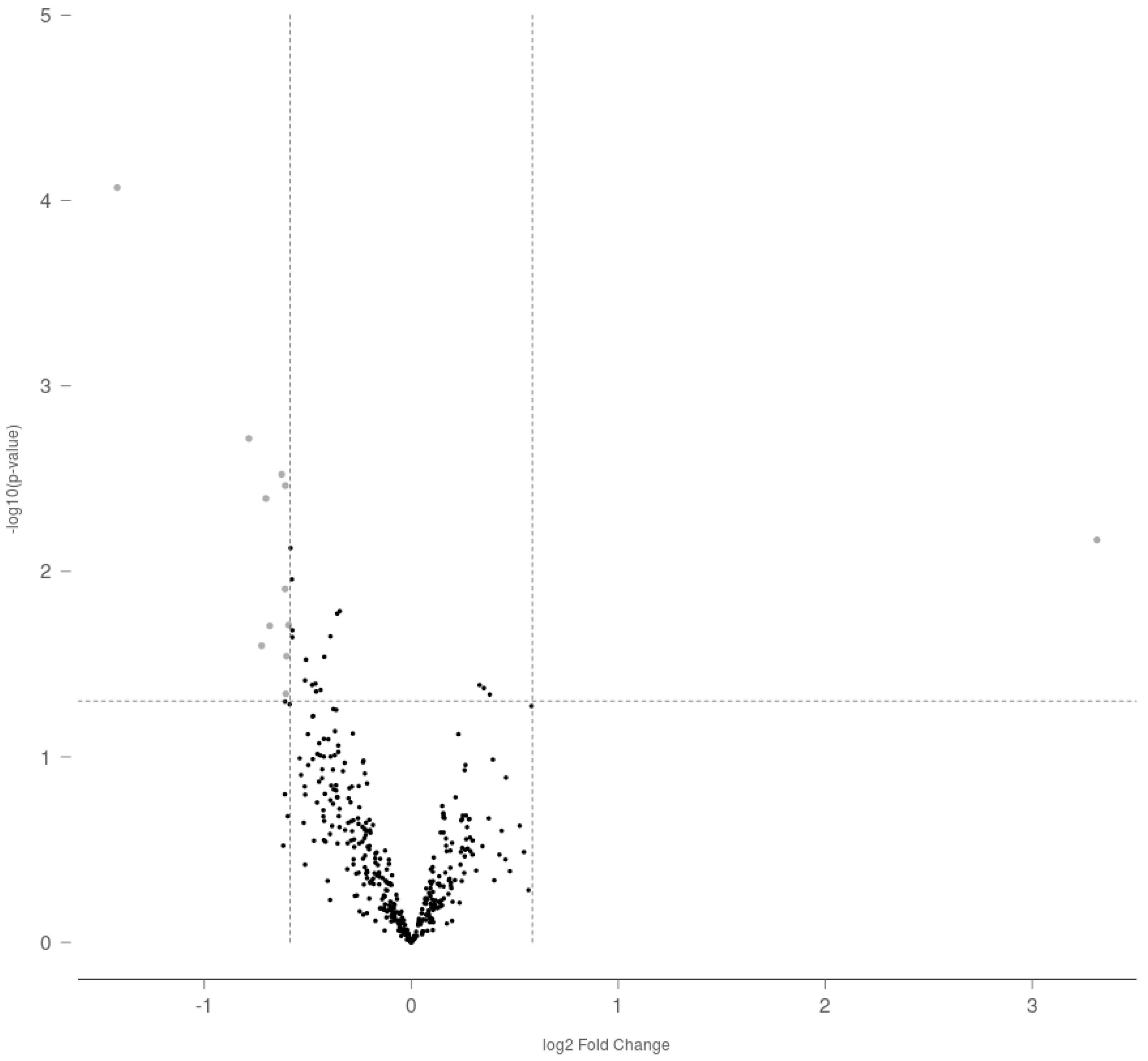
Figure 1. Volcano plot representation of differential miRNA expression as a result of gestational exposure to THC in adult rat ovaries. The dotted lines represent the fold change (≥1.5 or ≤−1.5) and p-value (<0.05) cutoff values selected to determine statistical difference.
Ijms 23 08000 g001
Figure 2. Differential expression of miR-122-5p (miR-122) in adult rat ovaries prenatally exposed to THC analyzed with (A): Rat v1.5 miRNA assay (Nanostring Technologies, Seattle, DC, USA), nSolver® and ROSALIND® analysis software, and (B): miRCURY™ LNA miRNA PCR assay system (Qiagen, Hilden, Germany) and SigmaPlot® (mean + SEM; N = 5, * p < 0.05).
Ijms 23 08000 g002
Figure 3. (A). Relative expression of Igf1r in prenatally THC-exposed adult rat ovaries. (B). Percentage of IGF-1R-positive ovarian follicular cells in prenatally THC-exposed adult rat ovaries. (C). IGF-1R protein levels in prenatally THC-exposed adult rat ovaries (mean + SEM; N = 5, * p < 0.05).
Ijms 23 08000 g003
Figure 4. (A). Relative expression of Casp3 in prenatally THC-exposed adult rat ovaries. (B). Percentage of cleaved caspase-3-positive ovarian follicular cells in prenatally THC-exposed adult rat ovaries. (C). Cleaved caspase-3 protein levels in prenatally THC-exposed adult rat ovaries (mean + SEM; N = 5, * p < 0.05).
Ijms 23 08000 g004
Figure 5. (A). Relative expression of Igf1r in rat ovary explants exposed to 3 μM THC for 48 h. (B). Relative expression of Igf1r in spontaneously immortalized rat granulosa cells exposed to 3 μM THC for 48 h (mean + SEM; N = 5, * p < 0.05).
Ijms 23 08000 g005
Table 1. List of differentially expressed miRNA targets in the adult rat ovary as a result of prenatal THC exposure.
Accession NumberTargetEffectFold Changep Value
MIMAT0000827rno-miR-122-5pUpregulated9.937410.00676
MIMAT0000856rno-miR-154-5pDownregulated−2.676588.50 × 10−5
MIMAT0000885rno-miR-214-3pDownregulated−1.721750.00192
MIMAT0017813rno-miR-3552Downregulated−1.650140.02515
MIMAT0000787rno-miR-18a-5pDownregulated−1.626610.00404
MIMAT0004742rno-miR-296-3pDownregulated−1.605930.01964
MIMAT0005284rno-miR-874-3pDownregulated−1.543350.00299
MIMAT0000882rno-miR-211-5pDownregulated−1.525230.01243
MIMAT0003212rno-miR-20b-3pDownregulated−1.524160.00344
MIMAT0005325rno-miR-598-3pDownregulated−1.521430.04569
MIMAT0004641rno-miR-330-5pDownregulated−1.517890.02862
MIMAT0005302rno-miR-190b-5pDownregulated−1.507450.01945
Table 2. Primer sequences for miR-122-5p, RNU5G and miR-191-5p.
Accession NumberRNASequence (5′-)
MIMAT0000421hsa-miR-122-5pUGGAGUGUGACAAUGGUGUUUG
NR_002852RNU5G snRNAAUACUCUGGUUUCUCUUCAGAUCGCAUAAAUCUUUCGCCUUUUACUAAAGAUUUCCGUGGAGAGGAACAACUCUGAGUCUUAACCCAAUUUUUUGAGCCUUGCUCCGACAAGGCUA
MIMAT0000440hsa-miR-191-5pCAACGGAAUCCCAAAAGCAGCUG
Table 3. Primer sequences for Igf1r, Casp3, B2m and Hprt.
Accession NumberGene NameSymbolForward (5′-3′)Reverse (5′-3′)
NM_052807.2Insulin-like growth factor 1 receptorIgf1rGGAATGGGTCGTGGACAGATACAATCAGCAGGATGGCAAC
NM_012922.2Caspase 3Casp3GAGCTTGGAACGCGAAGAAAAAGAGTCCATCGACTTGCTTCC
NM_012512.2Beta-2-microglobulinB2mAATTCACACCCACCGAGACCGCTCCTTCAGAGTGACGTGT
NM_012583.2Hypoxanthine phosphoribosyltransferase 1Hprt1GCAGTACAGCCCCAAAATGGGGTCCTTTTCACCAGCAAGCT
Publisher’s Note: MDPI stays neutral with regard to jurisdictional claims in published maps and institutional affiliations.