Brief Report

Hyaluronic Acid Alleviates Oxidative Stress and Apoptosis in Human Tenocytes via Caspase 3 and 7

by
Marialucia Gallorini
1,
Cristina Antonetti Lamorgese Passeri
2,
Amelia Cataldi
1,
Anna Concetta Berardi
2,*,† and
Leonardo Osti
3,†
1
Department of Pharmacy, University “G. d’Annunzio” Chieti-Pescara, Via dei Vestini 31, 66100 Chieti, Italy
2
Laboratory of Stem Cells, Department of Haematology, Transfusion Medicine and Biotechnologies, Santo Spirito Hospital, 65124 Pescara, Italy
3
Unit of Arthroscopy and Sports Medicine, Hesperia Hospital, 41125 Modena, Italy
*
Author to whom correspondence should be addressed.
These authors equally contributed to this work.
Int. J. Mol. Sci. 2022, 23(15), 8817; https://doi.org/10.3390/ijms23158817
Submission received: 24 June 2022 / Revised: 28 July 2022 / Accepted: 5 August 2022 / Published: 8 August 2022

Abstract

:
Rotator cuff tendinopathy (RCT) is the primary reason for shoulder surgery and its clinical management is still challenging. Hyaluronic acid (HA) has been shown to have anti-inflammatory effects in vitro and in vivo under RCT conditions, characterized by an exaggerated oxidative stress (OS). However, molecular mechanisms underlying HA-related effects are still partially disclosed. With these aims, a cell model of RCT was established by exposing primary human tenocytes to H2O2 for up to 72 h. Four different HAs by molecular weight were administered to measure nitric oxide (NO) and OS, apoptosis, and collagen 1 expression. In parallel, the well-known antioxidant ascorbic acid was administered for comparison. The present study highlights that HAs characterized by a low molecular weight are able to counteract the H2O2-induced OS by decreasing the percentage of apoptotic cells and reversing the activation of caspase 3 and 7. Likewise, NO intracellular levels are comparable to the ones of controls. In parallel, collagen 1 expression was ameliorated by HAs characterized by higher molecular weights compared to AA. These findings confirm that HA plays an antioxidant role comparable to AA depending on the molecular weight, and highlight the molecular mechanisms underlying the HA anti-apoptotic effects.

1. Introduction

Rotator cuff tendinopathy (RCT) is the most common cause of shoulder pain, with an increasing frequency which varies from 5% to 40% [1,2]. RCT is characterized by tendon structure disruption, ineffective neovascularization, and decreased collagen 1 production [3]. A role in the pathogenesis of tendinopathies for oxidative stress (OS) has been described in several studies [4]. It has been widely demonstrated that the accumulation of reactive oxygen species (ROS) stimulates the intracellular antioxidant system, which is tightly regulated by Nrf2 (nuclear factor erythroid 2 (NF-E2)-related factor 2) [5], and that a functional tendon-repair failure might be strongly associated with the activation of the Nrf2 pathway [6]. Recently, hyaluronic acid (HA) has been suggested for the clinical management of tendinopathies [7,8]. Our previous studies demonstrated that different HA preparations, depending on their molecular weight, counteracted apoptosis under H2O2-induced OS [9], enhanced cell recovery from H2O2 exposure in terms of decreased cytotoxicity and reduced Nrf2 expression, and increased the expression of the HA receptor CD44 [10].
HA, as one of the fundamental components of cartilage and tendons, contributes to their viscoelastic properties in vivo [8,11,12]. However, HA efficacy in the management of RCT is still debated and a comprehensive description of the underlying molecular mechanisms involved is still lacking [8].
In this light, to better understand the different HA biological outcomes under OS conditions, and to extend and improve HA pharmaceutical applications, the antioxidant effects of four different HAs by molecular weight on human rotator-cuff tendon-derived cells in terms of cytoprotection and caspase 3/7 activation were studied. Moreover, protein expression levels of collagen 1, the predominant collagen isoform involved in ECM deposition under inflammatory conditions, was investigated during HA administration. In addition, to evaluate whether the HA might have antioxidant functions, the well-known antioxidant ascorbic acid (AA) was used for comparison [13]. Moreover, AA is well known to participate in collagen biosynthesis [14], including in an Achilles tendon injury rat model [15].

2. Results

2.1. Effect of HAs on Cell Proliferation Rate and Viability

A cell proliferation analysis for up to 72 h of treatment was performed on tenocytes to verify the effects of HAs administered in parallel with H2O2 on their proliferation rate (Figure 1A). As expected, the percentage of H2O2-exposed proliferating tenocytes dramatically fell after 24 h, and the oxidative stressor administered had an even greater effect after 72 h, with proliferation percentages less than 5% in all the samples. When AA and HA preparations were administered in parallel with H2O2 after 24 h, the proliferation rate slightly but significantly increased in all the experimental conditions with respect to H2O2 alone. After 72 h, this effect was maintained.

2.2. Modulation of Superoxide Anions in the Presence of HAs

The intracellular generation of superoxide anions was measured in tenocytes after 6 h of exposure in all the experimental conditions (Figure 1B). When H2O2 is added to the cultures, a dramatic increase in the superoxide anion generation is registered, with the MFI ratio being 3-folds more than the one of the UC (Figure 1B). As expected, AA is able to significantly reduce the generation of superoxide in the presence of H2O2, with the MFI ratio halved with respect to H2O2 alone. In parallel, Hyalgan is the best in decreasing the amount of superoxide anions among the four HA preparations, followed by Hyalubrix and Artrosulfur HA.

2.3. Oxidative Stress-Induced Apoptosis Counteraction by HAs

Cell percentages in the various phases of cell death were assessed after 48 h in the presence of HAs under OS conditions (Figure 1C). After 48 h, viable cells appeared time-dependently decreased by H2O2 (38.37%) and early apoptotic cells dramatically increased (39.14%), as did late apoptotic tenocytes. When AA is added, cell viability significantly increases (56.60%) for late apoptotic cells and necrotic cells, whereas the percentage of early apoptosis is comparable to the one of H2O2 alone. In parallel, Artrosulfur HA and Synolis VA increase cell viability, decrease early apoptosis, but increase cell necrosis with respect to H2O2 alone (11.51%), with percentages measured as 21.31% and 24.57%, respectively. Cell viability is even more increased in the presence of Hyalubrix and Hyalgan (65.49% and 68.39%, respectively). In parallel, apoptosis is slightly decreased, and viability is slightly increased.

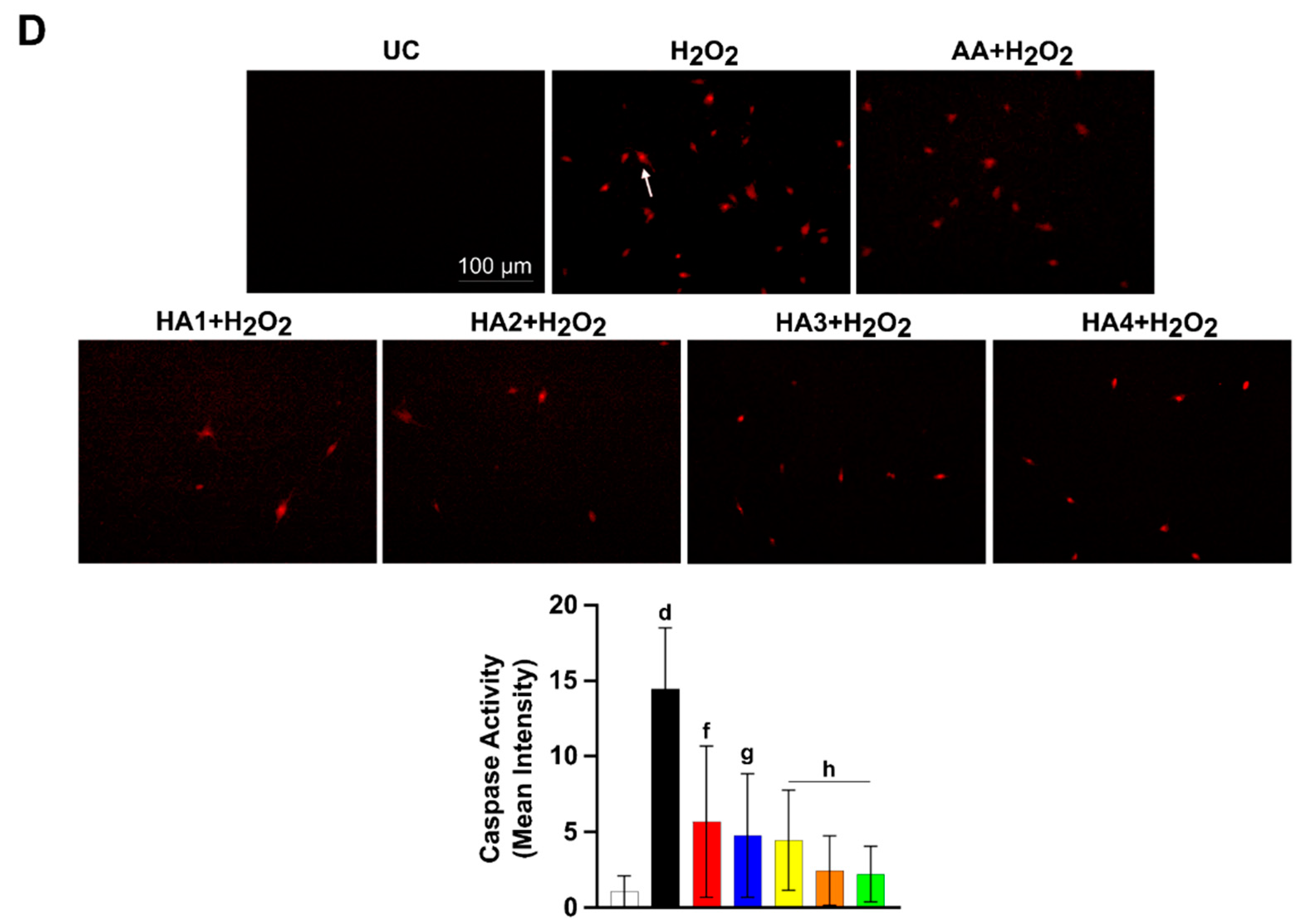
2.4. HAs Treatment Reduces the Activation of Caspase 3 and 7

To establish whether the apoptosis induction might be driven by the activation of caspases, the activity of caspase 3 and 7 was measured after 48 h under OS conditions (Figure 1D). By using the image-analyzing software, the mean fluorescent intensity related to caspase-positive (apoptotic) cells was measured (see Section 4). Compared to the UC control (MFI = 1.07), the H2O2 exposure dramatically increases the activation of caspase 3 and 7 (MFI = 14.46). In parallel, when the AA is added to the culture, the caspase 3 and 7 MFI decreases (5.69), indicating a reduced activity of caspases. Artrosulfur HA and Synolis VA slightly reduce caspase activity (MFI = 4.76 and 4.46, respectively) compared to AA, as shown in Figure 1D. When Hyalubrix and Hyalgan are administered, the activity of caspase 3 and 7 was even more reduced (MFI = 2.46 and 2.23, respectively) compared to AA. This suggests that HA counteracts the H2O2-induced apoptosis through the reduction of caspase 3 and 7 activation.

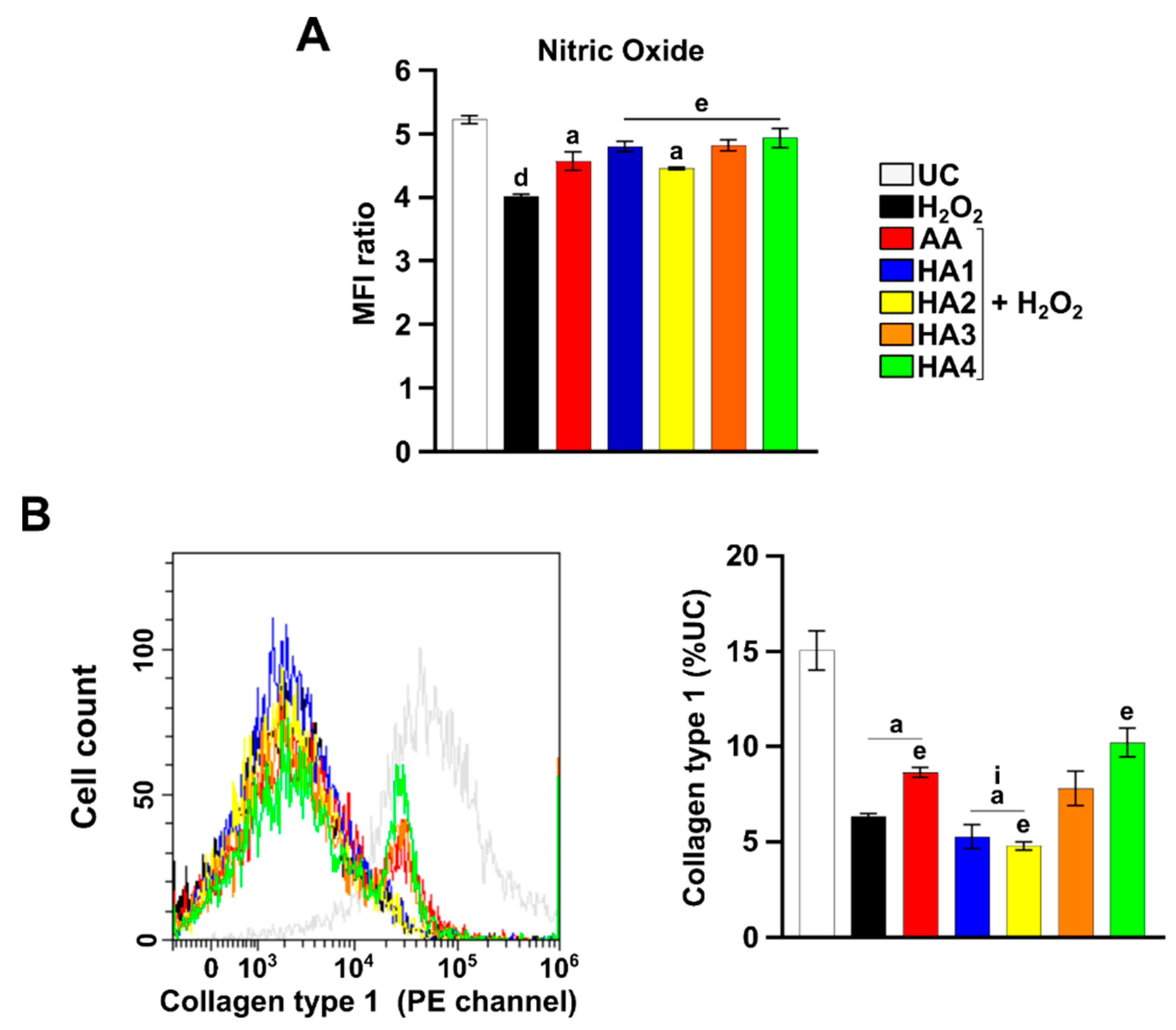
2.5. Modulation of Nitric Oxide (NO) in the Presence of HAs

The intracellular nitric oxide generated by tenocytes was measured in tenocytes after 6 h of exposure in all the experimental conditions (Figure 2A). H2O2 exposure triggers the NO fall (Figure 2A). When AA and the HA preparations are added, NO is slightly decreased with respect to UC, but it is maintained higher than H2O2 alone, mainly with Hyalubrix and Hyalgan.

2.6. Modulation of Collagen 1 Levels by HA Preparations

Collagen 1 expression levels were measured in tenocytes under OS conditions after 72 h (Figure 2B). As expected, the H2O2 exposure strongly decreases collagen 1 expression levels compared to the UC, whereas the presence of AA ameliorates collagen 1 expression levels under OS conditions. Data reveal that Hyalgan exerts a greater effect on tendon-derived cells exposed to H2O2 than all the other treatments in terms of collagen 1 expression.

3. Discussion

Oxidative stress (OS) is crucial under tendon injuries [16]. This might be attributed to the links among OS, inflammation, and apoptosis, all of which impair the structure and function of cells. An excessive OS results in the activation of redox-sensitive signal transduction pathways [17].
In the present work, OS was induced on tendon-derived cells by the H2O2 exposure. Our group already demonstrated that the HA could counteract apoptosis under OS induced by H2O2 [9]. Here, our previous results are consolidated, showing that all the four HAs used significantly counteracted the cytotoxic action of H2O2 in terms of proliferation (Figure 1A).
Since intracellular ROS is mostly produced in mitochondria, the superoxide mitochondrial production was measured to verify whether the HA might influence its modulation. Here, although all the HA preparations decrease the amount of superoxide anions, Hyalgan is revealed as the best preparation in counteracting their production (Figure 1B). Thus, our results suggest that HAs characterized by a lower molecular weight reduce the production of mitochondrial ROS.
Caspase 3 is a key protein in the stimulation of apoptosis, being the primary executioner in apoptotic cell death. In parallel, caspase 7 plays a supportive role in the execution phase of the process [18]. Therefore, we applicated a caspase 3/7 visualization to evaluate the HA efficacy in terms of anti-apoptotic cell responses under OS conditions [19]. Very interestingly, all the HAs have a similar behavior (Figure 1D). Consequently, it was highlighted that HA ameliorates H2O2-induced apoptosis via caspase 3/7, independently from the molecular weight.
The generation of NO plays a relevant role in repairing an injured tendon [20,21]. In addition, the use of a NO-releasing pro-drug was proven to be useful in the development of promising NO-based therapy [22]. For these reasons, the evaluation of NO expression is an important parameter to be considered in damaged tendons. Firstly, the H2O2 exposure caused a decrease in the production of NO. It is here shown that HAs induced a significant increase in the NO production compared to H2O2 alone-treated cells (Figure 2A). In detail, Artrosulfur, Hyalubrix, and Hyalgan were able to induce a higher NO production than the very well-known antioxidant AA [23]. This result is particularly intriguing since it was demonstrated that NO favors tendon healing at the molecular level by increasing collagen synthesis after 7 days of treatment in an in vitro tendon cell culture [24]. However, to offer deeper insights into the NO effects in tendinopathies, further studies are necessary.
Collagen is the major molecule secreted by tendon cells, participating in ECM remodeling and maintenance [25]. Physiologically, tendons are composed of 65–85% of collagen 1 [14]. In our previous studies, we already demonstrated that Synolis VA was able to stimulate collagen 1 synthesis in a dose dependent manner over 14 days [9]. In the present work we investigated the effect of the HA treatment at an earlier exposure time (72 h). Our results show that collagen 1 expression is downregulated during the H2O2 treatment. Under OS conditions, Hyalubrix and Hyalgan ameliorate the expression of collagen 1. It can be therefore speculated that these two HAs act earlier with respect to Synolis VA in terms of collagen 1 deposition, and this effect might be related to the difference in the molecular weight, as Synolis VA is the highest one.
In conclusion, the OS-induced apoptosis in tenocytes is driven by caspase 3 and 7, and this condition could be counteracted by the administration of HAs. Moreover, the administration of HAs with lower molecular weights leads to an earlier collagen 1 deposition and displays better outcomes in terms of OS counteraction, comparable to the well-known antioxidant AA. This in vitro study can lay the grounds for better outcomes regarding the clinical management of RCTs.

4. Materials and Methods

4.1. Cell Culture

Cryopreserved, tendon-derived cells, stored in vials in liquid nitrogen and isolated from the same patients used in our previous work, were used [26]. The cell isolation protocol was extensively described previously [9,10,26,27]. The tenocyte phenotype was confirmed by assessing the expression of tenocyte-specific genes [28]. For the present work, cells at passage 0 from three samples were thawed out and promptly cultured to avoid phenotype changes caused by later passages. Tenocytes were cultured as elsewhere [26] and used up to passage 5.

4.2. Cell Treatment

Tenocytes were seeded in 96- (0.5 × 10⁴ cells/well), 12- (0.2 × 10⁵ cells/well) or 6-well (0.5 × 10⁵ cells/well) tissue culture-treated plates (Falcon®, Corning Incorporated, Brooklyn, NY, USA) and were left to adhere for 24 h. Cultured cells were exposed to 2 mM of H2O2 (stock solution 30% v/v, Sigma-Aldrich, Milan, Italy), ascorbic acid (AA), 50 μg/mL (stock powder purchased from Sigma-Aldrich, Milan, Italy), and to four HAs, namely, Artrosulfur HA® (M.W. 1000 KDa) (HA1), Synolis VA® (M.W. 2200 KDa) (HA2), Hyalubrix® (M.W. 1600 KDa) (HA3), and Hyalgan® (M.W. 500–730 KDa) (HA4) at a final concentration of 1 mg/mL as reported previously [9,10]. Untreated cells (UC) were used as the control. Exposure times varied according to the various parameters analyzed, from 6 h up to 72 h.

4.3. Proliferation Assay (alamarBlue)

At the established exposure times (24 or 72 h), the medium was replaced with a fresh one containing 10% alamarBlue reagent (Invitrogen, Thermo Fisher Scientific, Waltham, MA, USA), and afterwards incubated for 4 h at 37 °C. The absorbance was measured at 570 and 600 nm by means of a Multiskan GO microplate spectrophotometer (Thermo Fisher Scientific). The value obtained without cells was established as the negative control.

4.4. Detection of Mitochondrial Superoxide Anions

After 6 h of exposure, the generation of mitochondrial superoxide anions was determined by flow cytometry using the fluorescent probe MitoSOX™ (MitoSOX™ Red Mitochondrial Superoxide Indicator, Invitrogen, Thermo Fisher Scientific, Waltham, MA, USA) based on an established procedure as elsewhere reported [29]. Relative fluorescence emissions were detected by a CytoFLEX flow cytometer (Beckman Coulter, Indianapolis, IN, USA) and they were expressed as mean fluorescence intensity ratios on the unstained control (not shown).

4.5. Determination of Apoptosis

The PE Annexin V/Dead Cell Apoptosis Kit with SYTOX® Green for Flow Cytometry (Invitrogen, Thermo Fisher Scientific, Waltham, MA, USA) was used to detect apoptosis by flow cytometry after 24 or 48 h of exposure as reported elsewhere [9]. Cell populations were detected by a CytoFLEX flow cytometer (Beckman Coulter, Indianapolis, IN, USA) and analyzed with the CytExpert software (Beckman Coulter, Indianapolis, IN, USA).

4.6. Detection of Caspase 3 and 7

Active caspase 3 and 7 were detected after 48 h of exposure using the Image-iT™ LIVE Red Caspase 3 and 7 Detection Kit (Invitrogen, Molecular Probes, Waltham, MA, USA), following the manufacturer’s instructions. The kit employs a novel approach to detect active caspases that is based on a fluorescent inhibitor of caspase (FLICA™) methodology [30]. Samples were observed through an ECLIPSE Ti-U inverted fluorescent microscope (Nikon Instruments Inc., Melville, NY, USA) equipped with a camera. For the image analysis, all digital images were captured with the NIS-Elements Imaging Software (Nikon Instruments Inc.). The total fluorescence intensity of an area ≥ 10 frames from each slide was determined. The intensity level was normalized with one of the untreated controls.

4.7. Detection of Intracellular Nitric Oxide

The Nitrixyte™ probe was used for detecting free NO in tenocytes (Cell Meter™ Fluorimetric Intracellular Nitric Oxide Assay Kit; AAT Bioquest, Inc., Sunnyvale, CA, USA) by flow cytometry as already reported [21]. Relative fluorescence emissions were detected by a CytoFLEX flow cytometer (Beckman Coulter, Indianapolis, IN, USA) and analyzed with the CytExpert software (Beckman Coulter, Indianapolis, IN, USA), and they were expressed as mean fluorescence intensity ratios on the unstained control (not shown).

4.8. Expression of Collagen 1 by Flow Cytometry

At the established exposure time (72 h), cell pellets were fixed, permeabilized, and stained as already reported [31]. Next, the mouse monoclonal anti-collagen 1A1 (Thermo Fisher Scientific, Waltham, MA, USA) antibody was added 1:50 to each sample and incubated for 1 h at 4 °C. Next, the secondary phycoerythrin (PE) horse anti-mouse IgG (H + L) antibody (Vector Laboratories, Inc., Burlingame, CA, USA) was added (1:50) and incubated at 4 °C in the dark for 45 min. Relative fluorescence emissions were detected as previously described [31].

4.9. Statistics

Statistical analysis was performed using the GraphPad 5.0 software (GraphPad Software, San Diego, CA, USA) by means of a t-test and ordinary one-way ANOVA, followed by post hoc Tukey’s multiple comparisons tests. Values of p < 0.05 were considered statistically significant.

5. Conclusions

Sustained OS leads to a decrease in tenocyte proliferation caused by an increase in cell death through apoptosis and a consequent diminished ECM remodeling due to a lack of collagen 1 deposition. In this light, understanding that the molecular pathway underlying the OS-induced apoptosis in tenocytes is driven by caspase 3 and 7, and that this condition could be counteracted by HAs characterized by lower molecular weights, can lay the groundwork for better outcomes about the management of RCTs.

Author Contributions

Conceptualization, A.C.B. and L.O.; methodology, M.G.; validation, M.G., A.C. and A.C.B.; formal analysis, M.G. and C.A.L.P.; data curation, M.G. and C.A.L.P.; writing—original draft preparation, M.G. and A.C.B.; writing—review and editing, M.G., A.C., A.C.B. and L.O.; project administration, A.C.B.; funding acquisition, L.O. All authors have read and agreed to the published version of the manuscript.

Funding

This work was supported by the University of Ferrara (Leonardo Osti) and Programma Operativo Nazionale (PON) Ricerca e Innovazione 2014–2020, Fondo Sociale Europeo, Azione I. 2 “Attrazione e Mobilità Internazionale dei Ricercatori” (Marialucia Gallorini).

Institutional Review Board Statement

The study used human cells represented by thawed material that was cryopreserved during our previous works [9,26]. In those papers (see Section 4), it was stated the all the procedures were approved by the Ethical Committee of Rome Tor Vergata University, that all patients gave written informed consent, and that all samples were anonymized before being processed. The study was conducted in accordance with the Declaration of Helsinki and approved by the Ethical Committee of Rome Tor Vergata University.

Informed Consent Statement

Informed consent was obtained from all subjects involved in the study.

Conflicts of Interest

The authors declare no conflict of interest.

References

  1. Yamamoto, A.; Takagishi, K.; Osawa, T.; Yanagawa, T.; Nakajima, D.; Shitara, H.; Kobayashi, T. Prevalence and risk factors of a rotator cuff tear in the general population. J. Shoulder Elb. Surg. 2010, 19, 116–120. [Google Scholar] [CrossRef] [PubMed]
  2. Oliva, F.; Osti, L.; Padulo, J.; Maffulli, N. Epidemiology of the rotator cuff tears: A new incidence related to thyroid disease. Muscles Ligaments Tendons J. 2014, 4, 309–314. [Google Scholar] [CrossRef] [PubMed]
  3. Thankam, F.G.; Roesch, Z.K.; Dilisio, M.F.; Radwan, M.M.; Kovilam, A.; Gross, R.M.; Agrawal, D.K. Association of inflammatory responses and ECM disorganization with HMGB1 upregulation and NLRP3 inflammasome activation in the injured rotator cuff tendon. Sci. Rep. 2018, 8, 8918. [Google Scholar] [CrossRef]
  4. Poulsen, R.C.; Knowles, H.J.; Carr, A.J.; Hulley, P.A. Cell differentiation versus cell death: Extracellular glucose is a key determinant of cell fate following oxidative stress exposure. Cell Death Dis. 2014, 5, e1074. [Google Scholar] [CrossRef] [PubMed] [Green Version]
  5. Gallorini, M.; Petzel, C.; Bolay, C.; Hiller, K.A.; Cataldi, A.; Buchalla, W.; Krifka, S.; Schweikl, H. Activation of the Nrf2-regulated antioxidant cell response inhibits HEMA-induced oxidative stress and supports cell viability. Biomaterials 2015, 56, 114–128. [Google Scholar] [CrossRef]
  6. Meng, J.; Yu, P.; Tong, J.; Sun, W.; Jiang, H.; Wang, Y.; Xue, K.; Xie, F.; Qian, H.; Liu, N.; et al. Hydrogen treatment reduces tendon adhesion and inflammatory response. J. Cell. Biochem. 2018, 120, 1610–1619. [Google Scholar] [CrossRef] [PubMed]
  7. Osti, L.; Buda, M.; Buono, A.D.; Osti, R.; Massari, L. Clinical evidence in the treatment of rotator cuff tears with hyaluronic acid. Muscles Ligaments Tendons J. 2016, 5, 270–275. [Google Scholar] [CrossRef]
  8. Oliva, F.; Marsilio, E.; Asparago, G.; Frizziero, A.; Berardi, A.C.; Maffulli, N. The Impact of Hyaluronic Acid on Tendon Physiology and Its Clinical Application in Tendinopathies. Cells 2021, 10, 3081. [Google Scholar] [CrossRef]
  9. Osti, L.; Berardocco, M.; di Giacomo, V.; Di Bernardo, G.; Oliva, F.; Berardi, A.C. Hyaluronic acid increases tendon derived cell viability and collagen type I expression in vitro: Comparative study of four different Hyaluronic acid preparations by molecular weight. BMC Musculoskelet. Disord. 2015, 16, 284. [Google Scholar] [CrossRef] [Green Version]
  10. Gallorini, M.; Berardi, A.C.; Gissi, C.; Cataldi, A.; Osti, L. Nrf2-mediated cytoprotective effect of four different hyaluronic acids by molecular weight in human tenocytes. J. Drug Target. 2020, 28, 212–224. [Google Scholar] [CrossRef]
  11. Muneta, T.; Koga, H.; Ju, Y.J.; Mochizuki, T.; Sekiya, I. Hyaluronan injection therapy for athletic patients with patellar tendinopathy. J. Orthop. 2012, 17, 425–431. [Google Scholar] [CrossRef]
  12. Flores, C.; Balius, R.; Alvarez, G.; Buil, M.A.; Varela, L.; Cano, C.; Casariego, J. Efficacy and Tolerability of Peritendinous Hyaluronic Acid in Patients with Supraspinatus Tendinopathy: A Multicenter, Randomized, Controlled Trial. Sports Med. Open 2017, 3, 22. [Google Scholar] [CrossRef] [Green Version]
  13. May, J.M.; Harrison, F.E. Role of vitamin C in the function of the vascular endothelium. Antioxid. Redox Signal. 2013, 19, 2068–2083. [Google Scholar] [CrossRef] [Green Version]
  14. Gallorini, M.; Carradori, S. Understanding collagen interactions and their targeted regulation by novel drugs. Expert Opin. Drug Discov. 2021, 16, 1239–1260. [Google Scholar] [CrossRef]
  15. Oliva, F.; Maffulli, N.; Gissi, C.; Veronesi, F.; Calciano, L.; Fini, M.; Brogini, S.; Gallorini, M.; Antonetti Lamorgese Passeri, C.; Bernardini, R.; et al. Combined ascorbic acid and T3 produce better healing compared to bone marrow mesenchymal stem cells in an Achilles tendon injury rat model: A proof of concept study. J. Orthop. Surg. Res. 2019, 14, 54. [Google Scholar] [CrossRef]
  16. Li, P.; Zhou, H.; Tu, T.; Lu, H. Dynamic exacerbation in inflammation and oxidative stress during the formation of peritendinous adhesion resulted from acute tendon injury. J. Orthop. Surg. Res. 2021, 16, 293. [Google Scholar] [CrossRef]
  17. Schweikl, H.; Gallorini, M.; Pöschl, G.; Urmann, V.; Petzel, C.; Bolay, C.; Hiller, K.A.; Cataldi, A.; Buchalla, W. Functions of transcription factors NF-κB and Nrf2 in the inhibition of LPS-stimulated cytokine release by the resin monomer HEMA. Dent. Mater. 2018, 34, 1661–1678. [Google Scholar] [CrossRef]
  18. Brentnall, M.; Rodriguez-Menocal, L.; De Guevara, R.L.; Cepero, E.; Boise, L.H. Caspase-9, caspase-3 and caspase-7 have distinct roles during intrinsic apoptosis. BMC Cell Biol. 2013, 14, 32. [Google Scholar] [CrossRef] [Green Version]
  19. Shim, M.K.; Yoon, H.Y.; Lee, S.; Jo, M.K.; Park, J.; Kim, J.H.; Jeong, S.Y.; Kwon, I.C.; Kim, K. Caspase-3/-7-Specific Metabolic Precursor for Bioorthogonal Tracking of Tumor Apoptosis. Sci. Rep. 2017, 7, 16635. [Google Scholar] [CrossRef] [Green Version]
  20. Bokhari, A.R.; Murrell, G.A. The role of nitric oxide in tendon healing. J. Shoulder Elb. Surg. 2012, 21, 238–244. [Google Scholar] [CrossRef]
  21. Moraes, S.A.; Oliveira, K.R.; Crespo-López, M.E.; Picanço-Diniz, D.L.; Herculano, A.M. Local NO synthase inhibition produces histological and functional recovery in Achilles tendon of rats after tenotomy: Tendon repair and local NOS inhibition. Cell Tissue Res. 2013, 353, 457–463. [Google Scholar] [CrossRef]
  22. Goshi, E.; Zhou, G.; He, Q. Nitric oxide detection methods in vitro and in vivo. Med. Gas Res. 2019, 9, 192–207. [Google Scholar] [CrossRef]
  23. di Giacomo, V.; Berardocco, M.; Gallorini, M.; Oliva, F.; Colosimo, A.; Cataldi, A.; Maffulli, N.; Berardi, A.C. Combined supplementation of ascorbic acid and thyroid hormone T3 affects tenocyte proliferation. The effect of ascorbic acid in the production of nitric oxide. Muscles Ligaments Tendons J. 2017, 7, 11–18. [Google Scholar] [CrossRef] [Green Version]
  24. Xia, W.; Szomor, Z.; Wang, Y.; Murrell, G.A. Nitric oxide enhances collagen synthesis in cultured human tendon cells. J. Orthop. Res. 2006, 24, 159–172. [Google Scholar] [CrossRef]
  25. James, R.; Kesturu, G.; Balian, G.; Chhabra, A.B. Tendon: Biology, biomechanics, repair, growth factors, and evolving treatment options. J. Hand Surg. 2008, 33, 102–112. [Google Scholar] [CrossRef]
  26. Oliva, F.; Berardi, A.C.; Misiti, S.; Verga Falzacappa, C.; Iacone, A.; Maffulli, N. Thyroid hormones enhance growth and counteract apoptosis in human tenocytes isolated from rotator cuff tendons. Cell Death Dis. 2013, 4, e705. [Google Scholar] [CrossRef] [Green Version]
  27. Yao, L.; Bestwick, C.S.; Bestwick, L.A.; Maffulli, N.; Aspden, R.M. Phenotypic drift in human tenocyte culture. Tissue Eng. 2006, 12, 1843–1849. [Google Scholar] [CrossRef]
  28. Berardi, A.C.; Oliva, F.; Berardocco, M.; la Rovere, M.; Accorsi, P.; Maffulli, N. Thyroid hormones increase collagen I and cartilage oligomeric matrix protein (COMP) expression in vitro human tenocytes. Muscles Ligaments Tendons J. 2014, 4, 285–291. [Google Scholar] [CrossRef]
  29. Gallorini, M.; Berardi, A.C.; Ricci, A.; Antonetti Lamorgese Passeri, C.; Zara, S.; Oliva, F.; Cataldi, A.; Carta, F.; Carradori, S. Dual Acting Carbon Monoxide Releasing Molecules and Carbonic Anhydrase Inhibitors Differentially Modulate Inflammation in Human Tenocytes. Biomedicines 2021, 9, 141. [Google Scholar] [CrossRef]
  30. Smolewski, P.; Bedner, E.; Du, L.; Hsieh, T.C.; Wu, J.M.; Phelps, D.J.; Darzynkiewicz, Z. Detection of caspases activation by fluorochrome-labeled inhibitors: Multiparameter analysis by laser scanning cytometry. Cytom. J. Int. Soc. Anal. Cytol. 2001, 44, 73–82. [Google Scholar] [CrossRef]
  31. Alagöz, M.A.; Özdemir, Z.; Uysal, M.; Carradori, S.; Gallorini, M.; Ricci, A.; Zara, S.; Mathew, B. Synthesis, Cytotoxicity and Anti-Proliferative Activity Against AGS Cells of New 3(2H)-Pyridazinone Derivatives Endowed with a Piperazinyl Linker. Pharmaceuticals 2021, 14, 183. [Google Scholar] [CrossRef]
Figure 1. Cell proliferation, generation of superoxide anions, apoptosis, and caspase 3/7 activity in tenocytes exposed to hyaluronic acids under OS conditions. (A) Bar graphs represent the % of alamarBlue reduction values after 24 and 72 h. Data are the means (±SD) of three different experiments. (B) Overlays of the peaks in the FL2 channel represent the fluorescent emissions of each sample. The bar graph shows the mean fluorescence intensity (MFI) related to the emission in the FL 2 channel which is proportional to the generation of superoxide anions. Values are the ratios of the MFIs generated from each sample on the unstained control (negative). (C) Bars represent the percentage of viable cells (unstained), cells in early apoptosis (annexin), cells in late apoptosis (annexin + SYTOX Green), and cells in necrosis (SYTOX Green). (D) Representative images from three independent experiments of caspase 3/7 activity (red cells). 1 cm = 100 μm. The bar graph represents the MFI related to stained cells for activated caspases. Untreated control (UC); hydrogen peroxide 2 mM (H2O2); ascorbic acid μg/mL (AA); Artrosulfur HA® (HA1), Synolis VA® (HA2), Hyalubrix® (HA3) and Hyalgan® (HA4) at a final concentration of 1 mg/mL. a: p ≤ 0.05; b: p = 0.001; c: p = 0.0001; d: p < 0.0001 versus UC; e: p ≤ 0.05; f: p = 0.001; g: p = 0.0001; h: p < 0.0001 versus H2O2; l: p < 0.0001 versus AA.
Ijms 23 08817 g001aIjms 23 08817 g001b
Figure 2. Generation of nitric oxide and collagen 1 expression in tenocytes exposed to hyaluronic acids under OS conditions. (A) The bar graph shows the mean fluorescence intensity (MFI) related to the emission in the FL2 channel, which is proportional to the generation of nitric oxide. Values are the ratios of the MFI generated from each sample on the unstained control (negative). (B) Overlays of the peaks in the FL2 channel represent the fluorescent emissions of each sample. The bar graph represents the % of expression related to collagen 1. Data are expressed as the % of the UC. Untreated control (UC); hydrogen peroxide 2 mM (H2O2); ascorbic acid μg/mL (AA); Artrosulfur HA® (HA1), Synolis VA® (HA2), Hyalubrix® (HA3), and Hyalgan® (HA4) at a final concentration of 1 mg/mL. a: p ≤ 0.05; d: p < 0.0001 versus UC; e: p ≤ 0.05 versus H2O2; i: p ≤ 0.05 versus AA.
Ijms 23 08817 g002
Publisher’s Note: MDPI stays neutral with regard to jurisdictional claims in published maps and institutional affiliations.

