Article

Common and Rare PCSK9 Variants Associated with Low-Density Lipoprotein Cholesterol Levels and the Risk of Diabetes Mellitus: A Mendelian Randomization Study

1
The First Cardiovascular Division, Department of Internal Medicine, Chang Gung Memorial Hospital and Chang Gung University College of Medicine, Taoyuan 33305, Taiwan
2
Department of Research, Taipei Tzu Chi Hospital, Buddhist Tzu Chi Medical Foundation, New Taipei City 23142, Taiwan
3
Department of Life Science, Chinese Culture University, Taipei City 11114, Taiwan
4
Cardiovascular Center, Division of Cardiology, Department of Internal Medicine, Taipei Tzu Chi Hospital, Buddhist Tzu Chi Medical Foundation, New Taipei City 23142, Taiwan
5
School of Medicine, Tzu Chi University, Hualien City 97004, Taiwan
*
Author to whom correspondence should be addressed.
Int. J. Mol. Sci. 2022, 23(18), 10418; https://doi.org/10.3390/ijms231810418
Submission received: 4 August 2022 / Revised: 1 September 2022 / Accepted: 5 September 2022 / Published: 8 September 2022
(This article belongs to the Special Issue Lipid Metabolism and Genes)

Abstract

:
PCSK9 is a candidate locus for low-density lipoprotein cholesterol (LDL-C) levels. The cause–effect relationship between LDL-C levels and diabetes mellitus (DM) has been suggested to be mechanism-specific. To identify the role of PCSK9 and genome-wide association study (GWAS)-significant variants in LDL-C levels and the risk of DM by using Mendelian randomization (MR) analysis, a total of 75,441 Taiwan Biobank (TWB) participants was enrolled for a GWAS to determine common and rare PCSK9 variants and their associations with LDL-C levels. MR studies were also conducted to determine the association of PCSK9 variants and LDL-C GWAS-associated variants with DM. A regional plot association study with conditional analysis of the PCSK9 locus revealed that PCSK9 rs10788994, rs557211, rs565436, and rs505151 exhibited genome-wide significant associations with serum LDL-C levels. Imputation data revealed that three rare nonsynonymous mutations—namely, rs151193009, rs768846693, and rs757143429—exhibited genome-wide significant association with LDL-C levels. A stepwise regression analysis indicated that seven variants exhibited independent associations with LDL-C levels. On the basis of two-stage least squares regression (2SLS), MR analyses conducted using weighted genetic risk scores (WGRSs) of seven PCSK9 variants or WGRSs of 41 LDL-C GWAS-significant variants revealed significant association with prevalent DM (p = 0.0098 and 5.02 × 10⁻⁷, respectively), which became nonsignificant after adjustment for LDL-C levels. A sensitivity analysis indicated no violation of the exclusion restriction assumption regarding the influence of LDL-C-level-determining genotypes on the risk of DM. Common and rare PCSK9 variants are independently associated with LDL-C levels in the Taiwanese population. The results of MR analyses executed using genetic instruments based on WGRSs derived from PCSK9 variants or LDL-C GWAS-associated variants demonstrate an inverse association between LDL-C levels and DM.

1. Introduction

Elevated low-density lipoprotein cholesterol (LDL-C) levels constitute a major risk factor for atherosclerotic cardiovascular disease, as well as cardiovascular and total mortality [1,2,3]. A reduction in LDL-C has been consistently associated with a decrease in major vascular events, with similar effectiveness in men and women, high- and low-risk subgroups, ethnically diverse populations, patients using statin or non-statin drugs, and children with familial hypercholesterolemia [2,3,4,5,6,7,8]. Mendelian randomization (MR) studies have demonstrated a cause–effect relationship between LDL-C levels and cardiovascular disease and mortality [9,10,11,12]. However, clinical trials and meta-analyses have revealed that statins increase the incidence of new-onset diabetes mellitus (DM) in a dose-dependent manner, especially in prediabetes [13,14,15,16]. Furthermore, MR studies using variants in HMGCR, NPC1L1, and PCSK9 have reported that decreased LDL-C levels were associated with an increased incidence of new-onset DM [9,10,17,18,19,20], but this finding was not supported by other studies [11,21]. Whether PCSK9 inhibitors increase the risk of diabetes is also still under debate [22].
Proprotein convertase subtilisin/kexin type 9 (PCSK9) is a multifaceted serine protease that plays a critical role in the regulation of hepatic LDL receptor (LDLR) function, and is involved in atherothrombosis and cardiovascular biology in addition to LDL regulation [23,24]. PCSK9 is primarily produced in the liver. Nevertheless, it is expressed in other organs, such as the kidneys, pancreas, and brain [25]; in various cell types involved in the development of atherosclerosis, such as endothelial cells, macrophages, and smooth muscle cells; and in human atherosclerotic plaques [26]. Human PCSK9 is located on the chromosome 1p32.3, which comprises 12 exons that encode a 692-amino-acid proteinase [26]. More than 850 PCSK9 mutations have been reported, and studies have demonstrated that the more common loss-of-function (LOF) mutations are associated with hypocholesterolemia and protection against coronary heart disease, but that the disease-related dominant gain-of-function (GOF) mutations cause familial hypercholesterolemia and coronary heart disease [23,24]. Both GOF and LOF mutations may occur as common or rare alternative allele frequencies, and through different mechanisms [23,24,27]. Research has also commonly observed differences in genetic variants in PCSK9 between Caucasian and East Asian populations, demonstrating the ethnic heterogeneity of such variants [28,29,30,31,32,33,34,35,36]. In contrast, noncoding PCSK9 sequence variants are rarely reported [10,37,38,39]. Therefore, searching for ethnicity-specific variants is crucial for precision medicine for each population. The Taiwan Biobank (TWB) population-based cohort study enrolled more than 100,000 volunteers aged 30–70 years with no history of cancer [40,41]. The objectives of the present study were to apply a regional plot association analysis and an MR analysis to identify the common and rare PCSK9 variants and other candidate variants associated with LDL-C in participants selected from the TWB cohort, and to test the cause–effect relationship between LDL-C levels and DM.

2. Results

2.1. Regional Plot Association Analysis for the PCSK9 Region

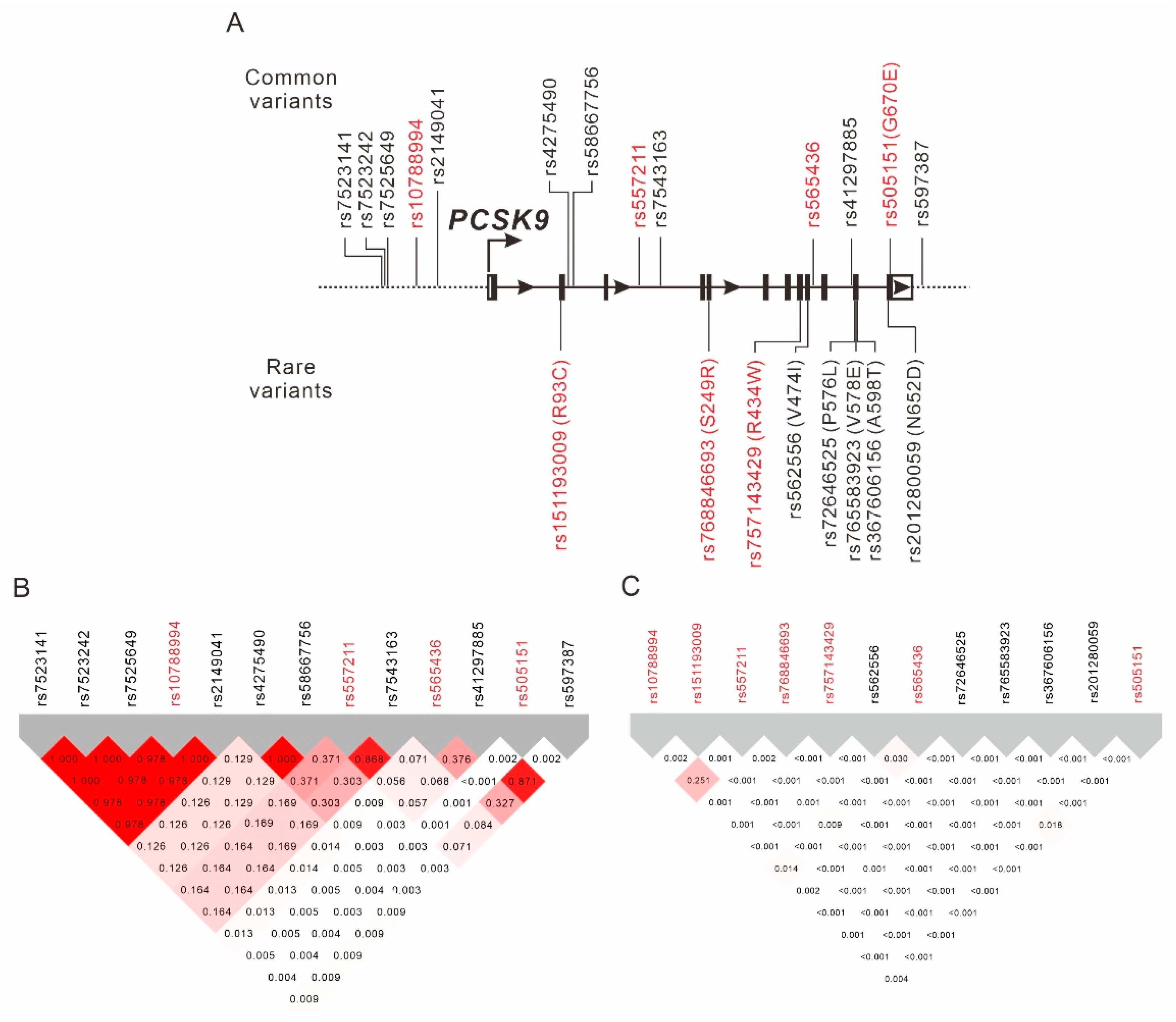
The selection process of the TWB participants is shown in Figure 1. Baseline characteristics of the study population are shown in Supplementary Table S1. Using genome-wide association study (GWAS) data obtained from 75,441 TWB participants, we performed a regional plot association study with conditional analysis to determine the association of LDL-C levels with 330 single-nucleotide variants (SNVs) located between 55.40 and 55.65 Mb on chromosome 1p32.3 within the PCSK9 region. A total of 66 SNVs exceeded the genome-wide significance threshold (p < 5 × 10⁻⁸), with the lead SNV being rs10788994 (p = 2.71 × 10⁻¹⁴; Figure 2A). We also performed stepwise analysis to clarify whether the associations between the other PCSK9 SNVs were independent of the lead SNV. After adjustment for the rs10788994 genotypes, we observed that rs565436 in the regional plot near the PCSK9 locus was still significantly associated with LDL-C levels (p = 3.28 × 10⁻⁹, Figure 2B). After further adjustment for both rs10788994 and rs565436 genotypes, we observed that rs505151 and rs12067569 were still associated with LDL-C levels (p = 1.83 × 10⁻⁷; Figure 2C). Because of the complete LD between the rs505151 and rs12067569 genotypes (Figure 3B), we included the rs505151 genotype in our subsequent analyses. We continuously adjusted for the rs10788994, rs565436, and rs505151 genotypes; however, none of the SNVs in the regional plot near the PCSK9 locus exhibited genome-wide significant associations with LDL-C levels (Figure 2D). Twelve SNVs located within the PCSK9 region reached genome-wide significance in initial regional association analysis, and were selected for linkage disequilibrium (LD) analysis. We observed that three SNVs—namely, rs10788994, rs557211, and rs565436—were strongly associated with LDL-C levels in low LD (pairwise r2 < 0.3), and all three SNVs together with the rs505151 variant were subsequently used for PCSK9-weighted genetic risk scores (WGRSs) and MR analysis (Figure 3, Supplementary Table S2).

2.2. Association between Rare PCSK9 Nonsynonymous Exonic Mutations and LDL-C Levels

From the pre-quality-control (QC) imputation data, we found eight rare (minor allele frequency < 0.01) exonic nonsynonymous mutations of PCSK9, and analyzed the association of these mutations with LDL-C levels (Supplementary Table S3). All eight mutations exhibited a weak LD with the other genotypes (r2 < 0.01) (Figure 3C). After adjustment for age, sex, body mass index (BMI), and current smoking status, we observed that the rare alleles of three PCSK9 variants—namely, rs151193009, rs768846693, and rs757143429—exhibited a genome-wide significant association with lower LDL-C levels (Table 1).

2.3. Stepwise Linear Regression Analysis

We applied additive models to execute a stepwise linear regression analysis using age, sex, BMI, current smoking status, and seven PCSK9 variants. The analysis revealed that the rs10788994, rs151193009, rs557211, rs768846693, rs757143429, rs565436, and rs505151 genotypes contributed to 0.07%, 0.13%, 0.01%, 0.06%, 0.04%, 0.05%, and 0.02% of the variation in total cholesterol levels, respectively, and to 0.09%, 0.14%, 0.01%, 0.11%, 0.05%, 0.05%, and 0.03% of the variation in LDL-C levels, respectively (Table 2).

2.4. Association between LDL-C Levels and DM Status

The associations between LDL-C levels and DM status are presented in Supplementary Table S4. After adjustment for age, sex, BMI, and current smoking status, we observed that circulating LDL-C levels were negatively associated with DM status (OR = 0.99 (95% CI: 0.99, 0.99) per mg/dL unit of LDL-C, or 0.78 (95% CI: 0.75, 0.81) per mmol/L unit of LDL-C; p = 6.76 × 10−64) (Supplementary Table S4).

2.5. GWAS Analysis for LDL-C Levels

After fitting our linear regression model for genotype trend effects and adjusting for age, sex, BMI, and current smoking status, we observed that APOE rs7412 located on chromosome 19q13.32 had the highest −log10 p value for LDL-C levels (p < 10−307), and that a total of 47 lead SNVs for each peak (the SNV with the lowest p-value for each locus) exhibited genome-wide significant associations (Supplementary Figure S1 and Supplementary Table S5). We also assessed the associations of individual LDL-C-level-determining SNVs with DM after further adjustment for LDL-C levels; the results indicated that six SNVs–namely, APOE rs7412, PMFBP1 rs3852789, TMEM8A rs375498857, SLC10A1 rs2296651, TRIM5 rs16934050, and MACO1 rs61775184—were significantly associated with DM (all p < 0.01). These six SNVs were excluded from the calculation of WGRSs for the analysis of DM status. Thus, 41 SNVs were used as LDL-C GWAS-associated variants for WGRS calculation, and the resulting association with LDL-C levels increased significantly (p < 10−307; Table 3).

2.6. Association between LDL-C-Level-Associated PCSK9 Genotypes and Clinical and Laboratory Parameters

Over 70,000 volunteers participated in the genotype–phenotype association analysis. We analyzed the association of the seven aforementioned LDL-C-level-associated PCSK9 genotypes with various clinical phenotypes and laboratory parameters. Using the Bonferroni correction and our additive model, and after adjusting for age, sex, BMI, and current smoking status, we observed no significant associations (p < 0.001) between the PCSK9 genotypes and phenotypes, apart from LDL-C levels (Supplementary Tables S5–S18).

2.7. MR Analysis along with 2SLS IV Regression for Determining the Association of Genetic Determinants of LDL-C Levels with DM Status

We performed two-stage least squares regression (2SLS) instrumental variable (IV) analysis to determine the direction and causality of the association between LDL-C levels and DM status. In this regression, we used the seven aforementioned PCSK9 variants associated with LDL-C levels to derive weighted genetic risk scores (WGRSs), and the scores revealed that LDL-C levels were significantly associated with a lower risk of DM (p = 0.0079; Table 3). The association of PCSK9-WGRS with DM status remained significant after adjustment for multiple parameters associated with LDL-C levels (p = 0.0098), but the association became nonsignificant after adjustment for LDL-C levels (p = 0.1526). From the GWAS for LDL-C levels, we selected 41 independent SNVs as IVs. All SNVs were significantly associated with LDL-C levels at the genome-wide significance level (Supplementary Table S19). We further tested the associations of the WGRSs of these 41 LDL-C-determining variants with DM status. In the 2SLS IV analysis, our data revealed significant associations between the WGRSs of the 41 gene variants and DM status (p = 0.0046), which remained significant even after further adjustment for multiple parameters associated with LDL-C levels (p = 5.02 × 10⁷); nevertheless, the associations became nonsignificant after adjustment for LDL-C levels (p = 0.7606). The F statistics derived for the instruments ranged from 716 to 729 for LDL-C-level-determining genotypes, demonstrating a low risk of weak instrument bias (Supplementary Table S20).

2.8. Scatter Plots and Test for Heterogeneity

As shown in Supplementary Figure S2, scatterplots of the effect sizes of the associations between the LDL-C-level-determining alleles and LDL-C levels versus the effect sizes of the association between the LDL-C-level-determining alleles and DM status in the MR analysis revealed that independent genetic variants in different gene regions were concordantly associated with the outcomes, supporting a causal relationship between LDL-C levels and DM (Supplementary Figure S2). Through visual inspection and by using Cochran’s Q test, we found no evidence of heterogeneity for causal estimates in each genetic variant during the various sensitivity analyses (Supplementary Table S21).

2.9. Sensitivity Analysis for Causal Inference from Standard Mendelian Randomization with Multiple Genetic Variants Determining LDL-C Levels

The causal effects calculated using sensitivity analyses in the standard MR study were almost identical to the 2SLS estimates (Supplementary Table S22). The symmetric funnel plots confirmed the absence of a directional pleiotropy effect for each set of IV estimates and the IV strength (Supplementary Figure S3). Moreover, our data indicated that the estimates of the effect size in standard MR analysis in the inverse-variance-weighted (IVW), median-based, and MR–Egger regression (slope) methods were all consistent with the estimates for the effect of the LDL-C-level-determining allele on DM in the 2SLS method (Supplementary Table S22). The intercept of all Egger regression analyses was close to zero. These results indicate the presence of balanced pleiotropy in our standard MR analysis.

3. Discussion

This study analyzed PCSK9 variants associated with LDL-C levels in Taiwanese participants. Seven PCSK9 coding and noncoding variants were significantly associated with both total and LDL-C levels, contributing 0.38% and 0.49% of the variations, respectively. Our genotype–phenotype association analysis revealed that these variants had no other pleiotropic effects. This study is the first to report a rare PCSK9 coding-sequence variant—namely, rs768846693 pS249R—as a possible LOF mutation. We performed an MR analysis using 2SLS regression and a PCSK9 WRGS genetic instrument, and the analysis results indicated that genetically determined LDL-C levels were inversely associated with the prevalence of DM. These results provide further evidence that PCSK9 variants play a crucial role in determining cardiometabolic outcomes in the Taiwanese population.

3.1. Ethnic Heterogeneity of PCSK9 Gain-of-Function and Loss-of-Function Mutations

As of April 2022, more than 850 unique PCSK9 variants have been listed in the ClinVar database (https://www.ncbi.nlm.nih.gov/clinvar/?term=PCSK9[gene] (accessed on 24 May 2022)), of which 643 are from exonic, 128 are from intronic, and 67 are from either 5′ or 3′ untranslated regions. Of the exonic variants, 384 are missense, 18 are nonsense, 22 are frame shifts, and 219 are synonymous mutations. A clinical classification indicated the existence of 34 pathogenic or likely pathogenic variants and 99 conflicting interpretations of pathogenic variants. In the present study, we observed that three rare PCSK9 coding variants—namely, rs151193009 pR93C, rs768846693 pS249R, and rs757143429 pR434W—were LOF mutations, and were significantly associated with lower LDL-C levels. In the ClinVar database, pR93C and pR434W were classified as variants of conflicting interpretations, and S249R was classified as a variant of uncertain significance. This study is the first to report that pS249R is an LOF mutation and is associated with low LDL-C levels. Although the three PCSK9 coding variants (rs151193009 pR93C, rs768846693 pS249R, and rs757143429 pR434W) are rare mutations (minor allele frequencies = 0.004–0.027%), their LDL-C-lowering effect per allele was noted to be significant, ranging from 13 to 29 mg/dL. A recent study reported that the PCSK9 R93C variant was associated with a 60% lower risk of premature myocardial infarction in the Chinese Han population [42]. The ASPirin in Reducing Events in the Elderly trial also demonstrated that PCSK9 pR434W is a member of lipid-lowering PCSK9 and APOB genetic variants that are carried by healthy older individuals (aged ≥ 70 years) and contribute to coronary-heart-disease-free survival [43]. However, PCSK9 pS249R was first identified as an LOF mutation, but its clinical implications warrant further investigation. In contrast, we found that the common variant rs505151 (E670G) was a GOF mutation and was significantly associated with higher LDL-C levels, which is consistent with previous reports [24,29,35,44].

3.2. Role of Noncoding PCSK9 Variants

Previous studies have focused mostly on the role of exonic PCSK9 mutations in LDL-C levels. Our data reveal that variants in the promoter region, intronic region, and 3′ untranslated region may play a crucial role in and be independently associated with LDL-C levels. Our study revealed the PCSK9 rs10788994 variant, located in the promoter region of PCSK9, as the lead SNV for LDL-C levels. However, a case–control study on premature myocardial infarction in the Italian population reported that this SNV was not associated with LDL-C levels [38]. Our data also revealed that the PCSK9 intron variants rs557211 and rs565436 were independently associated with LDL-C levels. The rs565436 variant was reported to be associated with familial hypercholesterolemia in Malaysia [45], but the association between rs557211 and LDL-C levels has not yet been reported. Additional functional studies are necessary to elucidate the mechanism underlying the influence of these noncoding PCSK9 variants on LDL-C levels.

3.3. Mendelian Randomization for LDL-C and DM

Previous studies including GWAS and MR analyses have demonstrated that a genetically induced elevation of circulated LDL-C levels is associated with a lower risk of DM [12,46], supporting the observation that lowering LDL-C levels with statins slightly increases the risk of DM [13,14,15,16]. However, the mechanism underlying these effects is still unknown. MR studies using LDL-C-lowering drugs’ target genes—such as HMGCR, PCSK9, and NPC1L1 variants—as genetic instruments have consistently demonstrated an inverse association between LDL-C and DM [10,18,19,20]. However, not all MR and genetic studies have revealed inverse associations between each LDL-C-level-lowering allele and an increased risk of DM [11,21,47]. Accordingly, the heterogeneity between genetic and MR studies suggests that the diabetogenic effect of LDL-C reduction is mechanism-specific, and may depend on the underlying reduction in LDL-C levels, such as pancreatic ß-cell dysfunction caused by increased intracellular cholesterol levels due to either increased LDLR expression or transmembrane cholesterol transport [17,48]. Furthermore, genetic studies may shed light on the effects of pleiotropy on the association between LDL-C-level-determining alleles and DM status. For example, some studies have revealed that LDL-C-lowering alleles of HMGCR and PCSK9 variants increase body weight and waist circumference [17,18,19]. GCKR, TM6SF2, and PNPLA3 variants were reported to be associated with diabetogenic traits such as high liver fat, in addition to being associated with reductions in LDL-C levels [49]. However, most of the reported MR studies have included only people of European descent rather than people of Asian descent. Our results also reveal an inverse association between genetically determined LDL-C levels and the prevalence of DM, and the corresponding effect size is similar to that reported in previous studies. According to a WGRS derived using PCSK9, we noted that an LDL-C reduction of 1 mmol/L was associated with an increased risk of DM (OR 1.61, 95% CI: 1.04–2.49), and that an LDL-C reduction of 1 mg/dL was associated with an increased risk of DM (OR 1.01, 95% CI: 1.00–1.02). We obtained a similar estimate (OR 1.32: 1.09–1.60 per 1 mmol/L) when we applied a WGRS derived using LDL-C GWAS-associated variants. PCSK9 mutations can increase or reduce LDL-C levels in the blood. If only a single lead SNV rs10788994 in the PCSK9 gene region was used as genetic instrument for reducing LDL-C, such as HMGCR rs3064191 alone, the effect size would be insufficient to increase the risk of diabetes. When we applied a WGRS derived by aggregating several common and rare PCSK9 variants, we noted that the diabetogenic effect of LDL-C reduction became evident. This finding was also supported by the association of the WGRS derived by aggregating 41 LDL-C GWAS-significant variants with DM status. Moreover, PCSK9 variants—both common and rare—were not linked to any other metabolic traits. Overall, our findings indicate that lower circulating LDL-C levels constitute a key mediator of increased risk of DM. The diabetogenic effect can be attributed to reduction in LDL-C levels, and the association of individual SNVs with the risk of DM may be proportional to its association with LDL-C reduction.

3.4. Limitations

The present study has some limitations. Because of the cross-sectional study design, we could not investigate the association of circulating LDL-C at baseline with the incidence of DM; thus, we could not avoid survival bias. Second, genetic association studies revealed ethnic genetic heterogeneity; therefore, our findings may not be applicable to other ethnic groups. Third, our study did not have a second cohort; the inclusion of a second cohort—particularly one with a larger sample size and longitudinal follow-up—would strengthen the validity of our findings.

4. Methods and Materials

4.1. TWB Participants

The TWB population-based cohort study recruited participants from centers across Taiwan between 2008 and 2020. A total of 107,494 participants who were genotyped using the Axiom Genome-Wide CHB 1 or 2 Array and had no history of cancer were enrolled. Of these participants, 32,053 were excluded from the analysis because 12,289 participants did not have imputation data; 10,956 participants exhibited second-degree relatedness, as indicated by an identity with descent PI_HAT of >0.187, which was determined from a QC analysis conducted in a GWAS; 2862 participants fasted for less than 6 h; and 5946 participants had a history of hyperlipidemia. Moreover, participants with a history of DM, hypertension, hyperlipidemia, or gout—as determined from an examination of glucose metabolism parameters, blood pressure status, lipid profiles, and serum uric acid levels, respectively—were excluded. Figure 1 illustrates the flowchart of participant enrollment. The definitions of DM, hypertension, hyperlipidemia, and current smoking status are provided in Supplementary Method S1. Ethical approval was received from the Research Ethics Committee of Taipei Tzu Chi Hospital, Buddhist Tzu Chi Medical Foundation (approval number: 08-XD-005), and Ethics and Governance Council of the Taiwan Biobank (approval number: TWBR10908-01). Each participant provided written informed consent.

4.2. Genomic DNA Extraction and Genotyping

After blood samples were collected, DNA was extracted using a PerkinElmer Chemagic 360 instrument, in accordance with the manufacturer’s instructions (PerkinElmer, Waltham, MA, USA). Custom TWB chips were used to execute single-nucleotide variation genotyping on an Axiom Genome-Wide Array Plate system (Affymetrix, Santa Clara, CA, USA).

4.3. Clinical Phenotypes and Laboratory Examinations

The following clinical phenotypes were used in this study: BMI; waist circumference; waist–hip ratio; and systolic, mean, and diastolic blood pressure. Additionally, the following biochemical and hematological parameters were used: glucose metabolism parameters such as hemoglobin A1c (HbA1c) and fasting plasma glucose levels; liver and renal function parameters such as serum creatinine, blood urea nitrogen, aspartate aminotransferase (AST), alanine aminotransferase (ALT), γ-glutamyl transferase (γ-GT), albumin, total bilirubin, and uric acid levels; lipid profiles such as total cholesterol, high-density lipoprotein cholesterol (HDL-C), and LDL-C and triglyceride levels; and platelet counts, white and red blood cell counts, and hemoglobin and hematocrit levels. The BMI and estimated glomerular filtration rate (eGFR) were calculated as reported previously [50]. Because of the absence of data related to urine creatinine levels, only spot urine albumin levels were used to evaluate albuminuria.

4.4. Regional Plot Analysis and GWAS

A regional plot association analysis was conducted with LocusZoom.js [51] to determine the lead SNVs providing genome-wide significant evidence of association within the PCSK9 region for LDL-C levels. First, we performed a QC analysis in the GWAS to assess the participants who were enrolled after the application of the exclusion criteria (Figure 1). In the analysis, we used Axiom Genome-Wide CHB 1 and 2 Array plates (Affymetrix, Santa Clara, CA, USA) with 24,927 and 69,529 participants and comprising 611,656 and 640,160 SNVs, respectively, for genome-wide genotype imputation. The reference panel for this imputation process was the East Asian population from the 1000 Genome Project Phase 3 study, and the imputation tools were SHAPEIT and IMPUTE2. After the imputation process, the QC analysis was performed by filtering SNVs with an IMPUTE2 imputation quality score of >0.3. Indels were removed using VCF tools. All samples included in the analysis had a call rate of ≥97%. In the SNV QC analysis, SNVs with a call rate of <97%, a minor allele frequency of <0.01, and a violation of the Hardy–Weinberg equilibrium (p < 10−6) were excluded from subsequent analyses. Thus, after the QC analysis, we included 75,441 participants as well as 330 SNVs located at positions between 55.40 and 55.65 Mb on chromosome 1p32.3 for the regional plot association analysis. Subsequently, we extended the analysis to execute a whole-genome genotyping and imputation. After the execution of the QC analysis and application of other exclusion criteria, the same 75,441 participants and 3,639,888 SNVs were included for the GWAS of LDL-C levels. A p-value of <5 × 10⁻⁸ was considered to be a genome-wide significant association.

4.5. Statistical Analysis

Continuous variables are presented herein as means ± standard deviations or medians and interquartile ranges. The Mann–Whitney U test was used to compare differences in the distribution of linear parameters between the sexes. Lipid profiles and urine albumin levels were transformed logarithmically when examined using an analysis of variance and regression. Categorical data are presented herein as percentages, and their distributions were compared using the chi-squared test. Under the assumption of an additive genetic effect, we used a general linear regression model to analyze the correlation between the studied phenotypes, genotypes, and confounders after adjustment for age, sex, BMI, and current smoking status. We also applied a multiple logistic regression analysis to evaluate the independent effects of the investigated genotypes on lifestyle risks and atherosclerotic risk factors. Moreover, a linear stepwise regression analysis was conducted to determine the independent correlates of LDL-C levels. Genome-wide scans were performed using the PLINK software package. A p-value of <5 × 10⁻⁸ was considered to indicate a genome-wide significant association. In our genotype–phenotype analysis, we applied the Bonferroni correction. Statistical significance was defined as p < 0.0015, which was calculated as 0.05/33 according to 33 traits analyzed. LD was calculated using LDmatrix software (https://analysistools.nci.nih.gov/LDlink/?tab=ldmatrix (accessed on on 25 May 2022)). All statistical analyses were performed using SPSS (version 22; SPSS, Chicago, IL, USA).

4.6. MR Analysis

2SLS regression with IVs was performed to examine whether the LDL-C-determining SNVs and WGRSs were associated with the risk of DM through their associations with LDL-C levels. During the first stage of the regression, LDL-C-determining SNVs and WGRSs were regressed to generate predicted LDL-C levels. During the second stage of the regression, the study parameters were regressed on LDL-C-determining SNVs and WGRSs to generate the predicted risk of DM. To create WGRSs, SNVs were weighted according to each allele score by using the β coefficients from our GWAS analysis, and the risk allele exhibiting directionally concordant associations with the target parameters was selected. The F statistic was used to assess the strength of the instruments, and was calculated using the following equation: F = R2(n − 2)/(1 − R2), where R2 is the proportion of the variability in genetically determined LDL-C levels accounted for by the SNVs, and n is the sample size [52]. An F statistic of >10 indicates a relatively low risk of weak instrumental bias in MR analyses [52].

4.7. Scatterplots and Testing for Heterogeneity

The concordance of IV effects in the generalized linear models was tested using scatterplots [53]. In general, a causal conclusion may seem reasonable if several independent genetic variants in different gene regions are concordantly associated with the outcome. In this study, heterogeneity was assessed either visually in the scatterplot or through Cochran’s Q test on the causal estimate from each genetic variant with the outcome against the genetic association of the exposure of interest, in addition to their confidence intervals [54,55].

4.8. Sensitivity Analysis

Details of the sensitivity analysis are provided in Supplementary Method S2, including a multivariate analysis, funnel plots, IVW methods, simple and weighted median methods, and MR–Egger regression, which were reported previously [50].

5. Conclusions

Common and rare PSCK9 variants are independently associated with LDL-C levels in the Taiwanese population. The results of MR analyses executed using genetic instruments based on WGRSs derived from PCSK9 variants or LDL-C GWAS-associated variants demonstrate a causally inverse association between LDL-C levels and DM. These results provide further evidence that PCSK9 variants and other LDL-C GWAS-associated variants may play a crucial role in determining cardiometabolic outcomes.

Supplementary Materials

The following supporting information can be downloaded at: https://www.mdpi.com/article/10.3390/ijms231810418/s1. References [56,57,58,59,60,61,62] are cited in the supplementary materials.

Author Contributions

Conceptualization, Y.-L.K. and L.-A.H.; methodology, Y.-L.K. and L.-A.H.; software and validation, L.-A.H. and M.-S.T.; formal analysis, M.-S.T., Y.-L.K. and S.W.; resources, Y.-L.K. and H.-H.C.; data curation, M.-S.T., H.-H.C. and S.W.; writing—original draft preparation, L.-A.H.; writing—review and editing, Y.-L.K. and L.-A.H.; visualization, L.-A.H. and H.-H.C.; project administration, Y.-L.K. and M.-S.T.; supervision, Y.-L.K.; funding acquisition, H.-H.C. and Y.-L.K. All authors have read and agreed to the published version of the manuscript.

Funding

This study was supported by grants from Buddhist Tzu Chi Medical Foundation (TCMF-EP 111-02), the Ministry of Science and Technology (MOST 108-2314-B-303-026-MY3), and Taipei Tzu Chi Hospital, Buddhist Tzu Chi Medical Foundation (TCRD-TPE-MOST-110-10) to Y. L. Ko, and from Taipei Tzu Chi Hospital, Buddhist Tzu Chi Medical Foundation (TCRD-TPE-109-RT-1) to H. H. Chou.

Institutional Review Board Statement

The study was conducted in accordance with the Declaration of Helsinki, and approved by the Institutional Review Board of Taipei Tzu Chi Hospital, Buddhist Tzu Chi Medical Foundation (approval number: 08-XD-005), and the Ethics and Governance Council of the Taiwan Biobank (approval number: TWBR10908-01).

Informed Consent Statement

Informed consent was obtained from all subjects involved in the study.

Data Availability Statement

The data presented in this study are available upon request from the corresponding author.

Acknowledgments

We greatly appreciate the technical support from the Core Laboratory of the Taipei Tzu Chi Hospital, Buddhist Tzu Chi Medical Foundation, and expert statistical analysis assistance from Tsung-Han Hsieh.

Conflicts of Interest

The authors declare no conflict of interest.

References

  1. Thomas, H.; Diamond, J.; Vieco, A.; Chaudhuri, S.; Shinnar, E.; Cromer, S.; Perel, P.; Mensah, G.A.; Narula, J.; Johnson, C.O.; et al. Global Atlas of Cardiovascular Disease 2000–2016: The Path to Prevention and Control. Glob. Heart 2018, 13, 143–163. [Google Scholar] [CrossRef]
  2. Mihaylova, B.; Emberson, J.; Blackwell, L.; Keech, A.; Simes, J.; Barnes, E.H.; Voysey, M.; Gray, A.; Collins, R.; Baigent, C. The effects of lowering LDL cholesterol with statin therapy in people at low risk of vascular disease: Meta-analysis of individual data from 27 randomised trials. Lancet 2012, 380, 581–590. [Google Scholar] [CrossRef] [PubMed]
  3. Baigent, C.; Blackwell, L.; Emberson, J.; Holland, L.E.; Reith, C.; Bhala, N.; Peto, R.; Barnes, E.H.; Keech, A.; Simes, J.; et al. Efficacy and safety of more intensive lowering of LDL cholesterol: A meta-analysis of data from 170,000 participants in 26 randomised trials. Lancet 2010, 376, 1670–1681. [Google Scholar] [CrossRef] [PubMed]
  4. Boekholdt, S.M.; Arsenault, B.J.; Mora, S.; Pedersen, T.R.; LaRosa, J.C.; Nestel, P.J.; Simes, R.J.; Durrington, P.; Hitman, G.A.; Welch, K.M.; et al. Association of LDL cholesterol, non-HDL cholesterol, and apolipoprotein B levels with risk of cardiovascular events among patients treated with statins: A meta-analysis. JAMA 2012, 307, 1302–1309. [Google Scholar] [CrossRef] [PubMed]
  5. Fulcher, J.; O’Connell, R.; Voysey, M.; Emberson, J.; Blackwell, L.; Mihaylova, B.; Simes, J.; Collins, R.; Kirby, A.; Colhoun, H.; et al. Efficacy and safety of LDL-lowering therapy among men and women: Meta-analysis of individual data from 174,000 participants in 27 randomised trials. Lancet 2015, 385, 1397–1405. [Google Scholar] [CrossRef]
  6. Luirink, I.K.; Wiegman, A.; Kusters, D.M.; Hof, M.H.; Groothoff, J.W.; de Groot, E.; Kastelein, J.J.P.; Hutten, B.A. 20-Year Follow-up of Statins in Children with Familial Hypercholesterolemia. N. Engl. J. Med. 2019, 381, 1547–1556. [Google Scholar] [CrossRef]
  7. Silverman, M.G.; Ference, B.A.; Im, K.; Wiviott, S.D.; Giugliano, R.P.; Grundy, S.M.; Braunwald, E.; Sabatine, M.S. Association Between Lowering LDL-C and Cardiovascular Risk Reduction Among Different Therapeutic Interventions: A Systematic Review and Meta-analysis. JAMA 2016, 316, 1289–1297. [Google Scholar] [CrossRef]
  8. Yusuf, S.; Lonn, E.; Pais, P.; Bosch, J.; López-Jaramillo, P.; Zhu, J.; Xavier, D.; Avezum, A.; Leiter, L.A.; Piegas, L.S.; et al. Blood-Pressure and Cholesterol Lowering in Persons without Cardiovascular Disease. N. Engl. J. Med. 2016, 374, 2032–2043. [Google Scholar] [CrossRef]
  9. Ference, B.A.; Robinson, J.G.; Brook, R.D.; Catapano, A.L.; Chapman, M.J.; Neff, D.R.; Voros, S.; Giugliano, R.P.; Davey Smith, G.; Fazio, S.; et al. Variation in PCSK9 and HMGCR and Risk of Cardiovascular Disease and Diabetes. N. Engl. J. Med. 2016, 375, 2144–2153. [Google Scholar] [CrossRef]
  10. Schmidt, A.F.; Holmes, M.V.; Preiss, D.; Swerdlow, D.I.; Denaxas, S.; Fatemifar, G.; Faraway, R.; Finan, C.; Valentine, D.; Fairhurst-Hunter, Z.; et al. Phenome-wide association analysis of LDL-cholesterol lowering genetic variants in PCSK9. BMC Cardiovasc. Disord. 2019, 19, 240. [Google Scholar] [CrossRef] [Green Version]
  11. Tragante, V.; Asselbergs, F.W.; Swerdlow, D.I.; Palmer, T.M.; Moore, J.H.; de Bakker, P.I.W.; Keating, B.J.; Holmes, M.V. Harnessing publicly available genetic data to prioritize lipid modifying therapeutic targets for prevention of coronary heart disease based on dysglycemic risk. Hum. Genet. 2016, 135, 453–467. [Google Scholar] [CrossRef] [PubMed]
  12. White, J.; Swerdlow, D.I.; Preiss, D.; Fairhurst-Hunter, Z.; Keating, B.J.; Asselbergs, F.W.; Sattar, N.; Humphries, S.E.; Hingorani, A.D.; Holmes, M.V. Association of Lipid Fractions With Risks for Coronary Artery Disease and Diabetes. JAMA Cardiol. 2016, 1, 692–699. [Google Scholar] [CrossRef] [PubMed]
  13. Kostapanos, M.S.; Agouridis, A.P.; Elisaf, M.S. Variable effects of statins on glucose homeostasis parameters and their diabetogenic role. Diabetologia 2015, 58, 1960–1961. [Google Scholar] [CrossRef] [PubMed]
  14. Navarese, E.P.; Buffon, A.; Andreotti, F.; Kozinski, M.; Welton, N.; Fabiszak, T.; Caputo, S.; Grzesk, G.; Kubica, A.; Swiatkiewicz, I.; et al. Meta-analysis of impact of different types and doses of statins on new-onset diabetes mellitus. Am. J. Cardiol. 2013, 111, 1123–1130. [Google Scholar] [CrossRef]
  15. Ridker, P.M.; Danielson, E.; Fonseca, F.A.; Genest, J.; Gotto, A.M., Jr.; Kastelein, J.J.; Koenig, W.; Libby, P.; Lorenzatti, A.J.; MacFadyen, J.G.; et al. Rosuvastatin to prevent vascular events in men and women with elevated C-reactive protein. N. Engl. J. Med. 2008, 359, 2195–2207. [Google Scholar] [CrossRef]
  16. Sattar, N.; Preiss, D.; Murray, H.M.; Welsh, P.; Buckley, B.M.; de Craen, A.J.; Seshasai, S.R.; McMurray, J.J.; Freeman, D.J.; Jukema, J.W.; et al. Statins and risk of incident diabetes: A collaborative meta-analysis of randomised statin trials. Lancet 2010, 375, 735–742. [Google Scholar] [CrossRef]
  17. Filippatos, T.D.; Panagiotopoulou, T.; Tzavella, E.; Elisaf, M.S. Hypolipidemic Drugs and Diabetes Mellitus-Mechanisms and Data From Genetic Trials. J. Cardiovasc. Pharmacol. Ther. 2018, 23, 187–191. [Google Scholar] [CrossRef]
  18. Schmidt, A.F.; Swerdlow, D.I.; Holmes, M.V.; Patel, R.S.; Fairhurst-Hunter, Z.; Lyall, D.M.; Hartwig, F.P.; Horta, B.L.; Hyppönen, E.; Power, C.; et al. PCSK9 genetic variants and risk of type 2 diabetes: A mendelian randomisation study. Lancet. Diabetes Endocrinol. 2017, 5, 97–105. [Google Scholar] [CrossRef]
  19. Swerdlow, D.I.; Preiss, D.; Kuchenbaecker, K.B.; Holmes, M.V.; Engmann, J.E.; Shah, T.; Sofat, R.; Stender, S.; Johnson, P.C.; Scott, R.A.; et al. HMG-coenzyme A reductase inhibition, type 2 diabetes, and bodyweight: Evidence from genetic analysis and randomised trials. Lancet 2015, 385, 351–361. [Google Scholar] [CrossRef]
  20. Lotta, L.A.; Sharp, S.J.; Burgess, S.; Perry, J.R.B.; Stewart, I.D.; Willems, S.M.; Luan, J.; Ardanaz, E.; Arriola, L.; Balkau, B.; et al. Association Between Low-Density Lipoprotein Cholesterol-Lowering Genetic Variants and Risk of Type 2 Diabetes: A Meta-analysis. JAMA 2016, 316, 1383–1391. [Google Scholar] [CrossRef] [Green Version]
  21. Pan, W.; Sun, W.; Yang, S.; Zhuang, H.; Jiang, H.; Ju, H.; Wang, D.; Han, Y. LDL-C plays a causal role on T2DM: A Mendelian randomization analysis. Aging 2020, 12, 2584–2594. [Google Scholar] [CrossRef] [PubMed]
  22. Monami, M.; Sesti, G.; Mannucci, E. PCSK9 inhibitor therapy: A systematic review and meta-analysis of metabolic and cardiovascular outcomes in patients with diabetes. Diabetes Obes. Metab. 2019, 21, 903–908. [Google Scholar] [CrossRef] [PubMed]
  23. Barale, C.; Melchionda, E.; Morotti, A.; Russo, I. PCSK9 Biology and Its Role in Atherothrombosis. Int. J. Mol. Sci. 2021, 22, 5880. [Google Scholar] [CrossRef] [PubMed]
  24. Sundararaman, S.S.; Doring, Y.; van der Vorst, E.P.C. PCSK9: A Multi-Faceted Protein That Is Involved in Cardiovascular Biology. Biomedicines 2021, 9, 793. [Google Scholar] [CrossRef]
  25. Seidah, N.G.; Benjannet, S.; Wickham, L.; Marcinkiewicz, J.; Jasmin, S.B.; Stifani, S.; Basak, A.; Prat, A.; Chretien, M. The secretory proprotein convertase neural apoptosis-regulated convertase 1 (NARC-1): Liver regeneration and neuronal differentiation. Proc. Natl. Acad. Sci. USA 2003, 100, 928–933. [Google Scholar] [CrossRef]
  26. Ragusa, R.; Basta, G.; Neglia, D.; De Caterina, R.; Del Turco, S.; Caselli, C. PCSK9 and atherosclerosis: Looking beyond LDL regulation. Eur. J. Clin. Investig. 2021, 51, e13459. [Google Scholar] [CrossRef]
  27. Norata, G.D.; Tavori, H.; Pirillo, A.; Fazio, S.; Catapano, A.L. Biology of proprotein convertase subtilisin kexin 9: Beyond low-density lipoprotein cholesterol lowering. Cardiovasc. Res. 2016, 112, 429–442. [Google Scholar] [CrossRef]
  28. Hori, M.; Ohta, N.; Takahashi, A.; Masuda, H.; Isoda, R.; Yamamoto, S.; Son, C.; Ogura, M.; Hosoda, K.; Miyamoto, Y.; et al. Impact of LDLR and PCSK9 pathogenic variants in Japanese heterozygous familial hypercholesterolemia patients. Atherosclerosis 2019, 289, 101–108. [Google Scholar] [CrossRef]
  29. Hsu, L.A.; Teng, M.S.; Ko, Y.L.; Chang, C.J.; Wu, S.; Wang, C.L.; Hu, C.F. The PCSK9 gene E670G polymorphism affects low-density lipoprotein cholesterol levels but is not a risk factor for coronary artery disease in ethnic Chinese in Taiwan. Clin. Chem. Lab. Med. 2009, 47, 154–158. [Google Scholar] [CrossRef]
  30. Huang, C.C.; Niu, D.M.; Charng, M.J. Genetic Analysis in a Taiwanese Cohort of 750 Index Patients with Clinically Diagnosed Familial Hypercholesterolemia. J. Atheroscler. Thromb. 2022, 29, 639–653. [Google Scholar] [CrossRef]
  31. Kim, H.I.; Ye, B.; Gosalia, N.; Köroğlu, Ç.; Hanson, R.L.; Hsueh, W.C.; Knowler, W.C.; Baier, L.J.; Bogardus, C.; Shuldiner, A.R.; et al. Characterization of Exome Variants and Their Metabolic Impact in 6716 American Indians from the Southwest US. Am. J. Hum. Genet. 2020, 107, 251–264. [Google Scholar] [CrossRef] [PubMed]
  32. Lee, C.J.; Lee, Y.; Park, S.; Kang, S.M.; Jang, Y.; Lee, J.H.; Lee, S.H. Rare and common variants of APOB and PCSK9 in Korean patients with extremely low low-density lipoprotein-cholesterol levels. PLoS ONE 2017, 12, e0186446. [Google Scholar] [CrossRef] [PubMed]
  33. Lu, X.; Li, J.; Li, H.; Chen, Y.; Wang, L.; He, M.; Wang, Y.; Sun, L.; Hu, Y.; Huang, J.; et al. Coding-sequence variants are associated with blood lipid levels in 14,473 Chinese. Hum. Mol. Genet. 2016, 25, 4107–4116. [Google Scholar] [CrossRef]
  34. Moon, S.; Kim, Y.J.; Han, S.; Hwang, M.Y.; Shin, D.M.; Park, M.Y.; Lu, Y.; Yoon, K.; Jang, H.M.; Kim, Y.K.; et al. The Korea Biobank Array: Design and Identification of Coding Variants Associated with Blood Biochemical Traits. Sci. Rep. 2019, 9, 1382. [Google Scholar] [CrossRef] [PubMed]
  35. Qiu, C.; Zeng, P.; Li, X.; Zhang, Z.; Pan, B.; Peng, Z.Y.F.; Li, Y.; Ma, Y.; Leng, Y.; Chen, R. What is the impact of PCSK9 rs505151 and rs11591147 polymorphisms on serum lipids level and cardiovascular risk: A meta-analysis. Lipids Health Dis. 2017, 16, 111. [Google Scholar] [CrossRef] [PubMed]
  36. Tang, C.S.; Zhang, H.; Cheung, C.Y.; Xu, M.; Ho, J.C.; Zhou, W.; Cherny, S.S.; Zhang, Y.; Holmen, O.; Au, K.W.; et al. Exome-wide association analysis reveals novel coding sequence variants associated with lipid traits in Chinese. Nat. Commun. 2015, 6, 10206. [Google Scholar] [CrossRef] [PubMed]
  37. Ding, K.; Kullo, I.J. Molecular population genetics of PCSK9: A signature of recent positive selection. Pharm. Genom. 2008, 18, 169–179. [Google Scholar] [CrossRef]
  38. Guella, I.; Asselta, R.; Ardissino, D.; Merlini, P.A.; Peyvandi, F.; Kathiresan, S.; Mannucci, P.M.; Tubaro, M.; Duga, S. Effects of PCSK9 genetic variants on plasma LDL cholesterol levels and risk of premature myocardial infarction in the Italian population. J. Lipid Res. 2010, 51, 3342–3349. [Google Scholar] [CrossRef]
  39. Musunuru, K.; Romaine, S.P.; Lettre, G.; Wilson, J.G.; Volcik, K.A.; Tsai, M.Y.; Taylor, H.A., Jr.; Schreiner, P.J.; Rotter, J.I.; Rich, S.S.; et al. Multi-ethnic analysis of lipid-associated loci: The NHLBI CARe project. PLoS ONE 2012, 7, e36473. [Google Scholar] [CrossRef]
  40. Chen, C.H.; Yang, J.H.; Chiang, C.W.K.; Hsiung, C.N.; Wu, P.E.; Chang, L.C.; Chu, H.W.; Chang, J.; Song, I.W.; Yang, S.L.; et al. Population structure of Han Chinese in the modern Taiwanese population based on 10,000 participants in the Taiwan Biobank project. Hum. Mol. Genet. 2016, 25, 5321–5331. [Google Scholar] [CrossRef] [Green Version]
  41. Fan, C.T.; Lin, J.C.; Lee, C.H. Taiwan Biobank: A project aiming to aid Taiwan’s transition into a biomedical island. Pharmacogenomics 2008, 9, 235–246. [Google Scholar] [CrossRef] [PubMed]
  42. Yang, L.; Pu, T.; Zhang, Y.; Yan, H.; Yu, H.; Gao, W. The R93C Variant of PCSK9 Reduces the Risk of Premature MI in a Chinese Han Population. Front. Genet. 2022, 13, 875269. [Google Scholar] [CrossRef] [PubMed]
  43. Lacaze, P.; Riaz, M.; Sebra, R.; Hooper, A.J.; Pang, J.; Tiller, J.; Polekhina, G.; Tonkin, A.; Reid, C.; Zoungas, S.; et al. Protective lipid-lowering variants in healthy older individuals without coronary heart disease. Open Heart 2021, 8, e001710. [Google Scholar] [CrossRef] [PubMed]
  44. Sinnott-Armstrong, N.; Tanigawa, Y.; Amar, D.; Mars, N.; Benner, C.; Aguirre, M.; Venkataraman, G.R.; Wainberg, M.; Ollila, H.M.; Kiiskinen, T.; et al. Genetics of 35 blood and urine biomarkers in the UK Biobank. Nat. Genet. 2021, 53, 185–194. [Google Scholar] [CrossRef] [PubMed]
  45. Lye, S.H.; Chahil, J.K.; Bagali, P.; Alex, L.; Vadivelu, J.; Ahmad, W.A.; Chan, S.P.; Thong, M.K.; Zain, S.M.; Mohamed, R. Genetic polymorphisms in LDLR, APOB, PCSK9 and other lipid related genes associated with familial hypercholesterolemia in Malaysia. PLoS ONE 2013, 8, e60729. [Google Scholar] [CrossRef]
  46. Fall, T.; Xie, W.; Poon, W.; Yaghootkar, H.; Mägi, R.; Knowles, J.W.; Lyssenko, V.; Weedon, M.; Frayling, T.M.; Ingelsson, E. Using Genetic Variants to Assess the Relationship Between Circulating Lipids and Type 2 Diabetes. Diabetes 2015, 64, 2676–2684. [Google Scholar] [CrossRef]
  47. Besseling, J.; Kastelein, J.J.; Defesche, J.C.; Hutten, B.A.; Hovingh, G.K. Association between familial hypercholesterolemia and prevalence of type 2 diabetes mellitus. JAMA 2015, 313, 1029–1036. [Google Scholar] [CrossRef]
  48. Xu, H.; Ryan, K.A.; Jaworek, T.J.; Southam, L.; Reid, J.G.; Overton, J.D.; Baras, A.; Puurunen, M.K.; Zeggini, E.; Taylor, S.I.; et al. Familial Hypercholesterolemia and Type 2 Diabetes in the Old Order Amish. Diabetes 2017, 66, 2054–2058. [Google Scholar] [CrossRef]
  49. Liu, D.J.; Peloso, G.M.; Yu, H.; Butterworth, A.S.; Wang, X.; Mahajan, A.; Saleheen, D.; Emdin, C.; Alam, D.; Alves, A.C.; et al. Exome-wide association study of plasma lipids in >300,000 individuals. Nat. Genet. 2017, 49, 1758–1766. [Google Scholar] [CrossRef]
  50. Hsu, L.A.; Chou, H.H.; Teng, M.S.; Wu, S.; Ko, Y.L. Circulating chemerin levels are determined through circulating platelet counts in nondiabetic Taiwanese people: A bidirectional Mendelian randomization study. Atherosclerosis 2021, 320, 61–69. [Google Scholar] [CrossRef]
  51. Boughton, A.P.; Welch, R.P.; Flickinger, M.; VandeHaar, P.; Taliun, D.; Abecasis, G.R.; Boehnke, M. LocusZoom.js: Interactive and embeddable visualization of genetic association study results. Bioinformatics 2021, 37, 3017–3018. [Google Scholar] [CrossRef] [PubMed]
  52. Palmer, T.M.; Lawlor, D.A.; Harbord, R.M.; Sheehan, N.A.; Tobias, J.H.; Timpson, N.J.; Davey Smith, G.; Sterne, J.A. Using multiple genetic variants as instrumental variables for modifiable risk factors. Stat. Methods Med. Res. 2012, 21, 223–242. [Google Scholar] [CrossRef] [PubMed]
  53. Dai, J.Y.; Chan, K.C.; Hsu, L. Testing concordance of instrumental variable effects in generalized linear models with application to Mendelian randomization. Stat. Med. 2014, 33, 3986–4007. [Google Scholar] [CrossRef]
  54. Bowden, J.; Spiller, W.; Del Greco, M.F.; Sheehan, N.; Thompson, J.; Minelli, C.; Davey Smith, G. Improving the visualization, interpretation and analysis of two-sample summary data Mendelian randomization via the Radial plot and Radial regression. Int. J. Epidemiol. 2018, 47, 1264–1278. [Google Scholar] [CrossRef] [PubMed]
  55. Burgess, S.; Bowden, J.; Fall, T.; Ingelsson, E.; Thompson, S.G. Sensitivity Analyses for Robust Causal Inference from Mendelian Randomization Analyses with Multiple Genetic Variants. Epidemiology 2017, 28, 30–42. [Google Scholar] [CrossRef]
  56. Grundy, S.M.; Cleeman, J.I.; Daniels, S.R.; Donato, K.A.; Eckel, R.H.; Franklin, B.A.; Gordon, D.J.; Krauss, R.M.; Savage, P.J.; Smith, S.C., Jr.; et al. Diagnosis and management of the metabolic syndrome: An American Heart Association/National Heart, Lung, and Blood Institute Scientific Statement. Circulation 2005, 11, 2735–2752. [Google Scholar] [CrossRef]
  57. Bowden, J.; Smith, G.D.; Haycock, P.C.; Burgess, S. Consistent Estimation in Mendelian Randomization with Some Invalid Instruments Using a Weighted Median Estimator. Genet. Epidemiol. 2016, 40, 304–314. [Google Scholar] [CrossRef]
  58. Bowden, J.; Del Greco, M.F.; Minelli, C.; Davey Smith, G.; Sheehan, N.; Thompson, J. A framework for the investigation of pleiotropy in two-sample summary data Mendelian randomization. Stat. Med. 2017, 36, 1783–1802. [Google Scholar] [CrossRef]
  59. Burgess, S.; Butterworth, A.; Thompson, S.G. Mendelian Randomization Analysis with Multiple Genetic Variants Using Summarized Data. Genet. Epidemiol. 2013, 37, 658–665. [Google Scholar] [CrossRef]
  60. Egger, M.; Smith, G.D.; Schneider, M.; Minder, C. Bias in meta-analysis detected by a simple, graphical test. BMJ 1997, 315, 629–634. [Google Scholar] [CrossRef] [Green Version]
  61. Johnson, T.; Uk, S. Efficient Calculation for Multi-SNP Genetic Risk Scores. 2012. Available online: https://citeseerx.ist.psu.edu/viewdoc/summary?doi=10.1.1.398.7674 (accessed on 24 May 2022).
  62. Suurmond, R.; van Rhee, H.; Hak, T. Introduction, comparison, and validation of Meta-Essentials: A free and simple tool for meta-analysis. Res. Synth. Methods 2017, 8, 537–553. [Google Scholar] [CrossRef] [PubMed] [Green Version]
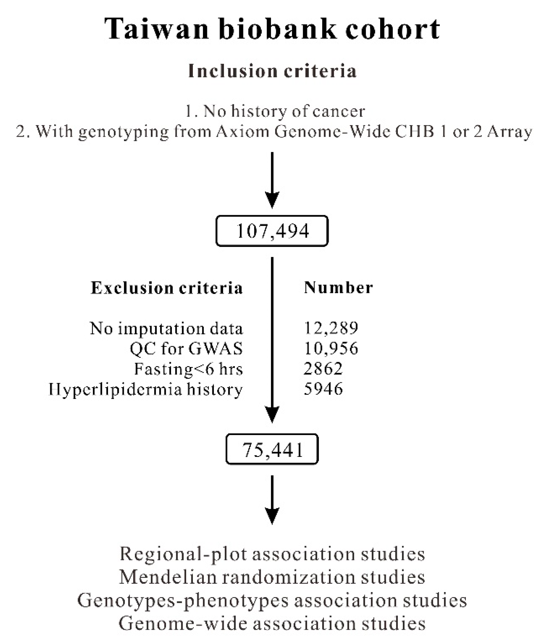
Figure 1. Participant selection flowchart.
Ijms 23 10418 g001
Figure 2. Regional association plots in a region of 200 kb surrounding the PCSK9 locus on chromosome 1p32 for low-density lipoprotein cholesterol levels. Regional plot associations are shown without (A) or with serial conditional analysis after further adjustment for rs10788994 (B), rs565436 (C), and rs505151 (D) genotypes.
Ijms 23 10418 g002
Figure 3. Location (A) and linkage disequilibrium (B,C) of common and rare PCSK9 variants. Shades of red and gray show the strength of the pairwise linkage disequilibrium based on r2, and numbers indicate the value of r2. Seven PCSK9 variants associated with LDL-C levels for weighted genetic risk scores are highlighted in red font.
Ijms 23 10418 g003
Table 1. Association between PCSK9 variants of low-density lipoprotein cholesterol (LDL-C) levels from Axiom Genome-Wide CHB 1 and 2 Array plates with genotype imputation, including common and rare PCSK9 variants.
PCSK9 VariantsChrPositionRef/AltFunc.refGeneGene.refGeneHWEMAFMMMmmmp ValueBetaSEp * Value
rs10788994155,500,976C/TintergenicBSND;PCSK90.76160.3475121.80 ± 31.26 (31,728)120.41 ± 30.84 (33,743)119.23 ± 30.86 (9015)6.44 × 10⁻¹³−0.00470.00061.99 × 10−14
rs151193009155,509,585C/Texon2:c.C277T:p.R93CPCSK90.44190.0027120.98 ± 30.98 (73,449)106.76 ± 28.53 (405)--1.76 × 10⁻¹⁸−0.05790.00571.19 × 10−24
rs557211155,514,215T/GIntron VariantPCSK90.84330.1916121.42 ± 31.179 (49,044)119.81 ± 30.701 (23,300)119.30 ± 30.654 (2740)1.69 × 10⁻¹¹−0.00520.00073.75 × 10−12
rs768846693155,518,412C/Aexon5:c.C747A:p.S249RPCSK90.90780.0004120.88 ± 31.02 (75,324)91.73 ± 28.18 (63)--1.18 × 10⁻⁹−0.13010.01431.12 × 10−19
rs757143429155,523,828C/Texon8:c.C1300T:p.R434WPCSK90.74540.0011120.89 ± 31.02 (75,239)107.81 ± 30.25 (175)--1.47 × 10⁻⁷−0.05090.00863.46 × 10−9
rs565436155,524,601G/AIntron VariantPCSK90.35770.1033121.22 ± 31.11 (60,123)119.41 ±30.65 (13,901)118.19 ± 31.05 (780)8.08 × 10⁻¹⁰−0.00670.00103.81 × 10−12
rs505151155529187G/Aexon12:c.G2009A:p.G670EPCSK90.92020.0534119.0 ± 31.00 (67,530)120.00 ± 31.21 (7603)121.00 ± 32.37 (215)6.00 × 10−60.00650.00135.89 × 10−7
p: unadjusted; p *: adjusted for age, sex, BMI, and current smoking status. Abbreviations: SNV, single-nucleotide variation; Chr: chromosome; Ref, reference allele; Alt, alternate allele; HWE, Hardy–Weinberg equilibrium; MAF, minor allele frequency; MM: homozygosity of major allele; Mm: heterozygosity of major and minor alleles; mm: homozygosity of minor allele; SE, standard error. Data are presented as the mean ± standard deviation (mg/dL) (number). LDL cholesterol values were logarithmically transformed before statistical testing to produce a normal distribution; however, the untransformed data are shown.
Table 2. Serum total and low-density lipoprotein cholesterol (LDL-C) levels: stepwise linear regression analysis, including PCSK9 genotypes.
Serum Total Cholesterol LevelSerum LDL-C Level
Betar2p ValueBetar2p Value
Age (years)0.00140.0362<10−3070.00150.0186<10−307
Sex (male vs. female)0.01540.00491.15 × 10−131------
Body mass index (kg/m2)0.00170.00661.09 × 10−1080.00510.0268<10−307
Current smoking status (%)0.00600.00048.49 × 10−9------
rs10788994 (TT vs.TC vs. CC)−0.00220.00079.21 × 10−6−0.00390.00096.80 × 10−8
rs151193009 (CC vs. CT)−0.03880.00131.47 × 10−24−0.06150.00143.03 × 10−27
rs557211 (TT vs.TG vs. GG)−0.00180.00010.0024−0.00240.00010.0052
rs768846693 (CC vs. CA)−0.06620.00068.4 × 10−12−0.12610.00113.87 × 10−18
rs757143429 (CC vs. CT)−0.03100.00047.69 × 10−8−0.05110.00053.43 × 10−9
rs565436 (AA vs. AG vs. GG)−0.00380.00057.55 × 10−9−0.00600.00051.19 × 10−9
rs505151 (AA vs. AG vs. GG)0.00360.00020.00010.00650.00031.23 × 10−6
Table 3. Summary of coefficients used for standard Mendelian randomization analysis: low-density lipoprotein cholesterol (LDL-C) levels and diabetes mellitus (DM).
TATBGATA-TBGA-TAGA-TBIVA-TB
BetaSEp aBetaSEp aBetaSEp aBetaSEp a (p b)
LDL-CDMWGRS_PCSK9_7SNVs−1.99930.11856.76 × 10−640.55990.03014.66 × 10−77−2.36850.89180.0079−4.22941.59260.0079 (0.0098 c)
WGRS_LDL-C_41SNVs−1.99930.11856.76 × 10−640.98230.0202<10−307−1.97430.69720.0046−1.97100.69610.0046 (5.02 × 10⁻⁷ c)
WGRS_PCSK9_7SNVs: weighted genetic risk scores (WGRSs) of seven PCSK9 variants; WGRS_LDL-C_41SNVs: WGRSs of 41 LDL-C genome-wide association study (GWAS)-significant variants; IVA and IVB: instrumental variables for GA and GB, respectively. a: Adjustment for age, sex, current smoking status, and BMI. b: After further adjustment of LDL-C levels, the p-values were 0.1256 for WGRS_PCSK9_7SNVs and 0.7606 for WGRS_LDL-C_41SNVs. c: Adjustment for age, sex, current smoking status, BMI, and other possible confounders, such as eGFR, platelet counts, hemoglobin, triglyceride, and AST.
Publisher’s Note: MDPI stays neutral with regard to jurisdictional claims in published maps and institutional affiliations.