Article

Loss of Serpina1 in Mice Leads to Altered Gene Expression in Inflammatory and Metabolic Pathways

1
Department of Respiratory Medicine, Platform Bioinformatics, Member of the German Center for Lung Research (DZL), Biomedical Research in Endstage and Obstructive Lung Disease (BREATH), Hannover Medical School, 30625 Hannover, Germany
2
Functional Genomics Unit, Rare Diseases Research Institute, Carlos III Health Institute, 28220 Majadahonda, Spain
3
Division of Experimental Pneumology, Member of the German Center for Lung Research (DZL), Biomedical Research in Endstage and Obstructive Lung Disease (BREATH), Hannover Medical School, 30625 Hannover, Germany
4
Department of Respiratory Medicine, Molecular Pneumology, Member of German Center for Lung Research (DZL), Biomedical Research School in Endstage and Obstructive Lung Disease (BREATH), Hannover Medical School, 30625 Hannover, Germany
*
Authors to whom correspondence should be addressed.
Int. J. Mol. Sci. 2022, 23(18), 10425; https://doi.org/10.3390/ijms231810425
Submission received: 8 August 2022 / Revised: 31 August 2022 / Accepted: 6 September 2022 / Published: 9 September 2022
(This article belongs to the Section Molecular Genetics and Genomics)

Abstract

:
The SERPINA1 gene encodes alpha1-antitrypsin (AAT), an acute phase glycoprotein and serine protease inhibitor that is mainly (80–90%) produced in the liver. Point mutations in the SERPINA1 gene can lead to the misfolding, intracellular accumulation, and deficiency of circulating AAT protein, increasing the risk of developing chronic liver diseases or chronic obstructive pulmonary disease. Currently, siRNA technology can knock down the SERPINA1 gene and limit defective AAT production. How this latter affects other liver genes is unknown. Livers were taken from age- and sex-matched C57BL/6 wild-type (WT) and Serpina1 knockout mice (KO) aged from 8 to 14 weeks, all lacking the five serpin A1a-e paralogues. Total RNA was isolated and RNA sequencing, and transcriptome analysis was performed. The knockout of the Serpina1 gene in mice changed inflammatory, lipid metabolism, and cholesterol metabolism-related gene expression in the liver. Independent single-cell sequencing data of WT mice verified the involvement of Serpina1 in cholesterol metabolism. Our results from mice livers suggested that designing therapeutic strategies for the knockout of the SERPINA1 gene in humans must account for potential perturbations of key metabolic pathways and consequent mitigation of side effects.

Graphical Abstract

1. Introduction

Defective human SERPINA1 genes are associated with liver, lung, and skin diseases due to mutations leading to low levels or polymerization of the alpha1-antitrypsin (AAT) protein and, in very rare cases, hemorrhagic disease due to the loss of the function of AAT (Pittsburgh mutation) [1]. Most of the available studies related to AAT and liver diseases focus on inherited misfolding of AAT protein leading to its polymerization, intracellular accumulation, and liver cell damage to different degrees. Therefore, pharmaceutical companies explore interventions using siRNA technology to knock down mutant SERPINA1 gene and to stop the production of misfolded AAT protein [2]. It is not currently clear whether such knockouts or knockdowns affect liver function at the transcriptional levels. To illuminate this, we compared liver transcriptome profiles of strain-, age- and sex-matched wild-type (WT) and Sepina1 knockout (KO) mice. We investigated the major changes in the expression of specific liver genes and pathways in KO mice compared to WT mice. Furthermore, we assessed whether these differences could help us predict how liver cells without the Serpina1 gene react to inflammatory stimuli (infection, trauma, etc.) and what can be the expected putative systemic consequences.
The human SERPINA1 (serpin family member 1) is a 12.2 kb inducible gene located on chromosome 14q32.1 [3]. The SERPINA1 gene is mainly expressed in cells originating from two different embryonal layers: hepatocytes from the endoderm and macrophages- from the mesoderm [4]. It encodes AAT, one of the most abundant circulating acute-phase proteins having anti-protease and immunomodulatory activities [5]. During inflammation, infections, and late pregnancy, serum levels of AAT can increase 4-6-fold above baseline levels, which are about 1–2 g/L in humans [6,7,8].
Murine AAT is encoded by a cluster of up to six individual Serpina1-related genes located on chromosome 12, depending on the mouse strain. In C57BL/6 mice, five Serpina1 paralogues, Serpina1a through Serpina1e, are present and expressed [9]. Recently, AAT-KO mice lacking all five Serpina1 paralogues were described to spontaneously develop emphysema with age and to exert an impaired lung antibacterial immunity but retain normal body weight, behavior, lifespan, and gender distribution [10,11].
Besides its anti-protease activity, AAT regulates cell proliferation and wound healing. Some studies demonstrated that AAT inhibits senescence-associated gene expression and aging-related pathways in both Drosophila and human cells [12]. A characteristic of senescence is an elevated expression of inflammatory cytokines and chemokines, growth factors, and proteases, which is termed senescence-associated secretory phenotype [12,13]. Different studies reported that treatment with AAT significantly decreased the expression of IL-6 and IL-8, two major senescence-associated factors [12,14,15]. In other experimental models, AAT was similarly found to inhibit the expression of IL-8, IL-6, TNF-α, and IL-1β and to suppress the activation of nuclear factor (NF)-κB, a factor regulating the expression of inflammatory cytokines [14,16].
Given that these prior studies have established anti-inflammatory and immunomodulatory roles of AAT, the transcriptome study of a Serpina1 knockout model presented here aimed at cataloging the transcriptional changes, which occurred in the absence of Serpina1, and interpreting these changes at the level of genes and pathways.

2. Results

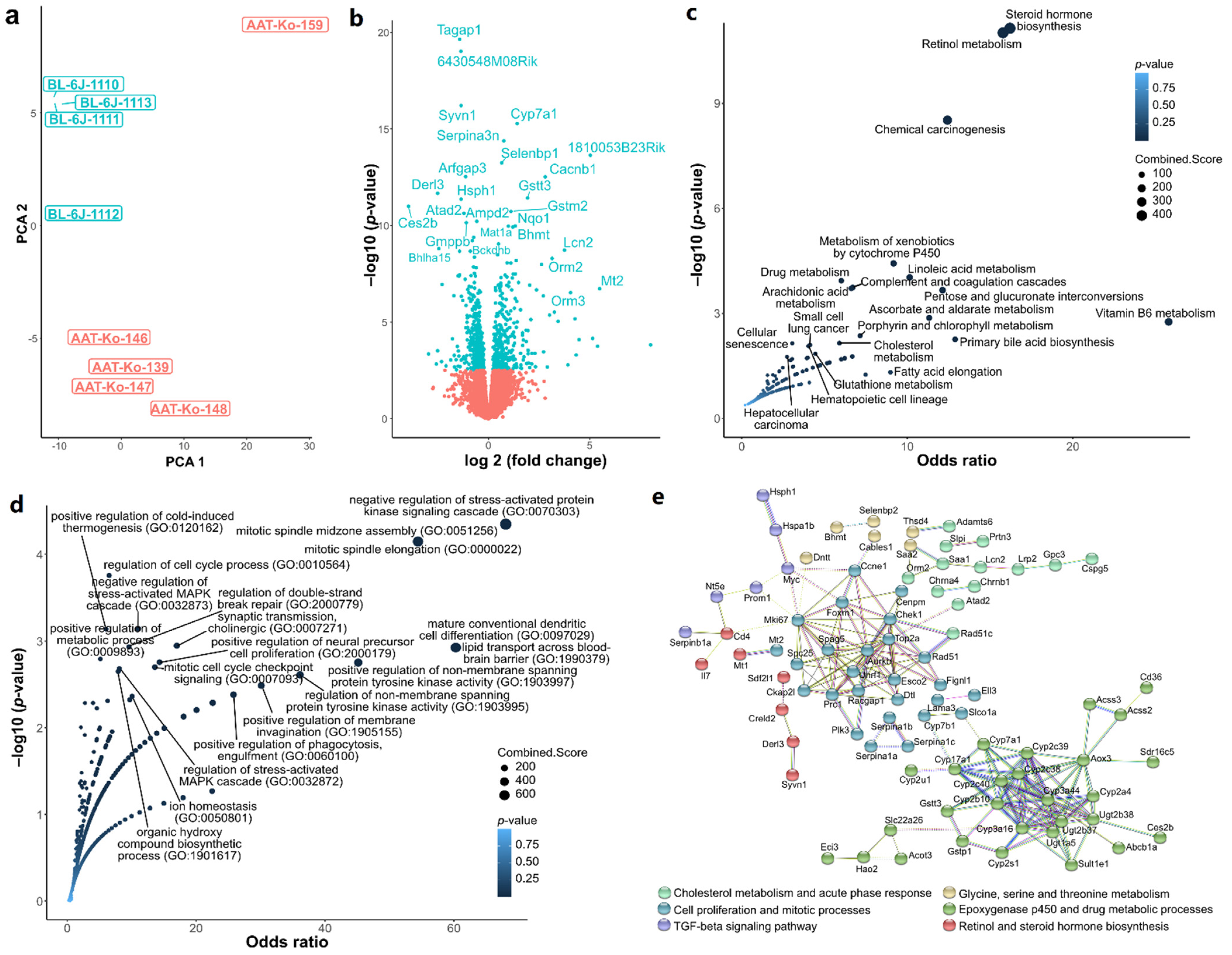
2.1. Transcriptome Quality Control and Differential Gene Expression

The initial 10 samples (five Serpina1 KO and five WT) were clustered using PCA to identify sample similarities and differences (Figure S1). The clustering showed an outlier (WT BL-6J-1109), which was removed to reduce a potentially detrimental source of variance amongst the differentially expressed genes (DEGs) (Figure 1a). Using DESeq2, 801 DEGs were found between the Serpina1 KO and WT mice livers. Among the latter, 438 were upregulated and 363 downregulated in the KO relative to WT mice (Figure 1b). Top downregulated genes in KO livers apart from Serpina1 included (by Log2Fc values) the following genes: Ces2b (carboxylic ester hydrolase 2B), Ugt2b38 (UDP glucuronosyltransferase two family, polypeptide B38), Mup2 (major urinary protein 2), Bglap3 (bone gamma-carboxyglutamate protein 3), and Derl3 (derlin-3). Among the top upregulated genes were Sult1e1 (sulfotransferase family 1E, member 1), Slc22a26 (Solute carrier family 22, member 26), Rgs16 (Regulator of G-protein signaling 16), Slc22a27 (solute carrier family 22, member 27), Mt1 (Metallothionein-1), and Orm3 (Alpha-1-acid glycoprotein 3). The full list of the DEGs is presented in Supplementary Table S1.

2.2. Hypothesis-Free Pathway Screening

The DEGs were further analyzed using EnrichR [17] to identify the gene terms (gene ontology, GO) [18] and pathways (KEGG) [19]. We observed changes associated with steroid hormone biosynthesis, retinol metabolism, and several other metabolic pathways associated with the KO phenotype in KEGG enrichment (Figure 1b). The GO analysis revealed that cholesterol metabolism, cytokine responses, and stress responses were associated with KO phenotype (Figure 1c).
To mine potential interactions based on protein-protein networks, we queried stringDB [20] using the DEGs. K-means clustering revealed six clusters (Figure 1d) linked with loss of Serpina1. The main interactions within the clusters involved the epoxygenase p450 (cytochrome p450 related genes) (Figure 1b,d), cell cycle-associated genes (Figure 1c,d), cholesterol metabolism, and acute-phase response pathways (Figure 1b,d).

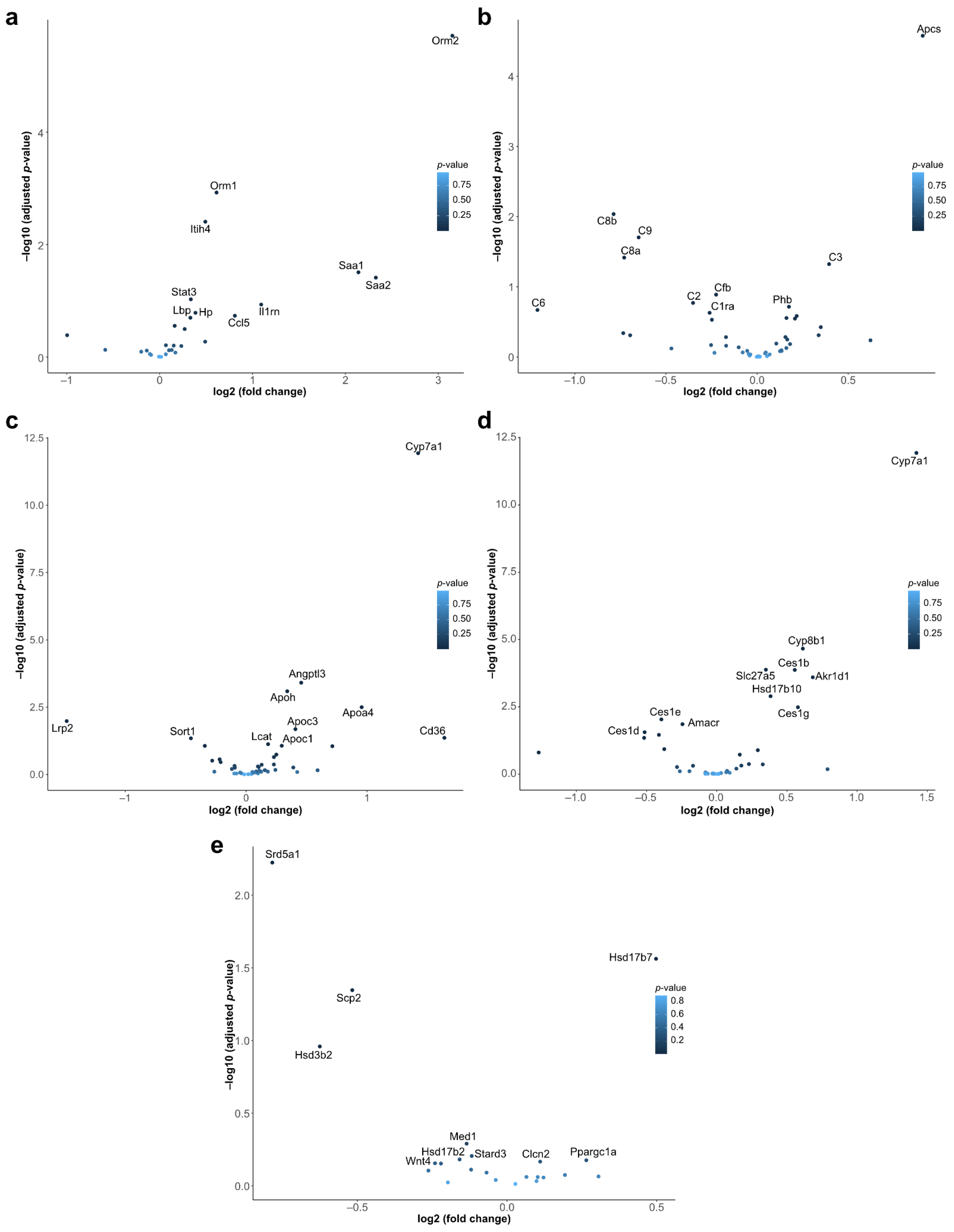
2.3. Hypothesis-Driven Pathway Analysis

In parallel to the hypothesis-free pathway analysis reported above, we conducted a targeted analysis of specific gene sets of interest. The former approach, although objective, suffered from the burden of multiple hypothesis corrections from hundreds of irrelevant gene sets. Based on prior literature and knowledge of SERPINA1, there were specific bioprocesses that were the object of our inquiry here. Human SERPINA1 plays an essential role in the modulation of acute-phase responses, inflammatory processes, and the lower respiratory tract protection from proteolytic damage. To examine these processes, we defined gene sets for these pathways of interest and calculated the overrepresentation of differentially expressed genes.
The p-values for the over-representation were as follows: (a) acute-phase response 0.0247, (b) complement pathway 0.1881, (c) cholesterol metabolism 0.0028, (d) bile acid metabolism 3.194e-07, and (e) steroid hormone biosynthesis 0.1421 (data not represented in plots).
We observed that genes associated with an acute-phase response, such as Orm2 (Alpha-1-acid glycoprotein 2), Saa1 (Serum amyloid A1), and Saa2 (Serum amyloid A2), were upregulated (Figure 2a). This was likely due to pro-inflammatory mice phenotype because of the loss of Serpina1 gene. Since the pro-inflammatory state was generally reflected by upregulated interleukins, we investigated the expression of interleukins. The transcriptional levels of Il6, Il7, Il15, and Il18 were only slightly upregulated (Figure S2), with no significant changes in transcription factors controlling the acute phase proteins. We also analyzed the complement pathway gene set (Figure 2b). Although, in general, this pathway remained not altered significantly in Serpina1 KO mice, specific genes were affected. For example, Apcs and C3 were slightly upregulated, whereas C4a and C8a were downregulated (Figure 2b). Hence, the parallel upregulation of pro-inflammatory interleukins and acute-phase proteins, and alterations in some of the complement pathway genes suggested a pro-inflammatory phenotype in KO mice.
As illustrated in Figure 2c, the expression of several cytochrome P450 proteins was disturbed in the KO vs. WT mice livers. Specifically, KO mice livers upregulated the Cyp7a1 (Cytochrome P450 Family 7 Subfamily A Member 1) gene that has an important role in cholesterol and bile acid metabolism and is associated with hypercholesterolemia. Several genes from bile acid metabolic processes were concomitantly upregulated in the livers of the KO vs. WT mice (Figure 2d). Among these genes were Cyp8b1 (sterol 12-alpha-hydrolase), Ces1g (carboxylesterase 1G, the major liver enzyme for the hydrolysis or transesterification of various xenobiotics), and Akr1d1 (aldo-keto reductase family 1 member D1, crucial for bile acid biosynthesis and important for steroid metabolism). Likewise, several genes of steroid hormone biosynthesis, like, Hsd17b7 (hydroxysteroid (17-beta) dehydrogenase 7), Scp1 (Synaptonemal complex protein 1), and Hsd3b2 (Hydroxy-Delta-5-Steroid Dehydrogenase, 3 Beta-Steroid Delta Isomerase 2) were significantly higher expressed in the livers of the Serpina1 KO vs. WT mice (Figure 2e). Genes related to linoleic acid and retinol metabolism (Figure S3) and vitamin D signaling were also altered in Serpina1 KO mice. Regarding glycolysis, most genes of canonical and anaerobic glycolysis were largely unaffected, with the exception of Pfkl and Pfkp gene members of the phosphofructokinase A family (Figures S4 and S5). It is noteworthy that both the hypothesis-driven and EnrichR results showed a predisposition towards altered metabolic processes in the KO mice (Figure 1b).

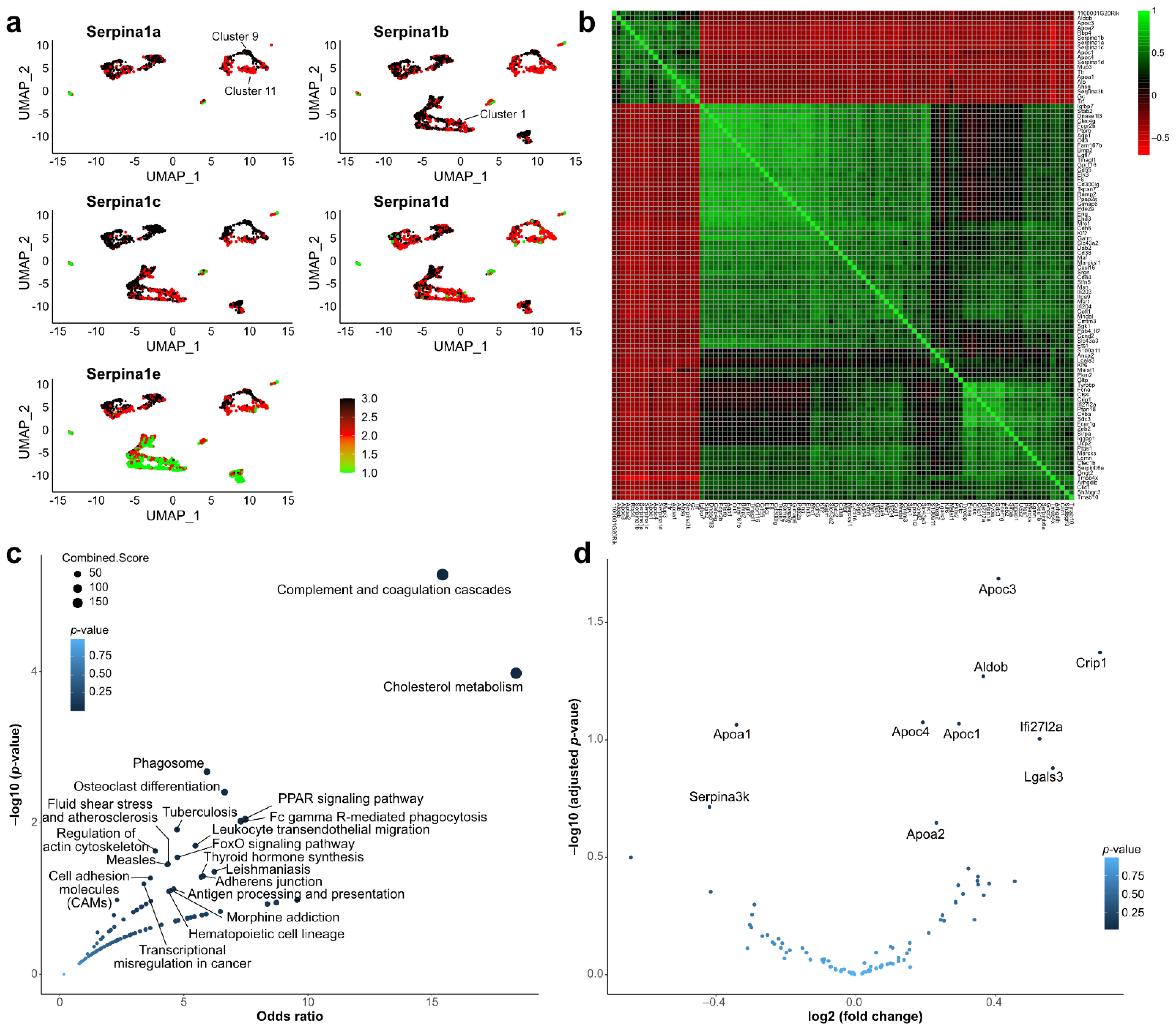
2.4. Single-Cell Sequencing Data Analysis

To complement and potentially validate our results, we performed a co-expression analysis of data from tabula muris, a compendium of single-cell transcriptome data from Mus musculus [21]. Among the three liver samples, we observed a global expression of four isoforms of Serpina1 (Figure 3a). We identified the top 100 correlated genes from this single-cell dataset and clustered them (Figure 3b). As expected, Serpina1a correlated with each and with other known Serpina1-related genes like Alb (Figure 3b). To summarize this set of Serpina1-associated genes, we performed an EnrichR analysis. We observed that complement and cholesterol pathways were enriched with Serpina1-associated genes (Figure 3c).
We further examined whether these Serpina1-associated genes were also differentially expressed in the KO experiment (Figure 3d). For example, Apoc3 was positively correlated in the WT mice and was downregulated in the KO mice. However, it must be noted that the fold-change values of most of these genes were relatively low. Overall, the analysis of the Serpina1 gene in single cells, along with pathways of the correlated genes, confirmed its roles in cholesterol metabolism and provided further indication of a role in the complement system.

3. Discussion

Our study examined the loss of the Serpina1 gene and its role in cellular pathways in the liver, the primary organ that produces the bulk of AAT. We found 801 DEGs, of which 55% were upregulated and 45% were downregulated in the KO relative to WT mice livers. By focusing on the transcript level, we characterized the consequences of loss of Serpina1 and which specific cellular processes were disrupted. Our data revealed changes in a broad range of cellular processes, including inflammation and acute phase response, metabolic processes, cellular differentiation, and molecular transport across membranes, to name a few.
Inflammatory processes are crucial to the survival of an organism through their roles in harmful stimuli, like pathogens, damaged cell response, and wound healing. Loss of Serpina1 in mice altered key inflammatory transcriptional signatures. Our results indicated a pro-inflammatory phenotype, likely via the alteration of acute-phase response and complement pathway mechanisms, among others. We showed that the expression of genes that modulate these responses, such as Il6, Il7, Apcs, C3, C4a, and C8a, were upregulated in KO mice. In addition to these transcripts, we showed a significant upregulation of Saa1 and Saa2, which are key transcripts of the acute-phase response and are implicated in the interaction between various cell types such as macrophages, hepatocytes, and myeloid-derived suppressor cells [22,23]. This complex interaction likely facilitates the binding of SAA1/2 to receptors, such as SR-B1, that participate in the cholesterol efflux [24,25]. Similarly, we show an upregulation of Orm2 coding for a key protein that has varied functions like transporter or modulator of the activity of the immune system depending on its glycosylation. Through the interaction with the farnesoid X receptor, orosomucoids also play a role in bile acid homeostasis [26].
A crucial physiological role of the liver is the metabolic control of biomolecules such as cholesterol and bile acid and its function as a biosynthesis hub for steroids and complement factors. Our analysis of KEGG enrichment showed that in Serpina1 KO mice, these pathways were enriched, indicating that alteration of Serpina1 expression can lead to changes in homeostatic conditions required for healthy liver function. Transcripts, such as St1e1, Cyp7a, Cyp8b1, Slc2a1, and Derl3, were shown in our data to be differentially expressed. St1e1, for example, coded for estrogen sulfotransferase, a key enzyme in estrogen homeostasis [27]. Gene ontology annotations related this gene to sulfotransferase and flavanol 3-sulfotransferase activities. Several of the cytochrome p450 enzymes played roles in key enzymatic and catalytic reactions, e.g., Cyp7a1 metabolized cholesterol to 7-alpha-hydroxycholesterol, the first step in bile acid synthesis. Dysregulation of bile acid homeostasis caused a wide spectrum of diseases usually associated with hepatic injury [28]. The transport of biomolecules is also a crucial step in which the liver participates. The DEG Derl3 is involved in the negative regulation of retrograde transport and has a regulatory role in controlling the regulation of Slc2a1 [29]. Similarly, the retinol metabolism and its associated genes, such as retinol dehydrogenases Rdh16, Rdh11, and short-chain dehydrogenase/reductase family 16C member 5 (Sdr16c5), are differentially expressed in the KO mice (Figure S2). Since retinol dehydrogenases control key enzymatic steps involved in retinol metabolism and other steroid metabolic processes [30], it seems likely that perturbation of Serpina1 expression in the liver alters immune regulatory and metabolic pathways which are involved in the homeostasis of the organism.
The analysis of Serpina1-correlated genes in the tabula muris scSEQ dataset provided an independent method to identify genes that could be co-regulated and functionally linked to Serpina1. KEGG enrichment analysis of these genes supports the hypothesis that Serpina1 expression affects immune responses and metabolism-associated pathways and primarily provides independent validation of the implicated role of Serpina1 in cholesterol metabolism. The single-cell dataset showed that the various Serpina genes in mice were generally co-expressed. Cluster 9 (Figure 3a) was one of the clusters expressing the highest levels of Serpina genes and was also associated with Mup3, Cyp2f2, and Scd1. Conversely, clusters tending for lower expression of Serpina genes, such as 1 and 11, showed higher levels of Cytochrome P450 genes. This may imply that liver cells specializing in xenobiotic metabolism express less Serpina, which is consistent with the pathway enrichment findings from the RNA-seq data from the knockout mice. Ultimately, further scRNA-seq studies specifically involving the Serpina knock-out mice would go much further to elucidate the role of these genes in the liver.
In conclusion, circulating AAT is not a single-function protein as an anti-protease but expresses broader anti-inflammatory effects. SERPINA1 regulation affects the modulation of various inflammation-related substances like chemokines, oxygen species, complement factors, lipoproteins, and others. A recent small cohort phase 2 trial using fazrisiran, an RNA interference against SERPINA1 “Z” mutation that caused degradation of AAT messenger RNA and abolished protein synthesis in hepatocytes, improved liver enzymes, and reduced fibrosis [31]. Our data suggested that knockdown therapies might be prone to side effects. Human studies should be carefully monitored to observe any such adverse long-term effects that could be due to alterations of key genes and pathways discussed above.
At first glance, Serpina1a to e knockout represents a similar situation as in AAT-Null patients. However, each of the mouse Serpina1 paralogues displayed specific functions. Amino acid polymorphisms in the reactive center loop determined AAT’s substrate specificity: Serpina1a and Serpina1b products AAT 1 and AAT 2 displayed anti-elastase activity, while Serpina1c and Serpina1d products AAT 3 and AAT 4 had anti-chymotryptic activity. The product of Serpina1e, AAT 5, lacks inhibitory activity against trypsin, chymotrypsin, and pancreatic as well as against neutrophilic elastase [32], suggesting that the results from the mouse AAT KO model may not reflect the situation of AAT-Null patients. Ultimately, the differences between the Serpina gene system in mice and humans create a limitation of these model-based studies. Further CRISPR-knock out or gene silencing of SERPINA1 in human-derived organoid models would be warranted.

4. Materials and Methods

4.1. Animal Model

WT mice (C57BL/6) were purchased from Janvier Labs (Janvier Labs, Le Genest-Saint-Isle, France). Serpina1-KO mice lacking all 5 serpin A1 a-e paralogues expressed in mice were used as previously described by [10]. As described earlier, Serpina1-KO mice have normal behavior, lifespan, and gender distribution. Only at the advanced age of more than 35 weeks do they spontaneously develop lung emphysema [11]. Livers from 10 male age-matched healthy mice (KO = 5 and WT = 5) aged from 10 to 12 weeks were isolated and frozen in nitrogen.

4.2. Total RNA Isolation from the Mice Livers

The isolation of the total RNA was performed using RNeasy Minikit (Qiagen, Hilden, Germany) as described in [33]. Livers were shortly collected in an RLT buffer supplemented with 1% mercaptoethanol. After an initial homogenization step using T10 basic ultra-turrax (Ika, Staufen im Breisgau, Germany), RNA isolation was continued with RNeasy Minikit according to the instructions of the manufacturer.

4.3. Transcriptome Analysis (RNA-seq)

Agilent 2100 Bioanalyzer based on an Agilent RNA 6000 Nano Kit was used to assess the quality of total RNA. RNASeq libraries were prepared by using 200 ng RNA of each sample. TruSeq Stranded mRNA (Illumina, San Diego, CA, USA) was used for library preparation according to the manufacturer’s protocol. After reverse transcription and adapter ligation, the cDNA products were purified with Ampure XP beads (Beckman Coulter, Brea, CA, USA), and enriched with 15 cycles of PCR to create the cDNA library. Sequencing was performed on a NovaSeq 6000 sequencer using 200 cycles in paired-end mode. RNA-Seq data was quality control analyzed with fastQC v0.11.8 and low-quality 30 ends were removed using Trimmomatic v0.38.

4.4. Data Preprocessing

The fastq files generated through high-throughput sequencing (HTS) were subjected to fastqc to assess the overall read quality. Lower quality reads were removed using trimmomatic. The processed files were aligned with STAR with hg19 as the reference, the output bam files were then subjected to HT-Seq to count the number of transcripts per gene.

4.5. Gene Expression Analysis

The principal component analysis (PCA) was performed to characterize the variation among the samples and to assess putative outliers. Differential expression analysis was performed using DEseq2 [34]. Expression values were normalized with rlog using only genes with average values greater than 3.4.

4.6. Gene Ontology and Pathway Analysis

For performing GO term analysis, we used genes meeting the cut-off criteria (log2FC +/− 1.2) post-normalization and log2FC calculations in the complete dataset. In total 221 genes were analyzed using EnrichR to obtain the GO terms (GO biological process 2021) and KEGG pathways (KEGG 2019 mouse). For mining the known interactions among differentially expressed genes, we used the STRING database with a full STRING network based on evidence through all available resources with a high confidence setting. The network was clustered into six clusters using k-means clustering algorithm.

4.7. Single-Cell Sequencing (scSEQ) Data Analysis

We utilized the publicly available tabula muris dataset for liver (10 × sequencing) [21]. The selected data consisted of three liver samples subjected to single-cell sequencing using the 10× platform. The data we processed with a Seurat and used a SCTransform to normalize variances in the scSEQ data [35,36]. The top 100 Serpina1-correlated genes were determined using Pearson’s r. This set of genes was then used to obtain KEGG pathways and volcano plots for differential expression.

Supplementary Materials

The following supporting information can be downloaded at: https://www.mdpi.com/article/10.3390/ijms231810425/s1.

Author Contributions

S.J., D.S.D. and S.H.M. conceptualized this project; B.M.-D., U.A.M., L.O., G.G.-M. and S.P.-L. contributed to the methodology; D.S.D. and S.H.M. performed the computational analysis; S.T. and S.W. contributed to the formal analysis and investigation. Resources were provided by S.J., B.M.-D., U.A.M. and T.W. The original draft was prepared by D.S.D., S.H.M. and S.J., D.S.D., S.H.M., S.W., S.J., T.W. and U.A.M. reviewed the manuscript. The project was supervised by D.S.D. and S.J. All authors have read and agreed to the published version of the manuscript.

Funding

RNA sequencing was supported by the grant ISCIII -AESI PI20CIII/00015.

Institutional Review Board Statement

As per the §4 animal welfare law in Germany, these animals were organ donors, and there is currently no requirement for laboratory animal organ donors to be approved by the Lower Saxony State Office for Consumer Protection and Food Safety (LAVES). Any further institutional reviews were not required.

Informed Consent Statement

Not applicable.

Data Availability Statement

Merged counts matrix for the study is available at 10.5281/zenodo.6974384 under restricted access.

Conflicts of Interest

The authors declare no conflict of interest.

References

  1. Hua, B.; Fan, L.; Liang, Y.; Zhao, Y.; Tuddenham, E.G. Alpha1-antitrypsin Pittsburgh in a family with bleeding tendency. Haematologica 2009, 94, 881–884. [Google Scholar] [CrossRef] [PubMed]
  2. Sehgal, A.; Bumcrot, D.; Bettencourt, B. Serpina1 Sirnas: Compositions of Matter and Methods of Treatment. U.S. Patent 10935917B2, 27 December 2012. [Google Scholar]
  3. Billingsley, G.D.; Walter, M.A.; Hammond, G.L.; Cox, D.W. Physical mapping of four serpin genes: Alpha 1-antitrypsin, alpha 1-antichymotrypsin, corticosteroid-binding globulin, and protein C inhibitor, within a 280-kb region on chromosome I4q32.1. Am. J. Hum. Genet. 1993, 52, 343–353. [Google Scholar] [PubMed]
  4. Perlino, E.; Cortese, R.; Ciliberto, G. The human alpha 1-antitrypsin gene is transcribed from two different promoters in macrophages and hepatocytes. EMBO J. 1987, 6, 2767–2771. [Google Scholar] [CrossRef] [PubMed]
  5. Kalsheker, N.; Morley, S.; Morgan, K. Gene regulation of the serine proteinase inhibitors alpha1-antitrypsin and alpha1-antichymotrypsin. Biochem. Soc. Trans. 2002, 30, 93–98. [Google Scholar] [CrossRef]
  6. Janciauskiene, S.; Wrenger, S.; Welte, T. Immunoregulatory Properties of Acute Phase Proteins—Specific Focus on α1-Antitrypsin. In Acute Phase Proteins; Janciauskiene, S., Ed.; Intechopen: London, UK, 2013; p. 190. [Google Scholar]
  7. Rotondo, J.C.; Aquila, G.; Oton-Gonzalez, L.; Selvatici, R.; Rizzo, P.; De Mattei, M.; Pavasini, R.; Tognon, M.; Campo, G.C.; Martini, F. Methylation of SERPINA1 gene promoter may predict chronic obstructive pulmonary disease in patients affected by acute coronary syndrome. Clin. Epigenet. 2021, 13, 79. [Google Scholar] [CrossRef]
  8. Rotondo, J.C.; Oton-Gonzalez, L.; Selvatici, R.; Rizzo, P.; Pavasini, R.; Campo, G.C.; Lanzillotti, C.; Mazziotta, C.; De Mattei, M.; Tognon, M.; et al. SERPINA1 Gene Promoter Is Differentially Methylated in Peripheral Blood Mononuclear Cells of Pregnant Women. Front. Cell Dev. Biol. 2020, 8, 550543. [Google Scholar] [CrossRef]
  9. Barbour, K.W.; Wei, F.; Brannan, C.; Flotte, T.R.; Baumann, H.; Berger, F.G. The murine alpha(1)-proteinase inhibitor gene family: Polymorphism, chromosomal location, and structure. Genomics 2002, 80, 515–522. [Google Scholar] [CrossRef]
  10. Ostermann, L.; Maus, R.; Stolper, J.; Schutte, L.; Katsarou, K.; Tumpara, S.; Pich, A.; Mueller, C.; Janciauskiene, S.; Welte, T.; et al. Alpha-1 antitrypsin deficiency impairs lung antibacterial immunity in mice. JCI Insight 2021, 6, e140816. [Google Scholar] [CrossRef]
  11. Borel, F.; Sun, H.; Zieger, M.; Cox, A.; Cardozo, B.; Li, W.; Oliveira, G.; Davis, A.; Gruntman, A.; Flotte, T.R.; et al. Editing out five Serpina1 paralogs to create a mouse model of genetic emphysema. Proc. Natl. Acad. Sci. USA 2018, 115, 2788–2793. [Google Scholar] [CrossRef]
  12. Yuan, Y.; DiCiaccio, B.; Li, Y.; Elshikha, A.S.; Titov, D.; Brenner, B.; Seifer, L.; Pan, H.; Karic, N.; Akbar, M.A.; et al. Anti-inflammaging effects of human alpha-1 antitrypsin. Aging Cell 2018, 17, e12694. [Google Scholar] [CrossRef] [Green Version]
  13. Lopes-Paciencia, S.; Saint-Germain, E.; Rowell, M.C.; Ruiz, A.F.; Kalegari, P.; Ferbeyre, G. The senescence-associated secretory phenotype and its regulation. Cytokine 2019, 117, 15–22. [Google Scholar] [CrossRef] [PubMed]
  14. Pott, G.B.; Chan, E.D.; Dinarello, C.A.; Shapiro, L. Alpha-1-antitrypsin is an endogenous inhibitor of proinflammatory cytokine production in whole blood. J. Leukoc. Biol. 2009, 85, 886–895. [Google Scholar] [CrossRef]
  15. Gottlieb, P.A.; Alkanani, A.K.; Michels, A.W.; Lewis, E.C.; Shapiro, L.; Dinarello, C.A.; Zipris, D. Alpha1-Antitrypsin therapy downregulates toll-like receptor-induced IL-1beta responses in monocytes and myeloid dendritic cells and may improve islet function in recently diagnosed patients with type 1 diabetes. J. Clin. Endocrinol. Metab. 2014, 99, E1418–E1426. [Google Scholar] [CrossRef]
  16. Shapiro, L.; Pott, G.B.; Ralston, A.H. Alpha-1-antitrypsin inhibits human immunodeficiency virus type 1. FASEB J. 2001, 15, 115–122. [Google Scholar] [CrossRef] [PubMed]
  17. Chen, E.Y.; Tan, C.M.; Kou, Y.; Duan, Q.; Wang, Z.; Meirelles, G.V.; Clark, N.R.; Ma’ayan, A. Enrichr: Interactive and collaborative HTML5 gene list enrichment analysis tool. BMC Bioinform. 2013, 14, 128. [Google Scholar] [CrossRef] [PubMed]
  18. Ashburner, M.; Ball, C.A.; Blake, J.A.; Botstein, D.; Butler, H.; Cherry, J.M.; Davis, A.P.; Dolinski, K.; Dwight, S.S.; Eppig, J.T.; et al. Gene ontology: Tool for the unification of biology. The Gene Ontology Consortium. Nat. Genet. 2000, 25, 25–29. [Google Scholar] [CrossRef] [PubMed]
  19. Kanehisa, M.; Furumichi, M.; Tanabe, M.; Sato, Y.; Morishima, K. KEGG: New perspectives on genomes, pathways, diseases and drugs. Nucleic Acids Res. 2017, 45, D353–D361. [Google Scholar] [CrossRef] [PubMed]
  20. Szklarczyk, D.; Gable, A.L.; Nastou, K.C.; Lyon, D.; Kirsch, R.; Pyysalo, S.; Doncheva, N.T.; Legeay, M.; Fang, T.; Bork, P.; et al. The STRING database in 2021: Customizable protein-protein networks, and functional characterization of user-uploaded gene/measurement sets. Nucleic Acids Res. 2021, 49, D605–D612. [Google Scholar] [CrossRef]
  21. Tabula Muris, C. A single-cell transcriptomic atlas characterizes ageing tissues in the mouse. Nature 2020, 583, 590–595. [Google Scholar] [CrossRef]
  22. Meek, R.L.; Benditt, E.P. Amyloid A gene family expression in different mouse tissues. J. Exp. Med. 1986, 164, 2006–2017. [Google Scholar] [CrossRef] [Green Version]
  23. Sack, G.H., Jr. Serum amyloid A—A review. Mol. Med. 2018, 24, 46. [Google Scholar] [CrossRef] [PubMed]
  24. Thomas, M.J.; Sorci-Thomas, M.G. SAA: A link between cholesterol efflux capacity and inflammation? J. Lipid Res. 2015, 56, 1383–1385. [Google Scholar] [CrossRef] [PubMed]
  25. Cai, L.; de Beer, M.C.; de Beer, F.C.; van der Westhuyzen, D.R. Serum amyloid A is a ligand for scavenger receptor class B type I and inhibits high density lipoprotein binding and selective lipid uptake. J. Biol. Chem. 2005, 280, 2954–2961. [Google Scholar] [CrossRef]
  26. Porez, G.; Gross, B.; Prawitt, J.; Gheeraert, C.; Berrabah, W.; Alexandre, J.; Staels, B.; Lefebvre, P. The hepatic orosomucoid/alpha1-acid glycoprotein gene cluster is regulated by the nuclear bile acid receptor FXR. Endocrinology 2013, 154, 3690–3701. [Google Scholar] [CrossRef]
  27. Cole, G.B.; Keum, G.; Liu, J.; Small, G.W.; Satyamurthy, N.; Kepe, V.; Barrio, J.R. Specific estrogen sulfotransferase (SULT1E1) substrates and molecular imaging probe candidates. Proc. Natl. Acad. Sci. USA 2010, 107, 6222–6227. [Google Scholar] [CrossRef] [PubMed]
  28. Chiang, J.Y.L.; Ferrell, J.M. Up to date on cholesterol 7 alpha-hydroxylase (CYP7A1) in bile acid synthesis. Liver Res. 2020, 4, 47–63. [Google Scholar] [CrossRef]
  29. Lopez-Serra, P.; Marcilla, M.; Villanueva, A.; Ramos-Fernandez, A.; Palau, A.; Leal, L.; Wahi, J.E.; Setien-Baranda, F.; Szczesna, K.; Moutinho, C.; et al. A DERL3-associated defect in the degradation of SLC2A1 mediates the Warburg effect. Nat. Commun. 2014, 5, 3608. [Google Scholar] [CrossRef]
  30. Kedishvili, N.Y. Enzymology of retinoic acid biosynthesis and degradation. J. Lipid Res. 2013, 54, 1744–1760. [Google Scholar] [CrossRef] [PubMed]
  31. Strnad, P.; Mandorfer, M.; Choudhury, G.; Griffiths, W.; Trautwein, C.; Loomba, R.; Schluep, T.; Chang, T.; Yi, M.; Given, B.D.; et al. Fazirsiran for Liver Disease Associated with Alpha1-Antitrypsin Deficiency. N. Engl. J. Med. 2022, 387, 514–524. [Google Scholar] [CrossRef]
  32. Paterson, T.; Moore, S. The expression and characterization of five recombinant murine alpha 1-protease inhibitor proteins. Biochem. Biophys. Res. Commun. 1996, 219, 64–69. [Google Scholar] [CrossRef]
  33. Frenzel, E.; Wrenger, S.; Immenschuh, S.; Koczulla, R.; Mahadeva, R.; Deeg, H.J.; Dinarello, C.A.; Welte, T.; Marcondes, A.M.; Janciauskiene, S. Acute-phase protein alpha1-antitrypsin-a novel regulator of angiopoietin-like protein 4 transcription and secretion. J. Immunol. 2014, 192, 5354–5362. [Google Scholar] [CrossRef] [PubMed]
  34. Love, M.I.; Huber, W.; Anders, S. Moderated estimation of fold change and dispersion for RNA-seq data with DESeq2. Genome Biol. 2014, 15, 550. [Google Scholar] [CrossRef] [PubMed]
  35. Hafemeister, C.; Satija, R. Normalization and variance stabilization of single-cell RNA-seq data using regularized negative binomial regression. Genome Biol. 2019, 20, 296. [Google Scholar] [CrossRef] [PubMed]
  36. Hao, Y.; Hao, S.; Andersen-Nissen, E.; Mauck, W.M., 3rd; Zheng, S.; Butler, A.; Lee, M.J.; Wilk, A.J.; Darby, C.; Zager, M.; et al. Integrated analysis of multimodal single-cell data. Cell 2021, 184, 3573–3587. [Google Scholar] [CrossRef] [PubMed]
Figure 1. Differential gene expression (DEGs) and pathway analysis of Serpina1 KO vs. WT. (a) Principal component analysis of samples selected for RNA-Seq data analysis and DEG exploration with WT labeled blue and KO labeled red. (b) Volcano plot of DEGs where x-axis represents the log2 fold _change and y-axis represents the significance level wherein p-values have been −log10 transformed. Significantly expressed genes are labeled blue (p-value ≤ 0.05), and non-significant genes are labeled in red. (c) A plot showing the KEGG pathways that were identified from DEGs between KO vs. WT mice using EnrichR. X-axis: odds ratio, Y-axis negative Log10 p-value. (d) Gene ontology analysis using EnrichR: x-axis: odds ratio, y-axis negative Log10 p-value. (e) Protein-protein interaction networks as generated with String-Db: Differentially expressed genes were subjected to k-means clustering (n = 6) with high confidence interaction settings.
Ijms 23 10425 g001
Figure 2. Differential expression of gene sets in Serpina1 KO vs. WT mice. Volcano plots depict the fold-change (x-axis) and significance (y-axis) for genes associated with: (a) acute phase response (n = 38); (b) complement pathway (n = 187); (c) cholesterol metabolism (n = 47); (d) bile acid metabolic processes (n = 44); and (e) steroid hormone biosynthesis (n = 30). The ten most highly significant DEGs in each pathway are annotated.
Ijms 23 10425 g002
Figure 3. scSEQ data analysis of Serpina1 gene expression. (a): Global expression of Serpina1 isoforms: Serpina1 was broadly expressed in the wild-type mice, with two among thirteen clusters that showed a lower expression. (b): Correlation matrix of Serpina1: Top 100 genes with Serpina1 highly correlated are shown in this plot with highly correlated genes in green and anti-correlated genes in red. (c): Pathway enrichment of correlated genes. (d): Expression changes in the KO dataset of the correlated genes (n = 100). The ten most highly significant DEGs are annotated.
Ijms 23 10425 g003
Publisher’s Note: MDPI stays neutral with regard to jurisdictional claims in published maps and institutional affiliations.

