Review

Drosophila as a Model Organism to Study Basic Mechanisms of Longevity

by
Anna A. Ogienko
1,
Evgeniya S. Omelina
1,2,
Oleg V. Bylino
3,
Mikhail A. Batin
4,
Pavel G. Georgiev
3 and
Alexey V. Pindyurin
1,*
1
Department of Regulation of Genetic Processes, Institute of Molecular and Cellular Biology SB RAS, 630090 Novosibirsk, Russia
2
Laboratory of Biotechnology, Novosibirsk State Agrarian University, 630039 Novosibirsk, Russia
3
Laboratory of Gene Expression Regulation in Development, Institute of Gene Biology RAS, 119334 Moscow, Russia
4
Open Longevity, 15260 Ventura Blvd., Sherman Oaks, Los Angeles, CA 91403, USA
*
Author to whom correspondence should be addressed.
Int. J. Mol. Sci. 2022, 23(19), 11244; https://doi.org/10.3390/ijms231911244
Submission received: 15 August 2022 / Revised: 20 September 2022 / Accepted: 20 September 2022 / Published: 24 September 2022
(This article belongs to the Section Molecular Genetics and Genomics)

Abstract

:
The spatio-temporal regulation of gene expression determines the fate and function of various cells and tissues and, as a consequence, the correct development and functioning of complex organisms. Certain mechanisms of gene activity regulation provide adequate cell responses to changes in environmental factors. Aside from gene expression disorders that lead to various pathologies, alterations of expression of particular genes were shown to significantly decrease or increase the lifespan in a wide range of organisms from yeast to human. Drosophila fruit fly is an ideal model system to explore mechanisms of longevity and aging due to low cost, easy handling and maintenance, large number of progeny per adult, short life cycle and lifespan, relatively low number of paralogous genes, high evolutionary conservation of epigenetic mechanisms and signalling pathways, and availability of a wide range of tools to modulate gene expression in vivo. Here, we focus on the organization of the evolutionarily conserved signaling pathways whose components significantly influence the aging process and on the interconnections of these pathways with gene expression regulation.

1. Introduction

Aging is a fundamental process that is characteristic to some extent for all multicellular organisms and is most studied and understood in the evolutionary line of animals. In mammals, it is characterized by the functional loss of cells, tissues, and organs and is accompanied by the increasing susceptibility to many diseases, including cancer, neurodegeneration, cardiovascular diseases, and metabolic disorders of an organism [1]. One of the most striking manifestations of the aging of process, both in humans and Drosophila, is that gradual deregulation and a violation in the functioning of signal transduction pathways can lead to diseases and death of organisms. Changes in the functioning of signaling pathways are known to result in reduction or lengthening of lifespan. For example, genetic manipulations with the insulin/insulin-like growth factor 1 (IGF-1) signaling (IIS) pathway and target of rapamycin (TOR) signaling pathway that receive signals from growth factors and nutrients (nutrient sensing), with the c-Jun NH2-terminal kinase (JNK) pathway (nutrient sensing and stress resistance), as well as with nuclear factor kappa B (NF-κB) pathway (inflammation and immune response) are associated with an increase in lifespan both in Drosophila and mammals [2,3,4]. Thus, signaling pathways can be efficient targets for interventions aimed at changing the lifespan of organisms.
Drosophila is one of the most studied model organisms, and in this review we describe in detail the organization of IIS, TOR, NF-κB, and JNK signaling pathways and lifespan control in Drosophila. We also draw parallels between Drosophila and humans. The analysis carried out in the review reveals general patterns in the functioning of four signaling pathways and allows us to draw conclusions about the conservation of lifespan control mechanisms between Drosophila and mammals. We also provide an overview of the mechanisms of epigenetic control of gene expression associated with the activity of these signaling pathways, where known.

2. Advantages of Drosophila as a Highly Informative Model Organism for Aging Studies

2.1. Advantages and Similarities of Drosophila and Mammals

Investigations using mammalian models (e.g., mice) require a large number of animals to be housed, maintained and subjected to invasive testing and therefore raise both economic and ethical concerns. Furthermore, studies on the aging of mammals are limited by their long lifespan. That is why non-vertebrate model organisms (e.g., yeast, worms, fruit flies) are popular among researchers in the aging field. Since the beginning of the last century, when the first gene in the history of genetics was discovered in Drosophila, fruit fly has been used as a model organism to identify conservative basic biological processes [5]. The first studies in the field of aging on Drosophila were carried out in the second decade of the last century, and since then this model animal has been extensively used for aging research [6,7]. High level of conservation of signaling transduction pathways between Drosophila and humans made Drosophila an indispensable model organism for identifying the pathways responsible for lifespan extension, as well as for understanding the complexity of the aging processes in eukaryotic organisms. Lastly, Drosophila and humans have similar classes of transcription factors [8] and, thus, share the mechanisms of gene expression regulation.

2.2. Features of the Biology of Drosophila as an Object for the Study of Aging

Drosophila has homologs to ~75% of human disease genes [9] and ~60% of all fly genes are conserved in humans [10,11]. Unlike mammals, handling and maintenance of fruit flies is easy and inexpensive over multiple generations, and experiments on flies are ethically acceptable. Flies are characterized by high fecundity (about 200 offspring per female), which allows for analysis of any feature of interest over hundreds of individuals to avoid statistical anomalies. Drosophila practically possesses one significant advantage over mammalian models in the aging field, that is short life cycle and lifespan of ~60–70 days for laboratory isogenic wild-type strains and ~120–130 days for non-isogenic flies from natural populations [12]. Drosophila overwinters in nature in the adult stage in vegetable warehouses, etc., wherever the temperature reaches 16 °C. At this temperature, the duration of lifespan of adult flies is significantly lengthened [13].
Health and longevity of fruit flies are affected by various factors such as diet, temperature, genetic and environmental interventions. Sexual differentiation has also significant effects on fly longevity. For instance, it was shown that male and female flies respond differently to dietary restriction (DR) [14]. Greater increase in lifespan was observed in females than in males in response to DR. Similarly, mating status also affects longevity of flies. Virgin females have the longest lifespan and mated females have the shortest lifespan while males are less affected [15]. Among possible mechanisms contributing to sex-specific lifespans may be genetic differences between the sexes, hormonal differences, and behavior [16]. Genetic background may also contribute to differences in lifespan between compared fly lines [17,18]. Therefore, studies of interventions aimed at understanding or targeting aging should be carried out using the closest genetic background between the compared fly lines as possible. Ideally, for lifespan tests, all fly lines should have the same genetic background. For that, mutations and transgenes should be backcrossed at a minimum of 10 generations to one or (preferably) two well-characterized, commonly available wild-type inbred strains (reviewed in [19]). Moreover, the compared fly lines should be free of the cytoplasmic endosymbiont, Wolbachia, since this infection can influence lifespan [20].

2.3. Statistical Analysis in Drosophila Aging Studies

Despite the fact that lifespan assays involve the simple counting of dead flies over time, it can be complicated by inconsistencies in approaches to estimating survival curves and other statistics. For instance, standard metrics in lifespan assay are the assessment of mean, median, or maximum lifespans within a Drosophila population. This is problematic, since variations in metrics between laboratories are quite common, leading to difficulties in comparison of lifespan effects. That is why comparison between survival curves of experimental and control flies and calculation of the significance level of differences between them (P) are performed using Kaplan–Meier method, log-rank test or Kolmogorov–Smirnov test [21].

2.4. Drosophila as a Model in the Study of Epigenetic Processes

Since the molecular mechanisms of gene regulation are rather evolutionarily conserved, Drosophila may be also a useful for studying the epigenetic mechanisms of gene expression control involved in development of aging and longevity (histone methylation and acetylation, microRNA expression, etc.). Epigenetic alterations in Drosophila are known to be controlled by the diet [22], temperature, illumination, and different forms of stress (heat shock, starvation). Therefore, Drosophila can be used as a model to study how exactly environmental stimuli affect epigenetic alterations. Understanding of how gradual deregulation of regulatory systems and imbalance of epigenetic mechanisms of gene regulation result in aging progression will contribute to a faster concretization of approaches aimed at combating aging. Thus, Drosophila turned out to be a convenient object for studying epigenetic mechanisms of longevity and aging.

2.5. Limitations in Using Drosophila as a Model in Aging Research

Drosophila, of cause, is one of the best analyzed/understood multicellular organism, more complex in comparison with C. elegans and yeast and is an ideal model system due to ease of genetic and environmental manipulations, completely sequenced genome, relatively low number of paralogous genes, the opportunity to investigate influence of the parameters such as gender ratio, resistance to pathogens, chemicals and environmental stress factors on longevity. In addition, Drosophila does not have programmed aging, as, for example, Arabidopsis, in which the death of an adult organism is associated with the maturation of seeds [23]. In Drosophila, aging apparently occurs as in humans: survival curves and mortality curves in these species look the same; moreover, the peak of reproduction in both species is shifted to early life periods [24]. However, several restrictions of using Drosophila exist as well. First, Drosophila goes through different developmental stages (embryo, larva, pupa, and adult) which mammals do not have [25]. Second, Drosophila differs substantially from mammals at the organismal level, brain anatomy, cardiovascular system, and respiration systems. Third, flies have a less complex and adaptive immune system than mammals and do not maintain their body temperature. This can lead to the fact that the effects of drugs/inhibitors/geroprotectors can vary greatly between Drosophila and mammals (the conversion of protoxins to toxins in the liver, as an example).
From a molecular point of view, despite the general good conservation of basic signaling pathways and cellular processes between mammals and Drosophila, low redundancy/reduced number of paralogous genes in Drosophila in comparison to mammals does not allow reproducing on this organism the regulatory complexity that is inherent in mammals, in which paralogization of regulatory genes is one of the main mechanisms for acquiring complexity [26,27,28]. In addition, poor conservation of some proteins/protein functions may be a problem as well.
Nevertheless, the comprehensibility (recognizability) of Drosophila as a model organism, as well as the rapid rate of generation turnover in this species, coupled with the conservation of the main regulatory and signaling pathways, makes Drosophila indispensable in aging studies. Below, we look in detail at the main signaling pathways in Drosophila known to be important for controlling aging.

3. Signaling Pathways Regulating Drosophila Lifespan

Below we briefly describe, from the Drosophila point of view and concentrating on the extracellular signals–gene expression regulation axis, evolutionary conserved signaling pathways, which dysfunctions result in altered fly lifespan. Thereby, direct effects on cellular protein synthesis, e.g., through modulation of activity of translation initiation factors that are not mediated by changes in expression level of genes encoding them, are not covered. More in-depth information on the signaling pathways, including their peculiarities in different species from yeast to humans as well as descriptions of their branches not directly related to gene expression regulation, is available in other reviews (see references below).

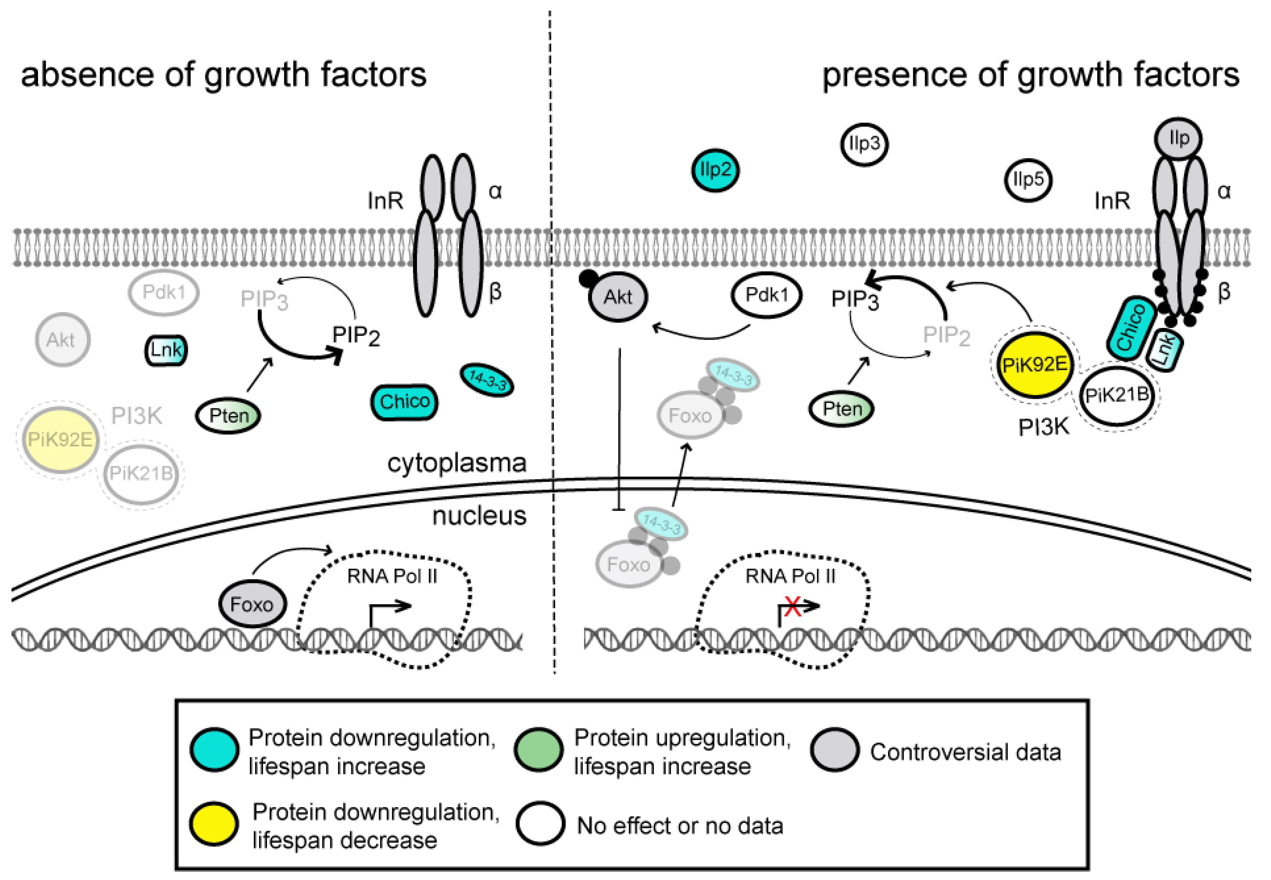
3.1. Insulin/IGF-1 Signaling (IIS) Pathway

The insulin/insulin-like growth factor 1 (IGF-1) signaling (IIS) pathway is involved in regulation of various physiological processes, including growth, development, metabolism, stress responses and reproduction (reviewed in [29,30,31,32]) (Figure 1). In total, hundreds of proteins are involved in the Drosophila insulin network [33]. The pathway is activated when one of the seven Drosophila insulin-like peptides (Ilp1–Ilp7, also known as DILP1–DILP7; information about human ortholog(s) of the fly proteins is provided in Supplementary Tables S1–S4) [34,35] binds to the insulin receptor (InR) [36,37,38] on the surface of the target cells.
This interaction stimulates tyrosine-protein kinase activity of InR [34,37,39] and results in a conformational change of the latter followed by its autophosphorylation and subsequent autoactivation [40]. Activated InR conveys signals further to Pi3K21B (Dp60) by its direct phosphorylation and through phosphorylation of the insulin receptor substrates Chico and Lnk that interact with Pi3K21B [41,42,43]. Regulatory Pi3K21B subunit together with catalytic Pi3K92E (Dp110) subunit form PI3K kinase that is responsible for conversion of phosphatidylinositol (3,4)-bisphosphate (PIP2) into phosphatidylinositol (3,4,5)-trisphosphate (PIP3) [44]. PIP3 serves as second messenger, activating protein kinase Pdk1, which in its turn phosphorylates protein kinase Akt (also known as PKB) at Thr342 and thus activates it [45,46,47,48]. Activated Akt phosphorylates transcription factor Foxo, which is responsible for upregulation of genes involved in longevity and stress resistance [49,50] (for details see below). As a result, phosphorylated Foxo becomes inactivated by nuclear exclusion with the help of 14-3-3ζ and 14-3-3ε chaperone proteins [51,52,53]. Regulation of Foxo activity by insulin/IGF pathway is evolutionarily conserved in humans [54,55], in which 14-3-3 protein promotes sequestering and inactivation of human FOXO3a after its phosphorylation by kinases such as Akt [56,57]. The IIS pathway is antagonized by phosphatase activities of Pten and PP2A, which dephosphorylate PIP3 and Akt, respectively [58,59,60,61,62,63] (Figure 1).
Some components of the IIS pathway are expressed at undetectable/very low to moderate levels with minimal variability across developmental stages and different tissues, for example, Ilp1, Ilp7, InR and Chico (Figure 2A and Supplementary Figure S1A). Other components generally expressed at very low to moderate levels are characterized by relatively high abundance at specific developmental stages and tissues. Among those are Ilp4, Ilp2 and Pi3K21B, which are actively expressed in early embryonic stages, larval/adult central nervous system (CNS) neuronal cells, and larval/adult digestive system, respectively. The 14-3-3 proteins are the only ones that are expressed at relatively stable extremely high levels, most probably due to their involvement in many other cellular processes apart from the IIS pathway. The IIS pathway can be functionally combined with the TOR pathway, so we summarize the conclusions from the heat map analysis drawn in Figure 2B and Supplementary Figure S1B in the TOR section (see below).
Downregulation of several pathway components that negatively regulate Foxo activity was shown to extend Drosophila lifespan (Supplementary Table S1). First, it is necessary to mention that thorough analysis of viable Ilp1-Ilp7 null mutants uncovered that most of the Ilps can act redundantly. Individual knockout of Ilp2, which has the highest homology to human insulin (IGF1, IGF2), results in a median adult lifespan extension of 9% and 13% for males and females, respectively [35]. Similarly, partial genetic ablation of the neurosecretory Ilp-producing cells that reside in the brain and are the primary producers of Ilp2, Ilp3 and Ilp5 in the adult body resulted in median lifespan increase by 10.5% in males and 18.5% and 33.5% in virgin and mated females, respectively [66]. Second, some InR, chico and 14-3-3ε mutations also extend lifespan [38,39,52,67,68]. Particularly, transheterozygous InR05545/InRE19 females demonstrated adult lifespan extension of 85%, while other homo- and transheterozygous InR mutants were found to be strongly or moderately short-lived [39]. Heterozygotes chico1 males and females showed increase of median adult lifespan of 13% and 36%, respectively. At the same time, the effect of homozygous chico1 mutation on the adult lifespan is completely different in males and females; the former are slightly short-lived, whereas the lifespan of the latter is extended by 48% [68].
Similarly, the Drosophila lifespan can be extended by upregulation of antagonistic IIS pathway regulators. Thus, Pten or Foxo overexpression in adult head fat body or adult male muscles increased the median lifespan of flies [69,70]. In addition, the extended lifespan was observed upon Foxo overexpression in female, but not male, adult fat body [71]. Interestingly, overexpression of Foxo or Pten in other tissues, e.g., in adult CNS or heart, had no positive effect on the lifespan [69,72]. Thus, the role of Foxo may be different depending on the tissue; in some cases, as for example in the CNS or dorsal vessel higher levels of Foxo have no effect on the longevity, but in the fat body or muscles it causes a life extension. Probably, the positive effect of Foxo on the lifespan is associated with maintenance of muscle tissue or with activation of lipases and mobilization of fat in fat body [73,74].
Several homozygous loss-of-function mutations or transheterozygous combinations of loss-of-function and hypomorphic mutations of the IIS pathway genes, e.g., InR05545/InRE19, chico1/chico1 and Akt1/Akt1, result in delayed development and a dwarf phenotype due to decreased cell number and cell size [34,42,75,76,77,78], which is even reflected in the name of the chico gene that means “small boy” in Spanish. In addition, the extension of female lifespan by modulating the expression level of the IIS pathway genes is often associated with reduced or delayed fecundity [38,42,66,68,71]. However, either dwarfism or reduced fecundity might not be necessarily coupled to the lifespan extension [68,69,79,80]. Moreover, in addition to Foxo, some other factors are very likely to be involved in the IIS pathway-mediated small body size and fecundity defects [78].
Thus, it can be concluded that the IIS pathway performs a “pro-nutritional role”. It activates/suppresses protein synthesis and cell proliferation through the TOR signaling pathway (see below) in response to the presence of food/fasting conditions, respectively, and, at the same time, it suppresses cell proliferation through Foxo, mobilizing fat reserve in adipose tissues via Foxo activity upon fasting. The effects of lengthening lifespan upon suppression of the IIS signaling pathway may be associated with the redistribution of nutrients under fasting conditions, as well as slowing down cell divisions with a decrease in protein synthesis.

3.2. Target of Rapamycin (TOR) Signaling Pathway

Being interconnected with the IIS pathway, the target of rapamycin (TOR) signaling pathway coordinates cell growth responses to the availability of nutrients and energy (reviewed in [81,82,83,84,85,86]) (Figure 3). Rapamycin is a natural compound with antifungal activity originally isolated from Streptomyces hygroscopicus bacteria [87]. It specifically inhibits Tor activity within TORC1, but not TORC2, complex in a wide range of organisms including Drosophila [88,89]. This property of rapamycin is reflected in the names of the Tor protein and the entire signaling pathway. The central component of the TOR pathway is the Ser/Thr kinase Tor, which functions in two distinct multiprotein complexes: Tor complex 1 (TORC1) and Tor complex 2 (TORC2) [90,91]. TORC1 consists of Tor, Lst8 and Raptor, while TORC2 consists of Tor, Lst8, Rictor and Sin1 [92,93,94]. The TORC1 activity is regulated by different upstream signals including, but not limited to, amino acids [95,96], growth factors via the Akt kinase (reviewed in [97]), signaling through AMP kinase (AMPK) pathway [98,99] and stress response through the regulation of the tuberous sclerosis complex (TSC) [100]. Much less is known about TORC2 regulation; in particular, this complex is activated upon heat stress [101,102] (Figure 3). It is important to mention that besides phosphorylating and thus regulating the activity of several cytoplasmic proteins, both Tor complexes were found to influence nuclear transcriptional programs, although much more evidence is available for TORC1 [103,104,105,106,107,108,109,110].
Briefly, when cells have sufficient amino acids, TORC1 is recruited from the cytoplasm to the lysosomal surface, where it is subsequently activated [111,112]. Each of these processes is described in more detail below. Amino acids imported into the cell by specialized transporters such as Slimfast (Slif) [113,114,115] are sensed by a number of proteins. One of them is Sestrin (Sesn) that in the presence of leucine and the absence of stresses dissociates from the multisubunit GATOR2 complex and, thus, activating it [116,117] (reviewed in [118,119]). GATOR2 is an inhibitor of GATOR1 complex, which is a GTPase-activating protein (GAP) toward the Ras-related GTP binding A/B (RagA-B) protein [116]. Inactivation of GATOR1 by GATOR2 allows RagA-B in the form of a heterodimer with RagC-D protein to target TORC1 to lysosomes [120,121]. At the lysosomal membrane, TORC1 can be subsequently activated by the GTPase Rheb [112,122] (Figure 3). TSC, which consists of the Tsc1 and Gigas (Gig) proteins and senses several different inputs including the presence of growth factors, energy and other stresses, is another important regulator of TORC1 activity (reviewed in [123]). In the absence of growth factors, TSC negatively regulates TOR signaling [124,125,126]. Upon activation of the IIS pathway, Akt phosphorylates both TSC components, although only Akt-dependent phosphorylation of Gigas at Ser924 and Thr1518 is conserved in mammals [127,128]. Based on the knowledge on mammalian counterparts, this likely results in acute sequestration of TSC from the lysosomal membrane to the cytoplasm by 14-3-3 chaperone proteins [129] (reviewed in [123]). Since Gigas is a GAP specific for Rheb, depletion of TSC at the lysosomal surface results in activation of Rheb, which is a key upstream activator of TORC1 [101,111,112,130,131]. The TORC1 activity is also indirectly regulated by another Tor complex, TORC2, which phosphorylates, among other targets, Akt at Ser505, thus increasing its kinase activity [101,132,133,134,135]. However, it is necessary to note that the functional significance of Akt-dependent phosphorylation of TSC in Drosophila is still not completely clear, as simultaneous mutations of the appropriate phosphorylation sites in the Tsc1 and Gigas proteins were reported to not affect the viability and development of flies [128,136], indicating the possible existence of redundant regulatory mechanism.
On the contrary, the TORC1 activity is inhibited by the AMPK complex, rapamycin and stress conditions like hypoxia and starvation (Figure 3). AMPK consists of the three components, AMPKα, Alicorn (Alc) and SNF4/AMP-activated protein kinase gamma subunit (SNF4Aγ) and is the main sensor of cellular energy status [137,138,139]. More specifically, this complex is activated by low energy levels (high AMP/ATP and ADP/ATP ratios) and then it phosphorylates, among other targets, Gigas at Ser1338 resulting in its functional activation [99]. In human cells, AMPK also directly phosphorylates Raptor, a regulatory component of TORC1 that leads to downregulation of TORC1 activity [98]. A high degree of conservation of the AMPK target site in Raptor suggests that AMPK might directly inhibit TORC1 in Drosophila cells as well [98]. Thus, AMPK inhibits TORC1 indirectly via TSC and Rheb and also possibly directly. In addition, high levels of steroid hormone 20-hydroxyecdysone, which controls developmental transitions in Drosophila, results in AMPK-dependent activation of heterotrimeric PP2A complex and, subsequently in inhibition of Akt activity [140]. On the other hand, the AMPK activity was shown to be positively dependent on Sestrin and the Lkb1 kinase [141,142]. Although the molecular mechanism of rapamycin-dependent TORC1 inhibition in Drosophila cells has not been elucidated yet, it likely involves intracellular FK506-binding proteins, such as Fkbp12, similarly to yeast and mammalian cells [143,144,145]. At present, rapamycin is most well-studied and most specific TORC1 inhibitor; its addition to fly food extends Drosophila lifespan [89,146,147]. The Scylla (Scyl) and Charybde (Chrb) proteins, both of which are upregulated under hypoxia or starvation, contribute to the activation of TSC, thus inhibiting TORC1 [148].
TORC1 regulates cell growth and adult fly lifespan in part through phosphorylation of cytoplasmic proteins that control protein synthesis and autophagy [89,149]. Consistently, changes in the expression levels of, for example, the eIF4E-binding protein (Thor), an inhibitor of cap-dependent protein synthesis, and ribosomal protein S6 kinase (S6k) that both are TORC1 targets, were shown to affect cells growth and Drosophila lifespan [150,151,152,153].
It was convincingly shown that activity of some transcription factors is regulated by TORC1, although the detailed molecular mechanisms regulating their activity are not always known (Figure 3). Among these transcription factors are Maf1, REPTOR and Mitf, which are active only when TORC1 is inactive. Maf1 is a repressor of RNA polymerase (RNA Pol) III activity. REPTOR together with its binding partner REPTOR-BP is required for expression of the vast majority of protein-coding genes, whose activity is induced by rapamycin treatment (promotes animal survival upon nutrient restriction) [107]. Mitf is a master regulator of transcription of genes encoding components of the vacuolar (H+)-ATPase (V-ATPase) holoenzyme that constitutes a part of the TOR signaling pathway at the lysosome membrane (not described above; see Figure 3) [108]. Activation of TORC1 eventually leads to phosphorylation of REPTOR, and presumably of Mitf, and their subsequent export from the nucleus to the cytoplasm by 14-3-3 proteins [107,108]. According to data available for Maf1 orthologs in other species, the similar mechanism could also work for Drosophila Maf1 [109,154,155]. In the absence of TORC1 activity, Maf1, REPTOR and Mitf become dephosphorylated by PP2A phosphatases and return to the nucleus, where they activate transcription of their target genes [107,108,155]. Contrariwise, transcription factors Myc, Nclb and Tif-IA are positively regulated by TORC1. Myc and Nclb (also known as PWP1) promote transcription mediated by all nuclear RNA Pols; although in the case of Myc and RNA Pol I the effect seems to be indirect and mediated, in part, by Tif-IA [104,105,110,115,156,157,158,159,160]. At least in the case of Nclb, it was found that this transcription factor is phosphorylated in a TORC1-dependent manner [110]. Overall, upon TORC1 activation, the activity of genes primarily involved in ribosome biogenesis is significantly upregulated. Among these genes are ribosomal, transfer and small nucleolar RNAs transcribed by RNA Pols I and III, as well as genes encoding ribosomal proteins that are transcribed by RNA Pol II. At the same time, transcription of autophagy-related genes (e.g., those encoding lysosomal proteins) becomes repressed. Besides that, inactivation of TORC1 results in elevated expression levels of histones H3 and H4 through non-canonical translation mediated by eIF3 [161] (Figure 3). More specifically, treatment of Drosophila with rapamycin increases levels of the histone proteins as well as the number of nucleosomes, ultimately altering chromatin structure and activating autophagy genes at least in the intestine [161]. Finally, TORC2 controls the nuclear localization and thus activity of Myc [106] as well as negatively regulating Foxo activity through Akt phosphorylation [132,134] (Figure 1 and Figure 3).
Similarly to the genes encoding the components of the IIS pathway, tissue-specific and developmental expression profiles of the TOR signaling pathway genes are not uniform (Figure 2B and Supplementary Figure S1B). The majority of genes encoding GATOR1, GATOR2, TORC1 and TORC2 components are ubiquitously expressed at very low to moderate levels. At the same time, a number of genes of the pathway exhibit tissue-specific expression patterns. For example, the amino-acid transporter gene slimfast is specifically expressed in the digestive tract, Malpighian (renal) tubules, ovaries and testes, while the Secretory 13 (Sec13) gene encoding a component of GATOR2 is predominantly active in salivary glands, fat body, accessory glands and spermathecae. The catalytic and scaffold subunits of the PP2A phosphatase complex, Mts and PP2A-29B, are both expressed at high levels during Drosophila development, probably due to the involvement of this complex in many different cellular processes. Lastly, transcription factors regulated by the TOR pathway demonstrate high variability in their tissue-specific and developmental expression profiles (Figure 2B and Supplementary Figure S1B).
Importantly, in all tissues and developmental stages, the same pattern of rather strongly expressed genes is observed. The results obtained for the transcriptome (Figure 2B and Supplementary Figure S1B) and proteome (Supplementary Figure S1B) are somewhat inconsistent, partly due to the lack of data for some proteins in the proteome, but by comparing them we can confidently say that at least both forms of chaperon 14-3-3 (promotes exclusion of phosphorylated Foxo from the nucleus), Sec13 (component of GATOR2, which activates the TORC1 signaling pathway through the inhibition of the GATOR1, thus, controlling the switch to cell proliferation and growth when food available and during female oocyte development), Mts, PP2A-29B, Wdb (catalytic, scaffold and regulatory subunit of PP2A phosphatase, respectively, which dephosphorylate Akt kinase, antagonizing the IIS pathway activity), and Fkbp12 (inhibitor of TOR) are ubiquitously expressed. Thus, it can be summarized that in all tissues and at all stages of development, a set of proteins is expressed that contributes to the activation of metabolism and protein synthesis upon food intake (14-3-3, GATOR2), and a set of proteins that contributes to the suppression/inactivation of metabolism and protein synthesis upon fasting (Mts, PP2A-29B, Wdb, Fkbp12). We concluded that the IIS and TOR pathways are always ready for both activation and repression in response to changing environmental conditions in terms of the availability of food resources. This may play a large adaptive role in the life of Drosophila, since the high energy expenditure associated with flight forces the flies to consume food frequently.
Inactivation of TORC1 caused by rapamycin treatment substantially increases the lifespan of Drosophila females and to a lesser extent of males [89]. Interestingly, the effect of rapamycin on the lifespan can be further enhanced by particular mutants of the IIS pathway (e.g., heterozygous chico1 background) as well as by DR [89]. Also, it was found that rapamycin treatment results in improved gut health in aged flies, in part through autophagy induction [147] and reduction of precursor tRNA (pre-tRNA) levels, suggesting the involvement of RNA Pol III [109]. Noticeably, gut-specific overexpression of Maf1, a key repressor of RNA Pol III-mediated transcription, slightly extends female lifespan [109]. Furthermore, knockdown of individual subunits of RNA Pol III either in the entire gut or specifically in gut intestinal stem cells (ISCs) prevents functional decline of the gut in aged flies as well as extending lifespan of flies; the latter effect is again much stronger for females then for males [109]. In this connection, it is necessary to mention that the homeostasis of ISCs was shown to be critical for Drosophila longevity [162].
Tor2L7/Tork17004 transheterozygous mutant flies with reduced activity of both TORC complexes are characterized by 20% longer median lifespan as well as a smaller body size than their counterparts carrying Tor+ rescue transgenic construct [163]. This situation is similar in mammals, where mutations in growth hormone or growth hormone receptor in mice cause a dwarf phenotype and extended lifespan [164,165]. Accordingly, genetic inactivation and overexpression of negative regulators of TORC1 result in shortened and extended adult fly lifespan, respectively (Supplementary Table S2). For example, ubiquitous RNAi-mediated knockdown of the AMPKα gene and null mutations of the Sestrin and Nprl2 genes (the latter one encodes a subunit of the GATOR1 complex) were each shown to shorten the lifespan of Drosophila [117,166,167]. In addition, ubiquitous or/and tissue-specific overexpression of Sestrin, Lkb1, Tsc1 or Gig increases the lifespan [117,142,152].
Besides Maf1 described above, other transcription factors regulated by TORC1 also contribute to the fly lifespan. First, REPTOR and REPTOR-BP knockout animals of both sexes have drastically reduced lifespan [107]. Second, Mitf overexpression in the nervous system leads to ~10% decrease in the lifespan of females and flies were not viable when Mitf was overexpressed in all tissues [168]. Third, Myc overexpression and haploinsufficiency substantially shortens and extends the lifespan, respectively [169]. Finally, single mutations in the Tif-IA and Polr1A genes encoding the essential activator of RNA Pol I and the largest specific subunit of this polymerase, respectively, were shown to extend the median lifespan by 8%, at least in females [170].
Thus, inactivation of transcription factors that promote animal survival upon nutrient restriction (REPTOR and REPTOR-BP), as well as an increase in the synthesis of ribosomal RNA genes due to Myc, reduces lifespan. On the contrary, moderate lengthening of lifespan results from a decrease in the expression of transcription factors that promote the synthesis of ribosomal RNA (Tif-IA or Polr1A). The negative effects of Mitf overexpression (induces lysosomal vacuolar (H+)-ATPase genes, essentially autophagy genes) are more difficult to explain: on the one hand, autophagy activation in neurons can be harmful to flies, and on the other hand, additional expression of lysosomal vacuolar (H+)-ATPase proteins may increase the pool of Rag GTPases associated with them on the lysosomal membrane, and these could activate TORC1 more readily leading to a subsequent drop in lifespan.

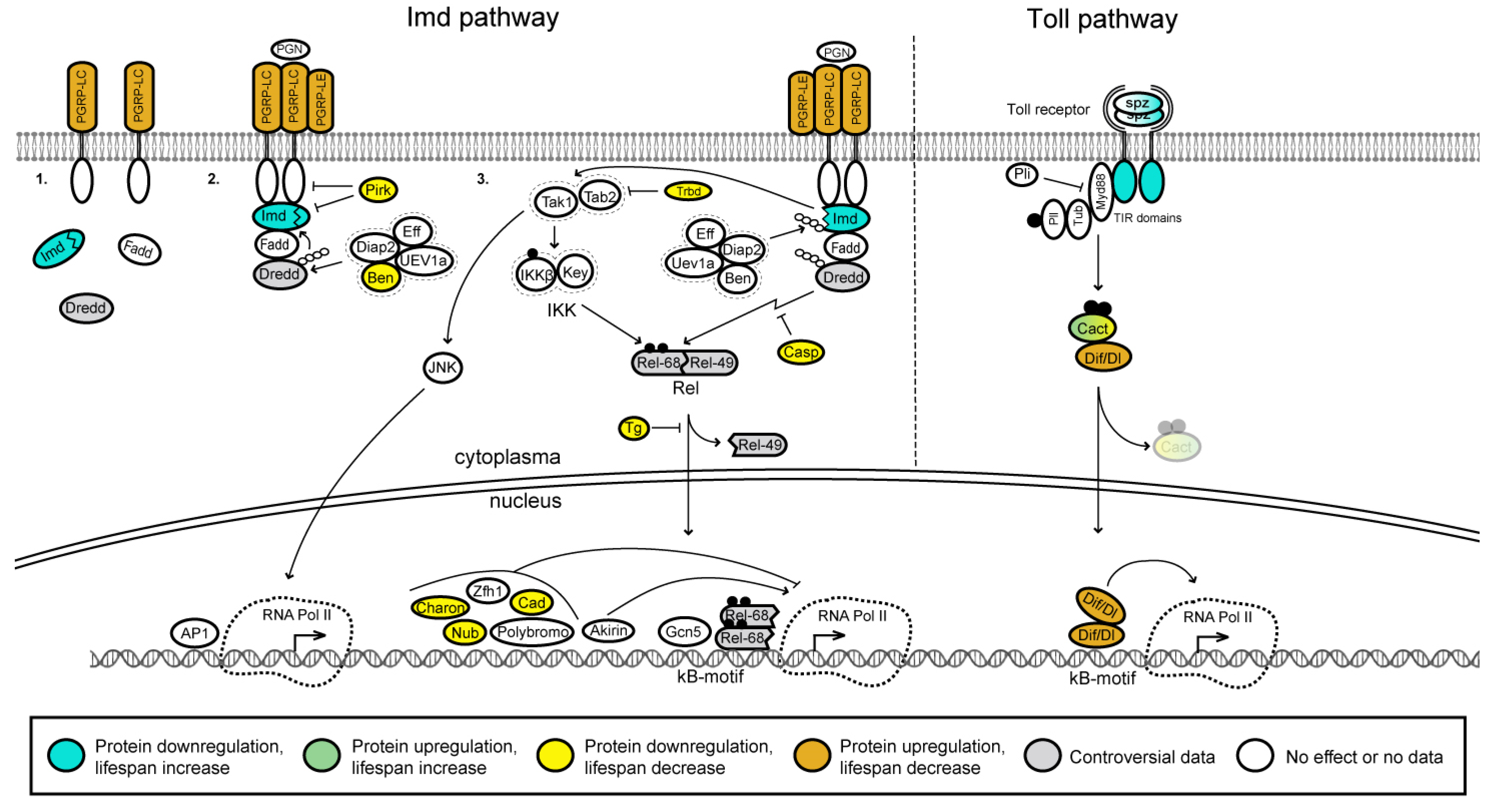
3.3. NF-κB Signaling Pathway

In different animal species, including Drosophila and mammals, aging is associated with the nuclear factor kappa B (NF-κB) signaling pathway [171]. It has been demonstrated that pharmacological inhibition of the NF-κB signaling prevents age-related pathologies and increases the median lifespan of Drosophila [171]. Fruit flies possess two NF-κB signaling pathways that are specifically active against different pathogens, the Imd (immune deficiency) signaling activated by most Gram-negative infections and the Toll signaling pathway activated in response to most Gram-positive bacterial and fungal infections [172,173,174] (Figure 4). The three Drosophila NF-κB factors—Dorsal (Dl), Dorsal-related immunity factor (Dif) and Relish (Rel)—regulate the insect humoral immunity that gets activated during infection [175,176,177].
Activation of the Imd pathway occurs through direct recognition of Gram-negative bacterial diaminopimelic acid (DAP)-type peptidoglycan (PGN) by the PGN recognition protein LC (PGRP-LC) at the cell surface or by PGRP-LE receptor intracellularly [178,179]. PGRP-LC has several isoforms, including three receptor isoforms (LCx, LCa and LCy). These isoforms have different specificity for extracellular polymeric or monomeric PGN fragments, which are referred to as tracheal cytotoxin (TCT) [180,181,182,183] and trigger the Drosophila Imd pathway being presented to PGRP-LE in the cytosol [183]. The PGRP-LB, PGRP-SC1, PGRP-SC2 receptors [184,185] and Pirk protein have been reported to suppress the Imd pathway [186]. Ligand binding causes the receptor to dimerize/multimerize and activates the recruitment of the adaptor proteins Imd and Fadd along with the Death related ced-3/Nedd2-like caspase (Dredd). The initiator caspase Dredd is activated by Drosophila inhibitor of apoptosis-2 (DIAP-2)-dependent polyubiquitylation [187]. Activated Dredd cleaves Imd, thus unmasking its domain of interaction with the DIAP-2 [188]. The E3 ubiquitin ligase DIAP-2 associates with cleaved Imd and rapidly conjugates Imd with K63-linked polyubiquitin. K63 polyubiquitin chains are suggested to function as a scaffold to activate transforming growth factor β (TGF-β)-activating kinase 1 (Tak1) and its binding adaptor Tab2 [188,189,190]. The deubiquitinase Trabid (Trbd) negatively regulates the pathway by inactivating Tak1 [190]. The Tak1/Tab2 complex mediates phosphorylation of the IKKβ subunit of the IκB kinase (IKK) on one hand and Jun nuclear kinase (JNK) (see below) on the other [191]. Activated IKKβ, in complex with Kenny (Key), phosphorylates Rel on serines 528 and 529. These phosphorylation sites are not required for Rel cleavage, nuclear translocation, or DNA binding. Instead, they are critical for recruitment of RNA Pol II and antimicrobial peptide (AMP) gene induction. Rel consists of a nuclear factor containing domain (Rel-68) and an inhibitory domain (Rel-49) responsible for keeping Rel in the cytoplasm. Cleavage of the full-length Rel by the caspase Dredd releases the N-terminal domain Rel-68 [192,193,194]. This allows phosphorylated Rel-68 to translocate to the nucleus where it binds as a dimer [195,196,197] to the κB sites within the promoters of genes encoding AMPs (Figure 4).
Phosphorylated Rel-68 forms a complex with PCAF (Gcn5) (histone acetyltransferase (HAT) p300/CBP-associated factor) at target genes and activates transcription [179] (Figure 4). Other regulators such as the transcription factors Caudal [198], Nubbin [199], Zfh1 [200] and Polybromo (or BAP180), a subunit of the chromatin-remodeling SWI/SNF complex [201], keep Rel activity in check, likely by directly antagonizing Rel binding to AMP regulatory DNA in the nucleus [183]. Thus, the activation of the Imd pathway is closely associated with epigenetic regulation and involves the recruitment of chromatin remodeling complexes such as SWI/SNF and HATs, such as PCAF.
Akirin is an NF-κB co-factor required for Imd target gene activation by the Rel transcription factor [202] (Figure 4). H3K4ac, an epigenetic mark of active gene transcription [203], is selectively enriched on Akirin-dependent, but not on Akirin-independent promoters. The presence of Akirin, BAP60 (component of SWI/SNF) and Rel is required at the same level on the promoters for an efficient transcription of Akirin-dependent genes. Akirin may link Rel and BAP60 for recruiting the SWI/SNF complex to the promoters of the Rel target genes [178]. Akirin is required for the transcription activation of direct immune effector (AMP) genes, but not for genes encoding negative regulators of the Imd pathway (except PGRP-SC2) [178]. Thus, Akirin links Rel-dependent Imd signaling to chromatin remodeling by SWI/SNF and this is accompanied by an increase in H3K4ac mark on promoters of Akirin-dependent genes.
Charon (also called Pickle) is a member of the IκB superfamily and inhibits the activity of Rel. Charon fulfills the functional and structural criteria for an IκB protein. It was shown to predominantly reside in the nuclear fraction and regulate the Imd pathway at the level of Rel68:Rel68 homodimer suppressing the Rel68-driven induction of genes encoding AMPs [204] (Figure 4). Caspar (Casp) inhibits Dredd-dependent cleavage of Rel [205]. During infection, it was suggested that certain targets of Rel inhibit JNK signaling through proteasomal degradation of Tak1 in Drosophila [206]. Transglutaminase (TG) suppresses the Imd pathway via protein-protein cross-linking that prevents Rel from entering the nucleus [207,208]. Downregulation of TG reduces the lifespan of flies raised under nonsterile conditions but not of flies reared under germ-free conditions [208].
The Toll signaling is activated in response to both Gram-positive cocci and fungi [173]. The pathway is initiated by recognition proteins circulating in the haemolymph, which include the PGN recognition protein SA (PGRP-SA), the Gram-negative binding protein 1 (GNBP1) [209,210]. Upon infection, PGRP-SA and GNBP1 induce a proteolytic cascade that ultimately processes the cytokine-growth factor-like polypeptide Spatzle (Spz, also called Spz-1) into a biologically active ligand, which binds to the ectodomain of Toll-1 receptor [211,212,213,214] (Figure 4). Besides that, Drosophila has eight other Toll receptors (Toll-2 to Toll-9) and five other Spz proteins (Spz-2 to Spz-6) [215]. The dimerized activated Toll receptor binds to the adaptor protein Myd88 via intracellular TIR domains [216,217,218]. Upon this interaction, Myd88 recruits Tube (Tub) and the Pelle (Pll) kinase forming the Myd88-Tub-Pll heterotrimeric plasma membrane associated complex [216,219,220] to induce the phosphorylation and subsequent degradation of the IκB inhibitor Cactus (Cact) [221,222]. Cact degradation frees the NF-κB factors Dif and/or Dorsal, which translocate(s) to the nucleus and bind(s) to κB motifs, resulting in transcription activation of genes encoding AMPs [223,224]. It was shown that Dif is the main transcription factor in the innate immunity response in adults and Dorsal is responsible for larvae immune response [224]. Deltex (Dx), a context-dependent regulator of Notch signaling, positively regulates the Toll signaling pathway by facilitating the nuclear localization of the transcription factor(s) Dorsal and/or Dif [225].
Tissue-specific and developmental expression profiles of the NF-κB signaling pathway genes are not uniform (Figure 5A and Supplementary Figure S2A). The majority of genes are ubiquitously expressed at moderate levels. Some genes are expressed at very low to moderate levels with minimal variability across developmental stages and different tissues, for example, imd, Dredd, Fadd and IKKβ (Figure 5A and Supplementary Figure S2A). Some other genes are generally expressed at low to moderate levels, but at specific developmental stages and tissues they are characterized by relatively high abundance. For instance, the pirk gene is very active in midgut of adult flies and larvae, the Tab2 gene is actively expressed in early embryonic stage (0–2 h), and the Transglutaminase (Tg) gene is highly expressed in crop of adult flies. As for the IIS and TOR pathways, only a few of the NF-κB pathway genes are active in all tissues and at all stages of development: effete (eff), bendless (ben) and Ubiquitin-conjugating enzyme variant 1A (Uev1A). Ben and Uev1A form a heterodimer that is required for Imd K63-linked polyubiquitinylation and its subsequent proteasome degradation [226,227,228]. Eff is also required for Imd ubiquitinylation and regulation of cell apoptosis [188]. Thus, it appears that in Drosophila, the Gram-positive bacterial response system that is triggered via the Imd pathway is constitutively ready to activate antimicrobial response genes. This may also have an adaptive value, and a decrease in the readiness of this system to fight infections due to mutations that disrupt the expression of its components, leads to a corresponding decrease in lifespan.
Downregulation of some components of the NF-κB signaling pathway was shown to slightly extend Drosophila lifespan (Supplementary Table S3). For instance, downregulation of the Toll (Tl) gene extends lifespan for about 12% and 2% in females and males, respectively [229]. Downregulation of imd leads to about 15% and 12% increase of mean lifespan in heterozygous male and female flies [230]. Downregulation of the cact, pirk, ben, trbd, casp, Tg, Charon and nubbin (nub) genes were demonstrated to cause moderate decrease of Drosophila lifespan (Figure 5A, Supplementary Table S3) [173,190,191,229,231,232,233]. The data on the influence of the Dredd and Rel mutations on fruit fly lifespan are controversial. Dredd mutation leads to 8–12% increase of the lifespan compared to control [191]. At the same time RNAi of the Dredd gene was found to cause 10 days increase and 15 days decrease of lifespan compared to control in male and female flies, respectively [234]. Rel-null flies were shown to have a statistically significant reduction (for about 23%) in the lifespan compared with control, but RNAi of Rel in glia, neurons and intestine resulted in about 60%, 38% and 35% increase of the lifespan, respectively [191].
Upregulation of the cact gene in neuroblasts increases the median lifespan of male and female flies for about 16% and 4%, respectively [173]. Overexpression of other genes (e.g., Dif, PGRP-LC, PGRP-LE, Rel) negatively regulates fly lifespan. Finally, shortened lifespan was observed in Dif, PGRP-LC, PGRP-LE and Rel mutants [173,212,235,236] (Figure 5A, Supplementary Table S3).

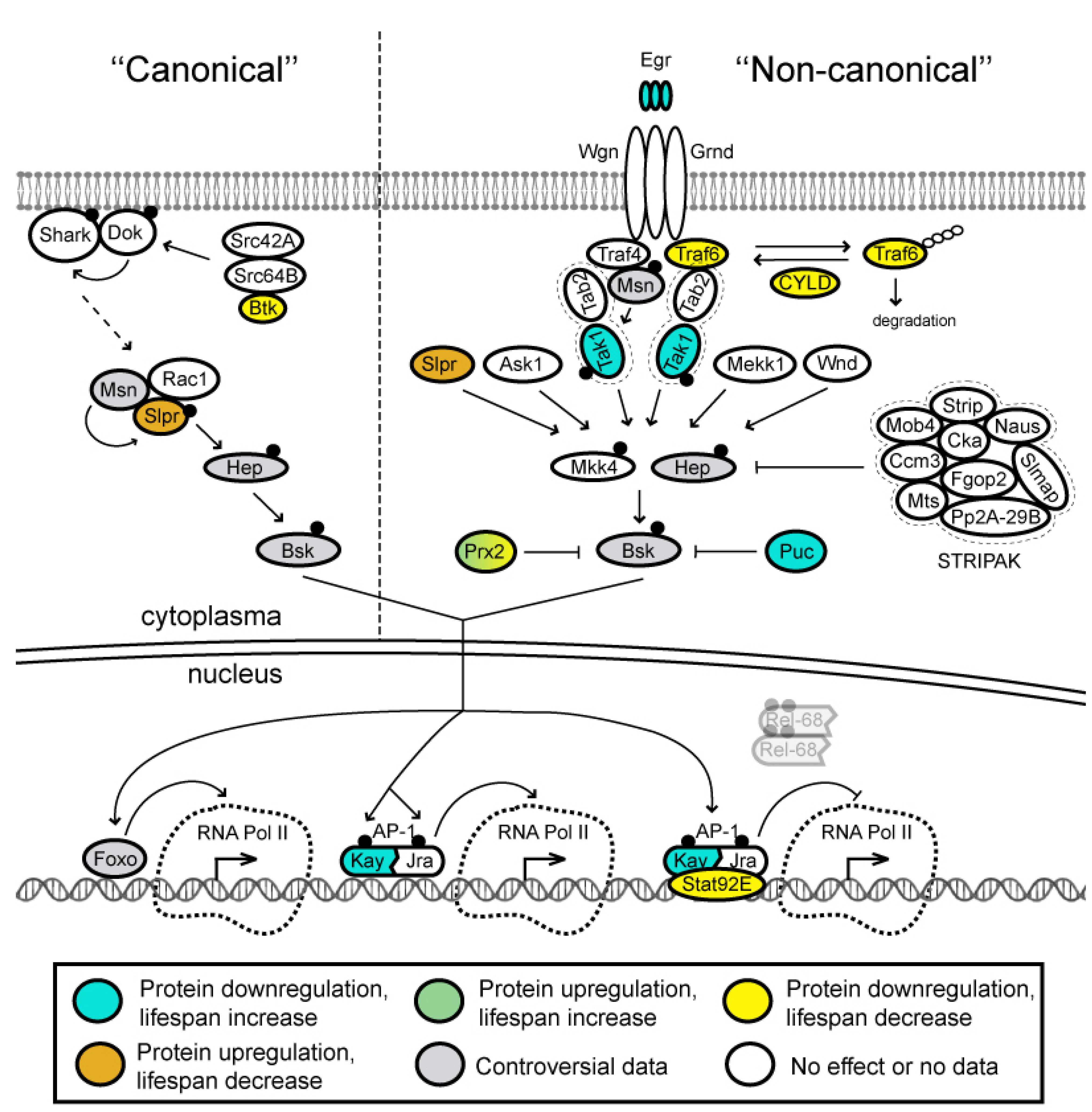
3.4. JNK Signaling Pathway

The c-Jun NH2-terminal kinase (JNK) is a conserved branch of the mitogen-activated protein kinase (MAPK) pathway [237]. JNK signaling is activated by cytokines and environmental stress [238] and regulates a number of cellular processes such as cellular proliferation, migration and apoptosis [239]. In Drosophila, the JNK signaling can be loosely classified into “canonical” and “non-canonical” pathways. The term ‘‘canonical’’ is used to describe pathway components activated during development, especially during embryonic dorsal closure, which are also required for normal wound closure [240,241]. “Non-canonical” signaling pathway is not required for development and is activated during stresses and wound healing [240,241].
“Non-canonical” signaling is activated by binding of the single tumor necrosis factor (TNF) ligand Eiger (Egr) to the TNF receptors Wengen (Wgn) and Grindelwald (Grnd) [242,243,244,245,246] that have been implicated in different cellular pathways [245] (Figure 6). Each TNF receptor forms hetero-hexamers with trimeric Egr ligands, although with different affinities [245]. Egr/Grnd activity is transduced via TNF receptor associated factor (TRAF) proteins and results in the activation of the JNK pathway [247]. Drosophila has only two TRAF proteins, Traf4 and Traf6 [248,249]. Originally, Traf4 was suggested to be the adaptor protein that links Wgn and the JNK cascade [250,251], since only Traf4, but not Traf6 binds to Mishappen (Msn), the first kinase in the JNK pathway [249]. However, further biochemical and genetic studies have revealed that Traf6, but not Traf4 plays this role [250,252,253,254]. Additionally, it was found that TRAF proteins transduce discrete signaling pathways in Drosophila. Overexpression of Traf4 causes JNK pathway activation, while overexpression of Traf6 leads to activation of Drosophila NF-κB proteins Dif and Rel [255]. In addition, deubiquitinating enzyme CYLD regulates JNK activation by deubiquitinating Traf6, thus, preventing it from ubiquitin-mediated proteolytic degradation in cytoplasma [252].
Ligand-receptor binding initiates a conserved signaling cascade of MAPK superfamily kinases, which includes at least five JNKK kinases (JNKKKs)—Slipper (Slpr), Wallenda (Wnd), Tak1, Mekk1 and Apoptotic signal-regulating kinase 1 (Ask1) as well as at least two JNK kinases (JNKKs)—Hemipterous (Hep) and MAP kinase kinase 4 (Mkk4) [256,257,258,259,260,261]. The ultimate goal of this JNK cascade is activation of the c-Jun N-terminal kinase Basket (Bsk) (Figure 6), which results in a variety of tissue-specific and context-specific cellular responses [243,262].
Activated (phosphorylated) Bsk translocates into the nucleus and, in its turn, activates its downstream transcription factor Jun-related antigen (Jra), which heterodimerizes with the transcription factor Kayak (Kay) forming an active AP-1 transcriptional complex in the nucleus [254,263] (Figure 6). AP-1 transcriptionally upregulates numerous target genes and AP-1 dimers bind to different types of palindromic sequences. Thus, Kay:Jun and Jun:Jun dimers preferentially bind to DNA motifs referred to as tetradeconyl phorbol acetate-responsive elements (TREs, also called AP-1 motifs) and, with slightly lower affinity, to cyclic adenosine monophosphate-responsive elements (CREs). One well-established AP-1 target gene is puckered (puc) [264] encoding a phosphatase that dephosphorylates Bsk in an induced-negative feedback loop (Figure 6). Other targets include the unpaired (upd) gene encoding the Drosophila interleukin-6 (IL-6) homolog [265] and the Matrix metalloproteinase 1 (Mmp1) gene encoding a protease that degrades the extracellular matrix [266]. AP-1 also forms a repressosome complex together with the Stat92E protein. This complex blocks the expression of immune effector genes (such as those encoding AMPs) by competitively blocking Rel binding and by recruiting histone deacetylase at their promoters [2,267].
Activation of the “canonical” and “non-canonical” JNK signaling pathways differs only upstream, but not downstream of the Hep component. The most upstream acting molecules identified for the “canonical” signaling during dorsal closure are nonreceptor tyrosine kinases of the Src family: Src42A, Src64B and Btk [240] (Figure 6). Src42A phosphorylates Downstream of kinase (Dok), which belongs to the Dok family of adaptor proteins. Dok is required for the appropriate tyrosine phosphorylation, membrane localization and activation of nonreceptor tyrosine kinase Shark [268]. How these kinases relay the signal to downstream components is still not well understood. However, it was shown that a serine threonine kinase Slipper (Slpr) interacts physically with Rac1 and with Msn, the latter of which is supposed to phosphorylate Slpr, thus activating it. Activated Slpr in its turn phosphorylates Hep. Then, the “canonical” and “non-canonical” pathways intersect [240,269].
It is also known that JNK antagonizes IIS, since JNK signaling promotes Foxo nuclear localization [270] (Figure 6). Foxo also regulates activity of numerous genes (see above). Among Foxo targets are cytoprotective genes such as Peroxiredoxin (Prx2), lethal (2) essential for life (l(2)efl) and Thor (eIF4E-binding protein, an inhibitor of cap-dependent protein synthesis), which activity advances stress defense [270,271]. Overexpression of Prx2 is able to suppress Bsk activity in neurons, indicating that Prx2 may also act as a negative feedback regulator of JNK in specific tissues [271].
Along with Puc and Prx2 inhibition, JNK signaling is downregulated by the STRIPAK complex components, Striatin interacting protein (Strip) and Connector of kinase to AP-1 (Cka), which bind to and inactivate Hep/JNKK acting as negative regulators of JNK signaling [272,273,274,275,276,277].
The JNK pathway is activated not only through Wgn and Grnd receptors but also from the Imd pathway through Tak1 (Figure 4 and Figure 6) [206,278]. JNK-regulated lipopolysaccharides-responsive genes exhibit transient expression, whereas IKK/Rel target genes demonstrate sustained expression pattern. Rel-dependent genes not only serve their own immune effector roles, but also participate in shaping the temporal pattern of the JNK-mediated immune response by inactivating Tak1. Thus, IKK/Rel branch plays an active role in turning off JNK activity [206].
Tissue-specific and developmental expression profiles of the JNK signaling pathway genes are not uniform (Figure 5B and Supplementary Figure S2B). Most genes of the pathway are expressed at moderate levels. The grnd, Traf6, wnd, Ask1, hep and Mkk4 genes are expressed at very low to moderate levels with minimal variability across developmental stages and different tissues (Figure 5B and Supplementary Figure S2B). The CYLD, Fgop2, Slmap and Strip genes are generally expressed at low levels, but they are very active in testis of adult males. The egr gene is active in Malpighian tubules of adult flies, while bsk expression first appears in CNS close to the end of the pupal stage and then it is kept active in the head, eye, brain and thoracic ganglion of adult flies. As with the other pathways we have described here, IIS, TOR and NF-κB, several genes of the JNK pathway (e.g., mts, Pp2A-29B, Prx2) are highly upregulated in virtually all tissues, and the corresponding products of these genes were found almost at all developmental stages (Figure 5B and Supplementary Figure S2B). The subunits of the PP2A phosphatase, Mts and Pp2A-29B (catalytic and scaffold subunit, respectively), which suppress the activity of the IIS pathway due to dephosphorylation of the Akt kinase, together with the Wdb regulatory subunit and Prx2 inhibitor appear to reliably inhibit the JNK cascade. This inhibition seems to be supported by the weak expression of the proteins of the STRIPAK complex, of which only Cka is expressed at an average level. At the same time, several main members of the JNK cascade, the kinases Msn, Mkk4 and Bsk, are ubiquitously expressed at moderate level. Thus, the JNK pathway is most likely inhibited in cells by default, but it is at the ready because all the kinases required for Egr signaling are constitutively present.
Downregulation of several components of the JNK signaling pathway (e.g., mutations of the egr, Tak1, kay and puc genes) slightly extend Drosophila lifespan (Figure 5B, Supplementary Table S4). On the contrary, mutations of the CYLD, Traf6, and Prx2 genes lead to the opposite effect on the Drosophila lifespan, causing its reduction. Upregulation of the slpr gene results in highly shortened lifespan of flies. At the same time, upregulation of the foxo and Prx2 genes causes slight extension of fly longevity (Figure 5B, Supplementary Table S4). It is rather difficult to understand the mechanisms of influence of the JNK pathway on lifespan, since this pathway has pleiotropic effects: it maintains stem cell renewal in homeostasis, induces apoptosis in case of damage, induces proliferation accompanied by switching the type of cell divisions and reprogramming of cells during regeneration [237]. However, the most significant increase in lifespan was observed with inhibition of the JNK pathway. Particularly, mutations of the egr gene, overexpression of Puc, and overexpression of the transcription factor Foxo gave the greatest increase in lifespan up to 62% (Supplementary Table S4). Moreover, Foxo, inducing genes associated with the suppression of proliferation and protein synthesis also induced the expression of another inhibitor of the JNK pathway, Prx2 [271]. Thus, although mutations and upregulation of some components of the JNK pathway lead to a sharp decrease in lifespan, suppression of signaling through the JNK pathway is generally associated with increased lifespan.

3.5. Interconnection between IIS, TOR, NF-κB and JNK Pathways

The interactions between the above-described pathways are much better studied in mammals. Signaling through the human IIS pathway involves kinases such as p38 (MAPK), JNK, AMPK, PKC and others and most of these kinases are able to phosphorylate several key regulators of glucose homeostasis as well as the insulin receptor, resulting in decreased activity of the PI3K/Akt [279]. Besides, it was shown that JNK antagonizes IIS, promoting FOXO nuclear localization [270]. This is how the interactions between the JNK, NF-κB and the IIS pathways can take place.
In Drosophila, the interactions between the pathways are rather poorly understood. The IIS pathway, as the central pathway associated with nutrient intake, interacts with all other pathways and plays an important role not only in the regulation of longevity, but also in the development of cancer in Drosophila [280]. As for the interaction between the IIS and the TOR pathways, it is obvious that the cap-dependent protein synthesis depends on the presence of amino acids and carbohydrates, and a decrease in their intake suppresses TOR, turns off protein synthesis, and reduces overall metabolism. This, in turn, leads to some extension of lifespan. An approach used by many investigators to reduce the activity of the insulin/TOR pathway is DR [3].
With regard to the the NF-κB and the JNK pathways, both of them appear to be involved in the immune response in Drosophila and are interconnected through the Tak1 kinase [206,227,278]. Activation of Rel seems to be associated with proteasomal degradation of Tak1 required for JNK signaling [206]. In turn, the degradation of Tak1 leads to the cessation of JNK signaling within 1 h. Thus, in the case of immune activation, a transient JNK and long-term NF-κB responses occur. The same relationship between the two pathways is observed in humans [206]. Interestingly, JNK pathway is not required for Rel activation, but is absolutely necessary for the immune response and production of antimicrobial peptides in Drosophila [278]. Recently, the role of the immune signaling in the fly brain and its association with longevity and neurodegenerative diseases has been demonstrated, placing the nervous system as the master system controlling lifespan [173,191]. The relationship between the immune and nervous systems in humans has also been discussed lately [281,282].

4. Concluding Remarks

Evolutionary conserved signaling pathways IIS, TOR, NF-κB and JNK play critical roles under stress conditions and aging. In Drosophila, each pathway consists of the numerous components, which are characterized by a larger number of paralogous subunits in human (Supplementary Tables S1–S4). This indicates the importance of these pathways and their development in evolution from a common ancestor of invertebrates and vertebrates.
In Drosophila, activation of the IIS and the TOR pathways leads to a shortening of lifespan, whereas activation of the JNK pathway suppresses the activity of TOR and causes an increase in lifespan. Violation of the NF-κB pathway results in a decrease in lifespan. The Imd sub-branch of the NF-κB pathway is constitutively ready for activation and both activating and repressing components of the IIS and the TOR pathways are constitutively expressed.
In closing, many questions regarding the links between the components of these signaling pathways and aging still remain. For instance, influence of downregulation or overexpression of genes on fly lifespan was studied only for 40–53% of the pathways’ components (Figure 2 and Figure 5). Moreover, some of these data seem to be controversial. Among constitutively expressed subunits of the pathways, effect on lifespan had been so far demonstrated only for 25% of them. In the future, it will be important to detect the pathways’ components with the highest magnitude of the effect on lifespan resulted from their misexpression.

Supplementary Materials

The following supporting information can be downloaded at: https://www.mdpi.com/article/10.3390/ijms231911244/s1. References [283,284,285,286,287,288,289,290,291,292,293,294,295,296,297,298,299,300,301] are cited in the Supplementary Materials.

Author Contributions

All authors contributed to the preparation of this review. A.A.O., E.S.O. and O.V.B. performed the most extensive critical analysis of the literature. A.A.O., E.S.O., O.V.B., M.A.B., P.G.G. and A.V.P. wrote the manuscript and carried out the final editing. All authors have read and agreed to the published version of the manuscript.

Funding

This work was mainly supported by the Russian Science Foundation Grant no. 19-74-30026 and in part of the gene expression analysis and data visualization by the Russian Science Foundation Grant no. 22-74-10118.

Conflicts of Interest

The authors declare no conflict of interest.

References

  1. Kennedy, B.K.; Berger, S.L.; Brunet, A.; Campisi, J.; Cuervo, A.M.; Epel, E.S.; Franceschi, C.; Lithgow, G.J.; Morimoto, R.I.; Pessin, J.E.; et al. Geroscience: Linking aging to chronic disease. Cell 2014, 159, 709–713. [Google Scholar] [CrossRef] [PubMed]
  2. Gan, T.; Fan, L.; Zhao, L.; Misra, M.; Liu, M.; Zhang, M.; Su, Y. JNK Signaling in Drosophila Aging and Longevity. Int. J. Mol. Sci. 2021, 22, 9649. [Google Scholar] [CrossRef] [PubMed]
  3. Green, C.L.; Lamming, D.W.; Fontana, L. Molecular mechanisms of dietary restriction promoting health and longevity. Nat. Rev. Mol. Cell Biol. 2022, 23, 56–73. [Google Scholar] [CrossRef] [PubMed]
  4. Miller, H.A.; Dean, E.S.; Pletcher, S.D.; Leiser, S.F. Cell non-autonomous regulation of health and longevity. Elife 2020, 9, e62659. [Google Scholar] [CrossRef]
  5. Green, M.M. 2010: A century of Drosophila genetics through the prism of the white gene. Genetics 2010, 184, 3–7. [Google Scholar] [CrossRef]
  6. Rogina, B. For the special issue: Aging studies in Drosophila melanogaster. Exp. Gerontol. 2011, 46, 317–319. [Google Scholar] [CrossRef]
  7. Promislow, D.E.L.; Flatt, T.; Bonduriansky, R. The Biology of Aging in Insects: From Drosophila to Other Insects and Back. Annu. Rev. Entomol. 2022, 67, 83–103. [Google Scholar] [CrossRef]
  8. Calissi, G.; Lam, E.W.; Link, W. Therapeutic strategies targeting FOXO transcription factors. Nat. Rev. Drug Discov. 2021, 20, 21–38. [Google Scholar] [CrossRef]
  9. Reiter, L.T.; Potocki, L.; Chien, S.; Gribskov, M.; Bier, E. A systematic analysis of human disease-associated gene sequences in Drosophila melanogaster. Genome Res. 2001, 11, 1114–1125. [Google Scholar] [CrossRef]
  10. Adams, M.D.; Celniker, S.E.; Holt, R.A.; Evans, C.A.; Gocayne, J.D.; Amanatides, P.G.; Scherer, S.E.; Li, P.W.; Hoskins, R.A.; Galle, R.F.; et al. The genome sequence of Drosophila melanogaster. Science 2000, 287, 2185–2195. [Google Scholar] [CrossRef] [Green Version]
  11. Rubin, G.M.; Yandell, M.D.; Wortman, J.R.; Gabor Miklos, G.L.; Nelson, C.R.; Hariharan, I.K.; Fortini, M.E.; Li, P.W.; Apweiler, R.; Fleischmann, W.; et al. Comparative genomics of the eukaryotes. Science 2000, 287, 2204–2215. [Google Scholar] [CrossRef] [PubMed]
  12. Yakovleva, E.U.; Naimark, E.B.; Markov, A.V. Adaptation of Drosophila melanogaster to Unfavorable Growth Medium Affects Lifespan and Age-Related Fecundity. Biochemistry 2016, 81, 1445–1460. [Google Scholar] [CrossRef] [PubMed]
  13. Shaposhnikov, M.V.; Guvatova, Z.G.; Zemskaya, N.V.; Koval, L.A.; Schegoleva, E.V.; Gorbunova, A.A.; Golubev, D.A.; Pakshina, N.R.; Ulyasheva, N.S.; Solovev, I.A.; et al. Molecular mechanisms of exceptional lifespan increase of Drosophila melanogaster with different genotypes after combinations of pro-longevity interventions. Commun. Biol. 2022, 5, 566. [Google Scholar] [CrossRef] [PubMed]
  14. Magwere, T.; Chapman, T.; Partridge, L. Sex differences in the effect of dietary restriction on life span and mortality rates in female and male Drosophila melanogaster. J. Gerontol. A Biol. Sci. Med. Sci. 2004, 59, 3–9. [Google Scholar] [CrossRef] [PubMed]
  15. Koliada, A.; Gavrilyuk, K.; Burdylyuk, N.; Strilbytska, O.; Storey, K.B.; Kuharskii, V.; Lushchak, O.; Vaiserman, A. Mating status affects Drosophila lifespan, metabolism and antioxidant system. Comp. Biochem. Physiol. Part A Mol. Integr. Physiol. 2020, 246, 110716. [Google Scholar] [CrossRef] [PubMed]
  16. Burger, J.M.; Promislow, D.E. Sex-specific effects of interventions that extend fly life span. Sci. Aging Knowl. Environ. 2004, 2004, pe30. [Google Scholar] [CrossRef]
  17. Toivonen, J.M.; Walker, G.A.; Martinez-Diaz, P.; Bjedov, I.; Driege, Y.; Jacobs, H.T.; Gems, D.; Partridge, L. No influence of Indy on lifespan in Drosophila after correction for genetic and cytoplasmic background effects. PLoS Genet. 2007, 3, e95. [Google Scholar] [CrossRef]
  18. Spencer, C.C.; Howell, C.E.; Wright, A.R.; Promislow, D.E. Testing an ‘aging gene’ in long-lived drosophila strains: Increased longevity depends on sex and genetic background. Aging Cell 2003, 2, 123–130. [Google Scholar] [CrossRef]
  19. He, Y.; Jasper, H. Studying aging in Drosophila. Methods 2014, 68, 129–133. [Google Scholar] [CrossRef]
  20. Li, Y.-Y.; Floate, K.D.; Fields, P.G.; Pang, B.-P. Review of treatment methods to remove Wolbachia bacteria from arthropods. Symbiosis 2014, 62, 1–15. [Google Scholar] [CrossRef]
  21. Yang, J.S.; Nam, H.J.; Seo, M.; Han, S.K.; Choi, Y.; Nam, H.G.; Lee, S.J.; Kim, S. OASIS: Online application for the survival analysis of lifespan assays performed in aging research. PLoS ONE 2011, 6, e23525. [Google Scholar] [CrossRef] [PubMed]
  22. Grandison, R.C.; Piper, M.D.; Partridge, L. Amino-acid imbalance explains extension of lifespan by dietary restriction in Drosophila. Nature 2009, 462, 1061–1064. [Google Scholar] [CrossRef] [PubMed]
  23. Huang, W.; MacLean, A.M.; Sugio, A.; Maqbool, A.; Busscher, M.; Cho, S.T.; Kamoun, S.; Kuo, C.H.; Immink, R.G.H.; Hogenhout, S.A. Parasitic modulation of host development by ubiquitin-independent protein degradation. Cell 2021, 184, 5201–5214.e12. [Google Scholar] [CrossRef]
  24. Jones, O.R.; Scheuerlein, A.; Salguero-Gómez, R.; Camarda, C.G.; Schaible, R.; Casper, B.B.; Dahlgren, J.P.; Ehrlén, J.; García, M.B.; Menges, E.S.; et al. Diversity of ageing across the tree of life. Nature 2014, 505, 169–173. [Google Scholar] [CrossRef]
  25. Fernández-Moreno, M.A.; Farr, C.L.; Kaguni, L.S.; Garesse, R. Drosophila melanogaster as a model system to study mitochondrial biology. Methods Mol. Biol. 2007, 372, 33–49. [Google Scholar] [CrossRef] [PubMed]
  26. Baker, C.R.; Hanson-Smith, V.; Johnson, A.D. Following gene duplication, paralog interference constrains transcriptional circuit evolution. Science 2013, 342, 104–108. [Google Scholar] [CrossRef]
  27. Diss, G.; Gagnon-Arsenault, I.; Dion-Coté, A.M.; Vignaud, H.; Ascencio, D.I.; Berger, C.M.; Landry, C.R. Gene duplication can impart fragility, not robustness, in the yeast protein interaction network. Science 2017, 355, 630–634. [Google Scholar] [CrossRef]
  28. Marlétaz, F.; Firbas, P.N.; Maeso, I.; Tena, J.J.; Bogdanovic, O.; Perry, M.; Wyatt, C.D.R.; de la Calle-Mustienes, E.; Bertrand, S.; Burguera, D.; et al. Amphioxus functional genomics and the origins of vertebrate gene regulation. Nature 2018, 564, 64–70. [Google Scholar] [CrossRef]
  29. Altintas, O.; Park, S.; Lee, S.J. The role of insulin/IGF-1 signaling in the longevity of model invertebrates, C. elegans and D. melanogaster. BMB Rep. 2016, 49, 81–92. [Google Scholar] [CrossRef]
  30. Nässel, D.R.; Vanden Broeck, J. Insulin/IGF signaling in Drosophila and other insects: Factors that regulate production, release and post-release action of the insulin-like peptides. Cell Mol. Life Sci. 2016, 73, 271–290. [Google Scholar] [CrossRef]
  31. Mathew, R.; Pal Bhadra, M.; Bhadra, U. Insulin/insulin-like growth factor-1 signalling (IIS) based regulation of lifespan across species. Biogerontology 2017, 18, 35–53. [Google Scholar] [CrossRef] [PubMed]
  32. Álvarez-Rendón, J.P.; Salceda, R.; Riesgo-Escovar, J.R. Drosophila melanogaster as a Model for Diabetes Type 2 Progression. Biomed. Res. Int. 2018, 2018, 1417528. [Google Scholar] [CrossRef] [PubMed]
  33. Vinayagam, A.; Kulkarni, M.M.; Sopko, R.; Sun, X.; Hu, Y.; Nand, A.; Villalta, C.; Moghimi, A.; Yang, X.; Mohr, S.E.; et al. An Integrative Analysis of the InR/PI3K/Akt Network Identifies the Dynamic Response to Insulin Signaling. Cell Rep. 2016, 16, 3062–3074. [Google Scholar] [CrossRef] [PubMed]
  34. Brogiolo, W.; Stocker, H.; Ikeya, T.; Rintelen, F.; Fernandez, R.; Hafen, E. An evolutionarily conserved function of the Drosophila insulin receptor and insulin-like peptides in growth control. Curr. Biol. 2001, 11, 213–221. [Google Scholar] [CrossRef]
  35. Grönke, S.; Clarke, D.F.; Broughton, S.; Andrews, T.D.; Partridge, L. Molecular evolution and functional characterization of Drosophila insulin-like peptides. PLoS Genet. 2010, 6, e1000857. [Google Scholar] [CrossRef]
  36. Ruan, Y.; Chen, C.; Cao, Y.; Garofalo, R.S. The Drosophila insulin receptor contains a novel carboxyl-terminal extension likely to play an important role in signal transduction. J. Biol. Chem. 1995, 270, 4236–4243. [Google Scholar] [CrossRef]
  37. Chen, C.; Jack, J.; Garofalo, R.S. The Drosophila insulin receptor is required for normal growth. Endocrinology 1996, 137, 846–856. [Google Scholar] [CrossRef]
  38. Yamamoto, R.; Palmer, M.; Koski, H.; Curtis-Joseph, N.; Tatar, M. Aging modulated by the Drosophila insulin receptor through distinct structure-defined mechanisms. Genetics 2021, 217, iyaa037. [Google Scholar] [CrossRef]
  39. Tatar, M.; Kopelman, A.; Epstein, D.; Tu, M.P.; Yin, C.M.; Garofalo, R.S. A mutant Drosophila insulin receptor homolog that extends life-span and impairs neuroendocrine function. Science 2001, 292, 107–110. [Google Scholar] [CrossRef]
  40. Fernandez, R.; Tabarini, D.; Azpiazu, N.; Frasch, M.; Schlessinger, J. The Drosophila insulin receptor homolog: A gene essential for embryonic development encodes two receptor isoforms with different signaling potential. EMBO J. 1995, 14, 3373–3384. [Google Scholar] [CrossRef]
  41. Weinkove, D.; Leevers, S.J.; MacDougall, L.K.; Waterfield, M.D. p60 is an adaptor for the Drosophila phosphoinositide 3-kinase, Dp110. J. Biol. Chem. 1997, 272, 14606–14610. [Google Scholar] [CrossRef] [PubMed]
  42. Werz, C.; Köhler, K.; Hafen, E.; Stocker, H. The Drosophila SH2B family adaptor Lnk acts in parallel to chico in the insulin signaling pathway. PLoS Genet. 2009, 5, e1000596. [Google Scholar] [CrossRef] [PubMed]
  43. Slack, C.; Werz, C.; Wieser, D.; Alic, N.; Foley, A.; Stocker, H.; Withers, D.J.; Thornton, J.M.; Hafen, E.; Partridge, L. Regulation of lifespan, metabolism, and stress responses by the Drosophila SH2B protein, Lnk. PLoS Genet. 2010, 6, e1000881. [Google Scholar] [CrossRef]
  44. Leevers, S.J.; Weinkove, D.; MacDougall, L.K.; Hafen, E.; Waterfield, M.D. The Drosophila phosphoinositide 3-kinase Dp110 promotes cell growth. EMBO J. 1996, 15, 6584–6594. [Google Scholar] [CrossRef] [PubMed]
  45. Alessi, D.R.; Deak, M.; Casamayor, A.; Caudwell, F.B.; Morrice, N.; Norman, D.G.; Gaffney, P.; Reese, C.B.; MacDougall, C.N.; Harbison, D.; et al. 3-Phosphoinositide-dependent protein kinase-1 (PDK1): Structural and functional homology with the Drosophila DSTPK61 kinase. Curr. Biol. 1997, 7, 776–789. [Google Scholar] [CrossRef]
  46. Verdu, J.; Buratovich, M.A.; Wilder, E.L.; Birnbaum, M.J. Cell-autonomous regulation of cell and organ growth in Drosophila by Akt/PKB. Nat. Cell Biol. 1999, 1, 500–506. [Google Scholar] [CrossRef] [PubMed]
  47. Cho, K.S.; Lee, J.H.; Kim, S.; Kim, D.; Koh, H.; Lee, J.; Kim, C.; Kim, J.; Chung, J. Drosophila phosphoinositide-dependent kinase-1 regulates apoptosis and growth via the phosphoinositide 3-kinase-dependent signaling pathway. Proc. Natl. Acad. Sci. USA 2001, 98, 6144–6149. [Google Scholar] [CrossRef]
  48. Kim, M.J.; O’Connor, M.B. Drosophila Activin signaling promotes muscle growth through InR/TORC1-dependent and -independent processes. Development 2021, 148, dev190868. [Google Scholar] [CrossRef]
  49. Webb, A.E.; Kundaje, A.; Brunet, A. Characterization of the direct targets of FOXO transcription factors throughout evolution. Aging Cell 2016, 15, 673–685. [Google Scholar] [CrossRef]
  50. Birnbaum, A.; Wu, X.; Tatar, M.; Liu, N.; Bai, H. Age-Dependent Changes in Transcription Factor FOXO Targeting in Female. Front. Genet. 2019, 10, 312. [Google Scholar] [CrossRef] [Green Version]
  51. Puig, O.; Marr, M.T.; Ruhf, M.L.; Tjian, R. Control of cell number by Drosophila FOXO: Downstream and feedback regulation of the insulin receptor pathway. Genes Dev. 2003, 17, 2006–2020. [Google Scholar] [CrossRef] [PubMed]
  52. Nielsen, M.D.; Luo, X.; Biteau, B.; Syverson, K.; Jasper, H. 14-3-3 Epsilon antagonizes FoxO to control growth, apoptosis and longevity in Drosophila. Aging Cell 2008, 7, 688–699. [Google Scholar] [CrossRef] [PubMed]
  53. Tzivion, G.; Dobson, M.; Ramakrishnan, G. FoxO transcription factors; Regulation by AKT and 14-3-3 proteins. Biochim. Biophys. Acta 2011, 1813, 1938–1945. [Google Scholar] [CrossRef]
  54. van der Horst, A.; Burgering, B.M. Stressing the role of FoxO proteins in lifespan and disease. Nat. Rev. Mol. Cell Biol. 2007, 8, 440–450. [Google Scholar] [CrossRef] [PubMed]
  55. Du, S.; Zheng, H. Role of FoxO transcription factors in aging and age-related metabolic and neurodegenerative diseases. Cell Biosci. 2021, 11, 188. [Google Scholar] [CrossRef]
  56. Brunet, A.; Kanai, F.; Stehn, J.; Xu, J.; Sarbassova, D.; Frangioni, J.V.; Dalal, S.N.; DeCaprio, J.A.; Greenberg, M.E.; Yaffe, M.B. 14-3-3 transits to the nucleus and participates in dynamic nucleocytoplasmic transport. J. Cell Biol. 2002, 156, 817–828. [Google Scholar] [CrossRef]
  57. Sunayama, J.; Tsuruta, F.; Masuyama, N.; Gotoh, Y. JNK antagonizes Akt-mediated survival signals by phosphorylating 14-3-3. J. Cell Biol. 2005, 170, 295–304. [Google Scholar] [CrossRef]
  58. Goberdhan, D.C.; Paricio, N.; Goodman, E.C.; Mlodzik, M.; Wilson, C. Drosophila tumor suppressor PTEN controls cell size and number by antagonizing the Chico/PI3-kinase signaling pathway. Genes Dev. 1999, 13, 3244–3258. [Google Scholar] [CrossRef]
  59. Huang, H.; Potter, C.J.; Tao, W.; Li, D.M.; Brogiolo, W.; Hafen, E.; Sun, H.; Xu, T. PTEN affects cell size, cell proliferation and apoptosis during Drosophila eye development. Development 1999, 126, 5365–5372. [Google Scholar] [CrossRef]
  60. Gao, X.; Neufeld, T.P.; Pan, D. Drosophila PTEN regulates cell growth and proliferation through PI3K-dependent and -independent pathways. Dev. Biol. 2000, 221, 404–418. [Google Scholar] [CrossRef] [Green Version]
  61. Leslie, N.R.; Batty, I.H.; Maccario, H.; Davidson, L.; Downes, C.P. Understanding PTEN regulation: PIP2, polarity and protein stability. Oncogene 2008, 27, 5464–5476. [Google Scholar] [CrossRef]
  62. Bánréti, Á.; Lukácsovich, T.; Csikós, G.; Erdélyi, M.; Sass, M. PP2A regulates autophagy in two alternative ways in Drosophila. Autophagy 2012, 8, 623–636. [Google Scholar] [CrossRef] [PubMed]
  63. Fischer, P.; Preiss, A.; Nagel, A.C. A triangular connection between Cyclin G, PP2A and Akt1 in the regulation of growth and metabolism in Drosophila. Fly 2016, 10, 11–18. [Google Scholar] [CrossRef] [PubMed]
  64. Graveley, B.R.; Brooks, A.N.; Carlson, J.W.; Duff, M.O.; Landolin, J.M.; Yang, L.; Artieri, C.G.; van Baren, M.J.; Boley, N.; Booth, B.W.; et al. The developmental transcriptome of Drosophila melanogaster. Nature 2011, 471, 473–479. [Google Scholar] [CrossRef] [PubMed]
  65. Leader, D.P.; Krause, S.A.; Pandit, A.; Davies, S.A.; Dow, J.A.T. FlyAtlas 2: A new version of the Drosophila melanogaster expression atlas with RNA-Seq, miRNA-Seq and sex-specific data. Nucleic Acids Res. 2018, 46, D809–D815. [Google Scholar] [CrossRef]
  66. Broughton, S.J.; Piper, M.D.; Ikeya, T.; Bass, T.M.; Jacobson, J.; Driege, Y.; Martinez, P.; Hafen, E.; Withers, D.J.; Leevers, S.J.; et al. Longer lifespan, altered metabolism, and stress resistance in Drosophila from ablation of cells making insulin-like ligands. Proc. Natl. Acad. Sci. USA 2005, 102, 3105–3110. [Google Scholar] [CrossRef]
  67. Tu, M.P.; Epstein, D.; Tatar, M. The demography of slow aging in male and female Drosophila mutant for the insulin-receptor substrate homologue chico. Aging Cell 2002, 1, 75–80. [Google Scholar] [CrossRef]
  68. Clancy, D.J.; Gems, D.; Harshman, L.G.; Oldham, S.; Stocker, H.; Hafen, E.; Leevers, S.J.; Partridge, L. Extension of life-span by loss of CHICO, a Drosophila insulin receptor substrate protein. Science 2001, 292, 104–106. [Google Scholar] [CrossRef]
  69. Hwangbo, D.S.; Gershman, B.; Gersham, B.; Tu, M.P.; Palmer, M.; Tatar, M. Drosophila dFOXO controls lifespan and regulates insulin signalling in brain and fat body. Nature 2004, 429, 562–566. [Google Scholar] [CrossRef]
  70. Demontis, F.; Perrimon, N. FOXO/4E-BP signaling in Drosophila muscles regulates organism-wide proteostasis during aging. Cell 2010, 143, 813–825. [Google Scholar] [CrossRef] [Green Version]
  71. Giannakou, M.E.; Goss, M.; Jünger, M.A.; Hafen, E.; Leevers, S.J.; Partridge, L. Long-lived Drosophila with overexpressed dFOXO in adult fat body. Science 2004, 305, 361. [Google Scholar] [CrossRef] [PubMed]
  72. Wessells, R.J.; Fitzgerald, E.; Cypser, J.R.; Tatar, M.; Bodmer, R. Insulin regulation of heart function in aging fruit flies. Nat. Genet. 2004, 36, 1275–1281. [Google Scholar] [CrossRef] [PubMed]
  73. Wang, B.; Moya, N.; Niessen, S.; Hoover, H.; Mihaylova, M.M.; Shaw, R.J.; Yates, J.R.; Fischer, W.H.; Thomas, J.B.; Montminy, M. A hormone-dependent module regulating energy balance. Cell 2011, 145, 596–606. [Google Scholar] [CrossRef]
  74. Lehmann, M. Endocrine and physiological regulation of neutral fat storage in Drosophila. Mol. Cell. Endocrinol. 2018, 461, 165–177. [Google Scholar] [CrossRef]
  75. Böhni, R.; Riesgo-Escovar, J.; Oldham, S.; Brogiolo, W.; Stocker, H.; Andruss, B.F.; Beckingham, K.; Hafen, E. Autonomous control of cell and organ size by CHICO, a Drosophila homolog of vertebrate IRS1-4. Cell 1999, 97, 865–875. [Google Scholar] [CrossRef]
  76. Scanga, S.E.; Ruel, L.; Binari, R.C.; Snow, B.; Stambolic, V.; Bouchard, D.; Peters, M.; Calvieri, B.; Mak, T.W.; Woodgett, J.R.; et al. The conserved PI3’K/PTEN/Akt signaling pathway regulates both cell size and survival in Drosophila. Oncogene 2000, 19, 3971–3977. [Google Scholar] [CrossRef] [PubMed]
  77. Rintelen, F.; Stocker, H.; Thomas, G.; Hafen, E. PDK1 regulates growth through Akt and S6K in Drosophila. Proc. Natl. Acad. Sci. USA 2001, 98, 15020–15025. [Google Scholar] [CrossRef] [PubMed]
  78. Slack, C.; Giannakou, M.E.; Foley, A.; Goss, M.; Partridge, L. dFOXO-independent effects of reduced insulin-like signaling in Drosophila. Aging Cell 2011, 10, 735–748. [Google Scholar] [CrossRef]
  79. Partridge, L.; Gems, D.; Withers, D.J. Sex and death: What is the connection? Cell 2005, 120, 461–472. [Google Scholar] [CrossRef]
  80. Giannakou, M.E.; Partridge, L. Role of insulin-like signalling in Drosophila lifespan. Trends Biochem. Sci. 2007, 32, 180–188. [Google Scholar] [CrossRef]
  81. Dobrenel, T.; Caldana, C.; Hanson, J.; Robaglia, C.; Vincentz, M.; Veit, B.; Meyer, C. TOR Signaling and Nutrient Sensing. Annu. Rev. Plant Biol. 2016, 67, 261–285. [Google Scholar] [CrossRef] [PubMed]
  82. Erdogan, C.S.; Hansen, B.W.; Vang, O. Are invertebrates relevant models in ageing research? Focus on the effects of rapamycin on TOR. Mech. Ageing Dev. 2016, 153, 22–29. [Google Scholar] [CrossRef] [PubMed]
  83. Antikainen, H.; Driscoll, M.; Haspel, G.; Dobrowolski, R. TOR-mediated regulation of metabolism in aging. Aging Cell 2017, 16, 1219–1233. [Google Scholar] [CrossRef] [PubMed]
  84. Gonzalez, S.; Rallis, C. The TOR Signaling Pathway in Spatial and Temporal Control of Cell Size and Growth. Front. Cell Dev. Biol. 2017, 5, 61. [Google Scholar] [CrossRef]
  85. Yao, Y.; Jones, E.; Inoki, K. Lysosomal Regulation of mTORC1 by Amino Acids in Mammalian Cells. Biomolecules 2017, 7, 51. [Google Scholar] [CrossRef]
  86. Bjedov, I.; Rallis, C. The Target of Rapamycin Signalling Pathway in Ageing and Lifespan Regulation. Genes 2020, 11, 1043. [Google Scholar] [CrossRef]
  87. Vézina, C.; Kudelski, A.; Sehgal, S.N. Rapamycin (AY-22,989), a new antifungal antibiotic. I. Taxonomy of the producing streptomycete and isolation of the active principle. J. Antibiot. 1975, 28, 721–726. [Google Scholar] [CrossRef]
  88. Jacinto, E.; Hall, M.N. Tor signalling in bugs, brain and brawn. Nat. Rev. Mol. Cell Biol. 2003, 4, 117–126. [Google Scholar] [CrossRef]
  89. Bjedov, I.; Toivonen, J.M.; Kerr, F.; Slack, C.; Jacobson, J.; Foley, A.; Partridge, L. Mechanisms of life span extension by rapamycin in the fruit fly Drosophila melanogaster. Cell Metab. 2010, 11, 35–46. [Google Scholar] [CrossRef]
  90. Oldham, S.; Montagne, J.; Radimerski, T.; Thomas, G.; Hafen, E. Genetic and biochemical characterization of dTOR, the Drosophila homolog of the target of rapamycin. Genes Dev. 2000, 14, 2689–2694. [Google Scholar] [CrossRef] [Green Version]
  91. Zhang, Y.; Billington, C.J.; Pan, D.; Neufeld, T.P. Drosophila target of rapamycin kinase functions as a multimer. Genetics 2006, 172, 355–362. [Google Scholar] [CrossRef] [PubMed]
  92. Bhaskar, P.T.; Hay, N. The two TORCs and Akt. Dev. Cell 2007, 12, 487–502. [Google Scholar] [CrossRef] [PubMed]
  93. Soulard, A.; Cohen, A.; Hall, M.N. TOR signaling in invertebrates. Curr. Opin. Cell Biol. 2009, 21, 825–836. [Google Scholar] [CrossRef] [PubMed]
  94. Glatter, T.; Schittenhelm, R.B.; Rinner, O.; Roguska, K.; Wepf, A.; Jünger, M.A.; Köhler, K.; Jevtov, I.; Choi, H.; Schmidt, A.; et al. Modularity and hormone sensitivity of the Drosophila melanogaster insulin receptor/target of rapamycin interaction proteome. Mol. Syst. Biol. 2011, 7, 547. [Google Scholar] [CrossRef]
  95. Sancak, Y.; Peterson, T.R.; Shaul, Y.D.; Lindquist, R.A.; Thoreen, C.C.; Bar-Peled, L.; Sabatini, D.M. The Rag GTPases bind raptor and mediate amino acid signaling to mTORC1. Science 2008, 320, 1496–1501. [Google Scholar] [CrossRef]
  96. Kim, E.; Goraksha-Hicks, P.; Li, L.; Neufeld, T.P.; Guan, K.L. Regulation of TORC1 by Rag GTPases in nutrient response. Nat. Cell Biol. 2008, 10, 935–945. [Google Scholar] [CrossRef]
  97. Avruch, J.; Hara, K.; Lin, Y.; Liu, M.; Long, X.; Ortiz-Vega, S.; Yonezawa, K. Insulin and amino-acid regulation of mTOR signaling and kinase activity through the Rheb GTPase. Oncogene 2006, 25, 6361–6372. [Google Scholar] [CrossRef]
  98. Gwinn, D.M.; Shackelford, D.B.; Egan, D.F.; Mihaylova, M.M.; Mery, A.; Vasquez, D.S.; Turk, B.E.; Shaw, R.J. AMPK phosphorylation of raptor mediates a metabolic checkpoint. Mol. Cell 2008, 30, 214–226. [Google Scholar] [CrossRef]
  99. Kim, M.; Lee, J.H. Identification of an AMPK phosphorylation site in Drosophila TSC2 (gigas) that regulate cell growth. Int. J. Mol. Sci. 2015, 16, 7015–7026. [Google Scholar] [CrossRef]
  100. Cully, M.; Genevet, A.; Warne, P.; Treins, C.; Liu, T.; Bastien, J.; Baum, B.; Tapon, N.; Leevers, S.J.; Downward, J. A role for p38 stress-activated protein kinase in regulation of cell growth via TORC1. Mol. Cell. Biol. 2010, 30, 481–495. [Google Scholar] [CrossRef] [Green Version]
  101. Yang, Q.; Inoki, K.; Kim, E.; Guan, K.L. TSC1/TSC2 and Rheb have different effects on TORC1 and TORC2 activity. Proc. Natl. Acad. Sci. USA 2006, 103, 6811–6816. [Google Scholar] [CrossRef] [PubMed]
  102. Jevtov, I.; Zacharogianni, M.; van Oorschot, M.M.; van Zadelhoff, G.; Aguilera-Gomez, A.; Vuillez, I.; Braakman, I.; Hafen, E.; Stocker, H.; Rabouille, C. TORC2 mediates the heat stress response in Drosophila by promoting the formation of stress granules. J. Cell Sci. 2015, 128, 2497–2508. [Google Scholar] [CrossRef] [PubMed]
  103. Guertin, D.A.; Guntur, K.V.; Bell, G.W.; Thoreen, C.C.; Sabatini, D.M. Functional genomics identifies TOR-regulated genes that control growth and division. Curr. Biol. 2006, 16, 958–970. [Google Scholar] [CrossRef] [PubMed]
  104. Grewal, S.S.; Evans, J.R.; Edgar, B.A. Drosophila TIF-IA is required for ribosome synthesis and cell growth and is regulated by the TOR pathway. J. Cell Biol. 2007, 179, 1105–1113. [Google Scholar] [CrossRef]
  105. Teleman, A.A.; Hietakangas, V.; Sayadian, A.C.; Cohen, S.M. Nutritional control of protein biosynthetic capacity by insulin via Myc in Drosophila. Cell Metab. 2008, 7, 21–32. [Google Scholar] [CrossRef]
  106. Kuo, Y.; Huang, H.; Cai, T.; Wang, T. Target of Rapamycin Complex 2 regulates cell growth via Myc in Drosophila. Sci. Rep. 2015, 5, 10339. [Google Scholar] [CrossRef]
  107. Tiebe, M.; Lutz, M.; De La Garza, A.; Buechling, T.; Boutros, M.; Teleman, A.A. REPTOR and REPTOR-BP Regulate Organismal Metabolism and Transcription Downstream of TORC1. Dev. Cell 2015, 33, 272–284. [Google Scholar] [CrossRef]
  108. Zhang, T.; Zhou, Q.; Ogmundsdottir, M.H.; Möller, K.; Siddaway, R.; Larue, L.; Hsing, M.; Kong, S.W.; Goding, C.R.; Palsson, A.; et al. Mitf is a master regulator of the v-ATPase, forming a control module for cellular homeostasis with v-ATPase and TORC1. J. Cell Sci. 2015, 128, 2938–2950. [Google Scholar] [CrossRef]
  109. Filer, D.; Thompson, M.A.; Takhaveev, V.; Dobson, A.J.; Kotronaki, I.; Green, J.W.M.; Heinemann, M.; Tullet, J.M.A.; Alic, N. RNA polymerase III limits longevity downstream of TORC1. Nature 2017, 552, 263–267. [Google Scholar] [CrossRef]
  110. Liu, Y.; Mattila, J.; Ventelä, S.; Yadav, L.; Zhang, W.; Lamichane, N.; Sundström, J.; Kauko, O.; Grénman, R.; Varjosalo, M.; et al. PWP1 Mediates Nutrient-Dependent Growth Control through Nucleolar Regulation of Ribosomal Gene Expression. Dev. Cell 2017, 43, 240–252.e5. [Google Scholar] [CrossRef] [Green Version]
  111. Demetriades, C.; Doumpas, N.; Teleman, A.A. Regulation of TORC1 in response to amino acid starvation via lysosomal recruitment of TSC2. Cell 2014, 156, 786–799. [Google Scholar] [CrossRef] [PubMed]
  112. Yang, S.; Zhang, Y.; Ting, C.Y.; Bettedi, L.; Kim, K.; Ghaniam, E.; Lilly, M.A. The Rag GTPase Regulates the Dynamic Behavior of TSC Downstream of Both Amino Acid and Growth Factor Restriction. Dev. Cell 2020, 55, 272–288.e5. [Google Scholar] [CrossRef] [PubMed]
  113. Colombani, J.; Raisin, S.; Pantalacci, S.; Radimerski, T.; Montagne, J.; Léopold, P. A nutrient sensor mechanism controls Drosophila growth. Cell 2003, 114, 739–749. [Google Scholar] [CrossRef]
  114. Goberdhan, D.C.; Meredith, D.; Boyd, C.A.; Wilson, C. PAT-related amino acid transporters regulate growth via a novel mechanism that does not require bulk transport of amino acids. Development 2005, 132, 2365–2375. [Google Scholar] [CrossRef]
  115. Ghosh, A.; Rideout, E.J.; Grewal, S.S. TIF-IA-dependent regulation of ribosome synthesis in Drosophila muscle is required to maintain systemic insulin signaling and larval growth. PLoS Genet. 2014, 10, e1004750. [Google Scholar] [CrossRef]
  116. Kim, J.S.; Ro, S.H.; Kim, M.; Park, H.W.; Semple, I.A.; Park, H.; Cho, U.S.; Wang, W.; Guan, K.L.; Karin, M.; et al. Sestrin2 inhibits mTORC1 through modulation of GATOR complexes. Sci. Rep. 2015, 5, 9502. [Google Scholar] [CrossRef]
  117. Lu, J.; Temp, U.; Müller-Hartmann, A.; Esser, J.; Grönke, S.; Partridge, L. Sestrin is a key regulator of stem cell function and lifespan in response to dietary amino acids. Nat. Aging 2021, 1, 60–72. [Google Scholar] [CrossRef]
  118. Lee, J.H.; Cho, U.S.; Karin, M. Sestrin regulation of TORC1: Is Sestrin a leucine sensor? Sci. Signal. 2016, 9, re5. [Google Scholar] [CrossRef]
  119. Saxton, R.A.; Sabatini, D.M. mTOR Signaling in Growth, Metabolism, and Disease. Cell 2017, 168, 960–976. [Google Scholar] [CrossRef]
  120. Sancak, Y.; Bar-Peled, L.; Zoncu, R.; Markhard, A.L.; Nada, S.; Sabatini, D.M. Ragulator-Rag complex targets mTORC1 to the lysosomal surface and is necessary for its activation by amino acids. Cell 2010, 141, 290–303. [Google Scholar] [CrossRef] [Green Version]
  121. Bar-Peled, L.; Schweitzer, L.D.; Zoncu, R.; Sabatini, D.M. Ragulator is a GEF for the rag GTPases that signal amino acid levels to mTORC1. Cell 2012, 150, 1196–1208. [Google Scholar] [CrossRef] [PubMed]
  122. Patel, P.H.; Thapar, N.; Guo, L.; Martinez, M.; Maris, J.; Gau, C.L.; Lengyel, J.A.; Tamanoi, F. Drosophila Rheb GTPase is required for cell cycle progression and cell growth. J. Cell Sci. 2003, 116, 3601–3610. [Google Scholar] [CrossRef] [PubMed]
  123. Huang, J.; Manning, B.D. The TSC1-TSC2 complex: A molecular switchboard controlling cell growth. Biochem. J. 2008, 412, 179–190. [Google Scholar] [CrossRef] [PubMed]
  124. Gao, X.; Pan, D. TSC1 and TSC2 tumor suppressors antagonize insulin signaling in cell growth. Genes Dev. 2001, 15, 1383–1392. [Google Scholar] [CrossRef]
  125. Tapon, N.; Ito, N.; Dickson, B.J.; Treisman, J.E.; Hariharan, I.K. The Drosophila tuberous sclerosis complex gene homologs restrict cell growth and cell proliferation. Cell 2001, 105, 345–355. [Google Scholar] [CrossRef]
  126. Potter, C.J.; Huang, H.; Xu, T. Drosophila Tsc1 functions with Tsc2 to antagonize insulin signaling in regulating cell growth, cell proliferation, and organ size. Cell 2001, 105, 357–368. [Google Scholar] [CrossRef]
  127. Potter, C.J.; Pedraza, L.G.; Xu, T. Akt regulates growth by directly phosphorylating Tsc2. Nat. Cell Biol. 2002, 4, 658–665. [Google Scholar] [CrossRef]
  128. Schleich, S.; Teleman, A.A. Akt phosphorylates both Tsc1 and Tsc2 in Drosophila, but neither phosphorylation is required for normal animal growth. PLoS ONE 2009, 4, e6305. [Google Scholar] [CrossRef] [PubMed]
  129. Cai, S.L.; Tee, A.R.; Short, J.D.; Bergeron, J.M.; Kim, J.; Shen, J.; Guo, R.; Johnson, C.L.; Kiguchi, K.; Walker, C.L. Activity of TSC2 is inhibited by AKT-mediated phosphorylation and membrane partitioning. J. Cell Biol. 2006, 173, 279–289. [Google Scholar] [CrossRef]
  130. Stocker, H.; Radimerski, T.; Schindelholz, B.; Wittwer, F.; Belawat, P.; Daram, P.; Breuer, S.; Thomas, G.; Hafen, E. Rheb is an essential regulator of S6K in controlling cell growth in Drosophila. Nat. Cell Biol. 2003, 5, 559–565. [Google Scholar] [CrossRef] [Green Version]
  131. Saucedo, L.J.; Gao, X.; Chiarelli, D.A.; Li, L.; Pan, D.; Edgar, B.A. Rheb promotes cell growth as a component of the insulin/TOR signalling network. Nat. Cell Biol. 2003, 5, 566–571. [Google Scholar] [CrossRef] [PubMed]
  132. Sarbassov, D.D.; Guertin, D.A.; Ali, S.M.; Sabatini, D.M. Phosphorylation and regulation of Akt/PKB by the rictor-mTOR complex. Science 2005, 307, 1098–1101. [Google Scholar] [CrossRef] [PubMed]
  133. Yang, Q.; Inoki, K.; Ikenoue, T.; Guan, K.L. Identification of Sin1 as an essential TORC2 component required for complex formation and kinase activity. Genes Dev. 2006, 20, 2820–2832. [Google Scholar] [CrossRef] [PubMed]
  134. Hietakangas, V.; Cohen, S.M. Re-evaluating AKT regulation: Role of TOR complex 2 in tissue growth. Genes Dev. 2007, 21, 632–637. [Google Scholar] [CrossRef] [PubMed]
  135. Wang, T.; Blumhagen, R.; Lao, U.; Kuo, Y.; Edgar, B.A. LST8 regulates cell growth via target-of-rapamycin complex 2 (TORC2). Mol. Cell. Biol. 2012, 32, 2203–2213. [Google Scholar] [CrossRef] [PubMed]
  136. Dong, J.; Pan, D. Tsc2 is not a critical target of Akt during normal Drosophila development. Genes Dev. 2004, 18, 2479–2484. [Google Scholar] [CrossRef] [PubMed]
  137. Pan, D.A.; Hardie, D.G. A homologue of AMP-activated protein kinase in Drosophila melanogaster is sensitive to AMP and is activated by ATP depletion. Biochem. J. 2002, 367, 179–186. [Google Scholar] [CrossRef]
  138. Hindupur, S.K.; González, A.; Hall, M.N. The opposing actions of target of rapamycin and AMP-activated protein kinase in cell growth control. Cold Spring Harb. Perspect. Biol. 2015, 7, a019141. [Google Scholar] [CrossRef]
  139. Sinnett, S.E.; Brenman, J.E. The Role of AMPK in Drosophila melanogaster. Exp. Suppl. 2016, 107, 389–401. [Google Scholar] [CrossRef]
  140. Yuan, D.; Zhou, S.; Liu, S.; Li, K.; Zhao, H.; Long, S.; Liu, H.; Xie, Y.; Su, Y.; Yu, F.; et al. The AMPK-PP2A axis in insect fat body is activated by 20-hydroxyecdysone to antagonize insulin/IGF signaling and restrict growth rate. Proc. Natl. Acad. Sci. USA 2020, 117, 9292–9301. [Google Scholar] [CrossRef]
  141. Lee, J.H.; Budanov, A.V.; Park, E.J.; Birse, R.; Kim, T.E.; Perkins, G.A.; Ocorr, K.; Ellisman, M.H.; Bodmer, R.; Bier, E.; et al. Sestrin as a feedback inhibitor of TOR that prevents age-related pathologies. Science 2010, 327, 1223–1228. [Google Scholar] [CrossRef] [PubMed]
  142. Funakoshi, M.; Tsuda, M.; Muramatsu, K.; Hatsuda, H.; Morishita, S.; Aigaki, T. A gain-of-function screen identifies wdb and lkb1 as lifespan-extending genes in Drosophila. Biochem. Biophys. Res. Commun. 2011, 405, 667–672. [Google Scholar] [CrossRef] [PubMed]
  143. Choi, J.; Chen, J.; Schreiber, S.L.; Clardy, J. Structure of the FKBP12-rapamycin complex interacting with the binding domain of human FRAP. Science 1996, 273, 239–242. [Google Scholar] [CrossRef]
  144. Albig, A.R.; Decker, C.J. The target of rapamycin signaling pathway regulates mRNA turnover in the yeast Saccharomyces cerevisiae. Mol. Biol. Cell 2001, 12, 3428–3438. [Google Scholar] [CrossRef]
  145. Ghartey-Kwansah, G.; Li, Z.; Feng, R.; Wang, L.; Zhou, X.; Chen, F.Z.; Xu, M.M.; Jones, O.; Mu, Y.; Chen, S.; et al. Comparative analysis of FKBP family protein: Evaluation, structure, and function in mammals and Drosophila melanogaster. BMC Dev. Biol. 2018, 18, 7. [Google Scholar] [CrossRef] [PubMed]
  146. Harrison, B.; Tran, T.T.; Taylor, D.; Lee, S.D.; Min, K.J. Effect of rapamycin on lifespan in Drosophila. Geriatr. Gerontol. Int. 2010, 10, 110–112. [Google Scholar] [CrossRef] [PubMed]
  147. Schinaman, J.M.; Rana, A.; Ja, W.W.; Clark, R.I.; Walker, D.W. Rapamycin modulates tissue aging and lifespan independently of the gut microbiota in Drosophila. Sci. Rep. 2019, 9, 7824. [Google Scholar] [CrossRef]
  148. Reiling, J.H.; Hafen, E. The hypoxia-induced paralogs Scylla and Charybdis inhibit growth by down-regulating S6K activity upstream of TSC in Drosophila. Genes Dev. 2004, 18, 2879–2892. [Google Scholar] [CrossRef]
  149. Chang, Y.Y.; Neufeld, T.P. An Atg1/Atg13 complex with multiple roles in TOR-mediated autophagy regulation. Mol. Biol. Cell 2009, 20, 2004–2014. [Google Scholar] [CrossRef]
  150. Montagne, J.; Stewart, M.J.; Stocker, H.; Hafen, E.; Kozma, S.C.; Thomas, G. Drosophila S6 kinase: A regulator of cell size. Science 1999, 285, 2126–2129. [Google Scholar] [CrossRef]
  151. Miron, M.; Lasko, P.; Sonenberg, N. Signaling from Akt to FRAP/TOR targets both 4E-BP and S6K in Drosophila melanogaster. Mol. Cell. Biol. 2003, 23, 9117–9126. [Google Scholar] [CrossRef] [PubMed]
  152. Kapahi, P.; Zid, B.M.; Harper, T.; Koslover, D.; Sapin, V.; Benzer, S. Regulation of lifespan in Drosophila by modulation of genes in the TOR signaling pathway. Curr. Biol. 2004, 14, 885–890. [Google Scholar] [CrossRef] [PubMed]
  153. Zid, B.M.; Rogers, A.N.; Katewa, S.D.; Vargas, M.A.; Kolipinski, M.C.; Lu, T.A.; Benzer, S.; Kapahi, P. 4E-BP extends lifespan upon dietary restriction by enhancing mitochondrial activity in Drosophila. Cell 2009, 139, 149–160. [Google Scholar] [CrossRef] [PubMed]
  154. Towpik, J.; Graczyk, D.; Gajda, A.; Lefebvre, O.; Boguta, M. Derepression of RNA polymerase III transcription by phosphorylation and nuclear export of its negative regulator, Maf1. J. Biol. Chem. 2008, 283, 17168–17174. [Google Scholar] [CrossRef] [PubMed]
  155. Noguchi, C.; Wang, L.; Shetty, M.; Mell, J.C.; Sell, C.; Noguchi, E. Maf1 limits RNA polymerase III-directed transcription to preserve genomic integrity and extend lifespan. Cell Cycle 2021, 20, 247–255. [Google Scholar] [CrossRef]
  156. Grewal, S.S.; Li, L.; Orian, A.; Eisenman, R.N.; Edgar, B.A. Myc-dependent regulation of ribosomal RNA synthesis during Drosophila development. Nat. Cell Biol. 2005, 7, 295–302. [Google Scholar] [CrossRef]
  157. Casper, A.L.; Baxter, K.; Van Doren, M. no child left behind encodes a novel chromatin factor required for germline stem cell maintenance in males but not females. Development 2011, 138, 3357–3366. [Google Scholar] [CrossRef]
  158. Gallant, P. Myc function in Drosophila. Cold Spring Harb. Perspect. Med. 2013, 3, a014324. [Google Scholar] [CrossRef]
  159. Herter, E.K.; Stauch, M.; Gallant, M.; Wolf, E.; Raabe, T.; Gallant, P. snoRNAs are a novel class of biologically relevant Myc targets. BMC Biol. 2015, 13, 25. [Google Scholar] [CrossRef]
  160. Liu, Y.; Cerejeira Matos, R.; Heino, T.I.; Hietakangas, V. PWP1 promotes nutrient-responsive expression of 5S ribosomal RNA. Biol. Open 2018, 7, bio037911c. [Google Scholar] [CrossRef] [Green Version]
  161. Lu, Y.X.; Regan, J.C.; Eßer, J.; Drews, L.F.; Weinseis, T.; Stinn, J.; Hahn, O.; Miller, R.A.; Grönke, S.; Partridge, L. A TORC1-histone axis regulates chromatin organisation and non-canonical induction of autophagy to ameliorate ageing. eLife 2021, 10, e62233. [Google Scholar] [CrossRef] [PubMed]
  162. Biteau, B.; Karpac, J.; Supoyo, S.; Degennaro, M.; Lehmann, R.; Jasper, H. Lifespan extension by preserving proliferative homeostasis in Drosophila. PLoS Genet. 2010, 6, e1001159. [Google Scholar] [CrossRef] [PubMed]
  163. Luong, N.; Davies, C.R.; Wessells, R.J.; Graham, S.M.; King, M.T.; Veech, R.; Bodmer, R.; Oldham, S.M. Activated FOXO-mediated insulin resistance is blocked by reduction of TOR activity. Cell Metab. 2006, 4, 133–142. [Google Scholar] [CrossRef] [PubMed]
  164. Endicott, S.J.; Boynton, D.N.; Beckmann, L.J.; Miller, R.A. Long-lived mice with reduced growth hormone signaling have a constitutive upregulation of hepatic chaperone-mediated autophagy. Autophagy 2021, 17, 612–625. [Google Scholar] [CrossRef]
  165. Bartke, A.; Westbrook, R. Metabolic characteristics of long-lived mice. Front. Genet. 2012, 3, 288. [Google Scholar] [CrossRef]
  166. Yang, S.; Long, L.H.; Li, D.; Zhang, J.K.; Jin, S.; Wang, F.; Chen, J.G. β-Guanidinopropionic acid extends the lifespan of Drosophila melanogaster via an AMP-activated protein kinase-dependent increase in autophagy. Aging Cell 2015, 14, 1024–1033. [Google Scholar] [CrossRef]
  167. Xi, J.; Cai, J.; Cheng, Y.; Fu, Y.; Wei, W.; Zhang, Z.; Zhuang, Z.; Hao, Y.; Lilly, M.A.; Wei, Y. The TORC1 inhibitor Nprl2 protects age-related digestive function in. Aging 2019, 11, 9811–9828. [Google Scholar] [CrossRef]
  168. Proshkina, E.N.; Shaposhnikov, M.V.; Shchegoleva, E.V.; Chernyshova, D.O.; Moskalev, A.A. Influence of Mitf gene overexpression on the life span of Drosophila melanogaster. Proc. Komi Sci. Cent. Ural. Div. Russ. Acad. Sci. 2020, 3, 41–46. [Google Scholar] [CrossRef]
  169. Greer, C.; Lee, M.; Westerhof, M.; Milholland, B.; Spokony, R.; Vijg, J.; Secombe, J. Myc-dependent genome instability and lifespan in Drosophila. PLoS ONE 2013, 8, e74641. [Google Scholar] [CrossRef]
  170. Martínez Corrales, G.; Filer, D.; Wenz, K.C.; Rogan, A.; Phillips, G.; Li, M.; Feseha, Y.; Broughton, S.J.; Alic, N. Partial Inhibition of RNA Polymerase I Promotes Animal Health and Longevity. Cell Rep. 2020, 30, 1661–1669.e4. [Google Scholar] [CrossRef] [Green Version]
  171. Moskalev, A.; Shaposhnikov, M. Pharmacological inhibition of NF-κB prolongs lifespan of Drosophila melanogaster. Aging 2011, 3, 391–394. [Google Scholar] [CrossRef] [PubMed]
  172. Myllymäki, H.; Valanne, S.; Rämet, M. The Drosophila imd signaling pathway. J. Immunol. 2014, 192, 3455–3462. [Google Scholar] [CrossRef] [PubMed]
  173. Khor, S.; Cai, D. Control of lifespan and survival by Drosophila NF-κB signaling through neuroendocrine cells and neuroblasts. Aging 2020, 12, 24604–24622. [Google Scholar] [CrossRef]
  174. Valanne, S.; Wang, J.H.; Rämet, M. The Drosophila Toll signaling pathway. J. Immunol. 2011, 186, 649–656. [Google Scholar] [CrossRef] [PubMed]
  175. Govind, S. Control of development and immunity by rel transcription factors in Drosophila. Oncogene 1999, 18, 6875–6887. [Google Scholar] [CrossRef] [PubMed]
  176. Hetru, C.; Hoffmann, J.A. NF-kappaB in the immune response of Drosophila. Cold Spring Harb. Perspect. Biol. 2009, 1, a000232. [Google Scholar] [CrossRef]
  177. Louradour, I.; Sharma, A.; Morin-Poulard, I.; Letourneau, M.; Vincent, A.; Crozatier, M.; Vanzo, N. Reactive oxygen species-dependent Toll/NF-κB activation in the Drosophila hematopoietic niche confers resistance to wasp parasitism. eLife 2017, 6, e25496. [Google Scholar] [CrossRef]
  178. Bonnay, F.; Nguyen, X.H.; Cohen-Berros, E.; Troxler, L.; Batsche, E.; Camonis, J.; Takeuchi, O.; Reichhart, J.M.; Matt, N. Akirin specifies NF-κB selectivity of Drosophila innate immune response via chromatin remodeling. EMBO J. 2014, 33, 2349–2362. [Google Scholar] [CrossRef]
  179. Harnish, J.M.; Link, N.; Yamamoto, S. as a Model for Infectious Diseases. Int. J. Mol. Sci. 2021, 22, 2724. [Google Scholar] [CrossRef]
  180. Chang, C.I.; Chelliah, Y.; Borek, D.; Mengin-Lecreulx, D.; Deisenhofer, J. Structure of tracheal cytotoxin in complex with a heterodimeric pattern-recognition receptor. Science 2006, 311, 1761–1764. [Google Scholar] [CrossRef] [Green Version]
  181. Kaneko, T.; Goldman, W.E.; Mellroth, P.; Steiner, H.; Fukase, K.; Kusumoto, S.; Harley, W.; Fox, A.; Golenbock, D.; Silverman, N. Monomeric and polymeric gram-negative peptidoglycan but not purified LPS stimulate the Drosophila IMD pathway. Immunity 2004, 20, 637–649. [Google Scholar] [CrossRef]
  182. Lim, J.H.; Kim, M.S.; Kim, H.E.; Yano, T.; Oshima, Y.; Aggarwal, K.; Goldman, W.E.; Silverman, N.; Kurata, S.; Oh, B.H. Structural basis for preferential recognition of diaminopimelic acid-type peptidoglycan by a subset of peptidoglycan recognition proteins. J. Biol. Chem. 2006, 281, 8286–8295. [Google Scholar] [CrossRef] [PubMed]
  183. Zhai, Z.; Huang, X.; Yin, Y. Beyond immunity: The Imd pathway as a coordinator of host defense, organismal physiology and behavior. Dev. Comp. Immunol. 2018, 83, 51–59. [Google Scholar] [CrossRef] [PubMed]
  184. Zaidman-Rémy, A.; Hervé, M.; Poidevin, M.; Pili-Floury, S.; Kim, M.S.; Blanot, D.; Oh, B.H.; Ueda, R.; Mengin-Lecreulx, D.; Lemaitre, B. The Drosophila amidase PGRP-LB modulates the immune response to bacterial infection. Immunity 2006, 24, 463–473. [Google Scholar] [CrossRef]
  185. Bischoff, V.; Vignal, C.; Duvic, B.; Boneca, I.G.; Hoffmann, J.A.; Royet, J. Downregulation of the Drosophila immune response by peptidoglycan-recognition proteins SC1 and SC2. PLoS Pathog. 2006, 2, e14. [Google Scholar] [CrossRef]
  186. Kleino, A.; Myllymäki, H.; Kallio, J.; Vanha-aho, L.M.; Oksanen, K.; Ulvila, J.; Hultmark, D.; Valanne, S.; Rämet, M. Pirk is a negative regulator of the Drosophila Imd pathway. J. Immunol. 2008, 180, 5413–5422. [Google Scholar] [CrossRef]
  187. Meinander, A.; Runchel, C.; Tenev, T.; Chen, L.; Kim, C.H.; Ribeiro, P.S.; Broemer, M.; Leulier, F.; Zvelebil, M.; Silverman, N.; et al. Ubiquitylation of the initiator caspase DREDD is required for innate immune signalling. EMBO J. 2012, 31, 2770–2783. [Google Scholar] [CrossRef]
  188. Paquette, N.; Broemer, M.; Aggarwal, K.; Chen, L.; Husson, M.; Ertürk-Hasdemir, D.; Reichhart, J.M.; Meier, P.; Silverman, N. Caspase-mediated cleavage, IAP binding, and ubiquitination: Linking three mechanisms crucial for Drosophila NF-kappaB signaling. Mol. Cell 2010, 37, 172–182. [Google Scholar] [CrossRef]
  189. Kim, C.H.; Paik, D.; Rus, F.; Silverman, N. The caspase-8 homolog Dredd cleaves Imd and Relish but is not inhibited by p35. J. Biol. Chem. 2014, 289, 20092–20101. [Google Scholar] [CrossRef]
  190. Fernando, M.D.; Kounatidis, I.; Ligoxygakis, P. Loss of Trabid, a new negative regulator of the drosophila immune-deficiency pathway at the level of TAK1, reduces life span. PLoS Genet. 2014, 10, e1004117. [Google Scholar] [CrossRef] [Green Version]
  191. Kounatidis, I.; Chtarbanova, S.; Cao, Y.; Hayne, M.; Jayanth, D.; Ganetzky, B.; Ligoxygakis, P. NF-κB Immunity in the Brain Determines Fly Lifespan in Healthy Aging and Age-Related Neurodegeneration. Cell Rep. 2017, 19, 836–848. [Google Scholar] [CrossRef] [PubMed]
  192. Stoven, S.; Silverman, N.; Junell, A.; Hedengren-Olcott, M.; Erturk, D.; Engstrom, Y.; Maniatis, T.; Hultmark, D. Caspase-mediated processing of the Drosophila NF-kappaB factor Relish. Proc. Natl. Acad. Sci. USA 2003, 100, 5991–5996. [Google Scholar] [CrossRef] [PubMed]
  193. Silverman, N.; Zhou, R.; Stöven, S.; Pandey, N.; Hultmark, D.; Maniatis, T. A Drosophila IkappaB kinase complex required for Relish cleavage and antibacterial immunity. Genes Dev. 2000, 14, 2461–2471. [Google Scholar] [CrossRef] [PubMed]
  194. Ertürk-Hasdemir, D.; Broemer, M.; Leulier, F.; Lane, W.S.; Paquette, N.; Hwang, D.; Kim, C.H.; Stöven, S.; Meier, P.; Silverman, N. Two roles for the Drosophila IKK complex in the activation of Relish and the induction of antimicrobial peptide genes. Proc. Natl. Acad. Sci. USA 2009, 106, 9779–9784. [Google Scholar] [CrossRef]
  195. Tanji, T.; Yun, E.Y.; Ip, Y.T. Heterodimers of NF-kappaB transcription factors DIF and Relish regulate antimicrobial peptide genes in Drosophila. Proc. Natl. Acad. Sci. USA 2010, 107, 14715–14720. [Google Scholar] [CrossRef]
  196. Han, Z.S.; Ip, Y.T. Interaction and specificity of Rel-related proteins in regulating Drosophila immunity gene expression. J. Biol. Chem. 1999, 274, 21355–21361. [Google Scholar] [CrossRef]
  197. Kunsch, C.; Ruben, S.M.; Rosen, C.A. Selection of optimal kappa B/Rel DNA-binding motifs: Interaction of both subunits of NF-kappa B with DNA is required for transcriptional activation. Mol. Cell. Biol. 1992, 12, 4412–4421. [Google Scholar] [CrossRef]
  198. Ryu, J.H.; Kim, S.H.; Lee, H.Y.; Bai, J.Y.; Nam, Y.D.; Bae, J.W.; Lee, D.G.; Shin, S.C.; Ha, E.M.; Lee, W.J. Innate immune homeostasis by the homeobox gene caudal and commensal-gut mutualism in Drosophila. Science 2008, 319, 777–782. [Google Scholar] [CrossRef] [PubMed]
  199. Dantoft, W.; Davis, M.M.; Lindvall, J.M.; Tang, X.; Uvell, H.; Junell, A.; Beskow, A.; Engström, Y. The Oct1 homolog Nubbin is a repressor of NF-κB-dependent immune gene expression that increases the tolerance to gut microbiota. BMC Biol. 2013, 11, 99. [Google Scholar] [CrossRef]
  200. Myllymäki, H.; Rämet, M. Transcription factor zfh1 downregulates Drosophila Imd pathway. Dev. Comp. Immunol. 2013, 39, 188–197. [Google Scholar] [CrossRef]
  201. He, X.; Yu, J.; Wang, M.; Cheng, Y.; Han, Y.; Yang, S.; Shi, G.; Sun, L.; Fang, Y.; Gong, S.T.; et al. Bap180/Baf180 is required to maintain homeostasis of intestinal innate immune response in Drosophila and mice. Nat. Microbiol. 2017, 2, 17056. [Google Scholar] [CrossRef] [PubMed]
  202. Goto, A.; Matsushita, K.; Gesellchen, V.; El Chamy, L.; Kuttenkeuler, D.; Takeuchi, O.; Hoffmann, J.A.; Akira, S.; Boutros, M.; Reichhart, J.M. Akirins are highly conserved nuclear proteins required for NF-kappaB-dependent gene expression in drosophila and mice. Nat. Immunol. 2008, 9, 97–104. [Google Scholar] [CrossRef] [PubMed]
  203. Guillemette, B.; Drogaris, P.; Lin, H.H.; Armstrong, H.; Hiragami-Hamada, K.; Imhof, A.; Bonneil, E.; Thibault, P.; Verreault, A.; Festenstein, R.J. H3 lysine 4 is acetylated at active gene promoters and is regulated by H3 lysine 4 methylation. PLoS Genet. 2011, 7, e1001354. [Google Scholar] [CrossRef] [PubMed]
  204. Salminen, T.S.; Rämet, M. Pickle Flavors Relish in Drosophila Immunity. Cell Host Microbe 2016, 20, 273–274. [Google Scholar] [CrossRef]
  205. Kim, M.; Lee, J.H.; Lee, S.Y.; Kim, E.; Chung, J. Caspar, a suppressor of antibacterial immunity in Drosophila. Proc. Natl. Acad. Sci. USA 2006, 103, 16358–16363. [Google Scholar] [CrossRef]
  206. Park, J.M.; Brady, H.; Ruocco, M.G.; Sun, H.; Williams, D.; Lee, S.J.; Kato, T.; Richards, N.; Chan, K.; Mercurio, F.; et al. Targeting of TAK1 by the NF-kappa B protein Relish regulates the JNK-mediated immune response in Drosophila. Genes Dev. 2004, 18, 584–594. [Google Scholar] [CrossRef]
  207. Maki, K.; Shibata, T.; Kawabata, S.I. Transglutaminase-catalyzed incorporation of polyamines masks the DNA-binding region of the transcription factor Relish. J. Biol. Chem. 2017, 292, 6369–6380. [Google Scholar] [CrossRef]
  208. Shibata, T.; Sekihara, S.; Fujikawa, T.; Miyaji, R.; Maki, K.; Ishihara, T.; Koshiba, T.; Kawabata, S. Transglutaminase-catalyzed protein-protein cross-linking suppresses the activity of the NF-κB-like transcription factor relish. Sci. Signal. 2013, 6, ra61. [Google Scholar] [CrossRef]
  209. Michel, T.; Reichhart, J.M.; Hoffmann, J.A.; Royet, J. Drosophila Toll is activated by Gram-positive bacteria through a circulating peptidoglycan recognition protein. Nature 2001, 414, 756–759. [Google Scholar] [CrossRef]
  210. Gobert, V.; Gottar, M.; Matskevich, A.A.; Rutschmann, S.; Royet, J.; Belvin, M.; Hoffmann, J.A.; Ferrandon, D. Dual activation of the Drosophila toll pathway by two pattern recognition receptors. Science 2003, 302, 2126–2130. [Google Scholar] [CrossRef] [Green Version]
  211. Jang, I.H.; Chosa, N.; Kim, S.H.; Nam, H.J.; Lemaitre, B.; Ochiai, M.; Kambris, Z.; Brun, S.; Hashimoto, C.; Ashida, M.; et al. A Spätzle-processing enzyme required for toll signaling activation in Drosophila innate immunity. Dev. Cell 2006, 10, 45–55. [Google Scholar] [CrossRef] [PubMed]
  212. DeVeale, B.; Brummel, T.; Seroude, L. Immunity and aging: The enemy within? Aging Cell 2004, 3, 195–208. [Google Scholar] [CrossRef] [PubMed]
  213. Morisato, D.; Anderson, K.V. The spätzle gene encodes a component of the extracellular signaling pathway establishing the dorsal-ventral pattern of the Drosophila embryo. Cell 1994, 76, 677–688. [Google Scholar] [CrossRef]
  214. Weber, A.N.; Tauszig-Delamasure, S.; Hoffmann, J.A.; Lelièvre, E.; Gascan, H.; Ray, K.P.; Morse, M.A.; Imler, J.L.; Gay, N.J. Binding of the Drosophila cytokine Spätzle to Toll is direct and establishes signaling. Nat. Immunol. 2003, 4, 794–800. [Google Scholar] [CrossRef]
  215. Chowdhury, M.; Li, C.F.; He, Z.; Lu, Y.; Liu, X.S.; Wang, Y.F.; Ip, Y.T.; Strand, M.R.; Yu, X.Q. Toll family members bind multiple Spätzle proteins and activate antimicrobial peptide gene expression in. J. Biol. Chem. 2019, 294, 10172–10181. [Google Scholar] [CrossRef]
  216. Sun, H.; Bristow, B.N.; Qu, G.; Wasserman, S.A. A heterotrimeric death domain complex in Toll signaling. Proc. Natl. Acad. Sci. USA 2002, 99, 12871–12876. [Google Scholar] [CrossRef]
  217. Horng, T.; Medzhitov, R. Drosophila MyD88 is an adapter in the Toll signaling pathway. Proc. Natl. Acad. Sci. USA 2001, 98, 12654–12658. [Google Scholar] [CrossRef]
  218. Tauszig-Delamasure, S.; Bilak, H.; Capovilla, M.; Hoffmann, J.A.; Imler, J.L. Drosophila MyD88 is required for the response to fungal and Gram-positive bacterial infections. Nat. Immunol. 2002, 3, 91–97. [Google Scholar] [CrossRef]
  219. Xiao, T.; Towb, P.; Wasserman, S.A.; Sprang, S.R. Three-dimensional structure of a complex between the death domains of Pelle and Tube. Cell 1999, 99, 545–555. [Google Scholar] [CrossRef]
  220. Moncrieffe, M.C.; Grossmann, J.G.; Gay, N.J. Assembly of oligomeric death domain complexes during Toll receptor signaling. J. Biol. Chem. 2008, 283, 33447–33454. [Google Scholar] [CrossRef] [Green Version]
  221. Imler, J.L.; Hoffmann, J.A. Signaling mechanisms in the antimicrobial host defense of Drosophila. Curr. Opin. Microbiol. 2000, 3, 16–22. [Google Scholar] [CrossRef]
  222. Fernandez, N.Q.; Grosshans, J.; Goltz, J.S.; Stein, D. Separable and redundant regulatory determinants in Cactus mediate its dorsal group dependent degradation. Development 2001, 128, 2963–2974. [Google Scholar] [CrossRef] [PubMed]
  223. Meng, X.; Khanuja, B.S.; Ip, Y.T. Toll receptor-mediated Drosophila immune response requires Dif, an NF-kappaB factor. Genes Dev. 1999, 13, 792–797. [Google Scholar] [CrossRef] [PubMed]
  224. Rutschmann, S.; Jung, A.C.; Hetru, C.; Reichhart, J.M.; Hoffmann, J.A.; Ferrandon, D. The Rel protein DIF mediates the antifungal but not the antibacterial host defense in Drosophila. Immunity 2000, 12, 569–580. [Google Scholar] [CrossRef]
  225. Sharma, V.; Mutsuddi, M.; Mukherjee, A. Deltex positively regulates Toll signaling in a JNK independent manner in Drosophila. Genes Cells 2021, 26, 254–263. [Google Scholar] [CrossRef]
  226. Bai, Z.; Wei, M.; Li, Z.; Xiao, W. Drosophila Uev1a is dually required for Ben-dependent DNA-damage response and fly mobility. Cell. Signal. 2020, 74, 109719. [Google Scholar] [CrossRef]
  227. Chen, L.; Paquette, N.; Mamoor, S.; Rus, F.; Nandy, A.; Leszyk, J.; Shaffer, S.A.; Silverman, N. Innate immune signaling in Drosophila is regulated by transforming growth factor β (TGF β)-activated kinase (Tak1)-triggered ubiquitin editing. J. Biol. Chem. 2017, 292, 8738–8749. [Google Scholar] [CrossRef]
  228. Ma, X.; Yang, L.; Yang, Y.; Li, M.; Li, W.; Xue, L. dUev1a modulates TNF-JNK mediated tumor progression and cell death in Drosophila. Dev. Biol. 2013, 380, 211–221. [Google Scholar] [CrossRef]
  229. Fabian, D.K.; Garschall, K.; Klepsatel, P.; Santos-Matos, G.; Sucena, É.; Kapun, M.; Lemaitre, B.; Schlötterer, C.; Arking, R.; Flatt, T. Evolution of longevity improves immunity in Drosophila. Evol. Lett. 2018, 2, 567–579. [Google Scholar] [CrossRef]
  230. Lin, Y.R.; Parikh, H.; Park, Y. Stress resistance and lifespan enhanced by downregulation of antimicrobial peptide genes in the Imd pathway. Aging 2018, 10, 622–631. [Google Scholar] [CrossRef] [Green Version]
  231. Ma, X.; Li, W.; Yu, H.; Yang, Y.; Li, M.; Xue, L.; Xu, T. Bendless modulates JNK-mediated cell death and migration in Drosophila. Cell Death Differ. 2014, 21, 407–415. [Google Scholar] [CrossRef] [PubMed]
  232. Kaduskar, B.; Trivedi, D.; Ratnaparkhi, G.S. Caspar SUMOylation regulates Drosophila lifespan. MicroPubl. Biol. 2020. [Google Scholar] [CrossRef]
  233. Morris, O.; Liu, X.; Domingues, C.; Runchel, C.; Chai, A.; Basith, S.; Tenev, T.; Chen, H.; Choi, S.; Pennetta, G.; et al. Signal Integration by the IκB Protein Pickle Shapes Drosophila Innate Host Defense. Cell Host Microbe 2016, 20, 283–295. [Google Scholar] [CrossRef] [PubMed]
  234. Parker, G.A.; Kohn, N.; Spirina, A.; McMillen, A.; Huang, W.; Mackay, T.F.C. Genetic Basis of Increased Lifespan and Postponed Senescence in Drosophila melanogaster. G3 Genes Genomes Genet. 2020, 10, 1087–1098. [Google Scholar] [CrossRef]
  235. Libert, S.; Chao, Y.; Chu, X.; Pletcher, S.D. Trade-offs between longevity and pathogen resistance in Drosophila melanogaster are mediated by NFkappaB signaling. Aging Cell 2006, 5, 533–543. [Google Scholar] [CrossRef] [PubMed]
  236. Badinloo, M.; Nguyen, E.; Suh, W.; Alzahrani, F.; Castellanos, J.; Klichko, V.I.; Orr, W.C.; Radyuk, S.N. Overexpression of antimicrobial peptides contributes to aging through cytotoxic effects in Drosophila tissues. Arch. Insect Biochem. Physiol. 2018, 98, e21464. [Google Scholar] [CrossRef]
  237. Herrera, S.C.; Bach, E.A. The Emerging Roles of JNK Signaling in Drosophila Stem Cell Homeostasis. Int. J. Mol. Sci. 2021, 22, 5519. [Google Scholar] [CrossRef]
  238. Weston, C.R.; Davis, R.J. The JNK signal transduction pathway. Curr. Opin. Cell Biol. 2007, 19, 142–149. [Google Scholar] [CrossRef]
  239. Davis, R.J. Signal transduction by the JNK group of MAP kinases. Cell 2000, 103, 239–252. [Google Scholar] [CrossRef]
  240. Ríos-Barrera, L.D.; Riesgo-Escovar, J.R. Regulating cell morphogenesis: The Drosophila Jun N-terminal kinase pathway. Genesis 2013, 51, 147–162. [Google Scholar] [CrossRef]
  241. Lesch, C.; Jo, J.; Wu, Y.; Fish, G.S.; Galko, M.J. A targeted UAS-RNAi screen in Drosophila larvae identifies wound closure genes regulating distinct cellular processes. Genetics 2010, 186, 943–957. [Google Scholar] [CrossRef]
  242. Kanda, H.; Igaki, T.; Kanuka, H.; Yagi, T.; Miura, M. Wengen, a member of the Drosophila tumor necrosis factor receptor superfamily, is required for Eiger signaling. J. Biol. Chem. 2002, 277, 28372–28375. [Google Scholar] [CrossRef] [PubMed]
  243. Sharma, V.; Mutsuddi, M.; Mukherjee, A. Deltex cooperates with TRAF6 to promote apoptosis and cell migration through Eiger-independent JNK activation in Drosophila. Cell Biol. Int. 2021, 45, 686–700. [Google Scholar] [CrossRef] [PubMed]
  244. Andersen, D.S.; Colombani, J.; Palmerini, V.; Chakrabandhu, K.; Boone, E.; Röthlisberger, M.; Toggweiler, J.; Basler, K.; Mapelli, M.; Hueber, A.O.; et al. The Drosophila TNF receptor Grindelwald couples loss of cell polarity and neoplastic growth. Nature 2015, 522, 482–486. [Google Scholar] [CrossRef] [PubMed]
  245. Palmerini, V.; Monzani, S.; Laurichesse, Q.; Loudhaief, R.; Mari, S.; Cecatiello, V.; Olieric, V.; Pasqualato, S.; Colombani, J.; Andersen, D.S.; et al. Drosophila TNFRs Grindelwald and Wengen bind Eiger with different affinities and promote distinct cellular functions. Nat. Commun. 2021, 12, 2070. [Google Scholar] [CrossRef]
  246. Igaki, T.; Kanda, H.; Yamamoto-Goto, Y.; Kanuka, H.; Kuranaga, E.; Aigaki, T.; Miura, M. Eiger, a TNF superfamily ligand that triggers the Drosophila JNK pathway. EMBO J. 2002, 21, 3009–3018. [Google Scholar] [CrossRef]
  247. Kodra, A.; de la Cova, C.; Singh, A.S.; Johnston, L.A. The TNF Egr participates in signaling during cell competition in the absence of a requirement for JNK. bioRxiv 2021. [Google Scholar] [CrossRef]
  248. Grech, A.; Quinn, R.; Srinivasan, D.; Badoux, X.; Brink, R. Complete structural characterisation of the mammalian and Drosophila TRAF genes: Implications for TRAF evolution and the role of RING finger splice variants. Mol. Immunol. 2000, 37, 721–734. [Google Scholar] [CrossRef]
  249. Liu, H.; Su, Y.C.; Becker, E.; Treisman, J.; Skolnik, E.Y. A Drosophila TNF-receptor-associated factor (TRAF) binds the ste20 kinase Misshapen and activates Jun kinase. Curr. Biol. 1999, 9, 101–104. [Google Scholar] [CrossRef]
  250. Geuking, P.; Narasimamurthy, R.; Basler, K. A genetic screen targeting the tumor necrosis factor/Eiger signaling pathway: Identification of Drosophila TAB2 as a functionally conserved component. Genetics 2005, 171, 1683–1694. [Google Scholar] [CrossRef] [Green Version]
  251. Moreno, E.; Yan, M.; Basler, K. Evolution of TNF signaling mechanisms: JNK-dependent apoptosis triggered by Eiger, the Drosophila homolog of the TNF superfamily. Curr. Biol. 2002, 12, 1263–1268. [Google Scholar] [CrossRef]
  252. Xue, L.; Igaki, T.; Kuranaga, E.; Kanda, H.; Miura, M.; Xu, T. Tumor suppressor CYLD regulates JNK-induced cell death in Drosophila. Dev. Cell 2007, 13, 446–454. [Google Scholar] [CrossRef] [PubMed]
  253. Kauppila, S.; Maaty, W.S.; Chen, P.; Tomar, R.S.; Eby, M.T.; Chapo, J.; Chew, S.; Rathore, N.; Zachariah, S.; Sinha, S.K.; et al. Eiger and its receptor, Wengen, comprise a TNF-like system in Drosophila. Oncogene 2003, 22, 4860–4867. [Google Scholar] [CrossRef] [PubMed]
  254. Igaki, T. Correcting developmental errors by apoptosis: Lessons from Drosophila JNK signaling. Apoptosis 2009, 14, 1021–1028. [Google Scholar] [CrossRef]
  255. Cha, G.H.; Cho, K.S.; Lee, J.H.; Kim, M.; Kim, E.; Park, J.; Lee, S.B.; Chung, J. Discrete functions of TRAF1 and TRAF2 in Drosophila melanogaster mediated by c-Jun N-terminal kinase and NF-kappaB-dependent signaling pathways. Mol. Cell. Biol. 2003, 23, 7982–7991. [Google Scholar] [CrossRef]
  256. Takatsu, Y.; Nakamura, M.; Stapleton, M.; Danos, M.C.; Matsumoto, K.; O’Connor, M.B.; Shibuya, H.; Ueno, N. TAK1 participates in c-Jun N-terminal kinase signaling during Drosophila development. Mol. Cell. Biol. 2000, 20, 3015–3026. [Google Scholar] [CrossRef]
  257. Ryabinina, O.P.; Subbian, E.; Iordanov, M.S. D-MEKK1, the Drosophila orthologue of mammalian MEKK4/MTK1, and Hemipterous/D-MKK7 mediate the activation of D-JNK by cadmium and arsenite in Schneider cells. BMC Cell Biol. 2006, 7, 7. [Google Scholar] [CrossRef]
  258. Neisch, A.L.; Speck, O.; Stronach, B.; Fehon, R.G. Rho1 regulates apoptosis via activation of the JNK signaling pathway at the plasma membrane. J. Cell Biol. 2010, 189, 311–323. [Google Scholar] [CrossRef]
  259. Kang, M.J.; Chung, J.; Ryoo, H.D. CDK5 and MEKK1 mediate pro-apoptotic signalling following endoplasmic reticulum stress in an autosomal dominant retinitis pigmentosa model. Nat. Cell Biol. 2012, 14, 409–415. [Google Scholar] [CrossRef]
  260. La Marca, J.E.; Richardson, H.E. Two-Faced: Roles of JNK Signalling During Tumourigenesis in the. Front. Cell Dev. Biol. 2020, 8, 42. [Google Scholar] [CrossRef] [Green Version]
  261. Kuranaga, E.; Kanuka, H.; Igaki, T.; Sawamoto, K.; Ichijo, H.; Okano, H.; Miura, M. Reaper-mediated inhibition of DIAP1-induced DTRAF1 degradation results in activation of JNK in Drosophila. Nat. Cell Biol. 2002, 4, 705–710. [Google Scholar] [CrossRef] [PubMed]
  262. Tafesh-Edwards, G.; Eleftherianos, I. JNK signaling in Drosophila immunity and homeostasis. Immunol Lett. 2020, 226, 7–11. [Google Scholar] [CrossRef] [PubMed]
  263. Robinson, B.S.; Moberg, K.H. Drosophila endocytic neoplastic tumor suppressor genes regulate Sav/Wts/Hpo signaling and the c-Jun N-terminal kinase pathway. Cell Cycle 2011, 10, 4110–4118. [Google Scholar] [CrossRef] [PubMed]
  264. Jiménez-Martínez, M.; Stamatakis, K.; Fresno, M. The Dual-Specificity Phosphatase 10 (DUSP10): Its Role in Cancer, Inflammation, and Immunity. Int. J. Mol. Sci. 2019, 20, 1626. [Google Scholar] [CrossRef]
  265. Pastor-Pareja, J.C.; Wu, M.; Xu, T. An innate immune response of blood cells to tumors and tissue damage in Drosophila. Dis. Model. Mech. 2008, 1, 144–154; discussion 153. [Google Scholar] [CrossRef]
  266. Uhlirova, M.; Bohmann, D. JNK- and Fos-regulated Mmp1 expression cooperates with Ras to induce invasive tumors in Drosophila. EMBO J. 2006, 25, 5294–5304. [Google Scholar] [CrossRef]
  267. Kim, L.K.; Choi, U.Y.; Cho, H.S.; Lee, J.S.; Lee, W.B.; Kim, J.; Jeong, K.; Shim, J.; Kim-Ha, J.; Kim, Y.J. Down-regulation of NF-kappaB target genes by the AP-1 and STAT complex during the innate immune response in Drosophila. PLoS Biol. 2007, 5, e238. [Google Scholar] [CrossRef]
  268. Biswas, R.; Stein, D.; Stanley, E.R. Drosophila Dok is required for embryonic dorsal closure. Development 2006, 133, 217–227. [Google Scholar] [CrossRef]
  269. Kučerová, L.; Kubrak, O.I.; Bengtsson, J.M.; Strnad, H.; Nylin, S.; Theopold, U.; Nässel, D.R. Slowed aging during reproductive dormancy is reflected in genome-wide transcriptome changes in Drosophila melanogaster. BMC Genom. 2016, 17, 50. [Google Scholar] [CrossRef]
  270. Wang, M.C.; Bohmann, D.; Jasper, H. JNK extends life span and limits growth by antagonizing cellular and organism-wide responses to insulin signaling. Cell 2005, 121, 115–125. [Google Scholar] [CrossRef] [Green Version]
  271. Lee, K.S.; Iijima-Ando, K.; Iijima, K.; Lee, W.J.; Lee, J.H.; Yu, K.; Lee, D.S. JNK/FOXO-mediated neuronal expression of fly homologue of peroxiredoxin II reduces oxidative stress and extends life span. J. Biol. Chem. 2009, 284, 29454–29461. [Google Scholar] [CrossRef] [PubMed]
  272. Chen, H.W.; Marinissen, M.J.; Oh, S.W.; Chen, X.; Melnick, M.; Perrimon, N.; Gutkind, J.S.; Hou, S.X. CKA, a novel multidomain protein, regulates the JUN N-terminal kinase signal transduction pathway in Drosophila. Mol. Cell. Biol. 2002, 22, 1792–1803. [Google Scholar] [CrossRef] [PubMed]
  273. Ashton-Beaucage, D.; Udell, C.M.; Gendron, P.; Sahmi, M.; Lefrançois, M.; Baril, C.; Guenier, A.S.; Duchaine, J.; Lamarre, D.; Lemieux, S.; et al. A functional screen reveals an extensive layer of transcriptional and splicing control underlying RAS/MAPK signaling in Drosophila. PLoS Biol. 2014, 12, e1001809. [Google Scholar] [CrossRef] [PubMed]
  274. Bond, D.; Foley, E. A quantitative RNAi screen for JNK modifiers identifies Pvr as a novel regulator of Drosophila immune signaling. PLoS Pathog. 2009, 5, e1000655. [Google Scholar] [CrossRef] [PubMed]
  275. Goudreault, M.; D’Ambrosio, L.M.; Kean, M.J.; Mullin, M.J.; Larsen, B.G.; Sanchez, A.; Chaudhry, S.; Chen, G.I.; Sicheri, F.; Nesvizhskii, A.I.; et al. A PP2A phosphatase high density interaction network identifies a novel striatin-interacting phosphatase and kinase complex linked to the cerebral cavernous malformation 3 (CCM3) protein. Mol. Cell. Proteom. 2009, 8, 157–171. [Google Scholar] [CrossRef]
  276. Ribeiro, P.S.; Josué, F.; Wepf, A.; Wehr, M.C.; Rinner, O.; Kelly, G.; Tapon, N.; Gstaiger, M. Combined functional genomic and proteomic approaches identify a PP2A complex as a negative regulator of Hippo signaling. Mol. Cell 2010, 39, 521–534. [Google Scholar] [CrossRef]
  277. La Marca, J.E.; Diepstraten, S.T.; Hodge, A.L.; Wang, H.; Hart, A.H.; Richardson, H.E.; Somers, W.G. Strip and Cka negatively regulate JNK signalling during Drosophila spermatogenesis. Development 2019, 146, dev174292. [Google Scholar] [CrossRef]
  278. Delaney, J.R.; Stöven, S.; Uvell, H.; Anderson, K.V.; Engström, Y.; Mlodzik, M. Cooperative control of Drosophila immune responses by the JNK and NF-kappaB signaling pathways. EMBO J. 2006, 25, 3068–3077. [Google Scholar] [CrossRef]
  279. Nandipati, K.C.; Subramanian, S.; Agrawal, D.K. Protein kinases: Mechanisms and downstream targets in inflammation-mediated obesity and insulin resistance. Mol. Cell. Biochem. 2017, 426, 27–45. [Google Scholar] [CrossRef]
  280. Weina, T.; Ying, L.; Yiwen, W.; Huan-Huan, Q. What we have learnt from Drosophila model organism: The coordination between insulin signaling pathway and tumor cells. Heliyon 2022, 8, e09957. [Google Scholar] [CrossRef]
  281. Dantzer, R. Neuroimmune Interactions: From the Brain to the Immune System and Vice Versa. Physiol. Rev. 2018, 98, 477–504. [Google Scholar] [CrossRef] [PubMed]
  282. Gernert, M.; Feja, M. Bypassing the Blood-Brain Barrier: Direct Intracranial Drug Delivery in Epilepsies. Pharmaceutics 2020, 12, 1134. [Google Scholar] [CrossRef] [PubMed]
  283. Dulac, A.; Issa, A.R.; Sun, J.; Matassi, G.; Jonas, C.; Chérif-Zahar, B.; Cattaert, D.; Birman, S. A Novel Neuron-Specific Regulator of the V-ATPase in Drosophila. eNeuro 2021, 8. [Google Scholar] [CrossRef] [PubMed]
  284. Tian, Y.; Yu, G.; Li, K.; Du, Y.; Yuan, Z.; Gao, Y.; Fan, X.; Yang, D.; Mao, X.; Yang, M. VhaAC39-1 regulates gut homeostasis and affects the health span in Drosophila. Mech. Ageing Dev. 2022, 204, 111673. [Google Scholar] [CrossRef] [PubMed]
  285. Strilbytska, O.M.; Semaniuk, U.V.; Storey, K.B.; Edgar, B.A.; Lushchak, O.V. Activation of the Tor/Myc signaling axis in intestinal stem and progenitor cells affects longevity, stress resistance and metabolism in Drosophila. Comp. Biochem. Physiol. B Biochem. Mol. Biol. 2017, 203, 92–99. [Google Scholar] [CrossRef]
  286. Hatfield, I.; Harvey, I.; Yates, E.R.; Redd, J.R.; Reiter, L.T.; Bridges, D. The role of TORC1 in muscle development in Drosophila. Sci. Rep. 2015, 5, 9676. [Google Scholar] [CrossRef]
  287. Chang, K.; Kang, P.; Liu, Y.; Huang, K.; Miao, T.; Sagona, A.P.; Nezis, I.P.; Bodmer, R.; Ocorr, K.; Bai, H. TGFB-INHB/activin signaling regulates age-dependent autophagy and cardiac health through inhibition of MTORC2. Autophagy 2020, 16, 1807–1822. [Google Scholar] [CrossRef]
  288. Zhan, L.; Xie, Q.; Tibbetts, R.S. Opposing roles of p38 and JNK in a Drosophila model of TDP-43 proteinopathy reveal oxidative stress and innate immunity as pathogenic components of neurodegeneration. Hum. Mol. Genet. 2015, 24, 757–772. [Google Scholar] [CrossRef]
  289. Ji, S.; Luo, Y.; Cai, Q.; Cao, Z.; Zhao, Y.; Mei, J.; Li, C.; Xia, P.; Xie, Z.; Xia, Z.; et al. LC Domain-Mediated Coalescence Is Essential for Otu Enzymatic Activity to Extend Drosophila Lifespan. Mol. Cell 2019, 74, 363–377. [Google Scholar] [CrossRef]
  290. Dantoft, W.; Lundin, D.; Esfahani, S.S.; Engström, Y. The POU/Oct Transcription Factor Pdm1/nub Is Necessary for a Beneficial Gut Microbiota and Normal Lifespan of Drosophila. J. Innate Immun. 2016, 8, 412–426. [Google Scholar] [CrossRef]
  291. Wu, K.; Tang, Y.; Zhang, Q.; Zhuo, Z.; Sheng, X.; Huang, J.; Ye, J.; Li, X.; Liu, Z.; Chen, H. Aging-related upregulation of the homeobox gene caudal represses intestinal stem cell differentiation in Drosophila. PLoS Genet. 2021, 17, e1009649. [Google Scholar] [CrossRef] [PubMed]
  292. Schneider, D.S.; Ayres, J.S.; Brandt, S.M.; Costa, A.; Dionne, M.S.; Gordon, M.D.; Mabery, E.M.; Moule, M.G.; Pham, L.N.; Shirasu-Hiza, M.M. Drosophila eiger mutants are sensitive to extracellular pathogens. PLoS Pathog. 2007, 3, e41. [Google Scholar] [CrossRef] [PubMed]
  293. Mishra, A.K.; Sachan, N.; Mutsuddi, M.; Mukherjee, A. Kinase active Misshapen regulates Notch signaling in Drosophila melanogaster. Exp. Cell. Res. 2015, 339, 51–60. [Google Scholar] [CrossRef] [PubMed]
  294. Nakayama, M.; Ishibashi, T.; Ishikawa, H.O.; Sato, H.; Usui, T.; Okuda, T.; Yashiro, H.; Ishikawa, H.; Taikou, Y.; Minami, A.; et al. A gain-of-function screen to identify genes that reduce lifespan in the adult of Drosophila melanogaster. BMC Genet. 2014, 15. [Google Scholar] [CrossRef] [PubMed]
  295. Wang, M.C.; Bohmann, D.; Jasper, H. JNK signaling confers tolerance to oxidative stress and extends lifespan in Drosophila. Dev. Cell 2003, 5, 811–816. [Google Scholar] [CrossRef]
  296. Biteau, B.; Hochmuth, C.E.; Jasper, H. JNK activity in somatic stem cells causes loss of tissue homeostasis in the aging Drosophila gut. Cell Stem Cell 2008, 3, 442–455. [Google Scholar] [CrossRef]
  297. Hamada, N.; Bäckesjö, C.M.; Smith, C.I.; Yamamoto, D. Functional replacement of Drosophila Btk29A with human Btk in male genital development and survival. FEBS Lett. 2005, 579, 4131–4137. [Google Scholar] [CrossRef]
  298. Kikuchi, M.; Sekiya, M.; Hara, N.; Miyashita, A.; Kuwano, R.; Ikeuchi, T.; Iijima, K.M.; Nakaya, A. Disruption of a RAC1-centred network is associated with Alzheimer’s disease pathology and causes age-dependent neurodegeneration. Hum. Mol. Genet. 2020, 29, 817–833. [Google Scholar] [CrossRef]
  299. Magwire, M.M.; Yamamoto, A.; Carbone, M.A.; Roshina, N.V.; Symonenko, A.V.; Pasyukova, E.G.; Morozova, T.V.; Mackay, T.F. Quantitative and molecular genetic analyses of mutations increasing Drosophila life span. PLoS Genet. 2010, 6, e1001037. [Google Scholar] [CrossRef]
  300. Soory, A.; Ratnaparkhi, G.S. SUMOylation of Jun fine-tunes the Drosophila gut immune response. PLoS Pathog. 2022, 18, e1010356. [Google Scholar] [CrossRef]
  301. Larson, K.; Yan, S.J.; Tsurumi, A.; Liu, J.; Zhou, J.; Gaur, K.; Guo, D.; Eickbush, T.H.; Li, W.X. Heterochromatin formation promotes longevity and represses ribosomal RNA synthesis. PLoS Genet. 2012, 8, e1002473. [Google Scholar] [CrossRef] [Green Version]
Figure 1. Drosophila insulin/IGF-1 signaling (IIS) pathway, a simplified schematic representation. The inactive and active states of the pathway are shown on the left and right, respectively. The binding of the insulin-like peptides (Ilp1-7) to insulin-like receptor (InR) initiates a phosphorylation cascade that results in the regulation of metabolism. Chico and Lnk, insulin receptor substrates; PI3K, phosphatidylinositol 3-kinase consisting of two subunits, catalytic Pi3K92E (Dp110) and regulatory Pi3K21B (Dp60); PIP2, phosphatidylinositol 4,5-bisphosphate; PIP3, phosphatidylinositol 3,4,5-trisphosphate; Pten, phosphatase and tensin homolog; Pdk1, 3-phosphoinositide dependent protein kinase-1; Akt, protein kinase B (PKB); Foxo, transcription factor Forkhead box O; 14-3-3, chaperone proteins. Arrows and bar-headed lines indicate activation and inhibition, respectively. Phosphate groups are depicted as solid black dots. Protein complexes are encircled by dashed lines. Inactive components of the pathway are shown in semi-transparent mode. Effects of mutations, depletion or overexpression of protein molecules on adult fly lifespan are color-coded according to the legend at the bottom; different simultaneous effects are shown as color gradients (for the references, see Supplementary Table S1).
Ijms 23 11244 g001
Figure 2. Heat map representation of the expression patterns of genes encoding components of Drosophila insulin/IGF-1 signaling (IIS) (A) and target of rapamycin (TOR) signaling (B) pathways during developmental stages (at the top) and across different tissues/organs (at the bottom). Gene expression data were taken from modENCODE and FlyAtlas 2 databases [64,65]. White and deep violet colors represent low and high gene expression levels, respectively. In addition, a summary of effects on fly lifespan caused by gene mutations, ubiquitous or tissue-specific knockdown or overexpression is shown between the gene expression top and bottom heat maps. Up and down arrows indicate increased and decreased adult fly lifespan, respectively; the size of arrows reflects the magnitude of the effect (for the references, see Supplementary Tables S1 and S2).
Ijms 23 11244 g002
Figure 3. Drosophila target of rapamycin (TOR) signaling pathway, a simplified schematic representation. The inactive and active states of the pathway are shown on the left and right, respectively. The anabolic kinase TOR functions in two distinct complexes, TORC1 and TORC2, and regulates cap-dependent translation as well as chromatin architecture and gene regulation. Through a number of intermediate steps, nutrients, particularly amino acids, and growth factors, such as insulin-like peptides, activate TORC1 (for additional details, see Figure 1). Akt, protein kinase B (PKB); Alc, Alicorn; AMPK, AMP-activated protein kinase; Chrb, Charybde; Fkbp12, FK506-binding protein 12kD; GATOR, GAP activity towards the Rags; Gig, Gigas; HP1, heterochromatin protein 1; Iml1, Increased minichromosome loss 1; Lkb1, Lkb1 kinase; Mts, Microtubule star; Nclb, No child left behind; Nprl2, Nitrogen permease regulator-like 2; Nprl3, Nitrogen permease regulator-like 3; Pp2A-29B, Protein phosphatase 2A at 29B; RagA-B, Ras-related GTP binding A/B; RagC-D, Ras-related GTP binding C/D; REPTOR, Repressed by TOR; REPTOR-BP, REPTOR-binding partner; Rheb, Ras homolog enriched in brain; Rictor, Rapamycin-insensitive companion of Tor; Scyl, Scylla; Sesn, a stress-inducible protein Sestrin; Sin1, SAPK-interacting protein 1; Slif, Slimfast; SNF4Aγ, SNF4/AMP-activated protein kinase gamma subunit; Tor, Target of rapamycin; V-ATPase, vacuolar-type ATPase; Wdb, Widerborst; 14-3-3, chaperone proteins. For the other proteins shown in the scheme, the symbols and the full names are identical. Arrows and bar-headed lines indicate activation and inhibition, respectively. Dotted arrows indicate that the molecular mechanisms are not fully understood. Phosphate groups are depicted as solid black dots. Protein complexes are encircled by dashed lines. Inactive components of the pathway are shown in semi-transparent mode. It should be noted that the indicated transcription factors might not always be required together for the activity of the target genes. Effects of mutations, depletion or overexpression of protein molecules on adult fly lifespan are color-coded according to the legend at the bottom; different simultaneous effects are shown as color gradients (for the references, see Supplementary Table S2).
Ijms 23 11244 g003
Figure 4. Drosophila NF-κB pathway, a simplified schematic representation. The Imd and Toll pathways are shown on the left and right, respectively. Upon infection with Gram-negative bacteria, the Imd pathway is activated through direct recognition of Gram-negative bacterial PGN by PGRP-LC at the cell surface. This creates a transient signaling platform resulting also in the transcription activation of genes encoding AMPs. 1, 2, and 3 indicate successive stages of the Imd pathway activation. In response to both Gram-positive cocci and fungi, Spz (Spatzle) is processed into a biologically active ligand and binds to the Toll receptor. This interaction initiates a proteolytic cascade that results in the transcription activation of genes encoding AMPs. AP-1, activator protein 1 transcription factor; Ben, Bendless (E2 ubiquitin-conjugating enzyme, an Ubc13 homolog); Cact, Cactus; Cad, Caudal; Diap2, Diap protein 2; Dif, transcription factor Dorsal-related immunity factor; Dl, transcription factor Dorsal; Dredd, Death related ced-3/Nedd2-like caspase (also known as Dcp2); Eff, Effete (E2 ubiquitin-conjugating enzyme, an Ubc5 homolog); Fadd, Fas-associated death domain; Gcn5, Gcn5 acetyltransferase (PCAF); IKK, IκB kinase complex; IKKβ, IκB kinase; Imd, Immune deficiency; JNK, c-Jun N-terminal kinase; Key, Kenny; Myd88, adaptor protein Myd88; Nub, Nubbin; Pirk, Poor Imd response upon knock-in; Pli, Pellino (a RING-domain-containing ubiquitin E3 ligase); Pll, Pelle, the serine/threonine kinase ortholog of IRAK; PGN, peptidoglycan; PGRP-LC, Peptidoglycan recognition protein LC; PGRP-LE, Peptidoglycan recognition protein LE; Rel, Relish; Tab2, Tak1-associated binding protein 2; Tak1, (TGF-β)-activating kinase 1; Tg, Transglutaminase; Trbd, Trabid; Tub, adaptor protein Tube; Uev1A, Ubiquitin-conjugating enzyme variant 1A (Ubc/E2 variant (Uev) homolog); Zfh1, Zn finger homeodomain 1. For the other proteins shown in the scheme, the symbols and the full names are identical. Arrows and bar-headed lines indicate activation and inhibition, respectively. Phosphate groups are depicted as solid black dots. Ubiquitin chains are shown as a string of white circles. Protein complexes are encircled by dashed lines. Inactive components of the pathway are shown in semi-transparent mode. Effects of mutations, depletion or overexpression of protein molecules on adult fly lifespan are color-coded according to the legend at the bottom; different simultaneous effects are shown as color gradients (for the references, see Supplementary Table S3).
Ijms 23 11244 g004
Figure 5. Heat map representation of the expression patterns of genes encoding components of Drosophila NF-κB (A) and JNK signaling (B) pathways during developmental stages (at the top) and across different tissues/organs (at the bottom). Gene expression data were taken from modENCODE and FlyAtlas 2 databases [64,65]. White and deep violet colors represent low and high gene expression levels, respectively. In addition, a summary of effects on fly lifespan caused by gene mutations, ubiquitous or tissue-specific knockdown or overexpression is shown between the gene expression top and bottom heat maps. Up and down arrows indicate increased and decreased adult fly lifespan, respectively; the size of arrows reflects the magnitude of the effect (for the references, see Supplementary Tables S3 and S4).
Ijms 23 11244 g005
Figure 6. The JNK pathway in Drosophila, a simplified schematic representation. Stress-induced JNK pathway is activated through Egr interaction with receptors Wgn or Grnd. This interaction initiates the hierarchical phosphorylation and subsequent activation of the MAPK cascade. AP-1, activator protein 1 transcription factor; Ask1, Apoptotic signal-regulating kinase 1; Bsk, Basket; Btk, Bruton tyrosine kinase; Ccm3, Cerebral cavernous malformation 3; Cka, Connector of kinase to AP-1; CYLD, Cylindromatosis; Dok, Downstream of kinase; Egr, Eiger; Fgop2, Fibroblast growth factor receptor 1 oncogene partner 2; Foxo, Forkhead box, sub-group O; Grnd, Grindelwald; Hep, Hemipterous; Jra, Jun-related antigen; Kay, Kayak; Mkk4, MAP kinase kinase 4; Mob4, MOB kinase activator 4; Msn, Misshapen; Naus, Nausicaa; Pp2A-29B, Protein phosphatase 2A at 29B; Prx2, Peroxiredoxin 2; Puc, Puckered; Shark, SH2 domain ankyrin repeat kinase; Slmap, Sarcolemma associated protein; Slpr, Slipper; Src42A, Src oncogene at 42A; Src64B, Src oncogene at 64B; Stat92E, Signal-transducer and activator of transcription protein at 92E; STRIPAK, Striatin-interacting phosphatase and kinase complex; Strip, Striatin interacting protein; Tab2, Tak1-associated binding protein 2; Tak1, TGF-β activated kinase 1; Traf4, TNF-receptor-associated factor 4; Traf6, TNF-receptor-associated factor 6; Wgn, Wengen; Wnd, Wallenda. Arrows and bar-headed lines indicate activation and inhibition, respectively. Phosphate groups are depicted as solid black dots. Ubiquitin chain is shown as a string of white circles. Protein complexes are encircled by dashed lines. Effects of mutations, depletion or overexpression of protein molecules on adult fly lifespan are color-coded according to the legend at the bottom; different simultaneous effects are shown as color gradients (for the references, see Supplementary Table S4).
Ijms 23 11244 g006
Publisher’s Note: MDPI stays neutral with regard to jurisdictional claims in published maps and institutional affiliations.

