Article

Developmental and Reproductive Impacts of Four Bisphenols in Daphnia magna

1
Innovation Center for Sustainable Forestry in Southen China, College of Forestry, Nanjing Forestry University, Nanjing 210037, China
2
Key Laboratory of Pesticide Environmental Assessment and Pollution Control, Nanjing Institute of Environmental Sciences, Ministry of Ecology and Environment, Nanjing 210042, China
*
Authors to whom correspondence should be addressed.
These authors contributed equally to this work.
Int. J. Mol. Sci. 2022, 23(23), 14561; https://doi.org/10.3390/ijms232314561
Submission received: 13 October 2022 / Revised: 12 November 2022 / Accepted: 16 November 2022 / Published: 23 November 2022

Abstract

:
Bisphenol A (BPA) is a typical endocrine-disrupting chemical (EDC) used worldwide. Considering its adverse effects, BPA has been banned or strictly restricted in some nations, and many analogs have been introduced to the market. In this study, we selected three representative substitutes, BPS, BPF, and BPAF, along with BPA, to assess the developmental and reproductive effects on Daphnia magna. The F0 generation was exposed to bisphenols (BPs) at an environmentally relevant concentration (100 μg/L) for 21 d; then the embryo spawn at day 21 was collected. Behavior traits, the activity of antioxidant enzymes, and gene transcription were evaluated at three developmental stages (days 7, 14, and 21). Notably, body length, heart rate, and thoracic limb beating were significantly decreased, and D. magna behaved more sluggishly in the exposed group. Moreover, exposure to BPs significantly increased the antioxidant enzymatic activities, which indicated that BPs activated the antioxidant defense system. Additionally, gene expression indicated intergenerational effects in larvae, particularly in the BPAF group. In conclusion, BPA analogs such as BPF and BPAF showed similar or stronger reproductive and developmental toxicity than BPA in D. magna. These findings collectively deepen our understanding of the toxicity of BPA analogs and provide empirical evidence for screening safe alternatives to BPA.

1. Introduction

Bisphenols (BPs) are a class of compounds used to synthesize materials such as polycarbonate and epoxy resin, which affect the daily lives of people worldwide [1]. Bisphenol A (BPA), an environmental endocrine disruptor, is one of the most widely used compounds [2,3]. Owing to its heavy estrogenic-like effects, its use has been banned or strictly restricted in some nations [4]. In recent years, with the ban or restriction of BPA, the production of BPA analogs with structures similar to those of BPA, including bisphenol S (BPS), bisphenol F (BPF), and bisphenol AF (BPAF), has continued to expand [5,6]. Owing to the rapidly expanding applications of bisphenol analogs, increasing attention has been paid to their toxicity and environmental side-effects.
Several studies have shown that BPs are commonly detected in various environmental media such as air [7], water [8], sediment [8], organisms [9], and human bodies [10,11]. Residual concentrations of BPA and its analogs have been reported to vary from nanograms per liter to milligrams per liter [5,8,12]. Previous studies reported that the highest levels of BPA in Africa were 251 ng/mL, 384.8 ng/mL, and 208.55 ng/mL for water, wastewater, and biological fluids, respectively [13]. Moreover, Wang, et al. [14] found that BPA, BPAF, and BPS were the most predominant analogs in Taihu Lake, China, with residual concentrations ranging from 49.7 to 3480 ng/L.
Generally, the toxicity of substitutes should be weaker than that of the original; however, studies have shown that BPs may have higher toxicity than BPA [14,15]. Several studies have shown that environmentally relevant concentrations of BPA and its analogs may cause abnormalities in mammalian or fish cell viability [16], growth [17], the digestive system [18], the nervous system [19], vascular development [20], and reproduction [21,22].
D. magna is a typical model organism used in toxicological studies to evaluate the environmental effects of pollution [23,24,25]. As a major consumer of algae and a primary food source for fish, D. magna plays a crucial role in food webs, suggesting that their reproduction may affect the ecosystem [26]. They are easy to handle and have a comparatively short longevity. Moreover, they are known to be quite sensitive to many chemicals [27]. Behavioral disorders in D. magna may influence the balance of the aquatic ecosystem. When exposed to environmental pollution, the behavioral responses of D. magna directly reflect the occurrence of in vivo sensitivity [28,29]. Liu, et al. [30] found that D. magna showed an overcompensatory effect on feeding behavior after short-term exposure to BPA, BPF, and BPS, whereas reproductive behavior did not return to normal levels in long-term exposure tests. Wolstenholme, et al. [31] demonstrated that gestational exposure to low-dose BPA may affect social interactions, particularly in females, but does not affect social preferences in juveniles. However, few studies have focused on highlighting the health concerns regarding the use of BPA alternatives in D. magna.
The aim of our study was to investigate the developmental and reproductive toxicity of D. magna exposed to BPA and its analogs, BPS, BPF, and BPAF. We exposed F0 generation D. magna to an environmentally relevant concentration of these chemicals (100 μg/L) for 21 d and collected the F1 generation on day 21 (d21). The developmental and reproductive indices, as well as behavioral traits, were recorded on days 7, 14, and 21 (d7, d14, and d21). The expression of genes related to growth and reproduction was detected on d21. Oxidative stress was also investigated.

2. Results

2.1. Development and Reproduction of the F0 Generation

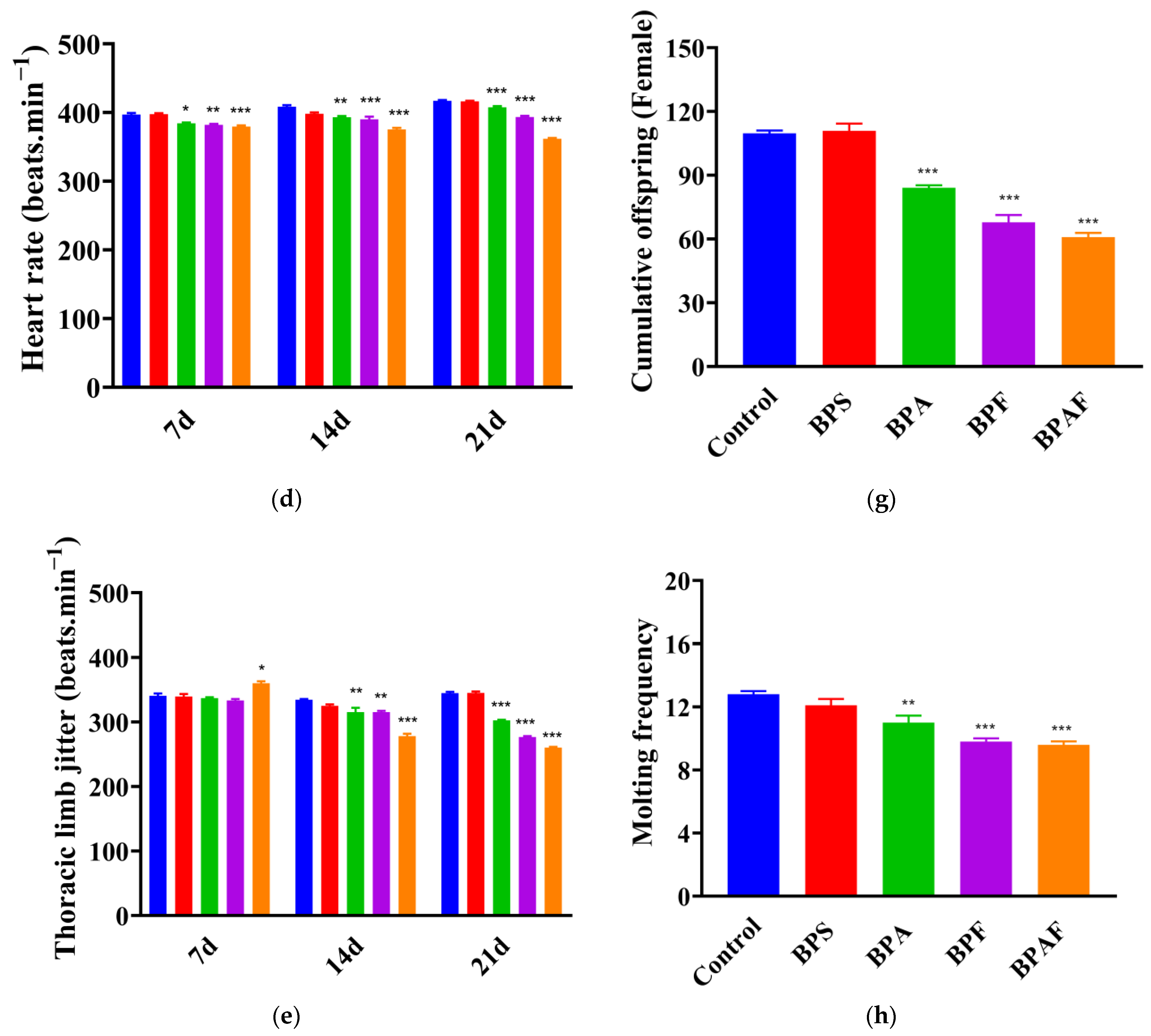
No mortality of the F0 generation was observed during the experimental period. The growth, development, and reproduction of the F0 generation are shown in Figure 1. Compared with the control group, the body length of D. magna was significantly shorter after exposure to both BPF and BPAF for 7 d (Figure 1b,c). As time progressed, D. magna in the BPA group became significantly shorter on 14 d and 21 d. Following BPA, BPF, and BPAF treatment, the heart rates were all significantly reduced at each time point (Figure 1d). Thoracic limb jittering, which can represent movement ability at some levels, exhibited a similar trend to heart rate (Figure 1e). With respect to reproduction, BPF and BPAF treatments may postpone the time to the first brood (Figure 1f). There was also a significant reduction in the number of cumulative offspring in the BPA, BPF, and BPAF groups (Figure 1g). D. magna reproduction is usually accompanied by molting. Molting frequency was also reduced in the three groups (Figure 1h). In addition, D. magna in the BPS treatment group did not show any significant differences in all of the six parameters.

2.2. Development and Reproduction of the F1 Generation

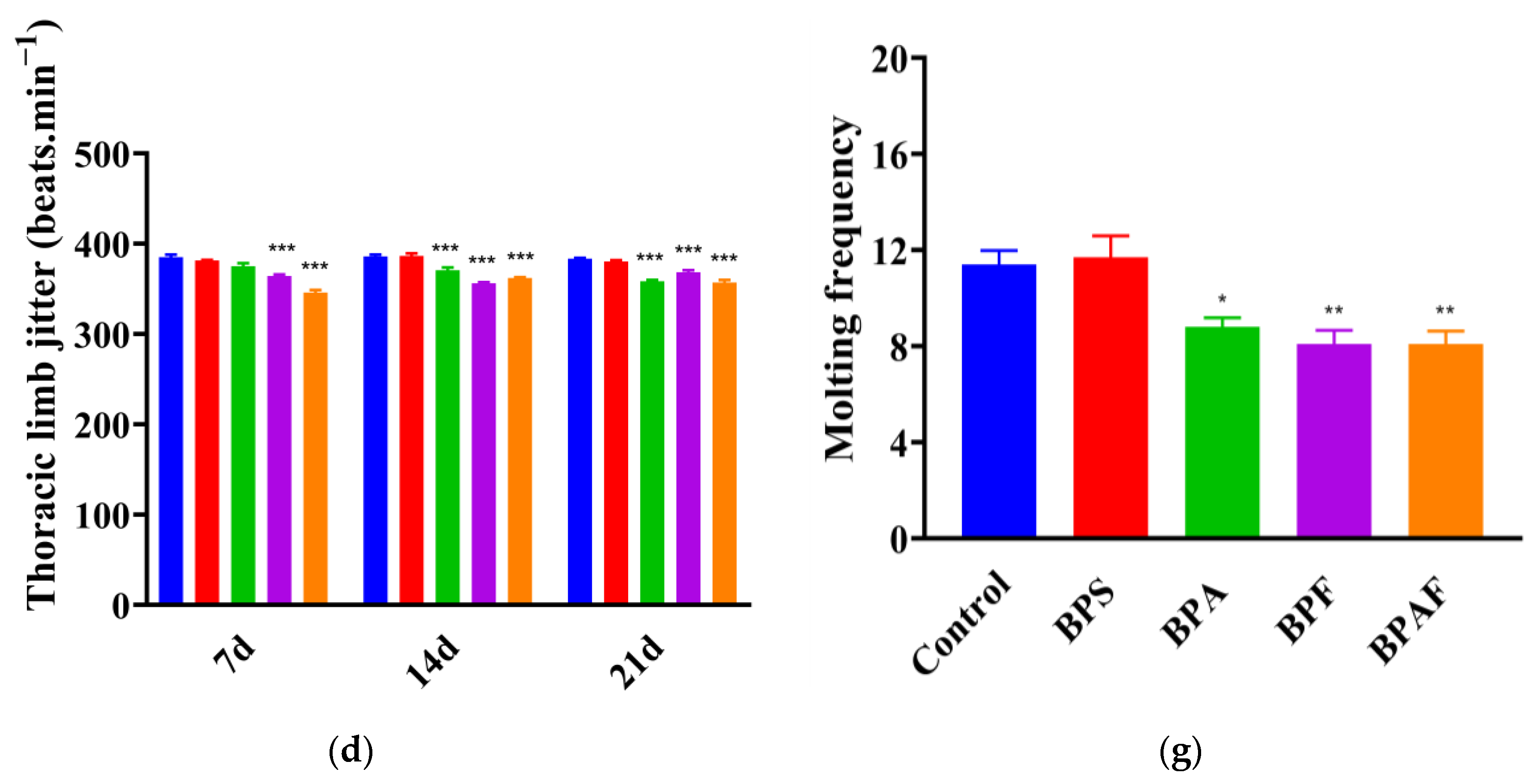
Figure 2 shows the growth, development, and reproduction of the F1 generation. The body length of the offspring was similar to that of the F0 generation, with D. magna exposed to BPF and BPAF for 14 or 21 d showing a significant decrease in body length (Figure 2a,b). Offspring of the BPA, BPF, and BPAF maternal treatment groups exhibited a reduced heart rate compared with that of the control group. Over time, this occurrence gradually became more evident (Figure 2c). The trend in thoracic limb tremors was similar to that of the heartbeat, both showing a reduction in the beating rate of the offspring after BPA, BPF, and BPAF treatment at 14 d and 21 d (Figure 2d). Additionally, the reproduction of the F1 generation was influenced by exposure to BPA and its analogs, and this manifested specifically as the deferred first brood time, reduction in cumulative offspring, and decreasing molting frequency (Figure 2e–g).

2.3. Behavioral Response

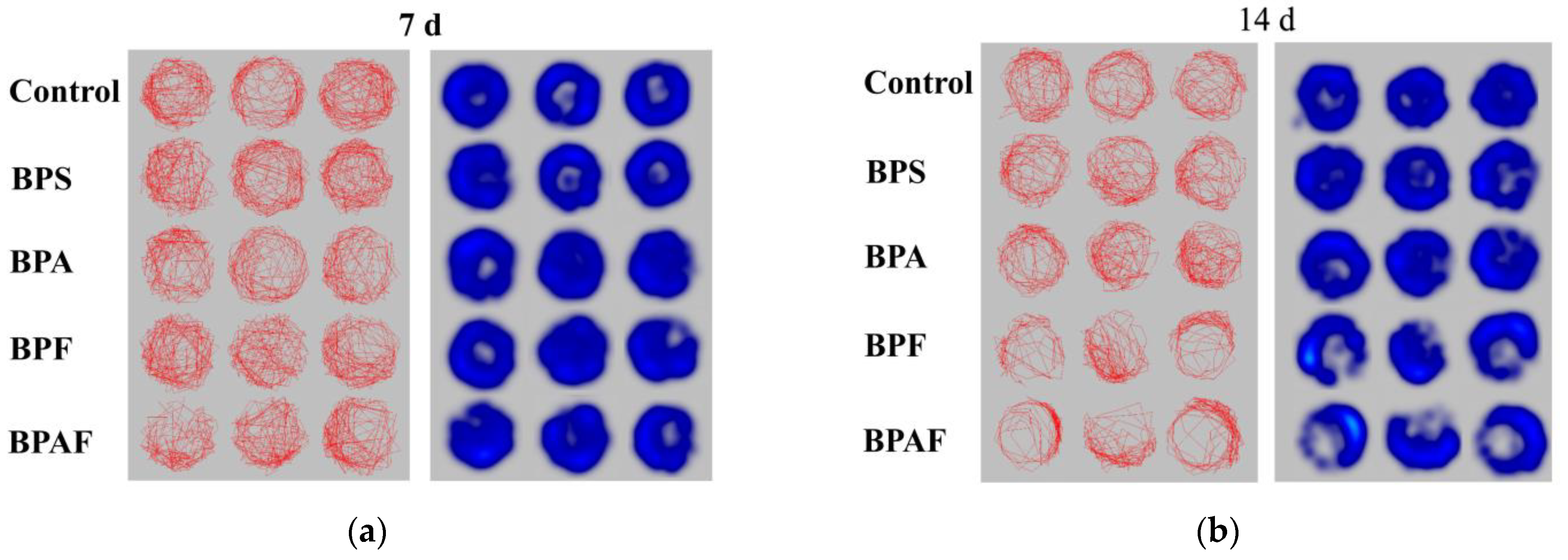
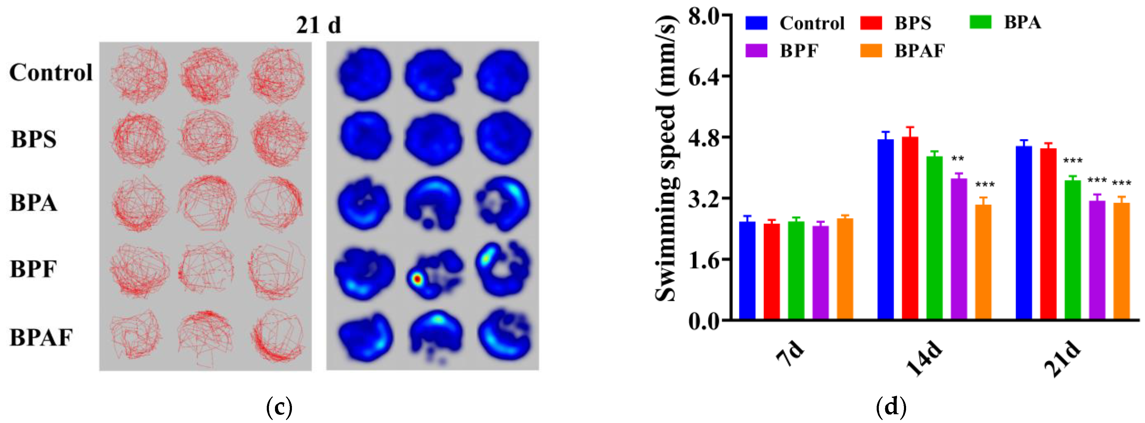
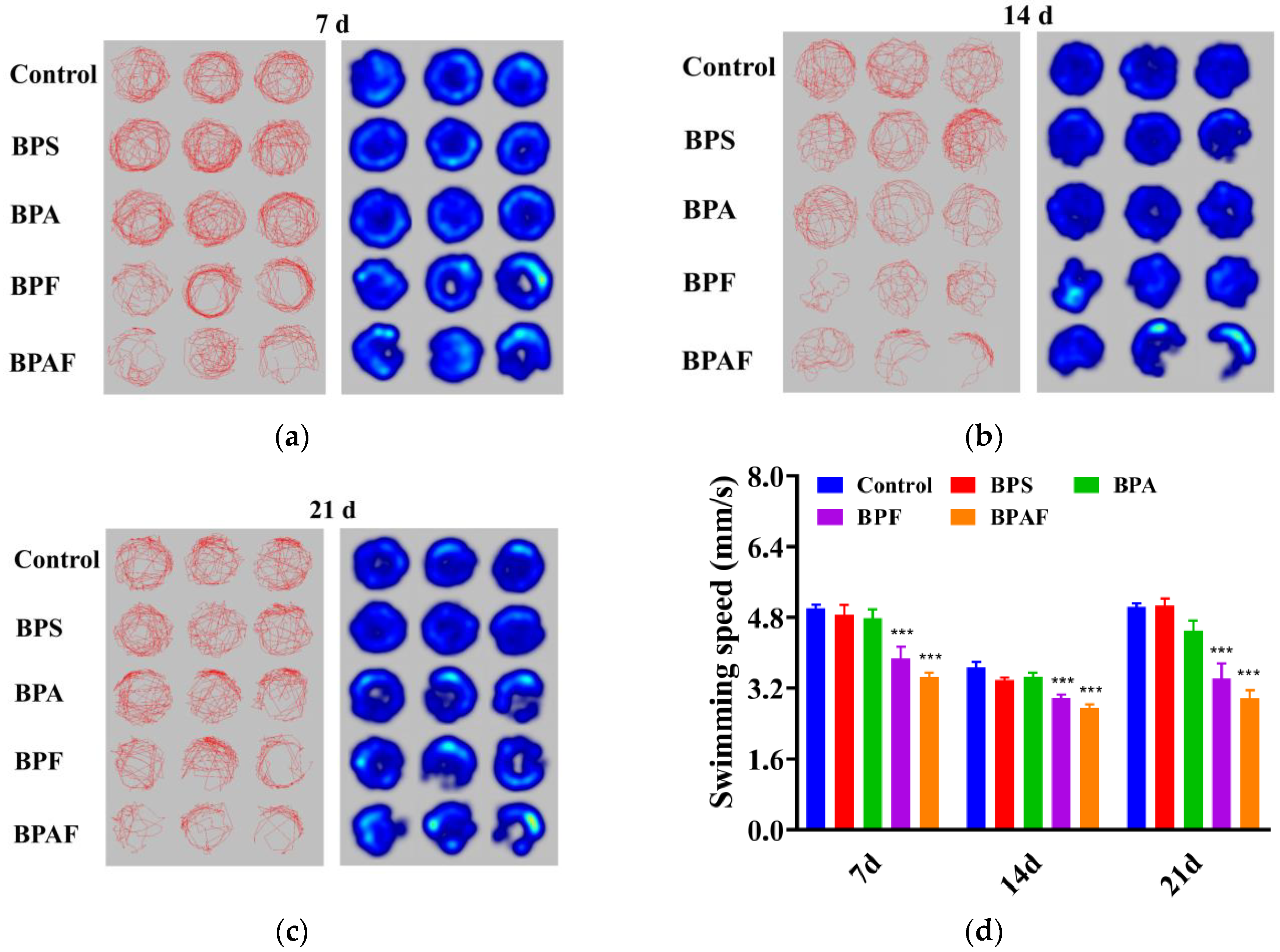
The behavioral response of the F0 generation is shown in Figure 3. As shown in Figure 3a–c, D. magna tended to be static in the treatment group, especially in the BPF and BPAF treatment groups, after 21 d of exposure. After 7 d of exposure, there was no significant difference in swimming speed between the control and exposed groups. The swimming speeds of D. magna treated with BPS, BPA, BPF, and BPAF were 1%, 9%, 22% (p < 0.05), and 36% (p < 0.01) of the control group, respectively, after 14 d of exposure and 1%, 20% (p < 0.001), 31% (p < 0.001), and 33% (p < 0.001) after 21 d (Figure 3d).
Figure 4 shows the behavioral response of the F1 generation. The behavioral traits of the F1 generation had a similar tendency to those of the F0 generation. Compared with the control group, the F1 generation in the exposed group moved more sluggishly, especially in the BPF and BPAF groups (Figure 4a–c). The F1 generation differed from the F0 generation with regard to a significant decrease in swimming speed after 7 d of exposure, which increased after 14 d and 21 d (Figure 4d). These results indicated that these chemicals had intergenerational effects on the behavioral response of D. magna.

2.4. Reflection of Antioxidant Enzymatic Activity

BPS, BPA, BPF, and BPAF may have a greater effect on the F0 generation than the F1 generation. The antioxidant enzymatic activities in almost all the samples collected from the F0 generation (except for the activity of MDA with BPS treatment) were significantly increased (Figure 5a–c), whereas only SOD in the BPA group, MDA in the BPF group, and CAT and MDA in the BPAF group showed significant increases among these indexes (Figure 5d–f).

2.5. Expressions of Genes Related to Development and Reproduction

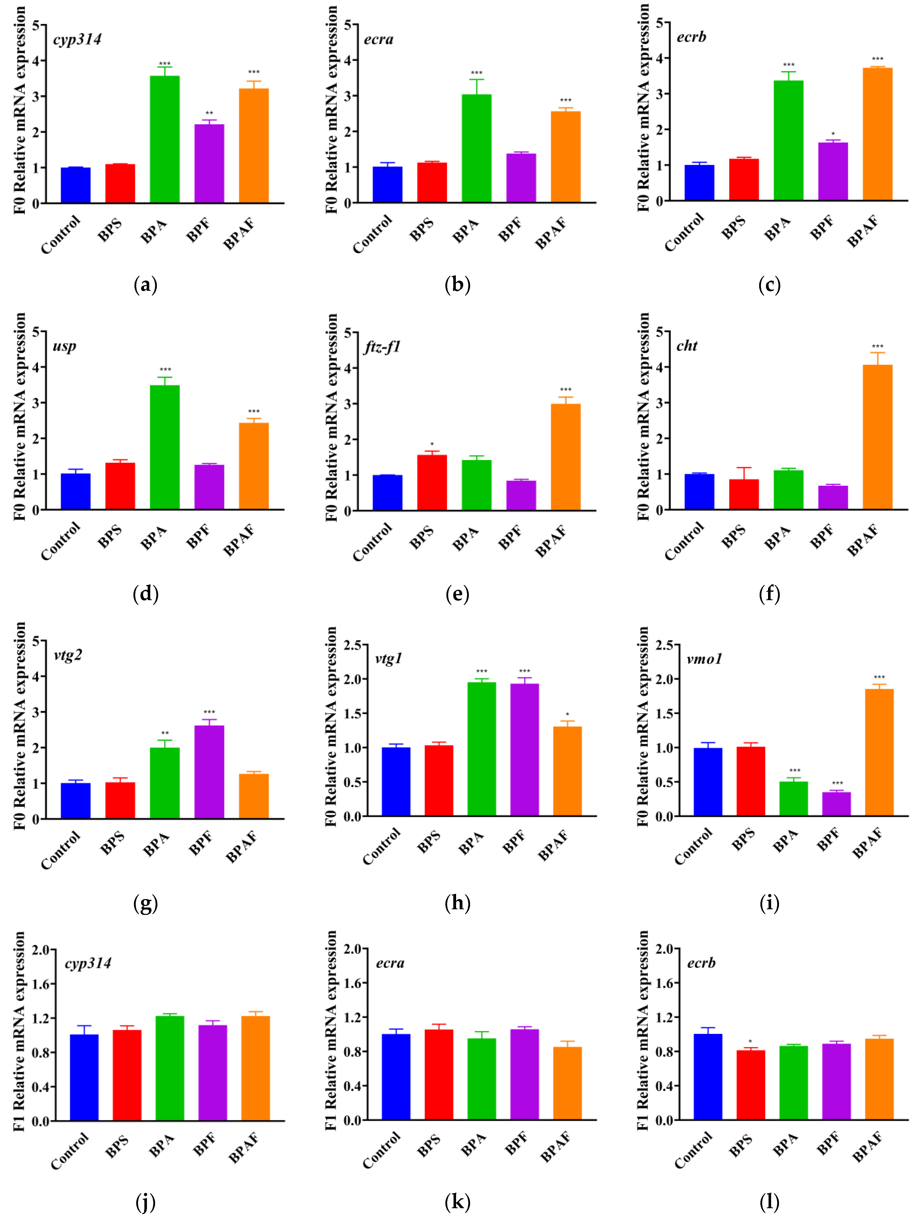
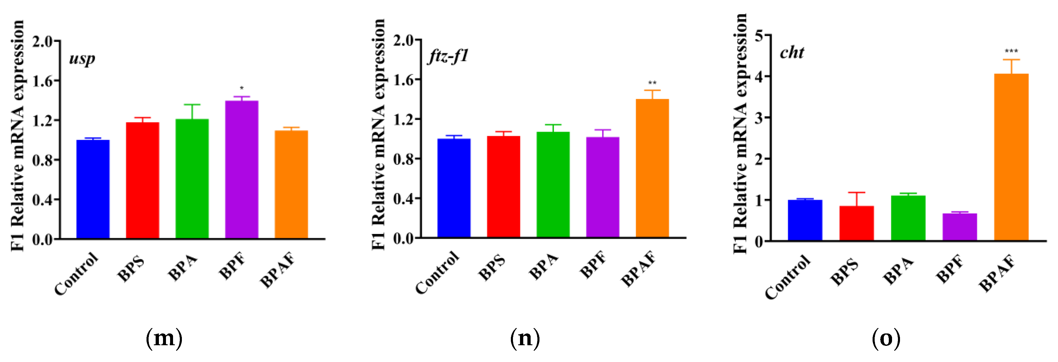
Figure 6 shows that, in the F0 generation, the expression of genes related to development significantly increased after BPAF exposure, whereas cyp314, ecra, ecrb, and usp expression significantly increased after BPA exposure. BPF significantly increased the expression of cyp314 and ecrb, whereas BPS significantly increased the expression of ftz-f1. The reproduction of D. magna was visibly influenced after exposure to BPA and its analogs; vtg1 and vtg2 expression improved significantly with BPA, BPF, and BPAF treatment, and vmo1 expression was inhibited by BPA and BPF treatment. Additionally, in the F1 generation, BPAF significantly increased the expression of ftz-f1 and cht in the offspring and BPF significantly increased the expression of usp, whereas BPS inhibited ecrb expression.

3. Discussion

With the strict restriction of BPA in recent years, BPA analogs have been increasingly used worldwide [5,6]. It has been reported that these chemicals potentially have toxic effects on vertebrates. Previous studies elaborated on the neurotoxicity of BPA and its analogs in zebrafish [15]. Ji, et al. [20] compared and evaluated the vascular toxicity and oxidative stress potency of the BPs in zebrafish and ranked them as follows: BPAF > BPF > BPA > BPS. Moreman, et al. [32] illustrated that BPA and its analogs (BPAF, BPF, and BPS) hindered the development of larval zebrafish, leading to cardiac edema, spinal malformation, and craniofacial deformities. However, few studies have tested the toxicity of BPs in arthropods. Here, we conducted a comprehensive study of the developmental and reproductive toxicity of BPA, BPS, BPF, and BPAF in D. magna and assessed their toxic effects. Our results showed that after exposure to BPA and its analogs, both the F0 and F1 generations of D. magna exhibited abnormalities in development and reproduction.
Body length can reflect the developmental situation at some levels. Our study showed that, compared with the control group, the body length in the exposed group was reduced, especially in the BPF and BPAF groups, and this inhibition became clearer over time. Heart rate and thoracic limb jittering are linked to feeding behavior or respiratory metabolism, indicating the health and growth of organisms and the stress response to pollutants [33]. Both were significantly decreased in the F0 and F1 generations after BPA, BPF, and BPAF treatment, whereas there was no significant difference in the group exposed to BPS. The thoracic limb is the food-intake organ of D. magna, and alterations in the thoracic limb may result in feeding difficulties. It is possible that the depression of heart rate led to a decrease in thoracic limb frequency or that BPAF affected the nervous system [34,35].
Reproductive ability is an important index for detecting the health of the population of D. magna and is widely used in contaminant toxicity risk assessment [30]. Although molting frequency is not directly related to spawning ability, some studies have shown a positive correlation between molting and spawning times [36]. We found that the time to the first brood was postponed, whereas the molting frequency was declining in the F0 generation. The cumulative offspring decreased in the F0 generation, which meant that reproduction was influenced by exposure. Moreover, this occurrence did not return to normal levels in juveniles, which indicated that the damage to reproductive capacity in the exposed group may have an intergenerational effect.
In this study, when exposed to BPA and its analogs, D. magna appeared to inhibit locomotor behavior in the F0 generation as well as in the F1 generation. Similar phenomena have also been observed in other animals, exhibiting inhibition of behavior. Gu illustrated that after BPS, BPF, BPA, and BPAF exposure, zebrafish appeared to suffer oxidative stress, larval hypoactivity, and dysregulated neuronal development [15]. Kim, et al. [37] demonstrated that exposure to BPS or other EDs can cause anxiety and reduced social behavior in juvenile mice. In terms of effects during generations, Wolstenholme, et al. [38] found that BPA exposure may influence the social recognition of both parent and descendant rats and has a long-lasting and transgenerational effect even after three generations of BPA exposure, which was consistent with our results.
It has been reported that BPs enhance reactive oxygen species (ROS) formation and damage the structures of lipids and proteins, which can lead to oxidative damage of cells [39,40]. Previous studies have shown that BPA may induce oxidization system disorders in the liver and nervous system [41,42]. Our results showed that exposure to all four BPs significantly increased the activity of CAT and SOD, and three of the BPs (except BPS) reciprocally increased the levels of MDA, indicating that BPs may induce oxidative stress in D. magna.
As mentioned previously, developmental effects and reproductive impairment may have intergenerational effects, which can often be demonstrated using RT-qPCR analysis. The development and reproduction of D. magna may be linked to molting, which can be regulated by ecdysteroids (20-hydroxyecdysone, 20-E) and juvenile hormones (JH) [36]. Cytochrome P450s (CYPs) are a superfamily of heme proteins that are vital for drug metabolism, bioactivation, and breakdown of xenobiotics [43,44]. Cyp314 belongs to this family and is responsible for synthesizing the molting hormone 20-E and converting it to its active form, which can regulate the molting and reproduction cycles [45,46]. In contrast to previous studies [47], our study showed that there is an up-regulation of cyp314 expression in D. magna of F0 generation after BPA, BPF, and BPAF exposure, which may account for the different exposure conditions. When ecdysone 20-E is combined with the protein ecdysone receptor (EcR) and ultraspiracle (USP) to form a nuclear heterodimer complex (EcR/USP-20E), the expression of ecra, ecrb, and usp is altered [45,48]. Moreover, ftz-f1 may also be expressed after the presentation of 20-E and incentivize the expression of downstream cht and other genes [49]. The cht gene is involved in cuticle metabolism [47]. The dysregulation of ecra, ecrb, usp, ftz-f1, and cht in both the F0 and F1 generations may indicate that BPA and its analogs indeed had endocrine effects and deteriorated the molting and metabolism of D. magna. Owing to the abnormal fluctuation still occurring in the F1 generation, it can be deduced that the damage may have intergenerational effects in terms of development.
Exposure to BPA and its analogs led to abnormalities in the gene expression of D. magna, which were related to both development and reproduction. As the precursor of vitellin, vitellogenin (vtg) is considered an energy source for the development of offspring in oviparous organisms [50,51]. Gene vom1 prevents the yolk from mixing with the albumen and protecting the egg from bacterial infection [52]. In our study, the expression of vtg1 and vtg2 improved significantly with BPA, BPF, and BPAF treatment, whereas the expression of vmo1 was inhibited by BPA and BPF treatment, which indicated that BP exposure impaired the reproductive system of D. magna. Several studies have demonstrated that endocrine disruptors can alter the expression of reproductive genes in organisms, and dysregulation of genes can be transferred during generations [50,53,54].

4. Materials and Methods

4.1. Experimental Animals

D. magna was provided by the Institute of Hydrobiology (Wuhan, China). The culturing method was performed as previously described [55]. Before the experiment, the organisms were incubated for ten generations in deionized water after aeration for 48 h (22 ± 1 °C, pH = 7.50 ± 0.15, salinity 200–230 mg/L). The lighting conditions were a 16 h light/8 h dark cycle under a light intensity of 1000 lx. D. magna were fed 2 mL of purebred Scenedesmus obliquus (approximately 106 cells mL−1 per organism) twice daily. The exposure solutions were changed every 2 d. The non-first larval D. magna was selected for toxicity exposure tests based on the OECD211 (Organization for Economic Co-operation and Development Guide standard method) [27].

4.2. Chemicals and Test Solutions

BPA (purity: 99%, CAS: 80-05-7), BPF (purity: 99%, CAS: 620-92-8), BPAF (purity: 99%, CAS: 147861-1), and BPS (purity: 99%, CAS: 80-09-1) were purchased from Bailingway Technology Co., Ltd. (Shanghai). Dimethyl sulfoxide (DMSO) was purchased from Sigma-Aldrich. Chlorella spp. and Plagiostellae spp. were purchased from Wuhan Institute of Hydrology. These four types of BPs were prepared in stock solutions at a concentration of 105 mg/L.

4.3. Chronic Toxicity Test

The concentrations were determined to be 100 μg/L according to the environmentally relevant concentration [13], and the stock solution was diluted to the final test solution with deionized water. For the exposure treatment, each group had ten replicates and each replicate had one D. magna (F0 generation), which were randomly separated into 100 mL sterile beakers with 80 mL test solution. All the control groups of the other chemicals received 0.01% (v/v) DMSO. D. magna fasted during the test, and other culture conditions were consistent with those described in Section 4.1. The number of new offspring was counted and recorded daily. The offspring (F1 generation) born on days 7, 14, and 21 after maternal exposure (7 d, 14 d, and 21 d) were collected and transferred into clean water [56,57]. Furthermore, molting, survival, and reproduction (both F0 and F1 generations) were recorded at 7 d, 14 d, and 21 d. In addition, behavior tests were performed at these three time points. After 21 d of culture, samples (both F0 and F1 generations) were collected for the following tests.

4.4. Heartbeat, Body Length, and Thoracic Limb Beat Frequency Measurement

A D. magna, which was able to move (including its antennae) at least every 15 s was considered to be alive [58]. Ten F1 generations were randomly selected from each replicate. F0 and F1 generations were observed by a Research Stereo Microscope (SMZ25, Nikon, Japan). The length from the top of the helmet to the base of the tail (excluding the tail spur) was measured as the body length of D. magna. The number of heart beats and the number of thoracic limb beats that survived for 1 min were recorded by manual counting as heart beats (one per minute) and thoracic limb shakes (one per minute).

4.5. Behavioral Tests

Behavioral tests were conducted using a DanioVision observation chamber (Noldus, Wageningen, Netherlands). According to the method of a previous study [23], 10 D. magna F0 or F1 generations were selected from each exposure treatment. A 48-well plate was used for detection, with one D. magna in each well filled with 1 mL of exposure solution. The test program was set for 35 min, including 5 min of dark acclimation and three 10-min light-and-dark cycles, which consisted of 5 min of light and 5 min of darkness. Movement traits, heat maps, and swimming speed were analyzed using EthoVision XT 15 video tracking software (Noldus, Wageningen, The Netherlands).

4.6. Real-Time Quantitative Reverse Transcription-Polymerase Chain Reaction (RT-qPCR)

After 21 d of exposure, ten D. magna from each exposure treatment were mixed for further study of gene transcription using TRIzol reagent (Thermo Fisher, Waltham, MA, USA), according to the manufacturer’s instructions. RNA concentration was measured using a NanoDrop2000 (Thermo Fisher), and cDNA was synthesized using a PrimeScript® RT reagent kit (TaKaRa, Shiga, Japan). The expression of the following genes was measured: cytochrome p450 314 (cyp314), ecdysone receptor a (ecra), ecdysone receptor b (ecrb), ultraspiracle (usp), fushi tarazu factor-1 (ftz-f1), chitinase (cht) (which are related to development) and vitellogenin 1 (vtg1), vitellogenin 2 (vtg2), vitelline outer layer membrane protein 1 (vmo1) (which are related to reproduction). The primers of these genes are shown in Table S2.

4.7. Determination of Antioxidant Enzymatic Activity

The lysate was homogenized using an ultrasonic crushing instrument and the protein concentration was determined using a protein detection kit (Biyuntian, Shanghai, China). Superoxide dismutase (SOD; Biyuntian) and catalase (CAT; Biyuntian) (MDA; Biyuntian) detection kits were used to determine the enzymatic activities of the different treatment groups.

4.8. Statistical Analysis

The data were analysed with one-way analysis of variance (ANOVA) using GraphPad Prism 8.0.1 software. The results are expressed as the mean value ± standard error of mean (SEM). Asterisks indicate significant differences between exposure and control, * indicates a significant difference (* p < 0.05), ** indicates a very significant difference (** p < 0.01), and *** indicates a highly significant difference (*** p < 0.001).

5. Conclusions

BPA has been strictly restricted because of its strong endocrine effects, and analogs have been invented for broad applications. However, the toxicity of these substitutes has not been fully evaluated. In the present study, we found that BPA alternatives induce similar toxic and estrogenic effects to BPA, and the toxicity of its three BPA substitutes (BPS, BPF, and BPAF) on the growth and reproduction of D. magna may be not weaker than those of BPA, which manifest as growth inhibition, oxidative stress, and altered gene expression. Considering all aspects, the toxicity of these four chemicals in Daphnia magna should be in the order BPAF > BPF ≈ BPA > BPS, which is consistent with the findings of previous studies conducted on zebrafish. Given that BPAF and BPF have toxic effects comparable to those of BPA, BPAF and BPF should be carefully considered as alternatives to BPA. In addition, we should be more concerned about the long-term low-dose effects of such alternatives at environmental concentrations. We can step up publicity to establish the perceptions of BPs’ impacts, adjust lifestyle habits to reduce daily exposure, and establish corresponding laws and regulations to cut off the emission of BPs [4].

Supplementary Materials

The following supporting information can be downloaded at: https://www.mdpi.com/article/10.3390/ijms232314561/s1, Table S1: Abbreviation and description of the parameters in this study; Table S2: Primers used for qPCR validation.

Author Contributions

Conceptualization, G.W. and L.Q.; methodology, G.W.; software, C.C., L.G. and X.Z. (Xiangling Zhang); formal analysis, L.Q.; investigation, J.D.; resources, L.G.; data curation, L.Q.; writing—original draft preparation, C.C.; writing—review and editing, C.C.; visualization, J.Z.; supervision, X.Z. (Xiaofei Zhang); project administration, X.Z. (Xiaofei Zhang). All authors have read and agreed to the published version of the manuscript.

Funding

This research was funded by Funds for Independent Innovation of Agricultural Science and Technology in Jiangsu Province (CX (20) 3084) and Jiangsu Social Development Project (Grant No.: BE2020781).

Institutional Review Board Statement

All the animal experiments were conducted in accordance with the guidelines for the care and use of laboratory animals of the Nanjing Institute of Environmental Sciences.

Informed Consent Statement

Not applicable.

Data Availability Statement

Not applicable.

Conflicts of Interest

The authors declare no conflict of interest.

References

  1. Fischnaller, M.; Bakry, R.; Bonn, G.K. A simple method for the enrichment of bisphenols using boron nitride. Food Chem. 2016, 194, 149–155. [Google Scholar] [CrossRef]
  2. Cipelli, R.; Harries, L.; Okuda, K.; Yoshihara, S.; Melzer, D.; Galloway, T. Bisphenol A modulates the metabolic regulator oestrogen-related receptor-alpha in T-cells. Reproduction 2014, 147, 419–426. [Google Scholar] [CrossRef] [Green Version]
  3. Huang, Y.Q.; Wong, C.K.; Zheng, J.S.; Bouwman, H.; Barra, R.; Wahlstrom, B.; Neretin, L.; Wong, M.H. Bisphenol A (BPA) in China: A review of sources, environmental levels, and potential human health impacts. Environ. Int. 2012, 42, 91–99. [Google Scholar] [CrossRef] [PubMed]
  4. Rahman, M.S.; Adegoke, E.O.; Pang, M.G. Drivers of owning more BPA. J Hazard Mater. 2021, 417, 126076. [Google Scholar] [CrossRef] [PubMed]
  5. Qiu, W.; Zhan, H.; Hu, J.; Zhang, T.; Xu, H.; Wong, M.; Xu, B.; Zheng, C. The occurrence, potential toxicity, and toxicity mechanism of bisphenol S, a substitute of bisphenol A: A critical review of recent progress. Ecotoxicol. Environ. Saf. 2019, 173, 192–202. [Google Scholar] [CrossRef] [PubMed]
  6. Zhang, L.; Fang, P.; Yang, L.; Zhang, J.; Wang, X. Rapid method for the separation and recovery of endocrine-disrupting compound bisphenol AP from wastewater. Langmuir 2013, 29, 3968–3975. [Google Scholar] [CrossRef] [PubMed]
  7. Xue, J.; Wan, Y.; Kannan, K. Occurrence of bisphenols, bisphenol A diglycidyl ethers (BADGEs), and novolac glycidyl ethers (NOGEs) in indoor air from Albany, New York, USA, and its implications for inhalation exposure. Chemosphere 2016, 151, 1–8. [Google Scholar] [CrossRef] [PubMed]
  8. Jin, H.; Zhu, L. Occurrence and partitioning of bisphenol analogues in water and sediment from Liaohe River Basin and Taihu Lake, China. Water Res. 2016, 103, 343–351. [Google Scholar] [CrossRef]
  9. Lu, L.; Yang, Y.; Zhang, J.; Shao, B. Determination of seven bisphenol analogues in reed and Callitrichaceae by ultra performance liquid chromatography-tandem mass spectrometry. J. Chromatogr. B Analyt. Technol. Biomed. Life Sci. 2014, 953–954, 80–85. [Google Scholar] [CrossRef]
  10. Jin, H.; Zhu, J.; Chen, Z.; Hong, Y.; Cai, Z. Occurrence and Partitioning of Bisphenol Analogues in Adults’ Blood from China. Environ. Sci. Technol. 2018, 52, 812–820. [Google Scholar] [CrossRef]
  11. Yang, Y.; Guan, J.; Yin, J.; Shao, B.; Li, H. Urinary levels of bisphenol analogues in residents living near a manufacturing plant in south China. Chemosphere 2014, 112, 481–486. [Google Scholar] [CrossRef]
  12. Liu, A.-F.; Qu, G.-B.; Yu, M.; Liu, Y.-W.; Shi, J.-B.; Jiang, G.-B. Tetrabromobisphenol-A/S and nine novel analogs in biological samples from the Chinese Bohai Sea: Implications for trophic transfer. Environ. Sci. Technol. 2016, 50, 4203–4211. [Google Scholar] [CrossRef] [PubMed]
  13. Rotimi, O.A.; Olawole, T.D.; De Campos, O.C.; Adelani, I.B.; Rotimi, S.O. Bisphenol A in Africa: A review of environmental and biological levels. Sci. Total Environ. 2021, 764, 142854. [Google Scholar] [CrossRef]
  14. Wang, Q.; Chen, M.; Shan, G.; Chen, P.; Cui, S.; Yi, S.; Zhu, L. Bioaccumulation and biomagnification of emerging bisphenol analogues in aquatic organisms from Taihu Lake, China. Sci. Total Environ. 2017, 598, 814–820. [Google Scholar] [CrossRef] [PubMed]
  15. Gu, J.; Guo, M.; Yin, X.; Huang, C.; Qian, L.; Zhou, L.; Wang, Z.; Wang, L.; Shi, L.; Ji, G. A systematic comparison of neurotoxicity of bisphenol A and its derivatives in zebrafish. Sci. Total Environ. 2022, 805, 150210. [Google Scholar] [CrossRef] [PubMed]
  16. Crump, D.; Sharin, T.; Chiu, S.; O’Brien, J.M. In Vitro Screening of 21 Bisphenol A Replacement Alternatives: Compared with Bisphenol A, the Majority of Alternatives Are More Cytotoxic and Dysregulate More Genes in Avian Hepatocytes. Environ. Toxicol. Chem. 2021, 40, 2026–2033. [Google Scholar] [CrossRef]
  17. Le Fol, V.; Ait-Aissa, S.; Sonavane, M.; Porcher, J.M.; Balaguer, P.; Cravedi, J.P.; Zalko, D.; Brion, F. In vitro and in vivo estrogenic activity of BPA, BPF and BPS in zebrafish-specific assays. Ecotoxicol. Environ. Saf. 2017, 142, 150–156. [Google Scholar] [CrossRef]
  18. Mu, X.; Qi, S.; Wang, H.; Yuan, L.; Wang, C.; Li, Y.; Qiu, J. Bisphenol analogues induced metabolic effects through eliciting intestinal cell heterogeneous response. Environ. Int. 2022, 165, 107287. [Google Scholar] [CrossRef]
  19. Gu, J.; Wu, J.; Xu, S.; Zhang, L.; Fan, D.; Shi, L.; Wang, J.; Ji, G. Bisphenol F exposure impairs neurodevelopment in zebrafish larvae (Danio rerio). Ecotoxicol. Environ. Saf. 2020, 188, 109870. [Google Scholar] [CrossRef]
  20. Ji, G.; Gu, J.; Guo, M.; Zhou, L.; Wang, Z.; Shi, L.; Gu, A. A systematic comparison of the developmental vascular toxicity of bisphenol A and its alternatives in vivo and in vitro. Chemosphere 2022, 291 Pt 2, 132936. [Google Scholar] [CrossRef]
  21. Gu, J.; Li, L.; Yin, X.; Liang, M.; Zhu, Y.; Guo, M.; Zhou, L.; Fan, D.; Shi, L.; Ji, G. Long-term exposure of zebrafish to bisphenol F: Adverse effects on parental reproduction and offspring neurodevelopment. Aquat. Toxicol. 2022, 248, 106190. [Google Scholar] [CrossRef] [PubMed]
  22. Chen, J.; Saili, K.S.; Liu, Y.; Li, L.; Zhao, Y.; Jia, Y.; Bai, C.; Tanguay, R.L.; Dong, Q.; Huang, C. Developmental bisphenol A exposure impairs sperm function and reproduction in zebrafish. Chemosphere 2017, 169, 262–270. [Google Scholar] [CrossRef]
  23. Yuan, S.; Liang, C.; Li, W.; Letcher, R.J.; Liu, C. A comprehensive system for detection of behavioral change of D. magna exposed to various chemicals. J. Hazard. Mater. 2021, 402, 123731. [Google Scholar] [CrossRef]
  24. Okamoto, A.; Yamamuro, M.; Tatarazako, N. Acute toxicity of 50 metals to Daphnia magna. J. Appl. Toxicol. 2015, 35, 824–830. [Google Scholar] [CrossRef] [PubMed]
  25. Wang, P.; Huang, B.; Chen, Z.; Lv, X.; Qian, W.; Zhu, X.; Li, B.; Wang, Z.; Cai, Z. Behavioural and chronic toxicity of fullerene to Daphnia magna: Mechanisms revealed by transcriptomic analysis. Environ. Pollut. 2019, 255 Pt 1, 113181. [Google Scholar] [CrossRef]
  26. Ten Berge, W.F. Breeding Daphnia magna. Hydrobiologia 1978, 59, 121–123. [Google Scholar] [CrossRef]
  27. OECD. Test No. 211: Daphnia Magna Reproduction Test; OECD: Paris, France, 2012. [Google Scholar]
  28. Cano, A.M.; Maul, J.D.; Saed, M.; Shah, S.A.; Green, M.J.; Canas-Carrell, J.E. Bioaccumulation, stress, and swimming impairment in Daphnia magna exposed to multiwalled carbon nanotubes, graphene, and graphene oxide. Environ. Toxicol. Chem. 2017, 36, 2199–2204. [Google Scholar] [CrossRef]
  29. Seda, B.C.; Ke, P.C.; Mount, A.S.; Klaine, S.J. Toxicity of aqueous C70-gallic acid suspension in Daphnia magna. Environ. Toxicol. Chem. 2012, 31, 215–220. [Google Scholar] [CrossRef] [PubMed]
  30. Liu, Y.; Yan, Z.; Zhang, L.; Deng, Z.; Yuan, J.; Zhang, S.; Chen, J.; Guo, R. Food up-take and reproduction performance of Daphnia magna under the exposure of Bisphenols. Ecotoxicol. Environ. Saf. 2019, 170, 47–54. [Google Scholar] [CrossRef]
  31. Wolstenholme, J.T.; Taylor, J.A.; Shetty, S.R.; Edwards, M.; Connelly, J.J.; Rissman, E.F. Gestational exposure to low dose bisphenol A alters social behavior in juvenile mice. PLoS ONE 2011, 6, e25448. [Google Scholar] [CrossRef]
  32. Moreman, J.; Lee, O.; Trznadel, M.; David, A.; Kudoh, T.; Tyler, C.R. Acute Toxicity, Teratogenic, and Estrogenic Effects of Bisphenol A and Its Alternative Replacements Bisphenol S, Bisphenol F, and Bisphenol AF in Zebrafish Embryo-Larvae. Environ. Sci. Technol. 2017, 51, 12796–12805. [Google Scholar] [CrossRef] [PubMed]
  33. Jeong, T.Y.; Yoon, D.; Kim, S.; Kim, H.Y.; Kim, S.D. Mode of action characterization for adverse effect of propranolol in Daphnia magna based on behavior and physiology monitoring and metabolite profiling. Environ. Pollut. 2018, 233, 99–108. [Google Scholar] [CrossRef]
  34. Hirano, M.; Ishibashi, H.; Matsumura, N.; Nagao, Y.; Watanabe, N.; Watanabe, A.; Onikura, N.; Kishi, K.; Arizono, K. Acute Toxicity Responses of Two Crustaceans, Americamysis bahia and Daphnia magna, to Endocrine Disrupters. J. Heath Sci. 2004, 50, 97–100. [Google Scholar] [CrossRef] [Green Version]
  35. Porter, K.G.; Orcutt, J.D.; Gerritsen, J. Functional Response and Fitness in a Generalist Filter Feeder, Daphnia Magna (Cladocera: Crustacea). Ecology 1983, 64, 735–742. [Google Scholar] [CrossRef]
  36. Dai, T.-H.; Sserwadda, A.; Song, K.; Zang, Y.-N.; Shen, H.-S. Cloning and Expression of Ecdysone Receptor and Retinoid X Receptor from Procambarus clarkii: Induction by Eyestalk Ablation. Int. J. Mol. Sci. 2016, 17, 1739. [Google Scholar] [CrossRef] [Green Version]
  37. Kim, B.; Colon, E.; Chawla, S.; Vandenberg, L.N.; Suvorov, A. Endocrine disruptors alter social behaviors and indirectly influence social hierarchies via changes in body weight. Environ. Health 2015, 14, 64. [Google Scholar] [CrossRef] [Green Version]
  38. Wolstenholme, J.T.; Goldsby, J.A.; Rissman, E.F. Transgenerational effects of prenatal bisphenol A on social recognition. Horm. Behav. 2013, 64, 833–839. [Google Scholar] [CrossRef] [Green Version]
  39. Michalowicz, J.; Mokra, K.; Bak, A. Bisphenol A and its analogs induce morphological and biochemical alterations in human peripheral blood mononuclear cells (in vitro study). Toxicol. In Vitro 2015, 29, 1464–1472. [Google Scholar] [CrossRef]
  40. Ray, P.D.; Huang, B.W.; Tsuji, Y. Reactive oxygen species (ROS) homeostasis and redox regulation in cellular signaling. Cell. Signal. 2012, 24, 981–990. [Google Scholar] [CrossRef] [Green Version]
  41. Huc, L.; Lemarie, A.; Gueraud, F.; Helies-Toussaint, C. Low concentrations of bisphenol A induce lipid accumulation mediated by the production of reactive oxygen species in the mitochondria of HepG2 cells. Toxicol. In Vitro 2012, 26, 709–717. [Google Scholar] [CrossRef] [PubMed]
  42. Lee, S.; Suk, K.; Kim, I.K.; Jang, I.S.; Park, J.W.; Johnson, V.J.; Kwon, T.K.; Choi, B.J.; Kim, S.H. Signaling pathways of bisphenol A-induced apoptosis in hippocampal neuronal cells: Role of calcium-induced reactive oxygen species, mitogen-activated protein kinases, and nuclear factor-kappaB. J. Neurosci. Res. 2008, 86, 2932–2942. [Google Scholar] [CrossRef]
  43. Snyder, M.J. Cytochrome P450 enzymes in aquatic invertebrates: Recent advances and future directions. Aquat. Toxicol. 2000, 48, 529–547. [Google Scholar] [CrossRef]
  44. Tyzack, J.D.; Kirchmair, J. Computational methods and tools to predict cytochrome P450 metabolism for drug discovery. Chem. Biol. Drug Des. 2019, 93, 377–386. [Google Scholar] [CrossRef]
  45. Song, Y.; Villeneuve, D.L.; Toyota, K.; Iguchi, T.; Tollefsen, K.E. Ecdysone Receptor Agonism Leading to Lethal Molting Disruption in Arthropods: Review and Adverse Outcome Pathway Development. Environ. Sci. Technol. 2017, 51, 4142–4157. [Google Scholar] [CrossRef] [PubMed] [Green Version]
  46. Shen, B.; Dong, H.-Q.; Tian, H.-S.; Ma, L.; Li, X.-L.; Wu, G.-L.; Zhu, C.-L. Cytochrome P450 genes expressed in the deltamethrin-susceptible and -resistant strains of Culex pipiens pallens. Pestic. Biochem. Phys. 2003, 75, 19–26. [Google Scholar] [CrossRef]
  47. Chen, S.; Li, X.; Li, H.; Yuan, S.; Li, J.; Liu, C. Greater toxic potency of bisphenol AF than bisphenol A in growth, reproduction, and transcription of genes in Daphnia magna. Environ. Sci. Pollut. Res. Int. 2021, 28, 25218–25227. [Google Scholar] [CrossRef]
  48. Nakagawa, Y.; Henrich, V.C. Arthropod nuclear receptors and their role in molting. FEBS J. 2009, 276, 6128–6157. [Google Scholar] [CrossRef] [PubMed] [Green Version]
  49. Seyoum, A.; Pradhan, A.; Jass, J.; Olsson, P.E. Perfluorinated alkyl substances impede growth, reproduction, lipid metabolism and lifespan in Daphnia magna. Sci. Total Environ. 2020, 737, 139682. [Google Scholar] [CrossRef] [PubMed]
  50. Houde, M.; Douville, M.; Giraudo, M.; Jean, K.; Lepine, M.; Spencer, C.; De Silva, A.O. Endocrine-disruption potential of perfluoroethylcyclohexane sulfonate (PFECHS) in chronically exposed Daphnia magna. Environ. Pollut. 2016, 218, 950–956. [Google Scholar] [CrossRef]
  51. Yang, Y.; Zheng, B.; Bao, C.; Huang, H.; Ye, H. Vitellogenin2: Spermatozoon specificity and immunoprotection in mud crabs. Reproduction 2016, 152, 235–243. [Google Scholar] [CrossRef]
  52. Giraudo, M.; Douville, M.; Cottin, G.; Houde, M. Transcriptomic, cellular and life-history responses of Daphnia magna chronically exposed to benzotriazoles: Endocrine-disrupting potential and molting effects. PLoS ONE 2017, 12, e0171763. [Google Scholar] [CrossRef] [Green Version]
  53. Du, Y.; Shi, X.; Liu, C.; Yu, K.; Zhou, B. Chronic effects of water-borne PFOS exposure on growth, survival and hepatotoxicity in zebrafish: A partial life-cycle test. Chemosphere 2009, 74, 723–729. [Google Scholar] [CrossRef] [PubMed]
  54. Ji, K.; Kim, Y.; Oh, S.; Ahn, B.; Jo, H.; Choi, K. Toxicity of perfluorooctane sulfonic acid and perfluorooctanoic acid on freshwater macroinvertebrates (Daphnia magna and Moina macrocopa) and fish (Oryzias latipes). Environ. Toxicol. Chem. 2008, 27, 2159–2168. [Google Scholar] [CrossRef] [PubMed]
  55. Yuan, S.; Li, H.; Dang, Y.; Liu, C. Effects of triphenyl phosphate on growth, reproduction and transcription of genes of Daphnia magna. Aquat. Toxicol. 2018, 195, 58–66. [Google Scholar] [CrossRef] [PubMed]
  56. Blewett, T.A.; Delompre, P.L.; He, Y.; Folkerts, E.J.; Flynn, S.L.; Alessi, D.S.; Goss, G.G. Sublethal and Reproductive Effects of Acute and Chronic Exposure to Flowback and Produced Water from Hydraulic Fracturing on the Water Flea Daphnia magna. Environ. Sci. Technol. 2017, 51, 3032–3039. [Google Scholar] [CrossRef] [Green Version]
  57. Bao, S.; Pan, B.; Wang, L.; Cheng, Z.; Liu, X.; Zhou, Z.; Nie, X. Adverse effects in Daphnia magna exposed to e-waste leachate: Assessment based on life trait changes and responses of detoxification-related genes. Environ. Res. 2020, 188, 109821. [Google Scholar] [CrossRef]
  58. Guo, J.; Liu, J.; Li, S. Influences of bisphenol AF on the reproduction and growth of Daphnia magna. China Environ. Sci. 2019, 39, 4394–4400. [Google Scholar]
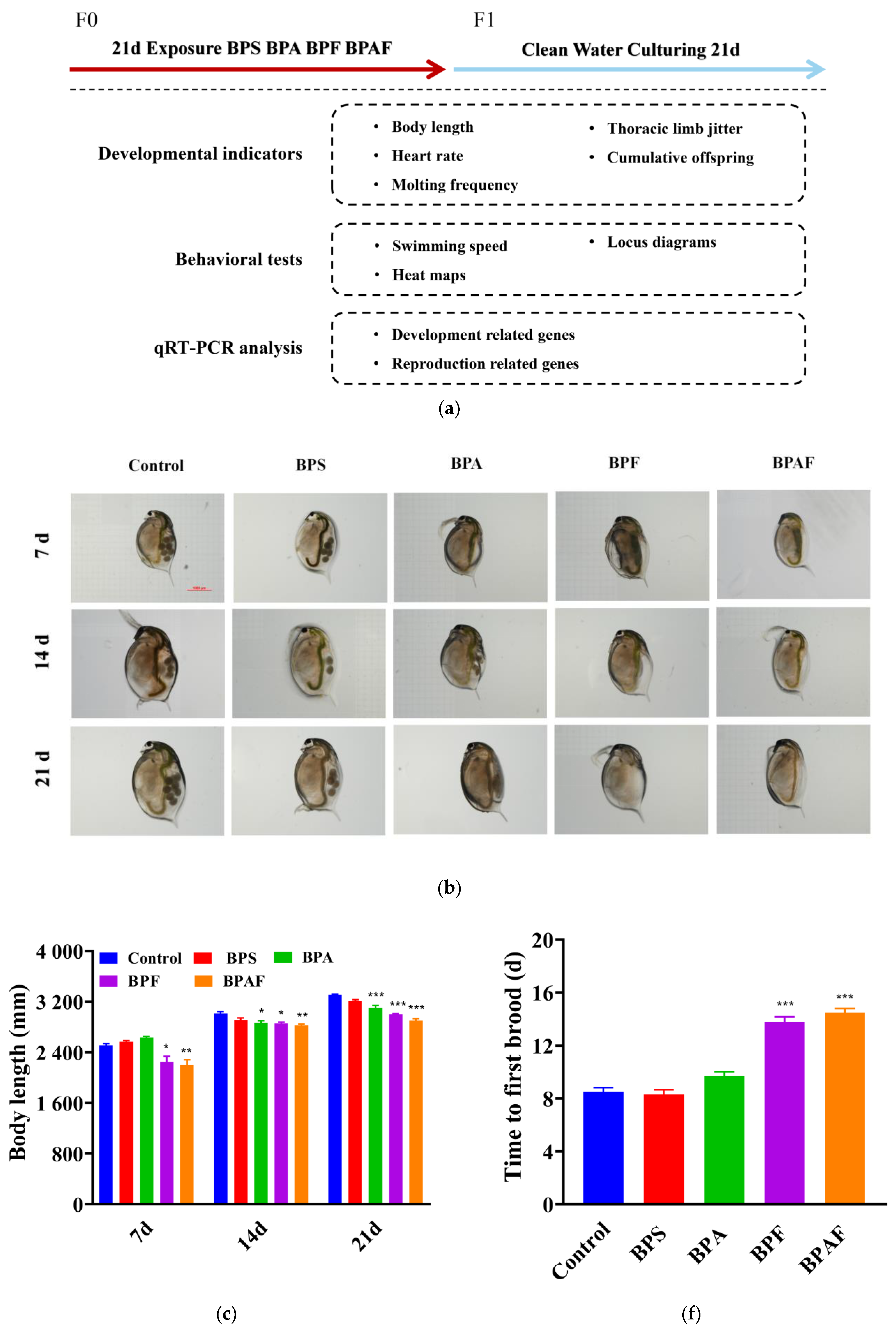
Figure 1. A schematic of the experimental design used (a). Development and reproduction of the F0 gener-ation induced by BPs: D. magna with microscopic observation (b), body length (c), heart rate (d), thoracic limb jittering (e), time to first brood (f), cumulative offspring (g), and molting frequency (h). Asterisks indicate significant differences between exposed and control groups (* p < 0.05, ** p < 0.01, *** p < 0.001).
Ijms 23 14561 g001aIjms 23 14561 g001b
Figure 2. Development and reproduction of the F1 generation induced by BPs. D. magna with microscopic observation (a), body length (b), heart rate (c), thoracic limb jittering (d), time to first brood (e), cumulative offspring (f), and molting frequency (g). Asterisks indicate significant differences be-tween exposed and control groups (* p < 0.05, ** p < 0.01, *** p < 0.001).
Ijms 23 14561 g002aIjms 23 14561 g002b
Figure 3. Behavioral response of the F0 generation. Locus diagrams and heat maps after 7 (a), 14 (b), and 21 (c) d exposure. Swimming speed (d). Asterisks indicate significant differences between the exposed and control groups (** p < 0.01, *** p < 0.001).
Ijms 23 14561 g003aIjms 23 14561 g003b
Figure 4. Behavioral response of the F1 generation. Locus diagrams and heat maps after 7 (a), 14 (b), and 21 (c) d exposure. Swimming speed (d). Asterisks indicate significant differences between the exposed and control groups (*** p < 0.001).
Ijms 23 14561 g004
Figure 5. Reflection of antioxidant enzymatic activity of the F0 generation (ac) and the F1 generation (df). Enzymatic activity of CAT (a,b), SOD (b,e), and MDA (c,f). Asterisks indicate significant differ-ences between the exposed and control groups (* p < 0.05, ** p < 0.01, *** p < 0.001).
Ijms 23 14561 g005
Figure 6. Expression of genes related to development (af,jo) and reproduction (gi) in the F0 generation (ai) and F1 generation (jo). Asterisks indicate significant differences between the exposed and control groups (* p < 0.05, ** p < 0.01, *** p < 0.001).
Ijms 23 14561 g006aIjms 23 14561 g006b
Publisher’s Note: MDPI stays neutral with regard to jurisdictional claims in published maps and institutional affiliations.