Article

The N6-Methyladenosine Regulator ALKBH5 Mediated Stromal Cell–Macrophage Interaction via VEGF Signaling to Promote Recurrent Spontaneous Abortion: A Bioinformatic and In Vitro Study

1
Shanghai Key Laboratory of Maternal Fetal Medicine, Shanghai First Maternity and Infant Hospital, School of Medicine, Tongji University, Shanghai 200092, China
2
Shanghai Key Laboratory of Maternal Fetal Medicine, Shanghai Institute of Maternal-Fetal Medicine and Gynecologic Oncology, Shanghai First Maternity and Infant Hospital, School of Medicine, Tongji University, Shanghai 200092, China
*
Author to whom correspondence should be addressed.
These authors contributed equally to this work.
Int. J. Mol. Sci. 2022, 23(24), 15819; https://doi.org/10.3390/ijms232415819
Submission received: 14 November 2022 / Revised: 29 November 2022 / Accepted: 6 December 2022 / Published: 13 December 2022
(This article belongs to the Special Issue Molecular Research in Uterine Biology and Pathophysiology)

Abstract

:
Successful conception requires the synchrony of multiple systems and organs. Dysregulation of stromal cell–immune cell interactions has been proposed to be associated with recurrent spontaneous abortion. However, the mechanism of this regulation has not been well elucidated. N6-methyladenosine is one of the most common RNA modifications, and is involved in many pathological processes. Our group has demonstrated that abnormal patterns of m6A modification inhibit trophoblast invasion and contribute to adverse pregnancy outcomes. The association between m6A regulators and stromal cell–immune cell interactions is unclear. We obtained RNA-seq profiles from a GEO dataset and identified differentially expressed m6A regulators between healthy controls and patients with a recurrent spontaneous abortion history. ROC curves, functional enrichment and subclassification analysis were applied to elucidate the role of m6A regulators in pregnancy. We verified the expression of m6A regulators and constructed an overexpression cell line in a coculture system to reveal ALKBH5 function in stromal cell–macrophage interactions. We identified 11 differentially expressed m6A regulators between healthy controls and patients with a recurrent spontaneous abortion history. Then, we identified the correlation between “eraser” genes and “writer” genes. We tested the predictive abilities of the 11 m6A regulators based on another dataset and verified their expression in primary human endometrial stromal cells. We then subclassified three distinct patterns using the 11 genes and visualized genes related to immune infiltration and macrophage function in each cluster. ALKBH5 was proven to be correlated with recurrent spontaneous abortion. To verify the role of ALKBH5 in RSA, we constructed an ALKBH5-overexpression cell line. Finally, we cocultured the overexpression cell line with THP-1 cells. A decrease in M2 differentiation was observed, and this bias could be attributed to the hyposecretion of VEGF in stromal cells. N6-methyladenosine regulators play a pivotal role in stromal cell–immune cell interactions at the maternal–fetal interface. Overexpression of the m6A “eraser” gene ALKBH5 in stromal cells resulted in the hyposecretion of VEGF. Dysregulation of VEGF might impair macrophage recruitment and M2 differentiation, which could be the potential cause of recurrent spontaneous abortion.

1. Introduction

Recurrent spontaneous abortion (RSA) is a distressing pregnancy disorder affecting ~2.5% of women who are trying to conceive. Successful conception requires the coordination of multiple systems and organs, and maternal immunological and endometrial synchrony is one of them [1]. Endometrial stromal cells (ESCs) are key components of the endometrium. During the window of implantation, ESCs surround and establish a direct interaction with the embryo. As the embryo develops, ESCs decidualize, and decidual stromal cells (DSCs) produce numerous factors to mediate the activity of endothelial vascular cells, epithelial cells and immune cells [2,3,4]. Endometrial immune dysfunction has been proposed to be associated with RSA. Among the immunocyte profiles, macrophages, with their flexible plasticity, play a key role in maintaining conception [5]. Although the dominance of macrophage differentiation status continues to shift throughout the process of pregnancy, disrupting the balance may impair the maternal–fetal interface immune microenvironment and result in embryonic loss [6]. ESC/DSC-derived cytokines are involved in macrophage polarization [3]. However, the mechanism of ESC/DSC–macrophage interactions is still unclear.
N6-methyladenosine (m6A) has been identified as one of the most common and abundant RNA modifications. m6A modification is catalyzed by corresponding enzymes called “writers,” “erasers” and “readers”, which can install, remove and recognize, respectively. More than 7000 coding and 300 noncoding RNAs are estimated to contain m6A, and they affect mRNA splicing, stabilization and translation [7]. Aberrancies of m6A modification are associated with a variety of diseases, including female fertility, follicle development, oocyte maturation and RSA. Recent studies found that abnormal patterns of m6A modification inhibited trophoblast invasion and impaired immunotolerance and immune-cell infiltration at the maternal–fetal interface [8,9]. However, there is limited evidence of the role of m6A modification in RSA-related stromal cell–immune cell interactions.
In this study, we comprehensively evaluated the expression level of m6A regulators in healthy controls and RSA patients based on the GSE165004 dataset from the Gene Expression Omnibus (GEO) database. We then identified the subtype classification based on the differentially expressed m6A regulators. A cluster was characterized by high expression of the m6A “eraser” gene ALKBH5 and impaired macrophage differentiation. We hypothesized that this cluster might be linked to RSA. The findings of our study could support a new theory for stromal cell–macrophage interactions and provide guidance for novel immune therapy for RSA patients.

2. Results

2.1. Landscape of the m6A Regulators in RSA

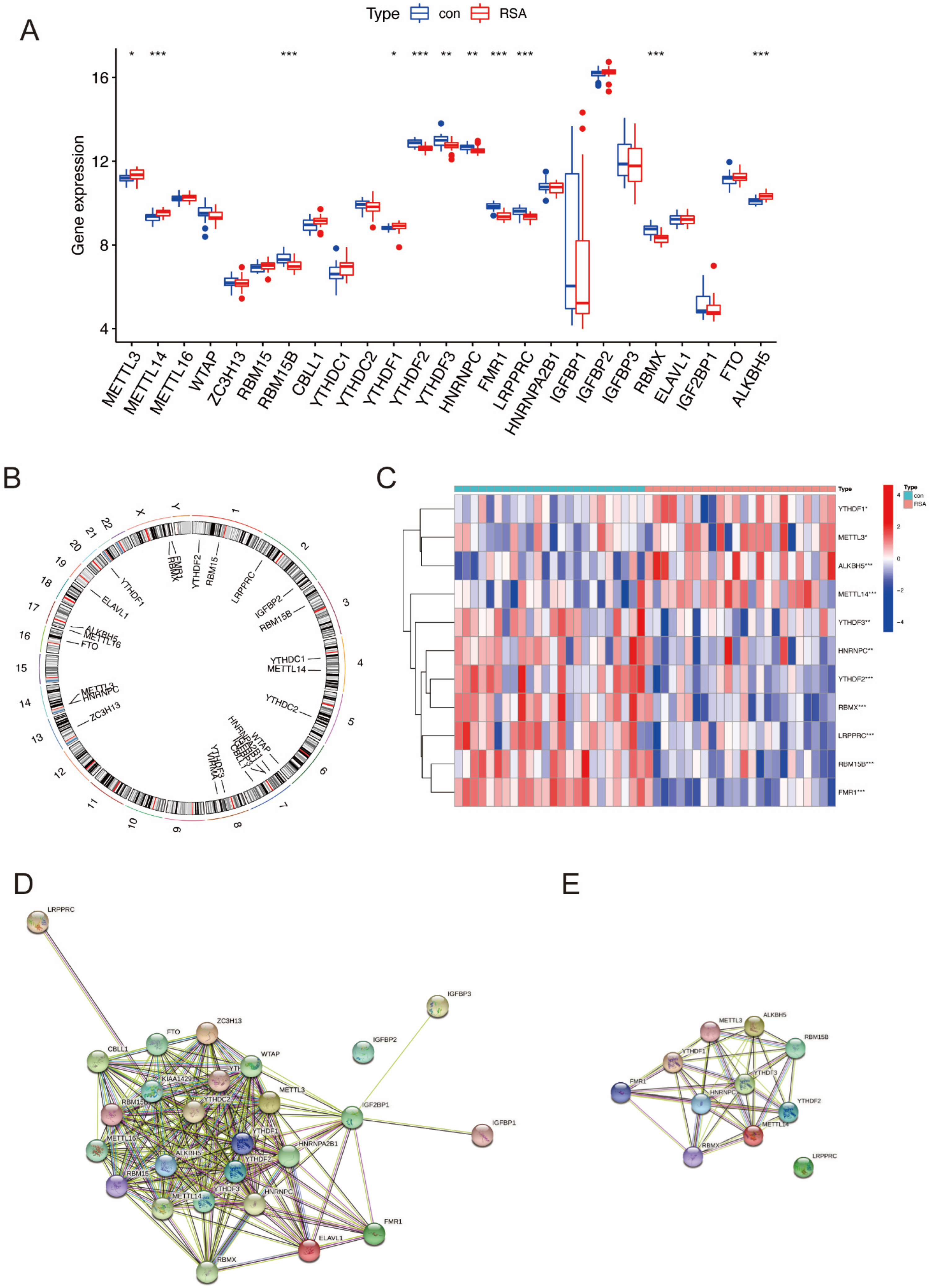
The workflow (Figure 1) was plotted to show the process of our study. A total of 25 m6A regulators between healthy controls and RSA patients were extracted utilizing the “limma” package in R (Figure 2A). The chromosomal position was visualized using the “RCircos” package (Figure 2B). Among the 25 m6A regulators, a total of 11 differentially expressed m6A regulators were visualized in a heatmap (Figure 2C). Protein-protein interactions between the 25 m6A regulators and the 11 differentially expressed m6A regulators are shown in Figure 2D,E, respectively.

2.2. Identification of the Correlation between Writers and Erasers in RSA

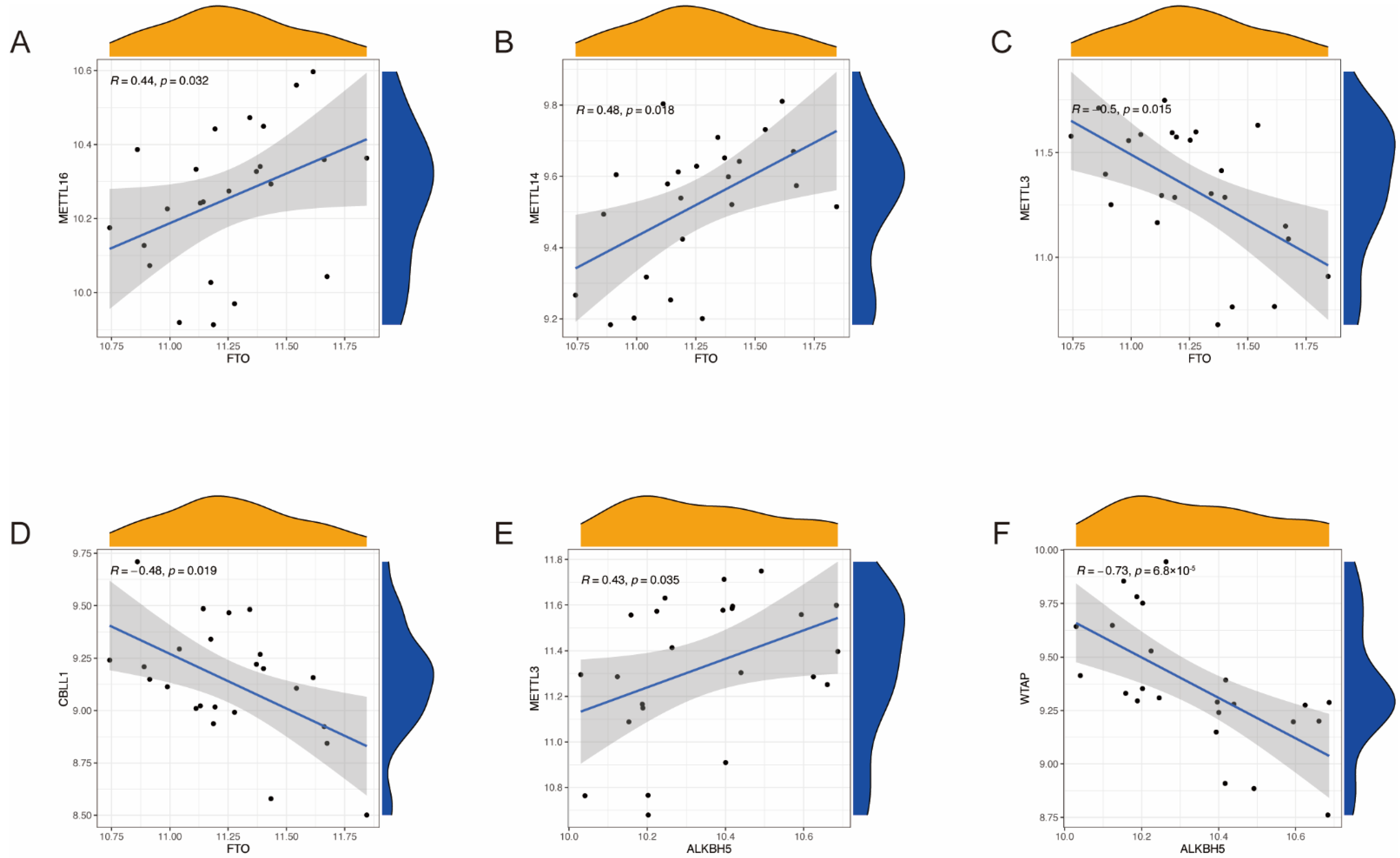
To explore whether low expression levels of eraser genes (ALKBH5 and FTO) match higher expression levels of writer genes (RBM15B, RBM15, METTL3, METTL14, WTAP, CBLL1, and ZC3H13) in RSA, we utilized linear regression analysis to construct the correlation between writers and erasers. We found that two writer genes were significantly correlated with the eraser gene ALKBH5, and four writer genes were significantly correlated with the eraser gene FTO (Figure 3).

2.3. Identification and Validation of the Predictive Ability of 11 m6A Regulators

To test the diagnostic value of the candidate m6A regulators, we constructed ROC curves based on GSE165004. The area under the ROC curve visualizes the predictive ability of each candidate gene. The AUCs of all 11 m6A regulators were greater than 0.65, and the AUCs of 5 m6A regulators (FMR1, RBMX, RBM15B, ALKBH5, and LRPPRC) were greater than 0.8 (Figure 4A). To further test the predictive accuracy of the candidate genes, we constructed ROC curves based on GSE26787. Those with AUCs greater than 0.8 are shown in Figure 4B.
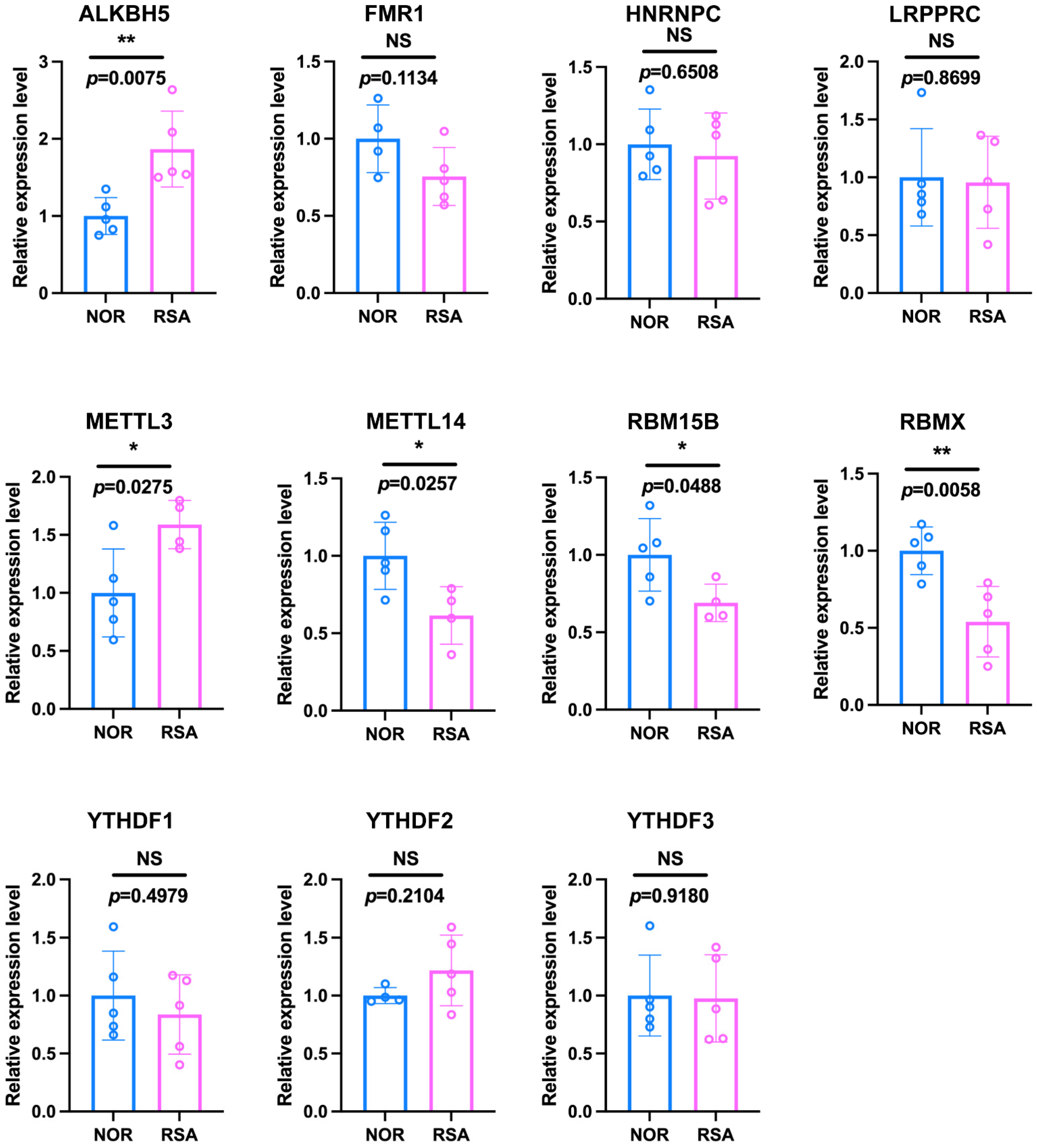
We used ESCs, the most abundant component of endometrial tissue, to validate the expression of m6A regulators. Consistent with the bioinformatic analysis, ALKBH5 and METTL3 were upregulated, while METTL14, RBM15B and RBMX were downregulated (Figure 5).

2.4. Identification of Three Distinct Patterns in RSA and Functional Enrichment Analysis

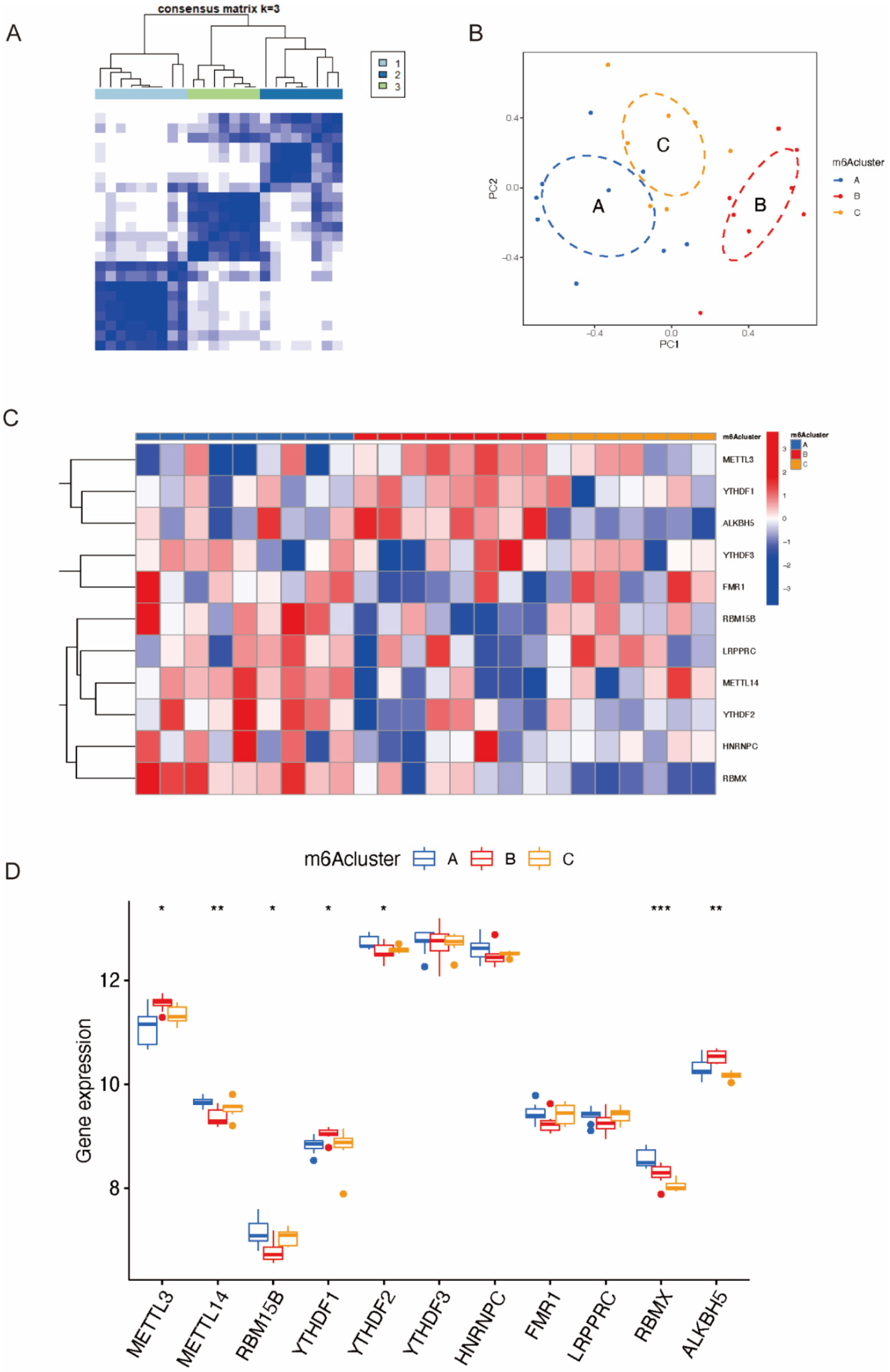
Utilizing the “ConsensusClustePlus” package, we distinguished three distinct m6A patterns based on the 11 candidate m6A regulators via an unsupervised clustering method (Figure 6A,B). According to the PCA, the three patterns were distinguished clearly with a stable distribution of samples (Figure 6C). The expression of significant m6A regulators in the three clusters is shown in Figure 6D, from which we found that ALKBH5 was overexpressed in Cluster B. Additionally, the process of constructing the consensus clustering is shown in Figure S1. We tried establishing the model with a consensus matrix from k = 2 to k = 9 (Figure S1A), and the consensus CDF curve and the relative change in the area under the CDF curve are shown in Figure S1B,C, respectively.
The differentially expressed genes between the normal control group and the RSA group were extracted and subjected to GSEA. The GSEA results (Figure 7A,B) show that the DEGs were mainly enriched in the activation of immune response in the control group and mainly involved in the cellular modified amino acid metabolic process in the RSA group. In addition, the DEGs among the three distinct m6A subtypes were also extracted and subjected to gene ontology enrichment analysis (Figure 7C–E). The results of GO analysis (Table 1) showed that the DEGs were mainly enriched in BPs, consisting of the olefinic compound metabolic process, the alcohol metabolic process and so on. In terms of CCs, the DEGs were mainly involved in lipid droplets and distal axons; in terms of MFs, they were enriched in catalytic activity acting on RNA and metabolism of NAD.

2.5. Genes Related to Immune Infiltration and Macrophage Function Related in Different m6A Patterns

ssGSEA was applied to calculate the abundance of immune cells in Clusters A–C (Figure 8A) and evaluate the correlation between 11 significant m6A regulators and immune cells (Figure 8B). We noticed that ALKBH5, the only differentially expressed m6A eraser gene in the dataset, was the most relevant m6A regulator with macrophage function. The analysis of ALKBH5 and immune cells (Figure 8C) indicated that upregulation of ALKBH5 in Cluster B might result in macrophage dysfunction. We also investigated the correlation between macrophage-function-related genes and m6A patterns (Figure 8D). The upregulation of IL6 and downregulation of CD163 and IL10 in Cluster B indicated that anti-inflammatory M2 differentiation was impaired, which could be a possible cause of RSA.

2.6. Overexpression of ALKBH5 in ESCs Affected Macrophage Differentiation via VEGF Secretion

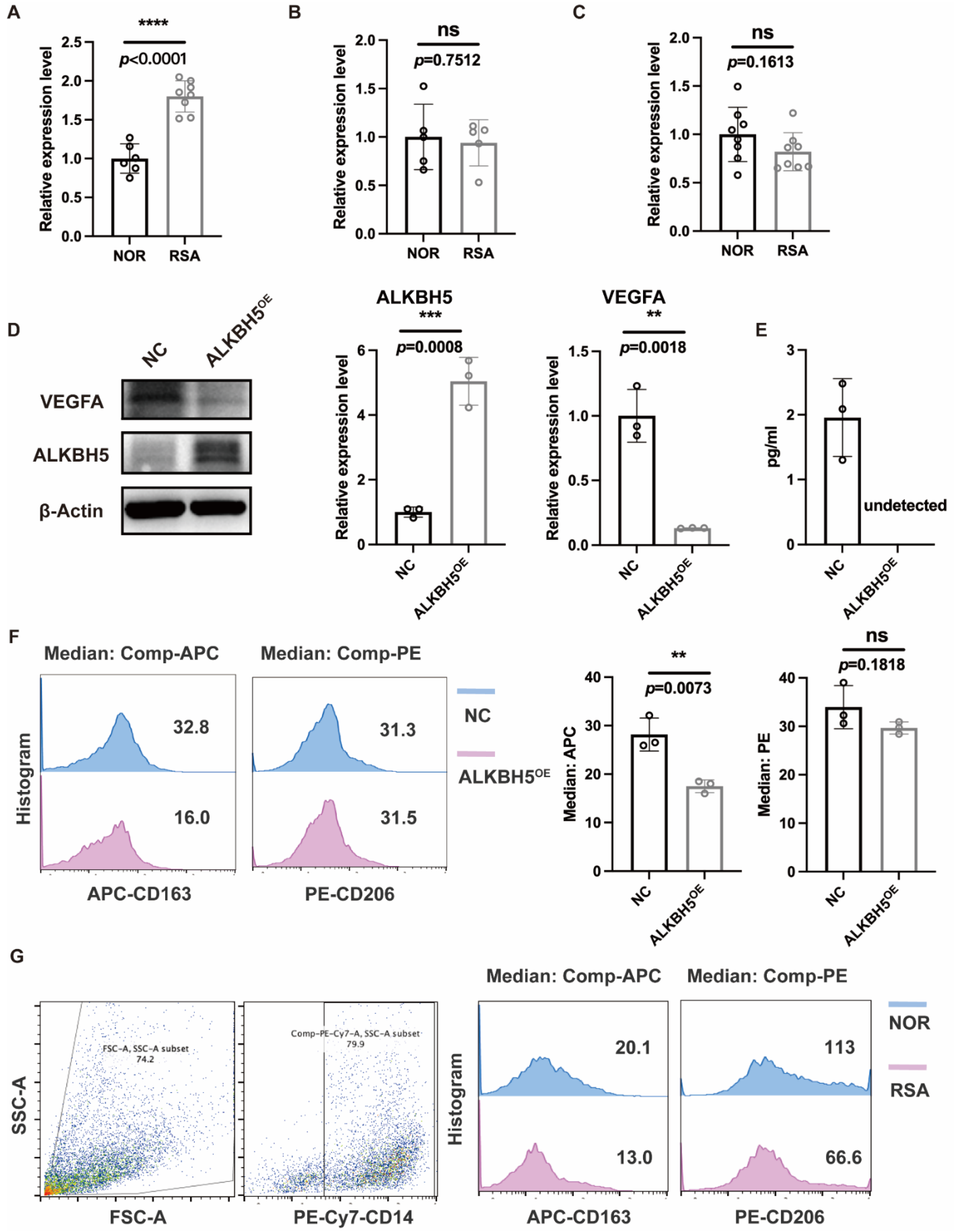
Due to the limited number of immune cells in endometrial tissue, we verified macrophage function in the decidua. ALKBH5 was also highly expressed in the DSCs (Figure 9A) of RSA patients, but not in decidual NK cells (Figure 9B) or decidual macrophages (Figure 9C). Therefore, we hypothesized that the dysfunction of macrophages was partially attributed to the overexpression of ALKBH5 in DSCs. We constructed an ALKBH5-overexpressing human ESC line to better understand its function. Based on the results shown in Figure 8D, we noticed that in the ALKBH5high cluster, the expression of VEGF was relatively low. Therefore, we measured both the intracellular and supernatant levels and confirmed the insufficient secretion of VEGF in the ALKBH5-overexpressing human ESC line (Figure 9D,E). Considering that VEGF signaling is an important mediator of macrophages, we established a coculture system of ALKBH5-overexpressing human ESC lines and THP-1 cells, and the M2 differentiation status of THP-1 cells was impaired in the ALKBH5-overexpression group (Figure 9F). This CD163low CD206low decidual macrophage was observed in some of the RSA patients (Figure 9G).

3. Discussion

Recurrent spontaneous abortion is prevalent but underestimated. A Japanese group found that the live birth rates of pregnant women with RSA history were similar in 2011–2018 to those who returned in 1994–2010 (63.8% vs. 67.8%) [10]. This clearly indicated that new ideas for RSA therapy were needed. Established evidence has shown that m6A regulators are involved in pregnancy maintenance [11]. However, the significance of m6A modification in RSA-related stromal cell–immune cell interactions remains unknown. In our study, we aimed to identify distinctive m6A regulators in RSA and reveal the mechanism of m6A regulator mediation of stromal cell–immune cell interactions.
We first identified 11 significant m6A regulators among 25 m6A regulators through differential expression analysis of normal and RSA patients. We then constructed ROC curves of the 11 candidate genes to demonstrate their predictive abilities for RSA. To test the predictive accuracy, we plotted the ROC curves of another RSA dataset and validated the expression of significant genes via qRT-PCR. ALKBH5, whose AUCs were greater than 0.8 in both datasets, was proven to be significantly overexpressed in RSA samples, indicating that ALKBH5 might be a potential biomarker for predicting RSA. Based on the 11 candidate genes, we further classified three distinct patterns, among which ALKBH5 was significantly upregulated in Cluster B. Therefore, we hypothesized that Cluster B was highly linked to RSA. Subsequently, we performed immune infiltration analysis and identified macrophage-function-related genes. ssGSEA indicated that ALKBH5 might jeopardize macrophage differentiation to promote RSA, and the mechanism might be the ESC-derived hyposecretion of VEGF. qRT-PCR, Western blotting, flow cytometry and ELISA experiments were subsequently performed to validate the bioinformatic results.
ALKBH5, a kind of primary m6A demethylase involved in alkylated DNA repair, is involved in different biological processes, including invasion, ossification, metastasis and proliferation [12,13]. The dysregulation of ALKBH5 plays a crucial role in various kinds of diseases by regulating or interacting with other genes [14]. ALKBH5 was reported to play an essential role in trophoblast invasion at the maternal–fetal interface [8,15], and was proven to be significantly overexpressed in infertile women [11].
Macrophages are involved in all stages of pregnancy, from embryonic implantation and placental formation to final delivery [16,17,18,19]. The precise macrophage differentiation status has not achieved consensus. However, this differentiation balance is carefully maintained, and the dysfunction of polarization is associated with adverse pregnancy outcomes. A decrease in CD163 and impaired M2 polarization activate macrophage apoptosis, contributing to the pathological process of RSA [20]. During the growing phase of pregnancy, the predominance of macrophage immune status was anti-inflammatory. Several studies revealed that as pregnancy advanced, M1 markers such as TLR9, IL1B, IL12RB2, CD48 and FGR were silenced, whereas the M2 surface marker CD206 and cytokines such as IL10, CCL13, CCL14 and IDO were upregulated [21,22]. Moreover, fetal-derived macrophages, Hofbauer cells, showed M2 characteristics with high expression of IL10, TGF-β, DC-SIGN, CD206 and CD163 [23]. VEGF is one of the pivotal growth factors produced in the decidua, and regulates the local immune microenvironment. VEGF in the decidua might contribute to macrophage recruitment and M2 polarization [24].
In our study, due to the limited number of immune cells in endometrial tissue, we focused on the interaction of DSCs and decidual immune cells. We recognized that decidualization is a complicated process and that the expression levels of many genes were altered. To minimize this effect, we confirmed ALKBH5 expression in primary human DSCs, which was consistent with the result in primary human ESCs. Additionally, excellent work has been published about ALKBH5 function in immune cells [25]. To rule out the potential mediation of ALKBH5 in decidual immune cells, we verified ALKBH5 expression in both decidual NK cells and macrophages, the top two immune-cell components in the decidua. We found that ALKBH5 was mainly differentially expressed in ESCs and DSCs, so we focused on stromal cell–immune cell mediation. In addition, VEGF is one of the major factors secreted by stromal cells, and evidence of its role in macrophage differentiation has been validated [26]. In our study, we analyzed and identified the crosslink between the ALKBH5high cluster and VEGFlow cluster. This result provides us with evidence that the phenomenon of ALKBH5-related macrophage dysfunction might be correlated with insufficient secretion of VEGF by endometrial stromal cells. One of the drawbacks of our study was that we were unable to identify the downstream molecules of ALKBH5 and the unidentified molecules that might regulate VEGF secretion. Further study should be performed to answer these scientific questions. Additionally, there was another limitation in our research. Though we chose a relatively reliable consensus matrix, there was still some over-fitting in the consensus clustering due to the limited sample numbers. We plan to carry out further research with larger sample numbers in order to generate a better consensus-clustering model.

4. Materials and Methods

4.1. Data Acquisition and PPI Network Construction

The GSE165004 dataset containing 24 healthy controls and 24 RSA patients was obtained from the GEO database. We extracted a total of 25 m6A regulators from the dataset. Eleven were differentially expressed, consisting of three writers (METTL3, METTL14, RBM15B), one eraser (ALKBH5) and seven readers (YTHDF1, YTHDF3, HNRNPC, YTHDF2, LRPPRC, FMR1, RBMX). Linear regression analysis was used to detect the correlation between erasers and writers. The 25 m6A regulators and 11 differentially expressed regulators were utilized to construct the protein-protein interaction network using STRING V11.5 (https://string-db.org/ (accessed on 3 March 2022)) [27]. In addition, GSE26787 [28], containing 5 RSA patients and 5 healthy controls, was utilized as the validation dataset.

4.2. Construction of Receiver Operating Characteristic (ROC) Curves

We constructed ROC curves of the candidate m6A regulators to identify their predictive ability via the “pROC” package. The area under the ROC curve (AUC) illustrates the predictive accuracy of each m6A regulator. In addition, the predictive ability of the significant m6A regulators was validated using the GSE26787 dataset.

4.3. Classification of Three Distinct m6A Subtypes and Extraction of DEGs

Consensus clustering is a useful method to subclassify members. Based on the significant m6A regulators, we utilized consensus clustering to classify the RSA patients into subtypes via the “ConsensusClustePlus” package [29]. Principal component analysis (PCA) was applied to distinguish and identify m6A patterns. Differentially expressed genes (DEGs) among the distinct m6A patterns, and between the control and RSA groups, were extracted using the “limma” package [30] with the criterion of p < 0.05.

4.4. Functional Enrichment Analysis of DEGs

Differentially expressed genes between the control and RSA groups were extracted for gene set enrichment analysis (GSEA) to reveal the potential mechanism of RSA. We downloaded and utilized “c2.cp.kegg.v7.4.symbols.gmt” as the reference gene set with the criterion of a false discovery rate (FDR) < 0.05 and adjusted p < 0.05.
We obtained 20 differentially expressed genes from the cluster differential expression analysis (Table S1), and the 20 DEGs among three distinct m6A patterns were utilized for gene ontology (GO) functional enrichment analysis via the “clusterProfiler” package [31]. Three aspects of the GO biological processes were visualized, the biological processes (BPs), cellular components (CCs) and molecular functions (MFs).

4.5. Single-Sample Gene Set Enrichment Analysis to Identify Immune-Cell Infiltration

The variety of immune cells plays an essential role in the mechanism of RSA. To identify the characteristics of immune cells in RSA samples, we applied single-sample gene set enrichment analysis (ssGSEA) [32]. The abundance of immune cells in RSA samples and the correlation between candidate m6A regulators and immune cell infiltration could be visualized through ssGSEA.

4.6. Cell Culture

Primary human ESCs were collected from uterine endometrial biopsies for benign gynecological disorders. Women undergoing hysterectomy provided informed consent and were anonymized during ESC isolation. This study was reviewed and approved by the Ethics Committee of Shanghai First Maternity and Infant Hospital. The isolation and purification procedure was performed as previously described [33], which supplied 98% pure ESCs. Both primary and immortalized human ESC lines were cultured in phenol red-free Dulbecco’s modified Eagle medium (DMEM)/Ham’s F12 with 10% fetal bovine serum (FBS), 100 U/mL penicillin and 100 μg/mL streptomycin.
Primary human decidual NK cells and macrophages were obtained from healthy pregnant women and patients with a diagnosed RSA history undergoing an elective abortion. All the RSA patients suffered a history of two or more spontaneous miscarriages. However, patients with uterine malformation, infections, chromosomal aberrations, or who underwent IVF treatment were excluded. The normal control group comprised women who chose to have elective abortions for nonmedical reasons. The isolation and purification procedure was performed as previously described [34]. The culture medium for primary decidual NK cells and the human macrophage cell line THP1 was RPMI 1640 with 10% fetal bovine serum, 100 U/mL penicillin and 100 μg/mL streptomycin. Primary decidual macrophages were cultured in DMEM with 10% fetal bovine serum, 100 U/mL penicillin and 100 μg/mL streptomycin. All cells were maintained in humidified conditions at 37 °C and 5% CO2.

4.7. Antibodies for Flow Cytometry (FCM)

To evaluate macrophage differentiation status, primary decidual macrophages and THP1 cells were stained with APC-conjugated anti-human CD163 antibody and PE-conjugated anti-human CD206 antibody for anti-inflammatory M2 bias. Macrophage purity was validated with PE-Cy7-CD14 (all from BioLegend, Beijing, China).

4.8. Construction of an ALKBH5-Overexpressing ESC Cell Line

We constructed an ALKBH5-overexpressing ESC cell line using the pCMV6-AN-DDK vector, and the protein level of ALKBH5 was confirmed by Western blotting analysis. The primers for the ALKBH5-overexpression plasmid were as follows: gatgacgataaggcgatcgcCATGGCGGCCGCCAGCGG; gagtgcggccgcttaacgcgtTCAGTGCCGCCGCATCTT. Gene transfection was performed using a Lipo3000 kit (Invitrogen, Shanghai, China) following the manufacturer’s protocol.

4.9. Enzyme-Linked Immunosorbent Assay

The VEGF concentration in the supernatant was measured using an enzyme-linked immunosorbent assay (ELISA) kit (MULTI SCIENCES, Hangzhou, China, #EK183-02).

4.10. Quantitative Real-Time PCR (qRT-PCR)

Total RNA was extracted from primary human ESCs. Lysates were prepared using RNAiso Plus (Takara, Osaka, Japan). One microgram of total RNA was reverse transcribed into cDNA utilizing a PrimeScript™ RT reagent kit (Takara, Osaka, Japan). The primer sequences of 11 significant m6A regulators are listed in Table 2.

4.11. Western Blot

RIPA (Beyotime Biotechnology, Haimen, China, #P0013B) supplemented with a protease inhibitor cocktail (Roche, Branford, CT, USA, #11697498001) and phosphatase inhibitor cocktail (Roche, Branford, CT, USA, #4906837001) was used for sample lysis. Proteins (15–30 µg) were loaded and separated by 10% SDS—PAGE and then transferred to polyvinylidene fluoride membranes. The membranes were incubated with the following primary antibodies at 4 °C overnight: ALKBH5 (1:1000; Abcam, Cambridge, UK), β-actin (1:5000; Cell Signaling Technology, Danvers, MA, USA) and VEGFA (1:1000, Abcam, Cambridge, UK).
The blots were further incubated with an HRP-conjugated goat anti-rabbit IgG secondary antibody (1:3000, Abmart, Shanghai, China) for 1 h at room temperature. Protein visualization was performed using an enhanced chemiluminescence solution (Merck Millipore, Darmstadt, Germany, #32106). The relative protein expression levels were analyzed by densitometry using ImageJ software. β-actin was used as a loading control.

4.12. Statistical Analysis

After collecting the data, R software (version 4.0.0) was used for the statistical analysis. The correlation between writers and erasers was explored utilizing linear regression analysis. All parametric analyses were based on two-tailed tests with significance set at p < 0.05. Differences between two groups were compared with Student’s t-test. Multiple groups were compared using Kruskal-Wallis tests.

5. Conclusions

Eleven significant m6A regulators between the control group and the RSA group were extracted and utilized to apply bioinformatic analysis consisting of subtype classification and functional enrichment. We identified and validated the upregulation of the m6A eraser ALKBH5 in ESCs and DSCs of RSA patients. After identifying three distinct m6A patterns, we focused on Cluster B, with a high expression level of ALKBH5 and insufficient macrophage infiltration. We recognized this cluster to be highly correlated with stromal cell–macrophage interactions. Experiments were conducted to validate the hypothesis; ALKBH5 overexpression in the ESC line caused the hyposecretion of VEGF and subsequently downregulated the macrophage M2 marker. Alteration of the M2 differentiation status potentially promoted RSA.

Supplementary Materials

The following supporting information can be downloaded at: https://www.mdpi.com/article/10.3390/ijms232415819/s1.

Author Contributions

L.J. had full access to all the data in the study and took responsibility for the integrity of the data and the accuracy of the data analysis. Y.Z. and J.S. are co-first authors. Conceptualization, L.J.; Methodology, Y.Z.; Software, J.S.; Validation, J.S., Y.Z. and L.J.; Formal analysis, J.S.; Investigation, Y.Z.; Resources, J.S.; Data curation, J.S.; Writing—original draft, J.S.; Writing—review & editing, Y.Z.; Visualization, J.S.; Supervision, L.J.; Project administration, L.J.; Funding acquisition, L.J. All authors have read and agreed to the published version of the manuscript.

Funding

This work was supported by the National Natural Science Foundation of China (81730039, 82071653) and Shanghai Municipal Health Commission (20194Y0477).

Institutional Review Board Statement

This study was conducted according to the guidelines of the Declaration of Helsinki, and approved by the Ethics Committee of Shanghai First Maternity and Infant Hospital (No KS18133; 02/25/2022).

Informed Consent Statement

Informed consent was obtained from all participants involved in the study.

Data Availability Statement

The datasets for this study can be found in the GEO database. Please see the https://www.ncbi.nlm.nih.gov/geo/ (accessed on 1 March 2022) for more details.

Acknowledgments

All the authors are thankful to Shanghai First Maternity and Infant Hospital and Tongji University for successful completion of this research.

Conflicts of Interest

The authors declare no conflict of interest.

References

  1. Lédée, N.; Petitbarat, M.; Prat-Ellenberg, L.; Dray, G.; Cassuto, G.N.; Chevrier, L.; Kazhalawi, A.; Vezmar, K.; Chaouat, G. Endometrial Immune Profiling: A Method to Design Personalized Care in Assisted Reproductive Medicine. Front. Immunol. 2020, 11, 1032. [Google Scholar] [CrossRef] [PubMed]
  2. Hess, A.P.; Hamilton, A.E.; Talbi, S.; Dosiou, C.; Nyegaard, M.; Nayak, N.; Genbecev-Krtolica, O.; Mavrogianis, P.; Ferrer, K.; Kruessel, J.; et al. Decidual stromal cell response to paracrine signals from the trophoblast: Amplification of immune and angiogenic modulators. Biol. Reprod. 2007, 76, 102–117. [Google Scholar] [CrossRef] [PubMed] [Green Version]
  3. Murata, H.; Tanaka, S.; Okada, H. Immune Tolerance of the Human Decidua. J. Clin. Med. 2021, 10, 351. [Google Scholar] [CrossRef] [PubMed]
  4. Nallasamy, S.; Li, Q.; Bagchi, M.K.; Bagchi, I.C. Msx Homeobox Genes Critically Regulate Embryo Implantation by Controlling Paracrine Signaling between Uterine Stroma and Epithelium. PLoS Genet. 2012, 8, e1002500. [Google Scholar] [CrossRef] [PubMed]
  5. Mor, G.; Cardenas, I.; Abrahams, V.; Guller, S. Inflammation and pregnancy: The role of the immune system at the implantation site. Ann. N. Y. Acad. Sci. 2011, 1221, 80–87. [Google Scholar] [CrossRef] [Green Version]
  6. Du, L.; Deng, W.; Zeng, S.; Xu, P.; Huang, L.; Liang, Y.; Wang, Y.; Xu, H.; Tang, J.; Bi, S.; et al. Single-cell transcriptome analysis reveals defective decidua stromal niche attributes to recurrent spontaneous abortion. Cell Prolif. 2021, 54, e13125. [Google Scholar] [CrossRef]
  7. Dai, D.; Wang, H.; Zhu, L.; Jin, H.; Wang, X. N6-methyladenosine links RNA metabolism to cancer progression. Cell Death Dis. 2018, 9, 124. [Google Scholar] [CrossRef] [Green Version]
  8. Li, X.-C.; Jin, F.; Wang, B.-Y.; Yin, X.-J.; Hong, W.; Tian, F.-J. The m6A demethylase ALKBH5 controls trophoblast invasion at the maternal-fetal interface by regulating the stability of CYR61 mRNA. Theranostics 2019, 9, 3853–3865. [Google Scholar] [CrossRef]
  9. Qiu, W.; Zhou, Y.; Wu, H.; Lv, X.; Yang, L.; Ren, Z.; Tian, H.; Yu, Q.; Li, J.; Lin, W.; et al. RNA Demethylase FTO Mediated RNA m6A Modification Is Involved in Maintaining Maternal-Fetal Interface in Spontaneous Abortion. Front. Cell Dev. Biol. 2021, 9, 617172. [Google Scholar] [CrossRef]
  10. Morita, K.; Ono, Y.; Takeshita, T.; Sugi, T.; Fujii, T.; Yamada, H.; Nakatsuka, M.; Fukui, A.; Saito, S. Risk Factors and Outcomes of Recurrent Pregnancy Loss in Japan. J. Obstet. Gynaecol. Res. 2019, 45, 1997–2006. [Google Scholar] [CrossRef]
  11. Zhao, S.; Lu, J.; Chen, Y.; Wang, Z.; Cao, J.; Dong, Y. Exploration of the potential roles of m6A regulators in the uterus in pregnancy and infertility. J. Reprod. Immunol. 2021, 146, 103341. [Google Scholar] [CrossRef]
  12. Wang, J.; Wang, J.; Gu, Q.; Ma, Y.; Yang, Y.; Zhu, J.; Zhang, Q. The biological function of m6A demethylase ALKBH5 and its role in human disease. Cancer Cell Int. 2020, 20, 347. [Google Scholar] [CrossRef]
  13. Zheng, G.; Dahl, J.A.; Niu, Y.; Fedorcsak, P.; Huang, C.-M.; Li, C.J.; Vågbø, C.B.; Shi, Y.; Wang, W.-L.; Song, S.-H.; et al. ALKBH5 Is a Mammalian RNA Demethylase that Impacts RNA Metabolism and Mouse Fertility. Mol. Cell 2013, 49, 18–29. [Google Scholar] [CrossRef] [Green Version]
  14. Chen, X.-Y.; Zhang, J.; Zhu, J.-S. The role of m6A RNA methylation in human cancer. Mol. Cancer 2019, 18, 103. [Google Scholar] [CrossRef] [Green Version]
  15. Zheng, Q.; Yang, F.; Gan, H.; Jin, L. Hypoxia induced ALKBH5 prevents spontaneous abortion by mediating m6A-demethylation of SMAD1/5 mRNAs. Biochim. Biophys. Acta 2022, 1869, 119316. [Google Scholar] [CrossRef]
  16. Care, A.S.; Diener, K.; Jasper, M.J.; Brown, H.M.; Ingman, W.V.; Robertson, S.A. Macrophages regulate corpus luteum development during embryo implantation in mice. J. Clin. Investig. 2013, 123, 3472–3487. [Google Scholar] [CrossRef] [Green Version]
  17. Co, E.C.; Gormley, M.; Kapidzic, M.; Rosen, D.B.; Scott, J.; Stolp, H.A.; McMaster, M.; Lanier, L.L.; Bárcena, A.; Fisher, S.J. Maternal Decidual Macrophages Inhibit NK Cell Killing of Invasive Cytotrophoblasts During Human Pregnancy. Biol. Reprod. 2013, 88, 155. [Google Scholar] [CrossRef]
  18. Erlebacher, A. Immunology of the Maternal-Fetal Interface. Annu. Rev. Immunol. 2013, 31, 387–411. [Google Scholar] [CrossRef]
  19. Nagamatsu, T.; Schust, D.J. The Immunomodulatory Roles of Macrophages at the Maternal—Fetal Interface. Reprod. Sci. 2010, 17, 209–218. [Google Scholar] [CrossRef]
  20. Yao, Y.; Hao, F.; Tang, L.-C.; Xu, X.-H.; Jin, L. Downregulation of HDAC8 expression decreases CD163 levels and promotes the apoptosis of macrophages by activating the ERK signaling pathway in recurrent spontaneous miscarriage. Mol. Hum. Reprod. 2020, 26, 521–531. [Google Scholar] [CrossRef]
  21. Kim, S.Y.; Romero, R.; Tarca, A.L.; Bhatti, G.; Kim, C.J.; Lee, J.; Elsey, A.; Than, N.G.; Chaiworapongsa, T.; Hassan, S.S.; et al. Methylome of Fetal and Maternal Monocytes and Macrophages at the Feto-Maternal Interface. Am. J. Reprod. Immunol. 2012, 68, 8–27. [Google Scholar] [CrossRef] [PubMed] [Green Version]
  22. Heikkinen, J.; Möttönen, M.; Komi, J.; Alanen, A.; Lassila, O. Phenotypic characterization of human decidual macrophages. Clin. Exp. Immunol. 2003, 131, 498–505. [Google Scholar] [CrossRef] [PubMed]
  23. Joerink, M.; Rindsjö, E.; van Riel, B.; Alm, J.; Papadogiannakis, N. Placental macrophage (Hofbauer cell) polarization is independent of maternal allergen-sensitization and presence of chorioamnionitis. Placenta 2011, 32, 380–385. [Google Scholar] [CrossRef] [PubMed]
  24. Wheeler, K.C.; Jena, M.; Pradhan, B.; Nayak, N.; Das, S.; Hsu, C.-D.; Wheeler, D.S.; Chen, K.; Nayak, N.R. VEGF may contribute to macrophage recruitment and M2 polarization in the decidua. PLoS ONE 2018, 13, e0191040. [Google Scholar] [CrossRef] [PubMed]
  25. Xu, F.; Zhang, Z.; Yuan, M.; Zhao, Y.; Zhou, Y.; Pei, H.; Bai, L. M6A Regulatory Genes Play an Important Role in the Prognosis, Progression and Immune Microenvironment of Pancreatic Adenocarcinoma. Cancer Investig. 2020, 39, 39–54. [Google Scholar] [CrossRef]
  26. Liu, X.; Guo, A.; Tu, Y.; Li, W.; Li, L.; Liu, W.; Zhou, Y.; Sang, A.; Zhu, M. Fruquintinib inhibits VEGF/VEGFR2 axis of choroidal endothelial cells and M1-type macrophages to protect against mouse laser-induced choroidal neovascularization. Cell Death Dis. 2020, 11, 1016. [Google Scholar] [CrossRef]
  27. Szklarczyk, D.; Franceschini, A.; Wyder, S.; Forslund, K.; Heller, D.; Huerta-Cepas, J.; Simonovic, M.; Roth, A.; Santos, A.; Tsafou, K.P.; et al. STRING v10: Protein–protein interaction networks, integrated over the tree of life. Nucleic Acids Res. 2015, 43, D447–D452. [Google Scholar] [CrossRef]
  28. Lédée, N.; Munaut, C.; Aubert, J.; Sérazin, V.; Rahmati, M.; Chaouat, G.; Sandra, O.; Foidart, J.M. Specific and extensive endometrial deregulation is present before conception in IVF/ICSI repeated implantation failures (IF) or recurrent miscarriages. J. Pathol. 2011, 225, 554–564. [Google Scholar] [CrossRef]
  29. Wilkerson, M.D.; Hayes, D.N. ConsensusClusterPlus: A class discovery tool with confidence assessments and item tracking. Bioinformatics 2010, 26, 1572–1573. [Google Scholar] [CrossRef] [Green Version]
  30. Ritchie, M.E.; Belinda, P.; Wu, D.; Hu, Y.; Law, C.W.; Shi, W.; Smyth, G.K. limma powers differential expression analyses for RNA-sequencing and microarray studies. Nucleic Acids Res. 2015, 43, e47. [Google Scholar] [CrossRef]
  31. Denny, P.; Feuermann, M.; Hill, D.P.; Lovering, R.C.; Plun-Favreau, H.; Roncaglia, P. Exploring autophagy with Gene Ontology. Autophagy 2018, 14, 419–436. [Google Scholar] [CrossRef]
  32. Rooney, M.S.; Shukla, S.A.; Wu, C.J.; Getz, G.; Hacohen, N. Molecular and Genetic Properties of Tumors Associated with Local Immune Cytolytic Activity. Cell 2015, 160, 48–61. [Google Scholar] [CrossRef] [Green Version]
  33. Chang, K.-K.; Liu, L.-B.; Jin, L.-P.; Zhang, B.; Mei, J.; Li, H.; Wei, C.-Y.; Zhou, W.-J.; Zhu, X.-Y.; Shao, J.; et al. IL-27 triggers IL-10 production in Th17 cells via a c-Maf/RORγt/Blimp-1 signal to promote the progression of endometriosis. Cell Death Dis. 2017, 8, e2666. [Google Scholar] [CrossRef] [Green Version]
  34. Hao, F.; Tang, L.-C.; Sun, J.-X.; Li, W.-X.; Zhao, Y.; Xu, X.-H.; Jin, L.-P. Decreased nitric oxide content mediated by asymmetrical dimethylarginine and protein l-arginine methyltransferase 3 in macrophages induces trophoblast apoptosis: A potential cause of recurrent miscarriage. Hum. Reprod. 2021, 36, 3049–3061. [Google Scholar] [CrossRef]
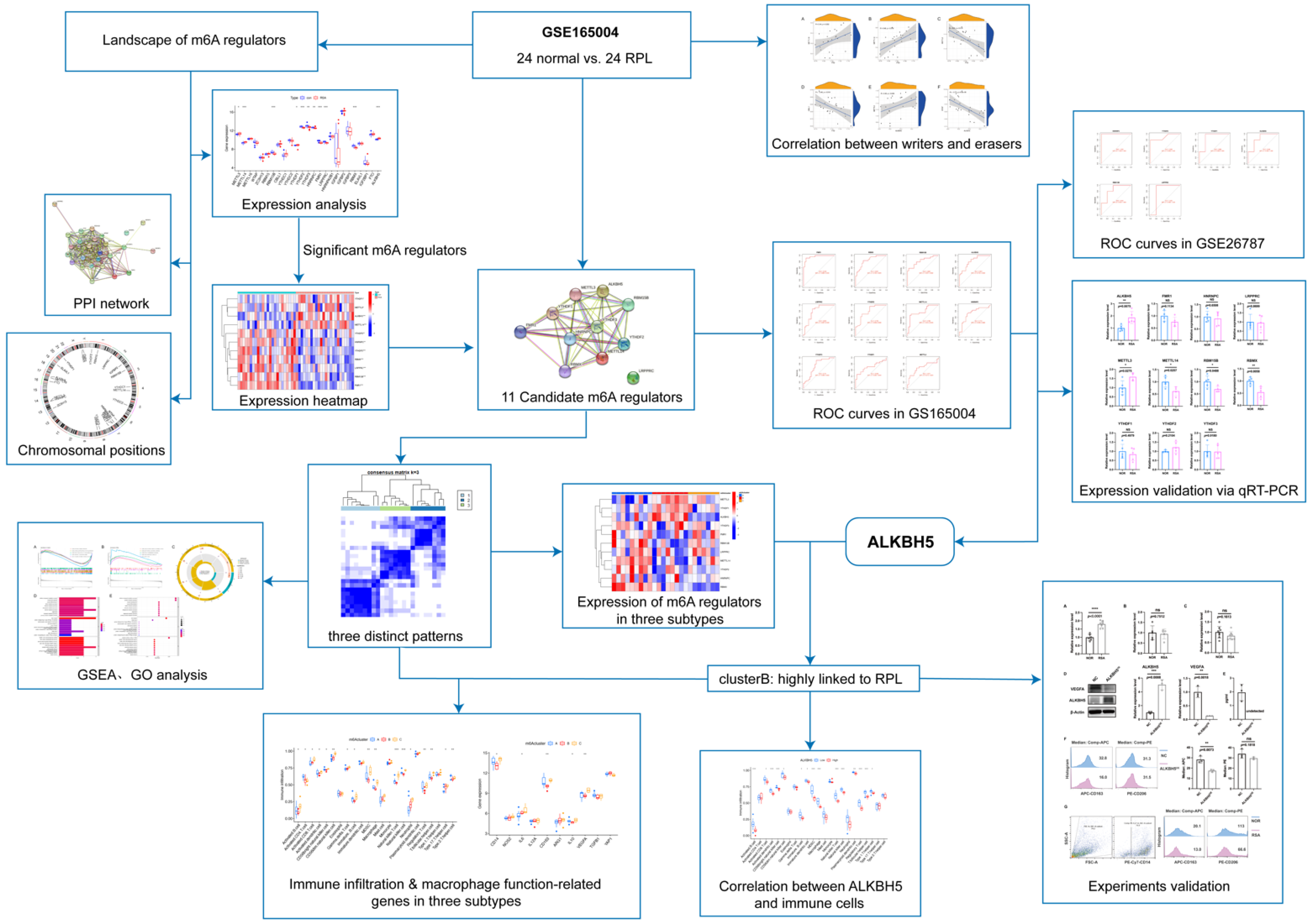
Figure 1. Workflow diagram illustrating the design and processes of our study.
Ijms 23 15819 g001
Figure 2. The landscape of the 25 m6A regulators in RSA. (A) Differential expression levels of the 25 m6A regulators between normal group and RSA patients. (B) Chromosomal positions of the 25 m6A regulators. (C) Expression heatmap of 11 significant m6A regulators. (D) PPI network of the 25 m6A regulators. (E) PPI network of the 11 significant genes. *: p < 0.05, **: p < 0.01, ***: p < 0.001.
Ijms 23 15819 g002
Figure 3. Correlation between writers and erasers in RSA. (AD) Writer genes: METTL16, METTL14, METTL3, and CBLL1; eraser gene: FTO. (E,F) Writer genes: METTL3 and WTAP; eraser gene: ALKBH5.
Ijms 23 15819 g003
Figure 4. Construction of ROC curves. (A) ROC curves of significant genes based on GSE165004. (B) ROC curves based on GSE26787.
Ijms 23 15819 g004
Figure 5. mRNA expression levels of significant m6A regulators in primary human ESCs. *: p < 0.05, **: p < 0.01, NS: Non-significance.
Ijms 23 15819 g005
Figure 6. Identification of three distinct m6A patterns. (A) Three distinct m6A patterns based on significant genes. (B) Principal component analysis. (C) Expression heatmap of 11 candidate genes in three subtypes. (D) Differential expression histogram of 11 significant genes in three subtypes. *: p < 0.05, **: p < 0.01, ***: p < 0.001, NS: Non-significance.
Ijms 23 15819 g006
Figure 7. GSEA and functional enrichment of DEGs. (A) Enrichment analysis in the control group via gene set enrichment analysis. (B) Enrichment analysis in the RSA group via gene set enrichment analysis. (C) GO functional enrichment analysis based on DEGs among three subtypes in the circle chart. (D) GO functional enrichment analysis based on DEGs among three subtypes in the barplot chart. (E) GO functional enrichment analysis based on DEGs among three subtypes in the bubble chart.
Ijms 23 15819 g007
Figure 8. Analysis of genes related to immune infiltration and macrophage function. (A) The relationship between three subtypes and the infiltration of 23 immune cells. (B) Correlation between significant genes and 23 kinds of immune cells. (C) Variation and difference in the immune-cell infiltration between low and high ALKBH5 expression groups. (D) Analysis of macrophage-function-related genes in three subtypes. *: p < 0.05, **: p < 0.01, ***: p < 0.001.
Ijms 23 15819 g008
Figure 9. ALKBH5 altered macrophage differentiation status via VEGF secretion. (A) mRNA expression level of ALKBH5 in primary human DSCs. (B) mRNA expression level of ALKBH5 in primary human decidual NK cells. (C) mRNA expression level of ALKBH5 in primary human decidual macrophage. (D) Protein level of ALKBH5 and VEGFA in ALKBH5-overexpression ESC line. (E) VEGF concentration of ALKBH5-overexpression ESC line supernatant. (F) CD163 and CD206 median fluorescence intensity of THP-1 after co-culturing with ALKBH5-overexpression ESC line. (G) CD163 and CD206 median fluorescence intensity of primary human decidual macrophage. **: p < 0.01, ***: p < 0.001, ****: p < 0.0001, NS: Non-significance.
Ijms 23 15819 g009
Table 1. The enriched GO terms of differentially expressed genes.
IDCategoryGO Namep. ValueCountGene ID
GO:0120254BPolefinic compound metabolic process0.000144113DHRS3/RDH10/CYP2J2
GO:0048599BPoocyte development0.000433232PLD6/ANG
GO:0009994BPoocyte differentiation0.000513822PLD6/ANG
GO:0042572BPretinol metabolic process0.000535022DHRS3/RDH10
GO:0005811CClipid droplet6.13 × 1053DHRS3/RDH10/CES1
GO:0052650MFNADP-retinol dehydrogenase activity4.83 × 1052DHRS3/RDH10
GO:0004745MFNAD-retinol dehydrogenase activity0.000105592DHRS3/RDH10
GO:0008106MFalcohol dehydrogenase (NADP+) activity0.000142442DHRS3/RDH10
GO:0004033MFaldo-keto reductase (NADP) activity0.000249522DHRS3/RDH10
GO:0004521MFendoribonuclease activity0.001414892PLD6/ANG
GO:0140098MFcatalytic activity, acting on RNA0.003472053PLD6/RAD54B/ANG
GO:0004540MFribonuclease activity0.003934612PLD6/ANG
GO:0016616MFoxidoreductase activity, acting on the CH-OH group of donors, NAD or NADP as acceptor0.004481632DHRS3/RDH10
GO:0004519MFendonuclease activity0.004840462PLD6/ANG
GO:0016298MFlipase activity0.004840462PLD6/CES1
GO:0016614MFoxidoreductase activity, acting on CH-OH group of donors0.005364492DHRS3/RDH10
GO:0015271MFoutward rectifier potassium channel activity0.010567861KCNK2
GO:0004518MFnuclease activity0.012385172PLD6/ANG
GO:0008191MFmetalloendopeptidase inhibitor activity0.012991751RARRES1
GO:0022841MFpotassium ion leak channel activity0.012991751KCNK2
GO:0008392MFarachidonic acid epoxygenase activity0.013798481CYP2J2
GO:0042625MFATPase-coupled ion transmembrane transporter activity0.013798481ATP6V0E2
GO:0044769MFATPase activity, coupled to transmembrane movement of ions, rotational mechanism0.013798481ATP6V0E2
GO:0046961MFproton-transporting ATPase activity, rotational mechanism0.013798481ATP6V0E2
GO:0022840MFleak channel activity0.01541011KCNK2
GO:0022842MFnarrow pore channel activity0.01541011KCNK2
GO:0009678MFpyrophosphate hydrolysis-driven proton transmembrane transporter activity0.016214981ATP6V0E2
GO:0008391MFarachidonic acid monooxygenase activity0.017019261CYP2J2
GO:0016866MFintramolecular transferase activity0.021832011CYP2J2
Table 2. The primer sequences of 11 significant m6A regulators.
Forward Primer Sequence (5′→3′)Reverse Primer Sequence (5′→3′)
YTHDF1ACCTGTCCAGCTATTACCCGTGGTGAGGTATGGAATCGGAG
METTL3CATTGCCCACTGATGCTGTGAGGCTTTCTACCCCATCTTGA
ALKBH5AGTTCCAGTTCAAGCCTATTCGTGAGCACAGTCACGCTTCC
METTL14GAACACAGAGCTTAAATCCCCATGTCAGCTAAACCTACATCCCTG
YTHDF3GCTATCCACCTAGTTCTCTTGGGATGCCAGGCACCTTACTCAAA
YTHDF2CCTTAGGTGGAGCCATGATTGTCTGTGCTACCCAACTTCAGT
RBMXCTTCAGGACCAGTTCGCAGTATCACGACCACTTGAGTAGAGAT
LRPPRCGCTCATAGGATATGGGACACACTCCAGGAAATCAGTTGGTGAGAAT
RBM15BTGATTGGTCGCAACCCCATTACAGTGACGTGTTAGGTCCCAG
FMR1TATGCAGCATGTGATGCAACTTTGTGGCAGGTTTGTTGGGAT
HNRNPCTCCTCCTCCTATTGCTCGGGGTGTTTCCTGATACACGCTGA
Publisher’s Note: MDPI stays neutral with regard to jurisdictional claims in published maps and institutional affiliations.