Article

MTARC1 and HSD17B13 Variants Have Protective Effects on Non-Alcoholic Fatty Liver Disease in Patients Undergoing Bariatric Surgery

by
Piotr Kalinowski
1,
Wiktor Smyk
2,
Małgorzata Nowosad
1,
Rafał Paluszkiewicz
1,
Łukasz Michałowski
3,
Bogna Ziarkiewicz-Wróblewska
4,
Susanne N. Weber
5,
Piotr Milkiewicz
6,
Frank Lammert
5,7,
Krzysztof Zieniewicz
1 and
Marcin Krawczyk
5,8,*
1
Department of General, Transplant and Liver Surgery, Medical University of Warsaw, 02-091 Warsaw, Poland
2
Department of Gastroenterology and Hepatology, Medical University of Gdansk, 80-210 Gdansk, Poland
3
Department of Pathology, Medical University of Warsaw, 02-091 Warsaw, Poland
4
Department of Pathology, Warsaw Medical University Clinical Center, 02-091 Warsaw, Poland
5
Department of Medicine II, Saarland University Medical Center, Saarland University, 66421 Homburg, Germany
6
Liver and Internal Medicine Unit, Department of General, Transplant and Liver Surgery, Medical University of Warsaw, 02-091 Warsaw, Poland
7
Hannover Health Science Campus, Hannover Medical School (MHH), 30625 Hannover, Germany
8
Laboratory of Metabolic Liver Diseases, Department of General, Transplant and Liver Surgery, Centre for Preclinical Research, Medical University of Warsaw, 02-091 Warsaw, Poland
*
Author to whom correspondence should be addressed.
Int. J. Mol. Sci. 2022, 23(24), 15825; https://doi.org/10.3390/ijms232415825
Submission received: 9 November 2022 / Revised: 8 December 2022 / Accepted: 9 December 2022 / Published: 13 December 2022
(This article belongs to the Special Issue Molecular Mechanism of Chronic Viral and Non-viral Liver Diseases)

Abstract

:
The severity of hepatic steatosis is modulated by genetic variants, such as patatin-like phospholipase domain containing 3 (PNPLA3) rs738409, transmembrane 6 superfamily member 2 (TM6SF2) rs58542926, and membrane-bound O-acyltransferase domain containing 7 (MBOAT7) rs641738. Recently, mitochondrial amidoxime reducing component 1 (MTARC1) rs2642438 and hydroxysteroid 17-beta dehydrogenase 13 (HSD17B13) rs72613567 polymorphisms were shown to have protective effects on liver diseases. Here, we evaluate these variants in patients undergoing bariatric surgery. A total of 165 patients who underwent laparoscopic sleeve gastrectomy and intraoperative liver biopsies and 314 controls were prospectively recruited. Genotyping was performed using TaqMan assays. Overall, 70.3% of operated patients presented with hepatic steatosis. NASH (non-alcoholic steatohepatitis) was detected in 28.5% of patients; none had cirrhosis. The increment of liver fibrosis stage was associated with decreasing frequency of the MTARC1 minor allele (p = 0.03). In multivariate analysis MTARC1 was an independent protective factor against fibrosis ≥ 1b (OR = 0.52, p = 0.03) and ≥ 1c (OR = 0.51, p = 0.04). The PNPLA3 risk allele was associated with increased hepatic steatosis, fibrosis, and NASH (OR = 2.22, p = 0.04). The HSD17B13 polymorphism was protective against liver injury as reflected by lower AST (p = 0.04) and ALT (p = 0.03) activities. The TM6SF2 polymorphism was associated with increased ALT (p = 0.04). In conclusion, hepatic steatosis is common among patients scheduled for bariatric surgery, but the MTARC1 and HSD17B13 polymorphisms lower liver injury in these individuals.

1. Introduction

Non-alcoholic fatty liver disease (NAFLD) is currently one of the most common liver diseases worldwide [1]. It is particularly common among obese individuals. Metabolic syndrome or diabetes mellitus represent the major risk factors for the development of a fatty liver [2]. As a result, increased hepatic steatosis is frequently reported in patients undergoing bariatric surgery [3]. Although fatty liver is benign in most affected individuals, it can progress toward liver fibrosis and cirrhosis [4]. The development and progression of NAFLD are mostly attributed to exogenous risk factors, but it is also modulated by genetic predisposition. Previous studies demonstrated that the patatin-like phospholipase domain containing 3 (PNPLA3; adiponutrin) p.I148M polymorphism is associated with liver steatosis [5]. Carriers of the minor PNPLA3 allele are also at risk of liver fibrosis, cirrhosis, and hepatocellular carcinoma (HCC) [6]. In addition, two variants, the transmembrane 6 superfamily member 2 (TM6SF2) p.E167K and membrane bound O-acyltransferase domain containing 7 (MBOAT7) p.G17E were previously linked to progressive NAFLD [7,8]. MBOAT7 is involved in the regulation of intracellular arachidonic acid [9], whereas carriers of the TM6SF2 polymorphisms have increased liver fat content but lower serum levels of triglycerides and cholesterol [10].
Recently, two common polymorphisms were shown to have protective effects in NAFLD. Indeed, carriers of the hydroxysteroid 17-beta dehydrogenase 13 (HSD17B13) rs72613567 and mitochondrial amidoxime reducing component 1 (MTARC1) p.A165T variants might have lower hepatic injury in the setting of chronic liver diseases. Abul-Husn et al. [11] analyzed exome sequence data and electronic health records from participants in the DiscovEHR human genetics study as well as 2391 human liver biopsies and demonstrated that in homozygous and heterozygous carriers of the HSD17B13 variant, the risks of NAFLD and NASH-cirrhosis were reduced by 30% and 17%, and 49% and 26%, respectively. The HSD17B13 variant ameliorated liver injury associated with the PNPLA3 risk allele [11]. The second protective variant, MTARC1, was shown to have beneficial effects on hepatic steatosis and cirrhosis and to correlate with improved plasma liver tests and lipoprotein profile [12]. Our candidate gene study demonstrated the protective effects of MTARC1 p.A165T polymorphism on liver injury in patients with autoimmune hepatitis (AIH) [13]. A brief report from Luukkonen et al. highlighted that individuals carrying the minor allele of the MTARC1 p.A165T polymorphism are characterized by higher blood phosphatidylcholine levels and decreased parameters of NAFLD severity as compared to carriers of the wild-type MTARC1 genotype [14]. Furthermore, the MTARC1 minor allele was also associated with a reduced risk of alcohol-related cirrhosis [15] and liver-related mortality [16].
Our current study analyzes a cohort of 165 prospectively recruited Caucasian Polish patients who underwent liver biopsy during weight loss surgery. In all patients, we genotyped the five mentioned variants and analyzed them in relation to liver biopsies and to the patient’s clinical data.

2. Results

Detailed baseline characteristics of the study cohort are presented in Table 1. We recruited 165 patients (median age 42 years, 66.7% women). Their mean body mass index (BMI) was 43.8 ± 5.7 kg/m2, and 48 (29.1%) had type 2 diabetes mellitus. The control cohort comprised 314 adult individuals without chronic liver diseases (median age 62 years, 68.3% women). A liver biopsy was performed intraoperatively in all patients undergoing bariatric surgery. In total, 116 patients in our cohort had NAFLD (i.e., steatosis ≥5%). Patients with NAFLD were characterized by significantly (Table 1) higher serum aspartate aminotransferase (AST), alanine aminotransferase (ALT), and gamma-glutamyl transferase (GGT) activities as compared to patients without NAFLD. Moreover, the presence of NAFLD was associated with higher triglycerides, homeostatic model assessment for insulin resistance (HOMA-IR), and lower high-density lipoprotein (HDL) cholesterol levels.
As shown in Table 2, 70.3% of included patients had hepatic steatosis, NASH was present in 28.5% of operated patients, 2.4% had liver fibrosis F3, and none of them had liver cirrhosis. All five selected variants were successfully genotyped in 165 patients and 314 controls included in the study. The distribution of all genotyped variants is presented in Table 3. Each variant was within the HWE, which underscores robust genotyping. Comparison of the PNPLA3 risk allele frequencies between patients undergoing bariatric surgery and controls demonstrated a significantly (p < 0.01) lower frequency of the minor allele in the latter group.
Comparison of the PNPLA3 risk allele frequencies between patients undergoing bariatric surgery and controls demonstrated a significantly (p < 0.01) lower frequency of the minor allele in the latter group.
As shown in Figure 1A, the increment of liver fibrosis was associated with decreasing frequency of the MTARC1 minor allele (p = 0.03). The second protective genetic variant, namely HSD17B13, influenced serum ALT and AST activities. As shown in Figure 1B,C, carriers of the HSD17B13 polymorphism had significantly lower serum ALT (p = 0.03, Figure 1B) and AST activities (p = 0.04, Figure 1C). Carriers of the TM6SF2 variant presented with increased serum ALT activity (p = 0.04).
We included the non-genetic risk factors and tested variants as covariates in regression models to analyze the determinants of liver steatosis and fibrosis. The results are presented in Table 4 and Table 5 (univariate analysis left panel, multivariate analysis right panel). In the multivariate model, variant MTARC1 was an independent protective factor against liver fibrosis ≥ 1b (OR = 0.52, 95% CI 0.29–0.92; p = 0.03) and ≥1c (OR = 0.51, 95% CI 0.28–0.92; p = 0.04). On the other hand, PNPLA3 and BMI represented independent risk factors for fibrosis stage ≥ 2 (OR = 3.09, 95% CI 1.49–6.40; p = 0.002; OR = 1.12, 95% CI 1.04–1.21; p = 0.003, respectively). In the multivariate models, the PNPLA3 represented a risk factor for steatosis grade ≥2 (OR = 2.27, 95% CI 1.24–4.15; p = 0.008) and grade 3 (OR = 3.69, 95% CI 1.56–8.70; p = 0.003). The TM6SF2 variant was associated with the risk for advanced steatosis (grade S3) (OR = 6.19, p = 0.001, Table 4). In the subgroup of PNPLA3 risk allele carriers (N = 48), regression analysis showed an association of the MTARC1 polymorphism with significantly reduced risk of fibrosis stage ≥ 1a (OR = 0.16; p = 0.02) and stage ≥ 1b (OR = 0.29; p = 0.03). None of the analyzed variants correlated significantly with liver inflammation or hepatocyte ballooning.
We performed regression analyses to detect risk factors for NASH in our cohort. The results are presented in Table 6. In the univariate model, we detected a trend for protection against NASH conferred by the MTARC1 polymorphism (p = 0.08), whereas the PNPLA3 variant, hyperlipidemia, and diabetes were all associated with a significantly increased risk of NASH. In a multivariate model including the PNPLA3 variant, hyperlipidemia, and diabetes, only the first two proved to represent independent risk factors for non-alcoholic steatohepatitis (Table 6).
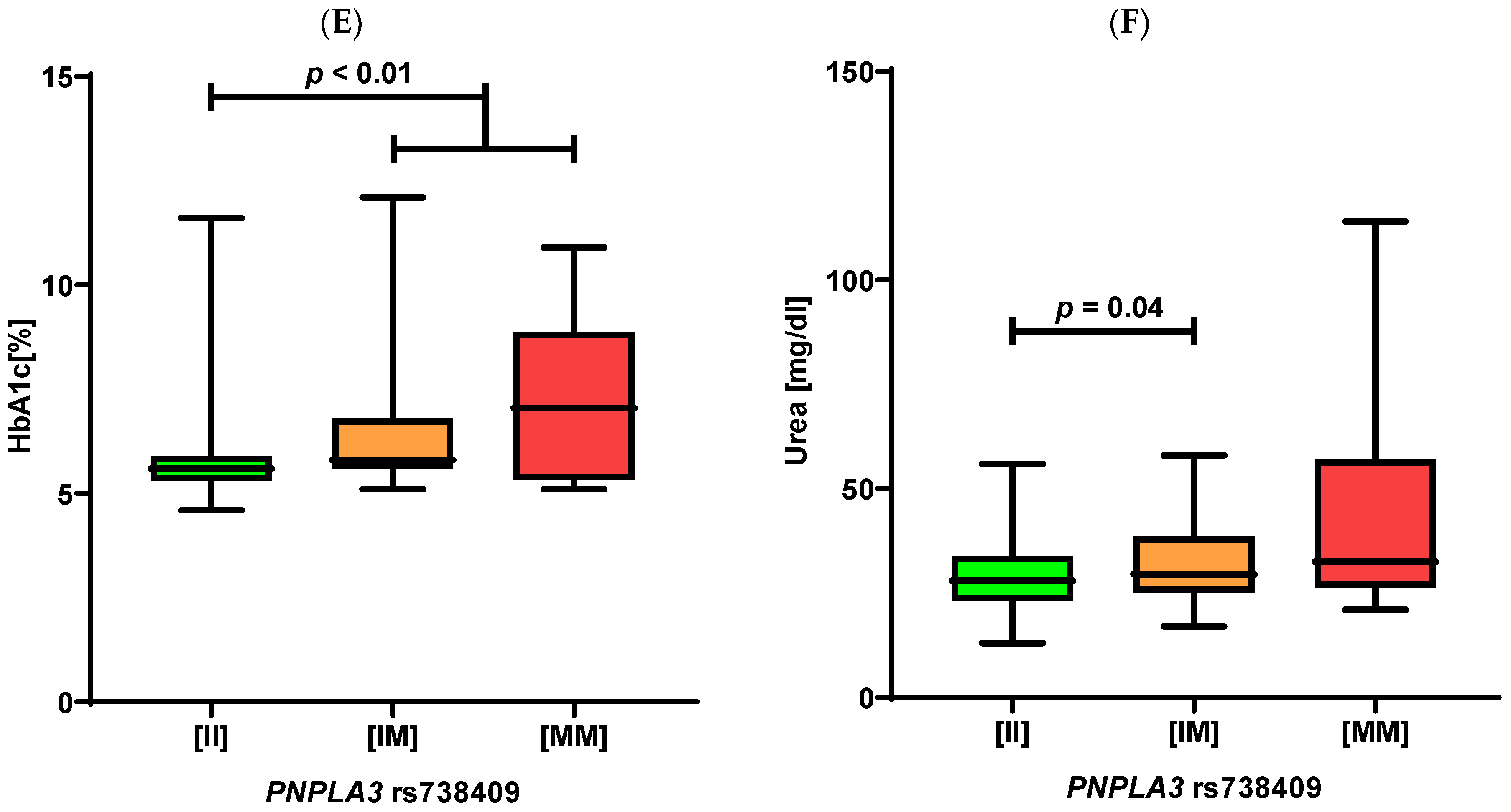
Concerning the metabolic profiles in our patients, we detected significant associations between the PNPLA3 p.I148M and increased fasting glucose (p = 0.03; Figure 1D), as well as, HbA1c levels (p < 0.01, Figure 1E). This polymorphism was also associated with an increased risk of developing type 2 diabetes (OR = 2.35, 95% CI 1.09–3.87; p = 0.03) and higher blood urea concentrations (p = 0.04, Figure 1F). Finally, we did not detect any association between patients’ phenotypes and the MBOAT7 polymorphism.

3. Discussion

Our current manuscript presents a comprehensive analysis of the genetic variants associated with NAFLD in patients undergoing bariatric surgery. To the best of our knowledge, this is one of the first such studies on individuals coming from Eastern Europe. In line with the previous reports, we detected harmful effects of the PNPLA3 p.I148M polymorphism on liver phenotypes and metabolic profiles in recruited patients. We also demonstrate that the common missense variant of MTARC1 p.A165T is protective against hepatic fibrosis in obese individuals and ameliorates the harmful effects of the PNPLA3 p.I148M minor allele. The protection conferred by the HSD17B13 rs72613567 polymorphism was less pronounced. It was associated with lower ALT and AST activities but not with the liver biopsy results.
We detected protective effects of the MTARC1 p.A165T polymorphism in our cohort. Luukkonen et al. reported previously that the MTARC1 variant is associated with a low SAF (steatosis-activity-fibrosis) score and decreased lobular inflammation, activity, and fibrosis [14]. Similar to our findings, the MTARC1 genotype did not correlate with steatosis in that study [14]. In our cohort, the protective effect of MTARC1 p.A165T minor allele on liver fibrosis was documented despite the absence of patients with cirrhosis, which could further increase the significance of the effect. Emdin et al. performed a large multicohort study and identified MTARC1 p.A165T as a protective variant against NAFLD-related cirrhosis and all-cause cirrhosis [12]. This variant was also associated with reduced hepatic steatosis diagnosed by physicians or assessed by imaging studies such as computed tomography [12] and magnetic resonance [17]. The decreased risk of developing NAFLD due to the MTARC1 variant was also confirmed in a recent analysis of 9491 cases with fatty liver [18]. MTARC1 is an enzyme located in the outer mitochondrial membrane that counterparts with other enzymatic systems and catalyzes the reduction of N-hydroxylated prodrugs, N-oxygenated compounds, and N(omega)-hydroxy-L-arginine [19,20]. The protective MTARC1 variant has been predicted by PolyPhen-2 to be deleterious to MTARC1 protein function [15]. Therefore, the reduced risk of liver injury associated with this polymorphism seems to originate from the loss of MTARC1 function.
HSD17B13 has a function of retinol dehydrogenase targeted to hepatic lipid droplets [21]. In our cohort, we did not detect any significant association between HSD17B13 variant and liver histology. However, lower activities of transaminases in carriers of the HSD17B13 polymorphism point to its potential protective effects. This observation is in line with the latest genome-wide association study (GWAS) by Gao et al. in Europeans, demonstrating that HSD17B13 and the MTARC1 and PNPLA3 polymorphisms substantially modulate serum transaminases [22]. The findings reported in our manuscript are partially in line with the GWAS by Anstee et al. [23]. This study demonstrated an association between the increased risk of biopsy-confirmed NAFLD and PNPLA3 as well as TM6SF2 variants and a trend towards a lower NAFLD risk in carriers of the HSD17B13 polymorphism.
Eventually, we confirmed that the PNPLA3 p.I148M polymorphism negatively affects the patients’ liver status. Indeed, not only was it associated with the NAFLD stage but also with a worse metabolic profile in analyzed patients. Interestingly, we detected a lower frequency of the PNPLA3 variant in patients scheduled for bariatric surgery compared to controls. We speculate that obese carriers of this variant might be less frequently scheduled for bariatric surgery due to their health status, but this needs to be evaluated in additional studies. Variant PNPLA3 p.I148M is by far the most frequently investigated and replicated genetic modifier of liver injury in NAFLD [5,24]. An example of the robust association between the PNPLA3 p.I148M variant and NAFLD severity was demonstrated in GWAS in the population of European ancestry [25]. Carriers of the minor PNPLA3 allele are known to be at risk of the entire spectrum of progressive liver steatosis from NAFLD to NASH, fibrosis, cirrhosis, and hepatocellular carcinoma [26]. Functional studies demonstrated that PNPLA3 is a lipase [27]. Alternatively, its retinyl-palmitate lipase activity in hepatic stellate cells was described [28]. PNPLA3 p.I148M and TM6SF2 p.E167K were associated with increased hepatic steatosis in our cohort. In contrast to PNPLA3, TM6SF2 did not affect fibrosis. This is in line with our previous studies in German and Polish patients with fatty liver [29,30]. TM6SF2 p.E167K was associated in both cohorts with hepatic steatosis, but a biopsy-based analysis of liver fibrosis in the German cohort did not yield a significant association between this variant and liver scarring [29]. In a large cohort study, Stender et al. found that adiposity amplified the genetic risk of NAFLD associated with SNPs (including PNPLA3 and TM6SF2) [31]. The effect of risk variants on hepatic steatosis assessed by magnetic resonance increased with BMI and was the greatest in severely obese individuals (BMI > 35 kg/m2). Hence, the risk of NAFLD development and progression at different stages, from steatosis to cirrhosis, is affected by the additive interaction between genetic variants and adiposity [31].
In our previous MRI-based analysis of Spanish patients undergoing bariatric surgery [32], we demonstrated that the PNPLA3, but not TM6SF2 or MBOAT7 variants, is associated with a greater reduction of hepatic fat content within 12 months after surgery. Overall, these results underscore the notion that obese individuals carrying the PNPLA3 minor allele are, on the one hand, at risk of a rapid progression of NAFLD, and they might be the ones who, in particular, profit from weight-loss therapy and lifestyle modifications [33]. Our findings suggest the possible reduction of detrimental PNPLA3 effects in obese individuals by MTARC1, but a small subgroup size is a limitation and requires confirmation. One possible solution is to consider MTARC1 p.A165T in assessing an individual’s risk and include it in a polygenic risk score. Such risk scores based on combinations of genetic risk variants have been published recently and showed significant associations with NAFLD severity [23,29]. In the future, we can expect novel NAFLD-risk modifiers that may emerge as more data is published from studies employing techniques such as GWAS or quantitative trait locus (QTL) mapping [25,34].
In conclusion, we demonstrate that MTARC1 p.A165T, and to a lesser extent, HSD17B13 rs72613567 polymorphism can be protective against NAFLD-related liver injury in patients with obesity scheduled for bariatric surgery. In particular, MTARC1 p.A165T can lower the harmful effects of the PNPLA3 p.I148M which remains the central risk factor for a progressive NAFLD. Since the function of MTARC1 is unclear, further studies in patients with chronic liver disease aimed at explaining the association between this genotype and clinical presentation are needed.

4. Materials and Methods

4.1. Study Cohort and Clinical Data

All patients were recruited at the Medical University of Warsaw. The study protocols (KB/140/2015 and KB/237/2015) were approved by the local ethics committee according to the ethical guidelines of the 1975 Declaration of Helsinki (latest revision, 2013), and written and informed consent was obtained from all participants. Inclusion criteria were based on clinical indications for bariatric surgery [35]. The criteria were fulfilled by 172 patients who were further evaluated and underwent laparoscopic sleeve gastrectomy and intraoperative liver biopsy. A group of adult controls without chronic liver diseases was recruited from outpatients in our hospital. A wedge liver biopsy was taken from the left lobe of the liver. Liver specimens collected intraoperatively were evaluated according to the NAFLD Activity Score (NAS) [36], which equals a sum of unweighted scores of steatosis (0–3), lobular inflammation (0–3), and ballooning (0–2). The NAS ≥ 5 corresponds with NASH. The stage of fibrosis was scored 0, 1a, 1b, 1c, 2, 3, and 4, as follows: 0—none; 1a—mild, zone 3, perisinusoidal; 1b—moderate, zone 3, perisinusoidal; 1c—portal/periportal; 2—perisinusoidal and portal/periportal; 3—bridging fibrosis; 4—cirrhosis. Exclusion criteria included previous history or positive serum markers of chronic liver disease. After the exclusion of 7 patients (4 positive for viral hepatitis, 3 with a history of alcohol abuse), 165 patients were finally included in the study.
All patients underwent clinical examination. Blood samples were drawn from fasted subjects, and liver function tests were determined by standard clinical-chemical assays in the central laboratory of our center.

4.2. Genotyping

DNA was extracted from peripheral blood mononuclear cells using the DNeasy Blood & Tissue Kit (Qiagen). DNA concentrations were measured using a NanoDrop spectrophotometer. Genotyping of the MTARC1 p.A165T (rs2642438), PNPLA3 p.I148M (rs738409), as well as other variants associated with the risk of NAFLD, namely TM6SF2 p.E167K (rs58542926), MBOAT7 p.G17E (rs641738), and HSD17B13 (rs72613567), was performed using TaqMan assays as described previously [25]. The fluorescence data were analyzed with allelic discrimination 7500 Software v.2.0.2.

4.3. Statistical Analyses

Statistical analyses were performed using SPSS (version 26.0; SPSS, Munich, Germany) and GraphPad Prism (version 8.0; GraphPad Software, San Diego, CA, USA). The nominal p-value < 0.05 was regarded as statistically significant. The consistency of genotyping distributions with the Hardy–Weinberg equilibrium (HWE) was tested by exact tests (https://ihg.helmholtz-muenchen.de/cgi-bin/hw/hwa1.pl accessed on 9 November 2022). The Shapiro–Wilk test was used to determine whether the set of observations followed normal distributions. Student t and Mann–Whitney-U tests were used to study normally and non-normally distributed parameters, respectively. The effects of genetic variants, age, BMI, and sex on patients’ phenotypes were tested by univariate and multivariate regression analyses.

Author Contributions

P.K., M.N. and R.P. performed bariatric surgeries; P.M. collected controls; Ł.M. and B.Z.-W. evaluated liver samples; S.N.W., F.L. and M.K. were responsible for genotyping; P.K., W.S. and M.K. analyzed the data; P.K., W.S. and M.K. drafted the first version of the manuscript which was edited by M.K.; K.Z. and M.K. supervised the project. All authors have read and agreed to the published version of the manuscript.

Funding

This research received no external funding.

Institutional Review Board Statement

The study was conducted in accordance with the Declaration of Helsinki, and approved by the Institutional Ethics Committee of Medical University of Warsaw (protocol codes: KB/140/2015 and KB/237/2015).

Informed Consent Statement

Informed consent was obtained from all subjects involved in the study. Written informed consent has been obtained from the patients to publish this paper.

Data Availability Statement

All data generated or analyzed during this study are included in this article. Further inquiries can be directed to the corresponding author.

Conflicts of Interest

The authors declare no conflict of interest.

References

  1. Estes, C.; Anstee, Q.M.; Arias-Loste, M.T.; Bantel, H.; Bellentani, S.; Caballeria, J.; Colombo, M.; Craxi, A.; Crespo, J.; Day, C.P.; et al. Modeling NAFLD disease burden in China, France, Germany, Italy, Japan, Spain, United Kingdom, and United States for the period 2016–2030. J. Hepatol. 2018, 69, 896–904. [Google Scholar] [CrossRef] [PubMed]
  2. Sarwar, R.; Pierce, N.; Koppe, S. Obesity and nonalcoholic fatty liver disease: Current perspectives. Diabetes Metab. Syndr. Obes. Targets Ther. 2018, 11, 533–542. [Google Scholar] [CrossRef] [PubMed] [Green Version]
  3. Lefere, S.; Onghena, L.; Vanlander, A.; van Nieuwenhove, Y.; Devisscher, L.; Geerts, A. Bariatric surgery and the liver—Mechanisms, benefits, and risks. Obes. Rev. 2021, 22, e13294. [Google Scholar] [CrossRef] [PubMed]
  4. Loomba, R.; Friedman, S.L.; Shulman, G.I. Mechanisms and disease consequences of nonalcoholic fatty liver disease. Cell 2021, 184, 2537–2564. [Google Scholar] [CrossRef] [PubMed]
  5. Krawczyk, M.; Liebe, R.; Lammert, F. Toward Genetic Prediction of Nonalcoholic Fatty Liver Disease Trajectories: PNPLA3 and Beyond. Gastroenterology 2020, 158, 1865–1880.e1861. [Google Scholar] [CrossRef]
  6. Dubuquoy, C.; Burnol, A.-F.; Moldes, M. PNPLA3, a genetic marker of progressive liver disease, still hiding its metabolic function? Clin. Res. Hepatol. Gastroenterol. 2013, 37, 30–35. [Google Scholar] [CrossRef]
  7. Kozlitina, J.; Smagris, E.; Stender, S.; Nordestgaard, B.G.; Zhou, H.H.; Tybjærg-Hansen, A.; Vogt, T.F.; Hobbs, H.H.; Cohen, J.C. Exome-wide association study identifies a TM6SF2 variant that confers susceptibility to nonalcoholic fatty liver disease. Nat. Genet. 2014, 46, 352–356. [Google Scholar] [CrossRef] [Green Version]
  8. Teo, K.; Abeysekera, K.W.; Adams, L.; Aigner, E.; Anstee, Q.M.; Banales, J.M.; Banerjee, R.; Basu, P.; Berg, T.; Bhatnagar, P.; et al. rs641738C>T near MBOAT7 is associated with liver fat, ALT and fibrosis in NAFLD: A meta-analysis. J. Hepatol. 2021, 74, 20–30. [Google Scholar] [CrossRef]
  9. Gijón, M.A.; Riekhof, W.R.; Zarini, S.; Murphy, R.C.; Voelker, D.R. Lysophospholipid Acyltransferases and Arachidonate Recycling in Human Neutrophils. J. Biol. Chem. 2008, 283, 30235–30245. [Google Scholar] [CrossRef] [Green Version]
  10. Li, T.-T.; Li, T.-H.; Peng, J.; He, B.; Liu, L.-S.; Wei, D.-H.; Jiang, Z.-S.; Zheng, X.-L.; Tang, Z.-H. TM6SF2: A novel target for plasma lipid regulation. Atherosclerosis 2018, 268, 170–176. [Google Scholar] [CrossRef]
  11. Abul-Husn, N.S.; Cheng, X.; Li, A.H.; Xin, Y.; Schurmann, C.; Stevis, P.; Liu, Y.; Kozlitina, J.; Stender, S.; Wood, G.C.; et al. A Protein-Truncating HSD17B13 Variant and Protection from Chronic Liver Disease. N. Engl. J. Med. 2018, 378, 1096–1106. [Google Scholar] [CrossRef]
  12. Emdin, C.A.; Haas, M.E.; Khera, A.V.; Aragam, K.; Chaffin, M.; Klarin, D.; Hindy, G.; Jiang, L.; Wei, W.-Q.; Feng, Q.; et al. A missense variant in Mitochondrial Amidoxime Reducing Component 1 gene and protection against liver disease. PLoS Genet. 2020, 16, e1008629. [Google Scholar] [CrossRef] [Green Version]
  13. Janik, M.K.; Smyk, W.; Kruk, B.; Szczepankiewicz, B.; Górnicka, B.; Lebiedzińska-Arciszewska, M.; Potes, Y.; Simões, I.C.M.; Weber, S.N.; Lammert, F.; et al. MARC1 p.A165T variant is associated with decreased markers of liver injury and enhanced antioxidant capacity in autoimmune hepatitis. Sci. Rep. 2021, 11, 24407. [Google Scholar] [CrossRef]
  14. Luukkonen, P.K.; Juuti, A.; Sammalkorpi, H.; Penttilä, A.K.; Orešič, M.; Hyötyläinen, T.; Arola, J.; Orho-Melander, M.; Yki-Järvinen, H. MARC1 variant rs2642438 increases hepatic phosphatidylcholines and decreases severity of non-alcoholic fatty liver disease in humans. J. Hepatol. 2020, 73, 725–726. [Google Scholar] [CrossRef]
  15. Innes, H.; Buch, S.; Hutchinson, S.; Guha, I.N.; Morling, J.R.; Barnes, E.; Irving, W.; Forrest, E.; Pedergnana, V.; Goldberg, D.; et al. Genome-Wide Association Study for Alcohol-Related Cirrhosis Identifies Risk Loci in MARC1 and HNRNPUL1. Gastroenterology 2020, 159, 1276–1289.e1277. [Google Scholar] [CrossRef]
  16. Schneider, C.V.; Schneider, K.M.; Conlon, D.M.; Park, J.; Vujkovic, M.; Zandvakili, I.; Ko, Y.-A.; Trautwein, C.; Carr, R.M.; Strnad, P.; et al. A genome-first approach to mortality and metabolic phenotypes in MTARC1 p.Ala165Thr (rs2642438) heterozygotes and homozygotes. Med 2021, 2, 851–863.e853. [Google Scholar] [CrossRef]
  17. Parisinos, C.A.; Wilman, H.R.; Thomas, E.L.; Kelly, M.; Nicholls, R.C.; McGonigle, J.; Neubauer, S.; Hingorani, A.D.; Patel, R.S.; Hemingway, H.; et al. Genome-wide and Mendelian randomisation studies of liver MRI yield insights into the pathogenesis of steatohepatitis. J. Hepatol. 2020, 73, 241–251. [Google Scholar] [CrossRef]
  18. Sveinbjornsson, G.; Ulfarsson, M.O.; Thorolfsdottir, R.B.; Jonsson, B.A.; Einarsson, E.; Gunnlaugsson, G.; Rognvaldsson, S.; Arnar, D.O.; Baldvinsson, M.; Bjarnason, R.G.; et al. Multiomics study of nonalcoholic fatty liver disease. Nat. Genet. 2022, 54, 1652–1663. [Google Scholar] [CrossRef]
  19. Gruenewald, S.; Wahl, B.; Bittner, F.; Hungeling, H.; Kanzow, S.; Kotthaus, J.; Schwering, U.; Mendel, R.R.; Clement, B. The Fourth Molybdenum Containing Enzyme mARC: Cloning and Involvement in the Activation of N-Hydroxylated Prodrugs. J. Med. Chem. 2008, 51, 8173–8177. [Google Scholar] [CrossRef]
  20. Kotthaus, J.; Wahl, B.; Havemeyer, A.; Kotthaus, J.; Schade, D.; Garbe-Schönberg, D.; Mendel, R.; Bittner, F.; Clement, B. Reduction of Nω-hydroxy-L-arginine by the mitochondrial amidoxime reducing component (mARC). Biochem. J. 2011, 433, 383–391. [Google Scholar] [CrossRef]
  21. Ma, Y.; Belyaeva, O.V.; Brown, P.M.; Fujita, K.; Valles, K.; Karki, S.; De Boer, Y.S.; Koh, C.; Chen, Y.; Du, X.; et al. 17-Beta Hydroxysteroid Dehydrogenase 13 Is a Hepatic Retinol Dehydrogenase Associated With Histological Features of Nonalcoholic Fatty Liver Disease. Hepatology 2019, 69, 1504–1519. [Google Scholar] [CrossRef] [PubMed]
  22. Gao, C.; Marcketta, A.; Backman, J.D.; O’Dushlaine, C.; Staples, J.; Ferreira, M.A.R.; Lotta, L.A.; Overton, J.D.; Reid, J.G.; Mirshahi, T.; et al. Genome-wide association analysis of serum alanine and aspartate aminotransferase, and the modifying effects of BMI in 388k European individuals. Genet. Epidemiol. 2021, 45, 664–681. [Google Scholar] [CrossRef] [PubMed]
  23. Anstee, Q.M.; Darlay, R.; Cockell, S.; Meroni, M.; Govaere, O.; Tiniakos, D.; Burt, A.D.; Bedossa, P.; Palmer, J.; Liu, Y.-L.; et al. Genome-wide association study of non-alcoholic fatty liver and steatohepatitis in a histologically characterised cohort. J. Hepatol. 2020, 73, 505–515. [Google Scholar] [CrossRef] [PubMed]
  24. Romeo, S.; Kozlitina, J.; Xing, C.; Pertsemlidis, A.; Cox, D.; Pennacchio, L.A.; Boerwinkle, E.; Cohen, J.C.; Hobbs, H.H. Genetic variation in PNPLA3 confers susceptibility to nonalcoholic fatty liver disease. Nat. Genet. 2008, 40, 1461–1465. [Google Scholar] [CrossRef] [PubMed] [Green Version]
  25. Namjou, B.; Lingren, T.; Huang, Y.; Parameswaran, S.; Cobb1, B.L.; Stanaway, I.B.; Connolly, J.J.; Mentch, F.D.; Benoit, B.; Niu, X.; et al. GWAS and enrichment analyses of non-alcoholic fatty liver disease identify new trait-associated genes and pathways across eMERGE Network. BMC Med. 2019, 17, 135. [Google Scholar] [CrossRef] [PubMed]
  26. Carlsson, B.; Lindén, D.; Brolén, G.; Liljeblad, M.; Bjursell, M.; Romeo, S.; Loomba, R. Review article: The emerging role of genetics in precision medicine for patients with non-alcoholic steatohepatitis. Aliment. Pharmacol. Ther. 2020, 51, 1305–1320. [Google Scholar] [CrossRef]
  27. Zechner, R.; Zimmermann, R.; Eichmann, T.O.; Kohlwein, S.D.; Haemmerle, G.; Lass, A.; Madeo, F. FAT SIGNALS—Lipases and Lipolysis in Lipid Metabolism and Signaling. Cell Metab. 2012, 15, 279–291. [Google Scholar] [CrossRef] [Green Version]
  28. Pirazzi, C.; Valenti, L.; Motta, B.M.; Pingitore, P.; Hedfalk, K.; Mancina, R.M.; Burza, M.A.; Indiveri, C.; Ferro, Y.; Montalcini, T.; et al. PNPLA3 has retinyl-palmitate lipase activity in human hepatic stellate cells. Hum. Mol. Genet. 2014, 23, 4077–4085. [Google Scholar] [CrossRef] [Green Version]
  29. Krawczyk, M.; Rau, M.; Schattenberg, J.M.; Bantel, H.; Pathil, A.; Demir, M.; Kluwe, J.; Boettler, T.; Lammert, F.; Geier, A. Combined effects of the PNPLA3 rs738409, TM6SF2 rs58542926, and MBOAT7 rs641738 variants on NAFLD severity: A multicenter biopsy-based study. J. Lipid Res. 2017, 58, 247–255. [Google Scholar] [CrossRef] [Green Version]
  30. Krawczyk, M.; Stachowska, E.; Milkiewicz, P.; Lammert, F.; Milkiewicz, M. Reduction of Caloric Intake Might Override the Prosteatotic Effects of the PNPLA3 p.I148M and TM6SF2 p.E167K Variants in Patients with Fatty Liver: Ultrasound-Based Prospective Study. Digestion 2016, 93, 139–148. [Google Scholar] [CrossRef]
  31. Stender, S.; Kozlitina, J.; Nordestgaard, B.G.; Tybjærg-Hansen, A.; Hobbs, H.H.; Cohen, J.C. Adiposity amplifies the genetic risk of fatty liver disease conferred by multiple loci. Nat. Genet. 2017, 49, 842–847. [Google Scholar] [CrossRef]
  32. Krawczyk, M.; Jiménez-Agüero, R.; Alustiza, J.M.; Emparanza, J.I.; Perugorria, M.J.; Bujanda, L.; Lammert, F.; Banales, J.M. PNPLA3 p.I148M variant is associated with greater reduction of liver fat content after bariatric surgery. Surg. Obes. Relat. Dis. 2016, 12, 1838–1846. [Google Scholar] [CrossRef]
  33. Shen, J.; Wong, G.L.-H.; Chan, H.L.-Y.; Chan, R.S.; Chan, H.-Y.; Chu, W.C.; Cheung, B.H.-K.; Yeung, D.K.-W.; Li, L.S.; Sea, M.M.-M.; et al. PNPLA3 gene polymorphism and response to lifestyle modification in patients with nonalcoholic fatty liver disease. J. Gastroenterol. Hepatol. 2015, 30, 139–146. [Google Scholar] [CrossRef]
  34. Delpero, M.; Arends, D.; Freiberg, A.; Brockmann, G.A.; Hesse, D. QTL-mapping in the obese Berlin Fat Mouse identifies additional candidate genes for obesity and fatty liver disease. Sci. Rep. 2022, 12, 10471. [Google Scholar] [CrossRef]
  35. Fried, M.; Yumuk, V.; Oppert, J.-M.; Scopinaro, N.; Torres, A.J.; Weiner, R.; Yashkov, Y.; Frühbeck, G. Interdisciplinary European Guidelines on Metabolic and Bariatric Surgery. Obes. Facts 2013, 6, 449–468. [Google Scholar] [CrossRef]
  36. Kleiner, D.E.; Brunt, E.M.; Van Natta, M.; Behling, C.; Contos, M.J.; Cummings, O.W.; Ferrell, L.D.; Liu, Y.-C.; Torbenson, M.S.; Unalp-Arida, A.; et al. Design and validation of a histological scoring system for nonalcoholic fatty liver disease. Hepatology 2005, 41, 1313–1321. [Google Scholar] [CrossRef]
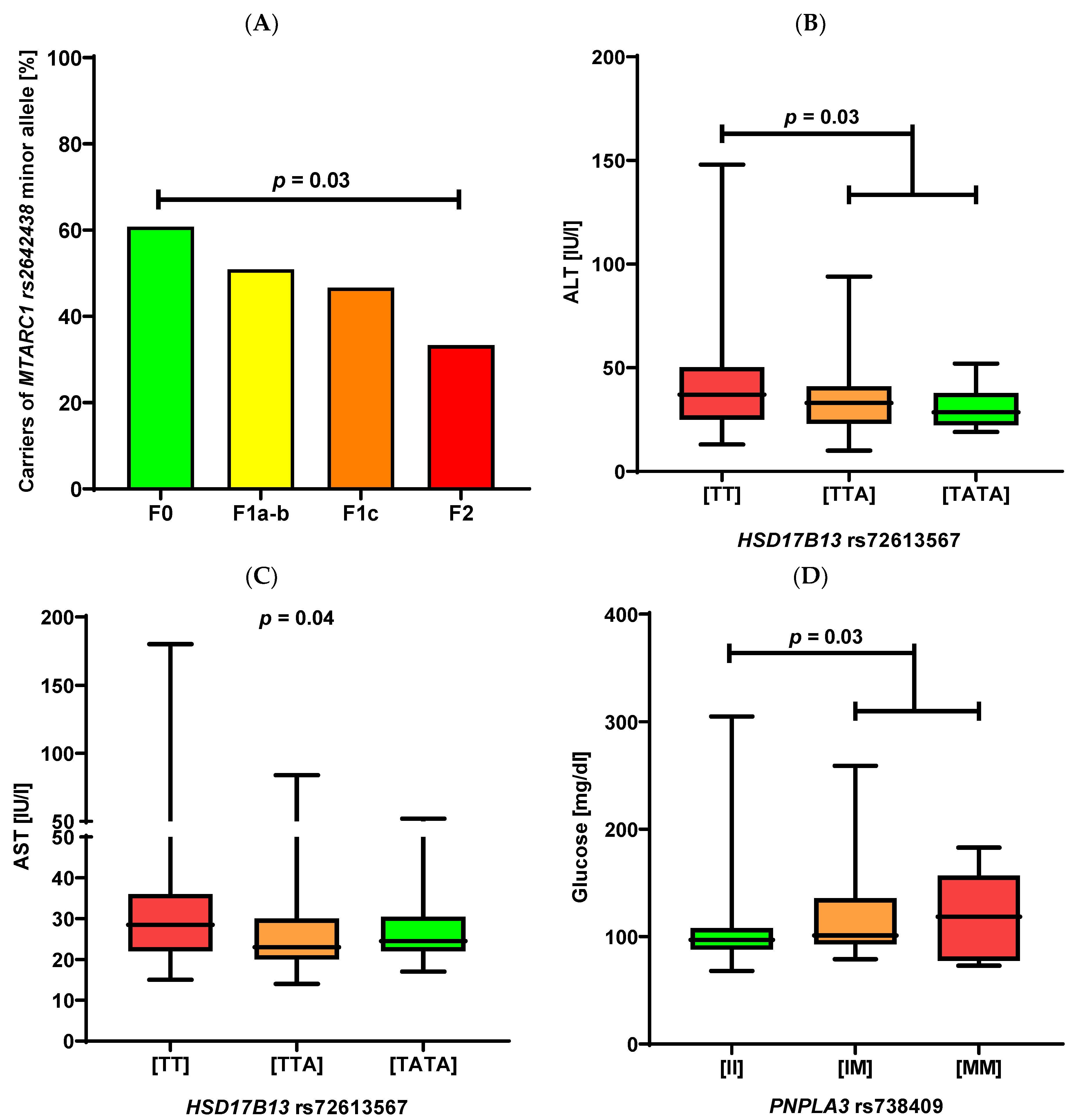
Figure 1. (A): Distribution of the MTARC1 minor allele in relation to liver fibrosis at liver biopsy in individuals undergoing bariatric surgery. F0 n = 31, F1a–b n = 29, F1c n = 14, F2 n = 7. (B,C): Association between the HSD17B13 polymorphism and serum ALT (B) and AST activities (C). Genotype frequencies: [TT] n = 102, [TTA] n = 51, [TATA] n = 12. (DF): Association between the PNPLA3 variant and glucose (D), HbA1c (E), and urea (F) levels. Genotype frequencies: [II] n = 117, [IM] n = 42, [MM] n = 6.
Ijms 23 15825 g001aIjms 23 15825 g001b
Table 1. Baseline characteristics of the study cohort.
Entire CohortPatients without NAFLDPatients with NAFLD
Total participants, n16549 (29.7%)116 (70.3%)
Female, n [%]110 (66.7%)39 (79.6%)71 (61.2%)
Age [years]42 (19–65)39 (19–63)42 (21–65)
BMI [kg/m2]43.8 (34.2–64.3)43.8 (34.2–56.0)43.7 (34.3–64.3)
Hypertension107 (64.8%)25 (51.0%)82 (70.7%)
Type 2 diabetes mellitus48 (29.1%)4 (8.2%)44 (37.9%)
Hyperlipidaemia109 (66.1%)23 (46.9%)86 (74.1%)
Haemoglobin [g/dL]14.1 (10.2–18.3)13.6 (10.2–18.3)14.2 (10.6–18.0)
Platelets [G/L]258.0 (122.0–426.0)266.0 (122.0–426.0)253.0 (131.0–425.0)
Creatinine [mg/dL]0.8 (0.5–3.6)0.8 (0.6–3.6)0.8 (0.5–2.1)
Urea [mg/dL]29.0 (13.0–114.0)28.0 (13.0–114.0)29.0 (14.0–58.0)
ALT [IU/L]34.0 (10.0–148.0)25.0 (13.0–94.0)38.0 (10.0–148.0)
AST [IU/L]26.0 (14.0–180.0)22.0 (15.0–108.0)29.0 (14.0–180.0)
ALP [IU/L]72.0 (25.0–133.0)73.0 (40.0–116.0)71.0 (25.0–133.0)
GGT [IU/L]31.0 (12.0–296.0)23.0 (12.0–192.0)35.5 (12.0–296.0)
Bilirubin [mg/dL]0.6 (0.1–1.9)0.5 (0.2–1.7)0.6 (0.1–1.9)
Amylase [IU/L]38.5 (17.0–144.0)37.0 (18.0–144.0)39.0 (17.0–89.0)
Lipase [IU/L]26.0 (13.0–180.0)25.0 (14.0–86.0)29.0 (13.0–180.0)
Total cholesterol [mg/dL]185.0 (97.0–273.0)176.0 (97.0–270.0)187.0 (115.0–273.0)
Triglycerides [mg/dL]151.0 (58.0–779.0)117.0 (58.0–263.0)163.5 (63.0–779.0)
HDL [mg/dL]44.0 (18.0–88.0)47.5 (29.0–88.0)42.0 (18.0–84.0)
LDL [mg/dL]105.0 (27.4–213.0)104.0 (29.0–213.0)105.0 (27.4–181.0)
FIB-4 [points]0.7 (0.2–5.0)0.7 (0.2–2.1)0.8 (0.3–5.0)
NFS [points]−0.85 (−4.0–4.8)−1.1 (−3.0–2.7)−0.7 (−4.0–4.8)
Glycaemia [mg/dL]97.0 (68.0–305.0)92.0 (73.0–126.0)101.0 (68.0–305.0)
HbA1c [%]5.7 (4.6–12.1)5.4 (4.9–7.1)5.8 (4.6–12.1)
C-peptide [ng/mL]3.8 (0.7–11.8)3.1 (2.0–5.5)3.9 (0.7–11.8)
Insulin [IU/mL]19.7 (4.8–177.0)14.5 (6.1–79.4)21.7 (4.8–177.0)
HOMA-IR [points]4.7 (1.0–74.0)3.4 (1.2–14.3)5.5 (1.0–74.0)
Values are presented as medians (ranges). Abbreviations: ALP, alkaline phosphatase; ALT, alanine aminotransferase; AST, aspartate aminotransferase; BMI, body mass index; FIB-4, fibrosis-4 score; GGT, Gamma-glutamyl transferase; HbA1c, glycated hemoglobin; HDL, High-density lipoprotein; HOMA-IR, homeostatic model assessment for insulin resistance; LDL, low-density lipoprotein; NAFLD, non-alcoholic fatty liver disease; NFS, NAFLD fibrosis score.
Table 2. Results of liver biopsies in 165 patients undergoing bariatric surgery.
HistologyScoreNumber of Patients%
Steatosis
  • <5%
04929.7
  • 5–33%
16438.8
  • >33–66%
23621.8
  • >66%
3169.7
Lobular inflammation
  • No foci
0159.1
  • <2 foci/200×
16941.8
  • 2–4 foci/200×
27143.0
  • >4 foci/200×
3106.1
Hepatocyte ballooning
  • None
04829.1
  • Few balloon cells
17746.7
  • Many cells/prominent ballooning
24024.2
NAS
  • 0–2
0–25130.9
  • 3–4
3–46740.6
  • ≥5
≥54728.5
Fibrosis
  • None
05130.9
  • Mild, zone 3, perisinusoidal
1a5734.6
  • Moderate, zone 3, perisinusoidal
1b21.2
  • Portal/periportal
1c3018.2
  • Perisinusoidal and portal/periportal
22112.7
  • Bridging fibrosis
342.4
  • Cirrhosis
400
Table 3. Genotype frequencies in all patients scheduled for bariatric surgery (cases) and in controls.
Cases
MTARC1
(rs2642438)
PNPLA3
(rs738409)
TM6SF2
(rs58542926)
MBOAT7
(rs641738)
HSD17B13
(rs72613567)
Wild-type80 (48.5%)117 (70.9%)149 (90.3%)56 (33.9%)102 (61.8%)
Heterozygous variant75 (45.4%)42 (25.5%)14 (8.5%)81 (49.1%)51 (30.9%)
Homozygous variant10 (6.1%)6 (3.6%)2 (1.2%)28 (17.0%)12 (7.3%)
Controls
Wild-type144 (45.9%)181 (57.6%)278 (88.5%)90 (28.6%)176 (56.1%)
Heterozygous variant142 (45.2%)114 (36.3%)35 (11.2%)166 (52.9%)110 (35.0%)
Homozygous variant28 (8.9%)19 (6.1%)1 (0.3%)58 (18.5%)28 (8.9%)
p0.37<0.010.790.300.23
OR0.850.641.060.870.84
The differences in genotype distributions between cases and controls were analyzed using Armitage’s trend test. Abbreviations: HSD17B13, 17β-Hydroxysteroid dehydrogenase type 13; MTARC1, mitochondrial amidoxime-reducing component 1; MBOAT7, membrane-bound O-acyltransferase domain-containing protein 7; OR, odds ratio; PNPLA3, patatin-like phospholipase domain-containing protein 3 also known as adiponutrin; TM6SF2, transmembrane 6 superfamily 2 human genes.
Table 4. Risk and protective factors for developing liver fibrosis.
Univariate Analysis Multivariate Analysis
FactorOdds Ratio (95% CI)pOdds Ratio (95% CI)p
Fibrosis Grade ≥ 1a
MTARC1 (rs2642438)0.56 (0.32–0.96)0.04NA
PNPLA3 (rs738409)1.65 (0.84–3.23)0.15NA
TM6SF2 (rs58542926)1.50 (0.52–4.35)0.45NA
MBOAT7 (rs641738)0.96 (0.60–1.55)0.87NA
HSD17B13 (rs72613567)1.37 (0.79–2.39)0.26NA
BMI (kg/m2)1.01 (0.95–1.07)0.74NA
Age (years)1.00 (0.97–1.04)0.95NA
Gender0.77 (0.37–1.58)0.48NA
Fibrosis Grade ≥ 1b
MTARC1 (rs2642438)0.55 (0.31–0.96)0.040.52 (0.29–0.92)0.03
PNPLA3 (rs738409)1.75 (0.98–3.13)0.06NA
TM6SF2 (rs58542926)1.79 (0.73–4.35)0.20NA
MBOAT7 (rs641738)1.16 (0.73–1.84)0.53NA
HSD17B13 (rs72613567)1.08 (0.65–1.79)0.78NA
BMI (kg/m2)1.03 (0.98–1.09)0.25NA
Age (years)1.04 (1.00–1.08)0.031.04 (1.01–1.08)0.02
Gender0.70 (0.36–1.37)0.30NA
Fibrosis Grade ≥ 1c
MTARC1 (rs2642438)0.55 (0.31–0.97)0.040.51 (0.28–0.92)0.04
PNPLA3 (rs738409)1.87 (1.04–3.35)0.041.79 (0.98–3.30)0.06
TM6SF2 (rs58542926)1.88 (0.77–4.58)0.17NA
MBOAT7 (rs641738)1.14 (0.72–1.82)0.58NA
HSD17B13 (rs72613567)1.07 (0.64–1.79)0.79NA
BMI (kg/m2)1.04 (0.98–1.10)0.18NA
Age (years)1.04 (1.00–1.07)0.041.04 (1.00–1.08)0.06
Gender0.72 (0.37–1.43)0.35NA
Fibrosis Grade ≥ 2
MTARC1 (rs2642438)0.54 (0.25–1.17)0.12NA
PNPLA3 (rs738409)2.86 (1.43–5.72)0.0033.09 (1.49–6.40)0.002
TM6SF2 (rs58542926)2.56 (0.97–6.72)0.06NA
MBOAT7 (rs641738)1.13 (0.61–2.08)0.70NA
HSD17B13 (rs72613567)0.64 (0.29–1.38)0.25NA
BMI (kg/m2)1.11 (1.04–1.20)0.0031.12 (1.04–1.21)0.003
Age (years)1.02 (0.97–1.06)0.46NA
Gender0.58 (0.25–1.39)0.22NA
NA—not applied; only statistically significant factors from the univariate analysis were included in the multivariate analysis. Abbreviations: see Table 1 and Table 3.
Table 5. Risk and protective factors for developing liver steatosis.
Univariate Analysis Multivariate Analysis
FactorOdds Ratio (95% CI)pOdds Ratio (95% CI)p
Steatosis grade ≥ S1
MTARC1 (rs2642438)1.20 (0.68–2.09)0.53NA
PNPLA3 (rs738409)1.38 (0.71–2.65)0.34NA
TM6SF2 (rs58542926)1.98 (0.60–6.58)0.26NA
MBOAT7 (rs641738)1.25 (0.77–2.04)0.37NA
HSD17B13 (rs72613567)0.77 (0.46–1.29)0.31NA
BMI (kg/m2)1.01 (0.95–1.07)0.81NA
Age (years)1.02 (0.98–1.05)0.35NA
Gender0.41 (0.18–0.89)0.03NA
Steatosis grade ≥ S2
MTARC1 (rs2642438)0.68 (0.38–1.19)0.17NA
PNPLA3 (rs738409)2.27 (1.25–4.12)0.0072.27 (1.24–4.15)0.008
TM6SF2 (rs58542926)2.51 (1.00–6.28)0.0492.51 (0.98–6.46)0.056
MBOAT7 (rs641738)1.25 (0.78–2.01)0.36NA
HSD17B13 (rs72613567)0.89 (0.52–1.51)0.66NA
BMI (kg/m2)1.01 (0.95–1.07)0.82NA
Age (years)1.02 (0.98–1.05)0.33NANA
Gender0.72 (0.36–1.43)0.34NA
Steatosis grade = S3
MTARC1 (rs2642438)1.65 (0.73–3.75)0.23NA
PNPLA3 (rs738409)3.46 (1.55–7.72)0.0023.69 (1.56–8.70)0.003
TM6SF2 (rs58542926)5.66 (1.99–16.11)0.0016.19 (2.00–19.17)0.001
MBOAT7 (rs641738)1.28 (0.61–2.67)0.52NA
HSD17B13 (rs72613567)0.95 (0.41–2.19)0.91NA
BMI (kg/m2)1.07 (0.99–1.17)0.11NA
Age (years)1.00 (0.95–1.05)0.96NA
Gender0.82 (0.28–2.38)0.71NA
NA—not applied; only statistically significant factors from the univariate analysis were included in the multivariate analysis. Abbreviations: see Table 1 and Table 3.
Table 6. Risk and protective factors for NASH.
Univariate Analysis Multivariate Analysis
FactorOdds Ratio (95% CI)pOdds Ratio (95% CI)p
MTARC1 (rs2642438)0.60 (0.33–1.08)0.08NA
PNPLA3 (rs738409)2.26 (1.24–4.13)0.0082.22 (1.03–4.80)0.04
TM6SF2 (rs58542926)2.32 (0.94–5.75)0.07NA
MBOAT7 (rs641738)1.06 (0.65–1.72)0.81NA
HSD17B13 (rs72613567)1.05 (0.61–1.79)0.86NA
Type 2 diabetes mellitus8.94 (3.94–20.28)<0.0017.91 (3.32–18.86)<0.001
Hyperlipidemia2.68 (1.14–6.30)0.022.93 (0.97–8.89)0.06
BMI (kg/m2)1.01 (0.95–1.07)0.75NA
Age (years)1.01 (0.98–1.05)0.57NA
Gender1.09 (0.53–2.25)0.81NA
NA—not applied; only statistically significant factors from the univariate analysis were included in the multivariate analysis. Abbreviations: see Table 1 and Table 3; NASH, non-alcoholic steatohepatitis.
Publisher’s Note: MDPI stays neutral with regard to jurisdictional claims in published maps and institutional affiliations.