
Tissue-Protective and Anti-Inflammatory Landmark of PRP-Treated Mesenchymal Stromal Cells Secretome for Osteoarthritis


Abstract
:1. Introduction
2. Results
2.1. PRP Characterization
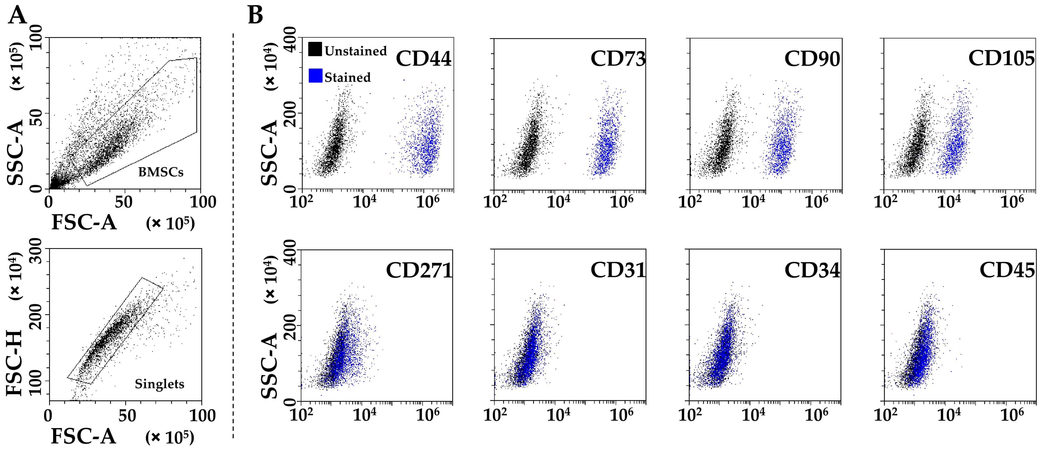
2.2. PRP-Treated BMSCs’ Characterization
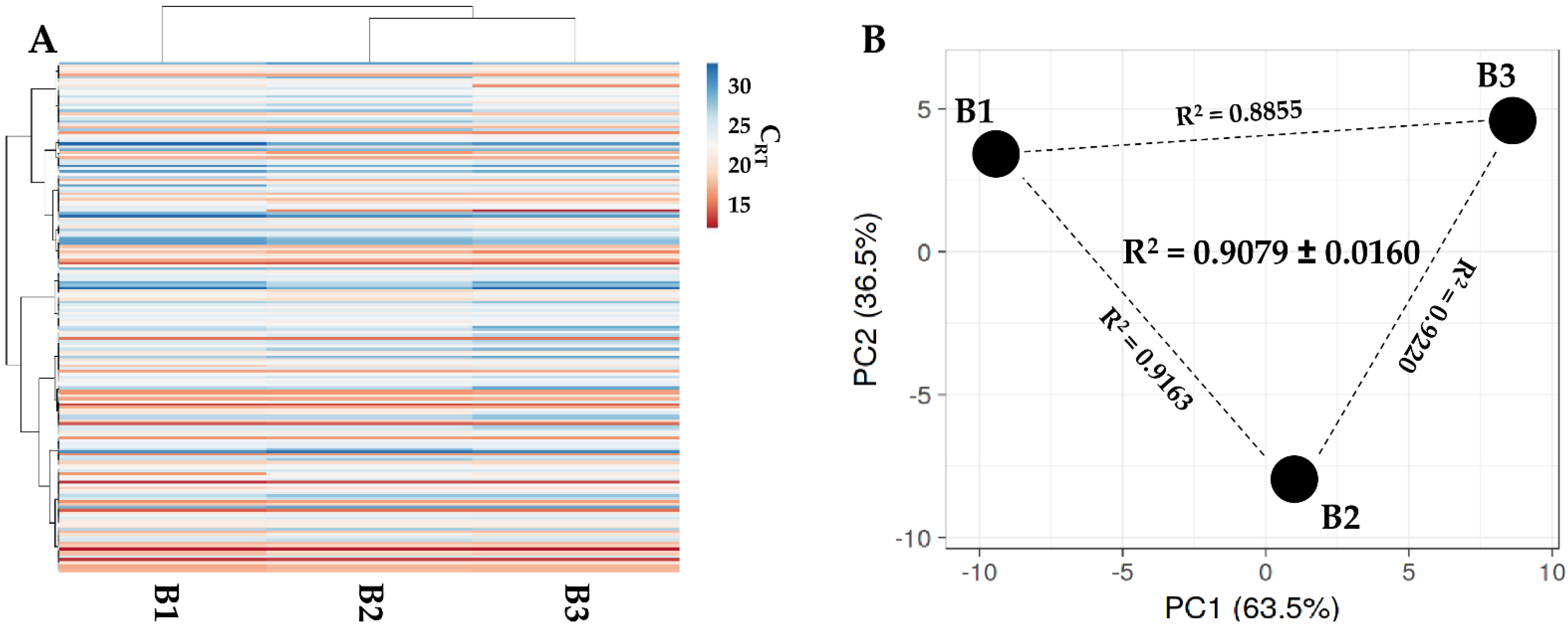
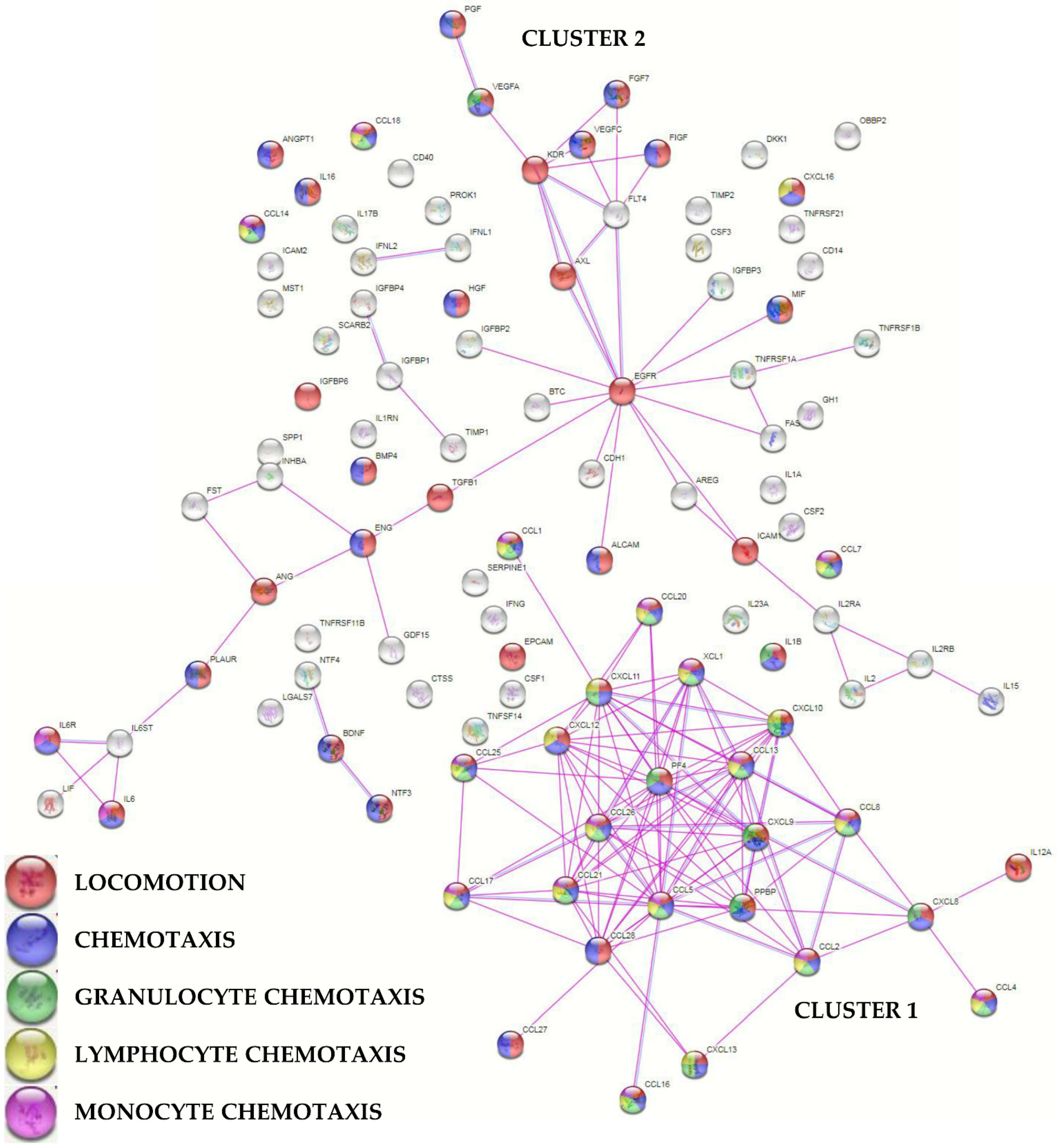
2.3. PRP-BMSCs’ Secreted Factors
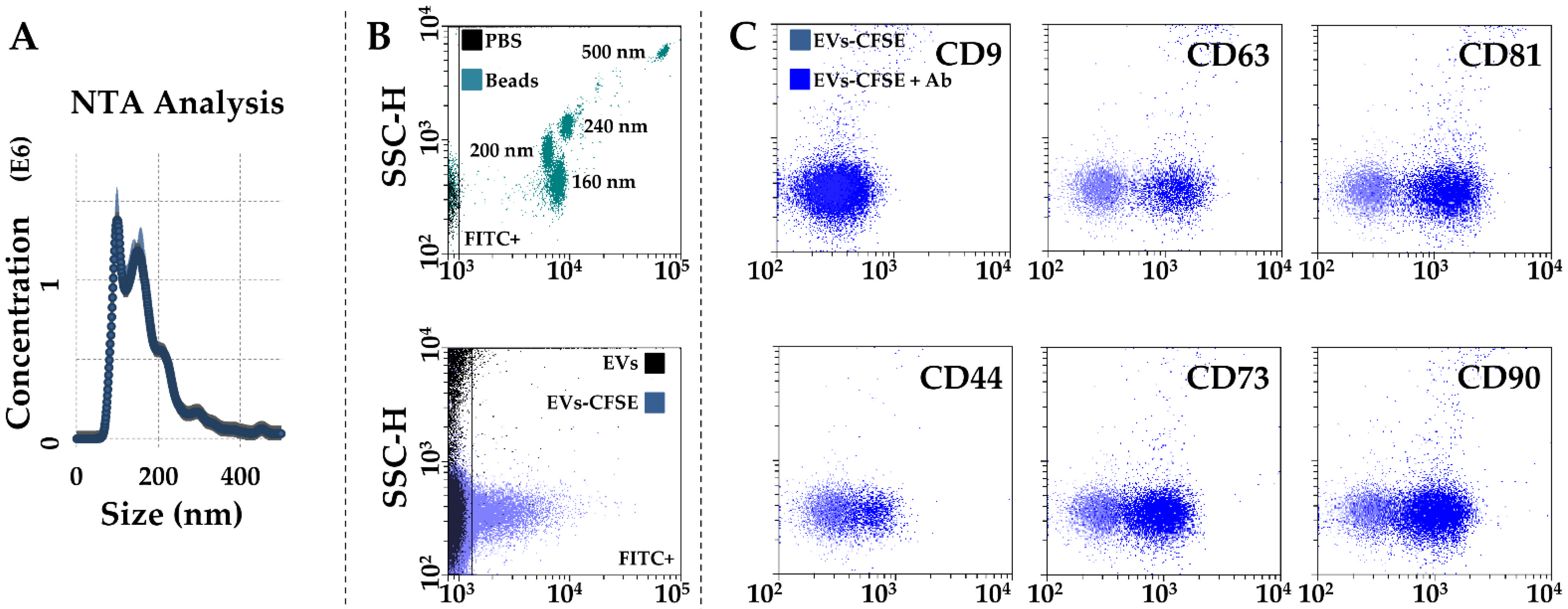
2.4. EVs’ Characterization
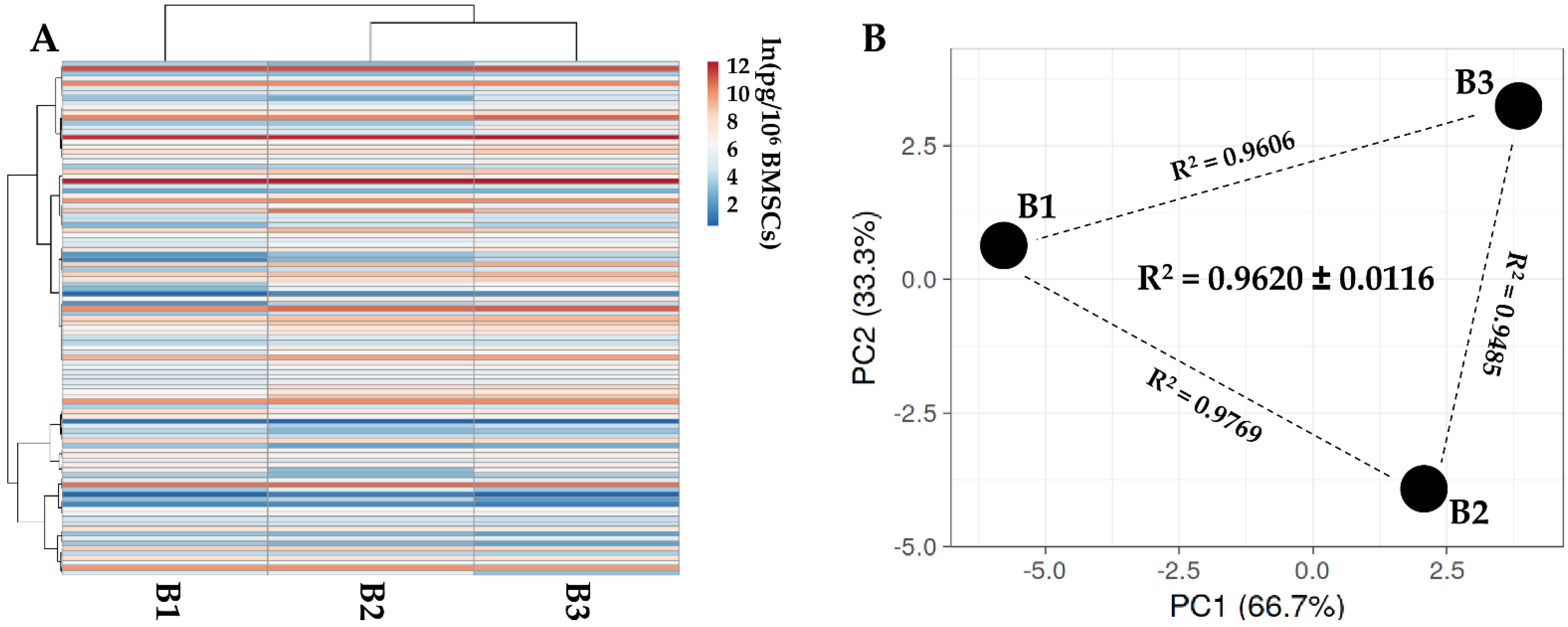
2.5. EV-miRNAs’ Identification
2.6. OA-Related Targets for PRP-BMSC EV-miRNAs
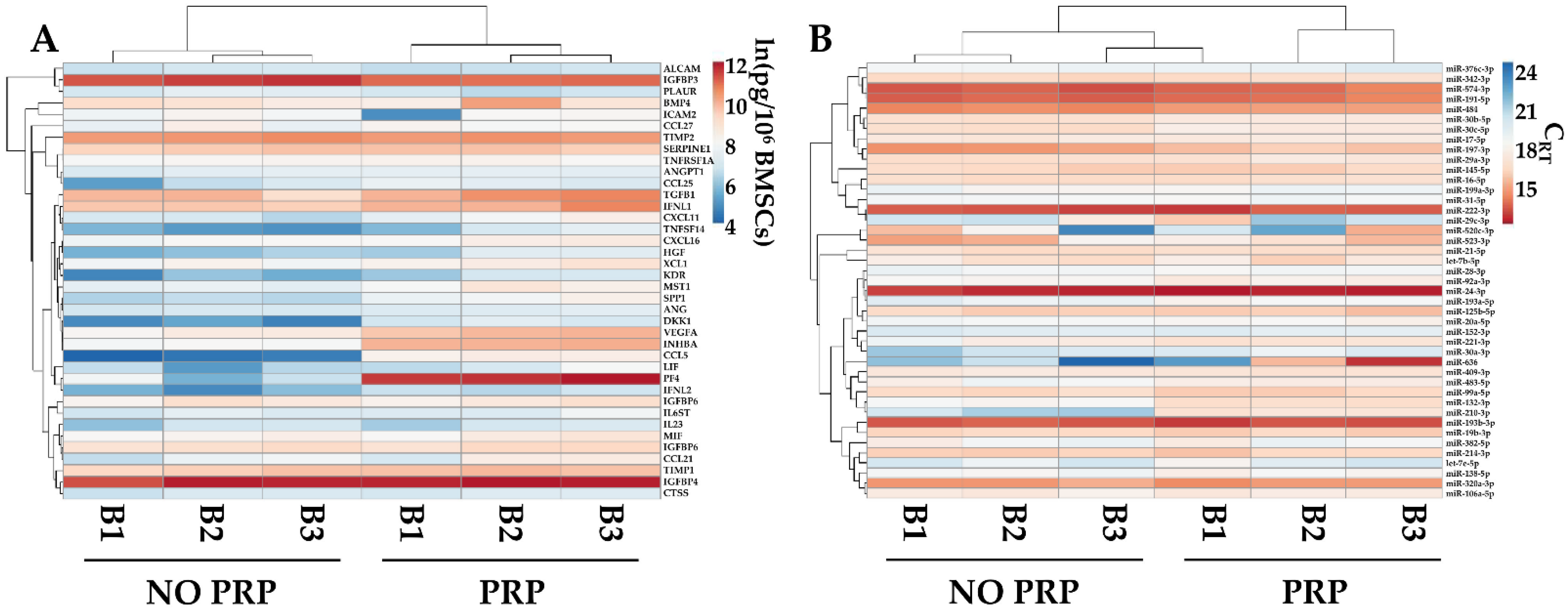
2.7. PRP Effects on BMSC-Secreted Factors and EV-miRNAs
3. Discussion
4. Materials and Methods
4.1. PRP Collection, Activation and Storage
4.2. ELISA Characterization of Activated PRP
4.3. Bone Marrow Collection, BMSCs’ Isolation and Expansion
4.4. Flow Cytometry Characterization of PRP-Treated BMSCs
4.5. ELISA Characterization of PRP-Treated BMSCs Secretome
4.6. Protein–Protein Interaction Networks
4.7. Characterization of EVs in PRP-Treated BMSCs’ Secretomes
4.8. Total RNA Isolation from EVs’ and miRNAs’ Quantification
4.9. Identification of miRNAs’ Target
4.10. Comparison of PRP-Treated vs. Untreated BMSCs’ Secretomes
4.11. Statistical and Computational Analyses
5. Conclusions
Supplementary Materials
Author Contributions
Funding
Institutional Review Board Statement
Informed Consent Statement
Data Availability Statement
Acknowledgments
Conflicts of Interest
References
- Hunter, D.J.; Bierma-Zeinstra, S. Osteoarthritis. Lancet 2019, 393, 1745–1759. [Google Scholar] [CrossRef] [PubMed]
- Brandt, K.D.; Radin, E.L.; Dieppe, P.A.; van de Putte, L. Yet more evidence that osteoarthritis is not a cartilage disease. Ann. Rheum. Dis. 2006, 65, 1261–1264. [Google Scholar] [CrossRef] [Green Version]
- Primorac, D.; Molnar, V.; Rod, E.; Jeleč, Z.; Čukelj, F.; Matišić, V.; Vrdoljak, T.; Hudetz, D.; Hajsok, H.; Borić, I. Knee Osteoarthritis: A Review of Pathogenesis and State-Of-The-Art Non-Operative Therapeutic Considerations. Genes 2020, 11, 854. [Google Scholar] [CrossRef] [PubMed]
- Gademan, M.G.J.; Hofstede, S.N.; Vlieland, T.P.M.V.; Nelissen, R.G.H.H.; De Mheen, P.J.M.-V. Indication criteria for total hip or knee arthroplasty in osteoarthritis: A state-of-the-science overview. BMC Musculoskelet. Disord. 2016, 17, 463. [Google Scholar] [CrossRef] [PubMed] [Green Version]
- Dhillon, M.S.; Behera, P.; Patel, S.; Shetty, V. Orthobiologics and platelet rich plasma. Indian J. Orthop. 2014, 48, 1–9. [Google Scholar] [CrossRef] [PubMed]
- Centeno, C.J.; Pastoriza, S.M. Past, current and future interventional orthobiologics techniques and how they relate to regen-erative rehabilitation: A clinical commentary. Int. J. Sport. Phys. Ther. 2020, 15, 301–325. [Google Scholar] [CrossRef]
- De Luca, P.; Kouroupis, D.; Viganò, M.; Perucca-Orfei, C.; Kaplan, L.; Zagra, L.; de Girolamo, L.; Correa, D.; Colombini, A. Human Diseased Articular Cartilage Contains a Mesenchymal Stem Cell-Like Population of Chondroprogenitors with Strong Immunomodulatory Responses. J. Clin. Med. 2019, 8, 423. [Google Scholar] [CrossRef] [Green Version]
- Sandonà, M.; Di Pietro, L.; Esposito, F.; Ventura, A.; Silini, A.R.; Parolini, O.; Saccone, V. Mesenchymal Stromal Cells and Their Secretome: New Therapeutic Perspectives for Skeletal Muscle Regeneration. Front. Bioeng. Biotechnol. 2021, 9, 652970. [Google Scholar] [CrossRef]
- Hwang, J.J.; Rim, Y.A.; Nam, Y.; Ju, J.H. Recent Developments in Clinical Applications of Mesenchymal Stem Cells in the Treatment of Rheumatoid Arthritis and Osteoarthritis. Front. Immunol. 2021, 12, 631291. [Google Scholar] [CrossRef]
- Wu, P.I.-K.; Diaz, R.; Borg-Stein, J. Platelet-Rich Plasma. Phys. Med. Rehabil. Clin. North Am. 2016, 27, 825–853. [Google Scholar] [CrossRef]
- Shen, L.; Yuan, T.; Chen, S.; Xie, X.; Zhang, C. The temporal effect of platelet-rich plasma on pain and physical function in the treatment of knee osteoarthritis: Systematic review and meta-analysis of randomized controlled trials. J. Orthop. Surg. Res. 2017, 12, 16. [Google Scholar] [CrossRef] [PubMed] [Green Version]
- Tang, X.-B.; Dong, P.-L.; Wang, J.; Zhou, H.-Y.; Zhang, H.-X.; Wang, S.-Z. Effect of autologous platelet-rich plasma on the chondrogenic differentiation of rabbit adipose-derived stem cells in vitro. Exp. Ther. Med. 2015, 10, 477–483. [Google Scholar] [CrossRef] [PubMed] [Green Version]
- Elder, S.; Thomason, J. Effect of Platelet-Rich Plasma on Chondrogenic Differentiation in Three-Dimensional Culture. Open Orthop. J. 2014, 8, 78–84. [Google Scholar] [CrossRef] [PubMed]
- Yun, S.; Ku, S.-K.; Kwon, Y.-S. Adipose-derived mesenchymal stem cells and platelet-rich plasma synergistically ameliorate the surgical-induced osteoarthritis in Beagle dogs. J. Orthop. Surg. Res. 2016, 11, 9. [Google Scholar] [CrossRef] [PubMed] [Green Version]
- Lamo-Espinosa, J.M.; Blanco, J.F.; Sánchez, M.; Moreno, V.; Granero-Moltó, F.; Sánchez-Guijo, F.; Crespo-Cullel, Í.; Mora, G.; Vicente, D.D.S.; Pompei-Fernández, O.; et al. Phase II multicenter randomized controlled clinical trial on the efficacy of intra-articular injection of autologous bone marrow mesenchymal stem cells with platelet rich plasma for the treatment of knee osteoarthritis. J. Transl. Med. 2020, 18, 356. [Google Scholar] [CrossRef] [PubMed]
- Pintat, J.; Silvestre, A.; Magalon, G.; Gadeau, A.P.; Pesquer, L.; Perozziello, A.; Peuchant, A.; Mounayer, C.; Dallaudière, B. Intra-articular Injection of Mesenchymal Stem Cells and Platelet-Rich Plasma to Treat Patellofemoral Osteoarthritis: Preliminary Results of a Long-Term Pilot Study. J. Vasc. Interv. Radiol. 2017, 28, 1708–1713. [Google Scholar] [CrossRef]
- Bui, K.H.-T.; Duong, T.D.; Nguyen, N.T.; Le, V.T.; Mai, V.T.; Phan, N.L.-C.; Van Pham, P. Symptomatic knee osteoarthritis treatment using autologous adipose derived stem cells and platelet-rich plasma: A clinical study. Biomed. Res. Ther. 2014, 1, 2–8. [Google Scholar] [CrossRef] [Green Version]
- Koh, Y.-G.; Kwon, O.-R.; Kim, Y.-S.; Choi, Y.-J. Comparative Outcomes of Open-Wedge High Tibial Osteotomy With Platelet-Rich Plasma Alone or in Combination With Mesenchymal Stem Cell Treatment: A Prospective Study. Arthrosc. J. Arthrosc. Relat. Surg. 2014, 30, 1453–1460. [Google Scholar] [CrossRef]
- Haleem, A.M.; El Singergy, A.A.; Sabry, D.; Atta, H.M.; Rashed, L.A.; Chu, C.R.; El Shewy, M.T.; Azzam, A.; Aziz, M.T.A. The Clinical Use of Human Culture–Expanded Autologous Bone Marrow Mesenchymal Stem Cells Transplanted on Platelet-Rich Fibrin Glue in the Treatment of Articular Cartilage Defects: A Pilot Study and Preliminary Results. Cartilage 2010, 1, 253–261. [Google Scholar] [CrossRef]
- Barilani, M.; Banfi, F.; Sironi, S.; Ragni, E.; Guillaumin, S.; Polveraccio, F.; Rosso, L.; Moro, M.; Astori, G.; Pozzobon, M.; et al. Low-affinity Nerve Growth Factor Receptor (CD271) Heterogeneous Expression in Adult and Fetal Mesenchymal Stromal Cells. Sci. Rep. 2018, 8, 9321. [Google Scholar] [CrossRef]
- Ragni, E.; Papait, A.; Orfei, C.P.; Silini, A.R.; Colombini, A.; Viganò, M.; Libonati, F.; Parolini, O.; de Girolamo, L. Amniotic membrane-mesenchymal stromal cells secreted factors and extracellular vesicle-miRNAs: Anti-inflammatory and regenerative features for musculoskeletal tissues. STEM CELLS Transl. Med. 2021, 10, 1044–1062. [Google Scholar] [CrossRef] [PubMed]
- Ragni, E.; Orfei, C.P.; Papait, A.; de Girolamo, L. Comparison of miRNA cargo in human adipose-tissue vs. amniotic-membrane derived mesenchymal stromal cells extracellular vesicles for osteoarthritis treatment. Extracell. Vesicles Circ. Nucleic Acids 2021, 2, 202–221. [Google Scholar] [CrossRef]
- Toh, W.S.; Lai, R.C.; Hui, J.H.P.; Lim, S.K. MSC exosome as a cell-free MSC therapy for cartilage regeneration: Implications for osteoarthritis treatment. Semin. Cell Dev. Biol. 2017, 67, 56–64. [Google Scholar] [CrossRef] [PubMed]
- Chevillet, J.R.; Kang, Q.; Ruf, I.K.; Briggs, H.A.; Vojtech, L.N.; Hughes, S.M.; Cheng, H.H.; Arroyo, J.D.; Meredith, E.K.; Gallichotte, E.N.; et al. Quantitative and stoichiometric analysis of the microRNA content of exosomes. Proc. Natl. Acad. Sci. USA 2014, 111, 14888–14893. [Google Scholar] [CrossRef] [Green Version]
- Schopman, N.C.T.; Heynen, S.; Haasnoot, J.; Berkhout, B. A miRNA-tRNA mix-up: tRNA origin of proposed miRNA. RNA Biol. 2010, 7, 573–576. [Google Scholar] [CrossRef]
- Chou, C.-H.; Jain, V.; Gibson, J.; Attarian, D.E.; Haraden, C.A.; Yohn, C.B.; Laberge, R.-M.; Gregory, S.; Kraus, V.B. Synovial cell cross-talk with cartilage plays a major role in the pathogenesis of osteoarthritis. Sci. Rep. 2020, 10, 10868. [Google Scholar] [CrossRef] [PubMed]
- Endisha, H.; Rockel, J.; Jurisica, I.; Kapoor, M. The complex landscape of microRNAs in articular cartilage: Biology, pathology, and therapeutic targets. J. Clin. Investig. 2018, 3, e121630. [Google Scholar] [CrossRef] [Green Version]
- Tavallaee, G.; Rockel, J.S.; Lively, S.; Kapoor, M. MicroRNAs in Synovial Pathology Associated With Osteoarthritis. Front. Med. 2020, 7, 376. [Google Scholar] [CrossRef]
- Xu, S.J.; Hu, H.T.; Li, H.L.; Chang, S. The Role of miRNAs in Immune Cell Development, Immune Cell Activation, and Tumor Immunity: With a Focus on Macrophages and Natural Killer Cells. Cells 2019, 8, 1140. [Google Scholar] [CrossRef] [Green Version]
- Galan, A.R.; Fernández-Messina, L.; Sánchez-Madrid, F. Control of Immunoregulatory Molecules by miRNAs in T Cell Activation. Front. Immunol. 2018, 9, 2148. [Google Scholar] [CrossRef]
- Ragni, E.; Orfei, C.P.; de Girolamo, L. Secreted Factors and Extracellular Vesicles Account for the Immunomodulatory and Tissue Regenerative Properties of Bone-Marrow-Derived Mesenchymal Stromal Cells for Osteoarthritis. Cells 2022, 11, 3501. [Google Scholar] [CrossRef]
- Qu, H.; Sun, S. Efficacy of mesenchymal stromal cells for the treatment of knee osteoarthritis: A meta-analysis of randomized controlled trials. J. Orthop. Surg. Res. 2021, 16, 11. [Google Scholar] [CrossRef] [PubMed]
- McLarnon, M.; Heron, N. Intra-articular platelet-rich plasma injections versus intra-articular corticosteroid injections for symptomatic management of knee osteoarthritis: Systematic review and meta-analysis. BMC Musculoskelet. Disord. 2021, 22, 550. [Google Scholar] [CrossRef] [PubMed]
- Luz-Crawford, P.; Djouad, F.; Toupet, K.; Bony, C.; Franquesa, M.; Hoogduijn, M.J.; Jorgensen, C.; Noël, D. Mesenchymal Stem Cell-Derived Interleukin 1 Receptor Antagonist Promotes Macrophage Polarization and Inhibits B Cell Differentiation. STEM CELLS 2016, 34, 483–492. [Google Scholar] [CrossRef] [Green Version]
- Deng, Y.; Zhang, Y.; Ye, L.; Zhang, T.; Cheng, J.; Chen, G.; Zhang, Q.; Yang, Y. Umbilical Cord-derived Mesenchymal Stem Cells Instruct Monocytes Towards an IL10-producing Phenotype by Secreting IL6 and HGF. Sci. Rep. 2016, 6, 37566. [Google Scholar] [CrossRef] [Green Version]
- Melief, S.M.; Schrama, E.; Brugman, M.H.; Tiemessen, M.M.; Hoogduijn, M.J.; Fibbe, W.E.; Roelofs, H. Multipotent stromal cells induce human regulatory T cells through a novel pathway involving skewing of monocytes toward anti-inflammatory macrophages. STEM CELLS 2013, 31, 1980–1991. [Google Scholar] [CrossRef]
- Schmidt, A.; Zhang, X.-M.; Joshi, R.N.; Iqbal, S.; Wahlund, C.; Gabrielsson, S.; Harris, R.A.; Tegnér, J. Human macrophages induce CD4+Foxp3+ regulatory T cells via binding and re-release of TGF-β. Immunol. Cell Biol. 2016, 94, 747–762. [Google Scholar] [CrossRef] [Green Version]
- Wang, D.; Huang, S.; Yuan, X.; Liang, J.; Xu, R.; Yao, G.; Feng, X.; Sun, L. The regulation of the Treg/Th17 balance by mesenchymal stem cells in human systemic lupus erythematosus. Cell. Mol. Immunol. 2017, 14, 423–431. [Google Scholar] [CrossRef] [Green Version]
- Duffy, M.M.; Ritter, T.; Ceredig, R.; Griffin, M.D. Mesenchymal stem cell effects on T-cell effector pathways. Stem Cell Res. Ther. 2011, 2, 34. [Google Scholar] [CrossRef] [Green Version]
- Davies, L.C.; Heldring, N.; Kadri, N.; Le Blanc, K. Mesenchymal Stromal Cell Secretion of Programmed Death-1 Ligands Regulates T Cell Mediated Immunosuppression. STEM CELLS 2017, 35, 766–776. [Google Scholar] [CrossRef]
- Di Nicola, M.; Carlo-Stella, C.; Magni, M.; Milanesi, M.; Longoni, P.D.; Matteucci, P.; Grisanti, S.; Gianni, A.M. Human bone marrow stromal cells suppress T-lymphocyte proliferation induced by cellular or nonspecific mitogenic stimuli. Blood 2002, 99, 3838–3843. [Google Scholar] [CrossRef] [PubMed]
- Zheng, S.; Huang, K.; Xia, W.; Shi, J.; Liu, Q.; Zhang, X.; Li, G.; Chen, J.; Wang, T.; Chen, X.; et al. Mesenchymal Stromal Cells Rapidly Suppress TCR Signaling-Mediated Cytokine Transcription in Activated T Cells Through the ICAM-1/CD43 Interaction. Front. Immunol. 2021, 12, 609544. [Google Scholar] [CrossRef]
- Bai, L.; Lennon, D.P.; Eaton, V.; Maier, K.; Caplan, A.; Miller, S.D.; Miller, R.H. Human bone marrow-derived mesenchymal stem cells induce Th2-polarized immune response and promote endogenous repair in animal models of multiple sclerosis. Glia 2009, 57, 1192–1203. [Google Scholar] [CrossRef] [PubMed] [Green Version]
- Fiorina, P.; Jurewicz, M.; Augello, A.; Vergani, A.; Dada, S.; La Rosa, S.; Selig, M.; Godwin, J.; Law, K.; Placidi, C.; et al. Immunomodulatory Function of Bone Marrow-Derived Mesenchymal Stem Cells in Experimental Autoimmune Type 1 Diabetes. J. Immunol. 2009, 183, 993–1004. [Google Scholar] [CrossRef] [Green Version]
- Jiang, D.; Muschhammer, J.; Qi, Y.; Kügler, A.; de Vries, J.C.; Saffarzadeh, M.; Sindrilaru, A.; Beken, S.V.; Wlaschek, M.; Kluth, M.A.; et al. Suppression of Neutrophil-Mediated Tissue Damage—A Novel Skill of Mesenchymal Stem Cells. STEM CELLS 2016, 34, 2393–2406. [Google Scholar] [CrossRef] [PubMed]
- Maldonado, M.; Nam, J. The Role of Changes in Extracellular Matrix of Cartilage in the Presence of Inflammation on the Pathology of Osteoarthritis. BioMed Res. Int. 2013, 2013, 284873. [Google Scholar] [CrossRef] [Green Version]
- Ellis, A.; Curry, V.; Powell, E.; Cawston, T. The Prevention of Collagen Breakdown in Bovine Nasal Cartilage by TIMP, TIMP-2 and a Low Molecular Weight Synthetic Inhibitor. Biochem. Biophys. Res. Commun. 1994, 201, 94–101. [Google Scholar] [CrossRef]
- Nagase, H.; Brew, K. Engineering of tissue inhibitor of metalloproteinases mutants as potential therapeutics. Arthritis Res. Ther. 2002, 4, S51–S61. [Google Scholar] [CrossRef]
- Blaney Davidson, E.N.; van der Kraan, P.M.; van den Berg, W.B. TGF-β and osteoarthritis. Osteoarthr. Cartil. 2007, 15, 597–604. [Google Scholar] [CrossRef] [Green Version]
- Wang, W.; Rigueur, D.; Lyons, K.M. TGFβ signaling in cartilage development and maintenance. Birth Defects Res. Part C: Embryo Today: Rev. 2014, 102, 37–51. [Google Scholar] [CrossRef]
- van der Kraan, P.M. Differential Role of Transforming Growth Factor-beta in an Osteoarthritic or a Healthy Joint. J. Bone Metab. 2018, 25, 65–72. [Google Scholar] [CrossRef] [PubMed]
- Mahmood, N.; Mihalcioiu, C.; Rabbani, S.A. Multifaceted Role of the Urokinase-Type Plasminogen Activator (uPA) and Its Receptor (uPAR): Diagnostic, Prognostic, and Therapeutic Applications. Front. Oncol. 2018, 8, 24. [Google Scholar] [CrossRef] [PubMed] [Green Version]
- Milner, J.M.; Patel, A.; Rowan, A.D. Emerging roles of serine proteinases in tissue turnover in arthritis. Arthritis Rheum. 2008, 58, 3644–3656. [Google Scholar] [CrossRef] [PubMed]
- Martel-Pelletier, J.; Faure, M.P.; Mccollum, R.; Mineau, F.; Cloutier, J.M.; Pelletier, J.P. Plasmin, plasminogen activators and inhibitor in human osteoarthritic cartilage. J. Rheumatol. 1991, 18, 1863–1871. [Google Scholar]
- Wen, C.; Xu, L.; Xu, X.; Wang, D.; Liang, Y.; Duan, L. Insulin-like growth factor-1 in articular cartilage repair for osteoarthritis treatment. Thromb. Haemost. 2021, 23, 277. [Google Scholar] [CrossRef]
- Galasso, O.; De Gori, M.; Nocera, A.; Brunetti, A.; Gasparini, G. Regulatory Functions of Insulin-like Growth Factor Binding Proteins in Osteoarthritis. Int. J. Immunopathol. Pharmacol. 2011, 24, 55–59. [Google Scholar] [CrossRef]
- Shum, L.; Wang, X.; Kane, A.A.; Nuckolls, G.H. BMP4 promotes chondrocyte proliferation and hypertrophy in the endochondral cranial base. Int. J. Dev. Biol. 2003, 47, 423–431. [Google Scholar]
- Bramlage, C.P.; Häupl, T.; Kaps, C.; Ungethüm, U.; Krenn, V.; Pruss, A.; Müller, G.A.; Strutz, F.; Burmester, G.-R. Decrease in expression of bone morphogenetic proteins 4 and 5 in synovial tissue of patients with osteoarthritis and rheumatoid arthritis. Arthritis Res. Ther. 2006, 8, R58. [Google Scholar] [CrossRef] [Green Version]
- Deng, Z.; Li, Y.; Gao, X.; Lei, G.; Huard, J. Bone morphogenetic proteins for articular cartilage regeneration. Osteoarthr. Cartil. 2018, 26, 1153–1161. [Google Scholar] [CrossRef] [Green Version]
- Chevalier, X.; Goupille, P.; Beaulieu, A.D.; Burch, F.X.; Bensen, W.G.; Conrozier, T.; Loeuille, D.; Kivitz, A.J.; Silver, D.; Appleton, B.E. Intraarticular injection of anakinra in osteoarthritis of the knee: A multicenter, randomized, double-blind, placebo-controlled study. Arthritis Rheum. 2009, 61, 344–352. [Google Scholar] [CrossRef]
- Cohen, S.B.; Proudman, S.; Kivitz, A.J.; Burch, F.X.; Donohue, J.P.; Burstein, D.; Sun, Y.-N.; Banfield, C.; Vincent, M.S.; Ni, L.; et al. A randomized, double-blind study of AMG 108 (a fully human monoclonal antibody to IL-1R1) in patients with osteoarthritis of the knee. Thromb. Haemost. 2011, 13, R125. [Google Scholar] [CrossRef] [PubMed] [Green Version]
- Fleischmann, R.M.; Bliddal, H.; Blanco, F.J.; Schnitzer, T.J.; Peterfy, C.; Chen, S.; Wang, L.; Feng, S.; Conaghan, P.G.; Berenbaum, F.; et al. A Phase II Trial of Lutikizumab, an Anti–Interleukin-1α/β Dual Variable Domain Immunoglobulin, in Knee Osteoarthritis Patients With Synovitis. Arthritis Rheumatol. 2019, 71, 1056–1069. [Google Scholar] [CrossRef] [PubMed]
- Aitken, D.; Laslett, L.L.; Pan, F.; Haugen, I.K.; Otahal, P.; Bellamy, N.; Bird, P.; Jones, G. A randomised double-blind placebo-controlled crossover trial of HUMira (adalimumab) for erosive hand OsteoaRthritis—The HUMOR trial. Osteoarthr. Cartil. 2018, 26, 880–887. [Google Scholar] [CrossRef] [PubMed] [Green Version]
- Kloppenburg, M.; Ramonda, R.; Bobacz, K.; Kwok, W.-Y.; Elewaut, D.; Huizinga, T.W.J.; Kroon, F.P.B.; Punzi, L.; Smolen, J.S.; Cruyssen, B.V.; et al. Etanercept in patients with inflammatory hand osteoarthritis (EHOA): A multicentre, randomised, double-blind, placebo-controlled trial. Ann. Rheum. Dis. 2018, 77, 1757–1764. [Google Scholar] [CrossRef] [PubMed]
- Cho, Y.; Jeong, S.; Kim, H.; Kang, D.; Lee, J.; Kang, S.-B.; Kim, J.-H. Disease-modifying therapeutic strategies in osteoarthritis: Current status and future directions. Exp. Mol. Med. 2021, 53, 1689–1696. [Google Scholar] [CrossRef]
- Holmbeck, K.; Bianco, P.; Caterina, J.; Yamada, S.; Kromer, M.; Kuznetsov, S.A.; Mankani, M.; Robey, P.G.; Poole, A.; Pidoux, I.; et al. MT1-MMP-Deficient Mice Develop Dwarfism, Osteopenia, Arthritis, and Connective Tissue Disease due to Inadequate Collagen Turnover. Cell 1999, 99, 81–92. [Google Scholar] [CrossRef] [Green Version]
- Hamilton, J.L.; Nagao, M.; Levine, B.R.; Chen, D.; Olsen, B.R.; Im, H.-J. Targeting VEGF and Its Receptors for the Treatment of Osteoarthritis and Associated Pain. J. Bone Miner. Res. 2016, 31, 911–924. [Google Scholar] [CrossRef] [Green Version]
- Xu, J.; Qian, X.; Ding, R. MiR-24-3p attenuates IL-1β-induced chondrocyte injury associated with osteoarthritis by targeting BCL2L12. J. Orthop. Surg. Res. 2021, 16, 371. [Google Scholar] [CrossRef]
- Meng, F.; Li, Z.; Zhang, Z.; Long, D.; He, A.; Liao, W. Microrna-193B modulates chondrogenesis and cartilage degeneration via histone deacetylase 3. Osteoarthr. Cartil. 2018, 26, S93–S94. [Google Scholar] [CrossRef] [Green Version]
- Ukai, T.; Sato, M.; Akutsu, H.; Umezawa, A.; Mochida, J. MicroRNA-199a-3p, microRNA-193b, and microRNA-320c are correlated to aging and regulate human cartilage metabolism. J. Orthop. Res. 2012, 30, 1915–1922. [Google Scholar] [CrossRef]
- Kopańska, M.; Szala, D.; Czech, J.; Gabło, N.; Gargasz, K.; Trzeciak, M.; Zawlik, I.; Snela, S. MiRNA expression in the cartilage of patients with osteoarthritis. J. Orthop. Surg. Res. 2017, 12, 51. [Google Scholar] [CrossRef] [PubMed] [Green Version]
- Song, J.; Jin, E.-H.; Kim, D.; Kim, K.Y.; Chun, C.-H. MicroRNA-222 regulates MMP-13 via targeting HDAC-4 during osteoarthritis pathogenesis. BBA Clin. 2014, 3, 79–89. [Google Scholar] [CrossRef] [PubMed]
- Fordham, J.B.; Naqvi, A.R.; Nares, S. Regulation of miR-24, miR-30b, and miR-142-3p during macrophage and dendritic cell differentiation potentiates innate immunity. J. Leukoc. Biol. 2015, 98, 195–207. [Google Scholar] [CrossRef] [PubMed] [Green Version]
- Jingjing, Z.; Nan, Z.; Wei, W.; Qinghe, G.; Weijuan, W.; Peng, W.; Xiangpeng, W. MicroRNA-24 Modulates Staphylococcus aureus-Induced Macrophage Polarization by Suppressing CHI3L1. Inflammation 2017, 40, 995–1005. [Google Scholar] [CrossRef] [PubMed]
- Forrest, A.R.R.; Kanamori-Katayama, M.; Tomaru, Y.; Lassmann, T.; Ninomiya, N.; Takahashi, Y.; De Hoon, M.J.L.; Kubosaki, A.; Kaiho, A.; Suzuki, M.; et al. Induction of microRNAs, mir-155, mir-222, mir-424 and mir-503, promotes monocytic differentiation through combinatorial regulation. Leukemia 2010, 24, 460–466. [Google Scholar] [CrossRef] [PubMed] [Green Version]
- Graff, J.W.; Dickson, A.M.; Clay, G.; McCaffrey, A.P.; Wilson, M.E. Identifying Functional MicroRNAs in Macrophages with Polarized Phenotypes. J. Biol. Chem. 2012, 287, 21816–21825. [Google Scholar] [CrossRef] [PubMed] [Green Version]
- Ying, X.; Wu, Q.; Wu, X.; Zhu, Q.; Wang, X.; Jiang, L.; Chen, X.; Wang, X. Epithelial ovarian cancer-secreted exosomal miR-222-3p induces polarization of tumor-associated macrophages. Oncotarget 2016, 7, 43076–43087. [Google Scholar] [CrossRef] [Green Version]
- Fayyad-Kazan, H.; Hamade, E.; Rouas, R.; Najar, M.; Fayyad-Kazan, M.; el Zein, N.; ElDirani, R.; Hussein, N.; Fakhry, M.; Al-Akoum, C.; et al. Downregulation of microRNA-24 and -181 parallels the upregulation of IFN-γ secreted by activated human CD4 lymphocytes. Hum. Immunol. 2014, 75, 677–685. [Google Scholar] [CrossRef]
- Chandran, P.A.; Keller, A.; Weinmann, L.; Seida, A.A.; Braun, M.; Andreev, K.; Fischer, B.; Horn, E.; Schwinn, S.; Junker, M.; et al. The TGF-β-inducible miR-23a cluster attenuates IFN-γ levels and antigen-specific cytotoxicity in human CD8+ T cells. J. Leukoc. Biol. 2014, 96, 633–645. [Google Scholar] [CrossRef]
- Ye, S.-B.; Zhang, H.; Cai, T.-T.; Liu, Y.-N.; Ni, J.-J.; He, J.; Peng, J.-Y.; Chen, Q.-Y.; Mo, H.-Y.; Cui, J.; et al. Exosomal miR-24-3p impedes T-cell function by targetingFGF11and serves as a potential prognostic biomarker for nasopharyngeal carcinoma. J. Pathol. 2016, 240, 329–340. [Google Scholar] [CrossRef]
- Scanzello, C.R. Chemokines and inflammation in osteoarthritis: Insights from patients and animal models. J. Orthop. Res. 2017, 35, 735–739. [Google Scholar] [CrossRef] [PubMed] [Green Version]
- Zhong, L.; Huang, X.; Rodrigues, E.; Leijten, J.; Verrips, T.; El Khattabi, M.; Karperien, M.; Post, J. DKK1 AND FRZB are necessary for chondrocyte (RE)differentiation and prevention of cell hypertrophy in 3D cultures of human chondrocytes and human mesenchymal stem cells. Osteoarthr. Cartil. 2016, 24, S142. [Google Scholar] [CrossRef]
- Wei, T.; Kulkarni, N.; Zeng, Q.; Helvering, L.; Lin, X.; Lawrence, F.; Hale, L.; Chambers, M.; Lin, C.; Harvey, A.; et al. Analysis of early changes in the articular cartilage transcriptisome in the rat meniscal tear model of osteoarthritis: Pathway comparisons with the rat anterior cruciate transection model and with human osteoarthritic cartilage. Osteoarthr. Cartil. 2010, 18, 992–1000. [Google Scholar] [CrossRef] [PubMed] [Green Version]
- Tang, H.; Zhu, W.; Cao, L.; Zhang, J.; Li, J.; Ma, D.; Guo, C. miR-210-3p protects against osteoarthritis through inhibiting subchondral angiogenesis by targeting the expression of TGFBR1 and ID4. Front. Immunol. 2022, 13, 982278. [Google Scholar] [CrossRef]
- Yang, M.; Yan, X.; Yuan, F.-Z.; Ye, J.; Du, M.-Z.; Mao, Z.-M.; Xu, B.-B.; Chen, Y.-R.; Song, Y.-F.; Fan, B.-S.; et al. MicroRNA-210-3p Promotes Chondrogenic Differentiation and Inhibits Adipogenic Differentiation Correlated with HIF-3α Signalling in Bone Marrow Mesenchymal Stem Cells. BioMed Res. Int. 2021, 2021, 6699910. [Google Scholar] [CrossRef] [PubMed]
- Zhang, D.; Cao, X.; Li, J.; Zhao, G. MiR-210 inhibits NF-κB signaling pathway by targeting DR6 in osteoarthritis. Sci. Rep. 2015, 5, 12775. [Google Scholar] [CrossRef]
- Zhang, W.; Hu, C.; Zhang, C.; Luo, C.; Zhong, B.; Yu, X. MiRNA-132 regulates the development of osteoarthritis in correlation with the modulation of PTEN/PI3K/AKT signaling. BMC Geriatr. 2021, 21, 175. [Google Scholar] [CrossRef] [PubMed]
- Gao, S.; Liu, L.; Zhu, S.; Wang, D.; Wu, Q.; Ning, G.; Feng, S. MicroRNA-197 regulates chondrocyte proliferation, migration, and inflammation in pathogenesis of osteoarthritis by targeting EIF4G2. Biosci. Rep. 2020, 40, BSR20192095. [Google Scholar] [CrossRef]
- Atashi, F.; Jaconi, M.; Pittet-Cuénod, B.; Modarressi, A. Autologous Platelet-Rich Plasma: A Biological Supplement to Enhance Adipose-Derived Mesenchymal Stem Cell Expansion. Tissue Eng. Part C Methods 2015, 21, 253–262. [Google Scholar] [CrossRef] [Green Version]
- Harrell, C.; Fellabaum, C.; Jovicic, N.; Djonov, V.; Arsenijevic, N.; Volarevic, V. Molecular Mechanisms Responsible for Therapeutic Potential of Mesenchymal Stem Cell-Derived Secretome. Cells 2019, 8, 467. [Google Scholar] [CrossRef] [Green Version]
- Ishikawa, J.; Takahashi, N.; Matsumoto, T.; Yoshioka, Y.; Yamamoto, N.; Nishikawa, M.; Hibi, H.; Ishigro, N.; Ueda, M.; Furukawa, K.; et al. Factors secreted from dental pulp stem cells show multifaceted benefits for treating experimental rheumatoid arthritis. Bone 2016, 83, 210–219. [Google Scholar] [CrossRef]
- Amodeo, G.; Niada, S.; Moschetti, G.; Franchi, S.; Savadori, P.; Brini, A.T.; Sacerdote, P. Secretome of human adipose-derived mesenchymal stem cell relieves pain and neuroinflammation independently of the route of administration in experimental osteoarthritis. Brain Behav. Immun. 2021, 94, 29–40. [Google Scholar] [CrossRef] [PubMed]
- Ragni, E.; Perucca Orfei, C.; De Luca, P.; Lugano, G.; Viganò, M.; Colombini, A.; Valli, F.; Zacchetti, D.; Bollati, V.; De Girolamo, L. Interaction with hyaluronan matrix and miRNA cargo as contributors for in vitro potential of mesenchymal stem cell-derived extracellular vesicles in a model of human osteoarthritic synoviocytes. Stem Cell Res. Ther. 2019, 10, 109. [Google Scholar] [CrossRef] [Green Version]
- Huang, H.-Y.; Lin, Y.-C.-D.; Li, J.; Huang, K.-Y.; Shrestha, S.; Hong, H.-C.; Tang, Y.; Chen, Y.-G.; Jin, C.-N.; Yu, Y.; et al. miRTarBase 2020: Updates to the experimentally validated microRNA–target interaction database. Nucleic Acids Res. 2020, 48, D148–D154. [Google Scholar] [CrossRef] [PubMed] [Green Version]
- Akoglu, H. User’s guide to correlation coefficients. Turk. J. Emerg. Med. 2018, 18, 91–93. [Google Scholar] [CrossRef]
- Metsalu, T.; Vilo, J. ClustVis: A web tool for visualizing clustering of multivariate data using Principal Component Analysis and heatmap. Nucleic Acids Res. 2015, 43, W566–W570. [Google Scholar] [CrossRef]


| TYPE | FACTOR | (pg/mL) | |
|---|---|---|---|
| CYT | ICAM2 | 790,983 | Intercellular adhesion molecule 2 |
| CYT | PLG | 182,992 | Plasminogen |
| REC | VCAM1 | 102,461 | Vascular cell adhesion protein 1 |
| CYT | IL13RA2 | 94,449 | Interleukin-13 receptor subunit alpha-2 |
| CHE | IL9 | 73,686 | Interleukin-9 |
| GF | IGFBP4 | 72,637 | Insulin-like growth factor binding protein 4 |
| REC | MICB | 66,893 | MHC class I polypeptide-related sequence B |
| GF | TGFB1 | 58,819 | Transforming growth factor beta-1 |
| CHE | PF4 | 56,183 | Platelet factor 4 |
| CYT | IL23A | 47,225 | Interleukin-23 subunit alpha |
| CYT | FLT1 | 47,188 | Vascular endothelial growth factor receptor 1 |
| INF | PDGFB | 47,171 | Platelet-derived growth factor subunit B |
| CHE | IL31 | 38,293 | Interleukin-31 |
| GF | BMP7 | 35,835 | Bone morphogenetic protein 7 |
| CYT | SIGLEC5 | 35,491 | Sialic-acid-binding Ig-like lectin 5 |
| GF | IGFBP3 | 34,608 | Insulin-like growth factor binding protein 3 |
| CHE | CCL26 | 34,511 | C-C motif chemokine 26 |
| GF | FGF4 | 33,301 | Fibroblast growth factor 4 |
| CHE | SPP1 | 30,604 | Osteopontin |
| GF | BMP4 | 28,461 | Bone morphogenetic protein 4 |
| GF | IGFBP2 | 27,669 | Insulin-like growth factor binding protein 2 |
| REC | IL21R | 26,521 | Interleukin-21 receptor |
| REC | SELL | 25,729 | L-selectin |
| GF | CSF1R | 24,163 | Macrophage-colony-stimulating factor 1 receptor |
| CYT | IL6ST | 23,811 | Interleukin-6 receptor subunit beta |
| INF | IL6R | 23,412 | Interleukin-6 receptor subunit alpha |
| CHE | CCL21 | 23,105 | C-C motif chemokine 21 |
| INF | TIMP2 | 22,795 | Metalloproteinase inhibitor 2 |
| REC | TNFRSF17 | 22,355 | Tumor necrosis factor receptor superfamily member 17 |
| CHE | IFNL1 | 19,506 | Interferon lambda-1 |
| CYT | CDH1 | 17,919 | Cadherin-1 |
| CYT | IL13RA1 | 16,766 | Interleukin-13 receptor subunit alpha-1 |
| GF | KDR | 15,881 | Vascular endothelial growth factor receptor 2 |
| CYT | TREM1 | 15,662 | Triggering receptor expressed on myeloid cells 1 |
| CYT | CTSS | 15,538 | Cathepsin S |
| REC | TNFRSF18 | 13,410 | Tumor necrosis factor receptor superfamily member 18 |
| CYT | LGALS7 | 12,956 | Galectin-7 |
| CYT | TNFRSF10D | 12,579 | Tumor necrosis factor receptor superfamily member 10D |
| CYT | PDGFA | 12,454 | Platelet-derived growth factor subunit A |
| INF | TNFRSF1A | 12,348 | Tumor necrosis factor receptor superfamily member 1A |
| GF | TGFA | 12,273 | Protransforming growth factor alpha |
| INF | TIMP1 | 12,041 | Metalloproteinase inhibitor 1 |
| GF | FGF7 | 11,984 | Fibroblast growth factor 7 |
| CYT | IL2RB | 11,830 | Interleukin-2 receptor subunit beta |
| CYT | SERPINE1 | 11,194 | Plasminogen activator inhibitor 1 |
| CHE | CXCL16 | 10,931 | C-X-C motif chemokine 16 |
| INF | ICAM1 | 10,930 | Intercellular adhesion molecule 1 |
| INF | CCL5 | 10,751 | C-C motif chemokine 5 |
| INF | TNFRSF1B | 10,501 | Tumor necrosis factor receptor superfamily member 1B |
| pg/Million Cells | |||||||
|---|---|---|---|---|---|---|---|
| TYPE | FACTOR | B1 | B2 | B3 | MEAN | SD | |
| GF | IGFBP4 | 146,361 | 166,965 | 160,221 | 157,849 | 8577 | Insulin-like growth factor binding protein 4 |
| CHE | PF4 | 118,491 | 127,082 | 177,384 | 140,985 | 25,975 | Platelet factor 4 |
| GF | IGFBP3 | 67,919 | 65,875 | 69,956 | 67,917 | 1666 | Insulin-like growth factor binding protein 3 |
| GF | TGFB1 | 25,432 | 43,973 | 51,332 | 40,245 | 10,897 | Transforming growth factor beta-1 |
| INF | TIMP2 | 37,691 | 43,548 | 37,878 | 39,706 | 2718 | Metalloproteinase inhibitor 2 |
| CHE | IFNL1 | 25,976 | 26,330 | 49,299 | 33,869 | 10,912 | Interferon lambda-1 |
| CYT | INHBA | 25,821 | 24,754 | 27,428 | 26,001 | 1099 | Inhibin beta A chain |
| GF | VEGFA | 18,261 | 24,211 | 24,511 | 22,328 | 2878 | Vascular endothelial growth factor A |
| INF | TIMP1 | 19,604 | 23,458 | 20,658 | 21,240 | 1626 | Metalloproteinase inhibitor 1 |
| CYT | SERPINE1 | 20,131 | 20,479 | 16,545 | 19,052 | 1778 | Plasminogen activator inhibitor 1 |
| GF | BMP4 | 6102 | 33,281 | 8863 | 16,082 | 12,213 | Bone morphogenetic protein 4 |
| GF | IGFBP6 | 9679 | 14,168 | 13,933 | 12,593 | 2063 | Insulin-like growth factor binding protein 6 |
| CRT | ||||||
|---|---|---|---|---|---|---|
| miRBase ID | B1 | B2 | B3 | Mean | SD | % Weight |
| hsa-miR-24-3p | 12.24 | 12.53 | 12.40 | 12.39 | 0.12 | 20.12142652 |
| hsa-miR-193b-3p | 12.96 | 13.40 | 13.28 | 13.21 | 0.18 | 11.35032031 |
| hsa-miR-222-3p | 12.93 | 13.66 | 13.57 | 13.38 | 0.32 | 10.08398807 |
| hsa-miR-574-3p | 13.72 | 13.83 | 14.34 | 13.96 | 0.27 | 6.762989192 |
| hsa-miR-191-5p | 13.75 | 13.90 | 14.25 | 13.97 | 0.21 | 6.730254585 |
| hsa-miR-1274B | 14.28 | 14.08 | 13.84 | 14.06 | 0.18 | 6.295528960 |
| hsa-miR-320a-3p | 14.35 | 14.78 | 14.81 | 14.65 | 0.21 | 4.197880228 |
| hsa-miR-484 | 14.82 | 14.88 | 14.95 | 14.89 | 0.05 | 3.563581189 |
| hsa-miR-197-3p | 15.65 | 16.11 | 15.73 | 15.83 | 0.20 | 1.854025114 |
| hsa-miR-125b-5p | 15.98 | 16.10 | 15.70 | 15.93 | 0.17 | 1.733067865 |
| hsa-miR-99a-5p | 15.98 | 15.97 | 16.34 | 16.09 | 0.17 | 1.542204224 |
| hsa-miR-145-5p | 15.98 | 15.95 | 16.49 | 16.14 | 0.25 | 1.494152546 |
| hsa-miR-19b-3p | 15.99 | 16.43 | 16.02 | 16.15 | 0.20 | 1.486233896 |
| hsa-miR-214-3p | 15.79 | 16.40 | 16.37 | 16.18 | 0.28 | 1.447598052 |
| hsa-miR-21-5p | 16.71 | 16.61 | 16.48 | 16.60 | 0.10 | 1.086479443 |
| hsa-miR-342-3p | 16.33 | 16.67 | 16.86 | 16.62 | 0.22 | 1.070779162 |
| hsa-miR-132-3p | 16.69 | 16.77 | 16.64 | 16.70 | 0.05 | 1.011849009 |
| hsa-miR-16-5p | 16.55 | 16.69 | 16.94 | 16.73 | 0.16 | 0.995615635 |
| hsa-miR-523-3p | 17.79 | 16.91 | 15.53 | 16.74 | 0.93 | 0.983952511 |
| hsa-miR-409-3p | 17.10 | 16.84 | 16.96 | 16.97 | 0.11 | 0.841476316 |
| hsa-miR-221-3p | 16.92 | 16.97 | 17.08 | 16.99 | 0.06 | 0.829124978 |
| hsa-miR-1274A | 17.21 | 16.95 | 16.94 | 17.03 | 0.12 | 0.803847975 |
| hsa-miR-636 | 22.93 | 15.48 | 12.76 | 17.06 | 4.30 | 0.790403154 |
| hsa-let-7b-5p | 17.78 | 16.10 | 17.35 | 17.08 | 0.71 | 0.780602681 |
| hsa-miR-210-3p | 16.83 | 17.27 | 17.13 | 17.08 | 0.18 | 0.779341548 |
| hsa-miR-29a-3p | 16.82 | 17.47 | 17.27 | 17.19 | 0.27 | 0.722629345 |
| hsa-miR-30b-5p | 17.38 | 17.40 | 17.32 | 17.36 | 0.03 | 0.639490414 |
| hsa-miR-106a-5p | 17.23 | 17.58 | 17.47 | 17.43 | 0.15 | 0.610894287 |
| hsa-miR-17-5p | 17.39 | 17.49 | 17.44 | 17.44 | 0.04 | 0.606534357 |
| hsa-miR-30c-5p | 17.78 | 17.39 | 17.33 | 17.50 | 0.20 | 0.582633817 |
| hsa-miR-720 | 17.94 | 17.54 | 17.49 | 17.66 | 0.20 | 0.521351303 |
| hsa-miR-92a-3p | 17.43 | 17.95 | 17.63 | 17.67 | 0.21 | 0.517749975 |
| hsa-miR-483-5p | 17.77 | 17.66 | 17.58 | 17.67 | 0.08 | 0.516555300 |
| hsa-miR-20a-5p | 18.22 | 18.29 | 17.88 | 18.13 | 0.18 | 0.376397024 |
| hsa-miR-138-5p | 17.69 | 18.53 | 18.18 | 18.13 | 0.34 | 0.375008235 |
| hsa-miR-193a-5p | 18.03 | 18.53 | 18.51 | 18.36 | 0.23 | 0.321448809 |
| hsa-miR-382-5p | 17.56 | 19.12 | 18.50 | 18.39 | 0.64 | 0.313599274 |
| hsa-miR-28-3p | 18.39 | 18.76 | 18.57 | 18.57 | 0.15 | 0.276431568 |
| hsa-miR-31-5p | 18.40 | 18.81 | 18.52 | 18.58 | 0.17 | 0.275793598 |
| hsa-miR-199a-3p | 18.20 | 18.97 | 18.89 | 18.69 | 0.35 | 0.255134201 |
| hsa-miR-376c-3p | 18.97 | 19.09 | 19.76 | 19.28 | 0.35 | 0.169888586 |
| hsa-miR-520e-3p | 20.11 | 22.56 | 15.28 | 19.32 | 3.03 | 0.165204860 |
| hsa-miR-29c-3p | 15.98 | 21.49 | 20.49 | 19.32 | 2.40 | 0.164899729 |
| hsa-let-7e-5p | 18.14 | 19.62 | 20.32 | 19.36 | 0.91 | 0.160575943 |
| hsa-miR-152-3p | 19.69 | 19.39 | 19.31 | 19.46 | 0.16 | 0.149097433 |
| hsa-miR-30a-3p | 19.88 | 18.79 | 19.82 | 19.50 | 0.50 | 0.145490183 |
| EXPRESSING CELL (>1%) | % WEIGHT | MAIN EV-miRNA (%) | FUNCTION | ||||
|---|---|---|---|---|---|---|---|
| C | S | H | T | ||||
| CYTOKINES | |||||||
| TNF | X | X | 2.34 | hsa-miR-125b-5p (1.73) | Pro-inflammatory | ||
| IL1B | X | X | 1.09 | hsa-miR-21-5p (1.09) | Pro-inflammatory | ||
| IL1A | X | X | 6.73 | hsa-miR-191-5p (6.73) | Pro-inflammatory | ||
| CSF1 | X | X | X | 0.15 | hsa-miR-152-3p (0.15) | Macrophage activator | |
| CXCL12 | X | X | 1.11 | hsa-miR-221-3p (0.83) | Articular cartilage matrix degeneration | ||
| CCL5 | X | X | X | 1.45 | hsa-miR-214-3p (1.45) | Cartilage erosion | |
| IL11 | X | X | X | 0.58 | hsa-miR-30c-5p (0.58) | Pro-inflammatory | |
| GROWTH FACTORS | |||||||
| TGFB1 | X | X | X | X | 26.88 | hsa-miR-24-3p (20.12) | Chondrocytes homeostasis, hypertrophy |
| IGF1 | X | X | 1.14 | hsa-miR-29a-3p (0.72) | Promote chondrocyte anabolic activity | ||
| FGF2 | X | X | 1.15 | hsa-miR-16-5p (1.00) | Promote catabolic and anti-anabolic effects | ||
| BMP2 | X | X | X | 1.22 | hsa-miR-106a-5p (0.61) | Promote cartilage regeneration | |
| VEGFA | X | X | X | 10.52 | hsa-miR-320a-3p (4.20) | Chondrocyte catabolism | |
| HGF | X | X | 1.26 | hsa-miR-16-5p (1.00) | Osteophyte formation | ||
| ANGPT2 | X | X | 3.22 | hsa-miR-125b-5p (1.73) | Abnormal angiogenesis in OA | ||
| CTGF | X | X | X | 2.07 | hsa-miR-145-5p (1.49) | Osteophyte formation and ECM degradation | |
| KITLG | X | X | X | 4.20 | hsa-miR-320a-3p (4.20) | Mast cell hyperplasia and inflammation | |
| TGFB2 | X | X | X | 2.58 | hsa-miR-145-5p (1.49) | High level in joint tissue during OA | |
| IGF2 | X | X | 1.73 | hsa-miR-125b-5p (1.73) | Promote cartilage matrix levels | ||
| BDNF | X | 2.79 | hsa-miR-132-3p (1.01) | Promote joint pain and inflammation | |||
| PROTEASES | |||||||
| ADAM12 | X | X | 0.88 | hsa-miR-29a-3p (0.72) | Proteinase involved in ECM degradation | ||
| ADAM17 | X | X | X | 1.64 | hsa-miR-145-5p (1.49) | Proteinase involved in ECM degradation | |
| ADAMTS9 | X | 0.72 | hsa-miR-29a-3p (0.72) | Proteinase involved in ECM degradation | |||
| MMP1 | X | 11.27 | hsa-miR-222-3p (10.08) | Proteinase involved in ECM degradation | |||
| MMP2 | X | X | 4.05 | hsa-miR-125b-5p (1.73) | Proteinase involved in ECM degradation | ||
| MMP9 | X | X | 1.17 | hsa-miR-132-3p (1.01) | Proteinase involved in ECM degradation | ||
| MMP14 | X | X | 21.61 | hsa-miR-24-3p (20.12) | Proteinase involved in ECM degradation | ||
| PLAU | X | X | 11.35 | hsa-miR-193b-3p (11.35) | ECM-degrading enzyme | ||
| PLAT | X | X | 1.09 | hsa-miR-21-5p (1.09) | ECM-degrading enzyme | ||
| APC | X | X | 2.34 | hsa-miR-125b-5p (1.73) | Promote MMP activity | ||
| TIMP2 | X | X | 0.99 | hsa-miR-106a-5p (0.61) | MMP inhibitor | ||
| TIMP3 | X | X | 12.61 | hsa-miR-222-3p (10.08) | MMP inhibitor | ||
| % WEIGHT | ROLE | |
|---|---|---|
| CARTILAGE | ||
| Protective | ||
| hsa-miR-24-3p | 20.12 | Prevents ECM degradation, increases chondrocyte viability |
| hsa-miR-193b-3p | 11.35 | Reduce cartilage degradation |
| hsa-miR-222-3p | 10.08 | Reduce cartilage degradation |
| hsa-miR-320a-3p | 4.20 | Increase chondrocyte viability |
| hsa-miR-125b-5p | 1.73 | Prevents aggrecan loss |
| hsa-miR-221-3p | 0.83 | Prevents ECM degradation |
| hsa-miR-210-3p | 0.78 | Promotes chondrocyte and ECM deposition |
| hsa-miR-17-5p | 0.61 | Induces autophagy |
| hsa-miR-92a-3p | 0.52 | Increases collagen deposition |
| hsa-miR-199a-3p | 0.26 | Anti-catabolic |
| hsa-miR-30a-3p | 0.15 | cartilage homeostasis |
| TOTAL | 50.62 | |
| Destructive | ||
| hsa-miR-21-5p | 1.09 | Negatively regulates chondrogenesis |
| hsa-miR-16-5p | 1.00 | Cartilage degradation |
| hsa-miR-30b-5p | 0.64 | Pro-apoptotic, ECM degradation |
| hsa-miR-483-5p | 0.52 | Chondrocyte hypertrophy, ECM degradation and cartilage angiogenesis |
| hsa-miR-138-5p | 0.38 | Cartilage degradation |
| TOTAL | 3.61 | |
| Dual | ||
| hsa-miR-145-5p | 1.49 | Regulates chondrocyte proliferation and fibrosis |
| SYNOVIUM | ||
| Protective | ||
| hsa-miR-29a-3p | 0.72 | Anti-fibrotic effects |
| MACROPHAGE | ||
| M1 | ||
| hsa-miR-125b-5p | 1.73 | Pro-M1 |
| hsa-miR-145-5p | 1.49 | Pro-M1 |
| TOTAL | 3.23 | |
| M2 | ||
| hsa-miR-24-3p | 20.12 | Pro-M2, anti-M1 |
| hsa-miR-222-3p | 10.08 | Pro-M2 |
| hsa-let-7b-5p | 0.78 | Pro-M2 |
| TOTAL | 30.99 | |
| T CELL | ||
| Pro-Activation | ||
| hsa-miR-19b-3p | 1.49 | Reduces PTEN repressor |
| hsa-miR-214-3p | 1.45 | Reduces PTEN repressor |
| hsa-miR-21-5p | 1.09 | Reduces PTEN repressor |
| hsa-miR-132-3p | 1.01 | Downregulate PIK3R1 |
| hsa-miR-221-3p | 0.83 | Downregulate PIK3R1 |
| hsa-let-7b-5p | 0.78 | Represses IL10 |
| hsa-miR-106a-5p | 0.61 | Represses IL10 |
| hsa-miR-17-5p | 0.61 | Reduces PTEN repressor and promotes IFNγ |
| hsa-let-7e-5p | 0.16 | Represses IL10 |
| TOTAL | 8.02 | |
| Anti-Activation | ||
| hsa-miR-24-3p | 20.12 | Represses IFNγ in activated CD4+ and CD8+ |
| hsa-miR-125b-5p | 1.73 | Maintains T cell naïve state |
| hsa-miR-342-3p | 1.07 | Downregulated upon activation |
| TOTAL | 22.93 | |
| Dual | ||
| hsa-miR-31-5p | 0.28 | Upregulates IL2, downregulated with activation |
| hsa-miR-210-3p | 0.78 | Upregulated in activated T cell, represses IL17 |
| TOTAL | 1.06 |
Publisher’s Note: MDPI stays neutral with regard to jurisdictional claims in published maps and institutional affiliations. |
© 2022 by the authors. Licensee MDPI, Basel, Switzerland. This article is an open access article distributed under the terms and conditions of the Creative Commons Attribution (CC BY) license (https://creativecommons.org/licenses/by/4.0/).
Share and Cite
Ragni, E.; Perucca Orfei, C.; De Luca, P.; Libonati, F.; de Girolamo, L. Tissue-Protective and Anti-Inflammatory Landmark of PRP-Treated Mesenchymal Stromal Cells Secretome for Osteoarthritis. Int. J. Mol. Sci. 2022, 23, 15908. https://doi.org/10.3390/ijms232415908
Ragni E, Perucca Orfei C, De Luca P, Libonati F, de Girolamo L. Tissue-Protective and Anti-Inflammatory Landmark of PRP-Treated Mesenchymal Stromal Cells Secretome for Osteoarthritis. International Journal of Molecular Sciences. 2022; 23(24):15908. https://doi.org/10.3390/ijms232415908
Chicago/Turabian StyleRagni, Enrico, Carlotta Perucca Orfei, Paola De Luca, Francesca Libonati, and Laura de Girolamo. 2022. "Tissue-Protective and Anti-Inflammatory Landmark of PRP-Treated Mesenchymal Stromal Cells Secretome for Osteoarthritis" International Journal of Molecular Sciences 23, no. 24: 15908. https://doi.org/10.3390/ijms232415908