Article

Exploring the Effectiveness and Durability of Trans-Kingdom Silencing of Fungal Genes in the Vascular Pathogen Verticillium dahliae

State Key Laboratory of Plant Genomics, Institute of Microbiology, Chinese Academy of Sciences, CAS Center for Excellence in Biotic Interactions, University of the Chinese Academy of Sciences, Beijing 100049, China
*
Authors to whom correspondence should be addressed.
Int. J. Mol. Sci. 2022, 23(5), 2742; https://doi.org/10.3390/ijms23052742
Submission received: 11 February 2022 / Revised: 25 February 2022 / Accepted: 26 February 2022 / Published: 1 March 2022
(This article belongs to the Special Issue RNA Interference-Based Tools for Plant Improvement and Protection)

Abstract

:
Host-induced gene silencing (HIGS) based on trans-kingdom RNA interference (RNAi) has been successfully exploited to engineer host resistance to pests and pathogens, including fungi and oomycetes. However, revealing the mechanisms underlying trans-kingdom RNAi between hosts and pathogens lags behind applications. The effectiveness and durability of trans-kingdom silencing of pathogenic genes are uncharacterized. In this study, using our transgenic 35S-VdH1i cotton plants in which dsVdH1-derived small RNAs (siVdH1) accumulated, small RNA sequencing analysis revealed that siVdH1s exclusively occur within the double-stranded (ds)VdH1 region, and no transitive siRNAs were produced beyond this region in recovered hyphae of Verticillium dahliae (V. dahliae). Accordingly, we found that VdH1 silencing was reduced over time in recovered hyphae cultured in vitro, inferring that once the fungus got rid of the 35S-VdH1i cotton plants would gradually regain their pathogenicity. To explore whether continually exporting dsRNAs/siRNAs from transgenic plants into recipient fungal cells guaranteed the effectiveness and stability of HIGS, we created GFP/RFP double-labeled V. dahliae and transgenic Arabidopsis expressing dsGFP (35S-GFPi plants). Confocal images visually demonstrate the efficient silencing of GFP in V. dahliae that colonized host vascular tissues. Taken together, our results demonstrate that HIGS effectively triggers long-lasting trans-kingdom RNAi during plant vasculature V. dahliae interactions, despite no amplification or transitivity of RNAi being noted in this soil-borne fungal pathogen.

1. Introduction

RNA interference (RNAi, or RNA silencing) is universal in eukaryotes, including animals, plants, and fungi. Compared with RNAi in animals and plants, the mechanisms of fungal RNAi are diverse based on studies in the model fungus, Neurospora crassa [1,2,3]. The existence of RNAi in plant phytopathogenic fungi is largely unknown [4,5,6]. In a few studies on V. dahliae [7], Botrytis cinerea [8,9], Fusarium [10,11,12], Magnaporthe oryzae [13,14,15,16,17,18], Puccinia [19,20], and Valsa mali [21,22,23], small RNAs (sRNAs) and microRNA (miRNA)-like sRNAs have been sequenced, and RNA silencing-related genes have been cloned. However, the function and mechanism of the regulation process have not been completely determined. RNAi in fungi is significantly diversified, and the numbers of RNA silencing proteins differ considerably among fungal species [24]. This diversification may lead to reduced efficacy of RNAi in some fungal species, such as Ustilago maydis, in which the entire RNA silencing machinery appears to have been lost [25].
Phytopathogenic fungi cause huge yield losses to crops worldwide. In addition to fungicides, which represent current approaches to control phytopathogenic fungi, alternative approaches based on RNAi named HIGS or spray-induced gene silencing (SIGS) are designated as in vivo approaches and in vitro approaches, respectively [26,27,28,29,30,31]. dsRNA or hairpin RNA (hpRNA) are introduced into host plants or through exogenous application to produce siRNAs targeting the essential genes of phytopathogens. In contrast to the few studies on the RNA silencing pathways of fungi, it has been demonstrated that this trans-kingdom RNAi approach is efficient in defending against many phytopathogenic fungi, such as Blumeria graminis [32] V. dahliae [33,34], B. cinerea [35], and M. oryzae [36]. Nevertheless, there is still an exception that transient HIGS is inefficient in inducing silencing in Zymoseptoria tritici, which is unable to take up dsRNA; however, this species encodes the core components of RNA silencing proteins [37,38]. Hence, for better application in crop protection, the mechanism and efficiency of these trans-kingdom siRNAs in fungal cells need to be investigated. Plants send miRNAs to fungal pathogens to silence virulence genes, indicating that natural trans-kingdom RNAi exists in host–pathogen interactions [39,40]. Further studies revealed that plants send sRNAs in extracellular vesicles to fungal pathogens to silence virulence genes [41]. Recently, endocytosis appeared to facilitate the uptake of dsRNA, as demonstrated in B. cinerea [40] and Sclerotinia sclerotiorum [42].
For soil-borne vascular fungi, the uptake of in vitro synthesized dsRNA seems invalid or less efficient due to the instability of naked RNA in the soil. However, pretreatment of Arabidopsis roots with dsRNA potentially inhibits the infection of V. dahliae [43]. HIGS, which is laborious but more stable, is still an efficient and competitive approach. Therefore, understanding the mechanisms of HIGS, for instance, in planta-delivered dsRNA or siRNAs concerning the spreading and efficient location of siRNAs, is critical in applying this profound approach for controlling pathogenic fungi, especially soil-borne fungi. We previously created transgenic cotton 35S-VdH1i expressing hairpin RNA specific to the hydrophobin (VdH1) gene of V. dahliae, a soil-borne vascular fungal pathogen [33]. These 35S-VdH1i cotton plants exhibited highly effective resistance to V. dahliae. The degradation of VdH1 mRNA and the accumulation of VdH1-derived siRNAs in hyphae recovered from infected transgenic cotton indicated plant-delivered siRNAs entered into and induced silencing in fungal cells [33]. Later, in tomato and Arabidopsis, HIGS was also demonstrated to be effective in defending against Verticillium wilt [34]. These results demonstrate the effectiveness of HIGS on V. dahliae, thereby conferring resistance to wilt disease in host plants. Three RNA-dependent RNA polymerases proteins (RDRs) are predicted in V. dahliae [7]. This prompted us to investigate whether these fungal RDRs would induce siRNA amplification of VdH1-derived siRNAs. In this study, through small RNA sequencing and Northern blot analysis, we found that fungal RDRs were not involved in the amplification of trans-kingdom RNAi signaling. By creating a visible silencing system, we further demonstrated that the highly effective trans-kingdom silencing of fungal targets took place inside the infected plants. All these results indicate that without amplification of RNAi, the efficacy of trans-kingdom silencing relies on persistent transporting dsRNA/siRNA in vascular tissues where V. dahliae colonized.

2. Results

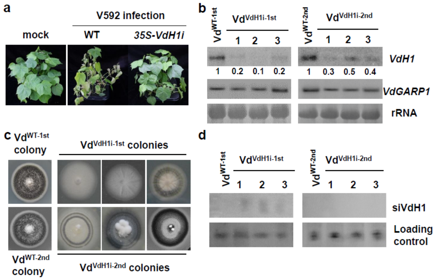
2.1. Transitive Silencing of the Trans-Kingdom Does Not Occur in Recipient V. dahliae

In plants, sRNA-mediated cleaved mRNAs may recruit RDRs to generate more dsRNAs and produce secondary siRNAs from regions surrounding the primary silencing trigger sequence, a silencing phenomenon termed silencing transitivity [44,45]. We have recently identified three RDRs in V. dahliae [7]. To investigate whether three fungal RDRs were involved in trans-kingdom RNAi for secondary siRNAs production in recipient cells, we utilized 35S-VdH1i-cotton line 3, which produces VdH1-derived siRNAs (siVdH1) [33] for infection with V. dahliae V592 and fungal recovery. The fungal hyphae recovered from wild-type and 35S-VdH1i cotton plants were named VdWT−1st and VdVdH1i−1st, respectively. Small RNAs isolated from VdWT−1st and VdVdH1i−1st colonies as well as 35S-VdH1i transgenic cotton plants were sequenced. siRNAs mapped to VdH1 were included in our analysis.
The sequencing data revealed significant amounts of siVdH1s of different lengths from 18 to 29 nucleotides (nt) produced in 35S-VdH1i transgenic cotton. A small amount of siVdH1s was detected in VdVdH1i−1st colonies but barely in VdWT−1st colonies (Figure 1a). Similar results were obtained in three individual sequencing libraries of each genotype of the colony (Supplementary Figure S1). The distribution of siVdH1s obtained from 35S-VdH1i transgenic cotton and VdVdH1i−1st colonies was aligned with VdH1 to the region used in designing hairpin RNA for creating 35S-VdH1i cotton plants [33]. siVdH1s were located alongside both strands of the VdH1i region (Figure 1a,b, and Supplementary Figure S1) but not beyond the VdH1i region, indicating that no transitive siRNAs beyond the RNAi trigger region were generated in recipient hyphae (Figure 1a). Interestingly, the length of siVdH1s was mainly 20 to 23 nt, especially 20 nt, in 35S-VdH1i transgenic cotton plants and VdVdH1i−1st colonies, and 24 nt siVdH1s were minimally detected (Figure 1b). This result prompted us to analyze endogenous known miRNAs in plants from sRNA sequencing libraries. As expected, the length of cotton endogenous known miRNAs from 35S-VdH1i transgenic cotton plants was mainly 21 and 22 nt (Figure 1c), indicating that the preparation and sequencing processes of sRNAs were appropriate. Our data suggest the possible existence of special DCL or relevant protein(s) implemented in processing exogenous RNAi constructs in cotton plants, resulting in the production of 20 to 23 nt siRNAs. Nevertheless, our data confirm that 35S-VdH1i transgene-derived siRNAs produced in cotton plants were exported to fungal hyphae to induce VdH1 gene silencing but did not trigger transitive target gene silencing in recipient V. dahliae.

2.2. Target Gene Silencing Is Reduced over Time in In Vitro Cultured Hyphae Recovered from Infected Plants

Given that no transitive silencing occurred in recipient fungal cells, we thus investigated whether target gene silencing in fungal cells would be lastingly maintained or transitorily after recovery from the infected host plants. The sixth generation of transgenic 35S-VdH1i cotton line 3 was used for V592 infection. Compared to the typical leaf wilt disease symptoms observed for wild-type cotton plants at 20 days postinoculation (dpi) (Figure 2a), transgenic 35S-VdH1i cotton plants exhibited significantly reduced disease grade in inoculated seedlings (Figure 2a). Consistent with our previous finding [33], VdH1 mRNA degradation in hyphae recovered from infected 35S-VdH1i cotton but not wild-type cotton plants was detected (Figure 2b). No significant difference in control VdGARP1 mRNA was detected in any of the recovered colonies (Figure 2b). The colony that grew from infected wild-type cotton plants (VdWT−1st) produced normal melanized microsclerotia (Figure 2c). In contrast, colonies that grew from 35S-VdH1i cotton plants (VdVdH1i−1st) developed VdH1 knockout mutant-like morphology that lacked or exhibited reduced development of melanized microsclerotia in 20-day-old plate cultures (Figure 2c).
To examine the maintenance of VdH1 silencing in recovered hyphae, we selected a patch of the first generational colonies from VdWT−1st and VdVdH1i−1st and transferred them to new plates for culture for 20 additional days. Intensive melanized microsclerotium growth was observed for VdWT−2nd. However, VdVdH1i−2nd colonies exhibited various degrees of melanized microsclerotium growth, and the morphologies differed from those of wild-type V592 (Figure 2c). Weak but clear signals of VdH1 mRNA were detected in VdVdH1i−2nd colonies (Figure 2b), which is consistent with the observation of the development of melanized microsclerotia in the VdVdH1i−2nd colonies. Furthermore, siVdH1s signals were observed in VdVdH1i−1st colonies but not in VdVdH1i−2nd colonies (Figure 2d) in accordance with VdH1 mRNA accumulation in VdVdH1i−1st and VdVdH1i−2nd colonies (Figure 2b). Taken together, our data demonstrate that target gene silencing may be maintained transitorily after fungal hyphae depart from host plants. As a result, we speculate that the sustained host-exported dsVdH1/siVdH1s were required and sufficient for silencing VdH1 in fungal cells inside the host plants, which maintained effective resistance of transgenic 35S-VdH1i cotton plants.
Figure 2. Examination of VdH1 mRNA and siVdH1 in hyphae recovered from infected cotton. (a) Disease symptoms of V592 infection on wild-type and transgenic cotton plants. (b) Detection of VdH1 mRNA in colonies recovered from wild-type and 35S-VdH1i transgenic cotton plants. The numbers below represent relative signal intensities. Hyphae grown from stems at 5 days post-culture were transferred to PDA medium to continue growth, named VdVdH1−1st colonies. Subculture colonies propagated from hyphae of VdVdH1−1st colonies were named VdVdH1−2nd colonies. (c) Morphologies of VdVdH1−1st colonies and VdVdH1−2nd colonies. Photographs were taken 20 days post-culture. (d) Small RNA hybridization of siVdH1 in VdVdH1i−1st colonies and VdVdH1i−2nd colonies.
Ijms 23 02742 g002

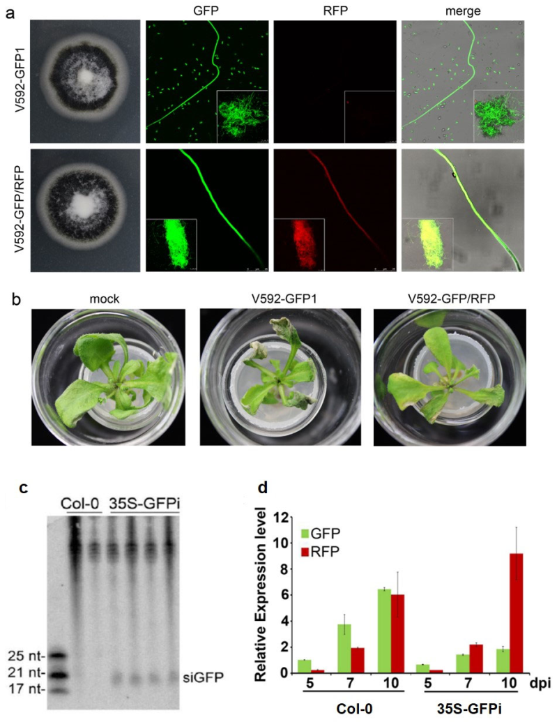
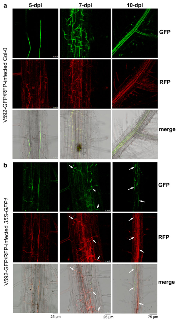
2.3. Vascular Tissue Is the Most Efficient Location for Trans-Kingdom RNAi between Plants and Vascular Fungi

To test our speculation on the efficient trans-kingdom silencing in fungal cells inside the infected plants, we first created a visible HIGS system. GFP-labeled V592 (V592-GFP1) [46] was retransformed with pSulPH-RFP, producing V592-GFP/RFP with similar growth morphology to V592-GFP1 (Figure 3a). Similar disease severity in Arabidopsis was caused by V592-GFP1 and V592-GFP/RFP. Leaf wilt was observed at 10 dpi (Figure 3b), revealing the normal infectivity of V592-GFP/RFP. To achieve visible HIGS of GFP in the hyphae, 35S-GFPi transgenic Arabidopsis was created. The production of GFP-derived siRNAs (siGFP) in 35S-GFPi transgenic Arabidopsis was confirmed (Figure 3c). Wild-type and 35S-GFPi transgenic Arabidopsis were inoculated with spores from V592-GFP/RFP by unimpaired root-dip inoculation [46]. Confocal laser scanning microscopy (CLSM) revealed that both GFP and RFP signals were observed in hyphae within the root cortical tissue in wild-type Arabidopsis by 5 dpi (Figure 4a). Both green and red fluorescence were also observed within the root cortical tissue in 35S-GFPi plants by 5 dpi; however, the GFP signal was obviously reduced, resulting in stronger red fluorescence in the merged images than that noted in wild-type Arabidopsis (Figure 4b). Similarly, while stronger GFP signals relative to RFP signals were observed in the hyphal net within the vascular tissue in wild-type Arabidopsis by 7 dpi (Figure 4a), stronger RFP relative to GFP signals in the hyphal net within the vascular tissue were observed in 35S-GFPi plants by 7 dpi (Figure 4b). Two signals overlapping in some hyphae were still observed in 35S-GFPi plants (Figure 4b, arrows). Again, intense GFP signals were observed in rapidly proliferating hyphae within the xylem vessel in wild-type Arabidopsis by 10 dpi (Figure 4a). In contrast, intense RFP signals were observed in most hyphae within the xylem vessel in 35S-GFPi plants (Figure 4b); only a few hyphae outside or around the vascular bundle maintained a clear GFP signal and overlapped with the RFP signal, as shown in the merged image (Figure 4b, arrows). Consistently, reduced levels of GFP mRNA but not RFP mRNA were detected by qRT–PCR of total RNAs extracted from the roots of the ten inoculated 35S-GFPi plants at various time points (Figure 3d). Taken together, our results clearly demonstrate that hyphae in 35S-GFPi plants were effectively and specifically triggered for GFP silencing, and GFP silencing was effectively sustained in the rapid proliferation of hyphae within the vascular tissue but possibly less effective within the cortical tissue of Arabidopsis roots where hyphae intercellularly crossed to reach the vascular tissue to realize colonization [46].

3. Discussion

The control strategy of fungal pathogens relies on the use of fungicides that are harmful to the environment. The newly developed HIGS is based on a sequence-specific RNAi mechanism that is friendly to the environment. Although considerable progress has been achieved in utilizing the HIGS strategy to protect against plant fungal pathogens, further research evaluating the efficiency, stability, and durability of resistance is needed in the field, thereby improving our understanding and application of this new approach.
In this study, small RNA sequencing results showed that siVdH1s from hyphae recovered from V. dahliae-infected 35S-VdH1i cotton matched but not beyond the VdH1i region (Figure 1a). Additionally, we showed that trans-kingdom VdH1i-derived siVdH1s-mediated degradation of VdH1 exists in hyphae fresh recovered from infected 35S-VdH1i cotton plants, given that the colonies of the VdVdH1i−2nd passages from recovered VdVdH1i−1st resumed normal VdH1 accumulation (Figure 2b). These data suggest the lack of transitive silencing in V. dahliae and indicate that three fungal RDR proteins are not involved in the trans-kingdom siRNA-induced silencing in V. dahlia [7]. This is partly similar to that in Rosellinia necatrix, a plant pathogenic fungus, in which systemic RNAi is not triggered by locally induced RNAi [47]. It has also been reported in F. asiaticum that siRNAs were only matched to the exogenous dsRNA triggers but not the target mRNA beyond the dsRNA trigger regions, indicating that RDR-dependent secondary siRNA amplification does not occur. Moreover, exogenous dsRNA exhibited a similar silencing efficiency in RDR mutants compared with that in wild-type [48]. These findings provide clues to explain the lack of secondary siRNA amplification as a divergent function of RDRs in fungi. Although there seems to be no RNAi amplification in V. dahliae, the siVdH1s generated persistently in transgenic 35S-VdH1i cotton are sufficient to silence the VdH1 gene to impart durable resistance. This finding differs from that noted for exogenously applied dsRNA or siRNAs, for which RNA amounts represent a limiting factor.
In plants, RNAi signals are transmitted locally from cell to cell through plasmodesmata (PD) and over long distances through the phloem [49,50]. The plant phloem may represent a site of accumulation of mobile sRNAs, the levels of which are modulated by stress conditions [51]. It is worth noting that the vascular fungal pathogen V. dahliae can efficiently take up host-derived sRNAs during infection [39], which may be due to the location of pathogen colonization. Hence, the plant vasculature might represent a target site for effective trans-kingdom RNAi. Notably, in V592-GFP/RFP-infected 35S-GFPi Arabidopsis roots, we observed strong GFP silencing of V592-GFP/RFP in vascular tissues, where strong red fluorescent hyphae were noted, but a lesser silencing effect in cortical tissues, where green/red fluorescent hyphae were present (Figure 4b). This finding is consistent with the role of mobile siRNAs in long-distance silencing through the plant vascular system. This result again suggests that silencing in hyphae is attributed to host-delivered siRNAs in fungal cells. Remarkably, whereas strong green hyphal proliferation was noted in wild-type Arabidopsis roots (Figure 4a), red hyphae were observed in the vascular tissue of 35S-GFPi Arabidopsis roots (Figure 4b). These results indicate that once GFP silencing in hyphae was established, it was maintained during hyphal growth and proliferation in vascular tissue. The formidable proliferation of V. dahliae hyphae in plant vascular tissue [46] together with the driving force of phloem flow underlying the long-distance transport of the silencing signal endows HIGS with high efficiency in cotton protection against the vascular pathogen V. dahliae.
In recent years, SIGS, or exogenously applied dsRNA or siRNAs in host plant protection, has become popular. A few studies revealed that SIGS for disease control was dependent on the efficiency of pathogen RNA uptake [43] and different dsRNA application approaches [52]. Additionally, an increasing number of studies have focused on improving strategies for prolonged dsRNA stability, efficacy, and scalability, such as Escherichia coli-derived anucleated minicells for dsRNA production and encapsulation [53], laser-assisted delivery of dsRNA [54], and nanoparticles for potential delivery of siRNA [54]. Although these GMO-free RNAi strategies are convenient and unlabored, the application frequency and amounts are factors that need to be considered and evaluated. Transgene-based HIGS possesses a distinctive advantage, such as constant delivery of siRNAs from plants to pathogens, ensuring the siRNA supply needed to trigger trans-kingdom RNAi. Nevertheless, exogenous application or constitutive expression of dsRNA or siRNAs is worthy of development for crop protection. Our study demonstrates that HIGS effectively triggers long-lasting trans-kingdom RNAi during interactions between transgenic cotton plants and V. dahliae, despite no amplification of RNAi being noted in this soil-borne fungal pathogen. Exploring the fungal endogenous RNAi pathways is needed to further reveal the molecular basis for this trans-kingdom RNAi, thus helping for better utilization of HIGS in crops. On the other hand, more pathogenic genes need to be tested for ideal targets in trans-kingdom RNAi. In all, our work provides further understanding of the efficacy of HIGS in defending against plant soil-borne vascular pathogens. In the future, fundamental knowledge on the molecular mechanisms of HIGS and SIGS will lead to novel integrative approaches or tailor-made solutions for controlling plant diseases [55].

4. Materials and Methods

4.1. Fungal Isolates, Culture Conditions, and Fungal Recovery and Infection Assays

A virulent defoliating V. dahliae isolate V592 from cotton was used in this study. The culture conditions of V592, the conidia production for infection assays, and the fungal recovery assay in cotton were described previously [33,56]. For plant infection assays in the laboratory, the “laboratory unimpaired root-dip inoculation method” described previously [56] was used for cotton and Arabidopsis root inoculation.

4.2. Cloning and Constructs

For the 35S-GFPi RNAi constructs, the sense and antisense sequences of the 3′-terminal 500 bp of GFP were amplified by PCR using sequence-specific primers as follows: forward primer, 5′-GGATCCATGCCGTGAGTGATCCCG-3′ (underlined letters: BamHI site); reverse primer, 5′-GAATTCGTGCTTCAGCCGCTACCC-3′ (underlined letters: EcoRI site). The antisense sequence was amplified using forward primer, 5′-GAGCTCATGCCGTGAGTGATCCCG-3′ (underlined letters: SacI site); reverse primer, 5′-AGATCTGTGCTTCAGCCGCTACCC-3′ (underlined letters: BglII site). Each of the PCR fragments was ligated into the pGEM-T vector (Tiangen). Sense and antisense sequences were inserted into an intron-containing intermediate construct (pSK-int) [57] to obtain sequence cassettes containing the inverted-repeat RNAi constructs as previously described [57], producing pSK-GFPi. A fragment of BamHI-SacI from pSK-GFPi was inserted into the binary vector pBI121 under the 35S promoter to generate 35S-GFPi for plant transformation.
To create double-labeled GFP and RFP of the V. dahliae isolate, pSulPH-RFP-NEO, which contains a neomycin (neo) resistance cassette, was generated as follows: the neo resistance cassette was amplified from pKOV21 [58] with primers 5′-GCTCTAGACAGCCGCCTTCGCAAGCGCT-3′ and 5′-GCTCTAGAGGCCAGCAGTAGACACTTGG-3′ (underlined letters: XbaI site). The PCR fragment was ligated into the pGEM-T easy vector, and the XbaI-digested fragment was inserted into XbaI-digested pSulPH, conferring the resulting pSulPH-NEO resistance to geneticin. The RFP fragment was amplified from the pGDR vector [59] with primers 5′-GCGGATCCATGGCCTCCTCCGAGAACGT-3′ (underlined letters: BamHI site) and 5′-GAATTCGCGATGTCCTTGTCCACCACCG-3′ (underlined letters: EcoRI site). The PCR fragment was inserted into pSulPH-NEO, resulting in pSulPH-RFP-NEO, which was retransformed to V592-GFP1 [46], producing the double-labeled V. dahliae isolate V592-GFP/RFP.

4.3. Fungal and Plant Transformation

The 35S-GFPi constructs were transformed into the Agrobacterium strain EHA105 for plant transformation. Arabidopsis (Columbia ecotype) was transformed according to the standard floral dip method [60]. The fungal transformation was performed as previously described [61].

4.4. RNA Extraction, RNA Gel Blotting, and Quantitative Real-Time PCR Analysis

Fungal isolates were grown in liquid Czapek–Dox medium for 3 days with shaking at 200 rpm and 26 °C in the dark, and the resulting mycelium was harvested for RNA isolation using TRIzol reagent (Invitrogen) according to the manufacturer’s instructions. For high molecular weight RNA gel blots, 20 µg of total RNA was separated on 1.2% agarose gels containing 6% formaldehyde and transferred to nylon N+ membranes. DNA probes were labeled with [a-32P] dCTP using the Rediprime II system (Amersham). For low molecular weight RNA gel blots, 40 µg of total RNA was separated by electrophoresis on 17% PAGE gels and electrically transferred to nylon N+ membranes. Then, [ɑ-32P] UTP-labeled gene-specific transcript sequences were used (New England Biolabs). For detection of the silencing of VdH1 in recovered hyphae, colonies recovered from the same infected plant at 20 days postinoculation were mixed for RNA isolation. For qRT–PCR, 2 μg of total RNA was reverse transcribed into cDNA using HiScript II Q RT SuperMix for qPCR (Vazyme). qRT–PCR analysis was performed with a 1000 series Thermal Cycling Platform (Bio-Rad) using SYBR qPCR Master Mix (Vazyme). The constitutively expressed elongation factor 1-α of V. dahliae (VdELF1) was used as an internal control. Gene-specific primers are listed below: VdELF1-qRT-F, CCATTGATATCGCACTGTGG and VdELF-qRT-R, TGGAGATACCAGCCTCGAAC; RFP-qRT-F, AGGACGGCTGCTTCATCTAC and RFP-qRT-R, CTTCAGGGCCTTGTGGGT; GFP-qRT-F, ATGGTGAGCAAGGGCGAGGAG and GFP-qRT-R, TAGGTCAGGGTGGTCACGAGG. At least three biological replicates and three technical replicates were performed in each experiment for each sample.

4.5. Confocal Laser Scanning Microscopy

A. thaliana roots were immersed in a conidial suspension (~105 conidia/mL in water solution) for 10 min and then transferred onto a 0.75% agar plate at 25 °C in the dark. Images were obtained under a confocal laser microscope (Leica TCS SP8; Leica Microsystems) with 100× oil immersion objective lenses. The excitation wavelengths and emission filters were as follows: 488 nm/bandpass 500 to 550 nm for GFP and 561 nm/bandpass 570 to 670 nm for RFP. Confocal images were captured with a Leica hybrid detector and analyzed with Leica LAS AF software.

4.6. Small RNA Sequencing

V. dahliae recovered from V592-infected cotton (VdWT−1st) and V592-infected 35S-VdH1i cotton (VdVdH1i−1st) were grown in liquid Czapek–Dox medium and harvested as described above for RNA extraction. 35S-VdH1i transgenic cotton plants grown at 26 °C with a 16 h light (8000 lux)/8 h dark cycle for approximately 3 weeks were subject to RNA extraction. RNA isolation, sRNA library construction, and sRNA sequencing were performed by BGI (http://www.bgitechsolutions.com/, accessed on 11 February 2022). The raw data were filtered to remove low-quality reads to obtain clean sequences. The sRNAs were aligned with VdH1 to the region used in designing hairpin RNA for creating 35S-VdH1i cotton plants. A Perl script was used to search for known miRNAs in cotton [62,63] with 18–30 nt clean sequences. The expression level of miRNAs was normalized by RPM.

Supplementary Materials

The following supporting information can be downloaded at: https://www.mdpi.com/article/10.3390/ijms23052742/s1.

Author Contributions

Data curation, T.Z. and Y.-Y.F.; formal analysis, T.Z., J.-H.Z., Y.-Y.F., and Y.J.; funding acquisition, H.-S.G.; methodology, Y.-Y.F.; project administration, J.-H.Z., H.-S.G. and Y.J.; software, J.-H.Z.; validation, T.Z.; writing—original draft, Y.J.; writing—review and editing, H.-S.G. All authors have read and agreed to the published version of the manuscript.

Funding

This research was funded by the National Natural Science Foundation of China, grant numbers 32020103003 and 31730078.

Institutional Review Board Statement

Not applicable.

Data Availability Statement

Not applicable.

Acknowledgments

We thank Y.L. Peng for plasmids pKOV21.

Conflicts of Interest

The authors declare no conflict of interest.

References

  1. Fulci, V.; Macino, G. Quelling: Post-transcriptional gene silencing guided by small RNAs in Neurospora crassa. Curr. Opin. Microbiol. 2007, 10, 199–203. [Google Scholar] [CrossRef]
  2. Lee, H.C.; Chang, S.S.; Choudhary, S.; Aalto, A.P.; Maiti, M.; Bamford, D.H.; Liu, Y. qiRNA is a new type of small interfering RNA induced by DNA damage. Nature 2009, 459, 274–278. [Google Scholar] [CrossRef] [PubMed] [Green Version]
  3. Lee, H.C.; Li, L.D.; Gu, W.F.; Xue, Z.H.; Crosthwaite, S.K.; Pertsemlidis, A.; Lewis, Z.A.; Freitag, M.; Selker, E.U.; Mello, C.C.; et al. Diverse Pathways Generate MicroRNA-like RNAs and Dicer-Independent Small Interfering RNAs in Fungi. Mol. Cell 2010, 38, 803–814. [Google Scholar] [CrossRef] [PubMed] [Green Version]
  4. Chang, S.S.; Zhang, Z.; Liu, Y. RNA interference pathways in fungi: Mechanisms and functions. Annu. Rev. Microbiol. 2012, 66, 305–323. [Google Scholar] [CrossRef] [PubMed] [Green Version]
  5. Nicolas, F.E.; Ruiz-Vazquez, R.M. Functional Diversity of RNAi-Associated sRNAs in Fungi. Int. J. Mol. Sci. 2013, 14, 15348–15360. [Google Scholar] [CrossRef] [Green Version]
  6. Torres-Martinez, S.; Ruiz-Vazquez, R.M. The RNAi Universe in Fungi: A Varied Landscape of Small RNAs and Biological Functions. Annu. Rev. Microbiol. 2017, 71, 371–391. [Google Scholar] [CrossRef]
  7. Jin, Y.; Zhao, J.H.; Zhao, P.; Zhang, T.; Wang, S.; Guo, H.S. A fungal milRNA mediates epigenetic repression of a virulence gene in Verticillium dahliae. Philos. Trans. R. Soc. Lond. Ser. B Biol. Sci. 2019, 374, 20180309. [Google Scholar] [CrossRef] [PubMed] [Green Version]
  8. Weiberg, A.; Wang, M.; Lin, F.M.; Zhao, H.W.; Zhang, Z.H.; Kaloshian, I.; Huang, H.D.; Jin, H.L. Fungal small RNAs suppress plant immunity by hijacking host RNA interference pathways. Science 2013, 342, 118–123. [Google Scholar] [CrossRef] [PubMed] [Green Version]
  9. Wang, M.; Weiberg, A.; Dellota, E.; Yamane, D.; Jin, H.L. Botrytis small RNA Bc-siR37 suppresses plant defense genes by cross-kingdom RNAi. Rna Biol. 2017, 14, 421–428. [Google Scholar] [CrossRef] [PubMed] [Green Version]
  10. Chen, R.; Jiang, N.; Jiang, Q.; Sun, X.; Wang, Y.; Zhang, H.; Hu, Z. Exploring microRNA-like small RNAs in the filamentous fungus Fusarium oxysporum. PLoS ONE 2014, 9, e104956. [Google Scholar] [CrossRef] [PubMed] [Green Version]
  11. Chen, Y.; Gao, Q.X.; Huang, M.M.; Liu, Y.; Liu, Z.Y.; Liu, X.; Ma, Z.H. Characterization of RNA silencing components in the plant pathogenic fungus Fusarium graminearum. Sci Rep. 2015, 5, 12500. [Google Scholar] [CrossRef] [PubMed] [Green Version]
  12. Son, H.; Park, A.R.; Lim, J.Y.; Shin, C.; Lee, Y.W. Genome-wide exonic small interference RNA-mediated gene silencing regulates sexual reproduction in the homothallic fungus Fusarium graminearum. PLoS Genet. 2017, 13, e1006595. [Google Scholar] [CrossRef]
  13. Gowda, M.; Nunes, C.C.; Sailsbery, J.; Xue, M.; Chen, F.; Nelson, C.A.; Brown, D.E.; Oh, Y.; Meng, S.; Mitchell, T.; et al. Genome-wide characterization of methylguanosine-capped and polyadenylated small RNAs in the rice blast fungus Magnaporthe oryzae. Nucleic Acids Res. 2010, 38, 7558–7569. [Google Scholar] [CrossRef] [PubMed] [Green Version]
  14. Nunes, C.C.; Gowda, M.; Sailsbery, J.; Xue, M.; Chen, F.; Brown, D.E.; Oh, Y.; Mitchell, T.K.; Dean, R.A. Diverse and tissue-enriched small RNAs in the plant pathogenic fungus, Magnaporthe oryzae. BMC Genom. 2011, 12, 288. [Google Scholar] [CrossRef] [PubMed] [Green Version]
  15. Raman, V.; Simon, S.A.; Romag, A.; Demirci, F.; Mathioni, S.M.; Zhai, J.; Meyers, B.C.; Donofrio, N.M. Physiological stressors and invasive plant infections alter the small RNA transcriptome of the rice blast fungus, Magnaporthe oryzae. BMC Genom. 2013, 14, 326. [Google Scholar] [CrossRef] [PubMed] [Green Version]
  16. Raman, V.; Simon, S.A.; Demirci, F.; Nakano, M.; Meyers, B.C.; Donofrio, N.M. Small RNA Functions Are Required for Growth and Development of Magnaporthe oryzae. Mol. Plant Microbe Interact. MPMI 2017, 30, 517–530. [Google Scholar] [CrossRef] [Green Version]
  17. Nguyen, Q.; Iritani, A.; Ohkita, S.; Vu, B.V.; Yokoya, K.; Matsubara, A.; Ikeda, K.I.; Suzuki, N.; Nakayashiki, H. A fungal Argonaute interferes with RNA interference. Nucleic Acids Res. 2018, 46, 2495–2508. [Google Scholar] [CrossRef] [Green Version]
  18. Li, Y.; Liu, X.; Yin, Z.; You, Y.; Zou, Y.; Liu, M.; He, Y.; Zhang, H.; Zheng, X.; Zhang, Z.; et al. MicroRNA-like milR236, regulated by transcription factor MoMsn2, targets histone acetyltransferase MoHat1 to play a role in appressorium formation and virulence of the rice blast fungus Magnaporthe oryzae. Fungal Genet. Biol. 2020, 137, 103349. [Google Scholar] [CrossRef]
  19. Mueth, N.A.; Ramachandran, S.R.; Hulbert, S.H. Small RNAs from the wheat stripe rust fungus (Puccinia striiformis f.sp. tritici). BMC Genom. 2015, 16, 718. [Google Scholar] [CrossRef] [PubMed] [Green Version]
  20. Wang, B.; Sun, Y.; Song, N.; Zhao, M.; Liu, R.; Feng, H.; Wang, X.; Kang, Z. Puccinia striiformis f. sp. tritici microRNA-like RNA 1 (Pst-milR1), an important pathogenicity factor of Pst, impairs wheat resistance to Pst by suppressing the wheat pathogenesis-related 2 gene. New Phytol. 2017, 215, 338–350. [Google Scholar] [CrossRef] [PubMed] [Green Version]
  21. Feng, H.; Xu, M.; Liu, Y.; Dong, R.; Gao, X.; Huang, L. Dicer-Like Genes Are Required for H2O2 and KCl Stress Responses, Pathogenicity and Small RNA Generation in Valsa mali. Front. Microbiol. 2017, 8, 1166. [Google Scholar] [CrossRef] [PubMed] [Green Version]
  22. Xu, M.; Guo, Y.; Tian, R.; Gao, C.; Guo, F.; Voegele, R.T.; Bao, J.; Li, C.; Jia, C.; Feng, H.; et al. Adaptive regulation of virulence genes by microRNA-like RNAs in Valsa mali. New Phytol. 2020, 227, 899–913. [Google Scholar] [CrossRef] [PubMed]
  23. Feng, H.; Xu, M.; Gao, Y.; Liang, J.; Guo, F.; Guo, Y.; Huang, L. Vm-milR37 contributes to pathogenicity by regulating glutathione peroxidase gene VmGP in Valsa mali. Mol. Plant Pathol. 2021, 22, 243–254. [Google Scholar] [CrossRef] [PubMed]
  24. Nakayashiki, H.; Nguyen, Q.B. RNA interference: Roles in fungal biology. Curr. Opin. Microbiol. 2008, 11, 494–502. [Google Scholar] [CrossRef]
  25. Laurie, J.D.; Linning, R.; Bakkeren, G. Hallmarks of RNA silencing are found in the smut fungus Ustilago hordei but not in its close relative Ustilago maydis. Curr. Genet. 2008, 53, 49–58. [Google Scholar] [CrossRef] [PubMed]
  26. Nunes, C.C.; Dean, R.A. Host-induced gene silencing: A tool for understanding fungal host interaction and for developing novel disease control strategies. Mol. Plant Pathol. 2012, 13, 519–529. [Google Scholar] [CrossRef] [PubMed]
  27. Koch, A.; Kogel, K.H. New wind in the sails: Improving the agronomic value of crop plants through RNAi-mediated gene silencing. Plant Biotechnol. J. 2014, 12, 821–831. [Google Scholar] [CrossRef]
  28. Hua, C.; Zhao, J.H.; Guo, H.S. Trans-Kingdom RNA Silencing in Plant-Fungal Pathogen Interactions. Mol. Plant 2018, 11, 235–244. [Google Scholar] [CrossRef] [Green Version]
  29. Rosa, C.; Kuo, Y.W.; Wuriyanghan, H.; Falk, B.W. RNA Interference Mechanisms and Applications in Plant Pathology. Annu. Rev. Phytopathol. 2018, 56, 581–610. [Google Scholar] [CrossRef]
  30. Hou, Y.; Ma, W. Natural Host-Induced Gene Silencing Offers New Opportunities to Engineer Disease Resistance. Trends Microbiol. 2020, 28, 109–117. [Google Scholar] [CrossRef]
  31. Das, P.R.; Sherif, S.M. Application of Exogenous dsRNAs-induced RNAi in Agriculture: Challenges and Triumphs. Front. Plant Sci. 2020, 11, 946. [Google Scholar] [CrossRef] [PubMed]
  32. Nowara, D.; Gay, A.; Lacomme, C.; Shaw, J.; Ridout, C.; Douchkov, D.; Hensel, G.; Kumlehn, J.; Schweizer, P. HIGS: Host-Induced Gene Silencing in the Obligate Biotrophic Fungal Pathogen Blumeria graminis. Plant Cell 2010, 22, 3130–3141. [Google Scholar] [CrossRef] [PubMed] [Green Version]
  33. Zhang, T.; Jin, Y.; Zhao, J.H.; Gao, F.; Zhou, B.J.; Fang, Y.Y.; Guo, H.S. Host-Induced Gene Silencing of the Target Gene in Fungal Cells Confers Effective Resistance to the Cotton Wilt Disease Pathogen Verticillium dahliae. Mol. Plant 2016, 9, 939–942. [Google Scholar] [CrossRef] [PubMed] [Green Version]
  34. Song, Y.; Thomma, B. Host-induced gene silencing compromises Verticillium wilt in tomato and Arabidopsis. Mol. Plant Pathol. 2018, 19, 77–89. [Google Scholar] [CrossRef] [Green Version]
  35. Xiong, F.J.; Liu, M.; Zhuo, F.P.; Yin, H.; Deng, K.X.; Feng, S.; Liu, Y.D.; Luo, X.M.; Feng, L.; Zhang, S.M.; et al. Host-induced gene silencing of BcTOR in Botrytis cinerea enhances plant resistance to grey mould. Mol. Plant Pathol. 2019, 20, 1722–1739. [Google Scholar] [CrossRef] [PubMed] [Green Version]
  36. Guo, X.Y.; Li, Y.; Fan, J.; Xiong, H.; Xu, F.X.; Shi, J.; Shi, Y.; Zhao, J.Q.; Wang, Y.F.; Cao, X.L.; et al. Host-Induced Gene Silencing of MoAP1 Confers Broad-Spectrum Resistance to Magnaporthe oryzae. Front. Plant Sci. 2019, 10, 433. [Google Scholar] [CrossRef]
  37. Kettles, G.J.; Hofinger, B.J.; Hu, P.S.; Bayon, C.; Rudd, J.J.; Balmer, D.; Courbot, M.; Hammond-Kosack, K.E.; Scalliet, G.; Kanyuka, K. sRNA Profiling Combined With Gene Function Analysis Reveals a Lack of Evidence for Cross-Kingdom RNAi in the Wheat Zymoseptoria tritici Pathosystem. Front. Plant Sci. 2019, 10, 892. [Google Scholar] [CrossRef]
  38. Ma, X.; Wiedmer, J.; Palma-Guerrero, J. Small RNA Bidirectional Crosstalk During the Interaction Between Wheat and Zymoseptoria tritici. Front. Plant Sci. 2019, 10, 1669. [Google Scholar] [CrossRef]
  39. Zhang, T.; Zhao, Y.L.; Zhao, J.H.; Wang, S.; Jin, Y.; Chen, Z.Q.; Fang, Y.Y.; Hua, C.L.; Ding, S.W.; Guo, H.S. Cotton plants export microRNAs to inhibit virulence gene expression in a fungal pathogen. Nat. Plants 2016, 2, 16153. [Google Scholar] [CrossRef]
  40. Wang, M.; Weiberg, A.; Lin, F.M.; Thomma, B.P.H.J.; Huang, H.D.; Jin, H.L. Bidirectional cross-kingdom RNAi and fungal uptake of external RNAs confer plant protection. Nat. Plants 2016, 2, 16151. [Google Scholar] [CrossRef]
  41. Cai, Q.; Qiao, L.L.; Wang, M.; He, B.Y.; Lin, F.M.; Palmquist, J.; Huang, S.N.D.; Jin, H.L. Plants send small RNAs in extracellular vesicles to fungal pathogen to silence virulence genes. Science 2018, 360, 1126–1129. [Google Scholar] [CrossRef] [PubMed] [Green Version]
  42. Wytinck, N.; Sullivan, D.S.; Biggar, K.T.; Crisostomo, L.; Pelka, P.; Belmonte, M.F.; Whyard, S. Clathrin mediated endocytosis is involved in the uptake of exogenous double-stranded RNA in the white mold phytopathogen Sclerotinia sclerotiorum. Sci Rep. 2020, 10, 12773. [Google Scholar] [CrossRef]
  43. Qiao, L.L.; Lan, C.; Capriotti, L.; Ah-Fong, A.; Sanchez, J.N.; Hamby, R.; Heller, J.; Zhao, H.W.; Glass, N.L.; Judelson, H.S.; et al. Spray-induced gene silencing for disease control is dependent on the efficiency of pathogen RNA uptake. Plant Biotechnol. J. 2021, 19, 1756. [Google Scholar] [CrossRef] [PubMed]
  44. Axtell, M.J.; Jan, C.; Rajagopalan, R.; Bartel, D.P. A two-hit trigger for siRNA biogenesis in plants. Cell 2006, 127, 565–577. [Google Scholar] [CrossRef] [Green Version]
  45. Carthew, R.W.; Sontheimer, E.J. Origins and Mechanisms of miRNAs and siRNAs. Cell 2009, 136, 642–655. [Google Scholar] [CrossRef] [PubMed] [Green Version]
  46. Zhao, P.; Zhao, Y.L.; Jin, Y.; Zhang, T.; Guo, H.S. Colonization process of Arabidopsis thaliana roots by a green fluorescent protein-tagged isolate of Verticillium dahliae. Protein Cell 2014, 5, 94–98. [Google Scholar] [CrossRef] [PubMed]
  47. Shimizu, T.; Yaegashi, H.; Ito, T.; Kanematsu, S. Systemic RNA interference is not triggered by locally-induced RNA interference in a plant pathogenic fungus, Rosellinia necatrix. Fungal Genet. Biol. 2015, 76, 27–35. [Google Scholar] [CrossRef] [PubMed]
  48. Song, X.S.; Gu, K.X.; Duan, X.X.; Xiao, X.M.; Hou, Y.P.; Duan, Y.B.; Wang, J.X.; Yu, N.; Zhou, M.G. Secondary amplification of siRNA machinery limits the application of spray-induced gene silencing. Mol. Plant Pathol. 2018, 19, 2543–2560. [Google Scholar] [CrossRef] [PubMed] [Green Version]
  49. Chen, X.M. Small RNAs and Their Roles in Plant Development. Annu Rev. Cell Dev. Bi. 2009, 25, 21–44. [Google Scholar] [CrossRef] [PubMed] [Green Version]
  50. Mermigka, G.; Verret, F.; Kalantidis, K. RNA silencing movement in plants. J. Integr. Plant. Biol. 2016, 58, 328–342. [Google Scholar] [CrossRef] [PubMed]
  51. Chitwood, D.H.; Timmermans, M.C. Small RNAs are on the move. Nature 2010, 467, 415–419. [Google Scholar] [CrossRef] [PubMed]
  52. Kiselev, K.V.; Suprun, A.R.; Aleynova, O.A.; Ogneva, Z.V.; Dubrovina, A.S. Physiological Conditions and dsRNA Application Approaches for Exogenously induced RNA Interference in Arabidopsis thaliana. Plants Basel 2021, 10, 264. [Google Scholar] [CrossRef] [PubMed]
  53. Islam, M.T.; Davis, Z.; Chen, L.S.; Englaender, J.; Zomorodi, S.; Frank, J.; Bartlett, K.; Somers, E.; Carballo, S.M.; Kester, M.; et al. Minicell-based fungal RNAi delivery for sustainable crop protection. Microb. Biotechnol. 2021, 14, 1847–1856. [Google Scholar] [CrossRef] [PubMed]
  54. Killiny, N.; Gonzalez-Blanco, P.; Gowda, S.; Martini, X.; Etxeberria, E. Plant Functional Genomics in A Few Days: Laser-Assisted Delivery of Double-Stranded RNA to Higher Plants. Plants Basel 2021, 10, 93. [Google Scholar] [CrossRef] [PubMed]
  55. Koch, A.; Wassenegger, M. Host-induced gene silencing—Mechanisms and applications. New Phytol. 2021, 231, 54–59. [Google Scholar] [CrossRef] [PubMed]
  56. Zhou, B.J.; Jia, P.S.; Gao, F.; Guo, H.S. Molecular characterization and functional analysis of a necrosis- and ethylene-inducing, protein-encoding gene family from Verticillium dahliae. Mol. Plant-Microbe Interact. MPMI 2012, 25, 964–975. [Google Scholar] [CrossRef] [PubMed] [Green Version]
  57. Guo, H.S.; Fei, J.F.; Xie, Q.; Chua, N.H. A chemical-regulated inducible RNAi system in plants. Plant J. Cell Mol. Biol. 2003, 34, 383–392. [Google Scholar] [CrossRef] [PubMed]
  58. Kong, L.A.; Yang, J.; Li, G.T.; Qi, L.L.; Zhang, Y.J.; Wang, C.F.; Zhao, W.S.; Xu, J.R.; Peng, Y.L. Different Chitin Synthase Genes Are Required for Various Developmental and Plant Infection Processes in the Rice Blast Fungus Magnaporthe oryzae. PLoS Pathog. 2012, 8, e1002526. [Google Scholar] [CrossRef] [PubMed] [Green Version]
  59. Goodin, M.M.; Dietzgen, R.G.; Schichnes, D.; Ruzin, S.; Jackson, A.O. pGD vectors: Versatile tools for the expression of green and red fluorescent protein fusions in agroinfiltrated plant leaves. Plant J. 2002, 31, 375–383. [Google Scholar] [CrossRef]
  60. Clough, S.J.; Bent, A.F. Floral dip: A simplified method for Agrobacterium-mediated transformation of Arabidopsis thaliana. Plant J. Cell Mol. Biol. 1998, 16, 735–743. [Google Scholar] [CrossRef] [PubMed] [Green Version]
  61. Gao, F.; Zhou, B.J.; Li, G.Y.; Jia, P.S.; Li, H.; Zhao, Y.L.; Zhao, P.; Xia, G.X.; Guo, H.S. A glutamic acid-rich protein identified in Verticillium dahliae from an insertional mutagenesis affects microsclerotial formation and pathogenicity. PLoS ONE 2010, 5, e15319. [Google Scholar] [CrossRef] [PubMed] [Green Version]
  62. Liu, N.; Tu, L.; Tang, W.; Gao, W.; Lindsey, K.; Zhang, X. Small RNA and degradome profiling reveals a role for miRNAs and their targets in the developing fibers of Gossypium barbadense. Plant J. Cell Mol. Biol. 2014, 80, 331–344. [Google Scholar] [CrossRef] [PubMed] [Green Version]
  63. Gong, L.; Kakrana, A.; Arikit, S.; Meyers, B.C.; Wendel, J.F. Composition and expression of conserved microRNA genes in diploid cotton (Gossypium) species. Genome Biol. Evol. 2013, 5, 2449–2459. [Google Scholar] [CrossRef] [PubMed] [Green Version]
Figure 1. Small RNA analysis of 35S-VdH1i transgenic cotton, VdVdH1i−1st, and VdWT−1st colonies. (a) RPM (reads per million sequences) and distribution of siVdH1s obtained by deep sequencing. The 5′-terminal 380bp VdH1i region is indicated. The y-axes represent the RPM of siVdH1s in small RNA libraries of 35S-VdH1i cotton, VdVdH1i−1st colonies, and VdWT−1st colonies related to positions along the VdH1 gene indicated in x-axes. siVdH1s (in red vertical lines) above and below the 0 value of y-axes indicate the sense and antisense orientation, respectively. (b) Length distribution of siVdH1s in 35S-VdH1i cotton and VdVdH1i−1st colony. (c) Length distribution of cotton endogenous known miRNAs from 35S-VdH1i transgenic plants. The repeats are provided in Supplementary data.
Ijms 23 02742 g001
Figure 3. Examination of V592-GFP/RFP in 35S-GFPi transgenic Arabidopsis. (a) Colony morphologies and confocal microscopy images of hyphae of V592-GFP1 and V592-GFP/RFP. (b) Similar infectivity of V592-GFP1 and V592-GFP/RFP in Arabidopsis. Photographs were taken at 10 days postinoculation. (c) Detection of GFP-derived siRNAs (siGFP) in 35S-GFPi transgenic Arabidopsis. Total RNAs isolated from wild-type Col-0 and 4 individual transformants were loaded. The GFP-specific sequence was used as a probe. The upper unspecific bands visible in the top blot serve as the loading control. Positions of 17, 21, and 25 nt siRNAs are indicated. (d) Expression of GFP and RFP mRNAs in infected Col-0 and 35S-GFPi Arabidopsis. Total RNAs isolated from roots of ten infected plants at indicated time points were quantified by qRT–PCR and normalized with the corresponding input RNA and VdELF1 (as the internal standard) levels. The value of GFP mRNA in V592-GFP/RFP-infected Col-0 was arbitrarily designated as 1. Error bars represent SD for three replicates. Reduced levels of GFP mRNA but not RFP mRNAs were detected in 35S-GFPi.
Ijms 23 02742 g003
Figure 4. Confocal laser scanning microscopy observation of GFP silencing in V. dahliae within infected Arabidopsis roots. (a,b) Confocal laser scanning microscopy images of GFP and RFP signals in hyphae within wild-type (Col-0) (a) and 35S-GFPi transgenic (b) Arabidopsis roots infected by double-labeled V. dahliae isolate V592-GFP/RFP. The merged images are compound micrographs of bright field transmission and the corresponding GFP and RFP fluorescence images. Examples of hyphae overlapped with both GFP and RFP signals are indicated by arrows. Six images for each root sample were observed and similar results were obtained.
Ijms 23 02742 g004
Publisher’s Note: MDPI stays neutral with regard to jurisdictional claims in published maps and institutional affiliations.

