Review

Peptide Human Neutrophil Elastase Inhibitors from Natural Sources: An Overview

1
Department of Pharmacy, “G. d’Annunzio” University of Chieti-Pescara, 66100 Chieti, Italy
2
Department of Biology, Science Faculty, Selcuk University, Konya 42130, Turkey
3
Department of Medical, Oral and Biotechnological Sciences, “G. d’Annunzio” University Chieti-Pescara, Via dei Vestini 31, 66100 Chieti, Italy
*
Author to whom correspondence should be addressed.
Int. J. Mol. Sci. 2022, 23(6), 2924; https://doi.org/10.3390/ijms23062924
Submission received: 11 February 2022 / Revised: 4 March 2022 / Accepted: 6 March 2022 / Published: 8 March 2022
(This article belongs to the Topic Bioactive Peptides as Functional Food Ingredients)

Abstract

:
Elastases are a broad group of enzymes involved in the lysis of elastin, the main component of elastic fibres. They are produced and released in the human body, mainly by neutrophils and the pancreas. The imbalance between elastase activity and its endogenous inhibitors can cause different illnesses due to their excessive activity. The main aim of this review is to provide an overview of the latest advancements on the identification, structures and mechanisms of action of peptide human neutrophil elastase inhibitors isolated from natural sources, such as plants, animals, fungi, bacteria and sponges. The discovery of new elastase inhibitors could have a great impact on the pharmaceutical development of novel drugs through the optimization of the natural lead compounds. Bacteria produce mainly cyclic peptides, while animals provide for long and linear amino acid sequences. Despite their diverse natural sources, these elastase inhibitors show remarkable IC50 values in a range from nM to μM values, thus representing an interesting starting point for the further development of potent bioactive compounds on human elastase enzymes.

1. Introduction

Elastic fibres compose an essential part of the extracellular matrix of connective tissue, where they allow its reversible and repetitive deformation. Each elastic fibre is mainly constituted by an amorphous core of elastin, which occupies more than 90% of the fibre, surrounded by an external envelope of microfibrils [1,2]. Fibrillin microfibrils ensure extensibility and elasticity during the deformation of tissues. A huge variety of fibrillin-binding proteins occurs for the assembly of elastic fibres [3], which play an important role in ensuring mechanical resilience, durability and cell interactivity within tissues [4].
Elastin is a hydrophobic protein that displays a huge cross-linking among Lys residues following the oxidation performed by the lysil-oxidase enzyme, which gives elastin a high level insolubility [1]. This protein contains a large quantity of hydrophobic amino acids. In particular, higher vertebrates’ elastin is composed of more than 30% Gly and about 75% Val, Ala and Pro residues [1,5]. The formation process of elastic fibres is called elastogenesis, comprising tropoelastin (elastin’s subunits) synthesis, coacervation, cross-linking and deposition [4].
Elastases are a group of enzymes that selectively lyse the amorphous component of elastic fibre, namely elastin [2]. Elastases are produced and released in different locations of the human body. Among them, human neutrophil elastase and pancreatic elastase show an important physiological and pathological role. In detail, pancreatic elastase measurement in faeces (faecal elastase-1) is used as an indirect test to determine the onset of the exocrine pancreatic insufficiency (EPI). Pancreatic elastase is secreted from pancreatic acinar cells and after binding to the bile salts goes through a tiny degradation into the gut. Clinicians can make use of two possible ELISA tests to measure faecal elastase-1 through monoclonal or polyclonal antibodies [6]. For example, the ScheBo Pancreatic Elastase 1 Stool Test (ScheBo Biotech, Giessen, Germany) is one of the most-used tests based on two monoclonal antibodies that measure the isoforms chymotrypsin-like elastase 3B (CELA3B) and, with lower efficiency, CELA3A [7].
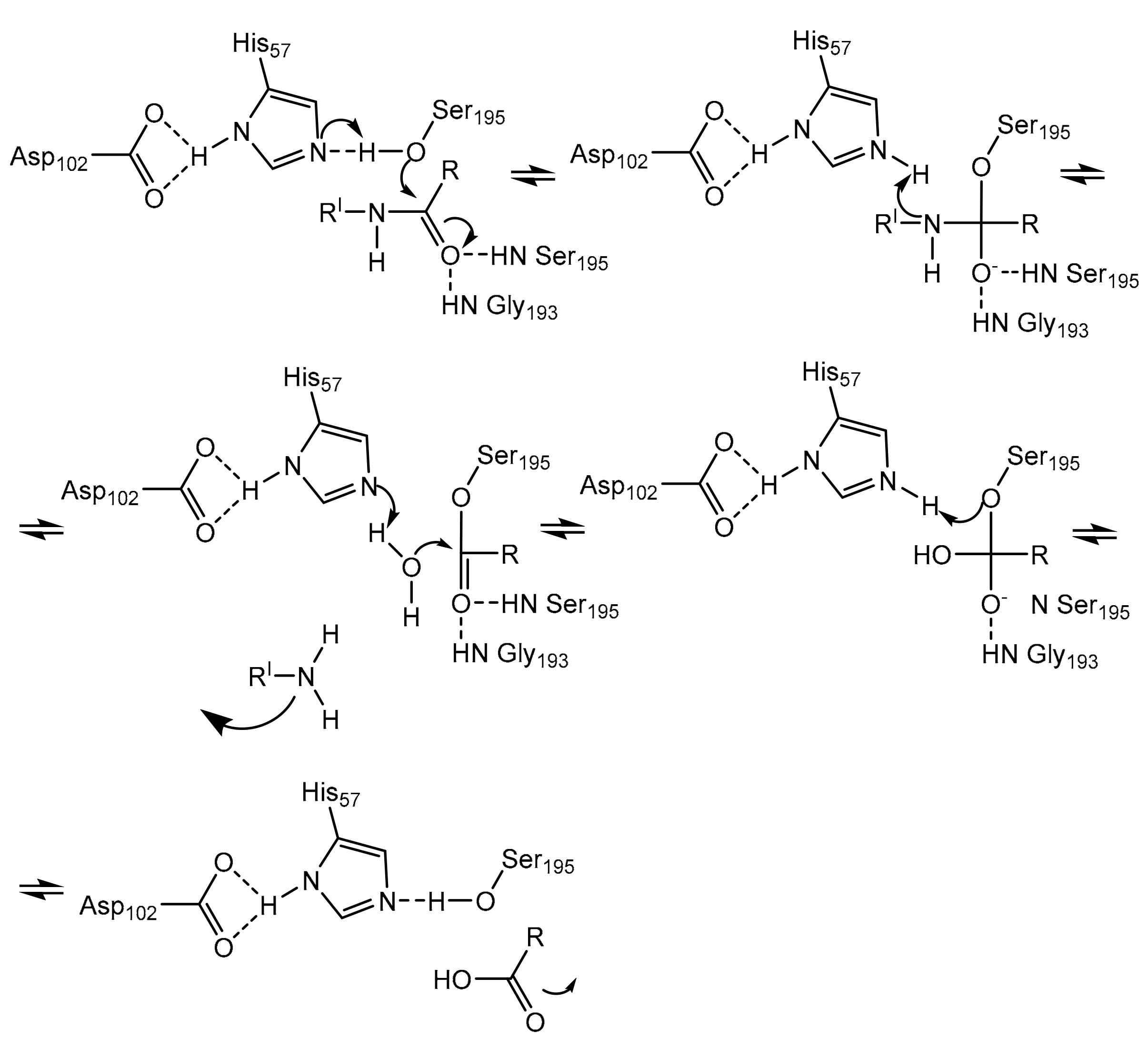
Neutrophils, a large population of leukocytes, play an important role in the regulation of the innate and adaptive immune responses, quickly achieving the inflammation/infection site [8]. During the degranulation process, neutrophils release human neutrophil elastase (HNE) (Figure 1), a glycoprotein composed of 218 amino acids, through extracellular neutrophil traps (NETs) [8,9]. NETs are composed of decondensed chromatin, neutrophil elastase and other enzymes [8]. HNE is a serine protease belonging to the chymotripsin family that is produced in the bone marrow during the stage of promyelocytes [9]. Neutrophil serine proteases are involved in inflammation and the immune response [10]. HNE is deposited as an active enzyme in the azurophilic granules of polymorphonuclear neutrophils. This enzyme has a catalytic site of three amino acids, called the catalytic triade: Asp102, His57 and Ser195. The mechanism of action of serine proteases, which includes neutrophil elastase, is illustrated in Scheme 1; when the target ligand achieves the enzymatic catalytic site, a proton transfer happens among the three amino acids, causing an increased nucleophile activity of Ser195 with a concomitant peptide bond cleavage [9,11]. In regular conditions, a balance between human neutrophil elastase activity and its endogenous inhibitors (e.g., elafin, serpins, α1-antitrypsin and secretory leukocyte proteinase inhibitor) is guaranteed, while the imbalance between elastase activity and its endogenous inhibitors can cause a variety of illnesses, such as chronic obstructive pulmonary disease, acute lung injury, acute respiratory distress syndrome and pulmonary fibrosis [12,13]. It has recently been hypothesized that neutrophil elastase inhibitors could be used to treat acute respiratory distress syndrome (ARDS) caused by COVID-19 infection, but their effective use is yet unproven, and more clinical studies should be conducted to evaluate the response and effectiveness of these types of inhibitors in COVID-19 patients [14].
The discovery of new elastase inhibitors from natural sources, such as plants, animals, fungi, bacteria and sponges, could have a great impact on the pharmaceutical development of novel drugs to prevent and to treat diseases caused by the excessive activity of elastase or the pathological degradation of elastin. Thus, the main aim of this review is to expose the latest advancements in the identification of peptide inhibitors of human elastase isolated from natural sources, trying for the first time, to the best of our knowledge, to outline all the natural peptide compounds active on human neutrophil elastase (also called human leukocyte elastase). We will provide an overview of their origins, structures and mechanisms of action where already known. This review could be the starting point for further investigations on the structural features necessary to promote a better interaction among peptide compounds and the active site of human neutrophil elastase, leading to the future synthesis of more potent peptide inhibitors. The development of novel peptide HNE inhibitors could be beneficial for the treatment of chronic obstructive pulmonary disease, acute lung injury, pulmonary fibrosis and disorders caused by COVID-19 infection, such as acute respiratory distress syndrome.

2. Natural Peptide Elastase Inhibitors Isolated from Animal Sources

2.1. ShSPI from Scolopendra Hainanum

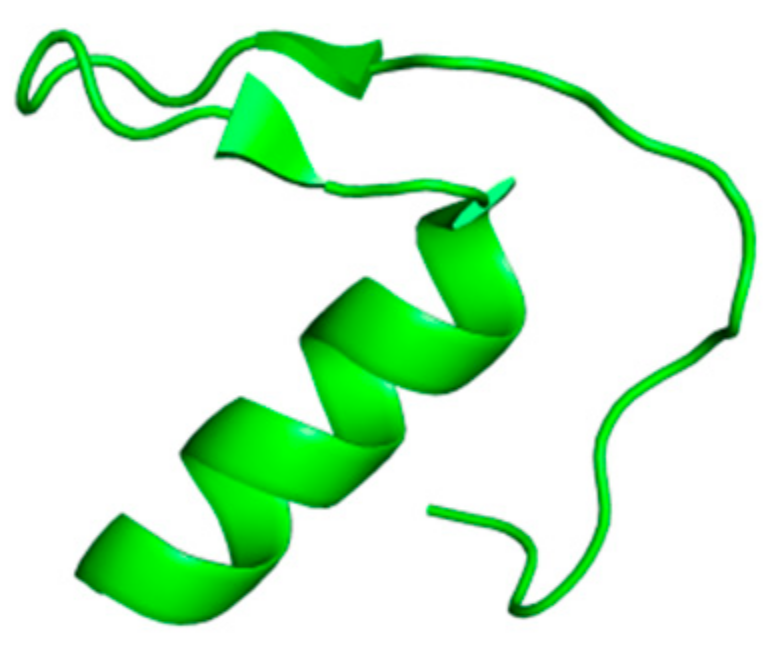
Luan et al. discovered a new serine protease inhibitor from the cDNA library of the venom glands of centipede Scolopendra Hainanum. This compound is called ShSPI, and it has been identified as an atypical kazal-type protease inhibitor that could be developed as a novel drug for the treatment of illnesses caused by human elastase. ShSPI is a bioactive peptide composed of 34 amino acids whose primary sequence is: CPQVCPAIYQPVFDEFGRMYSNSCEMQRARCLRG. The three-dimensional structure (Figure 2) displays a cystine-stabilized α-helix formed by Ser23 and Arg33 and a two-stranded anti-parallel β-sheet (Pro11 to Asp14 and Gly17 to Tyr20); the folding is guaranteed by the tertiary structure and the presence of two disulfide bonds among Cys1-Cys31 and Cys5-Cys24 [12]. Its inhibitory activity was compared to Sivelestat (ONO-5046), a human neutrophil elastase inhibitor tested as clinical drug for the treatment of ARDS and recently taken in consideration for the management of acute lung injury/acute respiratory distress syndrome (ALI/ARDS) or ARDS with coagulopathy caused by COVID-19 infection [12,17,18]. Both compounds inhibit HNE in a dose-dependent manner, but ShSPI exhibits a more effective activity than Sivelestat at the same concentration. ShSPI acts with a non-competitive inhibition mechanism against elastase, displaying a Ki value of 12.6 nM, and its equilibrium dissociation constant (KD) to HNE is 4.2 × 10⁻⁸; furthermore, ShSPI shows a huge stability after co-incubation with human plasma over 48 h [12].

2.2. AvKTI from Araneus Ventricosus

Peptide AvKTI has been isolated from a spider (Araneus ventricosus); it has the ability to inhibit trypsin, chymotrypsin, plasmin and human neutrophil elastase. It is composed of 170 amino acids and comprises a signal peptide of 19 units, a pro-peptide of 94 building blocks and a mature peptide of 57 amino acids that represents a Kunitz domain. The mature AvKTI has a peptide sequence (KDRCLLPKVTGPCKASLTRYYYDKDTKACVEFIYGGCRGNRNNFKQKDECEKACTDH) similar to other Kunitz-type serine protease inhibitors. This compound is the first Kunitz-type serine protease inhibitor isolated from spiders to be known for its antifibrinolytic and antielastolytic activity [19]. Kunitz-type inhibitors are usually composed of a single inhibitory domain or a multi-domain included in a single-chain compound [20]. It is produced in the spider’s epidermis; however, the physiological role of this compound in spiders is not yet clear [19,20]. AvKTI inhibits neutrophil elastase with an IC50 value of 446.93 nM and plasmin with an IC50 value of 10.07 nM, showing a lower neutrophil elastase inhibitory activity of about 44.4-fold. The Ki value of AvKTI against neutrophil elastase is 169.07 nM, while its Ki value against plasmin is 4.89 nM [19].

2.3. Gaumerin from Hirudo Nipponia

Guamerin is a cysteine-rich polypeptide composed of 57 amino acids (VDENAEDTHGLCGEKTCSPAQVCLNNECACTAIRCMIFCPNGFKVDENGCEYPCTCA) with a molecular weight of 6.110 that was isolated from a native Korean leech Hirudo nipponia by Jung et al. Its structure is characterized by ten cysteine residues that confer a high rigidity due to the formation of disulfide bridges. Guamerin showed stability at 25 °C in a range of pH varying from 1 to 11. It has the ability to inhibit human leukocyte elastase (HLE) with a Ki value of 8.1 × 10⁻¹⁴ M [21].

3. Natural Peptide Elastase Inhibitors Isolated from Fungi

3.1. Desmethylisaridin C2, Isaridin E, Isaridin C2 and Roseocardin from Beauveria Felina

Eight cyclodepsipeptides have been produced by the filamentous fungi Beauveria felina after the addition of suberoylanilide hydroxamic acid (SAHA), a histone deacetylase (HDAC) inhibitor, to the culture medium; three of them are new compounds, e.g., desmethylisaridin C2, desmethylisaridin E and isaridin F [22]. This is an example of an epigenetic tool used for obtaining inhibitor peptides from fungal sources [23]. Among them, Desmethylisaridin C2, isaridin E, isaridin C2 and roseocardin (Figure 3) are able to inhibit elastase release induced by formyl-L-methionyl-L-leucyl-L-phenylalanine (FMLP) in human neutrophils and to express an anti-inflammatory action without toxicity for human neutrophils. The IC50 values of these compounds are displayed in Table 1 [22].

3.2. AFUEI from Aspergillus fumigatus

AFUEI is an elastase inhibitor purified by Okumura et al. from Aspergillus fumigatus strain AFU-12 that came from the sputum of a patient afflicted with allergic bronchopulmonary aspergillosis. Its primary amino acid structure is composed of 68 residues (DPATCEKEAQFVKQELIGQPYTDAVANALQSNPIRVLHPGDMITMEYIASRLNIQVNENNEIISAHCA), and the molecular mass of its protein portion is 7526.2 Da. AFUEI could be effective against inflammatory diseases since it is able to inhibit human leukocyte elastase with an inhibitory activity of 83.8% [24]. Aspergillus fumigatus synthesizes A. fumigatus elastase (AFUE) and native A. fumigatus elastase inhibitor (N-AFUEI) in liquid medium, but N-AFUEI is barely produced. Therefore, for medical purposes, synthetic-AFUEI (S-AFUEI) has been produced by Peptide Institute Inc. (Osaka, Japan) since 2015. Further investigations about S-AFUEI’s biological and physiological activities have been reported by Fukui et al. [25].

4. Cyclic Peptides Derived from Bacteria as Elastase Inhibitors

4.1. Lyngbyastatin 4 and Lyngbyastatin 7

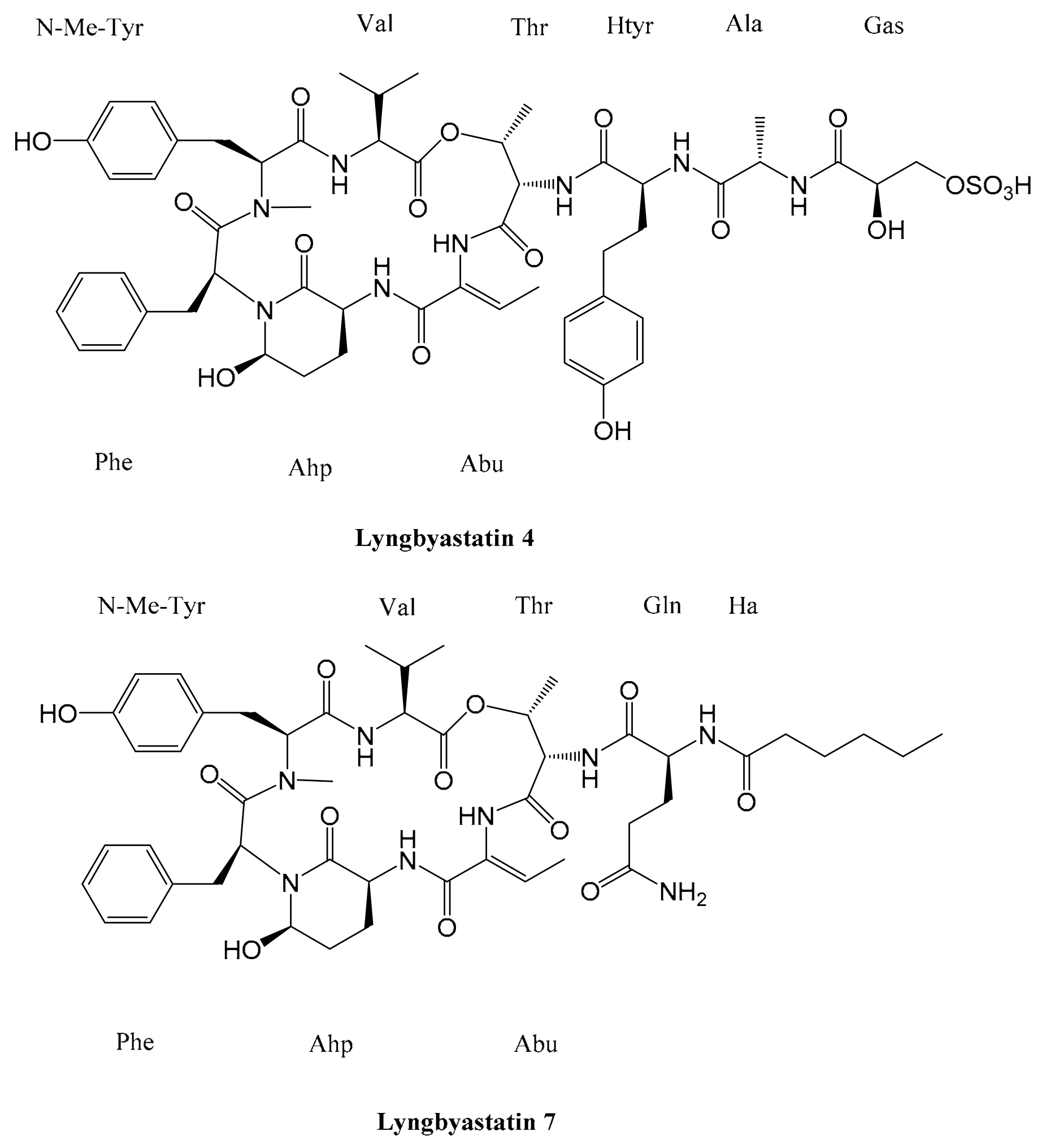
Cyanobacteria are ancient forms of life highly spread on our planet and seem to be very interesting in order to promote pharmaceutical advancements due to their secondary metabolites with a typical restricted conformation and their related metabolic stability and biological activities [26,27]. Cyclic depsipeptides isolated from cyanobacteria show similarities in their structures, which are usually composed of an amino acid sequence of six residues assembling a macrocyclic ring. The majority of these compounds display a residue of 3-amino-6-hydroxy-2-piperidone (Ahp). More than 20 depsipeptides show a hexadepsipeptide core, a 2-amino-2-butenoic acid (Abu) residue next to the Ahp ring and variable side chains (Figure 4). Another similar feature of these depsipeptides is the ester linkage between the hydroxyl group of L-threonine located in the β position and the carboxyl group of the last amino acid in C-terminal position [28].
Different peptides are produced by marine cyanobacteria belonging to genus Lyngbya [23]. Lyngbya confervoides coming from the Florida Atlantic coast is a producer of lyngbyastatin 4, a depsipeptide showing two peculiar residues of homotyrosine and Ahp; it has the ability to inhibit human neutrophil elastase, with an IC50 value of 49 nM. Moreover, it is not considered cytotoxic to several cancerous cell lines [29,30].
Lyngbyastatin 7 is a potent compound isolated from Lyngbya spp. whose structure has been defined by Taori et al. It has been tested by Salvador et al. as an inhibitor at a single concentration against 68 proteases. It exhibits a specific activity against serine protease elastase, proteinase K and chymotrypsin; it is able to inhibit HNE, with an IC50 value of 23 nM [29,31]. Its structure is characterized by a cyclic depsipeptide core composed of 19 residues, including the 3-amino-6-hydroxy-2-piperidone (Ahp) [9,31]. A low quantity of Lyngbyastatin 7 can be isolated from the natural source. Therefore, total synthesis methodologies have been developed to investigate its activity [32,33]. Salvador et al. elucidated the interaction between lyngbyastatin 7 and porcine pancreatic elastase through co-crystallization using the hanging drop vapor diffusion technique with the aim to better understand the mechanism of inhibition; Lyngbyastatin 7 and its related compounds act as substrate mimics. Indeed, the subsites S1 to S4 of the enzyme are occupied by the 2-amino-2-butenoic acid (Abu) and the N-terminal amino acids [29].

4.2. Tutuilamides A–C

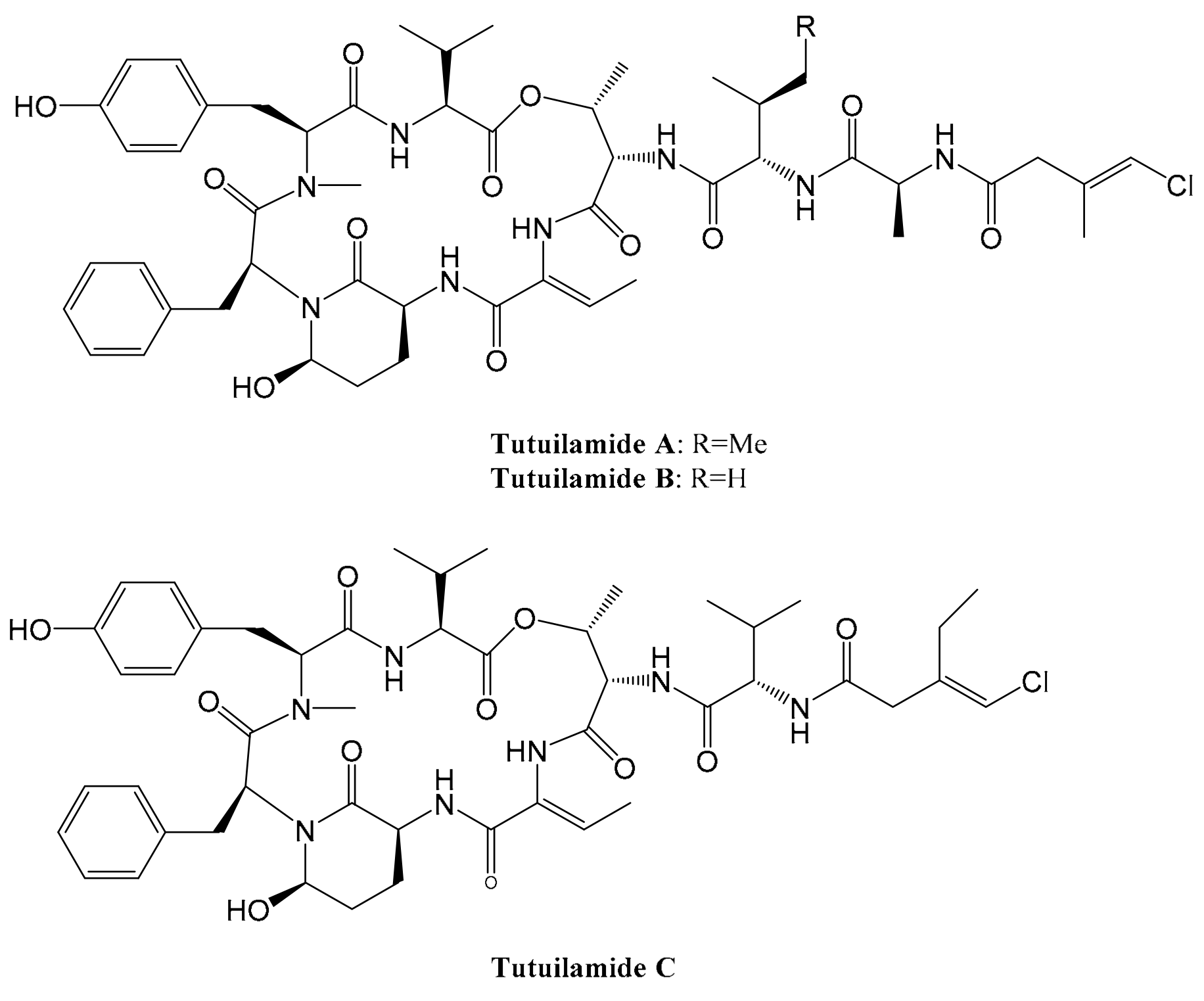
Keller et al. isolated three new peptides containing Ahp and Abu residues from two different bacteria, tutuilamide A and tutuilamide B from cyanobacterium Schizothrix sp. and tutuilamide C from Coleofasciculus sp. Their structures are characterized by an uncommon vinyl-chloride-containing residue. Tutuilamides A and B show the same cyclic core, but they differ for an amino acid (isoleucine in place of valine). The primary sequence of Tutuilamide A is Val-N-MeTyr-Phe-Ahp-Abu-Thr-Ile-Ala-Cmb. Tutuilamide C is similar to Tutuilamide B, except for the lack of an alanine residue and the presence of an additional methylene in the side chain (Figure 5). Tutuilamides A–C have been tested for the inhibition activity against serine proteases, e.g., porcine pancreatic elastase (PPE). Tutuilamides A and B are the most active as inhibitors against PPE, with an IC50 of 1-2 nM [28]. Keller et al. also investigated the crystallized structure of the complex between Tutuilamide A and porcine pancreatic elastase, revealing a good interaction in the substrate binding pocket and the presence of a supplemental H-bond formed between the carbonyl group of the Cmb residue and the backbone amide of Arg217 not observed in the lyngbyastatin 7 co-crystalized structure [28,29]. Therefore, Tutuilamide A shows a higher potency than lyngbyastatin 7 against PPE [28].
The total synthesis of Tutuilamide A has been reported through late-stage diversification, a convergent synthetic strategy similar to the total synthesis of lyngbyastatin 7 [32,33,34]. Its inhibitory activity against HNE has been evaluated, showing a high selectivity for this enzyme [34]. Tutuilamide A shows a higher inhibitory activity against PPE than lyngbyastatin 7. On the contrary, lyngbyastatin 7 seems to be equal or better than it in inhibiting HNE [28,34]. The IC50 values of Tutuilamide A and lyngbyastatin 7 against HNE are 0.73 nM and 0.85 nM, respectively [34].

4.3. Loggerpeptins A–C and Molassamide

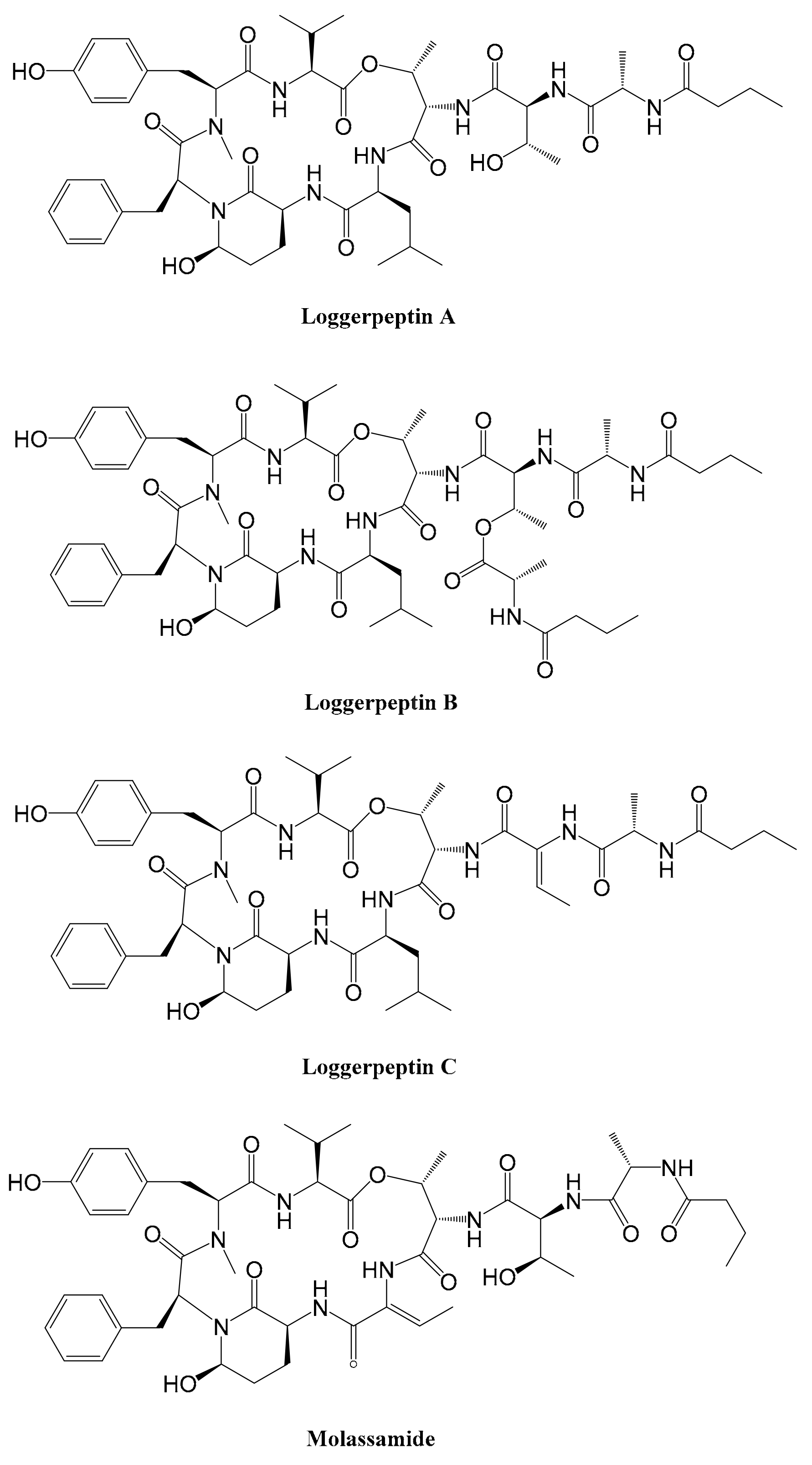
Al-Awadhi et al. identified four serine protease inhibitors from the marine cyanobacterium DRTO-73 of Loggerhead Key in Florida: Loggerpeptins A–C and molassamide. Loggerpeptins A–C have different molecular formulas: C50H72N8O13, C57H83N9O15 and C50H70N8O12, respectively [35]. Molassamide (C48H66N8O13) is a compound previously isolated from the cyanobacterium Dichothrix utahensis by Gunasekera et al. who elucidated its structure and inhibitory activity against porcine pancreatic elastase, α-chymotrypsin from bovine pancreas and trypsin from porcine pancreas (Figure 6) [36]. Loggerpeptins A–C and molassamide display a similar core structure composed of a 19-membered cyclic hexadepsipetide, including the Ahp residue and a modified glutamic acid residue. All the compounds have been tested against human neutrophil elastase and compared with sivelestat inhibition (IC50 = 0.06 μM), showing the ability to inhibit HNE with IC50 values of 0.29 μM, 0.89 μM, 0.62 μM and 0.11 μM, respectively. Among them, the most potent inhibitor is molassamide, while loggerpeptins B and C show less activity than loggerpeptin A. Loggerpeptin C differs from its analogues because of the presence of an Abu unit within the side chain [35].

4.4. Symplostatins 5–10

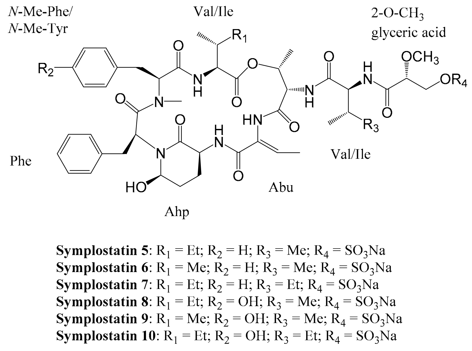
Symplostatins 5–10 are six cyclic depsipeptides containing an Ahp moiety derived from the red marine cyanobacterium Symploca sp. While symplostatins 8–10 contain an N-Me-Tyr residue, symplostatins 5–7 contain an N-Me-Phe residue (Figure 7). Salvador et al. evaluated their antiproteolytic activity against human neutrophil elastase. Among them, symplostatins 8–10 exhibit a more potent and effective inhibition of HNE (IC50 values of 41 nM, 28 nM and 21 nM, respectively). These three compounds, along with lyngbyastatins 4 and 7, are more effective in elastase inhibition than sivelestat, a specific human neutrophil elastase inhibitor. Further investigations on the inhibitory activity of symplostatins 5–10 towards human and bovine pancreatic chymotrypsin have been conducted, showing less potency compared to elastase inhibition [29].

4.5. Insulapeptolides A–H

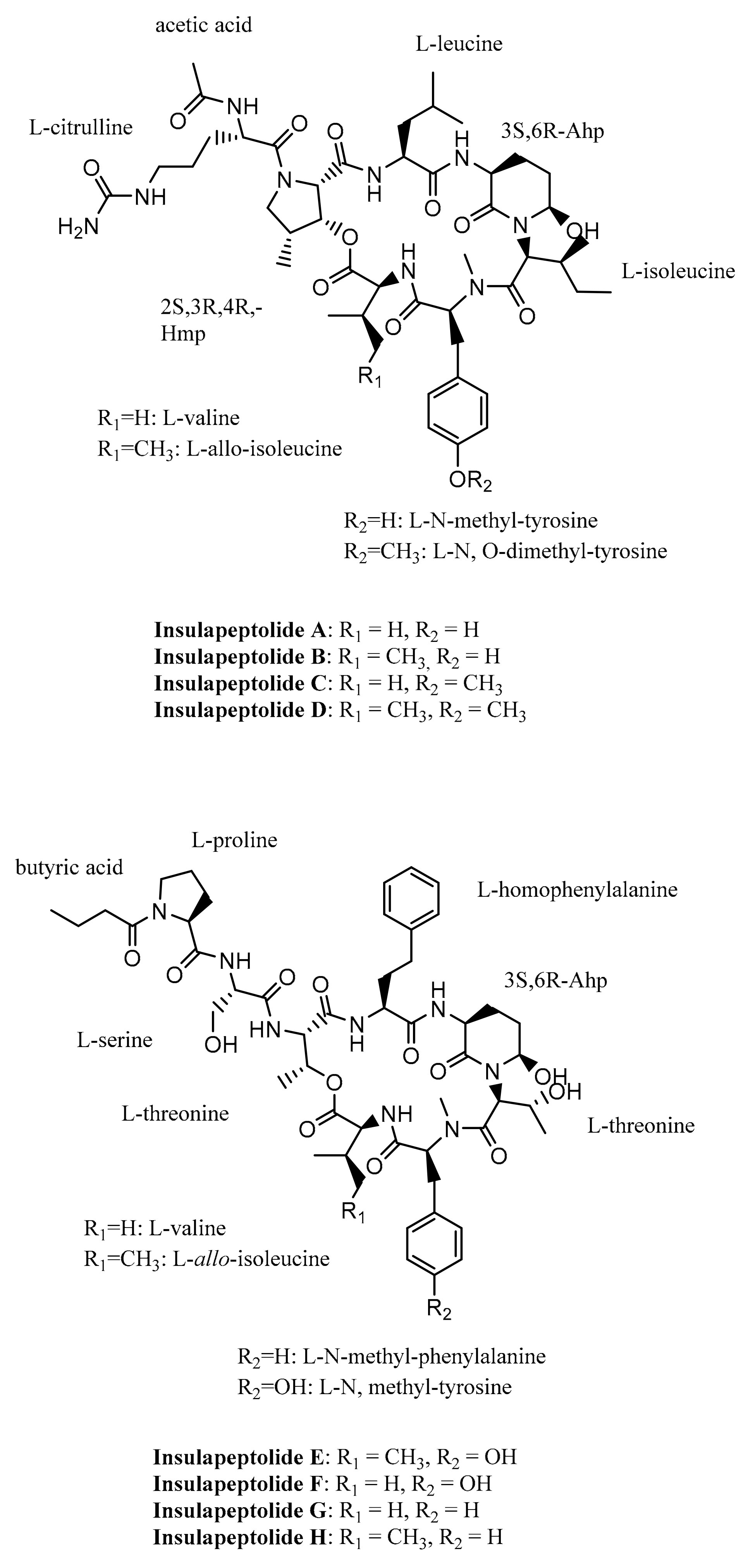
Following the evaluation of 17 cyanobacterial strains of various genera in a human leukocyte elastase inhibition assay, Mehner et al. identified the strain Nostoc insulare as an important source of HLE inhibitors. They isolated eight cyanopeptolins, namely insulapeptolides A–H, which show the ability to inhibit HLE, with IC50 values in a range from micro- to nanomolar values (Table 2). Insulapeptolides A–D exhibit two peculiar amino acids, 3-hydroxy-4-methyl-proline (Hmp) and citrulline, while insulapeptolides E–H display the L-threonine residue rather than Hmp moiety; these compounds show less inhibitory activity towards HLE (Figure 8). Moreover, Mehner et al. suggest that insulapeptolides act on HLE through a competitive inhibitory mechanism [26].

4.6. Brunsvicamides A–C

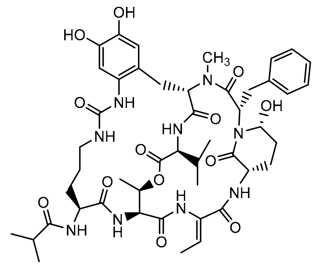
Brunsvicamides A–C, isolated from Tychonema sp., are cyclic peptides composed of six amino acids: five of them are enclosed in the 19-membered ring skeleton and the last one is linked to the α-amino group of the D-Lys thanks to the urea moiety (Figure 9). The cyclic ring is locked through an amide bond between Phe and D-Lys residues. Brunsvicamides A and B are characterized by the presence of L-amino acids, while brunsvicamide C shows an N-methyl-NI-formylkynurenine derived from tryptophan in the D-configuration [27,37]. These compounds show a selective activity against human leukocyte elastase, with Ki values of 1.1 μM, 0.70 μM and 1.6 μM, respectively, measured considering a competitive inhibition mechanism, and IC50 values of 3.12 μM, 2.00 μM and 4.42 μM, respectively [27].

4.7. FR901277

FR901277 is a natural compound identified and isolated from the bacterium filtrate of Streptomyces resistomycificus that acts as an HLE inhibitor (IC50 value of 1.8 × 10⁻⁷ M) (Figure 10). It displays a competitive inhibition mechanism against elastase (Ki = 1.2 × 10⁻⁸ M) [38]. It has a bicyclic structure with an isopropyl carbonyl N-terminus, four common amino acids and three unusual ones [39].

4.8. YM-47141 and YM-47142

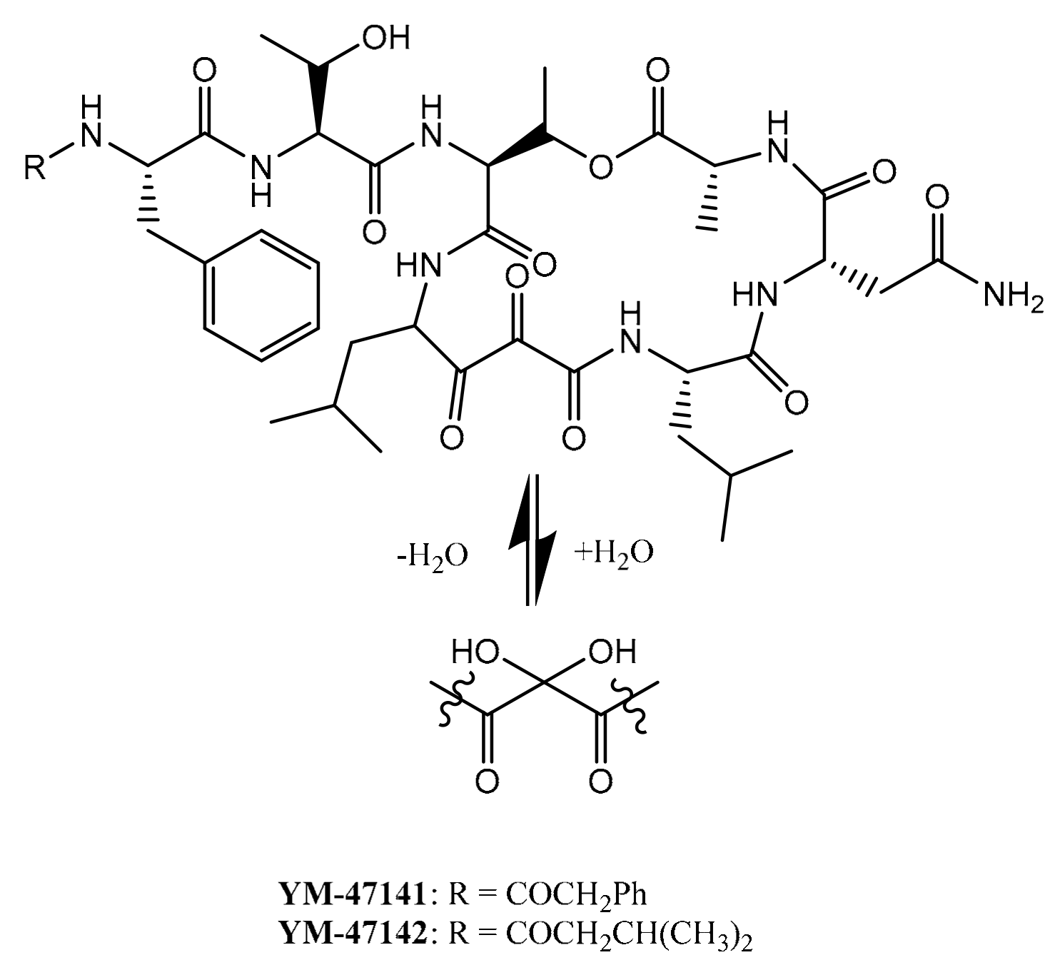
YM-47141 (C46H62N8O13) and YM-47142 (C43H64N8O13) are cyclic depsipeptides that were discovered by Yasumuro et al. from the fermented bacterium Flexibacter sp. strain Q17897 obtained from a soil sample and belonging to Gram-negative bacteria (Figure 11). These compounds are human leukocyte elastase inhibitors with IC50 values of 1.5 × 10⁻⁷ M and 3.0 × 10⁻⁷ M, respectively; among them, YM-47141 exhibits a superior inhibitory activity against HLE. Their structures are particularly interesting because of a vicinal tricarbonyl function; these are the first protease inhibitors containing this function discovered from a microbial source. They suggest that the elastase inhibitory activity is due to the formation of hemiketal between the serine residue on the catalytic enzymatic site and the 2-carbonyl group of 2,3-dioxo-4-amino-6-methyl-heptanoic acid contained in YM-47141 and YM-47142 [40].

5. Peptide Elastase Inhibitors Isolated from Plants

5.1. Ixorapeptide I and II from Ixora Coccinea

Ixorapeptide I and II are derivatized peptides isolated from the methanol extract of the aerial part of Ixora coccinea by Lee et al. (Figure 12). Only Ixorapeptide II has the ability to inhibit the elastase release in the anti-inflammatory assay (IC50 value of 0.27 μg/mL), showing a possible anti-inflammatory activity on neutrophils. It is 73 times more active on elastase release inhibition than phenylmethylsulfonyl fluoride (PMSF), a commercial reference compound, without the relevant cytotoxicity in the cancer cell lines [41].

5.2. Roseltide rT1 from Hibiscus sabdariffa

Roseltide rT1 (Figure 13) has been isolated from a medicinal plant called Hibiscus sabdariffa that belongs to the Malvaceae family. Loo et al. conducted a proteomic and transcriptomic analysis revealing eight cysteine-rich peptides called roseltides (rT1–rT8) containing six cysteine residues and derived from a three-domain precursor composed of 90 residues. The most abundant among them, Roseltide rT1, is composed of 27 residues, and it is defined as a knottin-type elastase inhibitor characterized by a cysteine-knot disulfide linkage with a cysteine spacing pattern of CX6CX6CCX4CX4CX. In total, 85% of its amino acids are hydrophobic. Overall, it is a positively charged peptide. It is able to inhibit human neutrophil elastase in a dose-dependent manner, showing an IC50 value of 0.47 μM. The presence of disulfide bridges in the structure of roseltide rT1 gives it an increased resistance against the acid degradation promoted by proteinases and human serum [42].
Moreover roseltide rT1 shows some features common to mitochondria-targeting compounds and helical peptides containing a huge quantity of Cys and Leu residues; it has been defined as a new class of mitochondria-targeting molecules different from the mitochondria-targeting helical peptides already known, showing a non-helical structure composed of four “clover-like” loops containing three disulfide bonds [43].

6. Cyclic Peptides from Marine Sponges

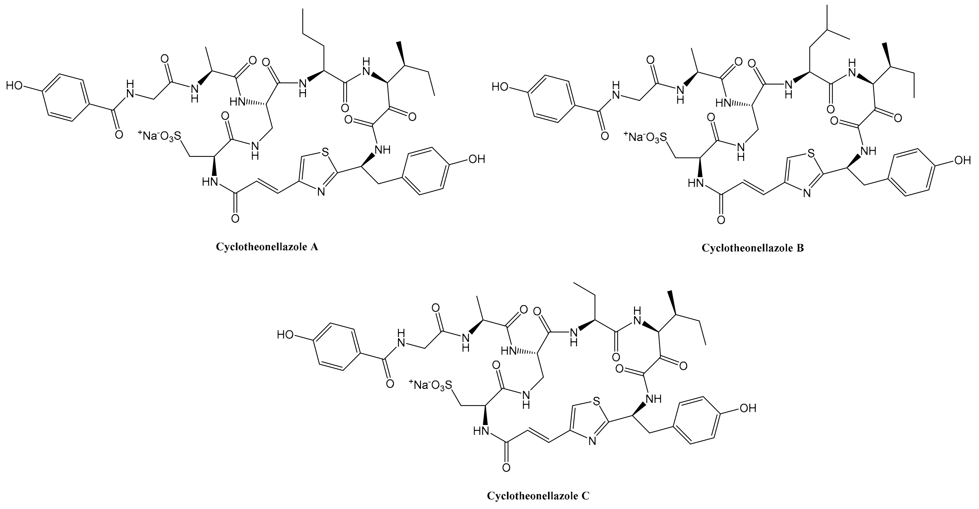
Peptides isolated from marine organisms often exhibit cyclic structures with substituted N- or C-termini. Therefore, they are relatively non-polar and easy to separate and purify from marine source. These compounds are often characterized by uncommon amino acids, including non-proteinogenic D-amino acids [44]. A huge number of bioactive secondary metabolites have been discovered from marine sponges of the genus Theonella, including peptides; Issac et al. isolated three novel cyclic peptides called cyclotheonellazoles A–C, in the sponge Theonella aff. Swinhoei from Dos de la Baleine, Madagascar [45]. They belong to the azole-homologated peptides (AHPs) that are characterized by the presence of azole-homologated amino acids. This class of peptides is produced by non-ribosomal synthase/polyketide synthase (NRPS-PKS) and are classified in two different types (I or II), depending on the presence of conserved tetrads of amino acids into the macrocyclic ring [44]. Cyclotheonellazole A (C44H54N9Na2O14S2), cyclotheonellazole B (C45H57N9NaO14S2) and cyclotheonellazole C (C43H52N9Na2O14S2) have 22 degrees of unsaturation, and their structures include few proteinogenic amino acids (two or three) and non-proteinogenic acids, e.g., 4-propenoyl-2-tyrosylthiazole (Ptt), 3-amino-4-methyl-2-oxohexanoic acid (Amoha) and diaminopropionic acid (Dpr). The 2-aminopentanoic acid contained in cyclotheonellazole A is replaced with leucine in cyclotheonellazole B and homoalanine in cyclotheonellazole C (Figure 14). These three compounds have the ability to inhibit elastase with IC50 values of 0.034 nM, 0.10 nM and 0.099 nM, respectively. The most effective among them against elastase activity is cyclotheonellazole A, probably due to a better interaction with the enzyme S2 subsite [45].
Cyclotheonellazole A was isolated in low yield from the natural source, so Cui et al. recently defined its chemical synthesis to further investigate its possible pharmaceutical development as lead compound. Moreover, they tested the synthesized Cyclotheonellazole A against human neutrophil elastase, showing an IC50 value of 0.321 µM. Therefore, they proved that this compound is a more effective inhibitor against elastase than sivelestat (IC50 = 0.704 µM), showing an inconsistent result compared with the previous study conducted by Issac et al.; this is probably due to different experimental conditions, such as the source and concentration of the enzyme, the substrate and the time of incubation [45,46]. Elastase has recently been considered as a potential target for the prevention of ARDS in patients afflicted with SARS2-CoV-2 infection [14]. The high elastase inhibitory activity of this compound could be particularly advantageous for patients with a D614G genotype infection [46].

7. Conclusions

A huge number of bioactive molecules have been isolated from natural sources [47,48]. Among them, distinctive peptides have been discovered so far from plants, fungi, animals, sponges and bacteria. Particularly important are peptide human elastase inhibitors that inhibit an enzyme able to lyse the main component (˃90%) of elastic fibres, called elastin [1,2]. The imbalance between human neutrophil elastase activity and its endogenous inhibitors can cause a broad range of diseases, such as chronic obstructive pulmonary disease, acute lung injury, acute respiratory distress syndrome and pulmonary fibrosis [12,13]. This is the main motivation of researchers to discover and isolate new and potent elastase inhibitors from natural sources. Furthermore, elastase has been recently considered as a potential target for the prevention of acute respiratory distress syndrome (ARDS) in COVID-19 patients and the possible use of neutrophil elastase inhibitors to prevent and to treat these disorders has been hypothesized. However, future clinical studies should be conducted to evaluate the effectiveness of elastase inhibitors on COVID-19 patients [14].
Peptide elastase inhibitors isolated from animal sources and fungi are often characterized by long and linear amino acid sequences [12,19,21,24]. Plants provide different types of peptide elastase inhibitors, such as the short peptide ixorapeptide II isolated from the MeOH extract of the aerial part of Ixora coccinea and the peptide roseltide rT1 from Hibiscus sabdariffa [41,42]. Bacterial sources provide cyclic peptides with peculiar residues that act as elastase inhibitors [26,28,29,30,31,35,36,37,38,40]. Marine sponges of the genus Theonella produce cyclotheonellazoles A–C, cyclic peptides belonging to azole-homologated peptides (AHPs) [44,45]. Despite the structural differences among them, these natural peptide elastase inhibitors show remarkable IC50 values in a range from nM to μM.
The future development of peptide analogues as HNE inhibitors would be a very innovative approach to fight a huge variety of disorders using very cheap, safe and easily produced compounds. Furthermore, the cyclic structure of the natural lead compounds could be advantageous for the design of novel pharmaceutical drugs because of their large surface area that is connected to their affinity and selectivity for the target; moreover, the restrained structure gives them less flexibility, enhancing their binding properties. Cyclic peptides are usually easily produced, modified and characterized and are considered to have a low toxicity [49].
In conclusion, the latest advancements on the isolation and characterization of peptide inhibitors against human neutrophil elastase will be the starting point for the future development of novel inhibitor drugs through the optimization of natural lead compounds and drug design processes. We consider the recent discoveries as a step forward to understand the structural features necessary to express the inhibitory activity against human neutrophil elastase and provide new commercial drugs against chronic obstructive pulmonary disease, acute lung injury, acute respiratory distress syndrome, pulmonary fibrosis and ARDS linked to COVID-19 infection.

Author Contributions

Conceptualization, L.M.; investigation, L.M. and G.S.; resources, G.S.; writing—original draft preparation, L.M., A.D.V. and A.S.; writing—review and editing, A.M., A.C. and G.Z.; supervision, A.M. All authors have read and agreed to the published version of the manuscript.

Funding

This research received no external funding.

Informed Consent Statement

Not applicable.

Data Availability Statement

Not applicable.

Conflicts of Interest

The authors declare no conflict of interest.

References

  1. Vrhovski, B.; Weiss, A.S. Biochemistry of Tropoelastin. Eur. J. Biochem. 1998, 258, 1–18. [Google Scholar] [CrossRef] [PubMed]
  2. Ross, R.; Bornstein, P. The Elastic Fiber I. The Separation and Partial Characterization of Its Macromolecular Components. J. Cell Biol. 1969, 40, 366–381. [Google Scholar] [CrossRef] [PubMed]
  3. Thomson, J.; Singh, M.; Eckersley, A.; Cain, S.A.; Sherratt, M.J.; Baldock, C. Fibrillin Microfibrils and Elastic Fibre Proteins: Functional Interactions and Extracellular Regulation of Growth Factors. Semin. Cell Dev. Biol. 2019, 89, 109–117. [Google Scholar] [CrossRef] [PubMed]
  4. Ozsvar, J.; Yang, C.; Cain, S.A.; Baldock, C.; Tarakanova, A.; Weiss, A.S. Tropoelastin and Elastin Assembly. Front. Bioeng. Biotechnol. 2021, 9, 643110. [Google Scholar] [CrossRef]
  5. Uitto, J. Biochemistry of the Elastic Fibers in Normal Connective Tissues and Its Alterations in Diseases. J. Investig. Dermatol. 1979, 72, 1–10. [Google Scholar] [CrossRef] [Green Version]
  6. Capurso, G.; Traini, M.; Piciucchi, M.; Signoretti, M.; Arcidiacono, P.G. Exocrine Pancreatic Insufficiency: Prevalence, Diagnosis, and Management. Clin. Exp. Gastroenterol. 2019, 12, 129–139. [Google Scholar] [CrossRef] [Green Version]
  7. Tóth, A.Z.; Szabó, A.; Hegyi, E.; Hegyi, P.; Sahin-Tóth, M. Detection of Human Elastase Isoforms by the ScheBo Pancreatic Elastase 1 Test. Am. J. Physiol.-Gastrointest. Liver Physiol. 2017, 312, G606–G614. [Google Scholar] [CrossRef]
  8. Taylor, S.; Dirir, O.; Zamanian, R.T.; Rabinovitch, M.; Thompson, A.A.R. The Role of Neutrophils and Neutrophil Elastase in Pulmonary Arterial Hypertension. Front. Med. 2018, 5, 217. [Google Scholar] [CrossRef]
  9. Donarska, B.; Łączkowski, K.Z. Recent Advances in the Development of Elastase Inhibitors. Future Med. Chem. 2020, 12, 1809–1813. [Google Scholar] [CrossRef]
  10. Crocetti, L.; Quinn, M.T.; Schepetkin, I.A.; Giovannoni, M.P. A Patenting Perspective on Human Neutrophil Elastase (HNE) Inhibitors (2014–2018) and Their Therapeutic Applications. Expert Opin. Ther. Pat. 2019, 29, 555–578. [Google Scholar] [CrossRef]
  11. Hajjar, E.; Broemstrup, T.; Kantari, C.; Witko-Sarsat, V.; Reuter, N. Structures of Human Proteinase 3 and Neutrophil Elastase—So Similar yet so Different. FEBS J. 2010, 277, 2238–2254. [Google Scholar] [CrossRef]
  12. Luan, N.; Zhao, Q.; Duan, Z.; Ji, M.; Xing, M.; Zhu, T.; Mwangi, J.; Rong, M.; Liu, J.; Lai, R. Identification and Characterization of ShSPI, a Kazal-Type Elastase Inhibitor from the Venom of Scolopendra Hainanum. Toxins 2019, 11, 708. [Google Scholar] [CrossRef] [Green Version]
  13. Rosenbloom, J.; Macarak, E.; Piera-Velazquez, S.; Jimenez, S.A. Human Fibrotic Diseases: Current Challenges in Fibrosis Research. In Fibrosis; Rittié, L., Ed.; Methods in Molecular Biology; Springer: New York, NY, USA, 2017; Volume 1627, pp. 1–23. ISBN 978-1-4939-7112-1. [Google Scholar]
  14. Mohamed, M.M.A.; El-Shimy, I.A.; Hadi, M.A. Neutrophil Elastase Inhibitors: A Potential Prophylactic Treatment Option for SARS-CoV-2-Induced Respiratory Complications? Crit. Care 2020, 24, 311. [Google Scholar] [CrossRef]
  15. Hansen, G.; Gielen-Haertwig, H.; Reinemer, P.; Schomburg, D.; Harrenga, A.; Niefind, K. Unexpected Active-Site Flexibility in the Structure of Human Neutrophil Elastase in Complex with a New Dihydropyrimidone Inhibitor. J. Mol. Biol. 2011, 409, 681–691. [Google Scholar] [CrossRef]
  16. The PyMOL Molecular Graphics System; Version 2.5.2; Schrödinger, LLC: Chiyoda, Tokyo, 2013.
  17. Aikawa, N.; Kawasaki, Y. Clinical Utility of the Neutrophil Elastase Inhibitor Sivelestat for the Treatment of Acute Respiratory Distress Syndrome. Ther. Clin. Risk Manag. 2014, 10, 621–629. [Google Scholar] [CrossRef] [Green Version]
  18. Sahebnasagh, A.; Saghafi, F.; Safdari, M.; Khataminia, M.; Sadremomtaz, A.; Talaei, Z.; RezaiGhaleno, H.; Bagheri, M.; Habtemariam, S.; Avan, R. Neutrophil Elastase Inhibitor (Sivelestat) May Be a Promising Therapeutic Option for Management of Acute Lung Injury/Acute Respiratory Distress Syndrome or Disseminated Intravascular Coagulation in COVID-19. J. Clin. Pharm. Ther. 2020, 45, 1515–1519. [Google Scholar] [CrossRef]
  19. Wan, H.; Lee, K.S.; Kim, B.Y.; Zou, F.M.; Yoon, H.J.; Je, Y.H.; Li, J.; Jin, B.R. A Spider-Derived Kunitz-Type Serine Protease Inhibitor That Acts as a Plasmin Inhibitor and an Elastase Inhibitor. PLoS ONE 2013, 8, e53343. [Google Scholar] [CrossRef]
  20. Mishra, M. Evolutionary Aspects of the Structural Convergence and Functional Diversification of Kunitz-Domain Inhibitors. J. Mol. Evol. 2020, 88, 537–548. [Google Scholar] [CrossRef]
  21. Jung, H.I.; Kim, S.I.; Ha, K.-S.; Joe, C.O.; Kang, K.W. Isolation and Characterization of Guamerin, a New Human Leukocyte Elastase Inhibitor from Hirudo Nipponia. J. Biol. Chem. 1995, 270, 13879–13884. [Google Scholar] [CrossRef] [Green Version]
  22. Chung, Y.-M.; El-Shazly, M.; Chuang, D.-W.; Hwang, T.-L.; Asai, T.; Oshima, Y.; Ashour, M.L.; Wu, Y.-C.; Chang, F.-R. Suberoylanilide Hydroxamic Acid, a Histone Deacetylase Inhibitor, Induces the Production of Anti-Inflammatory Cyclodepsipeptides from Beauveria Felina. J. Nat. Prod. 2013, 76, 1260–1266. [Google Scholar] [CrossRef]
  23. Ahmad, S.; Saleem, M.; Riaz, N.; Lee, Y.S.; Diri, R.; Noor, A.; Almasri, D.; Bagalagel, A.; Elsebai, M.F. The Natural Polypeptides as Significant Elastase Inhibitors. Front. Pharmacol. 2020, 11, 688. [Google Scholar] [CrossRef]
  24. Okumura, Y.; Matsui, T.; Ogawa, K.; Uchiya, K.; Nikai, T. Biochemical Properties and Primary Structure of Elastase Inhibitor AFUEI from Aspergillus Fumigatus. J. Med. Microbiol. 2008, 57, 803–808. [Google Scholar] [CrossRef] [Green Version]
  25. Fukui, Y.; Okumura, Y.; Uchiya, K.; Komori, Y.; Ogawa, K.; Nikai, T.; Hasegawa, Y. Biochemical and Cellular Activity of Chemically Synthesized Elastase Inhibitor (S-AFUEI) from Aspergillus Fumigatus. J. Mycol. Médicale 2019, 29, 345–351. [Google Scholar] [CrossRef]
  26. Mehner, C.; Müller, D.; Kehraus, S.; Hautmann, S.; Gütschow, M.; König, G.M. New Peptolides from the Cyanobacterium Nostoc Insulare as Selective and Potent Inhibitors of Human Leukocyte Elastase. ChemBioChem 2008, 9, 2692–2703. [Google Scholar] [CrossRef]
  27. Sisay, M.T.; Hautmann, S.; Mehner, C.; König, G.M.; Bajorath, J.; Gütschow, M. Inhibition of Human Leukocyte Elastase by Brunsvicamides A–C: Cyanobacterial Cyclic Peptides. ChemMedChem 2009, 4, 1425–1429. [Google Scholar] [CrossRef]
  28. Keller, L.; Canuto, K.M.; Liu, C.; Suzuki, B.M.; Almaliti, J.; Sikandar, A.; Naman, C.B.; Glukhov, E.; Luo, D.; Duggan, B.M.; et al. Tutuilamides A–C: Vinyl-Chloride Containing Cyclodepsipeptides from the Marine Cyanobacteria Schizothrix Sp. and Coleofasciculus Sp. with Potent Elastase Inhibitory Properties. ACS Chem. Biol. 2020, 15, 751–757. [Google Scholar] [CrossRef]
  29. Salvador, L.A.; Taori, K.; Biggs, J.S.; Jakoncic, J.; Ostrov, D.A.; Paul, V.J.; Luesch, H. Potent Elastase Inhibitors from Cyanobacteria: Structural Basis and Mechanisms Mediating Cytoprotective and Anti-Inflammatory Effects in Bronchial Epithelial Cells. J. Med. Chem. 2013, 56, 1276–1290. [Google Scholar] [CrossRef] [Green Version]
  30. Matthew, S.; Ross, C.; Rocca, J.R.; Paul, V.J.; Luesch, H. Lyngbyastatin 4, a Dolastatin 13 Analogue with Elastase and Chymotrypsin Inhibitory Activity from the Marine Cyanobacterium Lyngbya Confervoides. J. Nat. Prod. 2007, 70, 124–127. [Google Scholar] [CrossRef]
  31. Taori, K.; Matthew, S.; Rocca, J.R.; Paul, V.J.; Luesch, H. Lyngbyastatins 5–7, Potent Elastase Inhibitors from Floridian Marine Cyanobacteria, Lyngbya spp. J. Nat. Prod. 2007, 70, 1593–1600. [Google Scholar] [CrossRef]
  32. Luo, D.; Chen, Q.-Y.; Luesch, H. Total Synthesis of the Potent Marine-Derived Elastase Inhibitor Lyngbyastatin 7 and in Vitro Biological Evaluation in Model Systems for Pulmonary Diseases. J. Org. Chem. 2016, 81, 532–544. [Google Scholar] [CrossRef] [Green Version]
  33. Luo, D.; Luesch, H. Ahp-Cyclodepsipeptide Inhibitors of Elastase: Lyngbyastatin 7 Stability, Scalable Synthesis, and Focused Library Analysis. ACS Med. Chem. Lett. 2020, 11, 419–425. [Google Scholar] [CrossRef] [PubMed]
  34. Chen, Q.-Y.; Luo, D.; Seabra, G.M.; Luesch, H. Ahp-Cyclodepsipeptides as Tunable Inhibitors of Human Neutrophil Elastase and Kallikrein 7: Total Synthesis of Tutuilamide A, Serine Protease Selectivity Profile and Comparison with Lyngbyastatin 7. Bioorg. Med. Chem. 2020, 28, 115756. [Google Scholar] [CrossRef] [PubMed]
  35. Al-Awadhi, F.H.; Paul, V.J.; Luesch, H. Structural Diversity and Anticancer Activity of Marine-Derived Elastase Inhibitors: Key Features and Mechanisms Mediating the Antimetastatic Effects in Invasive Breast Cancer. ChemBioChem 2018, 19, 815–825. [Google Scholar] [CrossRef] [PubMed]
  36. Gunasekera, S.P.; Miller, M.W.; Kwan, J.C.; Luesch, H.; Paul, V.J. Molassamide, a Depsipeptide Serine Protease Inhibitor from the Marine Cyanobacterium Dichothrix Utahensis. J. Nat. Prod. 2010, 73, 459–462. [Google Scholar] [CrossRef]
  37. Müller, D.; Krick, A.; Kehraus, S.; Mehner, C.; Hart, M.; Küpper, F.C.; Saxena, K.; Prinz, H.; Schwalbe, H.; Janning, P.; et al. Brunsvicamides A−C: Sponge-Related Cyanobacterial Peptides with Mycobacterium Tuberculosis Protein Tyrosine Phosphatase Inhibitory Activity. J. Med. Chem. 2006, 49, 4871–4878. [Google Scholar] [CrossRef]
  38. Fujie, K.; Shinguh, Y.; Hatanaka, H.; Shigematsu, N.; Murai, H.; Fujita, T.; Yamashita, M.; Okamoto, M.; Okuhara, M. FR901277 A novel inhibitor of human leukocyte elastase from Streptomyces Resistomycificus Producing Organism, Fermentation, Isolation, Physico-Chemical and Biological Properties. J. Antibiot. 1993, 46, 908–913. [Google Scholar] [CrossRef]
  39. Nakanishi, I.; Kinoshita, T.; Sato, A.; Tada, T. Structure of Porcine Pancreatic Elastase Complexed with FR901277, a Novel Macrocyclic Inhibitor of Elastases, at 1.6 Å Resolution. Biopolymers 2000, 53, 434–445. [Google Scholar] [CrossRef]
  40. Yasumuro, K.; Suzuki, Y.; Shibazaki, M.; Teramura, K.; Abe, K. YM-47141 and YM-47142, New Elastase Inhibitors Produced by Flexibacter Sp. Q17897 I. Taxonomy, Fermentation, Isolation, Physico-Chemical Properties and Biological Activities. J. Antibiot. 1995, 48, 1425–1429. [Google Scholar] [CrossRef] [Green Version]
  41. Lee, C.-L.; Liao, Y.-C.; Hwang, T.-L.; Wu, C.-C.; Chang, F.-R.; Wu, Y.-C. Ixorapeptide I and Ixorapeptide II, Bioactive Peptides Isolated from Ixora Coccinea. Bioorg. Med. Chem. Lett. 2010, 20, 7354–7357. [Google Scholar] [CrossRef]
  42. Loo, S.; Kam, A.; Xiao, T.; Nguyen, G.K.T.; Liu, C.F.; Tam, J.P. Identification and Characterization of Roseltide, a Knottin-Type Neutrophil Elastase Inhibitor Derived from Hibiscus Sabdariffa. Sci. Rep. 2016, 6, 39401. [Google Scholar] [CrossRef] [Green Version]
  43. Kam, A.; Loo, S.; Dutta, B.; Sze, S.K.; Tam, J.P. Plant-Derived Mitochondria-Targeting Cysteine-Rich Peptide Modulates Cellular Bioenergetics. J. Biol. Chem. 2019, 294, 4000–4011. [Google Scholar] [CrossRef] [Green Version]
  44. Molinski, T.F. Cyclic Azole-Homologated Peptides from Marine Sponges. Org. Biomol. Chem. 2018, 16, 21–29. [Google Scholar] [CrossRef]
  45. Issac, M.; Aknin, M.; Gauvin-Bialecki, A.; De Voogd, N.; Ledoux, A.; Frederich, M.; Kashman, Y.; Carmeli, S. Cyclotheonellazoles A–C, Potent Protease Inhibitors from the Marine Sponge Theonella aff. Swinhoei. J. Nat. Prod. 2017, 80, 1110–1116. [Google Scholar] [CrossRef]
  46. Cui, Y.; Zhang, M.; Xu, H.; Zhang, T.; Zhang, S.; Zhao, X.; Jiang, P.; Li, J.; Ye, B.; Sun, Y.; et al. Elastase Inhibitor Cyclotheonellazole A: Total Synthesis and In Vivo Biological Evaluation for Acute Lung Injury. J. Med. Chem. 2022, 65, 2971–2987. [Google Scholar] [CrossRef]
  47. Mollica, A.; Locatelli, M.; Stefanucci, A.; Pinnen, F. Synthesis and Bioactivity of Secondary Metabolites from Marine Sponges Containing Dibrominated Indolic Systems. Molecules 2012, 17, 6083–6099. [Google Scholar] [CrossRef] [Green Version]
  48. Mollica, A.; Stefanucci, A.; Feliciani, F.; Lucente, G.; Pinnen, F. Synthesis of (S)-5,6-dibromo-tryptophan derivatives as building blocks for peptide chemistry. Tetrahedron Lett. 2011, 52, 2583–2585. [Google Scholar] [CrossRef]
  49. Zorzi, A.; Deyle, K.; Heinis, C. Cyclic Peptide Therapeutics: Past, Present and Future. Curr. Opin. Chem. Biol. 2017, 38, 24–29. [Google Scholar] [CrossRef] [Green Version]
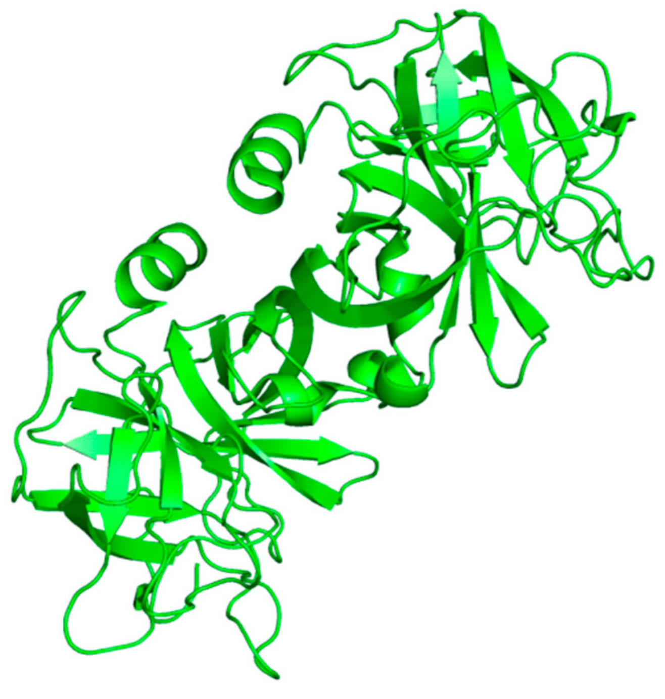
Figure 1. Structure of free human neutrophil elastase (HNE) from the RCSB Protein Data Bank (PDB-ID: 3Q76) [15,16].
Ijms 23 02924 g001
Scheme 1. Mechanism of action of serine proteases [11].
Ijms 23 02924 sch001
Figure 2. The solution structure of ShSPI (NMR ensemble) from the RCSB Protein Data Bank (PDB-ID: 6LF5) [12,16].
Ijms 23 02924 g002
Figure 3. Structures of Desmethylisaridin C2, isaridin E, isaridin C2 and roseocardin.
Ijms 23 02924 g003
Figure 4. Structures of Lyngbyastatin 4 and Lyngbyastatin 7.
Ijms 23 02924 g004
Figure 5. Structures of Tutuilamides A–C.
Ijms 23 02924 g005
Figure 6. Structures of Loggerpeptins A–C and Molassamide.
Ijms 23 02924 g006
Figure 7. Structures of Symplostatins 5–10.
Ijms 23 02924 g007
Figure 8. Structures of Insulapeptolides A–H.
Ijms 23 02924 g008
Figure 9. Structures of Brunsvicamides A–C.
Ijms 23 02924 g009
Figure 10. Structure of FR901277.
Ijms 23 02924 g010
Figure 11. Structures of YM-47141 and YM-47142.
Ijms 23 02924 g011
Figure 12. Structures of Ixorapeptide I and Ixorapeptide II.
Ijms 23 02924 g012
Figure 13. Structure of Roseltide rT1 (NMR ensemble) from the RCSB Protein Data Bank (PDB-ID: 5GSF) [16,42].
Ijms 23 02924 g013
Figure 14. Structures of Cyclotheonellazoles A–C.
Ijms 23 02924 g014
Table 1. IC50 values of Desmethylisaridin C2, Isaridin E, Isaridin C2 and Roseocardin in vitro on formyl-L-methionyl-L-leucyl-L-phenylalanine (FMLP)-induced human neutrophils [22].
CompoundsIC50 (μM)
Desmethylisaridin C210.01 ± 0.46
Isaridin E12.76 ± 1.00
Isaridin C212.12 ± 0.72
Roseocardin15.09 ± 0.28
Table 2. IC50 values of Insulapeptolides A–H for the inhibition of human leukocyte elastase (HLE) [26].
CompoundsIC50 (μM)
Insulapeptolide A0.14 ± 0.01
Insulapeptolide B0.10 ± 0.01
Insulapeptolide C0.090 ± 0.001
Insulapeptolide D0.085 ± 0.004
Insulapeptolide E3.2 ± 0.2
Insulapeptolide F1.6 ± 0.1
Insulapeptolide G3.5 ± 0.1
Insulapeptolide H2.7 ± 0.1
Publisher’s Note: MDPI stays neutral with regard to jurisdictional claims in published maps and institutional affiliations.