Article

The In Vitro Cytotoxicity of Eremothecium oil and Its Components—Aromatic and Acyclic Monoterpene Alcohols

1
Institute of Biochemical Technology, Ecology and Pharmacy, V.I. Vernadsky Crimean Federal University, 295007 Simferopol, Russia
2
Institute for Regenerative Medicine, Sechenov University, 119991 Moscow, Russia
3
Mazlumov All-Russian Research Institute of Sugar Beet and Sugar, 396030 Voronezh, Russia
4
Institute for Applied Synthetic Chemistry, Vienna University of Technology, Getreidemarkt 9/163, A-1060 Vienna, Austria
5
Faculty of Biology, Lomonosov Moscow State University, 119234 Moscow, Russia
6
World-Class Research Center “Digital Biodesign and Personalized Healthcare”, Sechenov University, 119991 Moscow, Russia
7
Chemistry Department, Lomonosov Moscow State University, 119991 Moscow, Russia
*
Author to whom correspondence should be addressed.
Int. J. Mol. Sci. 2022, 23(6), 3364; https://doi.org/10.3390/ijms23063364
Submission received: 29 January 2022 / Revised: 14 March 2022 / Accepted: 15 March 2022 / Published: 20 March 2022
(This article belongs to the Special Issue 25th Anniversary of IJMS: Advances in Biochemistry)

Abstract

:
The microscopic fungi Eremothecium ashbyi and E. gossypii are known for their ability to synthetize essential oil, which has a composition similar to that of rose oil. The development of Eremothecium oil technology enables the production of rose-scented products, which are demanded by pharmaceutical, food, and perfumery industries. This study focuses on assessing the in vitro cytotoxicity of Eremothecium oil, in comparison with that of rose oil, using a combination of methods and two cell types (3T3 mouse fibroblast cell line and bone-marrow-derived mesenchymal stromal cells (BM-MSCs)). The Eremothecium oil samples possessed cytotoxic effects that varied among strains and batches. The revealed cytotoxicity level may be used to tailor the qualitative and quantitative composition of Eremothecium oil to achieve a particular quality in its end products. These results require further analysis using other cell types and assays based on measuring other cell functions.

1. Introduction

Rose essential oil is highly demanded and has a wide range of applications in the pharmaceutical, food, and perfumery industries. However, the quantity of this oil is limited because of a shortage of raw materials, and the rose oil industry is mainly localized in Bulgaria and Turkey. Rose oil manufacturers are trying to solve this problem by improving farming techniques, using drip irrigation systems, and adding agrochemicals to the soil. This has led to a significant rise in the flower yield [1,2,3]. Nevertheless, rose oil production remains cost-intensive, mainly because of its laboriousness and time limitations. Harvesting rose flowers is the most labor-consuming part of the process and one of the decisive factors that influences the rose oil price [3]. Thus, there has been an increasing interest in biotechnological sources of fragrant products, especially rose-scented ones [4,5].
In 1986, Bugorskiy et al. revealed that homothallic ascomycetes—Eremothecium ashbyi Guillermond and E. gossypii (Ashby et Nowell) Kurtzman (synonym for Ashbya gossypii (Ashby et Nowell) Guilliermond)—were able to excrete rose-scented essential oil, which had a composition similar to that of natural rose essential oil [6,7]. They showed that it was possible to produce this oil by submerging E. ashbyi and E. gossypii cultures, achieving a yield of up to 5.7 g/L. Therefore, Eremothecium oil can be considered as a cheap high-grade alternative that can help to reduce the industry consumption of rose oil.
To promote Eremothecium oil applications, it should be extensively analyzed. This includes testing its activity and safety. Previously, we showed the antimicrobial effects of essential oils synthesized by E. ashbyi and E. gossypii in comparison with rose oil and its main components [8]. We revealed that the Eremothecium oil’s antibiotic activity against Escherichia coli, Pseudomonas aeruginosa, Myxococcus sp., Lactobacillus acidophilus, Lactococcus lactis ssp. lactis, Stenotrophomonas maltophilia, Acinetobacter baumannii, Klebsiella pneumoniae, Staphylococcus aureus, Bacillus subtilis, B. megatherium, and Candida albicans was similar to that of essential oil from rose petals. These effects were ensured by both its individual components and the synergism of their combination. The analysis of the Eremothecium oil’s bacteriostatic activity revealed reliable positive correlations between the level of growth suppression and lag phase lengthening at a concentration range from 0.49 μL/mL to 7.81 μL/mL (R = 0.73; p < 0.05), as well as between the level of growth suppression of multidrug-resistant strains (K. pneumoniae, P. aeruginosa, and A. baumannii), and total monoterpene alcohol concentration (R = 1.0; p < 0.05) [9].
Moreover, there are data on the Eremothecium oil toxicity assessed using only Paramecium caudatum [10]. It was shown that the toxicity of Eremothecium oil at a concentration higher than 860 μg/mL reached almost 100%, while the average lethal concentration was 210 μg/mL. The severity of the effects correlated with the concentrations of phenylethanol (R = −0.9; strong negative relationship), geraniol (R = 0.6; moderate positive relationship), nerol (R = −0.55; moderate negative relationship), linalool (R = −0.74; strong negative relationship), and the total monoterpene alcohol concentration (R = 0.5; moderate positive connection) in samples [11].
Nevertheless, the lack of cytotoxicity data can be obviously observed, and more findings are urgently required, especially using human and mammalian cell cultures. Thus, this study aims to reveal the in vitro cytotoxicity of Eremothecium oil and its components in comparison with that of rose oil, using a combination of methods and two cell types (3T3 mouse fibroblast cell line and mesenchymal stromal cells (MSCs)).

2. Results

The GS analysis of the component composition of Eremothecium and rose oils is presented in Table 1. The tested samples of oil produced by E. ashbyi, except EO-03, had low variations in component composition. Compared to them, the oil produced by E. gossypii had a high concentration of 2-phenylethanol; however, it did not exceed that of both rose oil samples. Interestingly, EO-03 had the lowest concentration of 2-phenylethanol, which can be explained by strain-determined synthetic activity. The highest concentration of monoterpene alcohols was achieved during the culturing of E. ashbyi strain VKM F-3009.
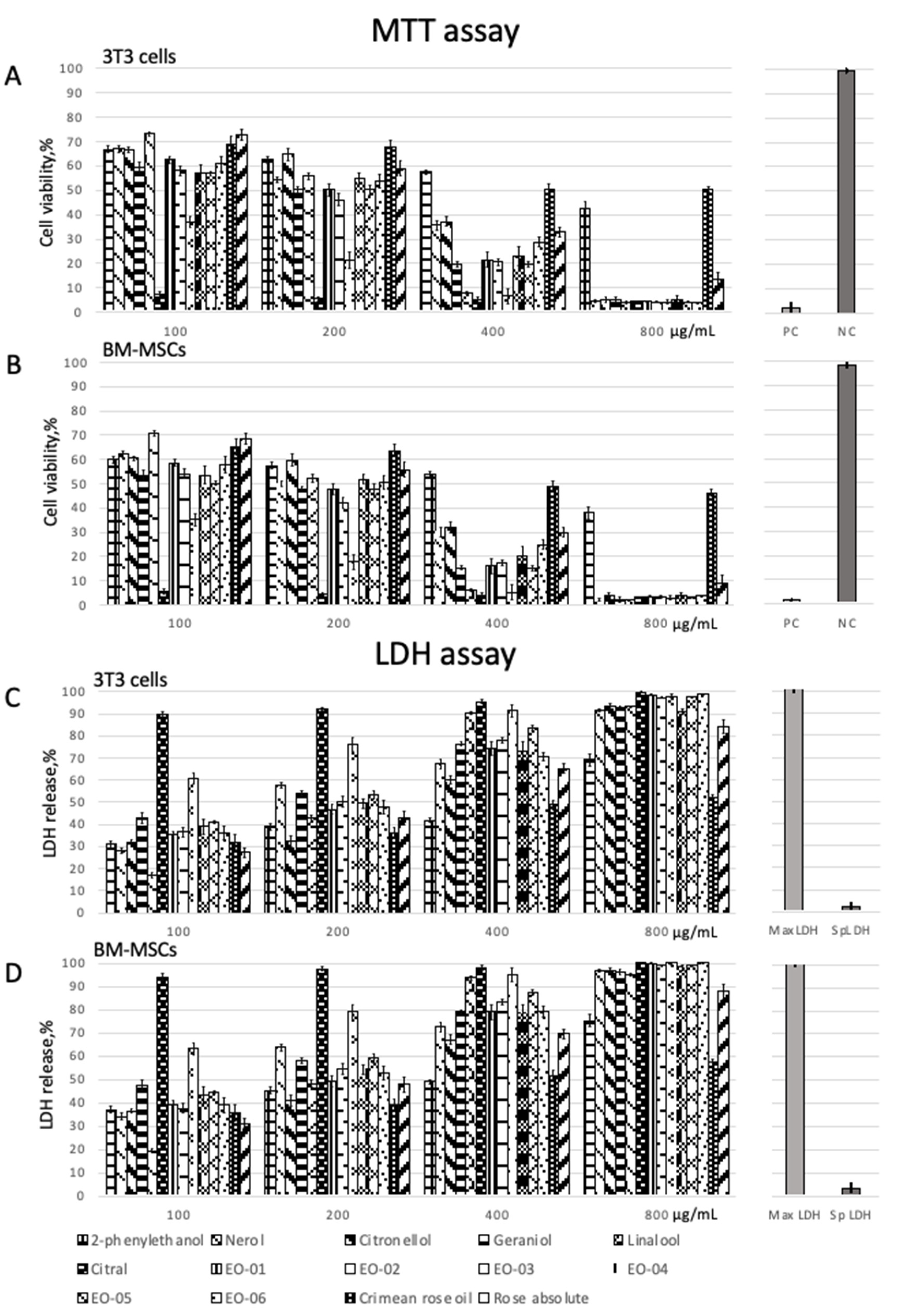
The MTT and LDH assay showed that all oil samples and their individual compounds were cytotoxic and caused a dose-dependent response in both tested cell cultures (Table 2 and Table 3, Figure 1 and Figure 2). The achieved data enabled us to calculate the toxicity parameters, such as LC50 and IC50 (Table 4). Both 3T3 cells and BM-MSCs were highly susceptible to the samples tested and controls; however, BM-MSCs showed higher sensitivity than 3T3 cells. Results of the MTT assay were in high correspondence with those of the LDH assay.
The lowest cytotoxicity for both 3T3 cells and BM-MSCs was revealed in testing Crimean rose oil, which had the highest percentage of 2-phenylethanol. For all samples, the cytotoxicity of Eremothecium oil was revealed to be higher than that of rose oil and similar to that of individual compounds. Despite differences in component composition, all Eremothecium oil samples had a similar level of cytotoxicity. However, having the minimum 2-phenylethanol content (4.40%), EO-03 caused the lowest cell viability among them. The highest cell viability was revealed for cells treated with EO-06, which contained the maximum percentage of 2-phenylethanol (35.89%). This may be due to the low toxicity of 2-phenylethanol compared to other compounds (Table 2 and Table 3, Figure 2).

3. Discussion

There is a growing interest in using essential oils as a promising antimicrobial agent, which could possess significant effects and potentiate the action of antibiotics [12]. Their hydrophobic components can enter into the periplasm through porin proteins on the outer membrane [13,14]. By accumulating on the cell membrane, essential oils disrupt it, which leads to an increase in membrane permeability for protons and other ions, causing a shift in the intercellular pH homeostasis. Moreover, in some cases, components of essential oils can change the conformation of proteins of the membrane [15]. These effects were shown for both non-toxic (0.05 μL/mL–0.5 μL/mL) and cytotoxic (≥3.125 μL/mL) concentrations [16,17]. It should be noted that the biological activity of essential oils is ensured by a specific combination of their components, which may possess even higher efficiency than individual compounds [18]. Therefore, approaches to regulate oil biosynthesis are highly demanded. As can be observed from Table 1, Eremothecium oil biotechnology provides a possibility to tune the content of particular components, e.g., 2-phenylethanol and monoterpene alcohols.
Here, we tried not only to evaluate the in vitro cytotoxicity of the Eremothecium oil samples but also to reveal the possible impact of their individual compounds on the observed effects. We showed that the cell cultures used (3T3 cells and BM-MSCs) were highly susceptible compared to those of animals (data from safety datasheets, GOST 30333-2007, and EU 1907/2006). We used two methods to test cytotoxicity: MTT and LDH assays. The first method is based on the formation of non-water-soluble colored formazan crystals due to the NAD-dependent oxidation of tetrazolium bromide ((3-(4,5-dimethylthiazol-2-yl)-2,5-diphenyltetrazolium bromide); MTT). This method mostly reflects the cells’ metabolic activity and has several limitations. Particularly, the cell responses may overlap, and the MTT oxidation in cells may be enhanced because of the adaptation to toxic agents [19]. Moreover, the formed formazan crystals cannot be properly dissolved, leading to errors in the assessed cytotoxicity level. Therefore, to adequately estimate in vitro cytotoxicity, we also used the second method—LDH assay; the released LDH is inversely proportional to the number of damaged cells. The achieved results had high correspondence with each other. 3T3 cells are one of the standard cell lines, and BM-MSCs are a common primary cell culture that are used to assess cytotoxicity. The cell viability, after treating with samples, significantly varied compared to parameters regulated by ISO 10993-5: 2009 (except citral).

4. Materials and Methods

Reagents. 2-phenylethanol, linalool, nerol, citronellol, geraniol, citral (including their GC analytical standards), and solvents were purchased from Sigma-Aldrich (Taufkirchen, Germany), rose absolute sample was purchased from Neal’s Yard Remedies (Dorset, UK), and Crimean rose oil sample was purchased from Krymskaya Rosa (Simferopol, Ukraine).
Fermentation and oil extraction. E. ashbyi (VKM F-3009, VKM F-4565, and VKM F-4566) and E. gossypii (VKM F-3296) strains were stored on potato dextrose, Sabouraud’s, and Czapek’s media [20,21,22]. To prepare the inoculum, we used the glucose–peptone liquid medium (glucose—7.5 g/L; peptone—4.0 g/L; sodium succinate—2.0 g/L; potassium phosphate dibasic—0.5 g/L; inositol—0.14 g/L; pH 6.5) and cultured it for 48 h at a temperature of 20–24 °C. Then, we seeded the fermentation medium (saccharose—10.0 g/L; soybean meal—20.0 g/L; pH 7.0) using the prepared inoculum at a concentration of 1–5% and cultured for 48–72 h at a temperature of 20–24 °C. We extracted Eremothecium oil with hexane as a solvent, which was removed using a rotor vacuum evaporator.
Oil analysis. Samples were analyzed by gas–liquid chromatography using a chromatograph Clarus 680 (PerkinElmer, Waltham, USA) equipped with a capillary polar column (INNOWAX®, 60 m × 0.32 mm) according to GOST ISO 7609:2014 “Essential oils—Analysis by gas chromatography on capillary columns—General method”. The column was kept at a temperature of 80°C for 5 min, and then the temperature was gradually increased up to 250 °C at a rate of 2 °C/min. The samples (0.2 μL) were injected at a temperature of 250 °C, and compounds were detected at a temperature of 250 °C using a flame ionization detector. Helium was used as a carrier gas at a flow rate of 0.5 mL/min. The main volatile compounds (geraniol, citronellol, nerol, 2-phenylethanol, linalool, and citral) were identified using retention times of their GC analytical standards, and their mass fractions were calculated.
Cell cultures. 3T3 mouse fibroblast cell lines and bone-marrow-derived mesenchymal stromal cells (BM-MSCs) were kindly provided by the Biobank at Sechenov University (Moscow, Russia). 3T3 cells were cultured in DMEM/F12 (Invitrogen, Carlsbad, USA) medium supplemented with penicillin (100 μg/mL), streptomycin (100 μg/mL), gentamycin (50 μg/mL), and fetal bovine serum (5%, FBS, HyClone, Logan, USA). BM-MSCs were cultured in DMEM/F12 medium (Invitrogen, USA) supplemented with L-glutamine (5 mg/mL, Gibco, USA), insulin–transferrin–sodium selenite (ProSpec, Israel), basic fibroblast growth factor (bFGF) (20 ng/mL, ProSpec, Israel), and gentamycin (50μg/mL, Paneco, Russia), as well as 10% FBS, under standard conditions (37 °C; 5% CO2).
MTT assay. The oil samples were diluted down to concentrations of 100, 200, 400, and 800 µg/mL using dimethyl sulfoxide (DMSO) and culture medium as described elsewhere [23]. The maximum DMSO concentration did not exceed 0.5%. 3T3 cells and BM-MSCs were seeded into 96-well cell culture plates (TPP, Switzerland) at a concentration of 2 × 10⁴ cells per well and were cultured for 24 h [24]. Then, the medium was removed from the wells, and 100 μL of either the sample or sodium dodecyl sulfate (SDS) (positive control) solution was added. The cells were incubated for 24 h. Changes in cell morphology caused by the sample cytotoxicity were revealed; however, these data were not used in the quantitative analysis. The supernatant was carefully removed, and MTT (0.5 mg/mL) solution was pipetted into each well. Plates were incubated for 2 h. The formazan crystals that formed were diluted by adding 100 µL DMSO. The plates were placed onto a plate shaker and then measured using a microplate reader Titertek Multiscan (Flow Laboratories, Finland) at a wavelength of 570 nm (reference wavelength 650 nm). The cell viability was calculated as follows:
Viability ,   %   = 100 × O D 570 e O D 570 b   ,
where OD570e—optical density value when the sample was added; OD570b—optical density value of non-treated cells.
Parameters such as LC50 (median lethal concentration) and IC50 (half-maximal inhibitory concentration) were calculated according to MR 1.1.726-98.
Lactate Dehydrogenase assay. The prepared dilutions of Eremothecium and rose oils, as well as their components, were tested using Pierce LDH Cytotoxicity assay kit (Thermo Scientific, Waltham, USA) in accordance with a manufacturer’s instructions. 3T3 cells and BM-MSCs were seeded into plates as described above and cultured for 24 h. LDH activity was measured after adding a substrate at a wavelength of 492 nm using a microplate reader Titertek Multiscan (Flow Laboratories, Helsinki, Finland). Cells treated with 1% Triton-X100 in phosphate-buffered saline (PBS) were used as a positive control, and non-treated cells as a negative control (spontaneous LDH release). Results were calculated in percent relative to the positive control.
Statistical analysis. All experiments were performed at least three times, and the results presented are those from single experiments yielding similar results to the triplicate experiments. Each data point shows the mean ± standard deviation. The analysis was conducted using ANOVA; the differences were assumed to be statistically significant if the p-value was less than 0.05 [25].

5. Conclusions

The approach described in this study can be successfully used to achieve preliminary results and screen the activity of essential oils. The assessed in vitro cytotoxicity, using a combination of methods and two cell types, may be applied to tune the qualitative and quantitative composition of Eremothecium oil samples. It can also be used to improve the quality of the end products. These findings should be verified using other cell types and assays based on measuring other cell functions (e.g., PicoGreen), including cell cycle and apoptosis.

Author Contributions

Conceptualization and methodology, E.S.; investigation, E.S. and A.S.; writing—original draft preparation, E.S. and N.M.; writing—review and editing, V.K. and V.P.; supervision, A.K. and W.L.; project administration, A.G. All authors have read and agreed to the published version of the manuscript.

Funding

Assessing cell cytotoxicity was financed by the Ministry of Science and Higher Education of the Russian Federation within the framework of state support for the creation and development of World-Class Research Centers "Digital biodesign and personalized healthcare" (agreement no. 075-15-2020-926).

Institutional Review Board Statement

Not applicable.

Informed Consent Statement

Not applicable.

Data Availability Statement

All data generated are included in the manuscript.

Acknowledgments

This study was carried out using the unique scientific facility, Transgenebank.

Conflicts of Interest

The authors declare no conflict of interest. The funder had no role in the design of the study, in the collection, analyses, or interpretation of data, in the writing of the manuscript, or in the decision to publish the results.

References

  1. Erdogan, G. Turkey rose oil production and marketing: A review on problem and opportunities. J. Appl. Sci. 2005, 5, 1871–1875. [Google Scholar]
  2. Nedkov, N.; Dobreva, A.; Kovacheva, N.; Bardarov, V.; Velcheva, A. Bulgarian rose oil of white oil-bearing rose. Bulg. J. Agric. Sci. 2009, 15, 318–322. [Google Scholar]
  3. Kovacheva, N.; Rusanov, K.; Atanassov, I. Industrial cultivation of oil bearing rose and rose oil production in Bulgaria during 21st century, directions and challenges. Biotechnol. Biotechnol. 2010, 2, 1793–1798. [Google Scholar] [CrossRef] [Green Version]
  4. Bomgardner, M.M. The sweet smell of microbes. Chem. Eng. New 2012, 29, 25–29. [Google Scholar] [CrossRef]
  5. Christen, P. Producción de aromas porfermentación en medio solido. Tópicos Investig. Posgrado 1995, 2, 102–109. [Google Scholar]
  6. Bugorski, P.S.; Semenova, E.F.; Rodov, V.S. Effect of hydrogen, potassium and sodium ions on the productivity of the Eremothecium ashbyi fungus. Microbiol. J. 1990, 3, 44–47. (In Russian) [Google Scholar]
  7. Bugorski, P.S.; Semenova, E.F. Fragrances produced by filamentous fungus Ashbya gossypii. Chem. Nat. Compd. 1991, 3, 428. (In Russian) [Google Scholar]
  8. Semenova, E.F.; Shpichka, A.I.; Presniakova, E.V. Pharmaceutical Mycology of Eremothecium Species; PSU Publishing House: Penza, Russia, 2013. (In Russian) [Google Scholar]
  9. Markelova, N.N.; Semenova, E.F. Sensitivity of nonfermentative gram-negative bacteria to essential oils of different origin. Microbiology 2017, 86, 610–617. [Google Scholar] [CrossRef]
  10. Semenova, E.F.; Shpichka, A.I.; Presniakova, E.V.; Presniakova, V.S.; Goncharov, M.A.; Goncharov, D.A. Development of a novel biotechnological fragrant product, Eremothecium oil. Indian J. Pharm. Educ. Res. 2017, 3, 136–138. [Google Scholar] [CrossRef] [Green Version]
  11. Niles, A.L.; Moravec, R.A.; Worzella, T.J.; Evans, N.J.; Riss, T.L. High-Throughput Screening Methods in Toxicity Testing; Steinberg, P., Ed.; John Wiley & Sons, Inc.: Hoboken, NJ, USA, 2013; pp. 107–127. [Google Scholar]
  12. Bakkali, F.; Averbeck, S.; Averbeck, D.; Idaomar, M. Biological effects of essential oils—a review. Food Chem. Toxicol. 2008, 2, 446–475. [Google Scholar] [CrossRef]
  13. May, J.; Chan, C.H.; King, A.; Williams, L.; French, G.L. Time–kill studies of tea tree oils on clinical isolates. J. Antimicrob Chemother. 2000, 5, 639–643. [Google Scholar] [CrossRef] [PubMed]
  14. Trombetta, D.; Castelli, F.; Sarpietro, M.G.; Venuti, V.; Cristani, M.; Daniele, C. Mechanisms of antibacterial action of three monoterpenes. Antimicrob. Agents Chemother. 2005, 6, 2474–2478. [Google Scholar] [CrossRef] [PubMed] [Green Version]
  15. Choi, H.W.; Lee, B.G.; Kim, N.H.; Park, Y.; Lim, C.W.; Song, H.K.; Hwang, B.K. A role for a menthone reductase in resistance against microbial pathogens in plants. Plant Physiol. 2008, 1, 383–401. [Google Scholar] [CrossRef] [PubMed] [Green Version]
  16. Brooke, J.S. Stenotrophomonas maltophilia: An emerging global opportunistic pathogen. Clinil. Microbiol. Rev. 2012, 1, 2–41. [Google Scholar] [CrossRef] [Green Version]
  17. Giamarellou, H. Multidrug-resistant Gram-negative bacteria: How to treat and for how long. Intern. J. Antimicrob. Agents 2010, 36, 50–54. [Google Scholar] [CrossRef] [Green Version]
  18. Markelova, N.N.; Semenova, E.F. Possible ways of overcoming the antibiotic resistance of nosocomial patogens Klebsiella pneumoniae, Acinetobacter baumannii, Pseudomonas aeruginosa, Stenotrophomonas maltophilia. Antibiot. Chemother. 2018, 11–12, 45–54. (In Russian) [Google Scholar]
  19. Sikkema, J.; De Bont, J.A.; Poolman, B. Mechanisms of membrane toxicity of hydrocarbons. Microbiol. Rev. 1995, 2, 201–222. [Google Scholar] [CrossRef]
  20. Semenova, E.; Presnyakova, V.; Goncharov, D.; Goncharov, M.; Presnyakova, E.; Presnyakov, S.; Moiseeva, I.; Kolesnikova, S. Spectrophotometric method for quantitative measuring essential oil in aromatic water and distillate with rose smell. J. Phys. Conf. Ser. 2017, 1, 012053. [Google Scholar] [CrossRef] [Green Version]
  21. Semenova, E.F.; Presniakova, V.S.; Kurakov, A.V.; Bezrukova, E.I. Composition and production rates of essential oil during the submerged cultivation of Eremothecium ashbyi and E. gossypii. Biotechnology 2020, 2, 12–15. (In Russian) [Google Scholar] [CrossRef]
  22. Semenova, E.F.; Shpichka, A.I.; Presniakova, E.V. Aromatic and monoterpene alcohol accumulation by Eremothecium ashbyi strains differing in riboflavinogenesis. Appl. Biochem. Microbiol. 2017, 3, 374–380. [Google Scholar] [CrossRef]
  23. Shakeri, A.; Khakdan, F.; Soheili, V.; Sahebkar, A.; Rassam, G.; Asili, J. Chemical composition, antibacterial activity, and cytotoxicity of essential oil from Nepeta ucrainica L. spp. kopetdaghensis. Ind. Crops Prod. 2014, 58, 315–321. [Google Scholar] [CrossRef]
  24. Shpichka, A.; Koroleva, A.; Kuznetsova, D.; Dmitriev, R.I.; Timashev, P. Fabrication and handling of 3D scaffolds based on polymers and decellularized tissues. In Multi-Parametric Live Cell Microscopy of 3D Tissue Models; Springer: Berlin/Heidelberg, Germany, 2017; Volume 1035, pp. 71–81. [Google Scholar]
  25. Platonov, A.E. Statistical Analysis in Medicine and Biology: Aims, Terminology, Logic, Computer Methods; RAMS Publishing House: Moscow, Russia, 2000; pp. 7–42. (In Russian) [Google Scholar]
Figure 1. 3T3 cells and BM-MSCs treated with Eremothecium oil samples. The top panel is non-treated cells (control). Phase-contrast microscopy. Scale bar = 30 µm.
Ijms 23 03364 g001
Figure 2. Viability of 3T3 cells (A) and BM-MSCs (B) treated with Eremothecium and rose oils, and their components (PC—positive control (SDS)); NC—negative control (non-treated cells). LDH release from 3T3 cells (C) and BM-MSCs (D) treated with Eremothecium and rose oils, and their components (MaxLDH—maximum LDH release (Triton X); SpLDH—spontaneous LDH release (non-treated cells)).
Ijms 23 03364 g002
Table 1. Components of essential oils produced by E. ashbyi and E. gossypii (GS analysis).
Strain, SpeciesSamplePEA, %Monoterpene Alcohols, %
GeraniolCitronellolNerolLinaloolTotal
VKM F-3009, E.a.EO-0113.2368.845.663.990.3478.83
VKM F-3009, E.a.EO-0213.8574.665.643.600.2584.15
VKM F-4565, E.a.EO-034.4043.305.501.850.0152.15
VKM F-4566, E.a.EO-0416.3267.176.224.020.8378.24
VKM F-4566, E.aEO-0515.8461.038.246.970.0176.24
VKM F-3296, E. g.EO-0635.8941.346.906.591.0855.91
Rosa gallicaCrimean rose oil 71.7715.803.905.900.1225.72
Rosa damascenaRose absolute67.335.628.543.500.2517.91
Note. E. a.E. ashbyi; E. g.—E. gossypii; PEA—phenylethyl alcohol.
Table 2. Viability of cells treated with Eremothecium and rose oils and their components. MTT assay (3T3/MSCs).
Concentration, μg/mLCell Viability, %
Sample100200400800
2-phenylethanol66.70 ± 1.3862.50 ± 1.5057.45 ± 0.9642.69 ± 2.62
60.06 ± 2.0857.43 ± 1.8753.87 ± 1.3438.02 ± 1.98
Nerol67.03 ± 1.3854.40 ± 1.2935.72 ± 2.134.27 ± 0.56
62.34 ± 1.7449.98 ± 2.3230.16 ± 1.872.43 ± 0.69
Citronellol66.88 ± 1.0864.77 ± 2.4737.26 ± 2.025.21 ± 1.18
60.45 ± 1.7859.79 ± 2.0332.07 ± 2.514.01 ± 1.24
Geraniol59.38 ± 2.3750.28 ± 1.2819.74 ± 0.945.03 ± 1.16
53.46 ± 2.1247.85 ± 1.0915.42 ± 1.832.10 ± 0.96
Linalool73.20 ± 1.0055.77 ± 1.548.00 ± 0.644.01 ± 0.45
70.76 ± 1.7952.34 ± 2.046.08 ± 0.911.98 ± 0.89
Citral7.38 ± 1.335.49 ± 0.745.13 ± 1.004.29 ± 0.29
5.67 ± 1.234.49 ± 0.984.12 ± 1.643.23 ± 0.54
EO-0162.54 ± 1.5050.21 ± 2.4021.51 ± 3.104.38 ± 0.34
58.64 ± 1.8747.65 ± 2.0916.27 ± 2.173.47 ± 0.39
EO-0258.07 ± 1.9946.19 ± 2.3520.61 ± 1.244.11 ± 0.40
54.06 ± 1.6542.19 ± 2.0417.56 ± 1.713.33 ± 0.69
EO-0337.05 ± 2.4521.31 ± 3.316.57 ± 2.904.21 ± 0.70
35.18 ± 1.6917.76 ± 1.875.32 ± 1.243.09 ± 0.76
EO-0457.21 ± 3.5055.02 ± 2.1023.09 ± 4.085.38 ± 1.18
53.61 ± 2.7451.89 ± 1.8619.96 ± 2.784.03 ± 1.56
EO-0556.98 ± 0.8050.20 ± 2.1019.45 ± 1.104.16 ± 0.35
50,18 ± 1,2147,71 ± 1,3215,40 ± 1,753,18 ± 0,59
EO-0660.90 ± 3.2053.90 ± 2.8028.89 ± 2.104.01 ± 0.18
57.83 ± 2.0350.67 ± 1.9424.91 ± 1.833.87 ± 0.67
Crimean rose oil68.65 ± 3.4068.02 ± 2.5050.58 ± 2.1050.43 ± 1.30
65.21 ± 2.4363.74 ± 2.0348.83 ± 1.9946.21 ± 3.52
Rose absolute72.93 ± 2.1059.10 ± 3.0933.01 ± 1.9613.23 ± 3.20
68.54 ± 1.8155.92 ± 2.4729.97 ± 1.589.15 ± 2.32
Table 3. LDH release in cells treated with Eremothecium and rose oils, and their components (3T3/MSCs).
Concentration,
μg/mL
LDH Release, %
Sample100200400800
2-phenylethanol31.02 ± 0.8739.15 ± 2.7141.79 ± 1.5469.18 ± 2.03
37.12 ± 0.9245.18 ± 1.2349.02 ± 1.0175.32 ± 2.94
Nerol28.17 ± 0.9657.93 ± 1.5367.32 ± 1.9891.54 ± 1.12
34.01 ± 1.2763.72 ± 0.6572.65 ± 1.1296.89 ± 2.01
Citronellol31.96 ± 1.4732.65 ± 2.3960.19 ± 3.1093.46 ± 1.88
36.52 ± 1.9141.07 ± 1.5467.12 ± 2.4797.19 ± 3.20
Geraniol43.10 ± 2.1554.21 ± 1.3476.34 ± 1.1292.57 ± 1.66
47.34 ± 2.8958.13 ± 1.7879.21 ± 1.9796.22 ± 1.09
Linalool16.99 ± 1.1942.85 ± 1.7890.59 ± 1.5493.18 ± 2.70
19.33 ± 1.5648.17 ± 1.8793.71 ± 2.0395.05 ± 2.54
Citral89.76 ± 1.9492.21 ± 1.6295.40 ± 1.3199.81 ± 0.79
94.28 ± 2.0197.70 ± 0.7998.13 ± 1.34102.34 ± 2.02
EO-0135.53 ± 1.3346.87 ± 1.7174.29 ± 2.4998.63 ± 0.67
39.18 ± 1.7749.19 ± 1.2979.05 ± 1.2399.98 ± 0.96
EO-0236.54 ± 1.4350.19 ± 2.0178.32 ± 1.5697.34 ± 0.96
37.63 ± 1.0854.76 ± 0.9683.33 ± 1.4199.01 ± 1.01
EO-0360.92 ± 1.8776.18 ± 2.3391.29 ± 1.1798.03 ± 0.64
63.10 ± 1.5579.09 ± 2.0395.14 ± 1.76100.21 ± 0.93
EO-0438.96 ± 1.9049.51 ± 1.6773.38 ± 3.2191.12 ± 1.94
43.21 ± 1.0453.99 ± 1.8878.13 ± 2.0598.36 ± 1.45
EO-0541.08 ± 1.0853.39 ± 1.1383.45 ± 1.6597.59 ± 1.83
44.50 ± 0.9659.04 ± 1.5987.62 ± 1.2399.19 ± 2.32
EO-0636.00 ± 2.0647.82 ± 1.1970.61 ± 1,5499.07 ± 1.19
39.02 ± 1.2852.90 ± 3.0379.27 ± 0.94100.31 ± 1.18
Crimean rose oil31.95 ± 1.5936.07 ± 1.8348.99 ± 2.9052.32 ± 1.69
35.68 ± 1.3239.06 ± 0,9951.82 ± 2,1657.23 ± 1.33
Rose absolute27.39 ± 1.7843.19 ± 2.1965.34 ± 1.6784.19 ± 2.01
31.03 ± 1.3747.82 ± 1,9369.71 ± 1.0187.90 ± 1.75
Table 4. In vitro toxicity parameters of Eremothecium and rose oils and their components (aromatic and monoterpene alcohols) (vs. in vivo *1–4).
SampleLC50, μg/mLLC50 *3, μg/mLIC50, μg/mLEC50 *4, μg/mLLD50 *, μg/mL
Orally 1Dermally 2
2-phenylethanol568.74<464.00150287.0–490.01600>2000
Nerol243.3120.320032.44500>5000
Citronellol299.9114.6622517.4834502650
Geraniol157.543.45–22.001507.75–13.13600>5000
Linalool216.1827.8015059.0–88.327905610
Citral-6.78756.8–103.83450–68002000–2250
EO-01139.21-125---
EO-02185.27-150---
EO-0312.02-5---
EO-04182,63-165---
EO-05144.78-125---
EO-06212.71-150---
Crimean rose oil707.82-300-<126003000
Rose absolute303.33-150->50002500
Note. *—according to the safety datasheets, GOST 30333-2007, and EU 1907/2006. LC50—median lethal concentration; IC50—half-maximal inhibitory concentration; EC50—half-maximal effective concentration; LD50—median lethal dose. 1—rat; 2—rabbit; 3—fish; 4—daphnia.
Publisher’s Note: MDPI stays neutral with regard to jurisdictional claims in published maps and institutional affiliations.