Review

The Morpho-Molecular Landscape of Spitz Neoplasms

by
Carlo Alberto Dal Pozzo
1 and
Rocco Cappellesso
2,*
1
Surgical Pathology and Cytopathology Unit, Department of Medicine (DIMED), University of Padua, 35121 Padua, Italy
2
Pathological Anatomy Unit, University Hospital of Padua, 35121 Padua, Italy
*
Author to whom correspondence should be addressed.
Int. J. Mol. Sci. 2022, 23(8), 4211; https://doi.org/10.3390/ijms23084211
Submission received: 28 February 2022 / Revised: 5 April 2022 / Accepted: 7 April 2022 / Published: 11 April 2022
(This article belongs to the Special Issue Tumor Microenvironment from a Precision Medicine Perspective 2.0)

Abstract

:
Spitz neoplasms are a heterogeneous group of melanocytic proliferations with a great variability in the histological characteristics and in the biological behavior. Thanks to recent discoveries, the morpho-molecular landscape of Spitz lineage is becoming clearer, with the identification of subtypes with recurrent features thus providing the basis for a more solid and precise tumor classification. Indeed, specific mutually exclusive driver molecular events, namely HRAS or MAP2K1 mutations, copy number gains of 11p, and fusions involving ALK, ROS, NTRK1, NTRK2, NTRK3, MET, RET, MAP3K8, and BRAF genes, correlate with distinctive histological features. The accumulation of further molecular aberrations, instead, promotes the increasing malignant transformation of Spitz neoplasms. Thus, the detection of a driver genetic alteration can be achieved using the appropriate diagnostic tests chosen according to the histological characteristics of the lesion. This allows the recognition of subtypes with aggressive behavior requiring further molecular investigations. This review provides an update on the morpho-molecular correlations in Spitz neoplasms.

1. Introduction

In 1948, the American pathologist Sophie Spitz published a landmark case series of melanocytic proliferations called “juvenile melanomas” or “melanomas of childhood” characterized by a combination of distinctive architectural and cytological features in association with peculiar epidermal changes (Figure 1) [1,2]. Such lesions are symmetric, often showing a dome-shaped, wedge-shaped or plaque-like silhouette, with sharp lateral borders and maturation towards the deep part [1,2]. The pattern of growth is predominantly nested. Nests may display varying cellularity, size, and shape are arranged in parallel to the rete ridges [1,2]. The overlying epidermis is hyperplastic and may be separated by the junctional nests and single melanocytes by clefts [1,2]. Pagetoid spread of the melanocytes into the epidermis, when present, usually occurs in bundles or nests of cells [1,2]. A variable number of small to large dull eosinophilic globules composed of amorphous filaments (so-called Kamino bodies) may be scattered throughout [1,2]. There is a predominance of enlarged epithelioid and/or spindle melanocytes with abundant eosinophilic or amphophilic cytoplasm with “ground-glass” appearance, round to oval to spindle nuclei with finely dispersed chromatin and distinct nucleoli [1,2]. Nuclear pseudoinclusions and multinucleated melanocytes may be encountered. Most lesions are amelanotic or paucimelanotic [1,2]. The mitotic activity is usually low. The inflammatory cell infiltrate is generally perivascular and dispersed throughout. These melanocytic proliferations typically occurred on the extremities of children and young adults and, despite the tendency to loco-regional nodal involvement, were hallmarked by an indolent clinical course, thus justifying their distinction from the adult melanoma in which the prognosis was dismal [1,3]. With time, it became evident that using these histological criteria it was possible to distinguish a group of benign melanocytic lesions – then named Spitz nevus (SN)–from melanoma and that these may be present also in adults [4].
However, cases occur that are more difficult to differentiate from melanoma because showing SN characteristics along with worrisome histological features, such as increased size, asymmetry, epidermal ulceration, lack of maturation, solid growth, diffuse pagetoid spread, hypodermic extension, marked cytological atypia, increased mitotic activity, deep and atypical mitoses. In 1959 Albert Bernard Ackerman recognized that melanocytic proliferations with Spitz histology encompassed a broad morphological spectrum of neoplasms ranging from completely benign, namely SN, to their fully malignant counterpart, namely malignant Spitz tumor (MST), passing through an intermediate category, namely atypical Spitz tumor (AST; Table 1) [5,6,7]. It must be highlighted that many MST diagnoses are achieved in the context of known synchronous metastasis or are initially defined as AST and classified as fully malignant after the detection of distant metastasis during the clinical follow-up. Indeed, it is well known that distinguishing AST from MST histologically is very difficult and at times impossible, even with the aid of common ancillary analyses. Spitz neoplasms may present at any age and at any site but most frequently affect the lower extremities and the face of patients under the age of 30 [8]. They are quite infrequent, accounting for about 1% of all resected melanocytic lesions and with an estimated annual incidence of little more than 1 case per 100,000 [8,9]. AST do not exceed the 6–8% of the number of SN and MST are very rare [10]. In the footsteps of Ackermann, the term “Spitzoid” was coined referred to melanocytic neoplasms sharing (at least some of) the distinctive SN features. This has led to confusion in the classification of melanocytic lesions since the term Spitzoid has been applied even to lesions with only the epidermal modifications typical of SN or to those with a small subpopulation of enlarged epithelioid and/or spindle melanocytes.
Until a few years ago, the genetic determinants of Spitz neoplasms development were largely unknown [11,12,13]. Thanks to the recent discoveries on the molecular landscape of the melanocytic Spitz lineage, the taxonomy of these tumors and the understanding of their biological behavior is becoming clearer [14]. Overall, both nevi and melanomas share the activation of some growth-promoting signaling pathways (considered the driver molecular events), mainly PI3K-AKT and RAF-MEK1/2-ERK1/2 [15,16]. In benign nevi these are almost the only molecular aberrations present [15,16]. In malignant melanomas, instead, there is also a variable number of additional molecular alterations (considered the promoting molecular events) able to block tumor-suppression mechanisms and to trigger further oncogenic signals, such as 9p21 deletion, 6p25 copy number gain, TP53 mutations, and TERT-promoter mutation [15,17,18,19] Thus, the driver molecular events seem to determine the specific histotype of each melanocytic neoplasm, while the type and amount of promoting aberrations seem to define their morphological and clinical aggressiveness [15,16]. According to this interpretation, the neoplasms regarded as melanocytic tumors of uncertain malignant potential result from the combination of the same driver molecular alterations of nevi and melanomas with a limited number of additional promoting genetic events determining a malignant potential lower than that of full-blown melanoma [15,16]. This seems to be true also in the Spitz setting. Indeed, the drivers of most common nevi and malignant melanomas are BRAF and NRAS activating mutations, but these alterations are virtually absent in Spitz neoplasms [20,21,22,23,24]. Instead, these harbor oncogenic HRAS or MAP2K1 mutations or kinase gene fusions involving ALK, BRAF, MET, NTRK1, NTRK2, NTRK3, RET, ROS1, and MAP3K8 in a mutually exclusive pattern [5,11,12,13,25,26,27,28,29,30,31,32,33,34,35,36,37]. The integration of such molecular data in the histology-based tumor classification of melanocytic neoplasms provides a way to identify the true Spitz neoplasms in the heterogeneous group of Spitzoid lesions [14]. Moreover, the existence of consistent genotype-phenotype relationships among the different subtypes of Spitz neoplasms (Table 2) can be used for the selection of the appropriate ancillary analyses to support a histological diagnosis of AST or MST and to better assess their malignant risk.
This review resumes the state-of-art in the knowledge of genotype-phenotype correlations in the field of Spitz neoplasms, especially focusing on subtypes harboring HRAS or MAP2K1 mutations, copy number gains of 11p, or fusions involving ALK, ROS, NTRK1, NTRK2, NTRK3, MET, RET, MAP3K8, and BRAF genes.

2. Spitz Neoplasms with HRAS Mutations or 11p Copy Number Gains

The RAS proto-oncogene family comprises three members, namely KRAS, NRAS, and HRAS, respectively located on the short arm (p) of chromosome 12, 1, and 11, and encoding the proteins KRAS4A, KRAS4B, NRAS, and HRAS [38,39]. These proteins are implicated in the signal transduction from the cell surface to the nucleus through the PI3K-AKT and RAF-MEK1/2-ERK1/2 pathways stimulating growth, differentiation, proliferation, and survival of the cell [40,41]. Missense single nucleotide point mutations usually occur in hotspot regions of the RAS active site leading to the production of aberrant proteins able to trigger downstream signaling without the need of extracellular cues [39]. Interestingly, the oncogenic role of RAS seems to be histotype-specific since different tumors are related to mutation of a precise RAS isoform [40]. This is particularly evident in melanocytic lesions where common nevi and malignant melanomas harbor almost exclusively NRAS mutations while Spitz neoplasms HRAS mutations [12,22,24]. Compared to the other RAS isoforms, HRAS appears to have a higher affinity for the PI3K-AKT pathway that is believed to be responsible for conferring the enlarged epithelioid or spindle phenotype to the melanocytes in Spitz neoplasms [42,43,44]. Copy number gains of the 11p region encompassing HRAS leads to overexpression of the protein product with similar results. Activating HRAS mutations and copy number gains of 11p occur in an exclusive or concurrent way in about 20% of Spitz neoplasms [22,45,46,47,48]. Most HRAS mutations commonly involve the codons 59–61 in exon 3 (mainly Q61R and Q61L) and rarely affect the codons 12 and 13 in exon 2 (mainly G13R) [5,13,22,48,49,50,51,52,53,54,55,56]. The detection of HRAS mutation requires sequencing analysis (classic or NGS), while FISH or CGH analysis is needed for the identification of 11p copy number alterations. The monoclonal antibody SP174 recognizes with high sensitivity the RAS Q61R mutant protein; unfortunately, however, it cross-reacts with both KRAS, NRAS, and HRAS isoforms resulting useless from a diagnostic point of view [57,58] Antibodies against the wild type HRAS protein may be of aid in highlighting Spitz neoplasms with HRAS overexpression due to 11p copy number gains.
Spitz neoplasms with HRAS aberrations tend to be predominantly intradermal symmetric lesions with infiltrative base characterized by epithelioid and spindle cells with abundant eosinophilic or amphophilic cytoplasm and slightly to moderately pleomorphic vesicular nuclei, intermingled with thick collagen bundles (desmoplasia) (Figure 2) [12,13,48]. Marked cytological atypia can be present in about 40% of HRAS-mutated cases [33]. Usually, mitoses are rare and not atypical. However, it must be highlighted that, on one hand, not all the Spitz neoplasms with these genomic alterations are desmoplastic and, on the other hand, desmoplasia has been reported also in Spitz neoplasms with ROS1, ALK, and BRAF gene fusions [13,59].
Most Spitz neoplasms with activating HRAS mutations and/or copy number gains of 11p are readily recognizable as SN, but cases occur with AST features; the prognosis is favorable [22,46,60,61].

3. Spitz Neoplasms with ALK Fusions

ALK resides on chromosome 2p and encodes a tyrosine kinase receptor involved in the PI3K-AKT, RAF-MEK1/2-ERK1/2, and JAK3-STAT3 pathways [62,63,64,65]. The reported proportion of cases with ALK fusions among SN and AST ranges from 10% to 20%, but it is restricted to approximately 1% of Spitz melanomas [66]. The ALK most frequent fusion partners are TPM3 and DCTN1 and it has been proposed that these rearrangements, compared with other fusions involved in the pathogenesis of Spitz neoplasms, lead to a very delayed oncogene-induced senescence resulting in large lesions [5,13,61,66,67,68,69,70,71,72]. Other recurrent fusion partners of ALK in the field of Spitz neoplasms include: MLPH, MYO5A, CLIP1, DDX3Y, KANK1, EEF2, GTF3C2, NPM1, PPFIBP1, SPTAN1, and TPR [68,69,70,73,74,75]. ALK immunohistochemistry with the monoclonal antibodies D5F3 and 5A4 serves as excellent surrogate for ALK fusions and is indicated in the appropriate morphological setting [11,66,70]. The expression is diffuse, strong, and granular in the cytoplasm of the melanocytes and may be present or not in the membrane or the nucleus [11,66,70]. NGS and FISH are the appropriate molecular techniques for the detection of the ALK fusions.
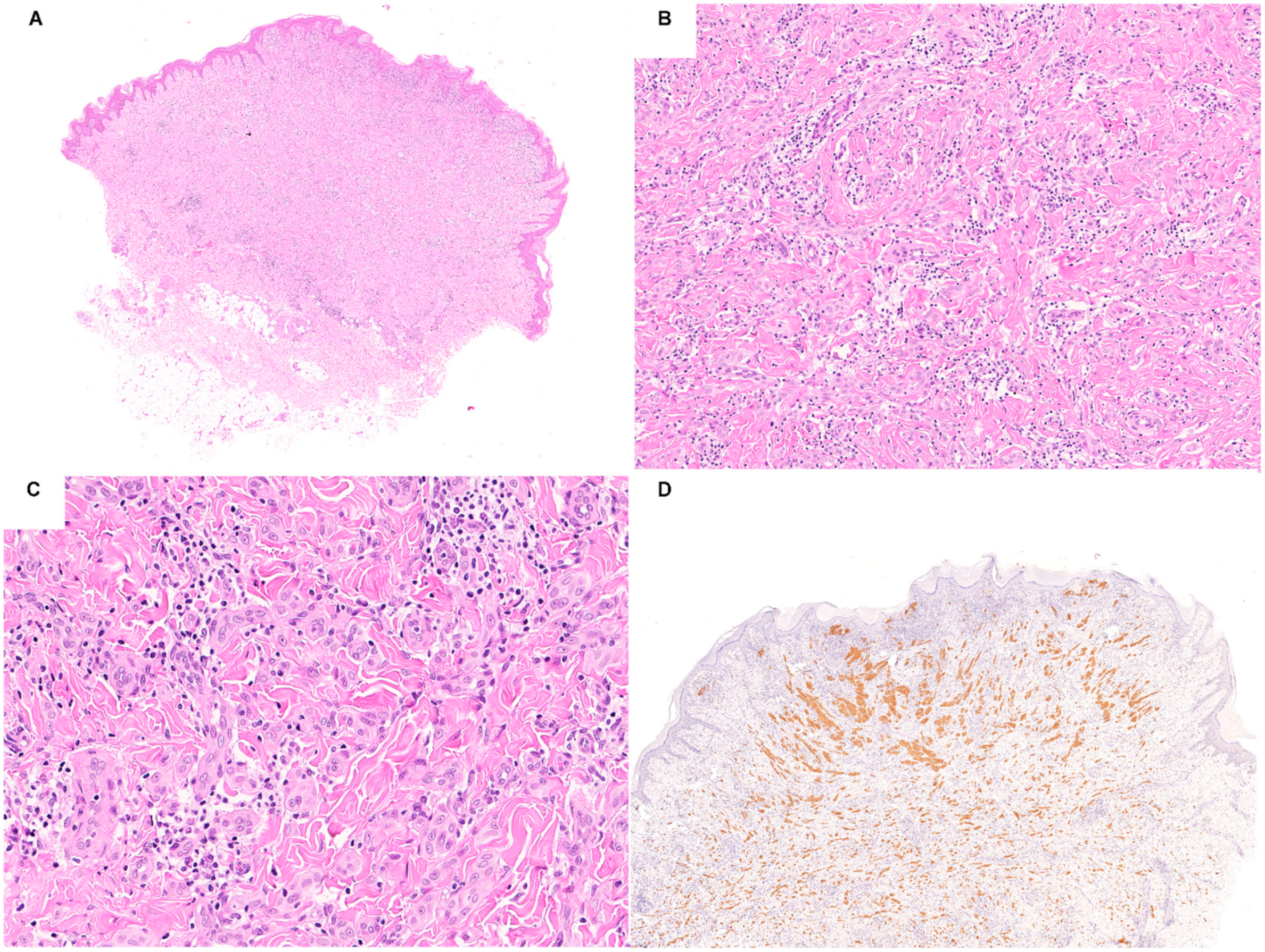
Clinically, ALK-fused Spitz neoplasms tend to be large and solitary papules or nodules arising on the extremities of young patients [3,11,70]. Histologically, most of these lesions share a distinctive pattern substantiated by a compound wedge shape silhouette with a bulbous and/or infiltrative base (Figure 3) [11,71,76]. The presence of non-pigmented, large, spindle melanocytes with pericellular clefts, amphophilic cytoplasm, vesicular nuclei, and prominent nucleoli, growing in plexiform intersecting fascicles, appears as a hallmark of ALK fusions [11,33,71,76]. Nevertheless, it is important to note that, although the described pattern is extremely sensitive in the prediction of ALK fusions, it is not equally specific. Other molecular subtypes of Spitz neoplasms may show overlapping features, such as NTRK1-fused cases [76]. Another pattern reported to be quite characteristic for ALK-fused Spitz neoplasm is the angiomatoid one [33]. The epidermis is often hyperplastic and pagetoid spread is usually absent [11]. Nuclear pleomorphism is usually mild and rarely moderate [11,33,71,76]. Worrisome features, such as ulceration, deep mitoses, and perineural invasions have been described [11,68,69,70,77]. Of note, combined Spitz neoplasms are more commonly ALK-fused [33].

4. Spitz Neoplasms with ROS1 Fusions

ROS1 proto-oncogene is located on the long arm (q) of chromosome 6 and encodes a tyrosine kinase receptor implicated in the PI3K-AKT, RAF-MEK1/2-ERK1/2, and JAK3-STAT3 pathways [78]. According to the analysis of large series of Spitz neoplasms, ROS1 fusions were found in 7–17% of cases [13,31]. Among these lesions, several fusion partners were reported, with PWWP2A (37% of cases) and TPM3 (31% of cases) being the most common in the series by Gerami et al. [59]. Other less common fusion partners of ROS1 among Spitz neoplasms are: PPFIBP1, CLIP1, ERC1, FIP1L1, HLA-A, MYH9, ZCCHC8, CAPRIN1, KIAA1598, MYH9, and MYO5A [5,13,59,73,79]. Immunohistochemistry with monoclonal antibody D4D6 against ROS1 is a fast, low-cost, and well-performing screening test for the identification of ROS1-fused Spitz neoplasms. Indeed, it showed 100% sensitivity and specificity when compared with FISH [80]. Although different ROS1 immunohistochemical patterns have been observed, such as diffuse or sparse granular cytoplasmic staining, dot-like staining, and nuclear staining, no specific correlations with the cellular localization of the various ROS1 fusions have been found [80]. In addition to FISH, NGS can be used to identify ROS1 fusions.
Clinically, ROS1-fused Spitz neoplasms are pink to red papules distributed throughout the body, mainly occurring in young adults of both sexes [59]. Histologically, there are not distinctive features specifically associated with ROS1 fusions. Indeed, many characteristics are shared with other molecular subtypes of Spitz neoplasms, particularly with those NTRK1-fused [59]. Nevertheless, a certain histological signature is enriched since most of the reported ROS1-fused lesions show a compound plaque-like or nodular silhouette with prominent expansile junctional nesting, with possible adnexal involvement and transepidermal elimination, composed of pure spindle melanocytes or of mixed spindle and epithelioid melanocytes, with mild to moderate nuclear pleomorphism, evidence of maturation, lack of pigmentation, associated with numerous Kamino bodies [13,33,59,79]. Recently, plexiform and angiomatoid patterns have been reported as more common in ROS1-fused Spitz neoplasms than in other molecular subtypes [33]. Cell sizes range from intermediate to large [13,59]. Mitoses may be not uncommon [79]. As above mentioned, cases occur in which ROS1 fusions have been found in desmoplastic SN [59].
Of great interest, in all published series, the presence of ROS1 fusions was associated with favorable outcome (no recurrence, no distant metastasis, and negative sentinel lymph node biopsy), although having been identified in cases diagnosed as MST [13]. No adverse events were observed in the available follow ups; thus, it is entirely reasonable to admit that ROS1-fused Spitz neoplasms usually have an indolent course [59].

5. Spitz Neoplasms with NTRK Fusions

The NTRK proto-oncogene family comprises three members, namely NTRK1, NTRK2, and NTRK3, respectively located on chromosomes 1q, 9q, and 15q and encoding the cell surface receptor tyrosine kinase proteins TRKA, TRKB, and TRKC (collectively referred as TRK proteins) involved in the PI3K-AKT, RAF-MEK1/2-ERK1/2, and PLCγ1 pathways [81,82,83]. These receptors are normally expressed in the nervous system and can be activated by the binding with several ligands, such as NGF, BDNF, and NT-3/4 [81]. TRK activation determines the autophosphorylation of the intracellular tyrosine residues and consequently the transmission of the signal through different pathways regulating the transcription of genes involved in neuronal survival and differentiation [81]. Fusions involving the NTRK gene family results in the production of TRK chimeric proteins with oncogenic properties since they couple constitutive expression with ligand-independent activation provided by the kinase domain preservation [83].
Among Spitz neoplasms, both NTRK1, NTRK2, and NTRK3 fusions have been reported, but NTRK1 alterations are by far the most prevalent [5,11,13,27,28,29,30,33,68,73,76,83,84,85,86,87,88].The partners of NTRK1 so far identified are LMNA, TPM3, TP53, and KHDRBS1 [13,28,73,88]. Those of NTRK3 are ETV6, MYO5A, MYH9, and SQSTM1 [27,29,73,83,85,88]. The only NTRK2 fusion identified in Spitz neoplasms has TFG as partner [30]. The frequency of NTRK fusions in Spitz neoplasms is approximately 10% [88].
Current guidelines for NTRK fusion detection in solid tumors state that a two-step testing approach must be followed [89]. Firstly, cases must be immunohistochemically screened using the monoclonal antibody EPR17341 that reacts against a C-terminal epitope conserved in wild-type and in all the chimeric TRK proteins (hence the definition of pan-TRK immunohistochemistry) [89,90,91]. Secondly, all the immunohistochemically positive cases must be analyzed using RNA-based NGS to prove the NTRK fusion [89]. This algorithm has been tested on a large AST series confirming the reliability of pan-TRK immunohistochemistry as screening test [88]. However, the application of RNA-based NGS to verify the presence of the gene fusion showed some weakness and it has been suggested to also perform FISH in all pan-TRK positive cases not confirmed by NGS analysis [88]. The staining pattern of pan-TRK can also provide information about the probable underlying NTRK fusion. Indeed, strong and diffuse nuclear immunostaining is quite specific for ETV6-NTRK3 fusion, while linear immunostaining in dendritic processes of the melanocytes directs towards MYO5A-NTRK3 fusion [29].

5.1. Spitz Neoplasms with NTRK1 Fusions

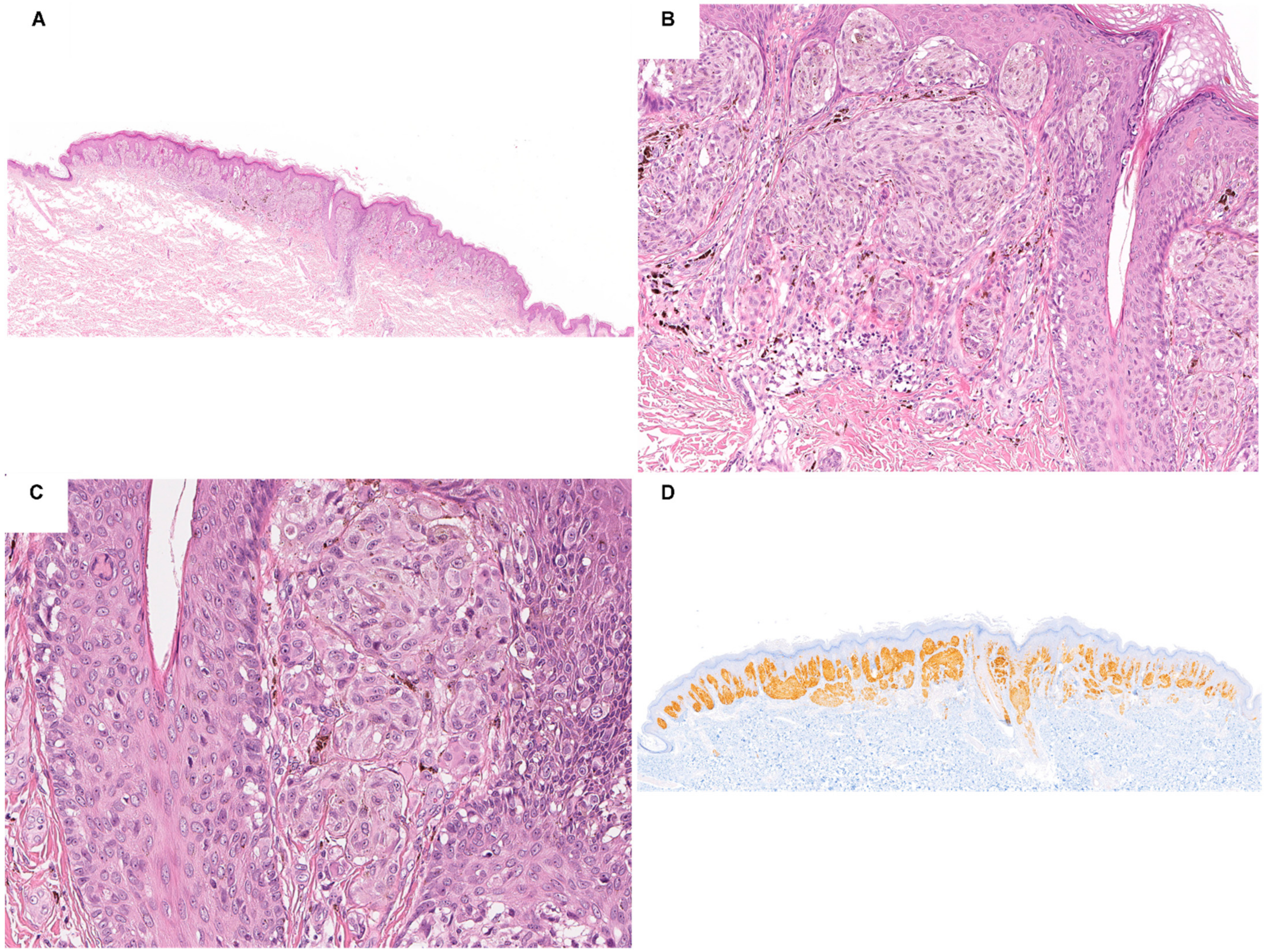
Most Spitz neoplasms harboring NTRK1 fusions are compound or dermal exophytic and symmetric lesions with thin and elongated rete ridges (filigree-like rete ridges), flat-base or wedge shape silhouette, lentiginous proliferation, lobulated nests, rosettes-like structures, and exaggerated maturation of epithelioid and/or spindle melanocytes with mild to moderate nuclear pleomorphism (Figure 4) [5,11,28,33]. Kamino bodies are frequently encountered, while mitoses are rare [5,11,13,28,33,76]. Like ALK-fused Spitz neoplasms, a plexiform pattern characterized by intersecting fascicles of spindle melanocytes can be observed in some cases [76].

5.2. Spitz Neoplasms with NTRK2 Fusions

The only reported NTRK2-fused SN belonged to histological variant of pigmented spindle cell nevus (aka nevus of Reed). The lesion is junctional, with hyperplastic epidermis, large nests with peripheral clefts composed of pigmented spindle melanocytes with abundant eosinophilic cytoplasm and elongated or oval nuclei [30]. Neither nuclear pleomorphism nor mitoses are observed [30]. Kamino bodies are widely distributed [30].

5.3. Spitz Neoplasms with NTRK3 Fusions

NTRK3-fused Spitz neoplasms are mostly compound or dermal, with epidermal hyperplasia and dome-shaped silhouette [29]. Depending on the underlying fusions, the lesions show different morphology. Indeed, cases with ETV6-NTRK3 fusions are predominantly composed of epithelioid melanocytes with distinct cell borders, abundant eosinophilic cytoplasm, and pleomorphic nuclei, arranged in large coalescing and lobulated nests [29,33]. The constituent melanocytes of MYO5A-NTRK3-fused lesions, instead, are homogeneously spindled and organized in a fascicular to plexiform growth pattern [29]. Palisading resembling Verocay bodies and rosettes-like structures are occasionally seen [29]. Lesions with MYH9-NTRK3 fusion are characterized by moderately large epithelioid melanocytes syncytially arranged with central desmoplastic stroma and peripheral collagen trapping [29].
The prognosis of Spitz neoplasms harboring NTRK1 or NTRK3 fusions is invariably favorable. Although rare cases of neoplastic cellular deposits in regional lymph nodes occur, no distant metastases and adverse outcomes have been reported so far [68,86].

6. Spitz Neoplasms with RET Fusions

RET proto-oncogene is located on chromosome 10q and encodes a tyrosine kinase receptor implicated in the PI3K-AKT, RAF-MEK1/2-ERK1/2, and PLCγ1 pathways [13,92]. RET fusions have been found in about 3–4% of Spitz neoplasms with the partner genes CCDC6, KIF5B, LMNA, GOLGA5, and MYO5A [13,31,85,93]. For their detection NGS or FISH analysis is required.
The few RET-fused Spitz neoplasms so far described are mainly compound symmetric lesions with epidermal hyperplasia, a plaque-like silhouette, large expansile nests of dyscohesive, intermediate-sized, and monotonous predominantly epithelioid melanocytes characterized by mild to moderate nuclear atypia [13,93].
Despite RET fusions have been found in SN, AST, and MST, the prognosis is favorable since the available follow up were uneventful [13,31,73,85,93].

7. Spitz Neoplasms with MET Fusions

MET proto-oncogene resides on chromosome 7q and encodes a tyrosine kinase receptor involved in the PI3K-AKT, RAF-MEK1/2-ERK1/2, PLCγ1, and β-catenin pathways [26,67]. Even though only a handful of MET-fused Spitz neoplasms have been reported up to date, the list of identified partner genes is quite long: TRIM4, ZKSCAN1, LRRFIP1, PPFIBP1, EPS15, and DCTN1 [26,31,87]. In this setting, NGS and FISH are the available options for the recognition of the MET fusions.
The morphologic features of Spitz neoplasms harboring MET fusions are not specific. Indeed, most of the reported cases are compound or intradermal, symmetric, and dome-shaped lesions with epidermal hyperplasia, large nests of intermediate to large epithelioid or spindle melanocytes with pericellular clefting [26].
MET fusions have been found in SN, AST, and MST, but all cases with available follow up behaved indolently [26,31,87].

8. Spitz Neoplasms with MAP2K1 Mutations

MAP2K1 proto-oncogene is located on chromosome 15q and encodes MEK1, the serine-threonine and tyrosine kinase directly downstream of RAF, which in turn phosphorylates ERK in the RAF-MEK1/2-ERK1/2 pathway [34]. The molecular background of MAP2K1 mutated lesions typically consists of in-frame deletions, that lead to an impaired MEK activation through RAF-dependent, RAF-regulated (conferring resistance to RAF inhibitors), or RAF-independent (insensitive to allosteric MEK inhibitors) possible mechanisms [37,94]. As for Spitz neoplasms, most MAP2K1 mutations involve exons 2 and 3, especially as in-frame deletions (p.E102_I103del. and p.I103_K104del), removing an autoinhibitory domain of the protein thus leading to a constitutive activated state unresponsive to feedback inhibition by RAS and RAF (class II in-frame deletions) [36]. However, these alterations seem to be extremely rare in this context. To date, only few cases of MAP2K1-mutated Spitz neoplasms have been described: Victor et al. reported a single case, Kerckhoffs et al. 4 cases, Sunshine et al. 6 cases, Donati et al. 4 cases, and Kervarrec T et al. a single case [33,34,35,36,37]. Sanger or NGS analysis is needed for the detection of the mutations.
Clinically, Spitz neoplasms harboring MAP2K1 mutations occur as small, flat or slightly elevated, pigmented lesions on the lower extremities of young patients, with a substantial female preponderance (M: F ratio 1: 2) [34,35,36,37]. Despite the attempts made to find relevant genotype-phenotype correlations within the framework of these lesions, this purpose has been greatly frustrated due to the very small sample size of the series, with the consequent wide variability of the observations [35,36]. Nevertheless, some morphologic features recur among the histological descriptions. Spitz neoplasms with MAP2K1 mutations seem to be hallmarked by a tendency toward a compound or intradermal wedge shape silhouette, with plexiform architecture and with convergence around the adnexa and the neurovascular bundles of nests composed of large epithelioid cells with vesicular nuclei and moderate to severe nuclear pleomorphism [34,35,36]. Other histological findings comprise heavy pigmentation, stromal accumulation of melanin and melanophages, lack of epidermal hyperplasia, and poor maturation [36,37]. Of interest, Donati and co-workers also described the association between the cytological features of a Spitz neoplasm and the architecture of a dysplastic nevus, matching the description of the so-called SPARK nevus [37]. It has been suggested that this morphological heterogeneity of Spitz neoplasms harboring MAP2K1 mutations resides in the effects of the secondary genetic hit on the phenotype [36]. Indeed, most cases harbor also other passenger mutations in well-known oncogenes and tumor-suppressor-genes, such as BRAF, IDH1, BAP1, and NF1 [36].
MAP2K1 mutations can be found in both benign and malignant Spitz lesions but are more common in AST and MST [34,36,37]. An overt malignant phenotype is mainly observed in cases with concurrent molecular aberrations involving HRAS, CDKN2A, ARID1A, or NOTCH2, or with copy number gains of 6p [35]. Nevertheless, no recurrences or adverse outcomes have been noted during the follow up of the patients [34,35,36,37].

9. Spitz Neoplasms with MAP3K8 Fusions

MAP3K8 proto-oncogene resides on chromosome 10p and encodes a serine-threonine and tyrosine kinase able to directly activate ERK1 and ERK2 in the RAF-MEK1/2-ERK1/2 pathway [95,96]. The kinase domain sequence of MAP3K8 is positioned between exons 1–8 of the gene, while the inhibitory C-terminal domain sequence is located in exon 9 [95,96]. This last exon is crucial because necessary for the proteasomal degradation of the enzyme [97]. Moreover, the C-terminus carries out its inhibitory activity by covering the kinase domain of MAP3K8 when is inactive, thus avoiding the phosphorylation of MEK1 and MEK2 and signal propagation [97,98]. The removal of the final exons of MAP3K8 through truncation or fusion with other gene partners invariably results in an oncogenic protein product with intact kinase domain but lacking its inhibitory controls that fuels the RAF-MEK1/2-ERK1/2 pathway [73]. Several MAP3K8 fusion partners have been reported so far among Spitz neoplasms: CDC42EP3, CUBN, STX7, SVIL, DIP2C (83), UBL3 (83), SPECC1, ATP2A2, CCNY, ZFP36L1, GNG2, LINC00703, MIR3681HG, PCDH7, PIP4K2A, PRKACB, SFMBT2, SLC4A4, and SUBN [5,31,32,34,73,99]. MAP3K8 fusions can be investigated with RNA-based NGS or FISH.
Clinically, MAP3K8-fused Spitz neoplasms usually present as exophytic pigmented lesions on the lower extremities of patients in a wide age range, with a slightly predominance of females [32,99]. Most cases appear as compound asymmetric lesions with epidermal hyperplasia, dome-shaped or nodular silhouette, with a predominantly nested junctional component [5,31,32,33,34,73,99]. Cells are almost always epithelioid, characterized by abundant eosinophilic cytoplasm, enlarged nuclei with a uniformly dispersed chromatin, and prominent nucleoli [32,33,99]. Worrisome features, such as epidermal ulceration, full thickness Pagetoid spread of melanocytes in the epidermis, moderate to severe nuclear pleomorphism, lack of maturation of the dermal component, deep mitoses, and presence of many scattered giant multinucleated melanocytes, are frequently observed in MAP3K8-fused Spitz neoplasms [32,33,99]. Indeed, most lesions are classified as AST or MST [32,33,99].
From a molecular point of view, AST and MST with MAP3K8 fusions regularly harbor additional genetic aberrations, mainly 9p21 deletion [5,31,32,33,34]. This can be easily showed by focal or diffuse homogeneous lack of immunostaining of the melanocytes for p16 [5,31,33,34]. Of note, Kervarrec et al. found that AST and MST with severe cellular atypia and p16 loss have a very high probability to be MAP3K8-fused [33].
Prognostically, Spitz neoplasms with MAP3K8 fusions associated with other molecular alterations may behave aggressively, with local tumor recurrence, lymph node involvement, and even patient death, albeit exceptional [32,34,73].

10. Spitz Neoplasms with BRAF Fusions

BRAF proto-oncogene is located on chromosome 7q and encodes the upstream serine-threonine and tyrosine kinase of the RAF-MEK1/2-ERK1/2 pathway [100,101]. The gene comprises a conserved region for the N -terminal cysteine-rich domains for the binding of RAS proteins, one for the serine-threonine-rich domains, and another one for the kinase domain [100,101]. The former two regions also have a kinase auto-inhibitory function [100,101]. Typically, in BRAF fusions these domains are lost and their controlling activity impaired [102,103]. The kinase, however, regularly work leading to an increased phosphorylation of the downstream MEK1, MEK2, ERK1, and ERK2 [102,103]. Various fusion partners are known: MAD1L1, MLANA, MYO5A, MZT1, AKAP9, AGK, CLIP2, SKAP2, SLC12A7, BAIAP2L1, CEP89, CUX1, DYNC1/2, LSM14A, NRF1, SOX6, TRIM24, ZKSCAN1, and EML4 [5,13,68,73,79,84,104,105,106]. RNA-based NGS or FISH analysis is required for the detection of BRAF fusions.
Clinically, BRAF-fused Spitz neoplasms usually occur as pink papules mainly on the lower extremities followed by the upper extremities [104]. The age range is wide, but most patients are young, even if slightly older than those with other subtypes of Spitz neoplasms [104]. There is a clear female predominance (M:F ratio 1:2) [104]. Histologically, these lesions are mostly compound or dermal, with epidermal hyperplasia, plaque-like, wedge shaped, or nodular silhouette, and are composed of intermediate to large epithelioid melanocytes with amphophilic cytoplasm, vesicular nuclei, and prominent nucleoli [5,11,33,68,79,84,104,106]. Nuclear pleomorphism is frequently marked [5,11,33,68,79,84,104,106]. Spitz neoplasms harboring BRAF fusions may show a typical pattern characterized by a superficial hyper-cellular dermal component with sheet-like architecture along with a deep hypo-cellular dermal component with prominent desmoplasia [11,13,33,104,106]. Moreover, nevoid feature has been observed more frequently in BRAF-fused Spitz neoplasms than in the other subtypes, excluded the NTRK1-fused one [33].
As for the MAP3K8-fused subtype of Spitz neoplasms, most cases with fusions involving BRAF are diagnosed as AST or MST, harbor additional genetic aberrations, especially 9p21 deletions, TERT promoter mutations, and 6p25 copy number gains, and may have a very aggressive behavior [5,11,13,31,33,68,73,79,84,86,87,104,105,106]. Indeed, distant metastases have been reported in patients with BRAF-fused MST [68,84,105].

11. Conclusions

The morpho-molecular landscape of Spitz neoplasms is becoming clearer, with the identification of specific subtypes with recurrent characteristics thus providing the basis for a more solid and precise tumor classification. Histological features may already guide the choice of the immunohistochemical and/or molecular investigations to be performed in AST and MST to identify the underlying driver genetic alterations. This allows to confirm the Spitz nature of the lesions and to highlight the need of further molecular analyses in cases with either MAP2K1 mutations or MAP3K8 and BRAF fusions since these subtypes is commonly associated with aggressive behavior. Future studies should provide long follow up data to substantiate this approach.

Author Contributions

Conceptualization, R.C.; writing—original draft preparation, C.A.D.P. and R.C.; writing—review and editing, R.C.; supervision, R.C. All authors have read and agreed to the published version of the manuscript.

Funding

This research received no external funding.

Institutional Review Board Statement

Not applicable.

Informed Consent Statement

Not applicable.

Conflicts of Interest

The authors declare no conflict of interest.

References

  1. Bastian, B.C. The molecular pathology of melanoma: An integrated taxonomy of melanocytic neoplasia. Annu. Rev. Pathol. 2014, 9, 239–271. [Google Scholar] [CrossRef] [PubMed] [Green Version]
  2. Spitz, S. Melanomas of childhood. Am. J. Pathol. 1948, 24, 591–609. [Google Scholar] [PubMed]
  3. Quan, V.L.; Panah, E.; Zhang, B.; Shi, K.; Mohan, L.S.; Gerami, P. The role of gene fusions in melanocytic neoplasms. J. Cutan. Pathol. 2019, 46, 878–887. [Google Scholar] [CrossRef] [PubMed] [Green Version]
  4. Connors, R.C.; Chalet, M.D.; Ackerman, A.B. Benign juvenile melanoma (Spitz nevus) vs. superficial spreading malignant melanoma: Criteria for histologic differentiation. J. Dermatol. Surg. 1975, 1, 14–15. [Google Scholar] [CrossRef] [PubMed]
  5. Raghavan, S.S.; Peternel, S.; Mully, T.W.; North, J.P.; Pincus, L.B.; LeBoit, P.E.; McCalmont, T.H.; Bastian, B.C.; Yeh, I. Spitz melanoma is a distinct subset of spitzoid melanoma. Mod. Pathol. 2020, 33, 1122–1134. [Google Scholar] [CrossRef] [PubMed]
  6. Sulzberger, M.B.; Kopf, A.W.; Witten, V.H. Pigmented nevi, benign juvenile melanoma and circumscribed precancerous melanosis. Postgrad. Med. 1959, 26, 617–631. [Google Scholar] [CrossRef]
  7. Kernen, J.A.; Ackerman, L.V. Spindle cell nevi and epithelioid cell nevi (so-called juvenile melanomas) in children and adults: A clinicopathological study of 27 cases. Cancer 1960, 13, 612–625. [Google Scholar] [CrossRef]
  8. Requena, C.; Requena, L.; Kutzner, H.; Sánchez Yus, E. Spitz nevus: A clinicopathological study of 349 cases. Am. J. Dermatopathol. 2009, 31, 107–116. [Google Scholar] [CrossRef]
  9. Weedon, D.; Little, J.H. Spindle and epithelioid cell nevi in children and adults. A review of 211 cases of the Spitz nevus. Cancer 1977, 40, 217–225. [Google Scholar] [CrossRef]
  10. Massi, D.; De Giorgi, V.; Mandalà, M. The complex management of atypical Spitz tumours. Pathology 2016, 48, 132–141. [Google Scholar] [CrossRef]
  11. Amin, S.M.; Haugh, A.M.; Lee, C.Y.; Zhang, B.; Bubley, J.A.; Merkel, E.A.; Verzì, A.E.; Gerami, P. A Comparison of Morphologic and Molecular Features of BRAF, ALK, and NTRK1 Fusion Spitzoid Neoplasms. Am. J. Surg. Pathol. 2017, 41, 491–498. [Google Scholar] [CrossRef] [PubMed]
  12. Bastian, B.C.; LeBoit, P.E.; Pinkel, D. Mutations and copy number increase of HRAS in Spitz nevi with distinctive histopathological features. Am. J. Pathol. 2000, 157, 967–972. [Google Scholar] [CrossRef] [Green Version]
  13. Wiesner, T.; He, J.; Yelensky, R.; Esteve-Puig, R.; Botton, T.; Yeh, I.; Lipson, D.; Otto, G.; Brennan, K.; Murali, R.; et al. Kinase fusions are frequent in Spitz tumours and spitzoid melanomas. Nat. Commun. 2014, 5, 3116. [Google Scholar] [CrossRef] [PubMed] [Green Version]
  14. Elder, D.E.; Massi, D.; Scolyer, R.A.; Willemze, R.; World Health Organization. WHO Classification of Skin Tumours, 4th ed.; International Agency for Research on Cancer: Lyon, France, 2018; p. 470. [Google Scholar]
  15. Urso, C. Melanocytic Skin Neoplasms: What Lesson from Genomic Aberrations? Am. J. Dermatopathol. 2019, 41, 623–629. [Google Scholar] [CrossRef] [PubMed]
  16. Shain, A.H.; Yeh, I.; Kovalyshyn, I.; Sriharan, A.; Talevich, E.; Gagnon, A.; Dummer, R.; North, J.; Pincus, L.; Ruben, B.; et al. The Genetic Evolution of Melanoma from Precursor Lesions. N. Engl. J. Med. 2015, 373, 1926–1936. [Google Scholar] [CrossRef] [PubMed]
  17. Network, C.G.A. Genomic Classification of Cutaneous Melanoma. Cell 2015, 161, 1681–1696. [Google Scholar] [CrossRef] [Green Version]
  18. Huang, F.W.; Hodis, E.; Xu, M.J.; Kryukov, G.V.; Chin, L.; Garraway, L.A. Highly recurrent TERT promoter mutations in human melanoma. Science 2013, 339, 957–959. [Google Scholar] [CrossRef] [Green Version]
  19. Potrony, M.; Badenas, C.; Aguilera, P.; Puig-Butille, J.A.; Carrera, C.; Malvehy, J.; Puig, S. Update in genetic susceptibility in melanoma. Ann. Transl. Med. 2015, 3, 210. [Google Scholar] [CrossRef]
  20. Gill, M.; Renwick, N.; Silvers, D.N.; Celebi, J.T. Lack of BRAF mutations in Spitz nevi. J. Investig. Dermatol. 2004, 122, 1325–1326. [Google Scholar] [CrossRef] [Green Version]
  21. Palmedo, G.; Hantschke, M.; Rütten, A.; Mentzel, T.; Hügel, H.; Flaig, M.J.; Yazdi, A.S.; Sander, C.A.; Kutzner, H. The T1796A mutation of the BRAF gene is absent in Spitz nevi. J. Cutan. Pathol. 2004, 31, 266–270. [Google Scholar] [CrossRef]
  22. Van Dijk, M.C.; Bernsen, M.R.; Ruiter, D.J. Analysis of mutations in B-RAF, N-RAS, and H-RAS genes in the differential diagnosis of Spitz nevus and spitzoid melanoma. Am. J. Surg. Pathol. 2005, 29, 1145–1151. [Google Scholar] [CrossRef] [PubMed]
  23. Pollock, P.M.; Harper, U.L.; Hansen, K.S.; Yudt, L.M.; Stark, M.; Robbins, C.M.; Moses, T.Y.; Hostetter, G.; Wagner, U.; Kakareka, J.; et al. High frequency of BRAF mutations in nevi. Nat. Genet. 2003, 33, 19–20. [Google Scholar] [CrossRef] [PubMed]
  24. Bauer, J.; Curtin, J.A.; Pinkel, D.; Bastian, B.C. Congenital melanocytic nevi frequently harbor NRAS mutations but no BRAF mutations. J. Investig. Dermatol. 2007, 127, 179–182. [Google Scholar] [CrossRef] [PubMed]
  25. Botton, T.; Yeh, I.; Nelson, T.; Vemula, S.S.; Sparatta, A.; Garrido, M.C.; Allegra, M.; Rocchi, S.; Bahadoran, P.; McCalmont, T.H.; et al. Recurrent BRAF kinase fusions in melanocytic tumors offer an opportunity for targeted therapy. Pigment. Cell Melanoma Res. 2013, 26, 845–851. [Google Scholar] [CrossRef] [Green Version]
  26. Yeh, I.; Botton, T.; Talevich, E.; Shain, A.H.; Sparatta, A.J.; de la Fouchardiere, A.; Mully, T.W.; North, J.P.; Garrido, M.C.; Gagnon, A.; et al. Activating MET kinase rearrangements in melanoma and Spitz tumours. Nat. Commun. 2015, 6, 7174. [Google Scholar] [CrossRef] [Green Version]
  27. Yeh, I.; Tee, M.K.; Botton, T.; Shain, A.H.; Sparatta, A.J.; Gagnon, A.; Vemula, S.S.; Garrido, M.C.; Nakamaru, K.; Isoyama, T.; et al. NTRK3 kinase fusions in Spitz tumours. J. Pathol. 2016, 240, 282–290. [Google Scholar] [CrossRef] [Green Version]
  28. Yeh, I.; Busam, K.J.; McCalmont, T.H.; LeBoit, P.E.; Pissaloux, D.; Alberti, L.; de la Fouchardière, A.; Bastian, B.C. Filigree-like Rete Ridges, Lobulated Nests, Rosette-like Structures, and Exaggerated Maturation Characterize Spitz Tumors with NTRK1 Fusion. Am. J. Surg. Pathol. 2019, 43, 737–746. [Google Scholar] [CrossRef]
  29. de la Fouchardière, A.; Tee, M.K.; Peternel, S.; Valdebran, M.; Pissaloux, D.; Tirode, F.; Busam, K.J.; LeBoit, P.E.; McCalmont, T.H.; Bastian, B.C.; et al. Fusion partners of NTRK3 affect subcellular localization of the fusion kinase and cytomorphology of melanocytes. Mod. Pathol. 2021, 34, 735–747. [Google Scholar] [CrossRef]
  30. Goto, K.; Pissaloux, D.; Tirode, F.; de la Fouchardière, A. Spitz nevus with a novel TFG-NTRK2 fusion: The first case report of NTRK2-rearranged Spitz/Reed nevus. J. Cutan. Pathol. 2021, 48, 1193–1196. [Google Scholar] [CrossRef]
  31. Quan, V.L.; Zhang, B.; Zhang, Y.; Mohan, L.S.; Shi, K.; Wagner, A.; Kruse, L.; Taxter, T.; Beaubier, N.; White, K.; et al. Integrating Next-Generation Sequencing with Morphology Improves Prognostic and Biologic Classification of Spitz Neoplasms. J. Investig. Dermatol. 2020, 140, 1599–1608. [Google Scholar] [CrossRef]
  32. Houlier, A.; Pissaloux, D.; Masse, I.; Tirode, F.; Karanian, M.; Pincus, L.B.; McCalmont, T.H.; LeBoit, P.E.; Bastian, B.C.; Yeh, I.; et al. Melanocytic tumors with MAP3K8 fusions: Report of 33 cases with morphological-genetic correlations. Mod. Pathol. 2020, 33, 846–857. [Google Scholar] [CrossRef] [PubMed]
  33. Kervarrec, T.; Pissaloux, D.; Tirode, F.; Samimi, M.; Jacquemus, J.; Castillo, C.; de la Fouchardière, A. Morphologic features in a series of 352 Spitz melanocytic proliferations help predict their oncogenic drivers. Virchows Arch. 2021, 480, 369–382. [Google Scholar] [CrossRef]
  34. Quan, V.L.; Zhang, B.; Mohan, L.S.; Shi, K.; Isales, M.C.; Panah, E.; Taxter, T.J.; Beaubier, N.; White, K.; Gerami, P. Activating Structural Alterations in MAPK Genes Are Distinct Genetic Drivers in a Unique Subgroup of Spitzoid Neoplasms. Am. J. Surg. Pathol. 2019, 43, 538–548. [Google Scholar] [CrossRef] [PubMed]
  35. Kerckhoffs, K.G.P.; Aallali, T.; Ambarus, C.A.; Sigurdsson, V.; Jansen, A.M.L.; Blokx, W.A.M. Expanding spectrum of “spitzoid” lesions: A small series of 4 cases with MAP2K1 mutations. Virchows Arch. 2021, 479, 195–202. [Google Scholar] [CrossRef] [PubMed]
  36. Sunshine, J.C.; Kim, D.; Zhang, B.; Compres, E.V.; Khan, A.U.; Busam, K.J.; Gerami, P. Melanocytic Neoplasms with MAP2K1 in Frame Deletions and Spitz Morphology. Am. J. Dermatopathol. 2020, 42, 923–931. [Google Scholar] [CrossRef] [PubMed]
  37. Donati, M.; Nosek, D.; Waldenbäck, P.; Martinek, P.; Jonsson, B.A.; Galgonkova, P.; Hawawrehova, M.; Berouskova, P.; Kastnerova, L.; Persichetti, P.; et al. MAP2K1-Mutated Melanocytic Neoplasms With a SPARK-Like Morphology. Am. J. Dermatopathol. 2021, 43, 412–417. [Google Scholar] [CrossRef]
  38. Hobbs, G.A.; Der, C.J.; Rossman, K.L. RAS isoforms and mutations in cancer at a glance. J. Cell Sci. 2016, 129, 1287–1292. [Google Scholar] [CrossRef] [Green Version]
  39. Maffeis, V.; Nicolè, L.; Cappellesso, R. RAS, Cellular Plasticity, and Tumor Budding in Colorectal Cancer. Front. Oncol. 2019, 9, 1255. [Google Scholar] [CrossRef] [Green Version]
  40. Prior, I.A.; Lewis, P.D.; Mattos, C. A comprehensive survey of Ras mutations in cancer. Cancer Res. 2012, 72, 2457–2467. [Google Scholar] [CrossRef] [Green Version]
  41. Makrodouli, E.; Oikonomou, E.; Koc, M.; Andera, L.; Sasazuki, T.; Shirasawa, S.; Pintzas, A. BRAF and RAS oncogenes regulate Rho GTPase pathways to mediate migration and invasion properties in human colon cancer cells: A comparative study. Mol. Cancer 2011, 10, 118. [Google Scholar] [CrossRef] [Green Version]
  42. Yan, J.; Roy, S.; Apolloni, A.; Lane, A.; Hancock, J.F. Ras isoforms vary in their ability to activate Raf-1 and phosphoinositide 3-kinase. J. Biol. Chem. 1998, 273, 24052–24056. [Google Scholar] [CrossRef] [PubMed] [Green Version]
  43. Ross, A.L.; Sanchez, M.I.; Grichnik, J.M. Molecular nevogenesis. Dermatol. Res. Pract. 2011, 2011, 463184. [Google Scholar] [CrossRef] [PubMed]
  44. Wiesner, T.; Kutzner, H.; Cerroni, L.; Mihm, M.C.; Busam, K.J.; Murali, R. Genomic aberrations in spitzoid melanocytic tumours and their implications for diagnosis, prognosis and therapy. Pathology 2016, 48, 113–131. [Google Scholar] [CrossRef] [PubMed] [Green Version]
  45. Bastian, B.C.; LeBoit, P.E.; Hamm, H.; Bröcker, E.B.; Pinkel, D. Chromosomal gains and losses in primary cutaneous melanomas detected by comparative genomic hybridization. Cancer Res. 1998, 58, 2170–2175. [Google Scholar] [PubMed]
  46. Bastian, B.C.; Wesselmann, U.; Pinkel, D.; Leboit, P.E. Molecular cytogenetic analysis of Spitz nevi shows clear differences to melanoma. J. Investig. Dermatol. 1999, 113, 1065–1069. [Google Scholar] [CrossRef] [PubMed] [Green Version]
  47. Ali, L.; Helm, T.; Cheney, R.; Conroy, J.; Sait, S.; Guitart, J.; Gerami, P. Correlating array comparative genomic hybridization findings with histology and outcome in spitzoid melanocytic neoplasms. Int. J. Clin. Exp. Pathol. 2010, 3, 593–599. [Google Scholar]
  48. Van Engen-Van Grunsven, A.C.; van Dijk, M.C.; Ruiter, D.J.; Klaasen, A.; Mooi, W.J.; Blokx, W.A. HRAS-mutated Spitz tumors: A subtype of Spitz tumors with distinct features. Am. J. Surg. Pathol. 2010, 34, 1436–1441. [Google Scholar] [CrossRef]
  49. Lazova, R.; Pornputtapong, N.; Halaban, R.; Bosenberg, M.; Bai, Y.; Chai, H.; Krauthammer, M. Spitz nevi and Spitzoid melanomas: Exome sequencing and comparison with conventional melanocytic nevi and melanomas. Mod. Pathol. 2017, 30, 640–649. [Google Scholar] [CrossRef] [Green Version]
  50. Bender, R.P.; McGinniss, M.J.; Esmay, P.; Velazquez, E.F.; Reimann, J.D. Identification of HRAS mutations and absence of GNAQ or GNA11 mutations in deep penetrating nevi. Mod. Pathol. 2013, 26, 1320–1328. [Google Scholar] [CrossRef] [Green Version]
  51. Kiyohara, T.; Sawai, T.; Kumakiri, M. Proliferative nodule in small congenital melanocytic naevus after childhood. Acta Derm. Venereol. 2012, 92, 96–97. [Google Scholar] [CrossRef] [Green Version]
  52. Li, J.Y.; Berger, M.F.; Marghoob, A.; Bhanot, U.K.; Toyohara, J.P.; Pulitzer, M.P. Combined melanocytic and sweat gland neoplasm: Cell subsets harbor an identical HRAS mutation in phacomatosis pigmentokeratotica. J. Cutan. Pathol. 2014, 41, 663–671. [Google Scholar] [CrossRef] [PubMed] [Green Version]
  53. Nemeth, K.; Szabo, S.; Cottrell, C.E.; McNulty, S.M.; Segura, A.; Sokumbi, O.; Browning, M.; Siegel, D.H. Mosaic pathogenic HRAS variant in a patient with nevus spilus with agminated Spitz nevi and parametrial-uterine rhabdomyosarcoma. Br. J. Dermatol. 2018, 178, 804–806. [Google Scholar] [CrossRef] [PubMed]
  54. Pontoizeau, J.; Stefan, A.; Comoz, F.; Houlier, A.; Haddad, V.; Pissaloux, D.; de la Fouchardiere, A. Agminated Spitz nevus arising in normal skin with redundant HRAS mutation. Eur. J. Dermatol. 2017, 27, 73–74. [Google Scholar] [CrossRef] [PubMed]
  55. Porubsky, C.; Teer, J.K.; Zhang, Y.; Deschaine, M.; Sondak, V.K.; Messina, J.L. Genomic analysis of a case of agminated Spitz nevi and congenital-pattern nevi arising in extensive nevus spilus. J. Cutan. Pathol. 2018, 45, 180–183. [Google Scholar] [CrossRef]
  56. Sarin, K.Y.; Sun, B.K.; Bangs, C.D.; Cherry, A.; Swetter, S.M.; Kim, J.; Khavari, P.A. Activating HRAS mutation in agminated Spitz nevi arising in a nevus spilus. JAMA Dermatol. 2013, 149, 1077–1081. [Google Scholar] [CrossRef] [Green Version]
  57. Massi, D.; Simi, L.; Sensi, E.; Baroni, G.; Xue, G.; Scatena, C.; Caldarella, A.; Pinzani, P.; Fontanini, G.; Carobbio, A.; et al. Immunohistochemistry is highly sensitive and specific for the detection of NRASQ61R mutation in melanoma. Mod. Pathol. 2015, 28, 487–497. [Google Scholar] [CrossRef] [Green Version]
  58. Felisiak-Goląbek, A.; Inaguma, S.; Kowalik, A.; Wasąg, B.; Wang, Z.F.; Zięba, S.; Pięciak, L.; Ryś, J.; Kopczynski, J.; Sarlomo-Rikala, M.; et al. SP174 Antibody Lacks Specificity for NRAS Q61R and Cross-Reacts with HRAS and KRAS Q61R Mutant Proteins in Malignant Melanoma. Appl. Immunohistochem. Mol. Morphol. 2018, 26, 40–45. [Google Scholar] [CrossRef]
  59. Gerami, P.; Kim, D.; Compres, E.V.; Zhang, B.; Khan, A.U.; Sunshine, J.C.; Quan, V.L.; Busam, K. Clinical, morphologic, and genomic findings in ROS1 fusion Spitz neoplasms. Mod. Pathol. 2021, 34, 348–357. [Google Scholar] [CrossRef]
  60. Da Forno, P.D.; Pringle, J.H.; Fletcher, A.; Bamford, M.; Su, L.; Potter, L.; Saldanha, G. BRAF, NRAS and HRAS mutations in spitzoid tumours and their possible pathogenetic significance. Br. J. Dermatol. 2009, 161, 364–372. [Google Scholar] [CrossRef] [Green Version]
  61. Hillen, L.M.; Van den Oord, J.; Geybels, M.S.; Becker, J.C.; Zur Hausen, A.; Winnepenninckx, V. Genomic Landscape of Spitzoid Neoplasms Impacting Patient Management. Front. Med. 2018, 5, 344. [Google Scholar] [CrossRef]
  62. Iwahara, T.; Fujimoto, J.; Wen, D.; Cupples, R.; Bucay, N.; Arakawa, T.; Mori, S.; Ratzkin, B.; Yamamoto, T. Molecular characterization of ALK, a receptor tyrosine kinase expressed specifically in the nervous system. Oncogene 1997, 14, 439–449. [Google Scholar] [CrossRef] [PubMed] [Green Version]
  63. Pulford, K.; Morris, S.W.; Turturro, F. Anaplastic lymphoma kinase proteins in growth control and cancer. J. Cell Physiol. 2004, 199, 330–358. [Google Scholar] [CrossRef] [PubMed]
  64. Slupianek, A.; Nieborowska-Skorska, M.; Hoser, G.; Morrione, A.; Majewski, M.; Xue, L.; Morris, S.W.; Wasik, M.A.; Skorski, T. Role of phosphatidylinositol 3-kinase-Akt pathway in nucleophosmin/anaplastic lymphoma kinase-mediated lymphomagenesis. Cancer Res. 2001, 61, 2194–2199. [Google Scholar] [PubMed]
  65. Zamo, A.; Chiarle, R.; Piva, R.; Howes, J.; Fan, Y.; Chilosi, M.; Levy, D.E.; Inghirami, G. Anaplastic lymphoma kinase (ALK) activates Stat3 and protects hematopoietic cells from cell death. Oncogene 2002, 21, 1038–1047. [Google Scholar] [CrossRef] [Green Version]
  66. Saraggi, D.; Salmaso, R.; Zamuner, C.; Munari, G.; Lanza, C.; Alaibac, M.S.; Bassetto, F.; Rugge, M.; Montesco, M.C.; Cerroni, L.; et al. Prevalence of ALK gene alterations among the spectrum of plexiform spitzoid lesions. J. Am. Acad. Dermatol. 2018, 79, 728–735. [Google Scholar] [CrossRef]
  67. Abounader, R.; Reznik, T.; Colantuoni, C.; Martinez-Murillo, F.; Rosen, E.M.; Laterra, J. Regulation of c-Met-dependent gene expression by PTEN. Oncogene 2004, 23, 9173–9182. [Google Scholar] [CrossRef] [Green Version]
  68. Lee, S.; Barnhill, R.L.; Dummer, R.; Dalton, J.; Wu, J.; Pappo, A.; Bahrami, A. TERT Promoter Mutations Are Predictive of Aggressive Clinical Behavior in Patients with Spitzoid Melanocytic Neoplasms. Sci. Rep. 2015, 5, 11200. [Google Scholar] [CrossRef] [Green Version]
  69. Kastnerova, L.; Martinek, P.; Grossmann, P.; Steiner, P.; Vanecek, T.; Kyclova, J.; Ferak, I.; Zalud, R.; Slehobr, O.; Svajdler, P.; et al. A Clinicopathological Study of 29 Spitzoid Melanocytic Lesions with ALK Fusions, Including Novel Fusion Variants, Accompanied by Fluorescence In Situ Hybridization Analysis for Chromosomal Copy Number Changes, and Both TERT Promoter and Next-Generation Sequencing Mutation Analysis. Am. J. Dermatopathol. 2020, 42, 578–592. [Google Scholar] [CrossRef]
  70. Yeh, I.; de la Fouchardiere, A.; Pissaloux, D.; Mully, T.W.; Garrido, M.C.; Vemula, S.S.; Busam, K.J.; LeBoit, P.E.; McCalmont, T.H.; Bastian, B.C. Clinical, histopathologic, and genomic features of Spitz tumors with ALK fusions. Am. J. Surg. Pathol. 2015, 39, 581–591. [Google Scholar] [CrossRef] [Green Version]
  71. Busam, K.J.; Kutzner, H.; Cerroni, L.; Wiesner, T. Clinical and Pathologic findings of Spitz nevi and atypical Spitz tumors with ALK fusions. Am. J. Surg. Pathol. 2014, 38, 925–933. [Google Scholar] [CrossRef] [Green Version]
  72. Melchers, R.C.; Willemze, R.; van Doorn, R.; Jansen, P.M.; Cleven, A.H.G.; Solleveld, N.; Vermeer, M.H.; Quint, K.D. Corresponding anaplastic lymphoma kinase-tropomyosin 3. JAAD Case Rep. 2019, 5, 970–972. [Google Scholar] [CrossRef] [PubMed]
  73. Newman, S.; Fan, L.; Pribnow, A.; Silkov, A.; Rice, S.V.; Lee, S.; Shao, Y.; Shaner, B.; Mulder, H.; Nakitandwe, J.; et al. Clinical genome sequencing uncovers potentially targetable truncations and fusions of MAP3K8 in spitzoid and other melanomas. Nat. Med. 2019, 25, 597–602. [Google Scholar] [CrossRef] [PubMed]
  74. Fujimoto, M.; Togashi, Y.; Matsuzaki, I.; Baba, S.; Takeuchi, K.; Inaba, Y.; Jinnin, M.; Murata, S.I. A case report of atypical Spitz tumor harboring a novel MLPH-ALK gene fusion with discordant ALK immunohistochemistry results. Hum. Pathol. 2018, 80, 99–103. [Google Scholar] [CrossRef] [PubMed]
  75. Chung, C.T.; Marrano, P.; Swanson, D.; Dickson, B.C.; Thorner, P.S. Fusion of ALK to the melanophilin gene MLPH in pediatric Spitz nevi. Hum. Pathol. 2019, 87, 57–64. [Google Scholar] [CrossRef] [PubMed]
  76. Kiuru, M.; Jungbluth, A.; Kutzner, H.; Wiesner, T.; Busam, K.J. Spitz Tumors: Comparison of Histological Features in Relationship to Immunohistochemical Staining for ALK and NTRK1. Int. J. Surg. Pathol. 2016, 24, 200–206. [Google Scholar] [CrossRef] [PubMed]
  77. Brown, R.A.; Wang, J.Y.; Raghavan, S.S.; Zhang, J.; Wan, D.C.; Born, D.; Koo, M.; Hazard, F.K.; Novoa, R.A.; Rieger, K.E. ALK-positive compound Spitz nevus with extensive perineural and intraneural neurotropism. J. Cutan. Pathol. 2021, 48, 154–159. [Google Scholar] [CrossRef]
  78. Drilon, A.; Jenkins, C.; Iyer, S.; Schoenfeld, A.; Keddy, C.; Davare, M.A. ROS1-dependent cancers-biology, diagnostics and therapeutics. Nat. Rev. Clin. Oncol. 2021, 18, 35–55. [Google Scholar] [CrossRef]
  79. Donati, M.; Kastnerova, L.; Martinek, P.; Grossmann, P.; Sticová, E.; Hadravský, L.; Torday, T.; Kyclova, J.; Michal, M.; Kazakov, D.V. Spitz Tumors With ROS1 Fusions: A ClinicoPathological Study of 6 Cases, Including FISH for Chromosomal Copy Number Alterations and Mutation Analysis Using Next-Generation Sequencing. Am. J. Dermatopathol. 2020, 42, 92–102. [Google Scholar] [CrossRef]
  80. Cesinaro, A.M.; Gallo, G.; Manfredini, S.; Maiorana, A.; Bettelli, S.R. ROS1 pattern of immunostaining in 11 cases of spitzoid tumour: Comparison with histoPathological, fluorescence in-situ hybridisation and next-generation sequencing analysis. Histopathology 2021, 79, 966–974. [Google Scholar] [CrossRef]
  81. Cocco, E.; Scaltriti, M.; Drilon, A. NTRK fusion-positive cancers and TRK inhibitor therapy. Nat. Rev. Clin. Oncol. 2018, 15, 731–747. [Google Scholar] [CrossRef]
  82. Rubin, J.B.; Segal, R.A. Growth, survival and migration: The Trk to cancer. Cancer Treat. Res. 2003, 115, 1–18. [Google Scholar] [CrossRef] [PubMed]
  83. Wang, L.; Busam, K.J.; Benayed, R.; Cimera, R.; Wang, J.; Denley, R.; Rao, M.; Aryeequaye, R.; Mullaney, K.; Cao, L.; et al. Identification of NTRK3 Fusions in Childhood Melanocytic Neoplasms. J. Mol. Diagn. 2017, 19, 387–396. [Google Scholar] [CrossRef] [PubMed]
  84. Wu, G.; Barnhill, R.L.; Lee, S.; Li, Y.; Shao, Y.; Easton, J.; Dalton, J.; Zhang, J.; Pappo, A.; Bahrami, A. The landscape of fusion transcripts in spitzoid melanoma and biologically indeterminate spitzoid tumors by RNA sequencing. Mod. Pathol. 2016, 29, 359–369. [Google Scholar] [CrossRef] [PubMed] [Green Version]
  85. VandenBoom, T.; Quan, V.L.; Zhang, B.; Garfield, E.M.; Kong, B.Y.; Isales, M.C.; Panah, E.; Igartua, C.; Taxter, T.; Beaubier, N.; et al. Genomic Fusions in Pigmented Spindle Cell Nevus of Reed. Am. J. Surg. Pathol. 2018, 42, 1042–1051. [Google Scholar] [CrossRef]
  86. Lee, C.Y.; Sholl, L.M.; Zhang, B.; Merkel, E.A.; Amin, S.M.; Guitart, J.; Gerami, P. Atypical Spitzoid Neoplasms in Childhood: A Molecular and Outcome Study. Am. J. Dermatopathol. 2017, 39, 181–186. [Google Scholar] [CrossRef]
  87. Zarabi, S.K.; Azzato, E.M.; Tu, Z.J.; Ni, Y.; Billings, S.D.; Arbesman, J.; Funchain, P.; Gastman, B.; Farkas, D.H.; Ko, J.S. Targeted next generation sequencing (NGS) to classify melanocytic neoplasms. J. Cutan. Pathol. 2020, 47, 691–704. [Google Scholar] [CrossRef]
  88. Cappellesso, R.; Nozzoli, F.; Zito Marino, F.; Simi, S.; Castiglione, F.; De Giorgi, V.; Cota, C.; Senetta, R.; Scognamiglio, G.; Anniciello, A.M.; et al. NTRK Gene Fusion Detection in Atypical Spitz Tumors. Int. J. Mol. Sci. 2021, 22, 2332. [Google Scholar] [CrossRef]
  89. Marchiò, C.; Scaltriti, M.; Ladanyi, M.; Iafrate, A.J.; Bibeau, F.; Dietel, M.; Hechtman, J.F.; Troiani, T.; López-Rios, F.; Douillard, J.Y.; et al. ESMO recommendations on the standard methods to detect NTRK fusions in daily practice and clinical research. Ann. Oncol. 2019, 30, 1417–1427. [Google Scholar] [CrossRef] [Green Version]
  90. Bourhis, A.; Redoulez, G.; Quintin-Roué, I.; Marcorelles, P.; Uguen, A. Screening for NTRK-rearranged Tumors Using Immunohistochemistry: Comparison of 2 Different pan-TRK Clones in Melanoma Samples. Appl. Immunohistochem. Mol. Morphol. 2020, 28, 194–196. [Google Scholar] [CrossRef]
  91. Hechtman, J.F.; Benayed, R.; Hyman, D.M.; Drilon, A.; Zehir, A.; Frosina, D.; Arcila, M.E.; Dogan, S.; Klimstra, D.S.; Ladanyi, M.; et al. Pan-Trk Immunohistochemistry Is an Efficient and Reliable Screen for the Detection of NTRK Fusions. Am. J. Surg. Pathol. 2017, 41, 1547–1551. [Google Scholar] [CrossRef]
  92. Šekoranja, D.; Pižem, J.; Luzar, B. An Update on Molecular Genetic Aberrations in Spitz Melanocytic Proliferations: Correlation with Morphological Features and Biological Behavior. Acta Med. Acad. 2021, 50, 157–174. [Google Scholar] [CrossRef] [PubMed]
  93. Kim, D.; Compres, E.V.; Zhang, B.; Khan, A.U.; Sunshine, J.C.; Quan, V.L.; Gerami, P. A Series of RET Fusion Spitz Neoplasms with Plaque-Like Silhouette and Dyscohesive Nesting of Epithelioid Melanocytes. Am. J. Dermatopathol. 2021, 43, 243–251. [Google Scholar] [CrossRef] [PubMed]
  94. Gao, Y.; Chang, M.T.; McKay, D.; Na, N.; Zhou, B.; Yaeger, R.; Torres, N.M.; Muniz, K.; Drosten, M.; Barbacid, M.; et al. Allele-Specific Mechanisms of Activation of MEK1 Mutants Determine Their Properties. Cancer Discov. 2018, 8, 648–661. [Google Scholar] [CrossRef] [PubMed] [Green Version]
  95. Hagemann, D.; Troppmair, J.; Rapp, U.R. Cot protooncoprotein activates the dual specificity kinases MEK-1 and SEK-1 and induces differentiation of PC12 cells. Oncogene 1999, 18, 1391–1400. [Google Scholar] [CrossRef] [Green Version]
  96. Salmeron, A.; Ahmad, T.B.; Carlile, G.W.; Pappin, D.; Narsimhan, R.P.; Ley, S.C. Activation of MEK-1 and SEK-1 by Tpl-2 proto-oncoprotein, a novel MAP kinase kinase kinase. EMBO J. 1996, 15, 817–826. [Google Scholar] [CrossRef]
  97. Gándara, M.L.; López, P.; Hernando, R.; Castaño, J.G.; Alemany, S. The COOH-terminal domain of wild-type Cot regulates its stability and kinase specific activity. Mol. Cell Biol. 2003, 23, 7377–7390. [Google Scholar] [CrossRef] [Green Version]
  98. Ceci, J.D.; Patriotis, C.P.; Tsatsanis, C.; Makris, A.M.; Kovatch, R.; Swing, D.A.; Jenkins, N.A.; Tsichlis, P.N.; Copeland, N.G. Tpl-2 is an oncogenic kinase that is activated by carboxy-terminal truncation. Genes Dev. 1997, 11, 688–700. [Google Scholar] [CrossRef] [Green Version]
  99. Newman, S.; Pappo, A.; Raimondi, S.; Zhang, J.; Barnhill, R.; Bahrami, A. Pathologic Characteristics of Spitz Melanoma With MAP3K8 Fusion or Truncation in a Pediatric Cohort. Am. J. Surg. Pathol. 2019, 43, 1631–1637. [Google Scholar] [CrossRef]
  100. Aramini, J.M.; Vorobiev, S.M.; Tuberty, L.M.; Janjua, H.; Campbell, E.T.; Seetharaman, J.; Su, M.; Huang, Y.J.; Acton, T.B.; Xiao, R.; et al. The RAS-Binding Domain of Human BRAF Protein Serine/Threonine Kinase Exhibits Allosteric Conformational Changes upon Binding HRAS. Structure 2015, 23, 1382–1393. [Google Scholar] [CrossRef] [Green Version]
  101. Richtig, G.; Hoeller, C.; Kashofer, K.; Aigelsreiter, A.; Heinemann, A.; Kwong, L.N.; Pichler, M.; Richtig, E. Beyond the BRAF. Br. J. Dermatol. 2017, 177, 936–944. [Google Scholar] [CrossRef]
  102. Hutchinson, K.E.; Lipson, D.; Stephens, P.J.; Otto, G.; Lehmann, B.D.; Lyle, P.L.; Vnencak-Jones, C.L.; Ross, J.S.; Pietenpol, J.A.; Sosman, J.A.; et al. BRAF fusions define a distinct molecular subset of melanomas with potential sensitivity to MEK inhibition. Clin. Cancer Res. 2013, 19, 6696–6702. [Google Scholar] [CrossRef] [PubMed] [Green Version]
  103. Kim, H.S.; Jung, M.; Kang, H.N.; Kim, H.; Park, C.W.; Kim, S.M.; Shin, S.J.; Kim, S.H.; Kim, S.G.; Kim, E.K.; et al. Oncogenic BRAF fusions in mucosal melanomas activate the MAPK pathway and are sensitive to MEK/PI3K inhibition or MEK/CDK4/6 inhibition. Oncogene 2017, 36, 3334–3345. [Google Scholar] [CrossRef] [PubMed]
  104. Kim, D.; Khan, A.U.; Compres, E.V.; Zhang, B.; Sunshine, J.C.; Quan, V.L.; Gerami, P. BRAF fusion Spitz neoplasms; clinical morphological, and genomic findings in six cases. J. Cutan. Pathol. 2020, 47, 1132–1142. [Google Scholar] [CrossRef] [PubMed]
  105. Ross, J.S.; Wang, K.; Chmielecki, J.; Gay, L.; Johnson, A.; Chudnovsky, J.; Yelensky, R.; Lipson, D.; Ali, S.M.; Elvin, J.A.; et al. The distribution of BRAF gene fusions in solid tumors and response to targeted therapy. Int. J. Cancer 2016, 138, 881–890. [Google Scholar] [CrossRef] [Green Version]
  106. Perron, E.; Pissaloux, D.; Neub, A.; Hohl, D.; Tartar, M.D.; Mortier, L.; Alberti, L.; de la Fouchardiere, A. Unclassified sclerosing malignant melanomas with AKAP9-BRAF gene fusion: A report of two cases and review of BRAF fusions in melanocytic tumors. Virchows Arch. 2018, 472, 469–476. [Google Scholar] [CrossRef]
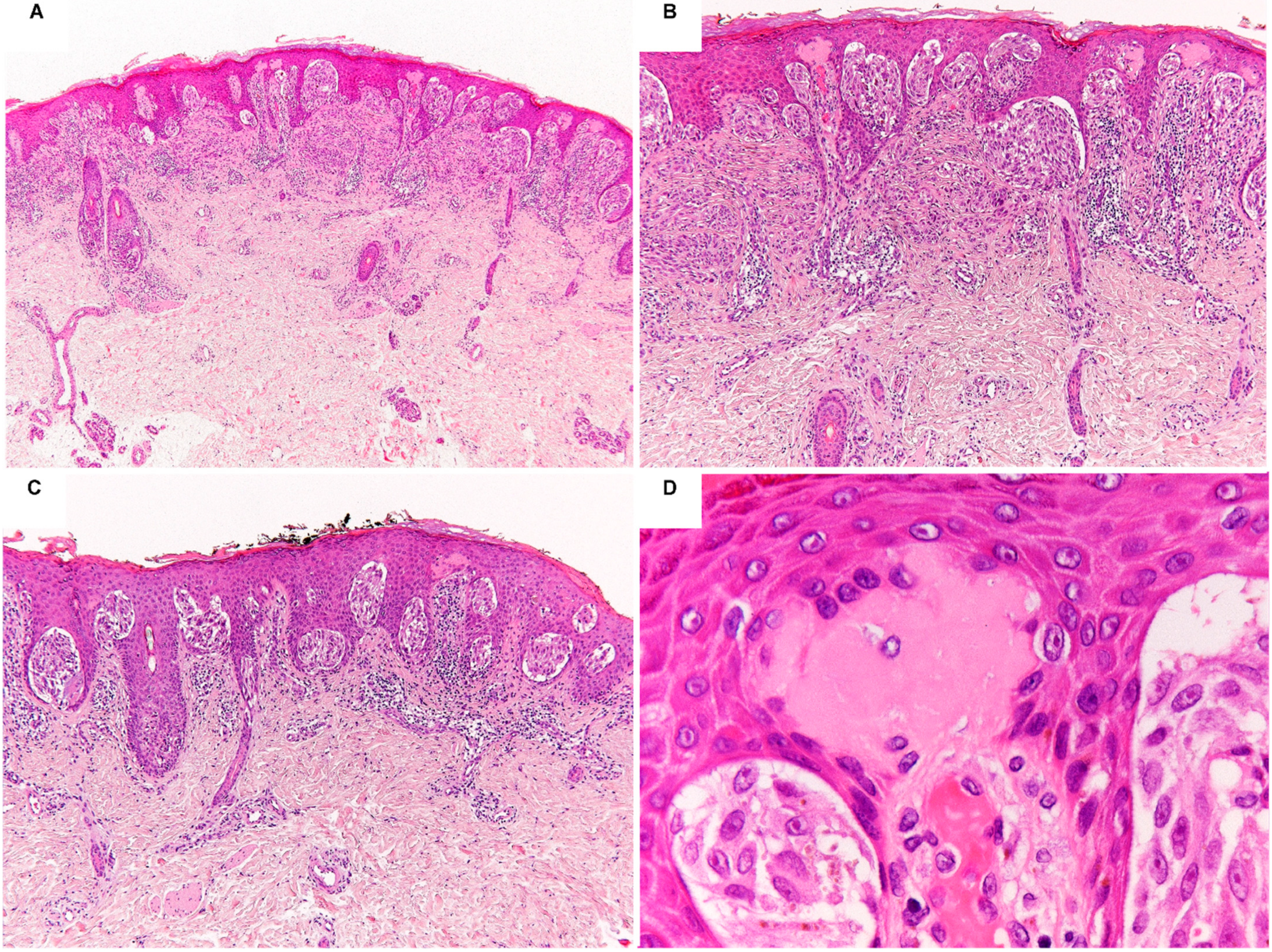
Figure 1. Photomicrographs of a prototypical Spitz nevus showing a symmetric, slightly raised, compound, and maturating melanocytic proliferation with epidermal hyperplasia, flat base, and scattered Kamino bodies, composed of large clefting nests of spindle and epithelioid melanocytes with abundant eosinophilic cytoplasm, moderately pleomorphic vesicular nuclei, and prominent nucleoli (AD) H&E staining; original magnification 10×, 100×, 100×, and 400×, respectively).
Ijms 23 04211 g001
Figure 2. Photomicrographs of a Spitz nevus harboring HRAS mutation showing a symmetric, exophytic and dermal melanocytic proliferation with flat base, composed of fascicles of large spindle melanocytes with amphophilic cytoplasm, moderately pleomorphic vesicular nuclei, and distinct nucleoli, intermingled with thick collagen bundles (AC) H&E staining; original magnification 10×, 100×, and 200×, respectively). Mass Array graphic output showing the HRAS p.Q61R c.182A > G mutation detected by mass spectrometry-based analysis (D).
Ijms 23 04211 g002
Figure 3. Photomicrographs of a Spitz nevus harboring ALK fusion showing a large, exophytic and dermal melanocytic proliferation with infiltrative base, composed of nonpigmented, large, epithelioid and spindle melanocytes with amphophilic cytoplasm, vesicular nuclei, and prominent nucleoli, arranged in plexiform intersecting fascicles (AC) H&E staining; original magnification 10×, 100×, and 200×, respectively). ALK (clone D5F3) immunohistochemistry showing a diffuse cytoplasmic staining ((D) original magnification 12.5×).
Ijms 23 04211 g003
Figure 4. Photomicrographs of an atypical Spitz tumor harboring NTRK1 fusion showing a slightly raised, compound, and symmetric melanocytic proliferation with filigree-like rete ridges, flat-base silhouette, and lobulated nests of epithelioid and spindle melanocytes with moderate nuclear pleomorphism (AC) H&E staining; original magnification 12.5×, 100×, and 200×, respectively). Pan-TRK (clone EPR17341) immunohistochemistry showing a diffuse cytoplasmic staining ((D) original magnification 12.5×).
Ijms 23 04211 g004
Table 1. Clinical, histological, immunohistochemical, and molecular features of Spitz neoplasms.
Spitz NevusAtypical Spitz TumorMalignant Spitz Tumor
Clinical Features
AgeMean and median age: 21 years (range 2–69 years)Can occur at any age; more common in younger patients (<40 years)Can occur at any age (often >40 years)
LocationMost commonly affects the extremitiesOccurs on extremities, trunkOccurs on extremities, trunk
DescriptionPink or reddish plaque, papule, or nodule.Plaque or nodule
Color variegation
Enlarged Plaque or nodule
Color variegation
Asymmetry
Evolving lesion
Histological Features
Size≤5 mm5–10 mm>5 mm
(often >10 mm)
SilhouetteSymmetricSymmetric or asymmetricOften asymmetric
CircumscriptionSharpOften poorPoor
UlcerationAbsentPossibleOften present
EpidermisHyperplasticOften effacedOften effaced
NestingVertically oriented with cleftingIrregularIrregular and confluent
Pagetoid spreadSometimes central and focalSometimes diffuseExtensive
MaturationPresentSometimes partial or absentAbsent
NecrosisAbsentUsually absentSometimes present
Kamino bodiesPresentOften absentAbsent
Deep marginPushingMostly pushingOften infiltrative
InflammationInconspicuousPatchyPatchy or band-like
Cytological Features
ShapeEnlarged epithelioid or spindle cellsEnlarged epithelioid or spindle cells with increasing atypiaEnlarged epithelioid or spindle cells with marked atypia
PleomorphismAbsent or mildMild to severeModerate to severe
CytoplasmGround glassGranularGranular
NucleusFinely dispersed chromatinHeterogeneous chromatinHyperchromasia
NucleolusDistinctIncreasingly prominentLarge
Nuclear/cytoplasmic ratioLowIntermediate to highHigh
PigmentSuperficial distributionVariable distributionVariable, often irregular distribution
Mitotic rate0–2/mm22–6 mitoses/mm22–6 mitoses/mm2
(often > 6 mitoses/mm2)
Atypical mitosesAbsentMostly absentPresent
Immunohistochemical Features
HMB45Diminished with depth in dermal componentDiminished or variable with depth in dermal componentDeep staining common
Ki-67<5%5–15%>15%
p16Present (checkerboard pattern)Sometimes diminished or absentOften diminished or absent
Molecular Features
CGH arrayIsolated gains of 7p and 11q, tetraploidyOften > 1 chromosomal abnormality
Gains of 6p25
Often > 1 chromosomal abnormality
Gains of 6p25
Loss of 9p21AbsentSometimes present (heterozygous or homozygous)Often present (homosygous)
TP53 mutationsAbsentSometimes presentOften present
TERT promoter mutationsAbsentSometimes presentOften present
Table 2. Morpho-molecular features of Spitz neoplasms and immunohistochemistry and/or molecular analyses useful for diagnostic confirmation. Histological features more characteristic of each molecular subtype are in bold.
Histological FeaturesDriver AlterationImmunohistochemistryMolecular Analyses
Symmetric plaque-like lesion
Infiltrative borders
Epithelioid and spindled large melanocytes
Low grade cytological atypia
Low mitotic rate
Desmoplastic stromal reaction
Predominantly intradermal growth
HRAS mutations
11p gains
HRASQ61R (clone SP174) not useful
HRASWT
NGS
CGH or FISH
Symmetrical dome/wedge-shaped large lesion
Epithelioid and spindle melanocytes
Mild to moderate cytological atypia
Low mitotic rate
Plexiform growth pattern
Absent or scant pigmentation
Absent or scant Kamino bodies
ALK fusionsALK (clones D5F3 and 5A4)FISH or NGS
Plaque-like or nodular lesion
Epithelioid and spindled melanocytes
Mild to moderate cytological atypia
Low mitotic rate
Prominent junctional component
Transepidermal elimination of nests
Adnexal involvement
Numerous Kamino bodies
ROS1 fusionsROS1 (clone D4D6)FISH or NGS
Lobulated nests
Rosette-like structures
Epithelioid and spindled melanocytes
Mild to moderate cytological atypia
Low mitotic rate
Extreme maturation
Filigree-like rete ridges
Predominantly junctional proliferation
Numerous Kamino bodies
NTRK1 fusionsPan-TRK (clone EPR17341)NGS (FISH suggested if pan-TRK is positive but NGS is negative)
Pattern ETV6-related:
Large coalescing and lobulated nests
Epithelioid melanocytes
Pleomorphic nuclei
Pattern MYO5A-related:
Spindle melanocytes
Fascicular to plexiform growth pattern
Palisading and rosettes-like structures
Pattern MYO5A-related:
Epithelioid melanocytes
Syncytial arrangement
Central desmoplastic stroma
Peripheral collagen trapping
NTRK3 fusionsPan-TRK (clone EPR17341)NGS (FISH suggested if pan-TRK is positive but NGS is negative)
Symmetrical, well-circumscribed proliferation with plaque-like silhouette
Small to intermediate-sized epithelioid and Spindle melanocytes
Low grade cytological atypia
Nested growth
RET fusionsNot availableFISH or NGS
Symmetric dome-shape lesion
Small to intermediate-sized epithelioid and spindle melanocytes
Low grade cytological atypia
Nested growth
MET fusionsNot availableFISH or NGS
Penetrating nevus/dysplastic nevus-like architecture
Infiltrative margins
Large epithelioid cells with relatively high degree of cito-nuclear atypia
Poor maturation
Lack of epidermal hyperplasia
Stromal accumulation of melanophages
Plexiform growing pattern
Hyperpigmentation
Absent or scant Kamino bodies
MAP2K1 mutationsNot availableNGS
Dome-shaped or nodular lesion
Predominantly nested junctional component Ulceration
Lack of maturation
Epithelioid melanocytes
Moderate to high grade cytological atypia
High mitotic rate
Giant multinucleated melanocytes
MAP3K8 fusionsNot availableFISH or NGS
Superficial dermal sheet-like architecture
Basal desmoplastic stromal reaction
Lack of maturation
Epithelioid morphology
Moderate to high grade cytological atypia
High mitotic rate
BRAF fusionsNot availableFISH or NGS
Publisher’s Note: MDPI stays neutral with regard to jurisdictional claims in published maps and institutional affiliations.