Article

Investigation of the Fuzzy Complex between RSV Nucleoprotein and Phosphoprotein to Optimize an Inhibition Assay by Fluorescence Polarization

by
Silva Khodjoyan
1,
Deborha Morissette
1,
Fortune Hontonnou
2,
Luis Checa Ruano
3,4,
Charles-Adrien Richard
2,
Olivier Sperandio
3,
Jean-François Eléouët
2,
Marie Galloux
2,
Philippe Durand
1,
Stéphanie Deville-Foillard
1 and
Christina Sizun
1,*
1
Institut de Chimie des Substances Naturelles, CNRS, Université Paris Saclay, F-91190 Gif-sur-Yvette, France
2
Virologie et Immunologie Moléculaires, INRAE, Université Paris-Saclay, F-78350 Jouy-en-Josas, France
3
Structural Bioinformatics Unit, Department of Structural Biology and Chemistry, Institut Pasteur, Université de Paris, CNRS UMR3528, F-75015 Paris, France
4
Collège Doctoral, Sorbonne Université, F-75005 Paris, France
*
Author to whom correspondence should be addressed.
Int. J. Mol. Sci. 2023, 24(1), 569; https://doi.org/10.3390/ijms24010569
Submission received: 30 November 2022 / Revised: 21 December 2022 / Accepted: 23 December 2022 / Published: 29 December 2022
(This article belongs to the Section Macromolecules)

Abstract

:
The interaction between Respiratory Syncytial Virus phosphoprotein P and nucleoprotein N is essential for the formation of the holo RSV polymerase that carries out replication. In vitro screening of antivirals targeting the N-P protein interaction requires a molecular interaction model, ideally consisting of a complex between N protein and a short peptide corresponding to the C-terminal tail of the P protein. However, the flexibility of C-terminal P peptides as well as their phosphorylation status play a role in binding and may bias the outcome of an inhibition assay. We therefore investigated binding affinities and dynamics of this interaction by testing two N protein constructs and P peptides of different lengths and composition, using nuclear magnetic resonance and fluorescence polarization (FP). We show that, although the last C-terminal Phe241 residue is the main determinant for anchoring P to N, only longer peptides afford sub-micromolar affinity, despite increasing mobility towards the N-terminus. We investigated competitive binding by peptides and small compounds, including molecules used as fluorescent labels in FP. Based on these results, we draw optimized parameters for a robust RSV N-P inhibition assay and validated this assay with the M76 molecule, which displays antiviral properties, for further screening of chemical libraries.

1. Introduction

Respiratory Syncytial virus (RSV) is the most common pathogen for acute pediatric low respiratory tract infections (ALRI) and bronchiolitis [1]. RSV is an important cause of death in infants in developing countries and a substantial burden on healthcare systems and hospitals worldwide. In 2005, RSV led to ~33 million RSV-associated ALRI episodes, 3 million hospitalizations and 55,000–199,000 deaths in children younger than 5 years [2,3]. RSV also represents a still underestimated risk of severe infection and mortality for immunocompromised and elderly persons [4].
There is still no licensed human RSV vaccine, even after more than 6 decades of research. The monoclonal antibody Palivizumab, which is not efficient against ongoing infection, has been administered for prophylaxis only to high-risk infants, due to high cost and limited efficacy [5,6,7]. Very recently Nirsevimab, a long-acting antibody for similar applications [8], was approved by the European Medicines Agency. More than 30 RSV prevention candidates are currently in clinical development [9,10]. The licensed therapeutic arsenal against RSV is even more limited. Ribavirin, a nucleoside analogue, has a low therapeutic index and is only used for high risk patients. Several drug molecules, antibodies and a siRNA, targeting the fusion process or the viral replication and transcription machinery, have recently entered clinical trials [5,7,11,12]. Fusion inhibitors targeting the RSV F protein, like the small compounds JNJ-53718678 [13], BTA-C585 (clinical trial NCT02718937), GS-5806 [14], and the ALX-0171 nanobody [15] completed phase 2 clinical trials. The viral RNA polymerase inhibitor ALS-008176 [16] also completed a phase 2 trial. Although promising, these drugs elicit known resistances, which calls for alternatives and/or combination therapies.
RSV is a nonsegmented single-stranded negative-sense RNA virus of the Mononegavirales order, Pneumoviridae family, and Orthopneumovirus genus [17]. Its genome consists of an RNA molecule enchased in a sheath made of RSV nucleoprotein (N protein), forming a helical ribonucleoprotein complex termed nucleocapsid [18,19,20]. Replication of the viral genome as well as transcription of viral mRNAs are carried out by the viral RNA polymerase machinery. The apo polymerase consists of the RSV L protein, a large multifunctional catalytic subunit, and its essential co-factor, the RSV phosphoprotein (P protein). The P protein mediates recognition of the genomic material by the polymerase via direct binding to the N protein [21,22,23]. This interaction between P and N proteins was also found to drive the formation of cytoplasmic condensates, which are involved in RSV replication [24,25]. Although the primary function of RSV P protein consists of tethering the polymerase onto its template, P is a multifunctional protein with several binding partners. These properties are closely linked to its mainly disordered structure, outside a short central tetramerization domain POD (Figure 1A) [26,27,28]. The N-terminal domain of P (PNTD) is nearly fully disordered and contains several short linear motifs that are recognized by different viral and cellular proteins [29]. Of note, the N-terminus of P is involved in the formation of the N0-P complex, where P serves as a chaperone for RNA-free N protein (N0) and maintains N competent for encapsidation of genomic RNA [30]. The C-terminal domain of P (PCTD) contains a large transiently structured region with α-helical propensity (P) [27], which is the binding site of the RSV L protein [23]. The 35–40 amino acid long C-terminal tail (PCtail) is fully disordered, and the 10 last amino acids were reported to be involved in the N-P interaction relevant for the holo polymerase complex [21,26,31].
The RSV N protein is an RNA-binding protein. When overexpressed as a recombinant protein in E. coli, N binds to bacterial RNA and forms ring-shaped N-RNA complexes comprising 10–11 N protomers (Figure 1B) [32]. The N protein consists of two globular N-and C-terminal domains, NNTD and NCTD, connected by a hinge, where RNA binds (Figure 1B). These two domains are flanked by flexible N- and C-terminal arms. The N-terminal arm (35 residues) engages into inter-protomer interactions in N-RNA rings [32]. NNTD contains the binding site of PCtail [21].
In contrast to full-length N, recombinant NNTD is produced as a monomeric and RNA-free protein. We previously provided a high resolution structural basis for the N-P interaction with X-ray crystal structures of complexes between NNTD and C-terminal P peptides, notably with the 2-mer Asp240Phe241 (P2 peptide in Figure 1A,C) [33]. The C-terminal Phe241 residue of P2 is anchored into a hydrophobic pocket at the surface of N (Figure 1C). Strikingly, for longer peptides, only this Phe241 residue was ordered in the crystal structures and adopted a well-defined position (Figure 1C). Nevertheless, there is evidence that the N-binding region of P extends beyond Phe241 in PCtail. A minigenome assay showed that in vitro replication of RSV was attenuated for the single Leu238Ala and the double Glu239Ala/Asp240Ala P mutants [21]. This result is in line with in vitro binding assays performed with recombinant N protein and GST-fused P mutants, which showed that up to 9 C-terminal P residues are necessary for N-binding [31]. This was corroborated by affinity measurements carried out by NMR with two C-terminal P peptides, P2 and the 12-mer P12 (Figure 1A). P12 displayed a 100-fold lower dissociation constant Kd (54 ± 9 µM) than P2 (4.2 ± 1 mM) [33]. We hypothesized that PCtail could make multiple contacts with the surface of N, increasing avidity of P and contributing to affinity. Since the position of these contacts is not well defined, NNTD-PCTD can be considered as a fuzzy complex [34,35]. Electrostatic interactions between the highly acidic PCtail and positively charged residues that are exposed at the surface of N around the P-binding pocket likely provide additional binding contribution. In cells, the negative charge of PCtail is increased by constitutive phosphorylation of the Ser232 and Ser237 residues [36,37,38,39]. Phosphorylation was shown to modulate RSV replication in vivo [39]. However, the Ser232 phosphorylation site was reported to be dispensable for viral RNA transcription and virus replication in vitro [36,38,39].
Since the NNTD-PCTD interaction is essential for the formation of the viral holo RNA polymerase complex and hence for RSV replication, we proposed that the RSV NNTD-PCTD protein-protein interaction (PPI) could be a new drug target for PPI inhibitors. We previously showed that PCTD could be displaced in vitro from NNTD by 1-benzyl pyrazole 3,5-dicarboxylate derivatives, which mimic the sidechain and C-terminal carboxylate of Phe241 (Figure 1C) [33]. After chemical modification to improve its membrane passage, the M76 molecule (Figure 1D) was able to inhibit replication of recombinant fluorescent RSV in cells. Results from other groups reinforce this hypothesis. Hesperitin, a flavanone that reduced intracellular RSV replication [40], was also shown to bind to the P-binding pocket of NNTD and to compete with the 11-mer P11 peptide [41]. The EDP-938 benzodiazepine, which demonstrated antiviral activity, elicited mutations on N close to the P-binding pocket, suggesting that it might be an N-P inhibitor [42].
Hesperitin and M76, which compete with P for N-binding in vitro and display antiviral activity [33,40,41], were identified from a limited set of molecules and different screening approaches: hesperitin was identified from a flavonoid series using an antiviral assay, whereas M76 was obtained from a subset of the ZINC database selected in silico with a stringent filter based on chemical structure. High-throughput screening (HTS) of chemical libraries would enable to more widely explore the potential of the RSV NNTD-PCTD PPI as an antiviral target. This requires a relevant model of the interaction combined to a robust HTS method. We have shown previously that the complex between NNTD and short C-terminal P peptides, derived from PCtail, affords a minimal complex mimicking the interaction between the P protein and the RSV nucleocapsid [33]. Fluorescence anisotropy (FA) [43], or equivalently fluorescence polarization (FP) (see Materials and Methods), are compatible with HTS [44]. FP has already been applied in the case of the RSV N-P interaction to probe inhibition by hesperitin [41] and by PCtail-derived peptides [45]. These FP measurements were performed under different experimental conditions, notably with different N protein constructs, different P peptides and different fluorescent labels. In this context, our aim was to obtain a deeper insight into the experimental requirements of an FP assay specific of the RSV N-P interaction. This led us to analyze the fuzzy complex formed between the N protein and the flexible PCtail.

2. Results

2.1. Phosphorylation of an RSV PCtail-Derived Peptide Increases Affinity for RSV NNTD

We previously analyzed the RSV NNTD-P12 complex using Nuclear Magnetic Resonance (NMR) [33]. As the isoelectric point of NNTD is 7.8, we used MES pH 6.5 buffer to ensure the stability of the protein. This pH is also suitable for amide-detected 2D NMR experiments. The buffer contained salt (250 mM NaCl) to further stabilize NNTD at protein concentrations required for NMR (50–100 µM). Under these experimental conditions we determined a Kd of 54 µM.
Here, as P protein is constitutively phosphorylated on Ser232 and Ser237 residues in cells, we first investigated the impact of phosphorylation on the affinity of the NNTD-PCtail complex. We performed an NMR titration experiment by adding increasing amounts of pP11 peptide, where Ser232 and Ser237 residues were replaced by O-phosphoserines (pSer, Figure 1A), to a solution of 15N-labeled NNTD, and measured 2D 1H-15N HSQC spectra (Figure 2A). We worked in the same buffer as with P12. At pH 6.5, the pSer sidechains are expected to be mostly negatively charged [46]. Addition of pP11 induced perturbations in the HSQC spectrum of NNTD, in particular chemical shift perturbations (CSPs). Saturation of this effect was reached after addition of ~6 molar equivalents of peptide. At this peptide:protein ratio, the protein can be considered to be in a fully bound form. We measured combined 1H and 15N amide CSPs (Equation (1)) for NNTD signals in a fast chemical exchange regime, i.e., signals that shifted in the spectrum without broadening at intermediate titration points (e.g., Ile129 in Figure 2A). Significant CSPs were located in and around the P-binding pocket, confirming that pP11 binds to the previously determined P-binding epitope on NNTD (Figure 2B,C) [33]. We fitted the NMR titration curves of these residues with a single site binding model to determine residue-specific Kds (Equation (2)), and calculated a mean Kd of 17.0 ± 4.5 µM from 26 individual values. Compared to the unphosphorylated P12 peptide, this represents a ~3-fold gain in affinity, which confirms the role of negative charges in PCtail for N-binding. Since measurements were made in a buffer with high ionic strength, the gain remains rather modest due to charge screening.
Overall, signals of bound NNTD were broader than those of unbound NNTD (Figure 2A). This can be explained by the increase of molecular weight, when the complex is formed. Line broadening at saturation was more marked for residues belonging to the P-binding site, suggesting exchange phenomena as additional sources of broadening. They may reflect local mobility of P in the complex and intermediate binding states. Some NNTD signals with large chemical shift differences between bound and unbound states (e.g., Y135 in Figure 2A) were broadened at intermediate titration points. Signals were recovered when saturation was reached, indicating that they are in an intermediate exchange regime. These residues were also located in or in the close vicinity of the P-binding pocket, and thus report on the same binding mode as residues in fast exchange. From chemical shift differences measured for these residues we determined an exchange rate kex of ~800 s−1 (from Equation (3)) between free and bound NNTD, which indicates a fast dynamic process. Since kex (koff + kon × [NNTD]) is a combination of association (kon) and dissociation (koff) rates, the relative contributions of dissociation and association could not be assessed by this method.

2.2. Fluorescence Polarization Reveals a Potential Secondary Binding Site on RSV NNTD for Fluorescein-Labeled RSV P11 Peptide

2D Protein-observed NMR is a powerful tool that gives detailed structural and thermodynamic insight, but requires large sample amounts and is time-consuming. Acquisition of a single titration point in Figure 2 took 15 min. It is therefore not adapted for medium to high-throughput assays. Fluorescence polarization (FP) is more time-effective. FP is routinely used in screening assays for binding inhibitors [43,44,47,48]. We thus tested if we could transpose the experimental NMR conditions to FP measurements. We first wondered if the NNTD construct, used for NMR, but also previously for isothermal titration calorimetry and surface plasmon resonance (SPR) experiments [33], was adapted for FP. As a general rule, fluorescence polarization increases with the apparent size of a fluorescent molecule. According to Perrin’s equation (Equation (5)), the theoretical FP of fluorescein (fluorescence lifetime ~4 ns) bound to NNTD (26.2 kDa) is ~400 mFP. This is close to 500 mFP, the theoretical maximal FP value, and indicates that the size of NNTD is compatible with an FP assay.
We started by labeling the pP11 peptide with N-terminally attached BODIPY FL, as described by Shapiro at al. [45]. However, we observed that the BODIPY FL-labeled peptide (BF2pP11) was prone to degradation, either via peptide bond cleavage or BODIPY FL-defluorination, under slightly acidic conditions. We then produced F11, another labelled peptide P11, by changing the fluorescent label to fluorescein-5-thiourea (FTU) by coupling with fluorescein-5-isothiocyanate (FITC). F11 proved to be stable. It is similar to the 6-carboxyfluorescein P peptide used by Sa et al. for competition with hesperitin [41]. To directly compare FP and NMR data, we made measurements in MES pH 6.5 buffer. Brij-35 detergent (0.01% w/v) was added below its critical micellar concentration (0.11% w/v) to prevent spurious binding of the peptide to the reading plates. To determine the binding affinity of F11 peptide for NNTD, we measured FP for 200 nM F11 by varying the concentration of NNTD, which ranged from 0.1 to 120 µM (Figure 3A). We tested buffer with and without salt.
In the presence of salt, FP measurements were stable for incubation times up to 48 h. FP of free F11 was 30–40 mFP. We fitted the binding curves with a single site binding model (Equation (10)). This yielded a maximal FP difference between bound and free F11 peptide ΔFPmax = 210–230 mFP and a dissociation constant Kd = 90 ± 15 µM (Figure 3A). Saturation of the binding curve could not be obtained within the applied NNTD concentration range, and this may induce an error on Kd determination. However, the Kd has the same order of magnitude as that determined by NMR for the P12 peptide under similar buffer conditions (Kd = 54 µM) [33]. The experimental FPmax value was lower than the theoretical value (~400 mFP), suggesting that the fluorescent label displays some mobility with respect to the complex. As the label is attached to the N-terminus of the peptide and hence to the opposite side of the anchoring Phe241 residue, this would be in line with a fuzzy NNTD-PCTD complex, where only the C-terminal Phe241 residue is well bound, whereas the rest of the P peptide remains disordered and only loosely attached.
In buffer without salt and in the presence of NNTD, F11 FP stabilized only after 30 min. Binding curves were obtained, but the FP data did not reach a plateau at high NNTD concentration. Instead, FP displayed a linear dependence with increasing NNTD:F11 molar ratios, suggesting a second binding mode with low affinity (Figure 3B). We fitted the curves with a combination of a single binding site for the first mode and a linear contribution for the second mode. This yielded ΔFPmax = 175 mFP and Kd = 0.6 ± 0.1 µM. ΔFPmax was of the same order of magnitude in buffers with and without salt, indicating that the dynamic range of FP was not affected by salt. This suggests that the dynamics of the fluorescent probe were similar in these two conditions. In contrast, the affinity was significantly higher in the absence of salt, by 2 orders of magnitude, confirming the importance of electrostatic interactions for the NNTD-P11 complex.
We previously showed by SPR that the M76 molecule (Figure 1D) could compete with P for NNTD binding in vitro [33]. We therefore tested competition between M76 and F11 by FP. Measurements were done in salt-free buffer to take advantage of the higher F11-NNTD affinity. The concentration of NNTD was set to 5 µM to benefit from a high range of FP values, comprised between FPmin and ~80% FPmax [48]. With increasing M76 concentration (up to 100 µM), F11 FP progressively decreased due to F11 displacement from NNTD by M76 (Figure 3C). However, even at high M76 concentration, FP did not reach the FPmin value. Saturation occurred at a high FP of ~105 mFP, suggesting that M76 was not able to fully displace F11. This corroborates the hypothesis of a secondary binding site on NNTD, for which M76 does not compete. We fitted the data with a Hill equation (Equation (12)), assuming that the maximal inhibition of F11 binding to the canonical site by M76 was obtained at saturation. We obtained an IC50 of 28 ± 5 µM. This was converted into an inhibition constant Ki of 2.5 µM using the IC50-to-Ki server [49], assuming a competitive mechanism and following values: Kd = 0.6 µM for F11, [NNTD] = 5 µM, and [F11] = 0.2 µM.

2.3. The Complex betweeen Fluorescein-Labeled C-Terminal P Peptides and Full-Length N Protein Provides a Robust Model of the RSV N-P Interaction for FP Measurements

As the FP data obtained with NNTD in salt-free buffer suggest the existence of a secondary binding site, we wanted to test if this was still the case with full-length RSV N protein. Wild-type N is produced in the form of ring shaped N-RNA complexes by overexpression in E. coli [32]. These rings mimic one turn of the helical nucleocapsid, which is the natural partner of PCtail. We therefore chose to test N-RNA as an alternative N form. N-RNA complexes contain 10–11 protomers and 7 ribonucleotides per N protomer [32]. Due to the high molecular weight of N-RNA rings (~0.5 MDa), a high fluorescence polarization is expected.
We carried out FP measurements using the fluorescent F11 peptide and N-RNA complex. As N-RNA is more stable at pH 8.0 than at pH 6.5, we worked in Tris buffer. No salt was added to promote high affinity. The FP binding data could be fitted with a single site binding model (Figure 4A). We obtained ΔFPmax = 125–130 mFP, which is less than determined with the NNTD construct, but again confirms the relative mobility of the fluorescent label. We determined a Kd of 7.6 ± 0.9 µM. Surprisingly, the Kd value obtained with N-RNA was one order of magnitude higher than with NNTD (0.6 µM), suggesting that binding to NNTD may not fully recapitulate binding to N-RNA.
We next performed inhibition experiments for the F11-(N-RNA) complex by M76 (Figure 4B). FP values decreased with increasing competitor concentration. At saturation, FP nearly reached the value of free F11, showing that the F11 peptide could be nearly completely displaced. Since the same concentration range was used for NNTD and N-RNA, poor solubility of M76 can be ruled out to explain the saturation observed before complete displacement of F11 in the presence NNTD (Figure 3C). This reinforces the hypothesis of a second binding site on NNTD, outside the canonical P-binding site. We fitted an IC50 of 12.6 µM (Figure 4B), which converts into a Ki of 2.4 µM (using Kd = 7.6 µM for F11, [N] = 10 µM and [F11] = 0.2 µM). The inhibition constant is similar to that obtained with NNTD in salt-free buffer, which suggests that M76 binds in a similar way to the P-binding site on NNTD and N-RNA.
M76 was dissolved in organic solvent such as ethanol or DMSO. This may affect the stability of the protein. We thus run a control experiment to evaluate the effect of solvent on FP measurements. We used the FTU-P7E peptide F7E (Table 1). Addition of up to 10% ethanol did not significantly affect FP measurements (Figure 4C). A slight decrease of FP was observed with 10% DMSO, but overall the FP assay is robust with respect to solvent. In summary, the N-RNA form appears to be suitable for FP measurements, as only specific binding of fluorescent P peptide and efficient inhibition by the M76 molecule were observed.

2.4. Measurement of the Binding Affinity of RSV Fluorescein-P Peptides for RSV N-RNA and Influence of Peptide Length on Fluorescent Label Mobility

Fluorescent labels can only be attached to the N-terminus of the P peptides, since the C-terminus of P directly anchors onto the N protein via the Phe241 residue (Figure 1C). Since C-terminal P peptides are flexible [26], the fluorescent label displays relative mobility, as reflected by the moderate ΔFPmax values reported in the previous paragraphs. This raises the question about how the P peptide length impacts binding affinity and FPmax. We thus designed a series of eight P peptides with increasing length, from 3 to 11 amino acids, and labeled them with FTU (Table 1). Moreover, to investigate the effect of phosphorylation, we produced phosphomimetic peptides, where either Ser237 alone or both Ser232 and Ser237 were replaced by glutamates.
We first measured binding of Fn peptides (200 nM) to N-RNA in Tris buffer by FP (Figure 5A,B). For Fn peptides with n ≥ 5, saturation of fluorescence anisotropy could be reached within the concentration range used for N-RNA, i.e., with 40 µM of total N protein concentration. The fully bound state of F3 and F4 peptides could not be obtained. We fitted the data with ΔFPmax and Kd as parameters. We observed that ΔFPmax decreased with the length of the peptide (Table 1), indicating that the dynamics of the fluorophore increase with its distance to the Phe241 anchor. Concomitantly, the affinity of Fn peptides increased with peptide length: Kds decreased from 62 µM (F3) to <1 µM (F11SE) (Table 1), highlighting the contribution of residues other than the two C-terminal P residues for N binding. In the 11-mer peptide series, phosphomimetic substitutions increased the affinity of F11, 9-fold for F11SE and 20-fold for F11EE (Figure 5B, Table 1). A closer inspection of the binding curves shows that F11SE and F11EE seem to display a second binding mode of weak affinity, which becomes visible once the canonical binding site has been saturated. For F11SE this mode could be fitted with a linear contribution (Figure 5B, Table 1).
To validate fluorescent peptide binding to N-RNA, we carried out competition experiments with unlabeled P peptides. We used 10 µM of N protein. We first probed competition of the F11 peptide by three P peptides of different lengths: P7E, P9E and P11EE (Figure 5C). These phosphomimetic peptides were able to fully displace F11, with IC50 values ranging from 22 µM for the longest P11EE peptide to 180 µM for the shortest P7E peptide (Table 2). The trend of IC50 suggests that the efficiency of P peptides in competing with F11 increases with peptide length. This is in line with the increase of affinity observed with increasing length of fluorescent P peptides (Table 2). Inhibition constants, calculated with Kd values determined for F11, ranged from 6 to 75 µM (Table 2). The trend of Ki was the same as that of IC50. It is noteworthy that Kis of unlabeled peptides were higher than the Kds of their FITC-labeled counterparts, suggesting that the fluorescent peptides may display higher affinities than the unlabeled ones.
We next made competition experiments between three fluorescent and unlabeled peptides with identical amino acid sequences: F5E vs. P5E, F7E vs. P7E and F11 vs. P11 (Figure 5D, Table 2). The unlabeled peptides were able to fully displace their fluorescent counterparts. IC50 and Ki values decreased with increasing peptide length, underlying the importance of P peptide length for inhibition of the N-P complex. Comparison of P11 with P11EE for inhibition of F11 binding (Table 2) showed that P11 competed less efficiently than P11EE with an IC50 of 86 versus 22 µM (Ki of 34 versus 6 µM). This confirms the role of additional negative charges for formation of the RSV N-P complex.
Finally, we probed inhibition by the M76 molecule of F11 and F7E in complex with N-RNA. M76 IC50 and Ki values were in the µM range, but still lower than those of any tested unlabeled P peptide, indicating that M76 is a more potent inhibitor than the peptides, including P11EE. Moreover, M76 appears to be more potent to inhibit the N-F11 complex than the N-F7E complex (Ki of 2.6 versus 8 µM). Although F11 and F7E cannot be directly compared because of their different lengths, this finding suggests that M76 is less efficient at competing with a phosphomimetic peptide. A rationale for this would be that M76 directly competes with Phe241, but not with Ser/Glu237 that binds outside of the cavity.
It must be noted that measurements with NNTD and N-RNA were done at different pH. Fluorescein fluorescence depends on pH, since the phenols and carboxylic acid groups can be ionized in a pH-dependent manner [50]. At pH 6.5, fluorescein dianion and monoanion forms are in a 1:1 equilibrium, whereas at pH 8.0 the dianion is predominant [51]. At pH 6.5 fluorescence emission at 490 nm is thus decreased by 30–40% as compared to pH 8 [52]. Nevertheless, FP should not be affected by pH, since fluorescence polarization is a ratio of emitted fluorescence intensities (Equation (4)). However, lower fluorescence leads to a lower signal-over-noise ratio at pH 6.5. In conclusion, N-RNA in a pH 8.0 buffer appears to be better suited for an FP inhibition assay than NNTD in a pH 6.5 buffer.

2.5. Fluorescein Binds to the RSV P-Binding Site on RSV N Protein

Although unlabeled P peptides were able to displace their fluorescent counterparts from RSV N-RNA, we wondered if the difference between the Ki of Fn peptides and the Kd of unlabeled peptides could originate in N-RNA binding of the fluorescein label. We therefore measured fluorescein FP in the presence of N-RNA. We obtained a binding curve (Figure 6B). Complete saturation of fluorescein by N-RNA was not reached in the N concentration range used for the experiment. We tentatively extracted a Kd of 91 µM (Table 3) from these binding data, assuming that FPmax reaches the maximal theoretical value (500 mFP). The binding affinity was lower than that measured for Fn peptides, but still significant. The contribution of fluorescein binding to FP could be non-negligible, especially for the shorter F3 and F4 peptides, for which we determined Kds of 62 and 35 µM, respectively (Table 1). However, the affinity of fluorescein was lower by at least of one order of magnitude as compared to peptides longer than F5E. Hence, for these peptides, the fluorescent label is not expected to strongly compete with the peptide part. To test if fluorescein binds to the P-binding site on the N-RNA, we carried out a competition experiment with M76 (Figure 6C). M76 was able to displace fluorescein rather efficiently, with an IC50 of 27 µM and a Ki of 13 µM (Table 3), indicating that fluorescein indeed targets the P-binding site of the N protein.
To validate binding of fluorescein to the P-binding site, we made an NMR titration experiment with 15N-labeled NNTD, using experimental conditions similar to those of the titration of NNTD by the pP11 peptide. Fluorescein induced CSPs located in the same region as those induced by pP11 (Figure 7A), indicating that fluorescein targets the P-binding site of NNTD (Figure 7B,C). To get atomic details about the NNTD-fluorescein complex, we run docking experiments using 2 docking softwares: MOE with GBVI/WSA dG scoring function [53] and SMINA with Vinardo scoring function [54]. Two different poses of fluorescein inserted into the P-binding pocket were predicted (Figure 7D). Fluorescein was modeled in its open, fluorescent form. In all cases, one of the xanthene rings was inserted into the hydrophobic P-binding pocket of NNTD, and the benzoic acid was positioned similarly to the pyrazole ring of M76, forming salt bridges with R150 or R132 (Figure 7D,E).
To avoid interference of the fluorophore with FP measurements, we sought to lower the affinity of fluorescein for RSV N protein, while retaining fluorescence property, by chemical modifications. We produced methyl and ethyl esters of the benzoic acid to change the charge and the size of the molecule (Figure 6A). A series of FP measurements was made with different N-RNA concentrations. The two fluorescein derivatives also bound to N-RNA, but displayed 2-fold lower affinity (Figure 6B, Table 3). M76 was able to displace these two variants (Figure 6C). Kis were of the same order of magnitude at the Ki of fluorescein (Table 3). These binding and inhibition experiments suggest that the behavior of the two fluorescein derivatives is close that of the original molecule. Indeed, fluorescein methyl ester adopted docking poses similar to those of fluorescein.

2.6. An FP Assay for the RSV N-P Interaction Using Full-Length N and BODIPY FL-Labeled P Peptides

The undesirable binding of fluorescein to N-RNA led us to reconsider the BODIPY FL as label for peptides, despite the preliminary mixed results obtained with this fluorophore. To get rid of the instability problems encountered with 4,4′ difluoro BODIPY FL scaffold, we considered its 4,4′ dicyano analogue reported as being more stable and with better photo-physical properties. Such derivatives were recently described as tags for bioconjugation [55,56], but, to our knowledge, not for an FP assay. While difluoro-4 and dicyano BODIPY FL 5 also bound to N-RNA, FP values were markedly lower than those of fluorescein at the same N-RNA concentrations (Figure 6B), suggesting that BODIPY FL is less prone to bind. The Kd for 4, obtained by fitting the ΔFP data with FPmax = 500 mFP, was also higher than that of fluorescein (Table 3). Altogether, these results show that the nature of the fluorophore must be taken into account in an FP assay with RSV N and P proteins, and that BODIPY FL is less prone than fluorescein to affect FP measurements. Moreover, as compared to difluoro BODIPY FL-labeled peptides, dicyano BODIPY FL-labeled peptides were chemically stable (either under the acidic conditions used for their purification by HPLC or under the conditions of the FP assay). To assess the properties of BODIPY FL as a fluorescent label for P peptides, we produced a new series of eight dicyano BODIPY FL-labeled (BCNn) peptides of variable length (Table 4) and tested them for N-RNA binding.
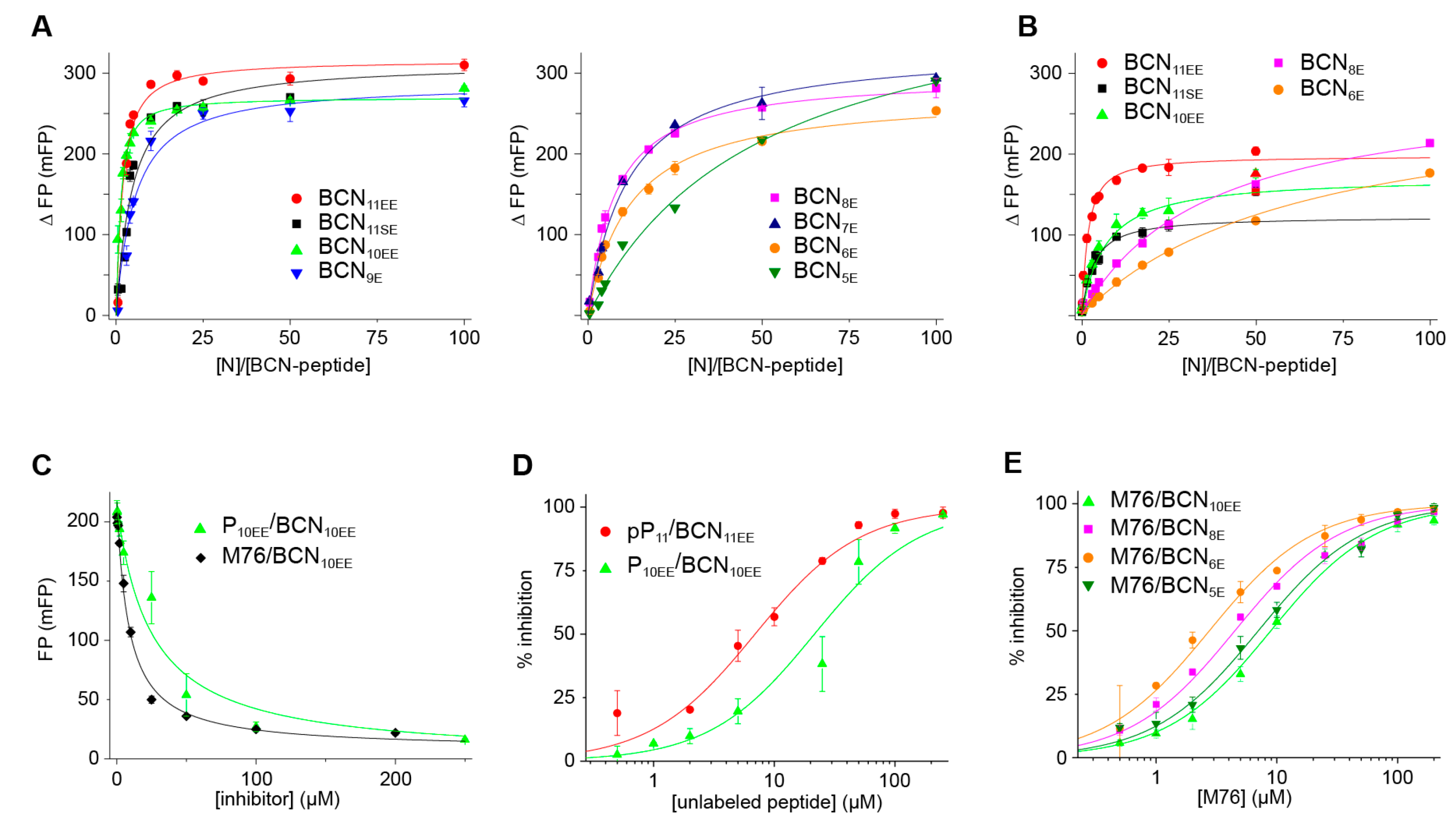
We first performed FP measurements in salt-free Tris pH 8 buffer (Figure 8A). We measured binding curves using 200 nM of fluorescent peptides. Measurements were stable within a time span of 1 h. Saturation by N-RNA was reached for all peptides, except for the shortest one BCN5E. Overall, the dynamic range of FP was more extended for BCNn peptides than for Fn peptides, with ΔFPmax of 270–430 mFP, as compared to 95–315 mFP for Fn peptides (Table 4). Fitted Kds were similar to those of their equivalent Fn peptides, suggesting that the fluorophore does not contribute to the affinity of P peptides longer than 5-mers. Overall, affinities increased with the length of the peptides. The BCNn peptide series exhibited a similar trend to that of the Fn series, highlighting the binding contribution of residues beyond Asp240 and Phe241. A gain of affinity was observed for BCN11EE vs. BCN11SE, showing that the phosphomimetic at position 232 contributes to binding.
To evaluate the quality of FP measurements, we performed a Z’ assay [57,58] with the BCN10EE peptide. An N-RNA concentration of 1 µM afforded a high dynamic range (ΔFPmax = 220 ± 13 mFP, with FPmin = 9 ± 3 mFP). We determined a Z’ value of 0.78 from seven data points, which validates the BCN10EE probe. We further tested robustness with respect to solvent. FP values were not significantly affected by addition of up to 5% ethanol and 10% DMSO.
As compared to salt-free buffer, the ΔFPmax range in salty buffer (100 mM NaCl) was significantly reduced for the long BCN10EE, BCN11SE and BCN11EE peptides (Figure 8B, Table 4). This is similar to what was observed for the F11 peptide. The fluorescent label of long P peptides thus appears to be more mobile under salty conditions, likely due to screening of electrostatic interactions involving acidic amino acids of P peptides. This was not observed for the shorter B6E and B8E peptides. However, it must be noted that the binding curves of BCN6E and BCN8E did not reach a plateau within the N-RNA concentration range used in our FP experiments, which may affect the fitted parameters. The affinity of BCN6E and BCN8E peptides decreased 5-fold in salty buffer, and that of BCN10EE 8-fold, as expected by screening of electrostatic interactions in a higher ionic strength buffer. Intriguingly, similar affinities were measured for BCN11EE and BCN11SE under both salt conditions, despite the phosphomimetic substitution in BCN11EE.
To further investigate the complex between BCNn peptides and N-RNA, we made competition experiments between unlabeled peptides and fluorescent peptides of same length and/or charge, using the P10EE/BCN10EE and pP11/BCN11EE pairs. The unlabeled peptides were able to fully displace their fluorescent equivalents (Figure 8C), and the inhibition curves could be fitted with a Hill equation (Figure 8D). The Kis of unlabeled peptides were in the µM range (Table 5).
To probe the potency of M76 as an inhibitor, we carried out competition experiments with the M76 molecule and several BCNn peptides. M76 was able to displace the fluorescent probes from N-RNA, and inhibition curves were obtained (Figure 8E). Since we used different N concentrations to have similar dynamic ranges of FP for the series of fluorescent peptides, we cannot directly compare M76 IC50s. However, Kis may be compared. Ki values were of the same order of magnitude, 0.5–1.8 µM, for peptides longer than B6E (Table 5), suggesting that the inhibition mechanism by M76 is the same for these BCNn peptides. Inhibition of BCN5E yielded slightly higher Ki values (3.6–4.8 µM), but the Kd determined from a non-saturating binding curve might bias the result. M76 appeared to be more potent than the P10EE peptide to displace BCN10EE from N-RNA with a Ki of 1.1 versus 2.9 µM (Table 5).

3. Discussion

Recognition of the RSV nucleocapsid by the RNA polymerase proceeds via a specific interaction between the essential RSV phosphoprotein polymerase cofactor P and the RSV nucleoprotein N in complex with genomic RNA. This interaction relies on the most C-terminal residue of P, Phe241, which inserts into a pocket at the surface of N. This view is supported by X-ray crystal structures of phenylalanine or short C-terminal P peptides in complex with the RSV NNTD construct, revealing the Phe241 aromatic moiety as a main structural element that drives P binding thanks to well-defined interactions with N atoms delineating a binding pocket. Recently, Phe241 was further confirmed to be the main determinant for P anchoring to N by in silico energetic analysis and mutational analysis of Phe241 in vitro [59]. This view is also supported by in vitro binding and polymerase activity assays. While the aromatic Phe241Trp substitution maintained N binding, deletion of Phe241 and substitution of Phe241 by smaller amino acids like alanine and aspartate impaired N binding [31]. In vitro polymerase activity, as assessed with a minigenome, was completely abrogated by the Phe241Ala mutation [21]. However, even though Phe241 acts as a linchpin, adjacent residues also significantly contribute to the RSV N-P complex. The Leu238Ala and the double Glu239Ala/Asp240Ala mutants reduced minigenome activity 2-fold [21]. Early investigations led to the conclusion that a tract of nine C-terminal residues was necessary and sufficient for N binding [31]. Later we showed that a 12-mer peptide displayed approximately the same affinity in vitro as the full PCTD domain, with Kd of 30–55 µM in salty buffer [33]. Affinity is expected to be rather weak, due to the requirement for polymerase processivity to elongate the newly synthesized RNA. Structural analysis of the RSV phosphoprotein by solution NMR indicated that PCtail remained unstructured in solution in the context of full-length P [26]. In contrast to other linear protein-binding motifs in P, like the binding sites for N0, M2-1 or L proteins, that transiently fold into α-helices and are stabilized in complex, PCtail did not display any propensity for any secondary structure, neither α-helical nor extended [29]. This is a strong indication that PCtail does not adopt any secondary structure in complex with N protein either. This implies that the entropic cost of binding likely remains moderate and that nearly all C-terminal amino acids of P may individually contribute to strengthen the N-P complex, even if they do not adopt a defined position.
The amino acid sequence of RSV N protein is rather conserved, and thus of particular interest for drug design [60,61]. The RSV N protein had already been identified before as a target for post-entry inhibitors. Notably the benzodiazepine RSV604, which reached phase II clinical trial before being discontinued, was proposed to bind to the N protein, since it elicited escape mutants on N [62,63]. Recently, EDP-938, another benzodiazepine in phase 2a clinical trial, was reported to target RSV N, also eliciting several mutations on N close to the P-binding pocket [42] leading to resistance. Based on the inhibitory potential of the M76 molecule we have suggested that the RSV P-binding pocket on N might be druggable [33]. This hypothesis was based on the assumption that a suitable inhibitor would be a direct competitor of the Asp240Phe241 dipeptide. If the interaction surface of the N-P complex extends beyond the Asp240Phe241 binding site and if up to 10 residues away from Phe241 contribute to the interaction energy, this raises the question if targeting the P-binding site with a small molecule would be sufficient to inhibit the N-P complex and displace PCtail and full-length P. In turn, this raises the question about how to design a meaningful assay to screen for inhibitors of the RSV N-P interaction. To address this question we sought for a more comprehensive view of the fuzzy complex formed between RSV N and C-terminal peptides to propose a relevant model of this interaction.
An inhibition assay by fluorescence polarization relies on the size difference between a free fluorescent probe and its complex with a target protein. We tested two forms of the nucleoprotein, the NNTD domain and the N-RNA complex. Our results suggest that the NNTD domain, although suitable in terms of size, induces binding biases, in particular one or more secondary interaction sites and binding of fluorescent labels to the P-binding pocket. In the N-RNA complex, which is structurally close to the nucleocapsid, the N protein surface is not fully accessible, which reduces unspecific binding, and even the P-binding site appears to be rather occluded (Figure 1B,C). N-RNA thus appears to be more relevant than NNTD and the monomeric RNA-free N0-like N mutant used by Shapiro et al. [45]. Interestingly, Shapiro et al. had suggested a difference in binding affinity between BODIPY FL-labeled and unlabeled P11 by comparing FP and SPR data, which hints at binding of the fluorescent label [45]. Here, we show that binding of BODIPY FL remains moderate as compared to fluorescein, and that the peptide part of BCNn peptides longer than 5-mers provides the main driving force for N-RNA binding. Steric hindrance in the N-RNA complex context could thus act as a filter to discriminate between specific and unspecific N binding.
Since C-terminal RSV P-peptides are highly negatively charged, electrostatic interactions likely play an important role for the affinity of the RSV N-P complex. We confirmed the role of electrostatic interactions by comparing binding affinities for several fluorescent P peptides in salty versus salt-free buffers. In salt-free buffer, sub-micromolar affinity could be achieved. This is an asset for an FP assay to eliminate low binding molecules and rank high-affinity inhibitors in an HTS inhibition assay. Shapiro et al. reported a 20-fold increase in affinity by phosphorylation of Ser232 and Ser237 residues in the P11 peptide [45]. We observed similar effect on affinity by using phosphomimetic peptides, which exacerbate electrostatic interactions with N.
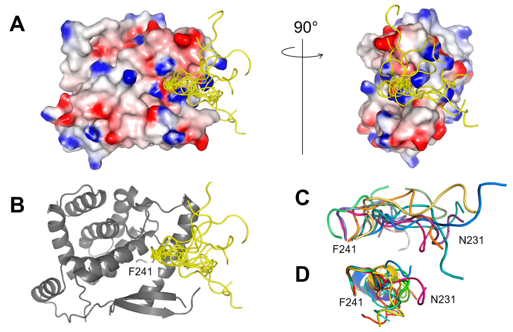
We previously assessed that the N-P complex was fuzzy, with the C-terminal Phe241 serving as a main anchor. To illustrate the dynamics at the P-binding site, we built a 3D structural model of the NNTD-P11 complex by docking the P11 peptide onto NNTD, using the Haddock webserver (Figure 9A–C). For this purpose, we used a structure of NNTD extracted from the NNTD-P2 complex X-ray structure (PDB 4uc9) and generated structural models of P11 using the PEP-FOLD3 server [64]. The structural ensemble of free P11 was highly disordered, with the exception of the C-terminus that displays an incomplete α-helical turn involving residues Glu239-Phe241 (Figure 9D). The 9 N-terminal residues of P11 (Asp231-Glu239) were allowed to remain flexible during docking. Docked P11 also displayed a high degree of disorder, and the α-helical turn partly unwound (Figure 9C). Overall, docked P11 structural models were more extended than free P11 models, but it cannot be excluded that this is due to the difference of algorithms implemented in Haddock and PEP-FOLD3.
The electrostatic surface potential of NNTD does not reveal a unique positively charged surface area, but rather a number of anchoring points afforded by positively charged residues. P11 displays neither defined conformation nor position, but seems to adopt a preferential orientation towards a positively charged patch located near the exit the cavity occupied by Phe241 (Figure 9A). This structural model provides a rationale for the overall increase of binding affinity observed with increasing peptide length up to 11 amino acids. Each amino acid contributes to binding affinity, either by electrostatic or van der Waals interactions. It also explains the concomitant decrease of FP in fluorescently labeled P peptides. As the label is attached to the opposite N-terminal side of the peptide, the fluorescent label experiences increased motional freedom, when the length of the tether increases. As the majority of C-terminal P peptide residues is charged, it is not surprising that a decrease in FP is observed in buffers with higher ionic strength.
As compared to difluoro BODIPY FL-labeled peptides, dicyano BODIPY FL-labeled peptides were more stable. The FP dynamic range was more extended for BCNn peptides than for Fn peptides: This is an advantage for an FP assay. An FP assay relies on a compromise between peptide length and mobility of the fluorescent label. In this respect, 10-mer or 11-mer BCNn peptides, which bind with sub-micromolar affinity in salt-free buffer and offer an extended dynamic FP range, appear to be suitable. Finally, M76 proved to be a suitable benchmark to screen for inhibitors, as it is able to fully displace even longer fluorescent P peptides.

4. Materials and Methods

4.1. Materials

All commercially available reagents and solvents were used as received unless otherwise noted. Fluorescein and fluorescein sodium salt were purchased from Sigma-Aldrich. Fluorescein-5-isothiocyanate (FITC) and all other reagent used for peptide synthesis were purchased from Fluorochem (Hadfield, UK) and Iris Biotech GmbH (Marktredwitz, Germany). pP11 and P11SS peptides (>95% purity assessed by HPLC) were purchased from GeneCust (Luxemburg). 1-(2,4-dichlorobenzyl)-1H-pyrazole-3,5-dicarboxylic acid (M76) was purchased from ChemBridge. 2-Chlorotritylchloride resin (theoretical loading of 1.6 mmol/g) was obtained from Merck (Molsheim, France). Flash chromatography purifications were performed using the automated chromatography Reveleris® Flash System (Grace, Büchi, Villebon-sur-Yvette, France) using prepacked normal phase cartridges from Interchim. Purifications were tracked with a dual λ absorbance UV detector and an ELSD detector.
HPLC analyses were performed on Hypersil C18 column (120 Å, 5 μm, 150 × 4.6 mm) using Waters Alliance 2690 separation module equipped with a Single Quadrupole Detector 2 (ESI quadrupole mass spectrometer), an ELS detector (Waters 2420) and a photodiode array detector (Waters 996). Reversed-phase ultra performance liquid chromatography-mass spectrometry (RP-UPLC-MS) analyses were performed on Waters equipment consisting of an ACQUITY UPLC H-Class separation module, photodiode array detector (eLambda detector), and a ESI triple quadrupole mass spectrometer (TQ detector). The analytical column used was the ACQUITY UPLC BEH C18 column (130 Å, 1.7 μm, 2.1 mm × 50 mm) operating at 0.6 mL·min−1 with linear gradient programs in 2.5 min run time (classical program: 5 to 100% of B in 2.5 min). UV monitoring was performed most of the time between 200 and 500 nm and was extracted at 214 nm. Solvent A consisted of H2O containing 0.1% (v/v) formic acid and solvent B was CH3CN containing 0.1% (v/v) FA. RP-HPLC purifications were performed on Waters equipment consisting of a 2545 quaternary pump, a photodiode array detector (Waters 2998), and an injector collector (Waters 2767). The preparative column, XBridge BEH C18 column (130 Å, 5 μm, 30 mm × 150 mm) was operated at 30 mL·min−1 with linear gradient programs in 15 min run time. The semi-preparative column, XBridge BEH C18 column (130 Å, 5 μm, 10 mm × 150 mm) was operated at 5 mL·min−1 with linear gradient programs in 15 min run time. Solvent C consisted of H2O containing 0.1% (v/v) TFA and solvent D consisted of CH3CN containing 9.9% (v/v) H2O and 0.1% TFA. Classical focused programs on preparative or semi-preparative column: 10 or 20% (v/v) slope of D in 15 min. Water was of Milli-Q quality and was obtained after filtration of distilled water through a Milli-Q cartridge system. CH3CN, FA, and TFA were of spectroscopic grade. High resolution mass spectra (HRMS-ESI) were obtained on a LCT Waters XE mass spectrometer equipped with an electrospray ionisation source. NMR spectra were performed on Bruker Avance spectrometers operating at 699 MHz for 1H NMR, 176 MHz for 13C NMR, 282 MHz for 19F and 160 MHz for 11B NMR experiments. The chemical shifts are reported in ppm relative to tetramethylsilane with the solvent resonance as the internal standard. Multiplicities were given as: s (singlet); d (doublets); t (triplets); q (quadruplets) m (multiplets). Coupling constants are reported as a J value in Hz.

4.2. Synthesis of Fluorescent Molecules

The fluorescein methyl ester 2 and ethyl ester 3 were synthetized in 73% and 40% yield from fluorescein according to Lu et al. [67] and C. Y. Ng et al. [68], respectively. Difluoro BODIPY FL 4 was synthesized according to Gießler et al. [69]. The dicyano BODIPY FL 5 was synthesized in two steps from the difluoro BODIPY FL methyl ester (Scheme 1). The latter was obtained according to K. Gießler et al. [69].
Synthesis of methyl 3-(5,5-dicyano-7,9-dimethyl-5H-4λ4,5λ4-dipyrrolo [1,2-c:2′,1′-f][1,3,2]diazaborinin-3-yl)propanoate (7): BF3·OEt2 (34.0 µL, 0.27 mmol) was added to a cooled solution of difluoro BODIPY FL derivative 6 (415 mg, 1.36 mmol) in anhydrous CH2Cl2 (70 mL) at 0 °C. The mixture was then stirred at 25 °C for 10 min and TMSCN (851 µL, 6.80 mmol) was added. The reaction mixture was stirred at room temperature for 2 h. A saturated aqueous NaHCO3 solution was added. The organic phase was washed with water and was dried over Na2SO4. The solvent was evaporated under reduced pressure to give the titled product as a red solid (0.43 g, 98%). 1H NMR (699 MHz, CDCl3) δ 7.22 (s, 1H), 7.06 (d, J = 4.30 Hz, 1H), 6.43 (d, J = 4.30 Hz, 1H), 6.31 (s, 1H), 3.73 (s, 3H), 3.45 (t, J = 7.20 Hz, 2H), 2.88 (t, J = 7.30 Hz, 2H), 2.74 (s, 3H), 2.31 (s, 3H); 13C NMR (176 MHz, CDCl3) δ 172.5, 161.2, 158.1, 145.3, 133.6, 131.7, 129.7, 126.02 (q, JCB = 75.2 Hz, 2 CN), 124.9, 122.1, 117.8, 52.3, 32.3, 24.3, 16.01, 11.7; 11B NMR (160 MHz, CD2Cl2) δ −16.86 (s); 19F NMR (282 MHz, CDCl3) no peaks observed. HRMS [ESI]: m/z calculated for C17H17BN4O2 [M − H] 319.1372; found: 319.1372.
Synthesis of 3-(5,5-dicyano-7,9-dimethyl-5H-4λ4,5λ4-dipyrrolo[1,2-c:2′,1′-f][1,3,2]diazaborinin-3-yl)propanoic acid (5): Concentrated HCl (16 mL) was added to a solution of methyl ester 7 (0.20 g, 0.62 mmol) in a mixture of THF (40 mL) and water (28 mL). The solution was stirred for 12 h at room temperature. Water (50 mL) and CH2CL2 (50 mL) were then added. The aqueous phase was extracted with CH2Cl2 (3 × 50 mL) and the combined organic phases were washed with brine and were dried over Na2SO4. The solvent was evaporated under reduced pressure to give the titled product as a red solid (0.17 g, 90%). M.p.: 211–232 °C. 1H NMR (500 MHz, DMSO-d6) δ 12.43 (s, 1H), 7.99 (s, 1H), 7.34 (d, J = 4.30 Hz, 1H), 6.63 (d, J = 4.20 Hz, 1H), 6.58 (s, 1H), 3.20 (t, J = 7.50 Hz, 2H), 2.82 (t, J = 7.50 Hz, 2H), 2.64 (s, 3H), 2.33 (s, 3H). 13C NMR (176 MHz, DMSO-d6) δ 173.1, 159.7, 157.5, 145.8, 132.8, 131.3, 130.3, 126.7, 125.5 (q, JCB = 74.0 Hz, 2CN), 121.9, 117.7, 31.2, 23.7, 15.1, 11.1. 11B NMR (160 MHz, CD2Cl2) δ −17.0 (s). 19F NMR (282 MHz, DMSO-d6) no peaks observed. HRMS [ESI]: m/z calculated for C16H15BN4O2 [M − H] 305,1288; found: 305.1198.
NMR spectra are given in Figures S1–S6. HPLC analysis of 5 and 7 is shown in Figures S31 and S32.

4.3. Peptide Synthesis

The BODIPY FL-labeled P peptide BF2pP11 was synthesized from purchased pP11 peptide and BODIPY FL as described in [45].
P3, P4, P5E, P5S, P7S, P7E, P9E, P9S, P11SE and P11EE peptides and their fluorescent dicyano BODIPY FL conjugates BCN5E, BCN6E, BCN7E, BCN8E, BCN9E, BCN10EE, BCN11SE, BCN11EE were synthesized as described below.
Assemblies of protected peptides were carried out manually on 2-Chlorotritylchloride resin (theoretical loading of 1.6 mmol/g—0.2 g) using the Fmoc/tBu strategy in a polypropylene reaction vessel fitted with polyethylene frits. After swelling of the resin for 15 min in CH2Cl2, the first amino acid was loaded through nucleophilic substitution by shaking the resin in a solution of Fmoc-Phe-OH (1 mmol/g) and DIPEA (1.5 equiv) in anhydrous CH2Cl2 (10 mL/g resin) for 30 min at room temperature. The un-reacted sites were then capped by shaking the resin in a mixture of MeOH/DIPEA/CH2Cl2 (2/1/17 v/v/v; 10.0 mL/g resin) for 10 min (repeated two times). The resin was then washed two times with DMF and three times with CH2Cl2 (10.0 mL/g resin) for 30 s. The overall process yielded a resin loading of around 0.65 mmol/g [determined from the dosage of the Fmoc released under the conditions of procedure A]. The number of reagent equivalents used in the following procedures is calculated from this resin loading. This step is followed by Fmoc removal according to procedure A. The elongation of the peptide was performed by cycle repetition of peptide coupling and Fmoc removal according to procedures B and A, respectively. Coupling of dicyano BODIPY FL, if required, was performed on the resin at the end of the peptide elongation on the amino terminus of the peptide according to procedure C. Cleavage of the peptide from the resin was carried out according to procedure D.

4.3.1. Procedure A: Fmoc Removal

N-α-Fmoc protecting groups were removed by shaking the resin in piperidine/DMF solution (1/4, v/v; 10.0 mL/g resin) for 5 min at room temperature. The process was repeated two times for 10 min, and the completeness of deprotection was monitored by ultraviolet absorption measurement of the filtrate at 299 nm. The resin was then washed five times with DMF and once with CH2Cl2 (10.0 mL/g resin) for 30 s.

4.3.2. Procedure B: Coupling Steps

Coupling reactions were performed by shaking with a solution of N-α-Fmoc-protected amino acid (2 equiv), PyBOP (2 equiv) and DIPEA (4 equiv) in DMF (10.0 mL/g resin) for 30 min at room temperature. The resin was then washed three times with DMF and once with CH2Cl2 (10.0 mL/g resin) for 30 s. Completeness of the coupling was controlled by TNBS and KAISER tests.

4.3.3. Procedure C: Coupling of Dicyano BODIPY FL

The resin was shaken with a solution of dicyano BODIPY FL 5 (1 equiv), DIPEA (4–8 equiv depending of the number of peptide acidic functions) and PyBOP (2 equiv) at room temperature in DMF (10.0 mL/g resin). The resin was then washed 14 times with DMF and 8 times with CH2Cl2 (10.0 mL/g resin) for 30 s.

4.3.4. Procedure D: Resin Cleavage and Protecting Groups Removal

The resin was first washed five times with CH2Cl2 (10.0 mL/g resin) before shaking in a mixture of TFA/TIS/H2O MQ (95/2.5/2.5 v/v/v; 10.0 mL/g resin) for 2 h at room temperature and then filtrated. The resin was rinsed once with the same acidic mixture of TFA/TIS/H2O MQ (95/2.5/2.5 v/v/v) for 1 min. The recovered solutions were concentrated under reduced pressure, and white solid was obtained by precipitation, triturating, and washing three times with diethyl ether. These crude peptides and crude dicyano BODIPY FL peptides, obtained as TFA salts, were purified by RP-HPLC on preparative or on semi-preparative column, respectively, using focused gradients, and freeze dried before their analysis by RP-UPLC-MS.

4.4. Peptide Purification and Yields

4.4.1. Unlabeled Peptides

UPLC traces and analysis for Pn peptides are given in Figures S7–S11.
  • P3 peptide was obtained as a white powder (34.0 mg, 50%). Analytical UPLC tr = 0.87 min; ESI-MS (positive mode) calculated for [C18H23N3O8], 409.2; found m/z, 410.4 (M + H)+. [Focused gradient 5–25% of B in 15 min].
  • P4 peptide was obtained as a white powder (49.0 mg, 59%). Analytical UPLC tr = 1.01 min; ESI-MS (positive mode) calculated for [C24H34N4O9], 522.2; found m/z, 523.5 (M + H)+. [Focused gradient 10–30% of B in 15 min].
  • P5E peptide was obtained as a white powder (59.0 mg, 59%). Analytical UPLC tr = 1.00 min; ESI-MS (positive mode) calculated for [C29H41N5O12], 651.3; found m/z, 652.6 (M + H)+. [Focused gradient 15–35% of B in 15 min].
  • P7E peptide was obtained as a white powder (43.0 mg, 33%). Analytical UPLC tr = 1.43 min; ESI-MS (positive mode) calculated for [C39H57N7O16], 879.4; found m/z, 880.8 (M + H)+. [Focused gradient 25–35% of B in 15 min].
  • P9E peptide was obtained as a white powder (27.0 mg, 17%). Analytical UPLC tr = 1.29 min; ESI-MS (positive mode) calculated for [C47H68N10O21], 1108.5; found m/z, 1109.7 (M + H)+. [Focused gradient 20–30% of B in 15 min].
  • P11EE: SPPS of P11EE peptide was performed on 150 mg of 2-CTC resin. The peptide P11EE was obtained as a white powder (28.0 mg, 20%). Analytical UPLC tr = 1.28 min; ESI-MS (positive mode) calculated for [C56H80N12O27], 1352.5; found m/z, 1353.9 (M + H)+. [Focused gradient 20–30% of B in 15 min].
  • P11SE: SPPS of P11SE peptide was performed on 15.0 mg of 2-CTC resin. P11SE was obtained as a white powder (4.50 mg) and was used without further purification for F11SE synthesis.

4.4.2. Dicyano BODIPY FL-Labeled BCNn Peptides

UPLC traces and analysis for BCNn peptides and blank are given in Figures S21–S30.
  • BCN5E: Starting from P5E on resin (8.71 µmol), BCN5E was obtained as a red powder (2.63 mg, 32%). Analytical UPLC tr = 1.61 min; ESI-MS (negative mode) calculated for [C45H54BN9O13], 939.8; found m/z, 939.4 (M − H). [Focused gradient 38–48% of B in 15 min].
  • BCN6E: Starting from P6E on resin (8.71 µmol), BCN6E was obtained as a red powder (1.60 mg, 18%). Analytical UPLC tr = 1.74 min; ESI-MS (negative mode) calculated for [C51H65BN10O14], 1052.5; found m/z, 1051.2 (M − H). [Focused gradient 43–53% of B in 15 min].
  • BCN7E: Starting from P7E on resin (8.71 µmol), BCN7E was obtained as a red powder (2.22 mg, 22%). Analytical UPLC tr = 1.66 min; ESI-MS (negative mode) calculated for [C55H70BN11O17], 1167.5; found m/z, 1166.9 (M − H). [Focused gradient 40–50% of B in 15 min].
  • BCN8E: Starting from P8E on resin (8.71 µmol), BCN8E was obtained as a red powder (2.00 mg, 18%). Analytical UPLC tr = 1.56 min; ESI-MS (positive mode) calculated for [C59H76BN13O19], 1282.6; found m/z, 1283.9 (M + H)+. [Focused gradient 36–46% of B in 15 min].
  • BCN9E: Starting from P9E on resin (8.71 µmol), B9E was obtained as a red powder (1.17 mg, 10%). Analytical UPLC tr = 1.53 min; ESI-MS (positive mode) calculated for [C63H81BN14O22], 1396.6; found m/z, 1398.2 (M + H)+. [Focused gradient 35–45% of B in 15 min].
  • BCN10EE: Starting from P10EE on resin (8.71 µmol), BCN10EE was obtained as a red powder (1.00 mg, 8%). Analytical UPLC tr = 1.50 min; ESI-MS (negative mode) calculated for [C68H88BN15O25], 1525.6; found m/z, 1525,1(M − H). [Focused gradient 34–44% of B in 15 min]. UV-Visible absorption spectrum and fluorescence excitation/emission spectra are given in Figure S33.
  • BCN11EE: Starting from P11EE on resin (8.71 µmol), BCN11EE was obtained as a red powder (1.01 mg, 11%). Analytical UPLC tr = 1.49 min; ESI-MS (negative mode) calculated for [C72H93BN16O28], 1640.6; found m/z, 1640.3 (M − H). [Focused gradient 33–43% of B in 15 min].
  • BCN11SE: Starting from P11SE on resin (4.35 µmol), BCN11SE was obtained as a red powder (1.12 mg, 16%). Analytical UPLC tr = 1.48 min; ESI-MS (negative mode) calculated for [C70H91BN16O27], 1598.6; found m/z, 1598.2 (M − H). [Focused gradient 32–42% of B in 15 min].

4.4.3. Fluorescein-Labeled Fn-Peptides

Due to reported instability of FTU-labelled peptides under acidic conditions [70], the coupling of FITC with synthesized P3, P4, P5E, P7E, P9E, P11SE, P11EE peptides and commercial pP11 and P11SS peptides was performed in solution. FITC (2 equiv) was added to a solution of the peptides (1 equiv) and DIPEA (9 equiv) in anhydrous DMF (0.06 mL/mg peptide). Solutions were stirred for 30 min to 1h depending on the peptide. Solutions were then purified by HPLC on semi-preparative column using focused gradients and freeze dried. UPLC traces and analysis for Fn peptides are given in Figures S12–S20.
  • F3: Starting from P3 (5.00 mg, 9.55 µmol), F3 was obtained as a yellow powder (3.00 mg, 48%). Analytical UPLC tr = 1.31 min; ESI-MS (negative mode) calculated for [C39H34N4O13S], 798.2; found m/z, 797.6 (M − H). [Focused gradient 25–45% of B in 15 min].
  • F4: Starting from P4 (5.00 mg, 7.85 µmol), F4 was obtained as a yellow powder (4.30 mg, 62%). Analytical UPLC tr = 2.12 min; ESI-MS (negative mode) calculated for [C45H45N5O14S], 911.3; found m/z, 910.6 (M − H). [Focused gradient 55–65% of solvent D consisted of MeOH containing 9.9% (v/v) H2O and 0.1% TFA in 15 min].
  • F5E: Starting from P5E (5.00 mg, 6.53 µmol), F5E was obtained as a yellow powder (3.60 mg, 53%). Analytical UPLC tr = 0.98 min; ESI-MS (positive mode) calculated for [C50H52N6O17S], 1040.3; found m/z, 1041.8 (M + H)+. [Focused gradient 35–45% of B in 15 min].
  • F7E: Starting from P7E (5.00 mg, 5.03 µmol), F7E was obtained as a yellow powder (2.60 mg, 50%). Analytical UPLC tr = 1.53 min; ESI-MS (positive mode) calculated for [C60H68N8O21S], 1268.4; found m/z, 1270.0 (M + H)+. [Focused gradient 30–50% of B in 15 min].
  • F9E: Starting from P9E (5.00 mg, 4.09 µmol), F9E was obtained as a yellow powder (1.40 mg, 22%) Analytical UPLC tr = 1.42 min; ESI-MS (positive mode) calculated for [C68H79N11O26S], 1497.5; found m/z, 1499.7 (M + H)+. [Focused gradient 30–50% of B in 15 min].
  • F11: Starting from commercial P11 (4.30 mg, 3.11 µmol), F11SS was obtained as a yellow powder (3.5 mg, 68%). Analytical UPLC tr = 1.45 min; ESI-MS (positive mode) calculated for [C73H87N13O30S], 1657.5; found m/z, 1659.8 (M + H)+. [Focused gradient 25–45% of B in 15 min].
  • F11SE: Starting from P11SE (4.50 mg, 3.43 µmol), F11SE was obtained as a yellow powder (0.90 mg, 15%). Analytical UPLC tr = 1.39 min; ESI-MS (negative mode) calculated for [C75H89N13O31S], 1699.6; found m/z, 1697.7 (M − H). [Focused gradient 25–45% of B in 15 min].
  • F11EE: Starting from P11EE (5 mg, 3.41 µmol), F11EE was obtained as a yellow powder (2.40 mg, 40%). Analytical UPLC tr = 1.39 min; ESI-MS (positive mode) calculated for [C77H91N13O32S], 1741.6; found m/z, 1742.6 (M + H)+. [Focused gradient 25–45% of B in 15 min].
  • pF11: Starting from commercial pP11 (4.20 mg, 2.93 µmol), pF11 was obtained as a yellow powder (1.10 mg, 20%). Analytical UPLC tr = 1.36 min; ESI-MS (negative mode) calculated for [C73H89N13O36P2S], 1817.5; found m/z, 908.7 (M − 2H)2−. [Focused gradient 25–45% of B in 15 min].

4.5. Bacterial Expression and Purification of RSV N Protein

Recombinant RSV N protein was produced in E. coli BL21(DE3) bacteria (Novagen, Madison, WI). The N-terminal domain NNTD (residues 31–252), containing a C-terminal 6x histidine tag, was overexpressed using the pET-N(31–252) plasmid and purified as described previously [21]. In a final step NNTD was dialyzed into MES 20 mM pH 6.5, NaCl 250 mM buffer [33]. Full-length N was produced by co-expression with the C-terminal domain of RSV P protein in E. coli BL21(DE3) co-transformed with pET-N and pGEX-P(161–241) plasmids [21]. Cultures were grown at 37 °C for 8 h in Luria-Bertani (LB) medium supplemented with 50 µg/mL kanamycin and 100 µg/mL ampicillin. An equivalent volume of LB was then added, and protein expression was induced with 80 µg/mL isopropyl-β-D-thio-galactoside (IPTG) for 15 h at 28 °C. Bacteria were harvested by centrifugation. The bacterial pellet was resuspended in lysis buffer (20 mM Tris-HCl pH 8.5, 150 mM NaCl, 1 mM EDTA, 2 mM dithiothreitol, 0.2% Triton X-100, 1 mg/mL lysozyme). Complete protease inhibitor cocktail (Roche, Mannheim, Germany) was added, and the suspension was incubated for 1 h on ice, sonicated and centrifuged at 4 °C for 30 min at 10,000× g. Glutathione-Sepharose 4B beads (GE Healthcare, Vélizy-Villacoublay, France) were added to the clarified supernatant and incubated at 4 °C for 3 h. The beads were washed twice in lysis buffer and three times in 20 mM Tris-HCl pH 8.5, 150 mM NaCl buffer. GST was cleaved from the N-P(161–241) complex by treating the beads with thrombin (Novagen) for 16 h at 20 °C. The supernatant was then loaded onto a Superdex 200 16/30 column (GE Healthcare) and eluted in 20 mM Tris-HCl pH 8.5, 150 mM NaCl buffer. Finally, the fractions containing the N protein in the form of an N-RNA complex were pooled and concentrated up to 2 mg/mL (45 µM of N protein). The protein was subsequently dialyzed into 20 mM Tris pH 8.0 buffer containing 0.01% Brij-35. The concentration of N protein was determined with a BCA assay (Thermo Fisher, Illkirch-Graffenstaden, France), calibrated with BSA.

4.6. NMR Measurements

NMR titration experiments with 15N-NNTD were performed in 20 mM MES pH 6.5, 250 mM NaCl buffer. pP11 peptide was solubilized at 1 mg/mL in MQ water by addition of 1 M NaOH until the pH became neutral. pP11 aliquots were lyophilized. For the titration experiment, pP11 was added stepwise to a 50 µM 15N-NNTD solution, using 0.2–6.2 molar equivalents. NMR data were acquired on a 14.1 T (600 MHz 1H frequency) Bruker Avance III NMR spectrometer equipped with a cryogenic TCI probe. A standard 2D 1H-15N HSQC spectrum was recorded for each titration point.
For titration with fluorescein, 0.5–8 molar equivalents of fluorescein were added from a 10 mM stock solution in water at pH 6.5 to 55 µM 15N-NNTD. NMR measurements were performed on a 16.4 T (700 MHz 1H frequency) Bruker NMR spectrometer equipped with a NEO console and a cryoTXO probe. All samples contained 7.5% 2H2O to lock the spectrometer frequency and 100 µM DSS to reference 1H chemical shifts. The temperature was set to 20 °C. NMR data were processed with TopSpin 4.0 (Bruker Biospin, Wissembourg, France) and analyzed with CcpNmr Analysis 2.4 software [71]. Backbone chemical shift assignment of NNTD was done previously [33].
Combined amide 1H and 15N chemical shift perturbations ΔδHN were calculated with a scaling factor of 1/10 for 15N, which corresponds to the ratio of 15N and 1H gyromagnetic ratios (Equation (1)):
Δ δ HN = ( ( δ 1 H δ 1 H ref ) 2 + ( δ 15 N δ 15 N ref ) 2 / 100 )
1H and/or 15N chemical shift perturbations were fitted in CcpNmr Analysis 2.4 as a function of the molar ligand:NNTD ratio r, using a single binding site model with a 1:1 stoichiometry, and assuming a fast chemical exchange regime (Equation (2)). Kd is the dissociation constant of the complex with NNTD. δfree and δbound (in ppm) are the chemical shifts of free and bound NNTD.
( δ δ ref ) = 1 2 ( δ bound δ free ) × ( K d [ N NTD ] tot + 1 + r ( K d [ N NTD ] tot + 1 + r ) 2 4 r )
For residues in intermediate exchange regime, the exchange rate between free and bound protein states kex (s−1) was estimated according to Equation (3), where BF is the Larmor frequency (MHz) of the observed nucleus.
k e x = π × Δ ν = π × ( δ bound δ free ) × BF  

4.7. Fluorescence Polarization Measurements

Fluorescence polarization measurements were carried out on a Paradigm Detection Platform (Beckman Coulter, Brea, CA, USA) operating with a microplate reader, a fluorescein detection cartridge (excitation range 485/20 nm, emission range 535/25 nm for both parallel and perpendicular components) and the SpectraMax Pro 6.1 software (Molecular Devices, San Jose, CA, USA). Samples were placed in polystyrene black flat bottom 96-well microplates (Cellstar, Greiner bio-one, Frickenhausen, Germany) with 20 µL final volume in each well. If not stated otherwise, the final concentration of fluorescent ligands was 200 nM. The buffer was either 20 mM MES pH 6.5, salt-free or with 250 mM NaCl, or 20 mM Tris pH 8.0 salt-free or with 100 mM NaCl, supplemented with 0.01% Brij-35. Mixing was done using the corresponding plate reader option. The temperature was set to 20 °C. Data were recorded in top reading mode. Measurements were done in triplicate and after an incubation time of 30 min, if not indicated otherwise. Fluorescence polarization (FP) in mFP units was determined from the parallel (I) and perpendicular (I) components of the emitted light according to Equation (4) [43]. A grating factor G of 1.2 was determined using 1 nM fluorescein in 1 mM NaOH and assuming a theoretical FP of 27 mFP [48]. Background signal was measured from wells containing only buffer, and the mean value was subtracted from each I and I component.
FP   ( mP ) = 1000 × ( I G I ) / ( I + G × I )
Theoretical fluorescence polarization values of a fluorophore bound to N protein were calculated from the fluorescence lifetime τ and the molecular weight MW using Perrin’s equation (Equation (5)). A is the fluorescence anisotropy related to FP by Equation (6) [43], A0 the intrinsic anisotropy (0.4), T the temperature in Kelvin, k the Planck constant, η the dynamic viscosity, and Vh the hydrated volume. Vh was calculated according to Equation (7), where N is the Avogadro number, v2 the partial specific volume of the protein (7.34 × 10⁻⁷ m3/g), δ the degree of hydration (0.75), and v10 the specific volume of water (10−6 m3/g). We used A0, v2, δ and v10 values reported for bovine serum albumine [72].
A = A 0 / ( 1 + kT × τ η V h )
FP = 1000 × 3 A / ( 2 + A )
V h = ( MW / N ) × ( v 2 + δ × v 1 0 )
We assumed that the measured fluorescence polarization FP is a linear combination of the FP of the fluorescent probe in the absence of N protein (FPmin) and the FP of N-bound fluorescent probe (FPmax), weighted by the molar fraction. The molar fraction x = [P]bound/[P]0 can then be expressed as a ratio of FP differences (Equation (8)).
x = ( FP FP min ) / ( FP max FP min ) = FP / FP max  
To determine the Kd value of a complex between a fluorescent probe and N protein, a series of samples with constant probe concentration and varying N concentration was measured. FP binding curves were fitted with a single binding site model and a 1:1 stoichiometry. The molar fraction of bound fluorescent probe was expressed as a function of the ratio of protein versus probe concentrations, r = [N]0/[P]0, according to Equation (9). Binding curves were fitted in Origin 7 software according to Equation (10), obtained by combining Equations (8) and (9).
x = 0.5 × [ 1 + r +   K d / [ P ] 0 ( 1 + r + K d / [ P ] 0 ) 2 4 r ]
FP = 0.5 × FP max × [ 1 + r +   K d / [ P ] 0 + ( 1 + r + K d / [ P ] 0 ) 2 4 r ]
To assess inhibition of a complex between a fluorescent probe and N protein by peptides or the M76 molecule, the concentration of N was set to work with an FP range comprised between FPmin and ~80% FPmax. Peptides were added from 1 or 2 mM stock solution in water at neutral pH. M76 was added from 10 or 20 mM stock solutions in ethanol or in DMSO. The percentage of inhibition (%i) was calculated as the relative change in FP (Equation (11)). %i was fitted as a function of inhibitor concentration with a Hill equation, where IC50 is the inhibitor concentration resulting in 50% inhibition (Equation (12)). Inhibition data were fitted with Origin 7, and in parallel with the Excel solver module. All data were compatible with an absence of cooperativity. FPmax and FPmin were fitted, and the maximal inhibition %i,max was set to 100%.
% i = 100 × [ 1 ( FP FP min ) / ( FP max FP min ) ]
% i = % i , max × [ inhibitor ] [ inhibitor ] + IC 50
Inhibition constants Ki were calculated on the IC50-to-Ki converter for a protein-ligand-inhibitor system (https://bioinfo-abcc.ncifcrf.gov/IC50_Ki_Converter/index.php, accessed on 24 August 2022) [49], assuming a competitive mechanism (Equation (13)). This tool takes into account the free concentrations of protein and ligand.
K i = ( IC 50 [ protein ] 2 ) / ( [ peptide ] K d + 1 )
A Z’-factor for FP measurements without test compound [58] was calculated from 7 data points for the condition 200 nM B10EE and 1 µM N-RNA, using Equation (14), where σ+ and σ- are the standard deviations of positive (fluorescent peptide bound to N protein) and negative (free fluorescent peptide) controls, respectively, and μ+ and μ- their mean values.
Z = 1 ( 3 σ + + 3 σ ) / | μ + μ |

4.8. Complex Modelling with Haddock

Docking of the P11 peptide onto NNTD was performed with the Haddock 2.4 software [65] on the WeNMR server [73]. A structure of P11 was built with the PEP-FOLD3 server [64] and allowed to be flexible (residues 1–9) during docking. The C-terminal Asp10 and Phe11 residues (equivalent to Asp240 and Phe241) were declared as active residues. The structure of NNTD was extracted from the X-ray structure of the NNTD-P2 complex (PDB 4uc9). N residues 46, 50, 53, 128, 131, 132, 135, 145, and 151, lining the P-binding pocket, were declared as active residues. Passive residues were automatically defined around active residues. Default scoring parameters for protein–protein complexes were used, except for Evdw 1, which was increased from 0.01 to 1. 1000 initial structures were generated. 200 final structures were refined in water and clustered according to the RMSD criterion. Haddock clustered 156 structures in 12 clusters. Cluster 1 with the best haddock score (−96.4 ± 4.9) contained 63 structures, i.e., 40% of clustered structures.

4.9. Docking of Small Compounds

Fluorescein and fluorescein methyl ester were docked on the P-binding pocket of NNTD (PDB 4ucc) using Smina software and MOE [53]. For Smina, Vinardo [54] scoring function was selected with an exhaustiveness of 8. The pose with the best scoring value was retained. For MOE, the placement method used was Triangle Matcher with London dG score and the number of poses was set to 30. These poses were then refined with GBVI/WSA dG score with rigid receptor, and the number of output poses was set to 5. The pose with the best GBVI/WSA dG score was retained.

Supplementary Materials

The following supporting information can be downloaded at: https://www.mdpi.com/article/10.3390/ijms24010569/s1.

Author Contributions

Conceptualization, C.S., S.D.-F. and P.D.; methodology, C.S., S.D.-F., O.S. and P.D.; formal analysis, D.M., S.K., C.S., S.D.-F. and P.D.; investigation, S.K., D.M., S.D.-F., P.D., C.S., L.C.R., C.-A.R., F.H. and M.G.; data curation, S.K., D.M. and C.S.; writing—original draft preparation, C.S.; writing—review and editing, C.S., S.K., S.D.-F., P.D., L.C.R., O.S., M.G. and J.-F.E.; visualization, S.K. and C.S.; supervision, C.S., S.D-.F., P.D., M.G., J.-F.E., O.S.; project administration, C.S.; funding acquisition, C.S., O.S., J.-F.E. and M.G. All authors have read and agreed to the published version of the manuscript.

Funding

This research was funded by Agence Nationale de la Recherche (grant ANR-19-CE18-0012-01), LabExLERMIT (Emergence PepRSV) and Sorbonne Université (doctoral fellowship to L.C.R.).

Institutional Review Board Statement

Not applicable.

Informed Consent Statement

Not applicable.

Data Availability Statement

Not applicable.

Acknowledgments

We gratefully acknowledge technical assistance from N. Hue and V. Steinmetz on the ICSN HPLC platform.

Conflicts of Interest

The authors declare no conflict of interest. The funders had no role in the design of the study; in the collection, analyses, or interpretation of data; in the writing of the manuscript; or in the decision to publish the results.

References

  1. O’Brien, K.L.; Baggett, H.C.; Brooks, W.A.; Feikin, D.R.; Hammitt, L.L.; Higdon, M.M.; Howie, S.R.; Knoll, M.D.; Kotloff, K.L.; Levine, O.S.; et al. Causes of severe pneumonia requiring hospital admission in children without HIV infection from Africa and Asia: The PERCH multi-country case-control study. Lancet 2019, 394, 757–779. [Google Scholar] [CrossRef] [PubMed] [Green Version]
  2. Shi, T.; McAllister, D.A.; O’Brien, K.L.; Simoes, E.A.F.; Madhi, S.A.; Gessner, B.D.; Polack, F.P.; Balsells, E.; Acacio, S.; Aguayo, C.; et al. Global, regional, and national disease burden estimates of acute lower respiratory infections due to respiratory syncytial virus in young children in 2015: A systematic review and modelling study. Lancet 2017, 390, 946–958. [Google Scholar] [CrossRef] [PubMed] [Green Version]
  3. Nair, H.; Nokes, D.J.; Gessner, B.D.; Dherani, M.; Madhi, S.A.; Singleton, R.J.; O’Brien, K.L.; Roca, A.; Wright, P.F.; Bruce, N.; et al. Global burden of acute lower respiratory infections due to respiratory syncytial virus in young children: A systematic review and meta-analysis. Lancet 2010, 375, 1545–1555. [Google Scholar] [CrossRef] [PubMed] [Green Version]
  4. Coultas, J.A.; Smyth, R.; Openshaw, P.J. Respiratory syncytial virus (RSV): A scourge from infancy to old age. Thorax 2019, 74, 986–993. [Google Scholar] [CrossRef] [PubMed] [Green Version]
  5. Cockerill, G.S.; Good, J.A.D.; Mathews, N. State of the Art in Respiratory Syncytial Virus Drug Discovery and Development. J. Med. Chem. 2019, 62, 3206–3227. [Google Scholar] [CrossRef]
  6. Elawar, F.; Oraby, A.K.; Kieser, Q.; Jensen, L.D.; Culp, T.; West, F.G.; Marchant, D.J. Pharmacological targets and emerging treatments for respiratory syncytial virus bronchiolitis. Pharmacol. Ther. 2021, 220, 107712. [Google Scholar] [CrossRef]
  7. Griffiths, C.; Drews, S.J.; Marchant, D.J. Respiratory Syncytial Virus: Infection, Detection, and New Options for Prevention and Treatment. Clin. Microbiol. Rev. 2017, 30, 277–319. [Google Scholar] [CrossRef] [Green Version]
  8. Hammitt, L.L.; Dagan, R.; Yuan, Y.; Baca Cots, M.; Bosheva, M.; Madhi, S.A.; Muller, W.J.; Zar, H.J.; Brooks, D.; Grenham, A.; et al. Nirsevimab for Prevention of RSV in Healthy Late-Preterm and Term Infants. N. Engl. J. Med. 2022, 386, 837–846. [Google Scholar] [CrossRef]
  9. Thornhill, E.M.; Salpor, J.; Verhoeven, D. Respiratory syntycial virus: Current treatment strategies and vaccine approaches. Antivir. Chem. Chemother. 2020, 28, 2040206620947303. [Google Scholar] [CrossRef]
  10. Mazur, N.I.; Terstappen, J.; Baral, R.; Bardají, A.; Beutels, P.; Buchholz, U.J.; Cohen, C.; Crowe, J.E.; Cutland, C.L.; Eckert, L.; et al. Respiratory syncytial virus prevention within reach: The vaccine and monoclonal antibody landscape. Lancet Infect. Dis. 2022, 23, e2–e21. [Google Scholar] [CrossRef]
  11. Heylen, E.; Neyts, J.; Jochmans, D. Drug candidates and model systems in respiratory syncytial virus antiviral drug discovery. Biochem. Pharmacol. 2017, 127, 1–12. [Google Scholar] [CrossRef] [Green Version]
  12. Nicholson, E.G.; Munoz, F.M. A Review of Therapeutics in Clinical Development for Respiratory Syncytial Virus and Influenza in Children. Clin. Ther. 2018, 40, 1268–1281. [Google Scholar] [CrossRef] [Green Version]
  13. Stevens, M.; Rusch, S.; DeVincenzo, J.; Kim, Y.-I.; Harrison, L.; Meals, E.A.; Boyers, A.; Fok-Seang, J.; Huntjens, D.; Lounis, N.; et al. Antiviral Activity of Oral JNJ-53718678 in Healthy Adult Volunteers Challenged with Respiratory Syncytial Virus: A Placebo-Controlled Study. J. Infect. Dis. 2018, 218, 748–756. [Google Scholar] [CrossRef] [Green Version]
  14. DeVincenzo, J.P.; Whitley, R.J.; Mackman, R.L.; Scaglioni-Weinlich, C.; Harrison, L.; Farrell, E.; McBride, S.; Lambkin-Williams, R.; Jordan, R.; Xin, Y.; et al. Oral GS-5806 activity in a respiratory syncytial virus challenge study. N. Engl. J. Med. 2014, 371, 711–722. [Google Scholar] [CrossRef] [Green Version]
  15. Detalle, L.; Stohr, T.; Palomo, C.; Piedra, P.A.; Gilbert, B.E.; Mas, V.; Millar, A.; Power, U.F.; Stortelers, C.; Allosery, K.; et al. Generation and Characterization of ALX-0171, a Potent Novel Therapeutic Nanobody for the Treatment of Respiratory Syncytial Virus Infection. Antimicrob. Agents Chemother. 2016, 60, 6–13. [Google Scholar] [CrossRef] [Green Version]
  16. DeVincenzo, J.P.; McClure, M.W.; Symons, J.A.; Fathi, H.; Westland, C.; Chanda, S.; Lambkin-Williams, R.; Smith, P.; Zhang, Q.; Beigelman, L.; et al. Activity of Oral ALS-008176 in a Respiratory Syncytial Virus Challenge Study. N. Engl. J. Med. 2015, 373, 2048–2058. [Google Scholar] [CrossRef] [Green Version]
  17. Amarasinghe, G.K.; Ayllon, M.A.; Bao, Y.; Basler, C.F.; Bavari, S.; Blasdell, K.R.; Briese, T.; Brown, P.A.; Bukreyev, A.; Balkema-Buschmann, A.; et al. Taxonomy of the order Mononegavirales: Update 2019. Arch. Virol. 2019, 164, 1967–1980. [Google Scholar] [CrossRef] [Green Version]
  18. Collins, P.L.; Karron, R.A. Respiratory Syncytial Virus and Metapneumovirus. In Fields Virology, 6th ed.; Knipe, D.M., Howley, P.M., Eds.; Lippinscot Williams & Wilkins, Wolters Kluwer: Philadelphia, PA, USA, 2013; pp. 1086–1123. [Google Scholar]
  19. Collins, P.L.; Melero, J.A. Progress in understanding and controlling respiratory syncytial virus: Still crazy after all these years. Virus Res. 2011, 162, 80–99. [Google Scholar] [CrossRef] [PubMed] [Green Version]
  20. Bakker, S.E.; Duquerroy, S.; Galloux, M.; Loney, C.; Conner, E.; Eleouet, J.F.; Rey, F.A.; Bhella, D. The respiratory syncytial virus nucleoprotein-RNA complex forms a left-handed helical nucleocapsid. J. Gen. Virol. 2013, 94, 1734–1738. [Google Scholar] [CrossRef]
  21. Galloux, M.; Tarus, B.; Blazevic, I.; Fix, J.; Duquerroy, S.; Eleouet, J.F. Characterization of a viral phosphoprotein binding site on the surface of the respiratory syncytial nucleoprotein. J. Virol. 2012, 86, 8375–8387. [Google Scholar] [CrossRef]
  22. Fearns, R. The Respiratory Syncytial Virus Polymerase: A Multitasking Machine. Trends Microbiol. 2019, 27, 969–971. [Google Scholar] [CrossRef] [PubMed]
  23. Gilman, M.S.A.; Liu, C.; Fung, A.; Behera, I.; Jordan, P.; Rigaux, P.; Ysebaert, N.; Tcherniuk, S.; Sourimant, J.; Eleouet, J.F.; et al. Structure of the Respiratory Syncytial Virus Polymerase Complex. Cell 2019, 179, 193–204 e114. [Google Scholar] [CrossRef] [PubMed] [Green Version]
  24. Galloux, M.; Risso-Ballester, J.; Richard, C.A.; Fix, J.; Rameix-Welti, M.A.; Eleouet, J.F. Minimal Elements Required for the Formation of Respiratory Syncytial Virus Cytoplasmic Inclusion Bodies In Vivo and In Vitro. MBio 2020, 11, e01202-20. [Google Scholar] [CrossRef] [PubMed]
  25. Lakdawala, S.; Lopez, N.; Camporeale, G.; Salgueiro, M.; Borkosky, S.S.; Visentín, A.; Peralta-Martinez, R.; Loureiro, M.E.; de Prat-Gay, G. Deconstructing virus condensation. PLoS Pathog. 2021, 17, e1009926. [Google Scholar] [CrossRef]
  26. Pereira, N.; Cardone, C.; Lassoued, S.; Galloux, M.; Fix, J.; Assrir, N.; Lescop, E.; Bontems, F.; Eleouet, J.F.; Sizun, C. New Insights into Structural Disorder in Human Respiratory Syncytial Virus Phosphoprotein and Implications for Binding of Protein Partners. J. Biol. Chem. 2017, 292, 2120–2131. [Google Scholar] [CrossRef] [Green Version]
  27. Cardone, C.; Caseau, C.-M.; Bardiaux, B.; Thureaux, A.; Galloux, M.; Bajorek, M.; Eléouët, J.-F.; Litaudon, M.; Bontems, F.; Sizun, C. A Structural and Dynamic Analysis of the Partially Disordered Polymerase-Binding Domain in RSV Phosphoprotein. Biomolecules 2021, 11, 1225. [Google Scholar] [CrossRef]
  28. Noval, M.G.; Esperante, S.A.; Molina, I.G.; Chemes, L.B.; Prat-Gay, G. Intrinsic disorder to order transitions in the scaffold phosphoprotein P from the respiratory syncytial virus RNA-polymerase complex. Biochemistry 2016, 55, 1441–1454. [Google Scholar] [CrossRef]
  29. Cardone, C.; Caseau, C.-M.; Pereira, N.; Sizun, C. Pneumoviral Phosphoprotein, a Multidomain Adaptor-Like Protein of Apparent Low Structural Complexity and High Conformational Versatility. Int. J. Mol. Sci. 2021, 22, 1537. [Google Scholar] [CrossRef]
  30. Galloux, M.; Gabiane, G.; Sourimant, J.; Richard, C.A.; England, P.; Moudjou, M.; Aumont-Nicaise, M.; Fix, J.; Rameix-Welti, M.A.; Eleouet, J.F. Identification and characterization of the binding site of the respiratory syncytial virus phosphoprotein to RNA-free nucleoprotein. J. Virol. 2015, 89, 3484–3496. [Google Scholar] [CrossRef] [Green Version]
  31. Tran, T.L.; Castagne, N.; Bhella, D.; Varela, P.F.; Bernard, J.; Chilmonczyk, S.; Berkenkamp, S.; Benhamo, V.; Grznarova, K.; Grosclaude, J.; et al. The nine C-terminal amino acids of the respiratory syncytial virus protein P are necessary and sufficient for binding to ribonucleoprotein complexes in which six ribonucleotides are contacted per N protein protomer. J. Gen. Virol. 2007, 88, 196–206. [Google Scholar] [CrossRef]
  32. Tawar, R.G.; Duquerroy, S.; Vonrhein, C.; Varela, P.F.; Damier-Piolle, L.; Castagne, N.; MacLellan, K.; Bedouelle, H.; Bricogne, G.; Bhella, D.; et al. Crystal structure of a nucleocapsid-like nucleoprotein-RNA complex of respiratory syncytial virus. Science 2009, 326, 1279–1283. [Google Scholar] [CrossRef]
  33. Ouizougun-Oubari, M.; Pereira, N.; Tarus, B.; Galloux, M.; Lassoued, S.; Fix, J.; Tortorici, M.A.; Hoos, S.; Baron, B.; England, P.; et al. A Druggable Pocket at the Nucleocapsid/Phosphoprotein Interaction Site of Human Respiratory Syncytial Virus. J. Virol. 2015, 89, 11129–11143. [Google Scholar] [CrossRef] [Green Version]
  34. Tompa, P.; Fuxreiter, M. Fuzzy complexes: Polymorphism and structural disorder in protein-protein interactions. Trends Biochem. Sci. 2008, 33, 2–8. [Google Scholar] [CrossRef]
  35. Sharma, R.; Raduly, Z.; Miskei, M.; Fuxreiter, M. Fuzzy complexes: Specific binding without complete folding. FEBS Lett. 2015, 589, 2533–2542. [Google Scholar] [CrossRef] [Green Version]
  36. Mazumder, B.; Barik, S. Requirement of casein kinase II-mediated phosphorylation for the transcriptional activity of human respiratory syncytial viral phosphoprotein P: Transdominant negative phenotype of phosphorylation-defective P mutants. Virology 1994, 205, 104–111. [Google Scholar] [CrossRef]
  37. Sanchez-Seco, M.P.; Navarro, J.; Martinez, R.; Villanueva, N. C-terminal phosphorylation of human respiratory syncytial virus P protein occurs mainly at serine residue 232. J. Gen. Virol. 1995, 76 Pt. 2, 425–430. [Google Scholar] [CrossRef]
  38. Villanueva, N.; Hardy, R.; Asenjo, A.; Yu, Q.; Wertz, G. The bulk of the phosphorylation of human respiratory syncytial virus phosphoprotein is not essential but modulates viral RNA transcription and replication. J. Gen. Virol. 2000, 81, 129–133. [Google Scholar] [CrossRef]
  39. Lu, B.; Ma, C.H.; Brazas, R.; Jin, H. The major phosphorylation sites of the respiratory syncytial virus phosphoprotein are dispensable for virus replication in vitro. J. Virol. 2002, 76, 10776–10784. [Google Scholar] [CrossRef] [Green Version]
  40. Kaul, T.N.; Middleton, E.; Ogra, P.L. Antiviral effect of flavonoids on human viruses. J. Med. Virol. 1985, 15, 71–79. [Google Scholar] [CrossRef]
  41. Sa, J.M.; Piloto, J.V.; Cilli, E.M.; Tasic, L.; Fossey, M.A.; Almeida, F.C.L.; Souza, F.P.; Caruso, I.P. Hesperetin targets the hydrophobic pocket of the nucleoprotein/phosphoprotein binding site of human respiratory syncytial virus. J. Biomol. Struct. Dyn. 2020, 40, 2156–2168. [Google Scholar] [CrossRef]
  42. Rhodin, M.H.J.; McAllister, N.V.; Castillo, J.; Noton, S.L.; Fearns, R.; Kim, I.J.; Yu, J.; Blaisdell, T.P.; Panarese, J.; Shook, B.C.; et al. EDP-938, a novel nucleoprotein inhibitor of respiratory syncytial virus, demonstrates potent antiviral activities in vitro and in a non-human primate model. PLoS Pathog. 2021, 17, e1009428. [Google Scholar] [CrossRef] [PubMed]
  43. Lakowicz, J.R. Fluorescence Anisotropy. In Principles of Fluorescence Spectroscopy; Kluwer Academic/Plenum Publisher: New York, NY, USA, 1999; pp. 291–319. [Google Scholar] [CrossRef]
  44. Zhang, H.; Wu, Q.; Berezin, M.Y. Fluorescence anisotropy (polarization): From drug screening to precision medicine. Expert Opin. Drug Discov. 2015, 10, 1145–1161. [Google Scholar] [CrossRef] [PubMed] [Green Version]
  45. Shapiro, A.B.; Gao, N.; O’Connell, N.; Hu, J.; Thresher, J.; Gu, R.F.; Overman, R.; Hardern, I.M.; Sproat, G.G. Quantitative investigation of the affinity of human respiratory syncytial virus phosphoprotein C-terminus binding to nucleocapsid protein. Virol. J. 2014, 11, 191. [Google Scholar] [CrossRef] [PubMed] [Green Version]
  46. Xie, Y.; Jiang, Y.; Ben-Amotz, D. Detection of amino acid and peptide phosphate protonation using Raman spectroscopy. Anal. Biochem. 2005, 343, 223–230. [Google Scholar] [CrossRef] [PubMed]
  47. Sportsman, J.R. Fluorescence anisotropy in pharmacologic screening. Methods Enzymol. 2003, 361, 505–529. [Google Scholar] [CrossRef]
  48. Hall, M.D.; Yasgar, A.; Peryea, T.; Braisted, J.C.; Jadhav, A.; Simeonov, A.; Coussens, N.P. Fluorescence polarization assays in high-throughput screening and drug discovery: A review. Methods Appl. Fluoresc. 2016, 4, 022001. [Google Scholar] [CrossRef] [Green Version]
  49. Cer, R.Z.; Mudunuri, U.; Stephens, R.; Lebeda, F.J. IC50-to-Ki: A web-based tool for converting IC50 to Ki values for inhibitors of enzyme activity and ligand binding. Nucleic Acids Res. 2009, 37, W441–W445. [Google Scholar] [CrossRef] [Green Version]
  50. Martin, M.M.; Lindqvist, L. The pH dependence of fluorescein fluorescence. J. Lumin. 1975, 10, 381–390. [Google Scholar] [CrossRef]
  51. Klonis, N.; Sawyer, W.H. Spectral properties of the prototropic forms of fluorescein in aqueous solution. J. Fluoresc. 1996, 6, 147–157. [Google Scholar] [CrossRef]
  52. Le Guern, F.; Mussard, V.; Gaucher, A.; Rottman, M.; Prim, D. Fluorescein Derivatives as Fluorescent Probes for pH Monitoring along Recent Biological Applications. Int. J. Mol. Sci. 2020, 21, 9217. [Google Scholar] [CrossRef]
  53. Chemical Computing Group. Molecular Operating Environment (MOE); Chemical Computing Group ULC: Montreal, QC, Canada, 2022. [Google Scholar]
  54. Quiroga, R.; Villarreal, M.A. Vinardo: A Scoring Function Based on Autodock Vina Improves Scoring, Docking, and Virtual Screening. PLoS ONE 2016, 11, e0155183. [Google Scholar] [CrossRef] [Green Version]
  55. Uriel, C.; Gómez, A.M.; García Martínez de la Hidalga, E.; Bañuelos, J.; Garcia-Moreno, I.; López, J.C. Access to 2,6-Dipropargylated BODIPYs as “Clickable” Congeners of Pyrromethene-567 Dye: Photostability and Synthetic Versatility. Org. Lett. 2021, 23, 6801–6806. [Google Scholar] [CrossRef]
  56. Blázquez-Moraleja, A.; Maierhofer, L.; Mann, E.; Prieto-Montero, R.; Oliden-Sánchez, A.; Celada, L.; Martínez-Martínez, V.; Chiara, M.-D.; Chiara, J.L. Acetoxymethyl-BODIPY dyes: A universal platform for the fluorescent labeling of nucleophiles. Org. Chem. Front. 2022, 9, 5774–5789. [Google Scholar] [CrossRef]
  57. Moerke, N.J. Fluorescence Polarization (FP) Assays for Monitoring Peptide-Protein or Nucleic Acid-Protein Binding. Curr. Protoc. Chem. Biol. 2009, 1, 1–15. [Google Scholar] [CrossRef]
  58. Zhang, J.-H.; Chung, T.D.Y.; Oldenburg, K.R. A Simple Statistical Parameter for Use in Evaluation and Validation of High Throughput Screening Assays. SLAS Discov. 1999, 4, 67–73. [Google Scholar] [CrossRef]
  59. Liu, H.; Shen, L.; Pan, C.; Huang, W. Structural modeling, energetic analysis and molecular design of a π-stacking system at the complex interface of pediatric respiratory syncytial virus nucleocapsid with the C-terminal peptide of phosphoprotein. Biophys. Chem. 2023, 292, 106916. [Google Scholar] [CrossRef]
  60. Collins, P.L.; Fearns, R.; Graham, B.S. Respiratory Syncytial Virus: Virology, Reverse Genetics, and Pathogenesis of Disease. In Challenges and Opportunities for Respiratory Syncytial Virus Vaccines; Springer: Berlin/Heidelberg, Germany, 2013; pp. 3–38. [Google Scholar] [CrossRef] [Green Version]
  61. Schobel, S.A.; Stucker, K.M.; Moore, M.L.; Anderson, L.J.; Larkin, E.K.; Shankar, J.; Bera, J.; Puri, V.; Shilts, M.H.; Rosas-Salazar, C.; et al. Respiratory Syncytial Virus whole-genome sequencing identifies convergent evolution of sequence duplication in the C-terminus of the G gene. Sci. Rep. 2016, 6, 26311. [Google Scholar] [CrossRef] [Green Version]
  62. Chapman, J.; Abbott, E.; Alber, D.G.; Baxter, R.C.; Bithell, S.K.; Henderson, E.A.; Carter, M.C.; Chambers, P.; Chubb, A.; Cockerill, G.S.; et al. RSV604, a novel inhibitor of respiratory syncytial virus replication. Antimicrob. Agents Chemother. 2007, 51, 3346–3353. [Google Scholar] [CrossRef]
  63. Challa, S.; Scott, A.D.; Yuzhakov, O.; Zhou, Y.; Tiong-Yip, C.L.; Gao, N.; Thresher, J.; Yu, Q. Mechanism of action for respiratory syncytial virus inhibitor RSV604. Antimicrob. Agents Chemother. 2015, 59, 1080–1087. [Google Scholar] [CrossRef] [Green Version]
  64. Lamiable, A.; Thevenet, P.; Rey, J.; Vavrusa, M.; Derreumaux, P.; Tuffery, P. PEP-FOLD3: Faster de novo structure prediction for linear peptides in solution and in complex. Nucleic Acids Res. 2016, 44, W449–W454. [Google Scholar] [CrossRef] [Green Version]
  65. van Zundert, G.C.P.; Rodrigues, J.; Trellet, M.; Schmitz, C.; Kastritis, P.L.; Karaca, E.; Melquiond, A.S.J.; van Dijk, M.; de Vries, S.J.; Bonvin, A. The HADDOCK2.2 Web Server: User-Friendly Integrative Modeling of Biomolecular Complexes. J. Mol. Biol. 2016, 428, 720–725. [Google Scholar] [CrossRef] [PubMed] [Green Version]
  66. Rocchia, W.; Alexov, E.; Honig, B. Extending the applicability of the nonlinear Poisson-Boltzmann equation: Multiple dielectric constants and multivalent ions. J. Phys. Chem. B 2001, 105, 6507–6514. [Google Scholar] [CrossRef]
  67. Lu, D.; Teng, F.; Liu, Y.; Lu, L.; Chen, C.; Lei, J.; Wang, L.; Zhang, J. Self-assembly of magnetically recoverable ratiometric Cu2+ fluorescent sensor and adsorbent. RSC Adv. 2014, 4, 18660–18667. [Google Scholar] [CrossRef]
  68. Ng, C.Y.; Kwok, T.X.W.; Tan, F.C.K.; Low, C.-M.; Lam, Y. Fluorogenic probes to monitor cytosolic phospholipase A2 activity. Chem. Commun. 2017, 53, 1813–1816. [Google Scholar] [CrossRef]
  69. Gießler, K.; Griesser, H.; Göhringer, D.; Sabirov, T.; Richert, C. Synthesis of 3′-BODIPY-Labeled Active Esters of Nucleotides and a Chemical Primer Extension Assay on Beads. Eur. J. Org. Chem. 2010, 2010, 3611–3620. [Google Scholar] [CrossRef]
  70. Jullian, M.; Hernandez, A.; Maurras, A.; Puget, K.; Amblard, M.; Martinez, J.; Subra, G. N-terminus FITC labeling of peptides on solid support: The truth behind the spacer. Tetrahedron Lett. 2009, 50, 260–263. [Google Scholar] [CrossRef]
  71. Vranken, W.F.; Boucher, W.; Stevens, T.J.; Fogh, R.H.; Pajon, A.; Llinas, M.; Ulrich, E.L.; Markley, J.L.; Ionides, J.; Laue, E.D. The CCPN data model for NMR spectroscopy: Development of a software pipeline. Proteins 2005, 59, 687–696. [Google Scholar] [CrossRef]
  72. Gonzalez Flecha, F.L.; Levi, V. Determination of the molecular size of BSA by fluorescence anisotropy. Biochem. Mol. Biol. Educ. 2003, 31, 319–322. [Google Scholar] [CrossRef]
  73. Honorato, R.V.; Koukos, P.I.; Jiménez-García, B.; Tsaregorodtsev, A.; Verlato, M.; Giachetti, A.; Rosato, A.; Bonvin, A.M.J.J. Structural Biology in the Clouds: The WeNMR-EOSC Ecosystem. Front. Mol. Biosci. 2021, 8, 708. [Google Scholar] [CrossRef]
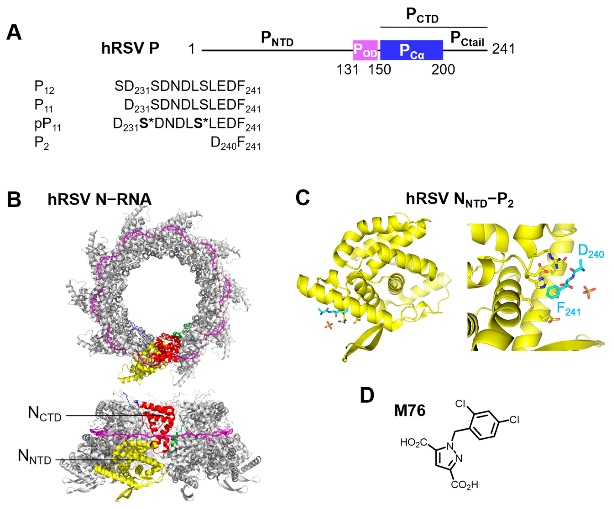
Figure 1. Human Respiratory Syncytial Virus (hRSV) phosphoprotein P and nucleoprotein N. (A) Domain organization of the RSV P protein (NTD; N-terminal domain, OD; oligomerization domain, CTD; C-terminal domain, itself divided into an α-helix-rich domain Cα and a disordered C-terminal tail, Ctail) and amino acid sequence of C-terminal P peptides. Phosphorylated serines are marked by *. (B) Two views of the X-ray crystal structure of an RSV N-RNA ring containing 10 N protomers (PDB 2wj8) in cartoon representation. The 7-mer RNA molecules are in magenta. One protomer is highlighted in color: N-terminal domain (NNTD) in yellow, C-terminal domain (NCTD) in red, N-terminal arm in green, C-terminal arm in blue. (C) Cartoon representation of the X-ray crystal structure of RSV NNTD (yellow) in complex with the C-terminal P2 peptide (cyan sticks) (PDB 4uc9). (D) Structure of M76 that inhibits the RSV NNTD-PCTD interaction.
Ijms 24 00569 g001
Figure 2. Binding of RSV pP11 peptide to RSV NNTD protein followed by NMR. (A) Selected region of superimposed 1H-15N HSQC spectra of 50 µM 15N-labeled NNTD, in 20 mM MES pH 6.5, 250 mM NaCl buffer, recorded during a titration experiment with the pP11 peptide. The magnetic field was 14.1 T, and the temperature 20 °C. The color code corresponds to the pP11:NNTD molar ratio as indicated below the spectrum. (B) Bar diagram of combined amide chemical shift perturbations (ΔδHN) measured for NNTD in the presence of 4.2 molar equivalents of pP11. Regions with significant CSPs are highlighted with a colored background: C-terminus of the αN1 helix (blue), β-hairpin (green), center of the αI2 helix (magenta), η1 loop (cyan), H151-loop (yellow). (C) Mapping of CSPs onto the X-ray structure of NNTD (PDB 4ucc): amide nitrogen atoms are represented as red spheres for residues with |ΔδHN| > mean + 2 SD, and orange spheres for |ΔδHN| > mean + SD. The color code is the same as in (B). The same view in surface representation highlights the PCTD binding pocket, indicated by a white arrow.
Ijms 24 00569 g002
Figure 3. Binding of F11 to RSV NNTD and inhibition by M76 assayed by fluorescence polarization. The buffer was MES pH 6.5, with 250 mM NaCl or without salt. The concentration of the fluorescent probe was 200 nM. (A) Binding of NNTD to F11 in buffer with salt: ΔFP = FP − FPmin (the FP of free F11 was FPmin = 30 mFP) was plotted as a function of the NNTD:F11 molar ratio. Error bars represent the difference between duplicate experiments. Data were fitted with a single site binding model (straight line) using Kd and ΔFPmax as parameters. R2 indicates the quality of the fit. (B) Binding of NNTD to F11 in salt-free buffer assayed by ΔFP. The area in the dotted contour box is expanded on the right side. Data were fitted with a single binding site model and an additional linear contribution. (C) Inhibition of NNTD (5 µm) binding to F11 by the M76 Molecule in salt-free Buffer. FP data were fitted with a Hill Equation. IC50 and R2 values are indicated. The dashed line indicates the mean FP value of free F11.
Ijms 24 00569 g003
Figure 4. Binding of Fn peptides to RSV N-RNA complex and inhibition by M76 assayed by fluorescence polarization. The buffer was Tris pH 8.0 without salt. The concentration of fluorescent probe was 200 nM. (A) Binding of N-RNA to F11 was assayed by ΔFP as a function of the N:F11 molar ratio. Data were fitted with a single site binding model. (B) Inhibition of N-RNA (10 µM) binding to F11 by M76: The left hand panel represents the experimental FP data, and the right hand panel the percentage of inhibition, calculated assuming FPmax = 120 mFP and FPmin = 40 mFP. (C) Influence of solvent addition on ΔFP measurements for the F7E peptide in the presence of N-RNA.
Ijms 24 00569 g004
Figure 5. Binding of FTU-labeled RSV P peptides (Fn) to RSV N-RNA and inhibition by unlabeled peptides assayed by FP. Data were acquired after 30 min incubation. Samples were in Tris pH 8 buffer without salt. The concentration of fluorescent probes was 200 nM. (A) Binding of N-RNA to Fn peptides was assayed by ΔFP as a function of the N:peptide molar ratio for peptides of various lengths. FP of free fluorescent peptide was FPmin ~30–40 mFP. Error bars represent the standard deviation of triplicate measurements. Curves were fitted with a single binding site model. (B) ΔFP binding curves for 11-mer fluorescent peptides (wild-type F11, Ser237Glu mutant F11SE and double Ser232Glu/Ser237Glu mutant F11EE) with N-RNA. Data were fitted with a single binding site model (straight lines). For F11SE a second calculated curve in broken line corresponds to a model with an additional linear component. (C) Inhibition curves of N-RNA (10 µM) binding to F11 by the unlabeled P7E, P9E and P11EE peptides. Data were fitted with a Hill equation. The dashed line indicates the mean FP value of free F11. (D) Inhibition curves of N-RNA (10 µM) binding to Fn peptides by unlabeled P peptides containing the same amino acid composition.
Ijms 24 00569 g005
Figure 6. Binding of fluorescent molecules to RSV N-RNA and inhibition by M76 assayed by fluorescence polarization. Measurements were carried out in Tris pH 8, Brij-35 buffer without salt. The concentration of fluorescent probes was 200 nM. (A) Chemical structure and name of fluorescent molecules. For fluorescein 1, the open form is shown. Fluorescein methyl ester 2; fluorescein ethyl ester 3; 4,4′ difluoro BODIPY FL 4; 4,4′ dicyano BODIPY FL 5. (B) ΔFP binding curves of fluorescent molecules as a function of the N:fluorescent molecule molar ratio. Fits were performed assuming FPmax = 500 mFP and FPmin = 0 mFP. (C) Inhibition of fluorescent molecule binding to N-RNA (20 µM) by M76.
Ijms 24 00569 g006
Figure 7. Analysis of fluorescein binding to RSV NNTD by NMR and by in silico docking. (A) Selected region of superimposed 1H-15N HSQC spectra of 15N-labeled RSV NNTD obtained during titration by fluorescein. NMR data were acquired at a magnetic field of 14.1 T, a temperature of 20 °C, and with 50 µM 15N-NNTD concentration. The color code corresponds to the fluorescein:NNTD molar ratio, as indicated on top. (B) Bar diagram of combined 1H and 15N amide chemical shift perturbations (ΔδHN) measured in the presence of 2 molar equivalents of fluorescein. (C) Mapping of CSPs onto the X-ray structure of NNTD (PDB 4ucc). Amide nitrogens are represented as red and orange spheres for residues with |ΔδHN| > mean + 2 SD and |ΔδHN| > mean + SD, respectively. Regions with significant CSPs are labeled with the same color code on the structure and on the diagram in (B). (D) Two docking poses of fluorescein binding to NNTD: the first pose was obtained with MOE (green) and the second with Smina (blue). (E) X-ray crystal structure of the NNTD-M76 complex (PDB 4ucc).
Ijms 24 00569 g007
Figure 8. Binding of dicyano BODIPY FL-labeled C-terminal RSV P peptides to RSV N-RNA and inhibition assayed by FP. Samples were in Tris pH 8 buffer, with or without salt. The concentration of fluorescent peptides was 200 nM. Error bars represent the standard deviation of measurements in triplicate. Fitted curves are in straight lines. (A) ΔFP binding curves of fluorescent BCNn peptides of various lengths to N-RNA in salt-free buffer (FPmin ~ 10 mFP). Data were fitted with a single binding site model. (B) ΔFP binding curves measured in buffer with salt (100 mM NaCl). (C) Inhibition of N-RNA (1 µM) binding to B10EE peptide by unlabeled P10EE peptide and by M76. Data were fitted with a Hill equation. (D) Inhibition of N-RNA (1 µM) binding to two BCNn peptides (BCN10EE and BCN11EE) by unlabeled P peptides with equivalent amino acid sequences (P10EE and pP11). (E) Inhibition by M76 of N-RNA (2 µM) binding to fluorescent peptides BCN5E, BCN6E, BCN8E, and BCN10EE.
Ijms 24 00569 g008
Figure 9. Structural model of the RSV NNTD-P11 complex. (A,B) Structures were generated with Haddock software [65], based on the crystal structure of the NNTD-P2 complex (PDB 4uc9) for NNTD and on a P11 model generated with PEP-FOLD3 [64]. 10 models were superimposed. The docked P11 peptides are in yellow cartoon. (A) Two views of NNTD rotated by 90° show the electrostatic surface potential calculated with Delphi software [66], with a scale ranging from −10 (red) to +10 kcal.mol−1 (blue). (B) The backbone of NNTD is shown in cartoon representation. (C) The P11 starting structure used for Haddock docking, in magenta, was aligned with the docked structures represented in other colors. The view is rotated, as compared to (A,B). (D) 20 P11 models generated with PEP-FOLD3 were structurally aligned along the three C-terminal residues Glu239-Phe241. The model used for Haddock docking is in magenta, and the other models in colors unrelated to those used in (C).
Ijms 24 00569 g009
Scheme 1. Synthesis of dicyano BODIPY FL 5. (a) BF3.OEt2, TMSCN, DCM, 2 h; (b) THF/H2O/HCl conc, 12 h.
Ijms 24 00569 sch001
Table 1. FTU-labeled RSV P peptides (Fn) designed for fluorescence polarization measurements with RSV N-RNA and binding parameters obtained from FP curve fits. ϕ designates the FTU label. Phosphomimetic peptides, where Ser232 and/or Ser237 were mutated into glutamates, are annotated with the letter E. FP measurements were done with 200 nM fluorescent probe in Tris pH 8.0 buffer without salt. Kd and ΔFPmax were determined by fitting binding curves to a single site binding model or a combination with a linear contribution (lines marked with *).
Fn PeptidePeptide SequenceKd (µM)ΔFPmax (mFP)
F11ϕ-DS232DNDLS237LEDF-OH7.6 ± 0.9130 ± 5
F11EEϕ-DE232DNDLE237LEDF-OH0.38 ± 0.12/0.15 ± 0.12 (*)114 ± 6/93 ±13 (*)
F11SEϕ-DS232DNDLE237LEDF-OH0.87 ± 0.26/0.62 ± 0.25 (*)111 ± 9/97 ± 12 (*)
F9Eϕ-DNDLE237LEDF-OH2.2 ± 0.3148 ± 5
F7Eϕ-DLE237LEDF-OH2.5 ± 0.3187 ± 7
F5Eϕ-E237LEDF-OH5.9 ± 0.5285 ± 7
F4ϕ-LEDF-OH35 ± 4291 ± 20
F3ϕ-EDF-OH62 ± 19315 ± 61
Table 2. Inhibition parameters obtained by FP for FTU-labeled RSV P peptides (Fn) in complex with RSV N-RNA. Samples were in Tris pH 8 buffer without salt. IC50 values were determined from inhibition experiments with 10 µM N-RNA and 200 nM fluorescent probe. Fitting of the inhibition data was done with a Hill equation. Ki was calculated from IC50 and Kd using the IC50-to-Ki server [49].
Fn PeptideInhibitorIC50 (µM)Ki (µM)Kd of Fn Peptide (µM) *Kd of Fn Equivalent to Inhibitor (µM) *
F11P11EE22 ± 467.60.38/0.15
F11P9E66 ± 6257.62.2
F11P7E180 ± 11757.62.5
F11P1186 ± 7347.67.6
F7EP7E376 ± 26742.52.5
F5EP5E433 ± 211585.95.9
F11M7613 ± 237.6
F7EM7648 ± 582.5
* Kd values of Fn peptides are from Table 1.
Table 3. Parameters for binding to RSV N-RNA and inhibition by M76 obtained for fluorescent molecules from FP. Apparent Kds were calculated assuming FPmax = 500 mFP. IC50s were determined from inhibition experiments with 20 µM N-RNA and 200 nM fluorescent probe. Kis were calculated from IC50 and Kd using the IC50-to-Ki server [49].
Fluorescent MoleculeKd (µM)M76 IC50 (µM)M76 Ki (µM)
Fluorescein 191 ± 527 ± 213
Fluorescein methyl ester 2200 ± 846 ± 732
Fluorescein ethyl ester 3250 ± 2765 ± 950
4,4′ difluoro BODIPY FL 4870 ± 140(*)(*)
(*) not determined.
Table 4. Dicyano BODIPY FL-labeled RSV P (BCN-P) peptides designed for FP measurements with RSV N-RNA and binding parameters obtained from FP. Samples were in Tris pH 8.0 buffer, without or with salt (100 mM NaCl). BCNn peptide concentration was 200 nM. Kd and ΔFPmax were obtained by fitting binding curves to a single site binding model.
Fluorescent BCNn PeptideFluorescent Peptide SequenceΔFPmax (mFP)Kd (µM)ΔFPmax (mFP)Kd (µM)
No SaltNo SaltWith SaltWith Salt
BCN11SEBCN-DSDNDLELEDF-OH312 ± 250.82 ± 0.22122 ± 20.55 ± 0.07
BCN11EEBCN-DEDNDLELEDF-OH316 ± 120.28 ± 0.07197 ± 50.26 ± 0.04
BCN10EEBCN-EDNDLELEDF-OH270 ± 70.16 ± 0.03171 ± 161.24 ± 0.31
BCN9EBCN-DNDLELEDF-OH288 ± 150.96 ± 0.13
BCN8EBCN-NDLELEDF-OH298 ± 51.43 ± 0.08284 ± 15 (*)7.1 ± 0.1 (*)
BCN7EBCN-DLELEDF-OH332 ± 82.2 ± 0.2
BCN6EBCN-LELEDF-OH275 ± 82.3 ± 0.2281 ± 19 (*)12.6 ± 1.6 (*)
BCN5EBCN-ELEDF-OH431 ± 159.8 ± 0.9
(*) Saturation of experimental binding curve not reached.
Table 5. Inhibition parameters obtained by FP for dicyano BODIPY FL-labeled RSV P peptides in complex with RSV N-RNA. Samples were in Tris pH 8 buffer. Fluorescent probe concentration was 200 nM. N concentration was between 1 and 20 µM as indicated. IC50 and FPmax were obtained by fitting the inhibition data with a Hill equation. Ki was calculated using the IC50-to-Ki server [49].
Fluorescent PeptideInhibitor[N-RNA] (µM)Fitted FPmax (mFP)Fitted IC50 (µM)Ki (µM)Kd (µM) of BCN-P Peptide (*)
BCN10EEP10EE121421 ± 42.90.16
BCN11EEpP1112346.9 ± 1.21.40.28
BCN11SEM76528017 ± 21.80.8
BCN10EEM7622578.9 ± 0.60.50.16
BCN10EEM7612198.5 ± 0.61.10.16
BCN8EM76519613 ± 22.01.4
BCN8EM7621664.5 ± 0.41.31.4
BCN6EM761017714 ± 11.12.3
BCN6EM7621042.7 ± 0.20.72.3
BCN5EM762028026 ± 23.69.8
BCN5EM762767.0 ± 0.64.89.8
(*) Kds of the BCNn peptides are from Table 4.
Disclaimer/Publisher’s Note: The statements, opinions and data contained in all publications are solely those of the individual author(s) and contributor(s) and not of MDPI and/or the editor(s). MDPI and/or the editor(s) disclaim responsibility for any injury to people or property resulting from any ideas, methods, instructions or products referred to in the content.