Article

Early-Life Exposure to Traffic-Related Air Pollutants Induced Anxiety-like Behaviors in Rats via Neurotransmitters and Neurotrophic Factors

1
Department of Immunology, School of Medicine, International University of Health and Welfare, 4-3, Kozunomori, Narita 286-8686, Chiba, Japan
2
Health and Environmental Risk Division, National Institute for Environmental Studies, Tsukuba 305-8506, Ibaraki, Japan
*
Author to whom correspondence should be addressed.
Int. J. Mol. Sci. 2023, 24(1), 586; https://doi.org/10.3390/ijms24010586
Submission received: 29 September 2022 / Revised: 6 December 2022 / Accepted: 22 December 2022 / Published: 29 December 2022
(This article belongs to the Special Issue Advances in the Research of Endocrine Disrupting Chemicals 2023)

Abstract

:
Recent epidemiological studies have reported significantly increasing hospital admission rates for mental disorders such as anxiety and depression, not only in adults but also in children and adolescents, indicating more research is needed for evaluation of the etiology and possible reduction and prevention of these disorders. The aim of the present study was to examine the associations between perinatal exposure to traffic-related air pollutants and anxiety-like behaviors and alterations in neurological and immunological markers in adulthood using a rat model. Sprague Dawley pregnant rats were exposed to clean air (control), diesel exhaust (DE) 101 ± 9 μg/m3 or diesel exhaust origin secondary organic aerosol (DE-SOA) 118 ± 23 μg/m3 from gestational day 14 to postnatal day 21. Anxiety-related behavioral tests including open field tests, elevated plus maze, light/dark transition tests and novelty-induced hypophagia were performed on 10-week-old rats. The hippocampal expression of neurotransmitters, neurotrophic factors, and inflammatory molecular markers was examined by real-time RT-PCR. Anxiety-like behaviors were observed in both male and female rat offspring exposed to DE or DE-SOA. Moreover, serotonin receptor (5HT1A), dopamine receptor (Drd2), brain-derived neurotrophic factor and vascular endothelial growth factor A mRNAs were significantly decreased, whereas interleukin-1β, cyclooxygenase-2, heme oxygenase-1 mRNAs and microglial activation were significantly increased in both male and female rats. These findings indicate that brain developmental period exposure to traffic-related air pollutants may induce anxiety-like behaviors via modulation of neurotransmitters, neurotrophic factors, and immunological molecular markers, triggering neuroinflammation and microglia activation in rats.

1. Introduction

Air pollution is has emerged as a global health problem, especially in rapidly developing countries. The World Health Organization estimates that over four million premature deaths in every year are due to air pollution. Particulate matter (PM)2.5 consists of diesel exhaust particles (DEP), which are major precursors of secondary organic aerosol (SOA) formation. Using commercial nano-sized carbon black and whole-body exposure chambers established in our research institute, we have reported the effect of exposure to air pollutants on alteration of brain neurotransmitters [1,2] and impaired behavior such as spatial learning and novel object recognition ability in mice and rats [3,4,5]. In addition, impaired social behavior and altered hypothalamic expression of social behavior-related genes have been observed in adult male mice offspring exposed to DE-SOA during the gestational and lactational periods [6].
In human studies, cognitive deficit, poor olfactory and auditory functions, anxiety, depression and other mental disorders have been reported [7,8,9,10]. EEG changes were found in subjects exposed to DE (300 μg/m3) for 1 h [11]. In addition, post-mortem examination of individuals exposed to high levels of air pollutants have shown increased oxidative stress markers and neuroinflammation [12,13]. A recent epidemiological study has demonstrated that short-term exposure to traffic-related pollutants, especially PM2.5, was significantly associated with hospital admissions for anxiety in the coastal city of Qingdao, China [14]. In that study, it was reported that females and younger individuals were more susceptible than males and elderly individuals.
In animal studies, it was reported that particles or gases in air pollutants can enter the brain by directly through the nasal olfactory mucosa [15,16], and by entering circulation and then cross the blood brain barrier [17,18]. Significantly increased proinflammatory cytokine tumor necrosis factor-α and microglia activation were found in mice exposed to DE compared with those exposed to filtered air [19]. Using the BrdU/NeuN co-localization method, Costa et al. showed that DE exposure significantly decreases neurogenesis in the hippocampal subgranular and subventricular zones [20]. Disruption of DNA methylation in the brain was observed in mice exposed to DE prenatally, by affecting genes involved in neuronal differentiation and neurogenesis [21]. In addition, using DE and DE-SOA as models of air pollutants, we have shown that perinatal exposure to DE-SOA induces autism-like behavior and dysregulation of neuroimmune responses in rats [22].
The hippocampus is the important brain area for cognitive functions such as episodic memory and spatial navigation. In addition, it is also involved in the pathogenesis of mood and anxiety disorders. The dorsal hippocampus contributes to cognitive functions, and the ventral hippocampus modulates emotional regulation [23,24]. The hippocampus, amygdala and prefrontal cortex are the brain areas which play major roles in emotional behaviors and functions. The neural connections projecting from ventral hippocampus to the prefrontal cortex are unidirectional. The ventral hippocampus also has bidirectional connections with the amygdala, and the amygdala has bidirectional connections with the prefrontal cortex. Because the amygdala and hippocampus are known to be involved in emotional and contextual memory processing, stress likely contributes to the dysregulation of these functions. Using freely moving calcium imaging and optogenetics, it was reported that optogenetic activation of ventral hippocampal terminals in the lateral hypothalamus, but not the basal amygdala, increased anxiety and avoidance. The ventral hippocampal area is enriched in anxiety cells; thus, the hippocampus can rapidly influence innate anxiety behavior directly via the ventral hippocampus–lateral hypothalamus pathway [25]. Transgenic animals with impaired hippocampal neurogenesis exhibit significantly increased anxiety-like behaviors [26]. Taken together, these studies indicate that the hippocampus is the essential brain region for anxiety-like behaviors that result from exposure to environmental insults. Thus, in the present study, the hippocampus was selected to examine environmental pollutant-induced anxiety-like behaviors in rats.
Currently, prevalence of mental disorders such as anxiety and depression is increasing. However, little is known about the association between environmental pollutant-exposure and anxiety. This prompted us to address this gap in the research concerning whether there is an association between developmental exposure to DE or DE-SOA and anxiety.

2. Results

2.1. Assessment of Overall Toxicity

At the time of sample collection, body weight and brain weight were measured in 10-week-old male and female rat offspring of the control and exposure groups to detect the overall toxicity. Body weight was not different between groups in both male and female rat offspring. However, brain weight was significantly increased in DE-SOA groups of both male and female rat offspring (p < 0.01, p < 0.05 vs. corresponding control, Figure 1).

2.2. Anxiety-Related Behavior Assessment

2.2.1. Open Field Test

The OFT is an experimental test used to determine general locomotor activity levels, anxiety, and exploratory behavior of rodents in scientific research. The male and female rats exposed to DE and DE-SOA groups showed a significantly decreased number of entries to the center and time stay in the center compared with the control group (p < 0.01; p < 0.05, Figure 2). Locomotor activity or distance traveled was not different between the control and exposure groups. However, grooming, rearing and defecation were increased in DE or DE-SOA exposed male and female rats. Our findings indicate that DE or DE-SOA exposure during the perinatal period may induce emotional insecurity and restlessness in male and female rats.

2.2.2. Elevated Plus Maze

The elevated plus maze (EPM) test was performed to assess the emotional level of the rats and rodents. The emotional security level was calculated based on two factors of entering the open arms and time spent in the arms. The male and female rats exposed to DE or DE-SOA groups showed a significantly decreased open arms entry compared with the control group (p < 0.01, p < 0.05, Figure 3) but no significant difference in time spent in open arms. In the EPM test, a decreased number of open arm entries, and reduced time spent in the open arms indicate increased anxiety responses.

2.2.3. Light Dark Transition Test

In the light dark transition (LDT) test, the number of entries and time spent in the light compartment were not different between the control and exposure groups in male rats. However, DE-exposed female rats showed an increased number of entries and time spent in the light compartment compared with the control rats (p < 0.01, p < 0.05, Figure 4). These findings indicate that DE exposure may affect some part of anxiety behavior in a sex-specific manner.

2.2.4. Novelty-Induced Hypophagia Test

Novelty-induced hypophagia (NIH) is a validated test for anxiety-like behavior induced by a new environment [27,28]. Latency to approach and latency to eat sweet potato were decreased in Day 2 and Day 3 compared with Day 1 in all groups. On novelty day, latency to approach and latency to eat sweet potato were delayed in DE- or DE-SOA-exposed male and female rats compared with the control group (p < 0.05, Figure 5). These findings indicate that perinatal exposure to DE or DE-SOA may induce anxiety-like behavior in male and female rats in new or novel environments.

2.3. Messenger RNA Expression Assay

2.3.1. Neurotransmitter Receptor Levels in the Hippocampus

Messenger RNA expression level of neurotransmitters such as serotonin receptor 5HT1A and dopamine receptor Drd2 mRNA in the hippocampus were examined. In male rats, 5HT1A and Drd2 mRNAs were significantly decreased in the DE- and DE-SOA-exposed groups compared with the control group (p < 0.01, p < 0.05 vs. control, Figure 6A). In contrast, in female rats, 5HT1A and Drd2 mRNAs were significantly decreased in the DE-SOA-exposed group only compared with the control group (p < 0.01, Figure 6B). These findings indicate that perinatal exposure to DE or DE-SOA may induce alteration in neurotransmitters in male and female rats in a sex-dependent manner.

2.3.2. Neurotrophin Levels in the Hippocampus

Brain-derived neurotrophic factor (BDNF) and vascular endothelial growth factor A (VEGFA) in the hippocampus were examined. In male rats, both BDNF and VEGFA mRNAs were significantly decreased in the DE-SOA-exposed group but not in the DE group compared with the control group (p < 0.05 vs. control, Figure 7A). However, in female rats, BDNF mRNA was significantly decreased in the DE-SOA-exposed group compared with the control group (p < 0.01 vs. control, Figure 7B). VEGFA mRNA in the female rats did not show significant difference between the control and exposure groups. These findings indicate that perinatal exposure to DE-SOA may induce reductions in BDNF levels in male and female rats.

2.3.3. Proinflammatory Cytokines and Oxidative Stress Markers in the Hippocampus

To detect DE or DE-SOA-induced inflammation in the hippocampus, the inflammatory molecular markers such as interleukin (IL)-1β, cyclooxygenase (COX)2 and heme oxygenase (HO)-1 in the hippocampus were examined. IL-1β, COX2 and HO1 mRNAs were significantly increased in the DE-SOA-exposed male rats compared with the control group (p < 0.05; Figure 8A). However, the expression levels of IL-1β mRNA only was remarkably increased in female rats exposed to DE-SOA compared with the control group (p < 0.05; Figure 8B). These results indicate that perinatal exposure to DE or DE-SOA may induce inflammatory responses in male and female rats in a sex-dependent manner.

2.4. Immunohistochemical Analyses

Microglial marker Iba1 was used to examine the activation of microglia in the hippocampus. Representative digital photomicrographs of Iba1-immunostained sections taken from the hippocampus of the control, DE- or DE-SOA-exposed groups are shown in Figure 9. Microglial activation was markedly increased in the hippocampus of the De- and DE-SOA-exposed group compared with that in the control group. Iba1-positive microglia in the DG area of the hippocampus were quantified under a high-power field using ImageJ software.

3. Discussion

The major findings of the present study were that the gestational and lactational exposure to DE-SOA induces the following responses: (1) anxiety-like behaviors such as decreased entry to the center and time stay in the center in OFT, decreased open arm entry and open arm time in EPM tests, and latency to approach and latency to eat food on the novelty day in NIH tests; (2) downregulation of anxiety-related gene serotonin receptor 5HT1A and neurotrophic factor BDNF; (3) upregulation of proinflammatory cytokine IL-1β, COX2 and oxidative stress marker HO-1; and (4) increased microglia activation in the hippocampus in male and female rats. Our findings suggest that the perinatal exposure to DE-SOA induces anxiety-like behavior in rats by triggering neuroinflammation and neuropsychological disorders via neurological and immunological biomarkers in the brain.
According to the WHO, air pollution is the fourth largest risk factor for global mortality [29]. Major air pollutants are organic hydrocarbons, particulate matter (PM), carbon monoxide (CO), nitrogen dioxide (NO2), carbon dioxide (CO2), and heavy metals [30]. The major sources of these air pollutants and heavy metals are from transportation [31]. Thus, traffic-related air pollution is a major contributor to global air pollution. Air pollution due to gasoline and diesel emissions from internal combustion engines of automobiles, trucks, locomotives, and ships leads to 800,000 premature deaths annually due to pulmonary, cardiovascular, and neurological complications [32]. People living and working in areas of heavy vehicle traffic have high susceptibility to mental health problems and learning and memory impairment such as anxiety, depression, and cognitive deficits.
Using animal studies, we have reported that exposure to diesel exhaust and diesel engine derived secondary organic aerosols is associated with adverse effects in the central nervous system (CNS) via increased oxidative stress and neuroinflammation. We have shown that the effect of exposure to the nano-sized carbon black (14 nm) and nanoparticle-rich diesel exhaust particles causes alteration of brain neurotransmitters [1,2] and impairs behavior such as spatial learning and novel object recognition ability [3,4,5] in mice and rats. In addition, impaired social behavior and altered hypothalamic expression of social behavior-related genes were observed in adult male mice offspring exposed to DE-SOA during the gestational and lactational periods [6]. In addition, using DE and DE-SOA as models of air pollutants, we have shown that perinatal exposure to DE-SOA induces autism-like behavior and dysregulation of neuroimmune responses in rats [22]. However, little is known about the association between environmental pollutant-induced autism and anxiety. This prompted us to address a research gap concerning the associations between developmental exposure to DE and/or DE-SOA and anxiety.
In preliminary studies, our research group investigated the critical period of exposure to nano-sized diesel exhaust particles for brain functions and behaviors in relation to different phases of developmental periods such as the whole gestational period, early, middle, and late gestational period, neonatal period, and perinatal period. We found that the perinatal period (GD 14 to PND21) was the most vulnerable period for the whole-body exposure to DE or DE-SOA. In addition, epidemiological studies have reported that exposure to air pollutants during prenatal and childhood periods is associated with neurodevelopmental and emotional disorders including ASD, ADHD, anxiety and depression [33,34,35]. Thus, in this study, we have selected the perinatal period to examine the effect of developmental exposure to DE or DE-SOA on neuropsychiatric diseases, especially anxiety-like behavior and neurological and immunological markers, using rat models.
In this study, four types of anxiety-like behavior tests including OFT, LDT test, EPM test and NIH tests were performed. In OFT, the center entry number and time spent in the center were significantly reduced in both male and female rats exposed to DE or DE-SOA. However, in the LDT test, only female rats exposed to DE showed increased entry number and time spent in the light compartment compared with the control group. In the EPM test, the male and female rats exposed DE and DE-SOA groups showed a significant decrease in the ability to enter the open arms, and shorter time spent in the open arms compared with the control group. Moreover, latency to approach and latency to eat in a novel environment were reduced significantly in both male and female rats exposed to DE or DE-SOA perinatally. Taken together, these results indicate that the male and female rats exposed to DE or DE-SOA during the perinatal period had induced anxiety-like behaviors.
The hypothalamic–pituitary–adrenal (HPA) axis and the serotonergic neuromodulatory system plays a crucial role in mediating anxiety behaviors [36]. 5-HT receptor abnormalities can induce a deficiency in brain serotoninergic activity, which causes increased vulnerability to anxiety. Among the 5HT receptors, it was reported that the 5-HT1A receptor knockout mice show increased anxiety [37,38], and in contrast, the 5-HT1A receptor overexpressed mice show decreased anxiety [39]. Although we did not detect the plasma corticosteroid levels in the present study, exposure to DE or DE-SOA can lead to the release of corticosteroids from the adrenal gland via the HPA axis, which can influence postsynatptic 5-HT1A function.
In human and animal studies, it was reported that dopamine D2 receptors play a major role in the pathogenesis of anxiety and depression [40,41,42]. In addition, D2Rs regulate anxiety-related behavior and elevate plus-maze-associative memory via the interactions of the dorsal hippocampus and the nucleus accumbens dopaminergic system [43]. In the present study, dopamine receptor Drd2, mRNAs in the hippocampus were remarkably reduced in DE-SOA-exposed male and female rats.
The vascular endothelial growth factor (VEGF)-A is essential for neurons because they require a large vascular supply and insufficient expression of VEGFA will lead to defects in the brain vascularization and neuronal apoptosis. VEGFA not only induces vascular permeability in tissues, including the blood brain barrier, but also facilitates the neurogenesis and proliferation of neurons, influences synaptic plasticity and modulates synaptic transmission in the adult hippocampus [44]. Moreover, VEGF can act as a neuroprotective factor in the adult brain, inhibiting apoptosis and inducing the growth of vascular and neuronal networks. Stress can decrease the hippocampal expression of VEGF [45]. In this study, VEGFA mRNAs in the hippocampus were significantly reduced in DE-SOA-exposed males, but not in female rats.
Brain-derived neurotrophic factor is a neurotrophin that influences cell growth, cell differentiation, and synaptic modification [46,47] and is highly expressed in the developing and adult hippocampus [48,49]. Recently, Val66Met, a single nucleotide polymorphism of the coding region of the BDNF gene has been identified as a risk factor for anxiety disorders and post-traumatic stress disorder [50,51]. In the present study, BDNF and VEGFA mRNAs in the hippocampus were remarkably reduced in DE-SOA-exposed male rats, and BDNF mRNA was remarkably reduced in DE-SOA-exposed male and female rats perinatally.
Microglia are natural immune cells in the brain and play a key role in neuroinflammation. Microglia detect changes in the surroundings and provide immunosurveillance activity. The functions of the microglia are maintaining neuronal synapses, identifying pathogens, and removing cellular debris. In this study, microglia marker Iba1 immunoreactivity was significantly increased in the dentate gyrus of the hippocampus of DE- or DE-SOA-exposed groups of male and female rats. This microglial activation was more remarkable in males compared with female rats.
In this study, some sex-dependent anxiety-like behavior (Table 1) and expression of molecular markers (Table 2) were observed. The exact etiology is not known, but females may possess additional protective activities compared with males. It has been reported that estrogen exerts protective actions by suppressing the neurotoxic stimulus or increasing the resilience of the brain to a given injury [52]. Moreover, paraoxonase-2 is a mitochondrial enzyme, possesses potent antioxidant and anti-inflammatory properties, and its expression in female monkeys was significantly higher than in male monkeys in multiple brain regions [53].
Significant differences in anxiety-like behaviors and neurological and immunological marker mRNAs between the control and DE-SOA groups were observed, but not between the DE and DE-SOA groups. Taken together, these results indicate that developmental exposure to DE or DE-SOA induces disturbances in the neurotransmitter system and neurotrophin production and also induces neuroinflammation via activated microglia. These release inflammatory molecules such as cytokines and oxidative stress markers, which may lead to anxiety-related behaviors in DE- or DE-SOA-exposed rats.

4. Materials and Methods

4.1. Animals

Twenty-four pregnant Sprague Dawley rats (gestational day (GD) 8) were purchased from Oriental Yeast Co., Ltd. (Tokyo, Japan) and exposed to clean air (control, n = 8), DE (n = 8), and DE-SOA (n = 8) from GD 14 to postnatal day (PND) 21 in whole-body exposure chambers. Food and water were given ad libitum. Date of birth was recorded as PND 0 and the offspring were housed in cages with dams under controlled environmental conditions (temperature, 22 ± 0.5 °C; humidity, 50 ± 5%; lights on 07:00–19:00 h). The pregnant rats were exposed clean air, DE or DE-SOA for 5 h per day (from 10:00 p.m. to 3:00 a.m.), 5 days a week excluding weekends from gestational day 14 to postnatal day 21 with their pups (Figure 10). The numbers of pups born were 253 (116 male and 137 female) in the control, 130 (68 male and 62 female) in the DE group, and 135 (63 male and 72 female) in the DE-SOA group. We used 3 male and 3 female pups from each dam (total 72 male and 72 female pups). Among them, 10 male and 10 female rats from 3 groups (total 60) were used for the anxiety-like behavior tests, and 5 male and 5 female rats from 3 groups (total 15) were used for immunohistochemical analyses.
The pups were weaned at PND 21 and 3~4 pups of the same sex were housed in a plastic cage. Anxiety-like behavior tests were performed at 10 weeks old. Behavioral testing was performed between 09:00 and 13:00 h. Before performing each test, the apparatus to be used was cleaned with 70% ethanol. After completing the anxiety behavioral tests, the rats were sacrificed under deep pentobarbital anesthesia. The hippocampus was collected from each animal, frozen quickly in liquid nitrogen, then stored at −80 °C until extraction of the total RNA. The experimental protocols were approved by the Ethics Committee of the Animal Care and Experimentation Council of the National Institute for Environmental Studies (NIES), Japan (AE-20-05, 9 March 2020). All efforts were made to minimize the number of animals used and their suffering.

4.2. Exposure to Clean Air, DE, or DE-SOA

The whole-body inhalation exposure chambers were established at the National Institute for Environmental Studies, Japan, as described previously [54]. Briefly, an 81-diesel engine (J08C; Hino Motors Ltd., Hino, Japan) was used to generate diesel exhaust. The engine was operated under a steady-state condition for 5 h a day. Our driving condition of the diesel engine was not simulated to any special condition in the real world. The engine operating condition (2000 rpm engine speed and 0 Nm engine torque) promoted the generation of high concentrations of nano-size particles. There were three chambers: a control chamber receiving clean air filtered through a high efficiency particulate air (HEPA) filter and a charcoal filter (referred to as “clean air”), the diluted exhaust (DE) chamber, which was without mixing of O3, and the DE-SOA chamber, which was generated by mixing DE with ozone at 0.6 ppm after secondary dilution. The secondary dilution ratios in the DE and DE-SOA chambers were the same, which resulted in the same particle and gaseous concentrations when O3 was not mixed. In fact, the concentrations of particles in DE-SOA were higher when O3 was mixed, and the concentrations of DE and DE-SOA were 101 ± 9 μg/m3 and 118 ± 23 μg/m3, respectively. The increased mass concentration was due to the generation of secondary particles. The temperature and relative humidity inside each chamber were adjusted to approximately 22 ± 0.5 °C and 50 ± 5%, respectively. In detail, sample air was taken from the inhalation chamber (2.25 m3) using stainless steel tubing. The gas concentrations (CO, CO2, NO, NO2, and SO2) were monitored using a gas analyzer (Horiba, Kyoto, Japan) (Table 3). The CO and NOx concentrations in both chambers were similar, but the NO and NO2 concentrations differed because NO was oxidized to NO2 by reaction with O3. The particle size distributions were measured using a scanning mobility particle sizer (SMPS 3034, TSI Instruments, Shoreview, MN, USA). The modal sizes of the particles used in the present study were 22.69 ± 1.47 nm for DE and 24.45 ± 1.21 nm for DE-SOA. The particles were collected using a Teflon filter (FP-500; Sumitomo Electric, Osaka, Japan) and a Quartz fiber filter (2500 QAT-UP; Pall, Pine Bush, NY, USA), and the particle mass concentrations were measured using a Teflon filter. The particle weights were measured using an electrical microbalance (UMX 2; Mettler-Toledo, Columbus, OH, USA; readability 0.1 μg) in an air-conditioned chamber (CHAM-1000; Horiba) under constant temperature and relative humidity conditions (21.5 °C, 35%). For the Quartz fiber filter, the quantities of elemental carbon (EC) and organic carbon (OC) were determined using a carbon analyzer (Desert Research Institute, Reno, NV, USA). The EC to OC ratios in the present study were 0.15 ± 0.06 for the control chamber, 0.36 ± 0.03 for the DE chamber, and 0.38 ± 0.03 for the DE-SOA exposure chamber.

4.3. Behavioral Assessment

All the animals were kept in the animal experimental room next to the behavior test room. The animals were placed in the testing room 30 min before the behavior test on the experimental day. Apart from the tested animal, other animals were kept in the animal experimental room during the whole experiment period. For every behavior test, a sufficient interval between test rats was maintained, and after cleaning with 70% ethanol, a hand-held fan was used to remove the smell of ethanol.

4.3.1. Open Field Test

The open field test is validated for use in the measurement of anxiety-related behaviors in rodents. The apparatus consisted of a square box (100 cm width × 100 cm length × 35 cm height) made of clear plastic. Inside the box, a center square (25 cm × 25 cm) was outlined in red tape. The apparatus was placed on the floor. Before and between trials, the box was cleaned with 70% ethanol and dried off. The rats were placed in the arena and allowed to explore it for 5 min. The movements were recorded by a video tracking system and stored on a computer. The video recordings were later scored for time spent in the center square, time spent moving throughout the box (locomotor time or distance travel), the number of entrances into the center, grooming (washing the face and body), rearing (standing on the hind limbs) and defecation (fecal pellets).

4.3.2. Elevated Plus Maze Test

The EPM apparatus was purchased from Muromachi Kikai (Tokyo, Japan) and consisted of two open and two closed arms. It was elevated 50.0 cm above the floor and each arm was 48.3 cm in length and 12.7 cm in width. The walls of the closed arms were 29.2 cm tall. On test day, the animals were moved to testing room 30 min before the experiment. At the beginning of testing, each rat was placed at the center of the crossing arms, facing the open arm and allowed to move freely within the maze for 5 min. Between trials, the apparatus was cleaned with 70% ethanol. Time spent in the open and closed arms, and entries into the open and closed arms were recorded. An animal was recorded as entering an arm when all four paws were placed in that arm. The illumination at the entrance into the open arms was set to ~90 lux.

4.3.3. Light Dark Transition Test

The apparatus contained two compartments of white and black opaque plexiglas (34 × 24 × 24 cm), with a rectangular aperture (8 cm diameter) between the two chambers permitting free movement between the chambers (Muromachi Kikai, Japan). The white compartment was named “the light compartment” and the black compartment was named “the dark compartment”. A black, non-translucent lid covered the dark compartment to exclude ambient room light. The light compartment was covered by a transparent plastic lid, which allowed room light into the apparatus. To begin testing, an animal was placed in the light compartment, facing away from the aperture between the chambers. The animal was allowed to investigate the chambers for 5 min and the number of entries and time spent in each compartment was recorded by video.

4.3.4. Novelty-Induced Hypophagia Test

The novelty-induced hypophagia (NIH) is a validated test for anxiety-like behavior induced by new environment [13,14]. Training Phase: Rats were removed from their home-cages and isolated for 30 min in a new standard cage (“training cage”), which was lined with fresh bedding and provided access to food and water, to acclimate to the novel environment (animals were not food or water restricted at any point during NIH testing). After the 30 min isolation period, a sweet potato slice was placed in the cage. The animal’s approaches to the sweet potato slice were recorded by video-assisted Any-maze software for 15 min. After 15 min, the rats were returned to their home-cages. The sweet potato slices were weighed before and after the training phase to determine how much of the sweet potato slice had been consumed by the rat. This process was repeated for the next 3 consecutive days using the same training cage.
Novelty Test: On the fourth day, rats were allowed to acclimate to their “training cage” for 30 min, as described previously for the training phase. Following this acclimation period, the animal was transferred to a novel plastic tub (58.42 cm (L) × 43.18 cm (H) × 31.75 cm (W) with no bedding/water/food) and a sweet potato slice was immediately placed in the novel tub. Explorations of the sweet potato slice in the novel environment were recorded by video for 15 min. After 15 min, the rats were returned to their home-cages. Sweet potato slices were weighed before and after the novelty test to determine the amount of consumption, as the amount of sweet potato slice consumed has also previously been reported as a measure of anxiety-like behavior [13]. Experimental videos were later analyzed for the time taken by the animal to approach and begin to consume the sweet potato (latency), as well as the amount of sweet potato consumed.

4.4. MessengerRNA Expression Assay

After completion of all anxiety-related behavioral tests, 10-week-old male and female rats (n = 10 from each group) were sacrificed under deep pentobarbital anesthesia and the hippocampus was collected from each group for mRNA analyses. Briefly, the total RNA was extracted from the hippocampal samples using the BioRobot EZ-1 and EZ-1 RNA tissue mini kits (Qiagen GmbH, Hilden, Germany). Then, the purity and quality of the total RNA were examined using the ND-1000 NanoDrop RNA Assay protocol (NanoDrop, Wilmington, DE, USA), as described previously [6]. Next, first-strand cDNA synthesis from the total RNA was made using SuperScript RNase H−Reverse Transcriptase II (Invitrogen, Carlsbad, CA, USA), according to the manufacturer’s protocol. The mRNA expression levels were examined by RT-PCR (Light Cycler 96, Roche, Mannheim, Germany). The tissue 18S rRNA level was used as an internal control. The following primer sequences were used in the present study. Some primers (BDNF, NM_012513; IL-1β, NM_008361; COX2, NM_011198; and HO-1, NM_010442) were purchased from Qiagen, Sample and Assay Technologies. Other primers were purchased from Hokkaido System Science (Sapporo, Japan) as follows: 18S (forward 5′-TACCACATCCAAAAGGCAG-3′, reverse 5′-TGCCCTCCAATGGATCCTC-3′), TNFα (forward 5′-GGTTCCTTTGTGGCACTTG-3′, reverse 5′-TTCTCTTGGTGACCGGGAG-3′), 5HT1A (forward 5′-CTGGGGACGCTCATTTTCT-3′, reverse 5′-CCAAGGAGCCGATGAGATAG-3′), Drd2 (forward 5′-TGTACAATACGCGCTACAGCTCCA-3′, reverse 5′-ATGCACTGCGTTCTGGTCTGCGTTA-3′), and VEGFA (forward 5′-CTGCTGTAACGATGAAGCCCTG-3′, reverse 5′-GCTGTAGGAAGCTCATCTCTCC-3′). Data were analyzed using the comparative threshold cycle method. Then, the relative mRNA expression levels were expressed as mRNA signals per unit of 18S rRNA expression.

4.5. Immunohistochemical Analyses

In the brain, microglia play a role as major immune cells, and microglial activation indicates neurotoxicity. The hippocampal tissue sections were immunostained with microglial marker Iba1. Briefly, the brain sections were immersed in absolute ethanol followed by 10% H2O2 for 10 min each at room temperature. After rinsing in 0.01-M phosphate buffer saline, the sections were blocked with 2% normal swine serum in PBS for 30 min at room temperature and then reacted with goat polyclonal anti-Iba1 (diluted 1:100; abcam: ab5076; Tokyo, Japan) in PBS for 1 h at 37 °C. Then, the sections were reacted with biotinylated donkey anti-rabbit IgG (1:300 Histofine; Nichirei Bioscience, Tokyo, Japan) in PBS for 1 h at 37 °C. The sections were then incubated with peroxidase-tagged streptavidin (1:300, ABC KIT) containing PBS for 1 h at room temperature. After a further rinsed in PBS, Iba1 immunoreactivity was detected using a Dako DAB Plus Liquid System (Dako Corp., Carpinteria, CA, USA). To detect the immunoreactivity of Iba1 in the hippocampus, photomicrographic digital images (150 dpi, 256 scales) of the hippocampal regions were taken using a CCD camera connected to a light microscope. Numbers of Iba1-positive microglia in the hippocampus were quantified in high power field using ImageJ software (5 fields per section and 3 sections per mouse, n = 5 rats/group).

4.6. Statistical Analysis

All the data were expressed as the mean ± standard error (SE). The statistical analyses were performed using the StatMate II statistical analysis system for Microsoft Excel, Version 5.0 (Nankodo Inc., Tokyo, Japan). The data were analyzed using a one-way analysis of variance with a post hoc analysis using the Bonferroni/Dunn method. Differences were considered significant at p < 0.05.

5. Conclusions

At least in part, anxiety-like behaviors were observed in both male and female rat offspring exposed to DE or DE-SOA. Moreover, hippocampal expression of neurotransmitters, including serotonin receptor 5HT1A, dopamine receptor Dr2d, BDNF and VEGFA mRNAs, were significantly decreased in both male and female rats. On the other hand, inflammatory markers, including IL-1β and COX2, oxidative stress marker HO1 and microglia activation in the hippocampus, were significantly increased in both male and female rat offspring exposed to DE or DE-SOA. Activated microglia were prominent in the hippocampus of both male and female rat offspring exposed to DE or DE-SOA during the perinatal period. Our results indicate that developmental exposure to air pollutants induces anxiety-like behaviors via modulation of neurotransmitters and neurotrophic factors in rats.
The differences between DE and DE-SOA are complex, as shown in Table 1 and Table 2. The effects of DE or DE-SOA are brain region-specific, and it would be preferable to examine the related brain regions simultaneously. Hippocampal volume, neurogenesis, apoptosis and autophagy are other mechanisms that contribute to hippocampus-dependent anxiety-like behaviors following air-pollutant exposure. The impact of exposure to DE-SOA appears greater than DE but is not statistically significant. DE is a precursor of DE-SOA, which contains ozone oxidized to form DE-SOA. Moreover, we could not determine exactly the differences between DE and DE-SOA in translocation to the brain or their effects on the anxiety-like behavior and molecular markers in the rat model. The limitation of this study was that only the hippocampus was examined. We plan to examine the constituents of DE or DE-SOA and their translocation to the brain and the different effects on other behaviors and molecular markers in future studies.

Author Contributions

T.-T.W.-S. designed the experiment, Y.F. performed exposure management, T.-T.W.-S. performed behavioral tests, C.K.-T.-T. performed histology and molecular analysis, C.K.-T.-T. wrote the manuscript, T.-T.W.-S. and S.H. critically revised the manuscript. All authors have read and agreed to the published version of the manuscript.

Funding

This research was funded by a Grant-in-Aid for Scientific Research (C) JSPS KAKENHI (Grant Number JP19K12361) andthe research project on health impacts of chemicals on children and future generations (National Institute for Environmental Studies, Japan: 1620AA041).

Institutional Review Board Statement

The study was conducted according to the guidelines of the Declaration of Helsinki, and approved by the Institutional Review Board of the National Institute for Environmental Studies, Tsukuba, Japan (AE-20-05 on 9 March 2020).

Informed Consent Statement

Not applicable.

Data Availability Statement

All data generated or analyzed during this study are available from the corresponding author on reasonable request.

Acknowledgments

The authors acknowledge Kaoru Onishi for her technical assistance.

Conflicts of Interest

The authors declare no conflict of interest.

Abbreviations

ASDautism spectrum disorder
DEdiesel exhaust
DE-SOAdiesel exhaust (DE)-origin secondary organic aerosol
5-HT1A5-hydroxytryptamine (serotonin) receptor 1A
Drd2dopamine receptor D2
BDNFbrain-derived neurotrophic factor
VEGFAvascular endothelial growth factor A
IL-1βinterleukin -1β
COX2cyclooxygenase 2
HO1heme oxygenase 1
TNFαtumor necrosis factor α
Iba1ionized calcium binding adaptor molecule 1
PNDpostnatal day

References

  1. Win-Shwe, T.T.; Mitsushima, D.; Yamamoto, S.; Fukushima, A.; Funabashi, T.; Kobayashi, T.; Fujimaki, H. Changes in neurotransmitter levels and proinflammatory cytokine mRNA expressions in the mice olfactory bulb following nanoparticle exposure. Toxicol. Appl. Pharmacol. 2007, 226, 192–198. [Google Scholar] [CrossRef] [PubMed]
  2. Win-Shwe, T.T.; Mitsushima, D.; Yamamoto, S.; Fujitani, Y.; Funabashi, T.; Hirano, S.; Fujimaki, H. Extracellular glutamate level and NMDA receptor subunit expression in mouse olfactory bulb following nanoparticle-rich diesel exhaust exposure. Inhal. Toxicol. 2009, 21, 828–836. [Google Scholar] [CrossRef] [PubMed]
  3. Win-Shwe, T.T.; Yamamoto, S.; Fujitani, Y.; Hirano, S.; Fujimaki, H. Spatial learning and memory function-related gene expression in the hippocampus of mouse exposed to nanoparticle-rich diesel exhaust. Neurotoxicology 2008, 29, 940–947. [Google Scholar] [CrossRef] [PubMed]
  4. Win-Shwe, T.T.; Yamamoto, S.; Fujitani, Y.; Hirano, S.; Fujimaki, H. Nanoparticle-rich diesel exhaust affects hippocampal-dependent spatial learning and NMDA receptor subunit expression in female mice. Nanotoxicology 2012, 6, 543–553. [Google Scholar] [CrossRef] [PubMed]
  5. Win-Shwe, T.T.; Fujimaki, H.; Fujitani, Y.; Hirano, S. Novel object recognition ability in female mice following exposure to nanoparticle-rich diesel exhaust. Toxicol. Appl. Pharmacol. 2012, 262, 355–362. [Google Scholar] [CrossRef] [PubMed]
  6. Win-Shwe, T.T.; Kyi-Tha-Thu, C.; Moe, Y.; Fujitani, Y.; Tsukahara, S.; Hirano, S. Exposure of BALB/c Mice to Diesel Engine Exhaust Origin Secondary Organic Aerosol (DE-SOA) during the Developmental Stages Impairs the Social Behavior in Adult Life of the Males. Front Neurosci. 2016, 9, 524. [Google Scholar] [CrossRef] [Green Version]
  7. Freire, C.; Ramos, R.; Puertas, R.; Lopez-Espinosa, M.J.; Julvez, J.; Aguilera, I.; Cruz, F.; Fernandez, M.F.; Sunyer, J.; Olea, N. Association of traffic-related air pollution with cognitive development in children. J. Epidemiol. Community Health 2010, 64, 223–228. [Google Scholar] [CrossRef]
  8. Calderon-Garciduenas, L.; Franco-Lira, M.; Henriquez-Roldan, C.; Osnaya, N.; Gomzalez-Maciel, A.; Reynoso-Robles, R.; Villareal-Calderon, R.; Herritt, L.; Brooks, D.; Keefe, S.; et al. Urban air pollution: Influences on olfactory function and pathology in exposed children and young adults. Exp. Toxicol. Pathol. 2010, 62, 91–102. [Google Scholar] [CrossRef] [Green Version]
  9. Fonken, L.K.; Xu, X.; Weil, Z.M.; Chen, G.; Sun, Q.; Rajagopalan, S.; Nelson, R.J. Air pollution impairs cognition, provokes depressive-like behaviors and alters hippocampal cytokine expression and morphology. Mol. Psychiatry 2011, 16, 987–995. [Google Scholar] [CrossRef] [Green Version]
  10. Guxens, M.; Sunyer, J. A review of epidemiological studies on neuropsychological effects of air pollution. Swiss Med. Wkly. 2012, 141, w13322. [Google Scholar] [CrossRef]
  11. Crüts, B.; Driessen, A.; van Etten, L.; Törnqvist, H.; Blomberg, A.; Sandström, T.; Mills, N.L.; Borm, P.J. Exposure to diesel exhaust induces changes in EEG I human volunteers. Part. Fibre Toxicol. 2008, 5, 4. [Google Scholar] [CrossRef] [PubMed] [Green Version]
  12. Calderón-Garcidueñas, L.; Solt, A.C.; Henríquez-Roldán, C.; Torres-Jardón, R.; Nuse, B.; Herritt, L.; Reed, W. Long-term Air Pollution Exposure Is Associated with Neuroinflammation, an Altered Innate Immune Response, Disruption of the Blood-Brain Barrier, Ultrafine Particulate Deposition, and Accumulation of Amyloid β-42 and α-Synuclein in Children and Young Adults. Toxicol. Pathol. 2008, 36, 289–310. [Google Scholar] [CrossRef] [PubMed]
  13. Levesque, S.; Surace, M.J.; McDonald, J.; Block, M.L. Air pollution and the brain: Subchronic diesel exhaust exposure causes neuroinflammation and elevates early markers of neurodegenerative disease. J. Neuroinflammation 2011, 8, 105. [Google Scholar] [CrossRef] [PubMed] [Green Version]
  14. Ji, Y.; Liu, B.; Song, J.; Cheng, J.; Wang, H.; Su, H. Association between traffic-related air pollution and anxiety hospitalizations in a coastal Chinese city: Are there potentially susceptible groups? Environ. Res. 2022, 209, 112832. [Google Scholar] [CrossRef] [PubMed]
  15. Oberdoerster, G.; Sharp, Z.; Atudorei, V.; Elder, A.; Gelein, R.; Kreyling, W.; Cox, C. Translocation of inhaled ultrafine particles to the brain. Inhal. Toxicol. 2004, 16, 437–445. [Google Scholar] [CrossRef] [PubMed]
  16. Peters, A.; Veronesi, B.; Calderón-Garcidueñas, L.; Gehr, P.; Chen, L.C.; Geiser, M.; Reed, W.; Rothen-Rutishauser, B.; Schürch, S.; Schulz, H. Translocation and potential neurological effects of fine and ultrafine particles a critical update. Part. Fibre Toxicol. 2006, 3, 13. [Google Scholar] [CrossRef] [Green Version]
  17. Oberdoerster, G.; Sharp, Z.; Atudorei, V.; Elder, A.; Gelein, R.; Lunts, A.; Kreyling, W.; Cox, C. Extra-pulmonary translocation of ultrafine carbon particles following whole body inhalation exposure of rats. J. Toxicol. Environ. Health A 2002, 65, 1531–1543. [Google Scholar] [CrossRef]
  18. Genc, S.; Zadeoglulari, Z.; Fuss, S.H.; Genc, K. The adverse effects of air pollution on the nervous system. J. Toxicol. 2012, 2012, 782462. [Google Scholar] [CrossRef] [PubMed] [Green Version]
  19. Cheng, H.; Davis, D.A.; Hasheminassab, S.; Sioutas, C.; Morgan, T.E.; Finch, C.E. Urban traffic-derived nanoparticulate matter reduces neurite outgrowth via TNFα in vitro. J. Neuroinflammation 2016, 26, 13–19. [Google Scholar] [CrossRef] [Green Version]
  20. Costa, L.G.; Cole, T.B.; Coburn, J.; Chang, Y.C.; Dao, K.; Roqué, P.J. Neurotoxicity of traffic-related air pollution. Neurotoxicology 2017, 59, 133–139. [Google Scholar] [CrossRef]
  21. Tachibana, K.; Takayanagi, K.; Akimoto, A.; Ueda, K.; Shinkai, Y.; Umezawa, M.; Takeda, K. Prenatal diesel exhaust exposure disrupts the DNA methylation profile in the brain of mouse offspring. J. Toxicol. Sci. 2015, 40, 1–11. [Google Scholar] [CrossRef] [PubMed] [Green Version]
  22. Win-Shwe, T.T.; Kyi-Tha-Thu, C.; Fujitani, Y.; Tsukahara, S.; Hirano, S. Perinatal Exposure to Diesel Exhaust-Origin Secondary Organic Aerosol Induces Autism-Like Behavior in Rats. Int. J. Mol. Sci. 2021, 22, 538. [Google Scholar] [CrossRef] [PubMed]
  23. Revest, J.M.; Dupret, D.; Koehl, M.; Funk-Reiter, C.; Grosjean, N.; Piazza, P.V.; Abrous, D.N. Adult hippocampal neurogenesis is involved in anxiety-related behaviors. Mol. Psychiatry 2009, 14, 959–967. [Google Scholar] [CrossRef] [PubMed] [Green Version]
  24. Fanselow, M.S.; Dong, H.W. Are the dorsal and ventral hippocampus functionally distinct structures? Neuron 2010, 65, 7–19. [Google Scholar] [CrossRef] [PubMed] [Green Version]
  25. Strange, B.A.; Witter, M.P.; Lein, E.S.; Moser, E.I. Functional organization of the hippocampal longitudinal axis. Nat. Rev. Neurosci. 2014, 15, 655–669. [Google Scholar] [CrossRef] [PubMed] [Green Version]
  26. Jimenez, J.C.; Su, K.; Goldberg, A.R.; Luna, V.M.; Biane, J.S.; Ordek, G.; Zhou, P.; Ong, S.K.; Wright, M.A.; Zweifel, L.; et al. Anxiety Cells in a Hippocampal-Hypothalamic Circuit. Neuron 2018, 97, 670–683.e6. [Google Scholar] [CrossRef] [PubMed] [Green Version]
  27. Dulawa, S.C.; Holick, K.A.; Gundersen, B.; Hen, R. Effects of chronic fluoxetine in animal models of anxiety and depression. Neuropsychopharmacology 2004, 29, 1321–1330. [Google Scholar] [CrossRef] [PubMed] [Green Version]
  28. Hunsberger, J.; Duman, C. Animal models for depression-like and anxiety-like behavior. Protoc. Exch. 2007, 1–13. [Google Scholar] [CrossRef]
  29. Vaughan, A. China Tops WHO List for Deadly Outdoor Air Pollution. The Guardian, 27 September 2016. [Google Scholar]
  30. Frampton, M.W.; Rich, D.Q. Does particle size matter? Ultrafine particles and hospital visits in Eastern Europe. Am. J. Respir. Crit. Care Med. 2016, 194, 1180–1182. [Google Scholar] [CrossRef]
  31. United States Environmental Protection Agency. 2014 National Emissions Inventory; United States Environmental Protection Agency: Washington, DC, USA, 2018.
  32. United States Environmental Protection Agency. Greenhouse Gas Emissions from a Typical Passenger Vehicle; United States Environmental Protection Agency: Washington, DC, USA, 2018.
  33. Forns, J.; Dadvand, P.; Foraster, M.; Alvarez-Pedrerol, M.; Rivas, I.; López-Vicente, M.; Suades-Gonzalez, E.; Garcia-Esteban, R.; Esnaola, M.; Cirach, M.; et al. Traffic-related air pollution, noise at school, and behavioral problems in Barcelona schoolchildren: A cross-sectional study. Environ. Health Perspect. 2016, 124, 529–535. [Google Scholar] [CrossRef]
  34. Yolton, K.; Khoury, J.C.; Burkle, J.; LeMasters, G.; Cecil, K.; Ryan, P. Lifetime exposure to traffic-related air pollution and symptoms of depression and anxiety at age 12 years. Environ. Res. 2019, 173, 199–206, Correction in Environ. Res. 2019, 176, 108519. [Google Scholar] [CrossRef] [PubMed]
  35. Brunst, K.J.; Ryan, P.H.; Altaye, M.; Yolton, K.; Maloney, T.; Beckwith, T.; Cecil, K.M.; LeMasters, G. Myo-inositol mediates the effects of traffic-related air pollution on generalized anxiety symptoms at age 12 years. Environ. Res. 2019, 175, 71–78. [Google Scholar] [CrossRef] [PubMed]
  36. Lanfumey, L.; Mongeau, R.; Cohen-Salmon, C.; Hamon, M. Corticosteroid-serotonin interactions in the neurobiological mechanisms of stress-related disorders. Neurosci. Biobehav. Rev. 2008, 32, 1174–1184. [Google Scholar] [CrossRef] [PubMed]
  37. Heisler, L.K.; Chu, H.-M.; Brennan, T.J.; Danao, J.A.; Bajwa, P.; Parsons, L.H.; Tecott, L.H. Elevated anxiety and antidepressant-like responses in serotonin 5-HT 1A receptor mutant mice. Proc. Natl. Acad. Sci. USA 1998, 95, 15049–15054. [Google Scholar] [CrossRef] [Green Version]
  38. Parks, C.L.; Robinson, P.S.; Sibille, E.; Shenk, T.; Toth, M. Increased anxiety of mice lacking the serotonin 1A receptor. Proc. Natl. Acad. Sci. USA 1998, 95, 10734–10739. [Google Scholar] [CrossRef] [PubMed] [Green Version]
  39. Kusserow, H.; Davies, B.; Hörtnagl, H.; Voigt, I.; Stroh, T.; Bert, B.; Theuring, F. Reduced anxiety-related behaviour in transgenic mice overexpressing serotonin 1A receptors. Brain Res. Mol. Brain Res. 2004, 129, 104–116. [Google Scholar] [CrossRef] [PubMed]
  40. Lawford, B.R.; Young, R.; Noble, E.P.; Kann, B.; Ritchie, T. The D2 dopamine receptor (DRD2) gene is associated with co-morbid depression, anxiety and social dysfunction in untreated veterans with post-traumatic stress disorder. Eur. Psychiatry 2006, 21, 180–185. [Google Scholar] [CrossRef] [PubMed]
  41. Block, M.L.; Wu, X.; Pei, Z.; Li, G.; Wang, T.; Qin, L.; Wilson, B.; Yang, J.; Hong, J.S.; Veronesi, B. Nanometer size diesel exhaust particles are selectively toxic to dopaminergic neurons: The role of microglia, phagocytosis, and NADPH oxidase. FASEB J. 2004, 18, 1618–1620. [Google Scholar] [CrossRef] [PubMed] [Green Version]
  42. Yokota, S.; Mizuo, K.; Moriya, N.; Oshio, S.; Sugawara, I.; Takeda, K. Effect of prenatal exposure to diesel exhaust on dopaminergic system in mice. Neurosci. Lett. 2009, 449, 38–41. [Google Scholar] [CrossRef]
  43. Ebrahimi-Ghiri, M.; Nasehi, M.; Zarrindast, M.R. The modulatory role of accumbens and hippocampus D2 receptors in anxiety and memory. Naunyn. Schmiedebergs Arch. Pharmacol. 2018, 391, 1107–1118. [Google Scholar] [CrossRef] [PubMed]
  44. McCloskey, D.P.; Croll, S.D.; Scharfman, H.E. Depression of synaptic transmission by vascular endothelial growth factor in adult rat hippocampus and evidence for increased efficacy after chronic seizures. J. Neurosci. 2005, 25, 8889–8897. [Google Scholar] [CrossRef] [PubMed] [Green Version]
  45. Ventriglia, M.; Zanardini, R.; Pedrini, L.; Placentino, A.; Nielsen, M.G.; Gennarelli, M.; Chiavetto, L.B. VEGF serum levels in depressed patients during SSRI antidepressant treatment. Prog. Neuropsychopharmacol. Biol. Psychiatry 2009, 33, 146–149. [Google Scholar] [CrossRef] [PubMed]
  46. Huang, E.J.; Reichardt, L.F. Neurotrophins: Roles in neuronal development and function. Annu. Rev. Neurosci. 2001, 24, 677–736. [Google Scholar] [CrossRef] [PubMed] [Green Version]
  47. Huang, E.J.; Reichardt, L.F. Trk receptors: Roles in neuronal signal transduction. Annu. Rev. Biochem. 2003, 72, 609–642. [Google Scholar] [CrossRef] [PubMed] [Green Version]
  48. Phillips, H.S.; Hains, J.M.; Laramee, G.R.; Rosenthal, A.; Winslow, J.W. Widespread expression of BDNF but not NT3 by target areas of basal forebrain cholinergic neurons. Science 1990, 250, 290–294. [Google Scholar] [CrossRef]
  49. Webster, M.J.; Herman, M.M.; Kleinman, J.E.; Shannon Weickert, C. BDNF and trkB mRNA expression in the hippocampus and temporal cortex during the human lifespan. Gene Expr. Patterns 2006, 6, 941–951. [Google Scholar] [CrossRef] [PubMed]
  50. Frielingsdorf, H.; Bath, K.G.; Soliman, F.; Difede, J.; Casey, B.J.; Lee, F.S. Variant brain-derived neurotrophic factor Val66Met endophenotypes: Implications for posttraumatic stress disorder. Ann. N. Y. Acad. Sci. 2010, 1208, 150–157. [Google Scholar] [CrossRef] [Green Version]
  51. Jiang, X.; Xu, K.; Hoberman, J.; Tian, F.; Marko, A.J.; Waheed, J.F.; Harris, C.R.; Marini, A.M.; Enoch, M.-A.; Lipsky, R.H. BDNF variation and mood disorders: A novel functional promoter polymorphism and Val66Met are associated with anxiety but have opposing effects. Neuropsychopharmacology 2005, 30, 1353–1361. [Google Scholar] [CrossRef] [Green Version]
  52. Wise, P.M.; Dubal, D.B.; Wilson, M.E.; Rau, S.W.; Böttner, M. Minireview: Neuroprotective effects of estrogen-new insights into mechanisms of action. Endocrinology 2001, 142, 969–973. [Google Scholar] [CrossRef]
  53. Jamwal, S.; Blackburn, J.K.; Elsworth, J.D. Sex-based disparity in paraoxonase-2 expression in the brains of African green monkeys. Free Radic. Biol. Med. 2021, 167, 201–204. [Google Scholar] [CrossRef]
  54. Fujitani, Y.; Hirano, S.; Kobayashi, S.; Tanabe, K.; Suzuki, A.; Furuyama, A.; Furuyama, A.; Kobayashi, T. Characterization of dilution conditions for diesel nanoparticle inhalation studies. Inhal. Toxicol. 2009, 21, 200–209. [Google Scholar] [CrossRef] [PubMed]
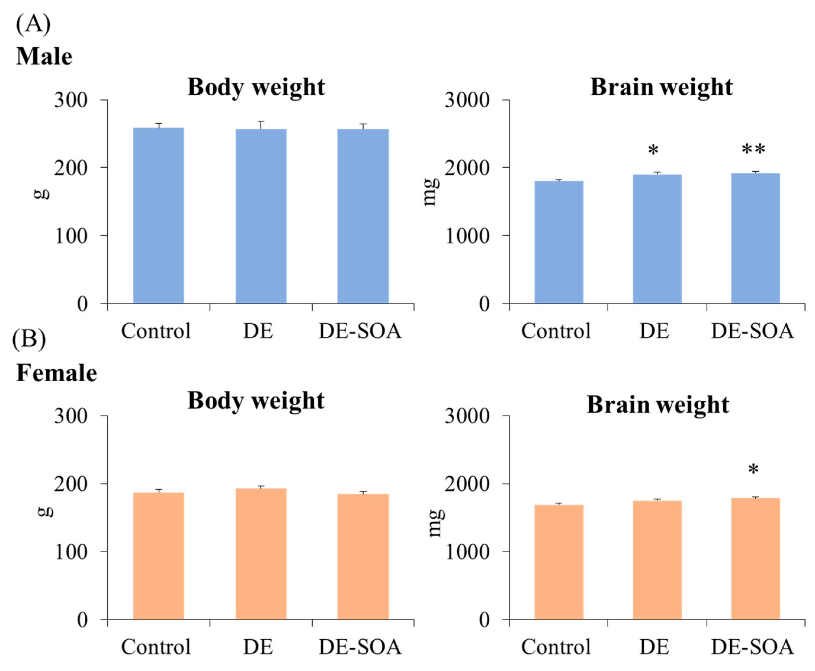
Figure 1. Assessment of overall toxicity. Body weight and brain weight in 10-week-old (A) male and (B) female rats of the control, DE- or DE-SOA-exposed groups (n = 10, ** p < 0.01, * p < 0.05 vs. corresponding control).
Ijms 24 00586 g001
Figure 2. Open field test. Number of entries to the center and time stay in the center in 10-week-old (A) male and (B) female rats of the control and the DE- or DE-SOA-exposed groups (n = 10, ** p < 0.01, * p < 0.05 vs. control). Table shows the number of grooming, rearing and defecation (fecal pellets) during open field test.
Ijms 24 00586 g002
Figure 3. Elevated plus maze test. Number of entries to the open arm and time spent in the open arm in 10-week-old (A) male and (B) female rats of the control and the DE- or DE-SOA-exposed groups (n = 10, ** p < 0.01, * p < 0.05 vs. corresponding control).
Ijms 24 00586 g003
Figure 4. Light dark transition test. Number of entries and time spent in the light compartment in 10-week-old (A) male and (B) female rats of the control and the DE- or DE-SOA-exposed groups (n = 10, ** p < 0.01, * p < 0.05 vs. corresponding control).
Ijms 24 00586 g004
Figure 5. Novelty-induced hypophagia test. Latency to approach and latency to eat in 10-week-old (A) male and (B) female rats of the control and the DE- or DE-SOA-exposed groups (n = 10, * p < 0.05 vs. corresponding Day 3 and # p < 0.05 vs. corresponding control group on novelty day).
Ijms 24 00586 g005
Figure 6. Messenger RNA expression levels of neurotransmitters in the hippocampus. Serotonin receptor (5HT1A) and dopamine receptor (Drd2) in the hippocampus of 10-week-old (A) male and (B) female rats of the control and the DE- or DE-SOA-exposed groups. (n = 10, ** p < 0.01, * p < 0.05 vs. control).
Ijms 24 00586 g006
Figure 7. Messenger RNA expression levels of neurotrophins in the hippocampus. BDNF and VEGFA in the hippocampus of 10-week-old (A) male and (B) female rats of the control and the DE- or DE-SOA-exposed groups. (n = 10, ** p < 0.01, * p < 0.05 vs. control).
Ijms 24 00586 g007
Figure 8. Messenger RNA expression levels of inflammatory and oxidative stress markers in the hippocampus. IL-1β, COX2 and HO1 mRNAs in the hippocampus of 10-week-old (A) male and (B) female rats of the control and the DE- or DE-SOA-exposed groups. (n = 10, * p < 0.05 vs. control).
Ijms 24 00586 g008
Figure 9. Representative photomicrographs of the dentate gyrus of the hippocampus. Microglia marker Iba1 immunoreactivity in 10-week-old (A) male and (B) female rats of the control and the DE- or DE-SOA-exposed groups. Blue arrows indicate activated microglia. (Scale bar = 50 μm). Number of Iba1-positive cells located within the DG area of the hippocampus was quantified in a high-power field under microscopy (histogram). (n = 5, ** p < 0.01 vs. control).
Ijms 24 00586 g009
Figure 10. Experimental protocol. Pregnant Sprague Dawley rats (GD 8) were exposed to clean air (control, n = 8), DE (n = 8), and DE-SOA (n = 8) from GD 14 to PND 21 in whole-body exposure chambers. 10 male and 10 female rats from 3 groups (total 60) were used for the anxiety-like behavior tests, 5 male and 5 female rats from 3 groups (total 15) were used for immunohistochemical analyses. Chronological order of anxiety-like behavior tests was expressed.
Ijms 24 00586 g010
Table 1. Summarized results of comparison of anxiety-like behavioral tests between DE and DE-SOA in male and female rats.
Behavioral TestParametersDE
(Compared to the Control)
DE-SOA
(Compared to the Control)
Male & Female DifferenceDE & DE-SOA Difference
MaleFemaleMaleFemale
OFTCenter entries↓↓↓↓↓↓NoNo
Center time↓↓↓↓↓↓↓↓NoNo
EPMOpen arm entries↓↓↓↓↓↓NoNo
Open arm timeNo
LDTLight entries↑↑YesYes
Light timeYesYes
NIHLatency to approach foodYesYes
Latency to eat foodYesYes
↓ Significantly decreased; ↑ Significantly increased.
Table 2. Summarized results of comparison of expression of molecular markers between DE and DE-SOA in male and female rats.
Molecular MarkersParametersDE
(Compared to the Control)
DE-SOA
(Compared to the Control)
Male & Female DifferenceDE & DE-SOA Difference
MaleFemaleMaleFemale
Neurotransmitters5HT1A↓↓↓↓YesNo
Drd2↓↓YesNo
NeurotrophinsBDNF↓↓↓↓NoNo
VEGFANoNo
Proinflammatory cytokinesIL-1βYesNo
COX2YesNo
Oxidative stress markerHO1YesNo
Microglia markerIba1↑↑↑↑↑↑↑↑NoNo
↓ Significantly decreased; ↑ Significantly increased.
Table 3. Characteristics of diesel exhaust particles and gaseous compounds in exposure chamber.
Diesel Exhaust ParticlesTemperatureRelative Humidity
Size
(nm)
Particle Number
(cm−3)
Concentration
(mg/m3)
(°C) (%) EC/OC WSOC/OC
Clean air0.87 ± 0.5713.2 ± 2.78 23.58 ± 0.2748.07 ± 0.770.15 ± 0.060.03 ± 0.04
DE-SOA24.45 ± 1.21 2.74 × 10⁶ ± 8.69 × 10⁴118.23 ± 31.1723.76 ± 0.1948.4 ± 0.850.38 ± 0.030.11 ± 0.05
DE22.69 ± 1.472.85 × 10⁶ ± 6.10 × 10⁴101.9 ± 19.4622.97± 0.2149.32 ± 0.870.36 ± 0.030.17 ± 0.11
Gaseous Compounds
CO (ppm)SO2 (ppm)NOx (ppm)NO2 (ppm)NO (ppm)O3 (ppm)CO2 (%)
Clean air0.21 ± 0.04 0.00 ± 0.00 0.00 ± 0.00 0.00 ± 0.00 0.00 ± 0.00 0.05 ± 0.00
DE-SOA2.51 ± 0.07 0.00 ± 0.00 1.14 ± 0.030.9 ± 0.030.15 ± 0.03 0.07 ± 0.00 0.07 ± 0.00
DE2.52 ± 0.07 0.01 ± 0.00 1.21 ± 0.03 0.43 ± 0.020.78 ± 0.020.07 ± 0.00
Data were expressed as mean ± SD.
Disclaimer/Publisher’s Note: The statements, opinions and data contained in all publications are solely those of the individual author(s) and contributor(s) and not of MDPI and/or the editor(s). MDPI and/or the editor(s) disclaim responsibility for any injury to people or property resulting from any ideas, methods, instructions or products referred to in the content.