Article

GmWAK1, Novel Wall-Associated Protein Kinase, Positively Regulates Response of Soybean to Phytophthora sojae Infection

1
Soybean Research Institute of Northeast Agricultural University/Key Laboratory of Soybean Biology of Chinese Education Ministry, Harbin 150030, China
2
Soybean Research Institute of Heilongjiang Academy of Agricultural Sciences/Key Laboratory of Soybean Cultivation of Ministry of Agriculture, Harbin 150030, China
3
Key Laboratory of Soybean Molecular Design Breeding, Northeast Institute of Geography and Agroecology, Chinese Academy of Sciences, Harbin 150030, China
*
Authors to whom correspondence should be addressed.
These authors contributed equally to this work.
Int. J. Mol. Sci. 2023, 24(1), 798; https://doi.org/10.3390/ijms24010798
Submission received: 11 December 2022 / Revised: 27 December 2022 / Accepted: 29 December 2022 / Published: 2 January 2023
(This article belongs to the Special Issue Soybean Molecular Breeding and Genetics)

Abstract

:
Phytophthora root rot is a destructive soybean disease worldwide, which is caused by the oomycete pathogen Phytophthora sojae (P. sojae). Wall-associated protein kinase (WAK) genes, a family of the receptor-like protein kinase (RLK) genes, play important roles in the plant signaling pathways that regulate stress responses and pathogen resistance. In our study, we found a putative Glycine max wall-associated protein kinase, GmWAK1, which we identified by soybean GmLHP1 RNA-sequencing. The expression of GmWAK1 was significantly increased by P. sojae and salicylic acid (SA). Overexpression of GmWAK1 in soybean significantly improved resistance to P. sojae, and the levels of phenylalanine ammonia-lyase (PAL), SA, and SA-biosynthesis-related genes were markedly higher than in the wild-type (WT) soybean. The activities of enzymatic superoxide dismutase (SOD) and peroxidase (POD) antioxidants in GmWAK1-overexpressing (OE) plants were significantly higher than those in in WT plants treated with P. sojae; reactive oxygen species (ROS) and hydrogen peroxide (H2O2) accumulation was considerably lower in GmWAK1-OE after P. sojae infection. GmWAK1 interacted with annexin-like protein RJ, GmANNRJ4, which improved resistance to P. sojae and increased intracellular free-calcium accumulation. In GmANNRJ4-OE transgenic soybean, the calmodulin-dependent kinase gene GmMPK6 and several pathogenesis-related (PR) genes were constitutively activated. Collectively, these results indicated that GmWAK1 interacts with GmANNRJ4, and GmWAK1 plays a positive role in soybean resistance to P. sojae via a process that might be dependent on SA and involved in alleviating damage caused by oxidative stress.

1. Introduction

Phytophthora root rot is a destructive disease in soybean worldwide, which is caused by the oomycete pathogen Phytophthora sojae (P. sojae) [1,2]. Its incidence is rapid, it spreads widely, and it can thus result in serious economic losses [3,4]. To date, using genetic resistance in soybean has been one of the most effective approaches to reduce the losses caused by P. sojae. Soybean cultivars ‘Dongnong 50’ and ‘Suinong 10’ are resistant and susceptible, respectively, to the predominant race 1 of P. sojae in Heilongjiang, China [5]; these materials are valuable for the study of plant resistance to P. sojae.
Plants have evolved numerous sensor proteins that perceive environmental cues and enable plants to appropriately respond to environmental stresses and pathogens [6]. Understanding how sensor proteins are used to monitor and respond to external signals in these processes is crucial for improving plant resistance to disease. Transmembrane receptor-like protein kinases (RLKs) play fundamental roles in cell–cell and plant–environment communication [7,8]. In plants, the first RLK was identified in Zea mays L. [7], and many others have been characterized from a variety of species [9,10,11,12]. RLKs are involved in developmental processes as well as responses to both abiotic and biotic stresses, particularly in plant–pathogen interactions [13,14,15,16].
Cell WAKs are a unique subfamily of plant RLKs that constitute the WAK-RLKs with the RLKs, which is a kinase structural domain responsible for activating the cytoplasmic signaling cascade [17,18]. Cell WAKs have a typical eukaryotic Ser/Thr kinase domain and an extracytoplasmic domain (ectodomain) containing several epidermal growth factor (EGF)-like repeats, which are the hallmark of the WAK subfamily [19,20,21]. Because of the unique structural features of WAK-RLKs, they can transfer signals from the cell wall to the cytoplasm well, whereas the extracellular structural domain can sense the stimulation of the cell wall and enable the transmission of signals via the cytoplasmic kinase structural domain [22,23,24].
WAKs are also involved in responses to pathogens [25,26,27]. In Arabidopsis, salicylic acid (SA) can induce the expression of AtWAK1; this induction requires the nonexpression of pathogenesis-related genes (NPR1) [24,25]. Moreover, AtWAK1 can be induced by methyl jasmonate and ethylene, which are related to fungal pathogens and defense-related signaling [26,27]. In addition to Arabidopsis, WAKs play a key role in the response to pathogens in other species [24], including tomato [28,29], wheat [30], and rice [31,32].
WAK proteins have been implicated in plant responses to minerals [33,34,35,36]. For example, WAKL4 plays an important role in Zn2+ accumulation in Arabidopsis shoots [35]. In terms of WAK function, these proteins appear to act as signal messengers in plant cells [23,33]. Via its extracellular domain, AtWAK1 acts as an oligogalacturonide receptor, which interacts with cell-wall-localized molecules [36,37,38]. Taken together, these results suggest a role of WAKs in signal transduction between the extracellular matrix and the cytoplasm during stress responses and development. However, the mechanisms of the WAK-mediated signal transduction pathway and the defense responses regulated by WAK are still unclear.
In our previous study, we found that GmLHP1 (like heterochromatin protein 1) is a negative regulator of the response to infection by P. sojae [39]. Through the transcriptome sequencing of soybean seedlings overexpressing GmLHP1, we detected notably enhanced expression of the gene encoding cell-wall-associated receptor protein kinase, designated GmWAK1 (GenBank accession no. XM_003534738), in the WAK-RLK family. As such, our aims in the current study included (a) using expression analysis and the phenotype to verify whether GmWAK1 is involved in the response to P. sojae infection; (b) finding the protein that interacts with GmWAK1 through a yeast two-hybrid assay and verifying its functionality; and (c) examining the physiological indicators and SA content to determine the molecular mechanism through which GmWAK1 responds to P. sojae. Our results provide a new perspective for in-depth analysis of the soybean response to P. sojae infection.

2. Results

2.1. GmWAK1 May Be Involved in Response to P. sojae Infection

Through RNA-sequencing (RNA-seq) analysis of the transcriptomes of wild-type (WT) and GmLHP1-overexpressing (OE) transgenic soybean lines, we found that the expression of a putative soybean wall-associated protein kinase gene, GmWAK1 (GenBank accession no. XM_003534738), significantly increased. However, GmLHP1 could not directly bind to the promoter region of GmWAK1 according to the results of the ChIP-qRT-PCR detection of GmLHP1-overexpressing and the WT plants (Figure S1). The expression of GmWAK1 was significantly higher in GmLHP1-overexpressing (OE) transgenic soybean plants than in the WT plants, and the expression of GmWAK1 in GmLHP1-RNAi transgenic soybean lines was markedly lower than that in the WT plants (Figure 1A). These results suggested that GmLHP1 can indirectly regulate the expression of GmWAK1.

2.2. Bioinformatics Analysis of GmWAK1

The full length of GmWAK1 is 2130 bp, which encodes a polypeptide of 709 amino acids. The predicted structure of GmWAK1 includes a conserved 111-residue GUB_WAK domain at amino acids 43–187 and a 265-residue PKc-like domain at amino acids 392–657 (Figure S2A). To find closely related proteins that might provide clues regarding the function of GmWAK1, we used a neighbor-joining algorithm to construct a phylogenetic tree based on the amino acid sequences of GmWAK1 and the 30 other members of the WAK family. The results of our comparison of GmWAK1 with the two neighboring WAKs with highest homology showed that it shares 68.76% amino acid identity with GmWAK8 (from Glycine max) (XP_003534785.2) and 68.22% amino acid identity with GsWAK9 (from Glycine soja) (KHN44651.1) (Figure S2B). The conserved 265 aa PKc-like domain of GmWAK1 is 96.64% identical to that of GmWAK8 and 93.62% identical to that of GsWAK9 (Figure S2C).

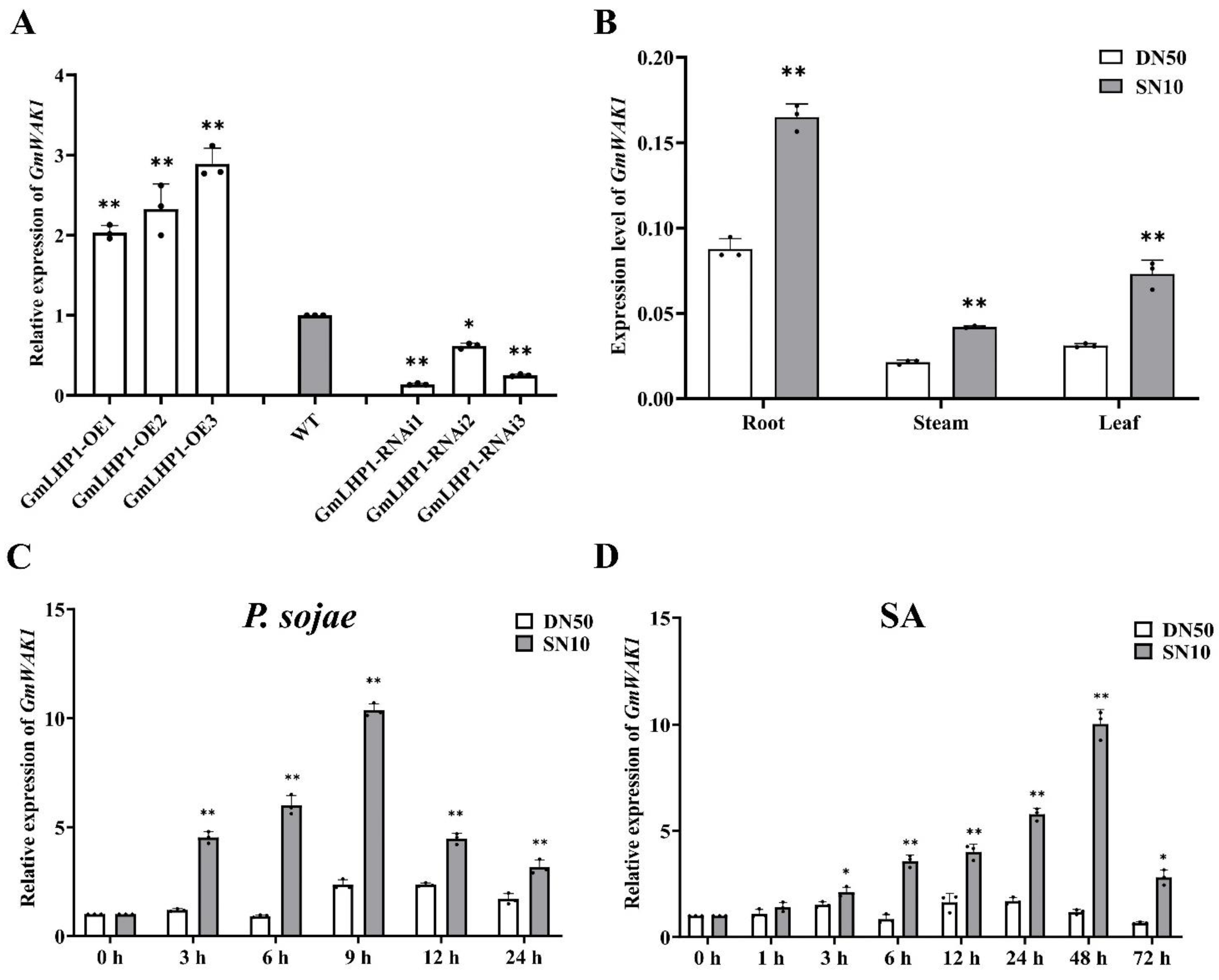
2.3. GmWAK1 Transcript Levels under Different Treatments

We evaluated the effect of biotic and abiotic stresses on GmWAK1 mRNA levels in ‘Suinong10’ and ‘Dongnong50’ by quantitative reverse transcription polymerase chain reaction (qRT-PCR). GmWAK1 was constitutively and most highly expressed in the roots, followed by the leaves and stems; the expression levels of GmWAK1 in the tissues of resistant soybean ‘Suinong 10’ were much higher than those in the tissues of susceptible soybean ‘Dongnong 50’ (Figure 1B). Inoculation of the resistant cultivar ‘Suinong 10’ with P. sojae caused a rapid increase in the levels of GmWAK1 mRNA, which reached a maximum at 48 h after inoculation, followed by a decline by 72 h. In contrast, inoculation with P. sojae resulted in no significant change in GmWAK1 mRNA levels in the susceptible cultivar ‘Dongnong 50’ (Figure 1C). GmWAK1 expression was also induced by SA, and the SA treatment caused the most rapid increase in GmWAK1 mRNA levels, which reached a maximum at 9 h with a subsequent rapid decline in expression (Figure 1D). These results suggested that GmWAK1 is primarily involved in the response to P. sojae and SA treatment.

2.4. Subcellular Localization of the GmWAK1

To verify the subcellular location of GmWAK1, we expressed a GmWAK1-GFP fusion protein in Arabidopsis protoplasts. We observed the green fluorescent protein (GFP) signal at the cell surface of protoplasts harboring GmWAK1-GFP; and marker protein P2300-35S-PIP2-mcherry in transgenic protoplasts showed a red fluorescence on the cell membrane (Figure 2A), similar to what was observed for the pathogenesis-related protein GmSnRK1 (Figure 2B) [40]. We observed the GFP signal in the entire cell of protoplasts transformed with 35S:GFP plasmids (Figure 2C). These results indicated that GmWAK1 is a membrane-localized protein.

2.5. GmWAK1 Is Positive Regulator in Soybean Resistance to P. sojae

To study the role of GmWAK1 in response to P. sojae, we generated GmWAK1-OE and GmWAK1-RNAi transgenic soybean lines (Figure S3). To test the P. sojae resistance of the T2 transgenic lines, we applied the zoospores of P. sojae to their cotyledons and leaves. After 3 days of incubation, we observed notable differences among the soybean lines. In the GmWAK1-RNAi lines, the cotyledons inoculated with P. sojae exhibited water-soaked lesions and were softer than those of WT; we observed almost no disease symptoms in the GmWAK1-OE lines. The P. sojae lesions on the GmWAK1-OE plants were significantly smaller than those on WT plants (p < 0.01), but larger than those on the GmWAK1-RNAi plants (Figure 3A). We obtained similar results after the inoculation of true leaves with P. sojae. After 3 days of incubation, the true leaves of WT and GmWAK1-RNAi transgenic soybean plants exhibited watery and rotting lesions, whereas those of the GmWAK1-OE transgenic soybean plants remained healthy (Figure 3B). As observed for the cotyledons, the lesion areas were significantly smaller in the GmWAK1-OE transgenic soybean plants and significantly larger in the GmWAK1-RNAi transgenic soybean plants relative to the WT plants (Figure 3C). The relative expression of GmTEF1 was significantly lower in in cotyledons and true leaves of GmWAK1-OE lines than in the WT plants but significantly higher in the GmWAK1-RNAi lines (p < 0.01) (Figure 3D,E). These results suggested that when GmWAK1 is overexpressed, the transgenic soybean plants acquire higher resistance to P. sojae infection.

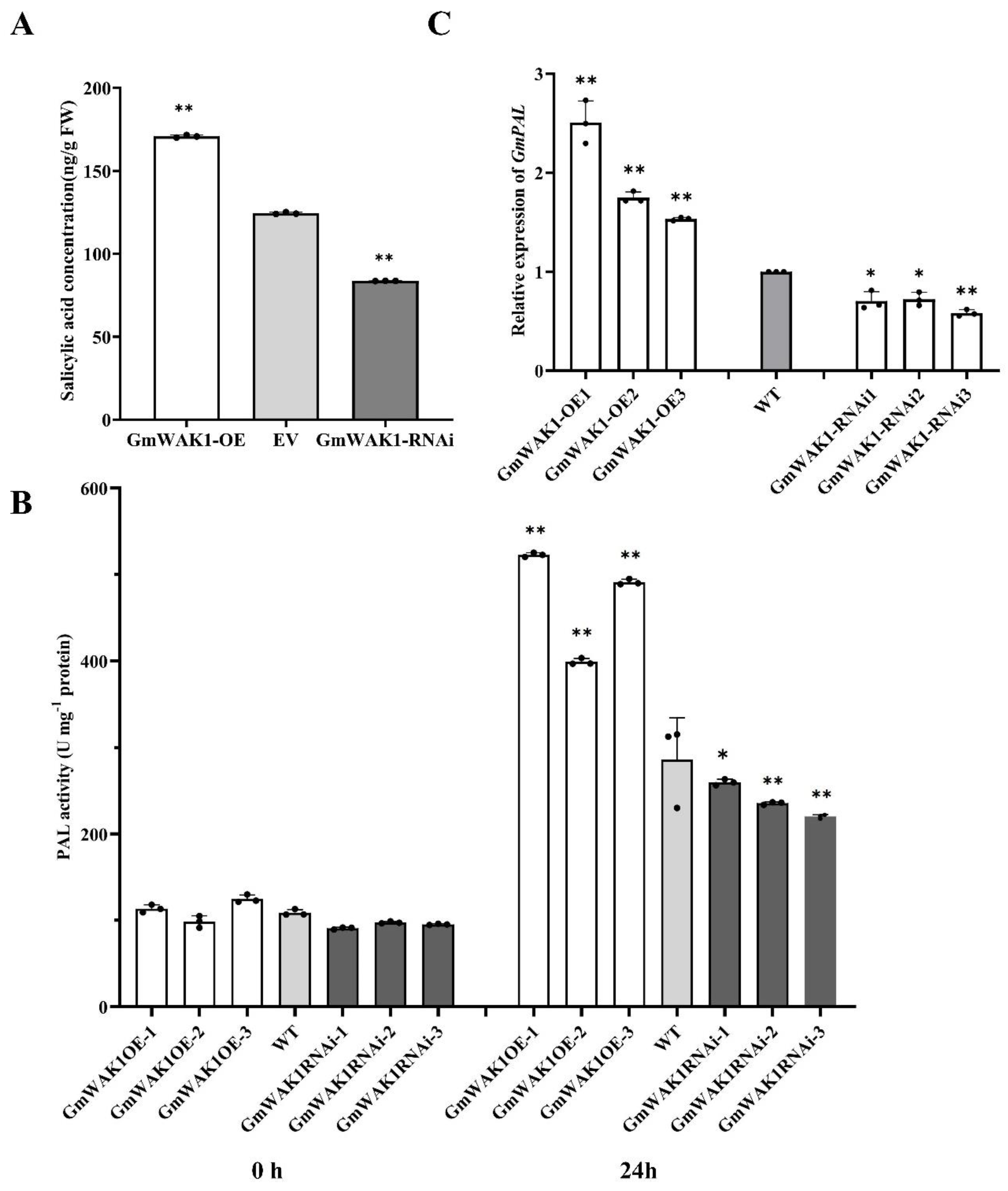
2.6. GmWAK1-Dependent SA Signaling in Response to P. sojae

To analyze whether the GmWAK1-regulated defense response was dependent on these phytohormones, we measured the SA content in the hairy roots of both GmWAK1 transgenic and WT plants. As shown in Figure 4A, the SA levels were significantly higher in GmWAK1-OE transgenic hairy roots than in the EV, and those of GmWAK1-RNAi were lower than those in the WT plants. The results showed that the phenylalanine ammonia-lyase (PAL) activity was significantly higher in the hairy roots of GmWAK1-OE transgenic plants than in those of WT plants after infection with P. sojae at 24 h postinoculation (hpi). The PAL activity in the hairy roots of GmWAK1-RNAi soybean was lower than that in the hairy roots of WT plants (Figure 4B). The expression of GmPAL in GmWAK1-OE was significantly higher than that in the WT plants; conversely, the RNAi of GmWAK1 led to reduced GmPAL activity relative to the control (Figure 4C). All these results showed that GmWAK1 plays an important role in the SA pathway.

2.7. Soybean GmWAK1-Regulated Defense Response to P. sojae Involves Alleviating Oxidative Stress Damage

Here, the superoxide dismutase (SOD) and peroxidase (POD) activities were much higher in GmWAK1 transgenic soybean plants than in the WT plants under both the mock treatment and at 24 hpi (Figure 5A,B). The expressions of GmSOD and GmPOD in GmWAK1-OE were significantly higher than those in the WT; the expressions in GmWAK1-RNAi were lower than those in the WT (Figure S4). Furthermore, the accumulation of ROS in GmWAK1-RNAi transgenic soybean plants was much higher than in the WT plants; the overexpression of GmWAK1 in soybean was lower than that in WT (Figure 5C). The accumulation of H2O2 in GmWAK1-OE transgenic plants was lower than in the WT plants, but it was higher in the transgenic GmWAK1-RNAi plants (Figure 5D). According to the results, we inferred that due to the elevated expression level of the corresponding enzyme genes in GmWAK1 transgenic soybean plants, the antioxidant enzyme activity increased, which may maintain certain lower steady-state levels of ROS, preventing harm from P. sojae infection.

2.8. GmWAK1 Interacts with GmANNRJ4

We screened the proteins that interact with GmWAK1 with immunoprecipitation-mass spectrometry. When induced with 0.8 mM IPTG at 37 °C for 4 h in LB medium, the GmWAK1-His protein expression levels were the highest. According to the results of SDS-PAGE analysis, the molecular weight of the inducible protein was about 120 kDa (Figure S5A,B). We performed Co-IP assays and LC-MS/MS analysis to identify proteins that interact with GmWAK1 (Figure S6). We identified 12 candidate GmWAK1-interacting proteins (Table S2). We selected a cDNA corresponding to annexin-like protein RJ4 (GmANNRJ4; LOC100813609) for further study because of its relatively high mass spectrometry score and its previously reported role in stress responses and pathogen resistance [41,42,43,44,45,46,47]. To confirm that these proteins interact in the plants, we used a bimolecular fluorescence complementation (BiFC) assay. We detected notable yellow fluorescence in the cytosol of Arabidopsis protoplasts after cotransformation with both N-terminal yellow fluorescent protein (YFPN)-tagged GmWAK1 and C-terminal YFP (YFPC)-tagged GmANNRJ4 (Figure 6A). In accordance with these results, the results of the glutathione S-transferase pull-down assay showed that its GmWAK1-his recombinant protein can be pulled down by GmANNRJ4-GST but not by GST alone (Figure 6B), further indicating that GmWAK1 interacts with GmANNRJ4 i. These results suggested that GmWAK1 directly interacts with GmANNRJ4.

2.9. GmANNRJ4 can Regulate Intracellular Ca2+ Concentrations and Promotes GmMPK6 Expression

GmANNRJ4 belongs to the annexin family of proteins, some of which are involved in regulating intracellular Ca2+ concentrations [48,49,50]. To determine whether GmANNRJ4 also has this function, we measured the cytoplasmic-free Ca2+ concentration in the hairy roots of transgenic transformed soybean with GmANNRJ4-overexpression or GmANNRJ4-RNAi constructs. The results showed that the concentration of free Ca2+ in the hairy roots of plants overexpressing GmANNRJ4 was higher than that in the hairy roots of WT plants; conversely, the RNAi of GmANNRJ4 led to a reduced free Ca2+ concentration relative to the control (Figure 6C).
To gain further insight into the function of GmANNRJ4, we used qPCR to analyze the expression of GmMPK6 in the hairy roots of GmANNRJ4-OE and GmANNRJ4-RNAi soybean (Figure 7) relative to that in the WT. The relative expression of GmMPK6 was significantly higher when GmANNRJ4 was overexpressed, whereas it was significantly lower in the hairy roots of GmANNRJ4-RNAi (Figure 6D). The expression of GmMKK4, which can phosphorylate and activate GmMPK6, was also significantly higher when GmANNRJ4 was overexpressed, whereas it was significantly lower in the hairy roots of GmANNRJ4-RNAi (Figure S8A). GmMKK4 and GmMPK6 were also highly expressed in the hairy roots of GmWAK1-OE soybean relative to those in the hairy roots of WT plants (Figure S8B,C). This finding suggested that GmANNRJ4 can positively regulate the expression of GmMPK6, and the regulation of GmMPK6 may be indirectly caused by the regulation of intracellular Ca2+ concentration by GmANNRJ4.

2.10. GmANNRJ4 Enhances Resistance to P. sojae and Positively Regulates Expression of PR Genes in Hairy Roots of Transgenic Soybean

We examined the response of GmANNRJ4-OE and GmANNRJ4-RNAi in the hairy roots of soybean to P. sojae infection. The hairy roots of GmANNRJ4-OE transgenic plants displayed significantly enhanced resistance compared with those of the WT plants after 2 days of P. sojae infection, and GmANNRJ4-RNAi showed the opposite results (Figure 7A). We measure the expression of PR genes in the hairy roots of GmANNRJ4-OE and GmANNRJ4-RNAi soybean by qRT-PCR. The expressions of GmPR1, GmPR2, and GmPR10 were significantly higher in the hairy roots of GmANNRJ4-OE (Figure 7B) and significantly lower in the hairy roots of GmANNRJ4-RNAi (Figure 7C) compared with those in the hairy roots of WT plants. However, GmPR3 expression was not affected by GmANNRJ4 over- or underexpression (Figure 7B,C). The above results suggest that the expression of GmANNRJ4 influences the expression of PR genes.

3. Discussion

GmLHP1 is an SA-inducible gene that inhibits the expression of GmWRKY40 and negatively regulates plant immunity [39,51]. Moreover, GmLHP1 interacts with GmBTB/POZ, promotes the ubiquitination and degradation of LHP1, and positively regulates the response of soybean to Phytophthora sojae infection and the expression of GmBTB/POZ [39,51]. In this study, the GmWAK1 gene was not directly regulated by GmLHP1 or the disease-resistance gene GmWRKY40, but it was upregulated in both overexpressed transgenic plants. These data suggested that some other genes that upregulate the expression of GmWAK1 and are involved in the regulation of P. sojae resistance are regulated by GmLHP1.
The plant cell wall not only maintains cell structure during plant growth and development but also responds to environmental stimuli and microbial pathogens [52]. The cell wall damage caused by pathogens is sensed by specific membrane proteins that transmit the signal across the membrane and activate the defense responses of the cell, thus improving plant pathogen resistance [19]. Among the membrane proteins involved in sensing cell wall damage are wall-associated receptor-like protein kinases (WAKs), which bind to the plant cell wall and have typical structure that consists of an extracellular domain, a transmembrane domain, and an intracellular kinase domain [53]. When plant cell walls are attacked by pathogens, oligogalacturonides (OGAs) are produced and then specifically recognized by AtWAK1 [20]. Activated AtWAK1 transmits this extracellular signal into the cell, which triggers the corresponding intracellular defense responses [38]. GmWAK1 is a membrane-localized protein; this location is consistent with a role in perceiving extracellular signals and transmitting these signals to the inside of the cell [18].
Here, we found that GmWAK1 interacted with GmANNRJ4. GmANNRJ4 is a member of the annexin protein (ANN) family. Annexins in plant membranes have a multisegment alpha-helix ANN domain that specifically binds to Ca2+; thus, they are also called Ca2+-dependent phospholipid binding proteins [54]. Annexins form permeable calcium channels on cell membranes and play a major role in regulating the concentrations of reactive oxygen species and free Ca2+ in plant cells [49,50,55]. Therefore, annexins are thought to directly form a Ca2+ flow pathway on the cell membrane and participate in various responses to adverse conditions by regulating Ca2+ concentrations and activating calmodulin- and Ca2+-dependent proteins [56]. Annexins play an important role in the response to abiotic and biotic stresses [57,58,59,60,61,62,63,64]. Until now, however, whether the annexin gene participates in the response to P. sojae had not been reported. As a candidate receptor for the transmembrane transmission of a disease-resistance signal, exploring the downstream proteins interacting with GmWAK1 is crucial to understanding its potential mechanism of action. An example is Arabidopsis AtWAK1, the extracellular domain of which binds to pectin ligands in the cell wall; AtWAK1 can activate MPK6-dependent stress responses by binding to the pectin fragments produced by the cell wall after infection by pathogens [21]. GmMKK4 could phosphorylate and activate GmMPK6 to enhance the resistance of soybean to P. sojae and promoted the expression of PR genes (GmPR1, GmPR2, GmPR5, and GmPR10) in response to P. sojae infection [65]. In this study, the expression of GmANNRJ4 in soybean significantly increased when infected by P. sojae, indicating that GmANNRJ4 participates in the process of soybean resistance to P. sojae. The hairy roots of transgenic soybean overexpressing GmANNRJ4 showed significantly increased resistance to P. sojae, indicating a positive regulation of soybean resistance to P. sojae. In determining the relationship between GmWAK1 and GmMPK6, we found that GmWAK1 can also increase the expressions of GmMKK4 and GmMPK6 in transgenic soybean plants overexpressing GmWAK1 and GmANNRJ4. We also found that the expressions of the PR genes (GmPR1, GmPR2, GmPR5 and GmPR10) were increased. Based on previous findings regarding annexin function, we hypothesized that GmWAK1 interacts with GmANNRJ4 and that both GmWAK1 and GmANNRJ4 are involved in regulating soybean resistance to P. sojae.
SA mediates systemic acquired resistance (SAR), which can control plant nutrition as well as the production of pathogens and improve disease resistance of organisms [66,67,68,69,70]. Zhang et al. [51] reported that GmBTB/POZ, via a process dependent on SA, plays a positive role in P. sojae resistance and the defense response in soybean. GmWAK1 is primarily involved in the response to P. sojae and SA treatment. SA accumulation and the transcript abundance of SA biosynthesis genes were much higher in GmWAK1 leaves than in those of WT plants. Herein, we determined that GmWAK1 played an important role in the SA pathway. As with SA, ROS also plays an important role in plant defense [71]. When under stress, a high concentration of H2O2 can kill pathogens and induce immune responses; low concentrations of H2O2 can also induce the expression of a series of genes encoding defense response proteins upon pathogen infestation, enhancing their resistance to pathogens [72]. The removal of excess ROS from plants can improve the resistance of plants to many pathogens [72,73,74,75]. Plants have well-developed antioxidant defense systems that efficiently scavenge ROS, involving the antioxidant enzymes SOD and POD [76]. SOD can reduce O2- levels by further dismutation to H2O2 [77,78]; POD can reduce H2O2 levels through the transport of electrons [79,80]. Maintaining high SOD and POD activities can result in reductions in H2O2 and ROS accumulation. In this study, the activities of SOD and POD were significantly increased, and the accumulations of ROS and H2O2 were kept markedly lower in GmWAK1-OE after P. sojae infection. The overexpression of GmWAK1 could enhance the resistance of soybean to P. sojae. This implied that the levels of SA and ROS might be critical for GmWAK1 in the response to P. sojae infection.
GmWAK1 is a receptor protein kinase associated with the cell wall in soybean. In this study, we examined the relationship between this protein and Phytophthora sojae infection. P. sojae significantly induced expression of GmWAK1 in ‘Suinong 10’, which is a soybean cultivar resistant to this pathogen, but only slightly induced the expression in ‘Dongnong 50’, the susceptible soybean cultivar. The SA levels were significantly higher in the hairy roots of transgenic GmWAK1-OE than in those of EV. This suggested that GmWAK1 may be a gene related to the P. sojae pathway in soybean. Transgenic soybean plants overexpressing GmWAK1 were significantly more resistant to P. sojae. Conversely, when the expression of GmWAK1 was suppressed by RNAi, the resistance to P. sojae was reduced. Based on the results, we propose the model depicted in Figure 8 for the promotion of resistance to P. sojae by GmWAK1 in soybean. GmLHP1 indirectly regulates GmWAK1. when soybean is infected by P. sojae, the expression of GmWAK1 increases, the expression of endogenous SA is upregulated, and the activities of SOD and POD are upregulated, which reduce the accumulation of O2− and H2O2, to be maintained at a certain lower steady-state level of ROS. When GmANNJ4 is overexpressed, calmodulator channels open to allow for the influx of calcium ions, whereas GmWAK1 interacts with GmANNRJ4 and activates the calmodulin-dependent kinase GmMPK6 by regulating intracellular Ca2+ concentrations. This ultimately promotes the expression of disease-related PR genes (GmPR1, GmPR2, and GmPR10) to enhance the resistance to P. sojae. This regulatory network provides a new perspective for in-depth analysis of the response of soybean to P. sojae infection and provides a new avenue for molecular breeding of disease resistance in soybean. In conclusion, GmWAK1 is induced by P. sojae infection and plays a positive regulatory role in the response of soybean to this pathogen.

4. Materials and Methods

4.1. Plant Materials, P. sojae Treatment, and Primers

In this study, we used soybean cultivars ‘Dongnong 50’ and ‘Suinong 10’, which are resistant and susceptible, respectively, to the predominant race of P. sojae, race 1, in Heilongjiang, China [5]. We sowed all the seeds in pots in a growth chamber that we set to 25/20 °C (night/day) and 80% relative humidity with a 16/8 h light (110 PAR light intensity)/dark cycle. Fourteen days after planting, we inoculated soybeans at the first-node stage (V1 [81]) with P. sojae race 1 following the method described by Ward et al. [82]. We removed unifoliate leaves at 0, 3, 6, 9, 12, 24, 48, and 72 h after inoculation. All primers are listed in Table S1.

4.2. Isolation and Sequence Analysis of GmWAK1

We isolated the total RNA from ‘Suinong 10’ and ‘Dongnong 50’ plants using TRIzol® reagent (Invitrogen, Shanghai, China; part number 15596026.). For reverse transcription, we used an M-MLV reverse-transcriptase kit (Takara, Dalian, China; part number 2641A). We isolated the full-length cDNA sequence of GmWAK1 from the cDNA of ‘Suinong 10’. We inserted PCR products into a pEASY® Blunt vector (Transgen Biotech, Beijing, China; part number CB101). To obtain the protein sequence data and analyze the nucleotides, we used the NCBI bioinformatics tools (http://www.ncbi.nlm.nih.gov/blast, accessed on 30 July 2022). We analyzed the predicted protein structure with SMART (http://smart.embl-heidelberg.de, accessed on 30 July 2021). We aligned the sequences using DNAMAN software (http://www.lynnon.com, accessed on 30 July 2022). Based on the nucleotide sequences of GmWAK1 and other WAK members, we generated phylogenetic trees using MEGA 5.1 software (http://www.megasoftware.net, accessed on 30 July 2022).

4.3. RT-PCR and qRT-PCR Analysis

We performed quantitative real-time PCR (qRT-PCR) to analyze the GmWAK1 transcript levels using a real-time PCR kit (Toyobo, Japan; part number FSQ-201) [83]. As a housekeeping gene, we used GmEF1β (GenBank accession no. NM_001248778) in soybean as an internal reference to normalize all data. We calculated relative transcript levels using the 2−ΔΔCT method. We performed three biological replicates per experiment per line.

4.4. Chromatin Immunoprecipitation (ChIP) qPCR Assay

For the ChIP assays, WT, p35S: Flag-GmLHP1 transgenic plants and p35S: Flag-Myc-GmWRK40 were subjected to chromatin extraction and immunoprecipitation as described by Saleh et al. [84]. The antibodies we used during the ChIP were anti-MYC-Tag Mouse mAb (Agarose Conjugated) (Abmart, code number M20012) and anti-DYKDDDDK-Tag Mouse mAb (Agarose Conjugated) (Abmart, code number M20018). The immunoprecipitated DNA samples were used as templates for qPCR assays. The primers used for this experiment are listed in Table S1.

4.5. Subcellular Localization of GmWAK1

We cloned the coding sequences (CDs) of GmWAK1 into the N-terminus of green fluorescent protein (GFP) under the control of the constitutive CaMV35S promoter using the primer pairs GmWAK1-GF and GmWAK1-GR (Table S1). We transformed the resulting expression vectors, 35S:GmWAK1-GFP and P2300-35S-PIP2-mcherry (RFP), into Arabidopsis protoplasts via polyethylene glycol (PEG)-mediated transfection as described by Yoo et al. [85]. After 16–20 h of incubation at 25 °C and under light, we imaged the GFP/RFP signal with a TCS SP8 confocal spectral microscope imaging system (Leica, Germany).

4.6. Inducing the Expression of GmWAK1 Fusion Protein

We constructed the His-tagged pET29b (+) -GmWAK1 recombinant fusion plasmid, which we transformed into E. coli BL21 (DE3) cells. We added 0.8 mM isopropyl-b-D-thiogalactoside (IPTG) at 37 °C, which we shook at 220 rpm with a Thermostatic Shaker in LB medium for 4 h. We analyzed the recombinant protein SDS-PAGE.

4.7. Vector Construction and Transformation of Soybean

We cloned the CDs of GmWAK1 into plant expression vector pCAMBIA3301-4Myc with gene-specific primers (Table S1). We amplified the cDNA fragment of GmWAK1 using the GmWAK1 RNAi-F/R primer set, which we inserted into vector pFGC5941 [86] (Table S1). We individually introduced the recombinant construct GmWAK1-OE and GmWAK1-RNAi vector into A. tumefaciens strain LBA4404, as previously described [87]. For the soybean transformation, we used susceptible cultivar ‘Dongnong 50’ as the explant the generation, following the Agrobacterium-mediated transformation method described by Paz et al. [88]. We constructed the plant overexpression vector 35S:GmANNRJ4 and vector GmANNRJ4-RNAi [86] (Table S1) to obtain transgenic hairy roots according to previously published methods [89,90]. We identified overexpression and RNAi transgenic soybean plants (T1) or transgenic hairy roots by PCR amplification, and we used a PAT/bar test strip to identify bar screening markers, which we developed for T2 transgenic soybean plants for the following analyses.

4.8. Assessment of Pathogen Resistance and the Disease Response

We grew the transgenic soybean seeds under a 16/8 h light/dark photoperiod at 25 °C and 70% relative humidity in a greenhouse. For disease resistance analysis, we treated the living cotyledons of the WT and transgenic soybean plants at the emergence stage (V1) [81] with P. sojae zoospores (approximately 1 × 10⁵ spores mL−1), following the methods described by Morrison and Thorne [91], and we inoculated the leaves following the procedure described by Dong et al. [92]. To study whether the GmANNRJ4 conferred resistance to pathogen infection, we performed artificial inoculation procedures as described by Ward et al. [82]. We determined the relative biomass of soybean in the infected root system after 3 d based on the transcript level of the soybean TEF1 gene, using soybean GmEF1β as an internal reference gene, according to Chacón et al. [93] (Table S1). Each experiment included three replicates, each with three technical replicates.

4.9. Bimolecular Fluorescence Complementation (BiFC) Assay

To validate the interactions between GmWAK1 and GmANNRJ4, we performed a BiFC assay based on yellow fluorescence protein (YFP). We separately cloned the coding sequences of GmWAK1 and GmANNRJ4 into serial pSAT6 vectors encoding either N- or C-terminal-enhanced yellow fluorescent protein fragments (Table S1). We used the resulting constructs for the transient assays via PEG transfection of Arabidopsis protoplasts, as described by Yoo et al. [85]. We imaged the transfected cells with a TCS SP8 confocal spectral microscopy imaging system (Leica).

4.10. Pull-Down Assays

We cloned GmWAK1 into the pET29b (+) expression vector and GmANNRJ4 into the pGEX-4T-1 expression vector. His-GmWAK1 and glutathione S-transferase (GST)-GmANNRJ4 proteins were separately produced in E. coli BL21 (DE3) cells, which we then harvested and purified using a GST-Sefinose kit (Sangon, China, code number C590927) or a His-bind Purification Kit (Merck Millipore, Burlington, MA, USA, code number 70239-3). We performed the pull-down assay as described by Yang et al. [94], with minor modifications. In a total volume of 1 mL of GST binding buffer (Sangon), we incubated the GST or GmANNRJ4-GST recombinant proteins for 1 h at 4 °C with 400 mL of GST resin (Sangon), after which we added an equal volume of the GmWAK1-His recombinant protein and then incubated for 6 h at 4 °C. We washed the binding reaction five times with binding buffer, each for 10 min at 4 °C; then, we eluted the pulled-down proteins by boiling, which we separated on a 12% SDS-PAGE gel and immunoblotted with anti-His antibody and anti-GST antibody (Abmart, Berkeley Heights, NJ, USA; part numbers M20020 and M20025).

4.11. Determination of Plant Hormone and Antioxidant Enzyme Activity Levels

We measured the SA levels referencing the method described by Pan et al. [95]. We measured SOD and POD enzyme activities according to the method described by Wang et al. [96]. We determined hydrogen peroxide (H2O2) accumulation according to the method of Velikova et al. [97]. We measured the rate of ROS generation according to the method described by Qian et al. [98]. Each experiment included three replicates, each with three technical replicates.

4.12. Intracellular Calcium Concentration Measurement

We individually introduced the 35S:GmANNRJ4-OE and 35S:GmANNRJ4-RNAi constructs into Agrobacterium tumefaciens strain K599 using the freeze–thaw method [86]. We generated transgenic soybean hairy roots following the methods described by Graham et al. [89] and Kereszt et al. [90] using A. rhizogenes-mediated transformation. We sampled the transgenic soybean hairy roots to extract intracellular free calcium, and we used a calcium ion fluorescence probe (Fluo-3 AM) to measure cytoplasmic free calcium concentrations according to the method described by Yu and Hinkle [99].

4.13. Statistical Analysis

In this study, we performed all the experiments three times. To analyze all the results and to determine the statistical significance between different measurements, we used Student’s t test. We considered a difference statistically significant when * p < 0.05 or ** p < 0.01.

5. Conclusions

In our study, we investigated the biological functions of soybean GmWAK1 in response to P. sojae infection. We found that when soybean was infected by P. sojae, the expression of GmWAK1 increased, the expression of endogenous SA was upregulated, and the activities of SOD/POD were upregulated. GmWAK1 also helped to maintain lower levels of H2O2 and ROS accumulation. When GmANNJ4 was overexpressed, Calmodulator channels opened to allow for calcium ion influx, whereas GmWAK1 interacted with GmANNRJ4 and activated the calmodulin-dependent kinase GmMPK6 by regulating intracellular Ca2+ concentrations. This ultimately promoted the expression of disease-related PR genes (GmPR1, GmPR2, and GmPR10) to enhance soybean resistance to P. sojae. This regulatory network provides a new perspective for in-depth analysis of the response of soybean to P. sojae infection and provides a possible new avenue for the molecular breeding of soybean disease resistance. In conclusion, GmWAK1 is induced by P. sojae infection and plays a positive regulatory role in the soybean response to this pathogen.

Supplementary Materials

The following supporting information can be downloaded at: https://www.mdpi.com/article/10.3390/ijms24010798/s1.

Author Contributions

P.X. and S.Z. designed the experiments; M.Z., N.L., S.C., J.W., S.H., Y.Z., X.W. and X.C. performed the experiments; C.Z., X.F., Y.S., B.S., S.L. and Y.L. analyzed the data; M.Z., P.X. and S.Z. wrote the manuscript; M.Z. and N.L contributed equally to this study. All authors have read and agreed to the published version of the manuscript.

Funding

This study was supported by the National Key R&D Program of China (2021YFD1201103), NSFC Projects (31971972), Natural Science Foundation of Heilongjiang Province (ZD2019C001), and Outstanding Talents and Innovative Team of Agricultural Scientific Research.

Institutional Review Board Statement

Not applicable.

Informed Consent Statement

Not applicable.

Data Availability Statement

The gene accession numbers are as follows: GmWAK1 (XM_003534738), GmANNRJ4 (XM_003542786), GmSOD1 (NM_001248369), GmPOD (XM_006575142), GmActin4 (AF049106), GmEF1β (NM_001248778), GmPAL (Glyma.19G182300), GmMKK4 (Glyma_07G003200), GmMPK6 (Glyma.02G138800), GmLHP1 (Glyma_16G079900), GmPR1 (Glyma.15G062400), GmPR2 (Glyma_03g132700), GmPR3 (Glyma_02G042500), GmPR10 (Glyma.15G145700), and GmWRKY40 (Glyma.15G003300).

Conflicts of Interest

The authors declare no conflict of interest.

References

  1. Wrather, J.A.; Anderson, T.R.; Arsyad, D.M.; Tan, Y.; Ploper, L.D.; PortaPuglia, A.; Ram, H.H.; Yorinori, J.T. Soybean disease loss estimates for the top 10 soybean producing countries in 1998. Plant Dis. 2001, 81, 107–110. [Google Scholar] [CrossRef] [PubMed] [Green Version]
  2. Tyler, B.M. Phytophthora sojae: Root rot pathogen of soybean and model oomycete. Mol. Plant Pathol. 2007, 8, 1–8. [Google Scholar] [CrossRef] [PubMed]
  3. Erwin, D.C. Phytophthora diseases worldwide. Plant Pathol. 1998, 47, 224–225. [Google Scholar] [CrossRef]
  4. Bernard, R.L.; Smith, P.E.; Kaufmann, M.J.; Schmitthenner, A.F. Inheritance of resistance to Phytophthora root and stem rot in soybean. Agron. J. 1957, 49, 391. [Google Scholar] [CrossRef]
  5. Zhang, S.Z.; Xu, P.F.; Wu, J.J.; Xue, A.G.; Zhang, J.X.; Li, W.B.; Chen, C.; Chen, W.Y.; Lv, H.Y. Races of Phytophthora sojae and their virulences on soybean cultivars in Heilongjiang, China. Plant Dis. 2010, 94, 87–91. [Google Scholar] [CrossRef] [Green Version]
  6. Dixon, R.A.; Lamb, C.J. Molecular communication in interactions between plants and microbial pathogens. Annu. Rev. Plant Phys. 1990, 41, 339–367. [Google Scholar] [CrossRef]
  7. Walker, J.C.; Zhang, R. Relationship of a putative receptor protein kinase from maize to the S-locus glycoproteins of Brassica. Nature 1990, 345, 743–746. [Google Scholar] [CrossRef]
  8. Walker, J.C. Receptor-like protein kinase genes of Arabidopsis thaliana. Plant J. 1993, 3, 451–456. [Google Scholar] [CrossRef]
  9. Shiu, S.H.; Karlowski, W.M.; Pan, R.; Tzeng, Y.H.; Mayer, K.F.X.; Li, W.H. Comparative analysis of the receptor-like kinase family in arabidopsis and rice. Plant Cell 2004, 16, 1220–1234. [Google Scholar] [CrossRef] [Green Version]
  10. Alexandre, L.; Sabine, C.; Philippe, D.; Philippe, S.; Patrick, T.; Séverine, T. Involvement of a putative Lycopersicon esculentum wall-associated kinase in the early steps of tomato–Orobanche ramosa interaction. Physiol. Mol. Plant Pathol. 2006, 69, 3–12. [Google Scholar] [CrossRef]
  11. Verica, J.A.; He, Z.H. The cell wall-associated kinase (WAK) and WAKlike kinase gene family. Plant Physiol. 2002, 129, 455–459. [Google Scholar] [CrossRef] [PubMed] [Green Version]
  12. Ma, Y.X.; Stafford, L.; Julian, R.; Antony, B.; Kim, L.J. WAKL8 regulates Arabidopsis stem secondary wall development. Plants 2022, 11, 2297. [Google Scholar] [CrossRef]
  13. Shiu, S.H.; Bleecker, A.B. Plant receptor-like kinase gene family: Diversity, function, and signaling. Science’s STKE 2001, 2001, re22. [Google Scholar] [CrossRef] [PubMed]
  14. Becraft, P.W. Receptor kinase signaling in plant development. Annu. Rev. Plant Phys. 2002, 18, 163–192. [Google Scholar] [CrossRef] [PubMed] [Green Version]
  15. Dievart, A.; Clark, S.E. Using mutant alleles to determine the structure and function of leucine-rich repeat receptor-like kinases. Curr. Opin. Plant. Biol. 2003, 6, 507–516. [Google Scholar] [CrossRef] [PubMed]
  16. Hua, D.P.; Wang, C.; He, J.N.; Liao, H.; Duan, Y.; Zhu, Z.Q.; Guo, Y.; Chen, Z.Z.; Gong, Z.Z. A plasma membrane receptor kinase, GHR1, mediates abscisic acid- and hydrogen peroxide-regulated stomatal movement in Arabidopsis. Plant Cell 2002, 24, 2546–2561. [Google Scholar] [CrossRef] [Green Version]
  17. He, Z.H.; Cheeseman, I.; He, D.; Kohorn, B.D. A cluster of five cell wall-associated receptor kinase genes, WAK1-5, are expressed in specific organs of Arabidopsis. Plant Mol. Biol. 1999, 39, 1189–1196. [Google Scholar] [CrossRef]
  18. He, Z.H.; Fujiki, M.; Kohorn, B.D. A cell wall associated receptor-like protein kinase. J. Biol. Chem. 1996, 271, 19789–19793. [Google Scholar] [CrossRef] [Green Version]
  19. Lally, D.; Ingmire, P.; Tong, H.Y.; He, Z.H. Antisense expression of a cell wall-associated protein kinase, WAK4, inhibits cell elongation and alters morphology. Plant Cell 2001, 13, 1317–1331. [Google Scholar] [CrossRef] [Green Version]
  20. Wagner, T.A.; Kohorn, B.D. Wall-associated kinases are expressed throughout plant development and are required for cell expansion. Plant Cell 2001, 13, 303–318. [Google Scholar] [CrossRef]
  21. Kohorn, B.D.; Kobayashi, M.; Johansen, S.; Riese, J.; Huan, L.F.; Koch, K.; Fu, S.; Dotson, A.; Byers, N. An Arabidopsis cell wall-associated kinase required for invertase activity and cell growth. Plant J. 2006, 46, 307–316. [Google Scholar] [CrossRef] [PubMed]
  22. Anderson, C.M.; Wagner, T.A.; Perret, M.; He, Z.H.; He, D.; Kohorn, B.D. Waks: Cell wall-associated kinases linking the cytoplasm to the extracellular matrix. Plant Mol. Biol. 2001, 47, 197–206. [Google Scholar] [CrossRef] [PubMed]
  23. Shiu, S.H.; Bleecker, A.B. Receptor-like kinases from Arabidopsis form a monophyletic gene family related to animal receptor kinases. Proc. Natl. Acad. Sci. USA 2001, 98, 10763–10768. [Google Scholar] [CrossRef] [PubMed] [Green Version]
  24. Verica, J.A.; Chae, L.; Tong, H.Y.; Ingmire, P.; Zheng, H.H. Tissue specific and developmentally regulated expression of a cluster of tandemly arrayed cell wall-associated kinase-like kinase genes in Arabidopsis. Plant Physiol. 2003, 133, 1732–1746. [Google Scholar] [CrossRef] [PubMed] [Green Version]
  25. He, Z.H.; He, D.; Kohorn, B.D. Requirement of the induced expression of a cell wall associated receptor kinase for survival during the pathogen response. Plant J. 1998, 14, 55–63. [Google Scholar] [CrossRef] [PubMed] [Green Version]
  26. Maleck, K.; Levine, A.; Eulgem, T.; Morgan, A.; Schmid, J.; Lawton, K.A.; Dangl, J.L.; Dietrich, R.A. The transcriptome of Arabidopsis thaliana during systemic acquired resistance. Nat. Genet. 2000, 26, 403–410. [Google Scholar] [CrossRef] [PubMed]
  27. Schenk, P.M.; Kazan, K.; Wilson, I.; Anderson, J.P.; Richmond, T.; Somerville, S.C.; Manners, J.M.; Kazan, K. Coordinated plant defense responses in Arabidopsis revealed by microarray analysis. Proc. Natl. Acad. Sci. USA 2000, 97, 11655–11660. [Google Scholar] [CrossRef] [PubMed] [Green Version]
  28. Rosli, H.G.; Zheng, Y.; Pombo, M.A.; Zhong, S.; Bombarely, A.; Fei, Z. Transcriptomics-based screen for genes induced by flagellin and repressed by pathogen effectors identifies a cell wall-associated kinase involved in plant immunity. Genome Biol. 2013, 14, 12. [Google Scholar] [CrossRef] [PubMed] [Green Version]
  29. Yu, H.F.; Zhang, W.N.; Fan, Y.L.; Yang, X.Y.; Shi, M.F.; Zhang, R.Y.; Wang, Y.; Qin, S.H. Genome-wide identification and expression analysis of wall-associated kinase (WAK) gene family in potato (Solanum tuberosum L.). Plant Biotechnol. Rep. 2022, 16, 317–331. [Google Scholar] [CrossRef]
  30. Xia, X.; Zhang, X.; Zhang, Y.; Wang, L.; An, Q.; Tu, Q.; Wu, L.; Jiang, P.; Zhang, P.; Yu, L.; et al. Characterization of the WAK Gene Family Reveals Genes for FHB Resistance in Bread Wheat (Triticum aestivum L.). Int. J. Mol. Sci. 2022, 23, 7157. [Google Scholar] [CrossRef]
  31. Delteil, A.; Gobbato, E.; Cayrol, B.; Estevan, J.; Michel-Romiti, C.; Dievart, A.; Kroj, T.; Morel, J.-B. Several wall-associated kinases participate positively and negatively in basal defense against rice blast fungus. BMC Plant Biol. 2016, 16, 17–26. [Google Scholar] [CrossRef] [PubMed] [Green Version]
  32. Hu, K.M.; Cao, J.B.; Zhang, J.; Xia, F.; Ke, Y.G.; Zhang, H.T. Improvement of multiple agronomic traits by a disease resistance gene via cell wall reinforcement. Nat. Plants 2017, 3, 17009–17017. [Google Scholar] [CrossRef] [PubMed]
  33. Morris, E.R.; Walker, J.C. Receptor-like protein kinases: The keys to response. Curr. Opin. Plant. Biol. 2003, 6, 339–342. [Google Scholar] [CrossRef]
  34. Hou, X.; Tong, H.; Selby, J.; Dewitt, J.; Peng, X.; He, Z.H. Involvement of a cell wall-associated kinase, WAKL4, in Arabidopsis mineral responses. Plant Physiol. 2005, 139, 1704–1716. [Google Scholar] [CrossRef] [Green Version]
  35. Sivaguru, M.; Ezaki, B.; He, Z.H.; Tong, H.; Osawa, H.; Baluska, F.; Hideaki, D.; Matsumoto, V. Aluminum induced gene expression and protein localization of a cell wall-associated receptor protein kinase in Arabidopsis thaliana. Plant Physiol. 2003, 132, 2256–2266. [Google Scholar] [CrossRef] [PubMed] [Green Version]
  36. Park, A.; Cho, S.; Yun, U.; Jin, M.; Lee, S.; Sachetto-Martins, G.; Park, O.K. Interaction of the Arabidopsis receptor protein kinase Wak1 with a glycine-rich protein, AtGRP3. J. Biol. Chem. 2003, 276, 26688–26693. [Google Scholar] [CrossRef] [Green Version]
  37. Decreux, A.; Messiaen, J. Wall-associated kinase WAK1 interacts with cell wall pectins in a calcium-induced conformation. Plant Cell Physiol. 2005, 46, 268–278. [Google Scholar] [CrossRef] [PubMed] [Green Version]
  38. Brutus, A.; Sicilia, F.; Macone, A.; Cervone, F.; Lorenzo, G.D. A domain swap approach reveals a role of the plant wall-associated kinase 1 (WAK1) as a receptor of oligogalacturonides. Proc. Natl. Acad. Sci. USA 2010, 107, 9452–9457. [Google Scholar] [CrossRef] [Green Version]
  39. Zhang, C.Z.; Cheng, Q.; Wang, H.Y.; Gao, H.; Fang, X.; Chen, X.; Zhao, M.; Wei, W.L.; Song, B.; Liu, S.S.; et al. GmBTB/POZ promotes the ubiquitination and degradation of LHP1 to regulate the response of soybean to Phytophthora sojae. Commun. Biol. 2021, 4, 372. [Google Scholar] [CrossRef]
  40. Wang, L.; Wang, H.Y.; He, S.F.; Meng, F.S.; Zhang, C.Z.; Fan, S.J.; Wu, J.J.; Xu, P.F.; Zhang, S.Z. GmSnRK1. 1, a sucrose non-fermenting-1 (SNF1)-related protein kinase, promotes soybean resistance to Phytophthora sojae. Front. Plant Sci. 2019, 10, 996. [Google Scholar] [CrossRef]
  41. Liu, Q.B.; Ding, Y.L.; Shi, Y.T.; Ma, L.; Yang, Y.; Song, C.P.; Wilkins, K.A.; Davies, J.M.; Knight, H.; Knight, M.R.; et al. The calcium transporter annexin1 mediates cold-induced calcium signaling and freezing tolerance in plants. EMBO J. 2020, 40, 2. [Google Scholar] [CrossRef] [PubMed]
  42. Mu, C.; Zhou, L.; Shan, L.B.; Li, F.J.; Li, Z.H. Phosphatase GhDsPTP3a interacts with annexin protein GhANN8b to reversely regulate salt tolerance in cotton (Gossypium spp.). N. Phytol. 2019, 223, 4. [Google Scholar] [CrossRef]
  43. Li, X.F.; Zhang, Q.; Yang, X.; Han, J.B.; Zhu, Z.G. OsANN3, a calcium-dependent lipid binding annexin is a positive regulator of aba-dependent stress tolerance in rice. Plant Sci. 2019, 284, 212–220. [Google Scholar] [CrossRef]
  44. Laohavisit, A.; Davies, J.M. Annexins. N. Phytol. 2011, 189, 40–53. [Google Scholar] [CrossRef] [PubMed]
  45. Carvalho-Niebel, F.D.; Timmers, A.C.; Chabaud, M.; Defaux-Petras, A.; Barker, D.G. The nod factor-elicited annexin MtANN1 is preferentially localised at the nuclear periphery in symbiotically activated root tissues of Medicago truncatula. Plant J. 2002, 32, 343–352. [Google Scholar] [CrossRef] [PubMed]
  46. Truman, W.; Bennett, M.H.; Kubigsteltig, I.; Turnbull, C.; Grant, M. Arabidpsis systemic immunity uses conserved defense signaling pathways and is mediated by jasmonates. Proc. Natl. Acad. Sci. USA 2007, 104, 1075–1080. [Google Scholar] [CrossRef] [PubMed] [Green Version]
  47. Vandeputte, O.; Lowe, Y.O.; Burssens, S.; Raemdonck, D.V.; Hutin, D.; Boniver, D.; Geelen, D.; Jaziri, E.M.; Baucher, M. The tobacco Ntann12 gene, encoding an annexin, is induced upon Rhodoccocus fascians infection and during leafy gall development. Mol. Plant Pathol. 2007, 8, 185–194. [Google Scholar] [CrossRef] [PubMed] [Green Version]
  48. Laohavisit, A.; Davies, J.M. Multifunctional annexins. Plant Sci. 2009, 177, 532–539. [Google Scholar] [CrossRef]
  49. Delmer, D.P.; Potikha, T.S. Structures and function of annexins in plants. Cell Mol. Life Sci. 1997, 53, 546–553. [Google Scholar] [CrossRef] [PubMed]
  50. Laohavisit, A.; Mortimer, J.C.; Demidchik, V.; Coxon, K.M.; Stancombe, M.A.; Macpherson, N. Zea mays annexins modulate cytosolic free Ca2+ and generate a Ca2+-permeable conductance. Plant Cell 2009, 21, 479–493. [Google Scholar] [CrossRef]
  51. Zhang, C.Z.; Gao, H.; Li, R.P.; Han, D.; Wang, L.; Wu, J.J.; Xu, P.F.; Zhang, S.Z. GmBTB/POZ, a novel BTB/POZ domain-containing nuclear protein, positively regulates the response of soybean to Phytophthora sojae infection. Mol. Plant Pathol. 2019, 20, 78. [Google Scholar] [CrossRef] [PubMed] [Green Version]
  52. Cantu, D.; Vicente, A.R.; Labavitch, J.M.; Bennett, A.B.; Powell, A.L.T. Strangers in the matrix: Plant cell walls and pathogen susceptibility. Trends Plant Sci. 2008, 13, 610–617. [Google Scholar] [CrossRef] [PubMed]
  53. Walker, J.C. Structure and function of the receptor-like protein kinases of higher plants. Plant Mol. Biol. 1994, 26, 1599–1609. [Google Scholar] [CrossRef] [PubMed]
  54. Moss, S.E.; Morgan, R.O. The annexins. Genome Biol. 2004, 5, 219. [Google Scholar] [CrossRef] [PubMed] [Green Version]
  55. Mohammad-Sidik, A.; Sun, J.; Shin, R.; Song, Z.; Ning, Y.; Matthus, E.; Wilkins, K.A.; Davies, J.M. Annexin 1 Is a Component of eATP-Induced Cytosolic Calcium Elevation in Arabidopsis thaliana Roots. Int. J. Mol. Sci. 2021, 22, 494. [Google Scholar] [CrossRef] [PubMed]
  56. Konopka-Postupolska, D.; Clark, G.; Goch, G.; Debski, J.; Floras, K.; Cantero, A.; Fijolek, B.; Roux, S.; Hennig, J. The role of annexin1 in drought stress in Arabidopsis. Plant Physiol. 2009, 150, 1394–1410. [Google Scholar] [CrossRef] [PubMed] [Green Version]
  57. Gorantla, M.; Babu, P.R.; Vbr, L.; Feltus, F.A.; Reddy, A.R. Functional genomics of drought stress response in rice: Transcript mapping of annotated unigenes of an indica rice (Oryza sativa L. cv. Nagina22). Curr. Sci. 2005, 89, 496–514. Available online: https://www.jstor.org/stable/24110798 (accessed on 15 July 2022).
  58. Tuomainen, M.; Tervahauta, A.; Hassinen, V.; Schat, H.; Koistinen, K.M.; Lehesranta, S.; Rantalainen, K.; Jukka Ha yrinen, J.; Auriola, S.; Anttonen, M.; et al. Proteomics of Thlaspi caerulescens accessions and an inter-accession cross segregating for zinc accumulation. J. Exp. Bot. 2010, 61, 1075–1087. [Google Scholar] [CrossRef]
  59. Zhou, M.L.; Yang, X.B.; Zhang, Q.; Zhou, M.; Zhao, E.Z.; Tang, Y.X.; Zhu, X.M.; Shao, J.R.; Wu, Y.M. Induction of annexin by heavy metal and jasmonic acid in Zea mays. Funct. Integr. Genomic. 2013, 13, 241–251. [Google Scholar] [CrossRef]
  60. Qiao, B.; Zhang, Q.; Liu, D.L.; Wang, H.Q.; Yin, J.Y.; Wang, R.; He, M.; Cui, M.; Shang, Z.; Wang, D.; et al. A calcium-binding protein, rice annexin OsANN1, enhances heat stress tolerance by modulating the production of H2O2. J. Exp. Bot. 2015, 66, 5853–5866. [Google Scholar] [CrossRef] [Green Version]
  61. Jami, S.K.; Clark, G.B.; Turlapati, S.A.; Handley, C.; Roux, S.J.; Kirti, O.B. Ectopic expression of an annexin from Brassica juncea confers tolerance to abiotic and biotic stress treatments in transgenic tobacco. Plant Physiol. Bioch. 2008, 46, 1019–1030. [Google Scholar] [CrossRef]
  62. He, M.J.; Yang, X.L.; Cui, S.L.; Mu, G.J.; Hou, M.Y.; Chen, H.Y.; Liu, L.F. Molecular cloning and characterization of annexin genes in peanut (Arachis hypogaea L.). Gene 2015, 568, 40–49. [Google Scholar] [CrossRef] [PubMed]
  63. Yan, H.F.; Luo, Y.; Jiang, Z.R.; Wang, F.; Xu, Q.J. Cloning and expression characterization of four annexin genes during germination and abiotic stress in Brassica rapa subsp. rapa ‘Tsuda’. Plant Mol. Biol. Rep. 2015, 34, 467–482. [Google Scholar] [CrossRef]
  64. Szalonek, M.; Sierpien, B.; Rymaszewski, W.; Gieczewska, K.; Garstka, M.; Lichocka, M.; Sass, L.; Paul, K.; Vass, I.; Vankova, R.; et al. Potato annexin STANN1 promotes drought tolerance and mitigates light stress in transgenic Solanum tuberosum L. plants. PLoS ONE 2015, 10, e0132683. [Google Scholar] [CrossRef] [PubMed] [Green Version]
  65. Gao, H.; Jiang, L.; Du, B.; Ning, B.; Ding, X.; Zhang, C.; Song, B.; Liu, S.S.; Zhao, M.; Zhao, Y.X.; et al. GmMKK4-activated GmMPK6 stimulates GmERF113 to trigger resistance to Phytophthora sojae in soybean. Plant J. 2022, 111–112, 473–495. [Google Scholar] [CrossRef] [PubMed]
  66. Durrant, W.E.; Dong, X. Systemic acquired resistance. Annu. Rev. Phytopathol. 2004, 42, 185–209. [Google Scholar] [CrossRef]
  67. Malamy, J.; Carr, J.P.; Klessig, D.F.; Raskin, I. Salicylic acid: A likely endogenous signal in the resistance response of tobacco to viral infection. Science 1990, 250, 1002–1004. [Google Scholar] [CrossRef] [Green Version]
  68. Sanchez, L.; Courteaux, B.; Hubert, J.; Kauffmann, S.; Renault, J.H.; Clement, C.; Baillieul, F.; Dorey, S. Rhamnolipids elicit defense responses and induce disease resistance against biotrophic, hemibiotrophic, and necrotrophic pathogens that require different signaling pathways in arabidopsis and highlight a central role for salicylic acid. Plant Physiol. 2012, 160, 1630. [Google Scholar] [CrossRef] [Green Version]
  69. Chen, Z.X.; Klessig, D.F. Identification of a soluble salicylic acid-binding protein that may function in signal transduction in the plant disease-resistance response. Proc. Natl. Acad. Sci. USA 1991, 88, 8179–8183. [Google Scholar] [CrossRef] [Green Version]
  70. Dong, X. SA, JA, ethylene, and disease resistance in plants. Curr. Opin. Plant. Biol. 1998, 1, 316–323. [Google Scholar] [CrossRef]
  71. Qi, J.; Wang, J.; Gong, Z.; Zhou, J.M. Apoplastic ROS signaling in plant immunity. Curr. Opin. Plant. Biol. 2017, 38, 92–100. [Google Scholar] [CrossRef]
  72. Foyer, C.H.; Shigeoka, S. Understanding oxidative stress and antioxidant functions to enhance photosynthesis. Plant Physiol. 2011, 155, 93–100. [Google Scholar] [CrossRef] [Green Version]
  73. Hückelhoven, R.; Kogel, K.H. Reactive oxygen intermediates in plant-microbe interactions: Who is who in powdery mildew resistance? Planta 2003, 216, 891–902. [Google Scholar] [CrossRef]
  74. Shetty, N.P.; Kristensen, B.K.; Newman, M.A.; Moller, K.; Gregersen, P.L.; Jorgensen, H.J. Association of hydrogen peroxide with restriction of Septoria tritici in resistant wheat. Physiol. Mol. Plant Pathol. 2003, 62, 333–346. [Google Scholar] [CrossRef]
  75. Shetty, N.P.; Jorgensen, H.J.; Jensen, J.D.; Collinge, D.B.; Shetty, H.S. Roles of reactive oxygen species in interactions between plants and pathogens. Eur. J. Plant Pathol. 2008, 121, 267–280. [Google Scholar] [CrossRef]
  76. Du, X.M.; Yin, W.X.; Zhao, Y.X.; Zhang, H. The production and scavenging of reactive oxygen species in plants. Sheng Wu Gong Cheng Xue Bao (Chin. J. Biotechnol.) 2001, 17, 121–125. [Google Scholar] [PubMed]
  77. Quan, L.J.; Zhang, B.; Shi, W.W.; Li, H.Y. Hydrogen peroxide in plants: A versatile molecule of the reactive oxygen species network. J. Integr. Plant Biol. 2008, 50, 2–18. [Google Scholar] [CrossRef] [PubMed]
  78. Moller, I.M. Plant mitochondria and oxidative stress: Electron transport, NADPH turnover, and metabolism of reactive oxygen species. Annu. Rev. Plant Physiol. Mol. Biol. 2001, 52, 561–591. [Google Scholar] [CrossRef] [Green Version]
  79. Hiraga, S.; Sasaki, K.; Ito, H.; Ohashi, Y.; Matsui, H. A large family of class III plant peroxidases. Plant Cell Physiol. 2001, 42, 462–468. [Google Scholar] [CrossRef] [Green Version]
  80. Passardi, F.; Cosio, C.; Penel, C.; Dunand, C. Peroxidases have more functions than a Swiss army knife. Plant Cell Rep. 2005, 24, 255–265. [Google Scholar] [CrossRef]
  81. Fehr, W.R.; Caviness, C.E.; Burmood, D.T.; Pennington, J. Stage of development descriptions for soybeans. Glycine max (L.) Merrill. Crop Sci. 1971, 11, 929–931. [Google Scholar] [CrossRef]
  82. Ward, E.W.B.; Lazarovits, G.; Unwin, C.H.; Buzzell, R.I. Hypocotyl reactions and glyceollin in soybeans inoculated with zoospores of Phytophthora megasperma var. sojae. Phytopathology 1979, 69, 951–955. [Google Scholar] [CrossRef]
  83. Zhang, Z.G.; Liu, X.; Wang, X.D.; Zhou, M.P.; Zhou, X.Y.; Ye, X.G.; Wei, X.N. An R2R3 MYB transcription factor in wheat, TaPIMP1, mediates host resistance to Bipolaris sorokiniana and drought stresses through regulation of defenseand stress-related genes. N. Phytol. 2012, 196, 1155–1170. [Google Scholar] [CrossRef] [PubMed]
  84. Saleh, A.; Alvarez-Venegas, R.; Avramova, Z. An effificient chromatin immunoprecipitation (ChIP) protocol for studying histone modififications in Arabidopsis plants. Nat. Protoc. 2008, 3, 1018–1025. [Google Scholar] [CrossRef] [PubMed]
  85. Yoo, S.; Cho, Y.; Sheen, J. Arabidopsis mesophyll protoplasts: A versatile cell system for transient gene expression analysis. Nat. Protoc. 2007, 2, 1565–1572. [Google Scholar] [CrossRef] [PubMed] [Green Version]
  86. Kerschen, A.; Napoli, C.A.; Jorgensen, R.A.; Müller, A.E. Effectiveness of RNA interference in transgenic plants. FEBS Lett. 2004, 566, 223–228. [Google Scholar] [CrossRef]
  87. Holsters, M.; De, W.D.; Depicker, A.; Messens, E.; Van, M.M.; Schell, J. Transfection and transformation of Agrobacterium tumefaciens. Mol. Gen. Genet. 1978, 163, 181–187. [Google Scholar] [CrossRef]
  88. Paz, M.M.; Shou, H.; Guo, Z.; Zhang, Z.; Banerjee, A.K.; Wang, K. Assessment of conditions affecting Agrobacterium-mediated soybean transformation using the cotyledonary node explant. Euphytica 2004, 136, 167–179. [Google Scholar] [CrossRef]
  89. Graham, T.L.; Graham, M.Y.; Subramanian, S.; Yu, O. RNAi silencing of genes for elicitation or biosynthesis of 5-deoxyisofavonoids suppresses race-specific resistance and HR cell death in Phytophthora sojae infected tissues. Plant Physiol. 2007, 144, 728–740. [Google Scholar] [CrossRef] [Green Version]
  90. Kereszt, A.; Li, D.; Indrasumunar, A.; Nguyen, C.D.; Nontachaiyapoom, S.; Kinkema, M.; Gresshoff, P.M. Agrobacterium rhizogenes-mediated transformation of soybean to study root biology. Nat. Protoc. 2007, 2, 948–952. [Google Scholar] [CrossRef]
  91. Morrison, R.H.; Thorne, J.C. Inoculation of detached cotyledons for screening soybeans against two races of Phytophthora megasperma Var. sojae 1. Crop Sci. 1978, 18, 1089–1091. [Google Scholar] [CrossRef]
  92. Dong, L.D.; Cheng, Y.X.; Wu, J.J.; Cheng, Q.; Li, W.B.; Fan, S.J.; Jiang, L.Y.; Xu, Z.L.; Kong, F.J.; Zhang, D.; et al. Overexpression of GmERF5, a new member of the soybean EAR-motifcontaining ERF transcription factor, enhances resistance to Phytophthora sojae in soybean. J. Exp. Bot. 2015, 66, 2635–2647. [Google Scholar] [CrossRef]
  93. Chacón, O.; González, M.; López, Y.; Portieles, R.; Pujol, M.; González, E.; Schoonbeek, H.J.; Métraux, J.P.; Borrás-Hidalgo, O. Over-expression of a protein kinase gene enhances the defense of tobacco against Rhizoctonia solani. Gene 2010, 452, 54–62. [Google Scholar] [CrossRef] [PubMed] [Green Version]
  94. Yang, L.; Jiang, Y.; Wu, S.F.; Zhou, M.Y.; Wu, Y.L.; Chen, G.Q. CCAAT/enhancer-binding protein alpha antagonizes transcriptional activity of hypoxia-inducible factor 1 alpha with direct proteinprotein interaction. Carcinogenesis 2008, 29, 291–298. [Google Scholar] [CrossRef] [PubMed] [Green Version]
  95. Pan, X.; Welti, R.; Wang, X. Quantitative analysis of major plant hormones in crude plant extracts by high-performance liquid chromatography-mass spectrometry. Nat. Protoc. 2010, 5, 986–992. [Google Scholar] [CrossRef]
  96. Wang, S.D.; Zhu, F.; Yuan, S.; Yang, H.; Xu, F.; Shang, J.; Jia, S.D.; Zhang, Z.W.; Wang, J.H.; Xi, D.H.; et al. The roles of ascorbic acid and glutathione in symptom alleviation to SA-deficient plants infected with RNA viruses. Planta 2011, 234, 171–181. [Google Scholar] [CrossRef]
  97. Velikova, V.; Yordanov, I.; Edreva, A. Oxidative stress and some antioxidant systems in acid rain-treated bean plants: Protective role of exogenous polyamines. Plant Sci. 2000, 151, 59–66. [Google Scholar] [CrossRef]
  98. Qian, H.F.; Chen, W.; Sun, L.W.; Jin, Y.X.; Liu, W.P.; Fu, Z.W. Inhibitory effffects of paraquat on photosynthesis and the response to oxidative stress in Chlorella vulgaris. Ecotoxicology 2009, 18, 537–543. [Google Scholar] [CrossRef]
  99. Yu, R.; Hinkle, P.M. Rapid turnover of calcium in the endoplasmic reticulum during signaling: Studies with cameleon calcium indicators. J. Biol. Chem. 2000, 275, 23648–23653. [Google Scholar] [CrossRef]
Figure 1. Analysis of expression of GmWAK1 by quantitative real-time PCR (qRT-PCR). (A) Expression of GmWAK1 genes, GmLHP1-overexpressing (OE), RNAi transgenic plants, and WT plants. (B) Tissue specificity analysis of ‘Dongnong50’ and ‘Suinong10’ of GmWAK1 gene. (C) Relative GmWAK1 expression at different times (0, 6, 12, 24, 48, and 72 h) after Phytophthora sojae (P. sojae) infection of ‘Suinong 10’ and ‘Dongnong 50’. (D) Relative GmWAK1 expression at different times (0, 3, 6, 9, 12, and 24 h) after salicylic acid (SA) (0.5 mM) treatment in ‘Suinong 10’ and ‘Dongnong 50’. Three biological replicates, each with three technical replicates; * p < 0.05, ** p < 0.01, Student’s t test. Error bars indicate standard deviations.
Ijms 24 00798 g001
Figure 2. Subcellular localization of GmWAK1. GmWAK1-GFP fusion protein was transiently expressed in Arabidopsis protoplasts; plasmid containing p2300-35S-H2B-mCherry (recombinant histone H2B) was cotransformed as a nuclear marker; p2300-35S-PIP2-mCherry (plasma membrane intrinsic proteins) was cotransformed as a cell membrane marker. Images of bright-field, green fluorescent protein (GFP) fluorescence (green) only, red fluorescent protein (mCherry) fluorescence (red) only, chlorophyll autofluorescence (purple) only, and their combination. Scale bars: 10 µm.
Ijms 24 00798 g002
Figure 3. Phenotype identification of GmWAK1 under P. sojae influence in transgenic soybean plants. (A,B) Disease symptoms on living cotyledons and leaves of transgenic and WT plants treated with P. sojae inoculum at 0 and 3 days, respectively. (C) Lesion area of transgenic lines and WT plants detected after 3 days of incubation with P. sojae. (D,E) Results of qRT-PCR analysis of relative biomass of P. sojae in GmWAK1 transgenic lines and WT soybean based on P. sojae TEF1 transcript levels. Three biological replicates, each with three technical replicates; ** p < 0.01, Student’s t test; error bars indicate standard deviation.
Ijms 24 00798 g003
Figure 4. Investigation of relationship between GmWAK1 and salicylic acid (SA). (A) SA contents in leaves of transgenic and WT soybean. (B) The phenylalanine ammonia-lyase (PAL) activity in transgenic soybean plants and mock-treated WT plants at 0 and 24 h after P. sojae infection. (C) Relative expression GmPAL gene in leaves of transgenic and WT plants. Three biological replicates, each with three technical replicates; * p < 0.05, ** p < 0.01, Student’s t test; error bars indicate standard deviation.
Ijms 24 00798 g004
Figure 5. Activities of superoxide dismutase (SOD) (A), peroxidase (POD) (B), reactive oxygen species (ROS) (C), and hydrogen peroxide (H2O2) (D) accumulation. Relative activity of POD and SOD and relative contents of total ROS and H2O2 in transgenic soybean plants and mock-treated WT plants at 0 and 24 h after P. sojae infection. Three biological replicates, each with three technical replicates; * p < 0.05, ** p < 0.01, Student’s t test; error bars indicate standard deviation.
Ijms 24 00798 g005
Figure 6. Interaction of GmWAK1 with GmANNRJ4. (A) Results of Bimolecular fluorescence complementation (BiFC) analysis of interaction between GmWAK1 and GmANNRJ4 in Arabidopsis protoplast cells. Yellow fluorescent protein (GFP) fluorescence (yellow) was observed by confocal laser microscopy. Scale bars: 10 µm. (B) Interaction between GmWAK1 and GmANNRJ4 as revealed by pull-down assay. (C) Results of analysis of intracellular Ca2+ concentration regulation. Calcium ion concentration in cytoplasm of hairy roots of GmANNRJ4 transgenic soybean (D). Relative expression of GmMPK6 in GmANNRJ4 transgenic soybean plants. Results of analysis of relative expression of GmMPK6 in GmANNRJ4 transgenic soybean plants by qRT−PCR. Three biological replicates, each with three technical replicates; * p < 0.05, ** p < 0.01, Student’s t test; error bars indicate standard deviation.
Ijms 24 00798 g006
Figure 7. Response of GmANNRJ4 to P. sojae in hairy roots of transgenic soybean. (A) Phenotype identification of GmANNRJ4 under P. sojae influence in hairy roots of transgenic soybean. (B,C) Relative expression of GmPR1 (Glyma.15G062400), GmPR2 (Glyma.03g132700), GmPR3 (Glyma.02G042500), and GmPR10 (Glyma.15G145700) in GmANNRJ4-OE and GmANNRJ4-RNAi transgenic plants. Three biological replicates, each with three technical replicates; ** p < 0.01, Student’s t test; error bars indicate standard deviation.
Ijms 24 00798 g007
Figure 8. Working model illustrating how GmWAK1 and GmANNRJ4 activate defense-related genes during P. sojae infection.
Ijms 24 00798 g008
Disclaimer/Publisher’s Note: The statements, opinions and data contained in all publications are solely those of the individual author(s) and contributor(s) and not of MDPI and/or the editor(s). MDPI and/or the editor(s) disclaim responsibility for any injury to people or property resulting from any ideas, methods, instructions or products referred to in the content.

