Article

Probiotic Yoghurt Enriched with Mango Peel Powder: Biotransformation of Phenolics and Modulation of Metabolomic Outputs after In Vitro Digestion and Colonic Fermentation

1
School of Agriculture, Food and Ecosystem Sciences, Faculty of Science, The University of Melbourne, Parkville, VIC 3010, Australia
2
Melbourne Veterinary School, Faculty of Science, The University of Melbourne, Parkville, VIC 3010, Australia
*
Author to whom correspondence should be addressed.
Int. J. Mol. Sci. 2023, 24(10), 8560; https://doi.org/10.3390/ijms24108560
Submission received: 25 April 2023 / Revised: 6 May 2023 / Accepted: 9 May 2023 / Published: 10 May 2023

Abstract

:
This study investigated the health-promoting effects and prebiotic functions of mango peel powder (MPP) both as a plain individual ingredient and when incorporated in yoghurt during simulated digestion and fermentation. The treatments included plain MPP, plain yoghurt (YA), yoghurt fortified with MPP (YB), and yoghurt fortified with MPP and lactic acid bacteria (YC), along with a blank (BL). The identification of polyphenols in the extracts of insoluble digesta and phenolic metabolites after the in vitro colonic fermentation were performed employing LC-ESI-QTOF-MS2. These extracts were also subjected to pH, microbial count, production of SCFA, and 16S rRNA analyses. The characterisation of phenolic profiles identified 62 phenolic compounds. Among these compounds, phenolic acids were the major compounds that underwent biotransformation via catabolic pathways such as ring fission, decarboxylation, and dehydroxylation. Changes in pH indicated that YC and MPP reduced the media pH from 6.27 and 6.33 to 4.50 and 4.53, respectively. This decline in pH was associated with significant increases in the LAB counts of these samples. The Bifidobacteria counts were 8.11 ± 0.89 and 8.02 ± 1.01 log CFU/g in YC and MPP, respectively, after 72 h of colonic fermentation. Results also showed that the presence of MPP imparted significant variations in the contents and profiles of individual short chain fatty acids (SCFA) with more predominant production of most SCFA in the MPP and YC treatments. The 16s rRNA sequencing data indicated a highly distinctive microbial population associated with YC in terms of relative abundance. These findings suggested MPP as a promising ingredient for utilisation in functional food formulations aiming to enhance gut health.

1. Introduction

It has been suggested that consuming more fruits and vegetables is associated with a lower risk of certain chronic diseases such as cancers, metabolic syndrome, cardiovascular diseases (CVD), and other degenerative diseases [1,2]. Thus, diet management has been recommended as a practical approach to decrease the prevalence and progression of these chronic disorders. Nevertheless, explicating the association between nutrition and wellbeing involves a precise insight into the digestive processes through which food products are altered by their interaction with the gut microbiota to exercise biological functioning [3]. Alongside digestion, various enzymatic and chemical reactions, in combination with mechanical agitations, convert food macronutrients (carbohydrates, lipids, and proteins) into absorbable components in the intestinal lumen [3]. Though digestion is a very effective process, some proportion of food components escape the hydrolytic action of enzymes and successive absorption, hence moving to the colonic phase, where the colonic microflora can further interact with them.
Mango peels are a good source of distinct phytochemical compounds including carotenes, phytosterols, and polyphenols [1]. The major complex carbohydrates in mangoes are dietary fiber (DF) and its constituents such as pectin, cellulose, and hemicelluloses, while polyphenols are another key phytochemical substance. These constituents represent the main elements of the non-absorbable fraction of a food matrix. They remain linked to DFs, which are not hydrolysed by human digestive enzymes and is instead transported to the colon where it is subsequently fermented by the gut microbiota [4]. These gut microbes alter complex phenolic substances into low molecular weight components, which exert their advantageous effects through their prebiotic-like actions of modulating the beneficial gut flora such as Lacticaseibacillus and Bifidobacterium [5]. The gut microbiota will also ferment the DFs to produce short chain fatty acids (SCFA) which exert enhanced beneficial effects on the immune system [6]. During the process of fermentation, colonic bacteria can generate a broad array of compounds that can exert positive or negative influences on gut physiology [7]. Sayago-Ayerdi et al. [8] used a dynamic large intestine fermentation system (TIM-2 model) to demonstrate changes in the gut microbiota induced by mango peels. They reported that the main detected genera were Bifidobacterium, Lacticaseibacillus, Dorea, and Lactococcus, and their abundance was dependent on the time of fermentation, whereby Bifidobacteria were reported to be highly abundant at 24 h of colonic fermentation.
The present study investigated the stability and faecal bioconversion of polyphenols in plain mango peel powder (MPP) and MPP in yoghurt as a model food system using an in vitro colonic fermentation protocol. Furthermore, the generation of metabolites such as short chain fatty acids (SCFA), the acidification profile, and the corresponding transformation in microbial diversity were analysed.

2. Results and Discussions

2.1. Identification of Precursor Polyphenols and Phenolic Catabolites in MPP and MPP Fortified Yoghurts before and after In Vitro Colonic Fermentation

In the present work, MPP, YA, YB, and YC sample residues obtained after a simulated in vitro digestion were submitted to in vitro colonic fermentation in the presence of an inoculum prepared from human faeces. The tentative identification of phenolic compounds in the insoluble digesta and phenolic metabolites after in vitro faecal fermentation was monitored at various incubation intervals: 0, 24, 48, and 72 h. Similar to a previous related study [9], no phenolics were detected in the samples of YA except gallic acid. The main sources of phenolic compounds in plain yoghurt (YA) without any added MPP are milk proteins and peptides, usually in very small quantities. Therefore, the sample extracts of YA were not subjected to LC-ESI-QTOF-MS2 analyses during the in vitro faecal fermentation stages.
The phenolics detected in experimental blanks (FS + CFM) and present in the samples (MPP, YA, YB, YC) were disregarded, therefore, allowing only those compounds which were particular to the action of the colonic faecal microbiota. The polyphenols detected at time 0h (just after the homogenisation of FS with insoluble digesta) were referred to the fraction of phenolics that remained insoluble during simulated in vitro digestion. However, a wide range of phenolic metabolites were detected during 72 h of fermentation. The LC-ESI-QTOF-MS2 used in this study allowed for the identification of 62 phenolic compounds (Figure S1), including phenolic acids, flavonoids, and other polyphenols (Table 1).

2.1.1. Phenolic Acids

Compounds 18 were detected as hydroxybenzoic acids. The major fragmentation patterns associated with these phenolic acids were [M-CO2] and [M-CO2-H2O]. Compound 1, with characteristic parent ions at m/z 300.9991 and fragment ions at m/z 257 and m/z 229, was tentatively identified as ellagic acid [10]. Compounds 2, 5, 6, and 7, with precursor ions at m/z 153.0193, 197.0455, 137.0244 and 169.0142, were identified as protocatechuic acid, syringic acid, 4-hydroxybenzoic acid, and gallic acid, respectively, based on authentic standards (Figure S2). Compound 8 exhibited parent ions at m/z 410.1644 [M-H] which dissociated into m/z 105 and m/z 77, corresponding to 2- hydroxy hippuric acid. Hydroxy hippuric acids are the common compounds associated with the microbial degradation of polyphenols in the colon [11]. A total of seven hydroxycinnamic acids were identified in MPP and yoghurt sample extracts before and after colonic fermentation. Compounds 9, 11, and 15 were identified as caffeic acid 4-O-glucoronide (m/z 355.0653), ferulic acid (m/z 193.0508), and 3-sinapoylquinic acid (m/z 397.1134), respectively, based on their characteristic molecular weight and fragment ions. These hydroxycinnamic acids were detected only in the samples of MPP (before faecal fermentation) (Table 1).
Compounds 16, 17, 18, and 19 displayed [M-H] ions at m/z 181.0505, 193.0869, 209.0828, and 165.0555, and were tentatively identified as, respectively, homovanillic acid, 3-hydroxyphenylvaleric acid, 5-(3′,4′-dihydroxyphenyl)-valeric acid, and 4-hydroxyphenyl-2-propionic acid, based on their MS2 fragment ions and molecular weights. These phenolic acids could come from the microbial metabolism of monomeric polyphenols in the gut by following various catabolic pathways such as C-ring fission, dihydroxylation, and oxidation reactions [12,13].

2.1.2. Flavonoids

A total of 16 flavonoids were tentatively identified in the tested samples (Table 1). Compound 26, a flavone in the samples after the faecal fermentation of MPP, YB, and YC, was tentatively identified as cirsilineol based on its characteristic precursor ions at [M-H] with m/z 343.0823 and fragments at m/z 328 and m/z 297. This compound has previously been reported to show binding affinities with various cancer biomarkers while stimulating reactive oxygen species apoptosis [14]. Compounds 33 and 34 displayed parent ions at m/z 255.0663 and m/z 287.0925, with MS2 ions at m/z 227, 135, and m/z 119, and were tentatively identified as 2-dehydro-O-desmethylangolensin and 5′-Methoxy-O-desmethylangolensin, respectively. Setchell et al. [15] demonstrated the production of 2-dehydro-O-desmethylangolensin during the in vitro anaerobic faecal incubation of soy isoflavones, particularly daidzein. Compound 37 showed molecular ions at [M-H] m/z 301.0717, labelled as hesperetin with MS2 ions at m/z 283 and m/z 177, and was detected in the colonic fermentation of MPP extracts. Past studies reported that the microbial metabolism of hesperidin and naringenin in lemon peel increased the release of hesperetin during solid-state fermentation [16].

2.1.3. Other Polyphenols

Two compounds [38 (m/z 211.0472) and 39 (m/z 227.0351)] were putatively identified as hydroxycoumarins and corresponded to urolithin B and urolithin A (Table 1). Compound 40 was tentatively identified as p-anisaldehyde with m/z 134.0451 [M-H] ions that fragmented to produce ions at m/z 122 and m/z 109. Compounds 54 to 62 were recognised as lignans and detected before and after the colonic fermentation of MPP, YB, and YC. Lignans are fibre associated polyphenols that act as strong antioxidants.

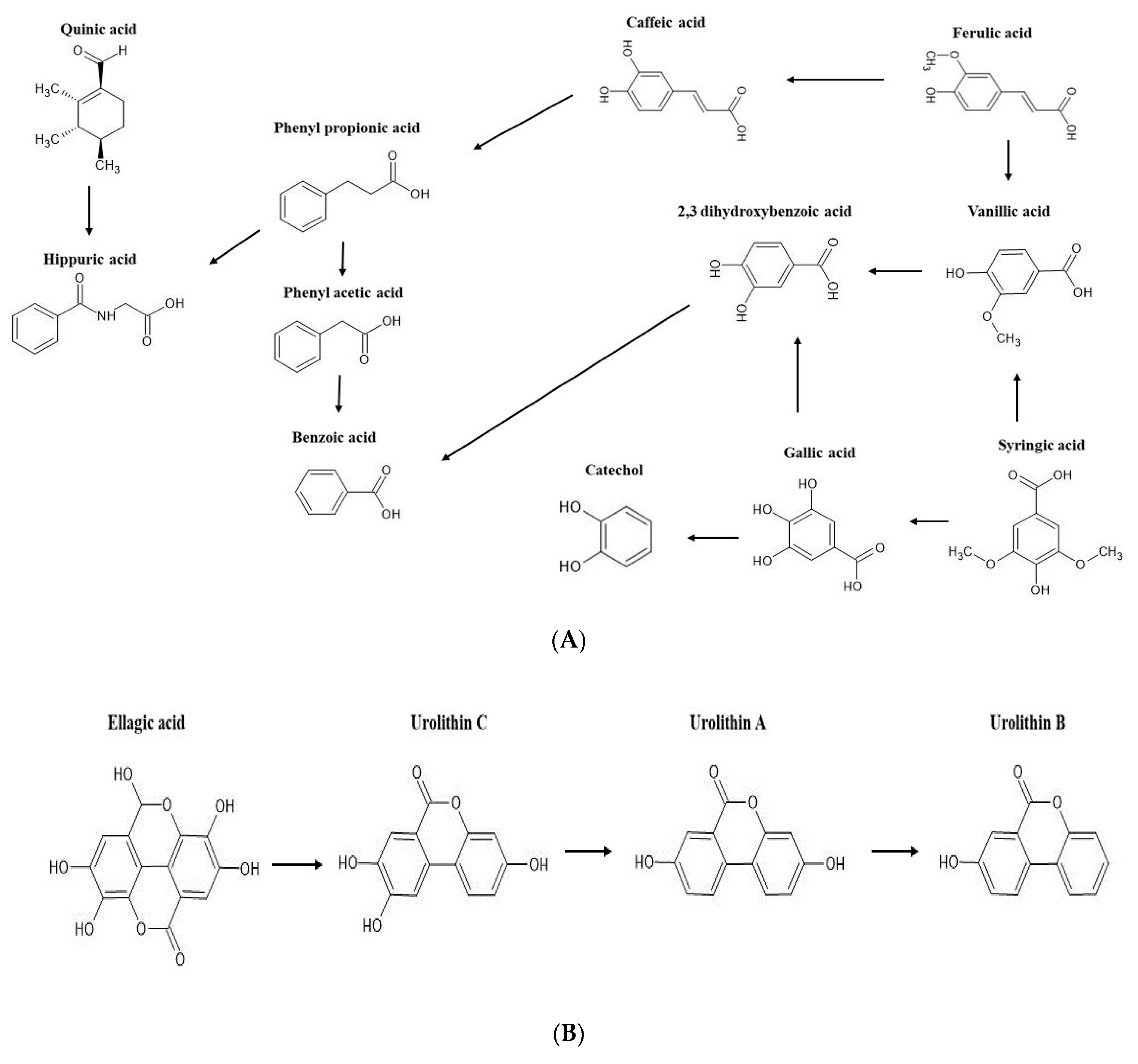
2.2. Bioconversion of Phenolics during Colonic Fermentation of Mango-Based Yoghurts

The mechanisms of interactions between gut flora and dietary polyphenols are characterised by compositional variations in the gut microbiota and/or the production of bioavailable metabolites via the action of gut flora that can modify the potential pharmacological properties of polyphenols. Substantial variations were observed in the phenolic profiles during incubation from 0 to 72 h with faecal matter. For example, ellagic acid was detected only in the insoluble residues of MPP, and protocatechuic acid was found throughout the 72 h of fermentation, whereas some hydroxycinnamic acids such as cinnamic and ferulic acids (m/z 147.0451 and m/z 193.0506, respectively) were not detected after 48 h of fermentation. Hernandez-Maldonado et al. [13] reported the rapid fermentation of ferulic, cinnamic, and chlorogenic acids during the colonic fermentation of mango fortified cereal bars. Another study by Dong et al. [17] reported that the methylation and dehydroxylation of syringic acid resulted in the formation of gallic acid during faecal fermentation, which upon further catabolism yielded catechol through decarboxylation and dehydroxylation. Moreover, catechol could also originate from protocatechuic acid, which is a bio-transformation product of gallic acid (compound 7, m/z 169.0142) through a dehydroxylation reaction (Figure 1A) [12,18]. Almeida et al. [19] reported that the faecal metabolism of quercetin or its glycosides led to the formation of protocatechuic acid and dihydroxyphenyl acetic acid.
Moreover, the in vitro colonic fermentation of indigestible fractions of MPP, YB, and YC resulted in the appearance of hydroxycoumarins, such as urolithins. As illustrated in Figure 1B, urolithins are the catabolites of ellagic acid. The microbiota mediated conversion of ellagic acid into urolithins is characterised by the hydrolysis of one lactone moiety and the release of water molecule followed by decarboxylation. The generation of urolithins through the degradation of ellagic acid was reported by Mosele et al. [12] and Garcia-Villalba et al. [20] in digested Arbutus unedo fruits and pomegranate.
With respect to flavonoids, no detection of flavanols was reported after 24 h of fermentation, but 5-(3′,4′-dihydroxyphenyl)-valeric acid ([M-H] at m/z 209.0819) and 3-hydroxyphenylvaleric acid ([M-H] at m/z 193.087) derivatives could be produced by dihydroxylation and ring cleavage conversions [21]. Previous studies have shown that catechin and its epimer, epicatechin, are catabolised by intestinal microbiota and converted into dihydroxyphenylpropan-2-ol and dihydroxyphenyl valeric acids, which then undergo dehydroxylation by the action of the colonic microflora, resulting in the generation of (hydroxyl) phenyl propionic acid, phenyl acetic acid, and benzoic acids [18,22].
Lignans, particularly dimethylmatairesinol and lariciresinol, were detected in the residue (solid) fraction of MPP, YB, and YC samples after simulated digestion. In vitro digestion studies suggested that stomach acids and intestinal enzymes do not play a major role in the initial hydrolysis of these lignans, leaving them intact [23]. However, they were biodegraded to enterolignans such as enterodiol and enterolactone in the later stages of faecal fermentation (Figure 1C). Though the precursor compounds have certain physiological effects, their bioconversion into metabolites have considerably greater biological outcomes. The potential anticancer effects of these enterolactones via antioxidant and antiestrogenic activities were proposed in previous studies [24,25]. The release of norathyriol ([M-H] at m/z 259.0248) at 48 and 72 h of fermentation indicated the likely breakdown of mangiferin (Figure 1D). According to Li et al. [26] norathyriol inhibits the production of uric acid by targeting organic anion transporters.

2.3. Variations in pH and Microbiological Population during Colonic Fermentation

2.3.1. pH

Changes in pH are important factors that reflect the degree of fermentation. As shown in Figure 2, the initial pH values of the blank, MPP, YA, YB, and YC were about 6.93, 6.83, 6.94, 6.73, and 6.76, respectively, with no significant differences (p ≥ 0.05) among them. However, a significant decline (p ≤ 0.05) in pH was recorded in the YA, YB, YC, and MPP treatments after the first 24 h of faecal fermentation. The highest decrease (2.19) in pH was observed in MPP samples, while YB, YA, and YC showed a decline of 1.84, 1.69, and 1.99, respectively. A gradual, but less severe, decrease in pH was recorded during the rest of the fermentation in all tested samples, with YB and YC exhibiting the lowest pH of 4.50, followed by MPP, with pH values of 4.53 at 72 h (Figure 2). The lowest acidification (less changes in pH) was detected in the blank (BL) throughout the fermentation period (72 h). These results may suggest that the phenolics and sugars present in MPP and yoghurt were used as sources of carbon for gut microbiota growth and metabolism, which could have triggered the decline in pH. These observations agreed with those reported by Tang et al. [27], who reported a significant decline in the pH of citrus fruits as probiotic fermentation proceeded.

2.3.2. Microbial Population

The quantitative variations in faecal microbiota during the in vitro colonic fermentation of plain MPP and MPP enriched yoghurt were assessed using the spread plate method. Plate count agar (PCA), MRS agar, and MRS agar enriched with cysteine were applied to examine lactic acid bacteria count (LAB), bifidobacteria, and total anaerobes, respectively (Figure 3A–C). The Bifidobacteria and LAB were selected as they belong to the predominant class of health promoting bacteria and are the producers of SCFA [28]. The recorded microbial abundance elicited by all treatments (YA, YB, YC, and MPP) in comparison with their respective blanks are given in Figure 3. The blank samples [colonic fermentation medium (CFM) + faecal slurry (FS)] showed a significant decline (p ≤ 0.05) in the log colony counts of all tested bacteria throughout the fermentation period in comparison with YC and MPP. Similarly, both the YA and YB treatments revealed similar LAB counts (Figure 3A), Bifidobacteria (Figure 3B), and total anaerobic (Figure 3C), but smaller counts than those in YC and MPP at each time of sampling during the 72 h of fermentation.
The initial Bifidobacteria counts in yoghurt enriched with MPP and probiotics (YC) and in plain MPP (Figure 3B) were very close, such as 8.36 and 8.58 log CFU/g, respectively. Furthermore, the Bifidobacteria counts in these two treatments were increased significantly to the maximum counts of 10.06 and 9.44 log CFU/g in YC and plain MPP, respectively, after 24 h of fermentation, followed by a continuous decline. However, the final counts of Bifidobacteria in these treatments remained greater than the counts in YA and TB after 72 h of fermentation (Figure 3B).
Similar patterns of changes in the total anaerobic and LAB counts were detected during the 72 h of fermentation. For example, after the initial increase in the LAB counts in YC at 24 h of fermentation, the counts decreased to 8.95 and 7.88 log CFU/g after 48 h and 72 h of fermentation, respectively (Figure 3A). These results suggested that the presence of mango peel powder in the YC treatments had significant positive prebiotic effects on the growth of LAB during the in vitro fermentation. Such prebiotic effects of MPP were not as significant in yoghurt enriched with MPP only (the YB treatment). This may be due to the fact that prebiotic effects were triggered by the presence of probiotic bacteria in the YC treatment.
The detected positive effect of plain MPP on the population of all tested bacteria could be attributed to the content of phenolic compounds and dietary fibre present in MPP. However, such positive effect was not detected in the YB treatment, where yoghurt was enriched with mango at 2% only. The effect of phenolic compounds on the gut microbiota has been reported by Boto-Ordóñez et al. [29], who suggested that foods rich in phenolic compounds may affect gut microbiota composition and activity by stimulating or inhibiting specific bacterial groups. Another investigation by Gutiérrez-Sarmiento et al. [3] reported that the colonic fermentation of mango-based bars revealed a considerable increase in Firmicutes and Actinobacteria during the initial 24 h with a reduced abundance until 48 h of fermentation.

2.3.3. Changes in Faecal Microbial Diversity in the Presence of MPP

It has been well established that the human gut microbiota is associated with metabolism, disease development, and immune functions in the body. From the results of the colony count methods and the changes in pH discussed above, it was evident that MPP and YC imparted some positive influences; therefore, fermentation media containing MPP, YC, and YA were assessed for compositional analysis through 16S rRNA sequencing at 24 and 72 h of fermentation. The relative abundance (RA) of Firmicutes, Proteobacteria, Bacteroidetes, Verrucomicrobiota, and Actinobacteria was over 99% at the phyla level (Figure 4B). Higher RA (85.06%) of Firmicutes was measured in YC as compared to YA (32.01%) and MPP (27.10%), particularly at 24 h of colonic fermentation, with Streptococcus being the most abundant genera (Figure 4A). The presence of probiotics and MPP in yoghurt (YC) triggered changes in the abundance and composition of the microbial population and was different to that of YA and MPP. The shift in microbial diversity was most likely promoted by dietary fibre and bound polyphenols that escaped small intestinal digestion. In general, the interactions between phenolics and the gut microbiota during fermentation are demonstrated to generate a series of metabolites, which in turn may contribute to modulating the normal microflora balance [17].
A complete statistical analysis of alpha- and beta-diversity was not possible in this study due to the pilot nature of investigating the effect of the different media on microbiome changes. Therefore, the results of estimated alpha- and beta-diversity are given in the supplementary section of this manuscript (Section S2.3.3, Figures S3 and S4). The primary aim of this test was to stipulate an overview on the role of gut microbiota in the metabolism of polyphenol and probiotic fermented diets. However, in vivo experiments are needed with larger sample sizes to validate the results of the present study.

2.4. Short Chain Fatty Acids Production during In Vitro Colonic Fermentation of Yoghurt Enriched with Mango Peel Powder

Short chain fatty acids (SCFAs) are produced by the gut microbiota via the fermentation of compounds that remain undigested when passing through the intestinal tract. The concentration of SCFAs released in the large intestine depends upon the intestinal transit time, composition of the host’s diet, and microbiota [30]. Figure 5 shows the concentrations of SCFAs released during the in vitro colonic fermentation of MPP, YB, YC, and their associated negative control (YA) and blank (FS + CFM).
Data in Figure 5 show that acetic acid was the most prevalent fatty acid in all tested samples, followed by propionic acid and butyric acid. A substantial increase in the levels of acetic acid was observed in MPP (307.31 ± 5.47 mM) and YB (261.25 ± 22.28 mM) after 24 h of fermentation, whereas the production efficacy of AA in YC delivered higher concentrations (252.46 ± 4.43 mM) subjected to 48h of colonic fermentation (Figure 5). On the other hand, significantly (p ≤ 0.05) lower concentrations of acetic acid were produced in YA (123.30 ± 0.62 mM) and blank (45.05 ± 0.36 mM) during the first 24 h of incubation, with an increase of 27% in the concentration of acetic acid in YA during the next 24 h (48 h of fermentation). This higher generation of acetic acid in the presence of MPP and YB could be attributed to the fibres present in MPP. This predominant formation of acetic acid is in accordance with the studies of Hernandez-Maldonado et al. [13] and Freire et al. [31] in which mango-based bars and goat milk fortified with grape juice pomace, respectively, were subjected to colonic fermentation. It is also evident from Figure 5 that the concentration of acetic acid was affected by fermentation time and declined significantly (p ≤ 0.05) when the fermentation time extended from 48 to 72 h. These results align well with the findings of Granado-Serrano et al. [32] and Hossain et al. [33], where the authors reported maximum generation of SCFAs between 0 and 48 of colonic fermentation. Previous studies indicated increased production of SCFAs in the presence of different probiotic strains, e.g., B. animalis subsp. lactis GCL2505 was reported to greatly enhance the levels of SCFAs in the gut [34]. In addition, the enhanced release of acetic acid during the colonic fermentation of MPP and MPP fortified yoghurts could also be attributed to mango flavonoids, which were bio-degraded to various small molecule phenolics such as phenyl valeric acids, phenyl propionic acid, and phenyl acetic acids. These findings were substantiated by recent studies on citrus flavanones, indicating the reduced bioavailability of citrus flavanones leading them to surpass the digestive tract to the colon and subsequently undergo hydrolysis by the gut microbiota into phenolic catabolites, consequently enhancing the levels of SCFAs [27,35].
Figure 5 also reveals that the highest concentrations of propionic acid and butyric acid were achieved after 24 h of fermentation, followed by a gradual decline until 72 h in all treatments except MPP, which showed a small but insignificant (p ≥ 0.05) increase of 0.67 mM in the levels of BA between 48 and 72 h of fermentation. However, compared to acetic acid, significantly (p ≤ 0.05) lower amounts of butyric and propionic acid were produced by all the tested samples. The contents of propionic acid were highest at 24 h of colonic fermentation in all the tested substrates except YA, where it reached a maximum value of 11.53 ± 1.89 mM after 48 h of colonic incubation. The maximum concentration of butyric acid was achieved by MPP (32.79 ± 2.01 mM) during the first 24 h of colonic fermentation, which was 2.2, 2.8, and 3.6 folds higher than its concentrations in YC, YB, and YA, respectively (Figure 5). The enhanced release of these acids from MPP could be possibly due the stimulus provided by the gut microbiota to the phenolics (flavanols) and dietary fibres in MPP [36,37]. Interestingly, synergism between SCFAs and phenolics, for example, BA and gallic acid, has been linked to the modulation of inflammatory signals for treating inflammatory disorders [38]. Analogous to the acetic acid, lower amounts of butyric acid were released in the blank (8.16 ± 0.23 mM) with a gradual decline in production efficacy until 72 h of colonic fermentation. These findings are supported by the study by Herrera-Cazares et al. [37], who reported that the in vitro colonic fermentation of mango bagasse and mango bagasse based functional confections produced increased concentrations of acetate and butyrate.
Regarding the contents of valeric acid, a similar production pattern to that of propionic acid was observed. However, significantly (p ≤ 0.05) lower amounts of valeric acid were detected in comparison to propionic acid. The highest amounts of valeric acid (6.52 ± 0.96 mM) were achieved in MPP at 24 h, followed by YC (4.47 ± 0.35 mM), YB (3.38 ± 0.71 mM), YA (1.79 ± 0.14 mM), and blank (0.59 ± 0.15 mM).
Data in Figure 5 also show the production of varying concentrations of isovaleric and isobutyric acids with respect to the different treatments. Unlike MPP, the highest amounts of isovaleric and isobutyric acids were produced by YC at 10.59 ± 0.78 mM and 8.65 ± 0.53 mM, respectively. This could possibly be explained by the fact that varying proportions of certain non-digestible fibres in the substrates may result in various patterns of SCFA production [39]. These results confirmed that amongst the SCFAs formed during the in vitro colonic fermentation, acetic, butyric, and propionic acids were the major SCFAs produced by the colonic microbiota, with higher concentrations, while valeric, isobutyric, and isovaleric acids were the minor SCFAs, in low concentrations. These findings agree with recent results by Loo et al. [40] and Tamargo et al. [41], who investigated in vitro and in vivo colonic fermentation using sugarcane polyphenol and cranberry extracts as substrates. However, Rios-Covian et al. [30] reported that in the hindgut, SCFAs were produced in the order of acetate > propionate > butyrate, which was different from the order of acetate > butyrate > propionate in the present study (Figure 5). This could possibly be attributed to the synergism/antagonism between SCFAs and other phytochemicals and/or microbial cross-feeding in different fermentation systems [37].

3. Materials and Methods

3.1. Materials

A commercial yoghurt starter culture (YOFLEX®—consisting of Streptococcus thermophilus and Lactobacillus delbrueckii subsp. bulgaricus) and three probiotic strains (Lacticaseibacillus rhamnosus LGG, Lacticaseibacillus casei 431, Bifidobacterium animalis subsp. lactis BB-12) were kindly supplied by Chr. Hansen, Bayswater, VIC, Australia. Powdered skim milk (Australian instant skim milk powder, Coles) was obtained from a local supermarket in Melbourne, VIC, Australia. Pancreatin was purchased from Alfa Aesar (Ward Hill, MA, USA). Mangiferin, pyrogallol, gallic acid, caffeic acid, 3-hydroxy phenyl acetic acid, 3-(2-hydroxyphenyl) propionic acid, porcine pepsin, α-amylase from aspergillus oryzae, p-nitrophenyl-α-d-glucopyranoside, bile salts, LC-MS grade formic acid, acetic acid, 4-methyl valeric acid, guar, casein, tryptone, cysteine HCl, peptone, mucin, pectin, potato starch, yeast extract, and MRS agar were acquired from Sigma–Aldrich (Castle Hill, NSW, Australia). Sodium carbonate (anhydrous), HCl, NaHCO3, NaOH, (NH4)2CO3, KCl, NaCl, KH2PO4, MgCl2(H2O)6, CaCl2, K2HPO4, Na3PO4, MgSO4.7H2O Tween 80, orthophosphoric acid, acetonitrile, and methanol of HPLC grade were acquired from Chem-supply Pty Ltd. (Melbourne, VIC, Australia).

3.2. Methods

3.2.1. Preparation of Probiotic Cultures and Mango Peel Powder

The probiotic strains of L. rhamnosus (LGG®), L. casei (431®), B. lactis (Bb-12®), and yoghurt starter culture (SC) (YOFLEX®) were activated under anaerobic conditions in de Man Rogosa Sharp (MRS) broth at 37 °C for 48 h, and the cultures were harvested via centrifugation as described by Zahid et al. [42,43]. The mango peels were transformed into powder form using a freeze-drying operation at −48 °C (Dynavac engineering FD3 freeze-drier, Belmont, Australia) and subsequently ground to a uniform particle size of 250 µm [42] using a laboratory coffee grinder (Multigrinder EMO405, Sunbeam, Melbourne, Australia).

3.2.2. Yoghurt Samples Preparation

Yoghurt mixes (stirred type) were prepared in triplicates and labelled yoghurt A (YA), yoghurt B (YB), and yoghurt C (YC) according to the method of Zahid et al. [43]. YA represented the negative control and contained a starter culture (SC) only, YB consisted of SC along with added MPP at a predetermined concentration of 2%, and YC consisted of yoghurt B enriched with each of the tested probiotics at a concentration of 1% (v/v). Plain MPP was also included in the study as a positive control. All the treatments were prepared in triplicate.

3.2.3. In Vitro Gastrointestinal Digestion and Colonic Fermentation of MPP and MPP Fortified Yoghurts

The simulated fluids for oral, gastric, and small intestinal digestion phases were prepared using a mix of electrolytes (Cl, Na+, K+, H2PO4, Mg2+, Ca2+, NH4+, and HCO3) at variable concentrations. All the samples and controls were subjected to a three-step sequential digestion model using INFOGEST protocol as explained in our previous study [44]. After in vitro digestion, the residue (indigestible fraction) was exposed to colonic fermentation under anaerobic conditions [44].
The procedure of this in vitro colonic fermentation study was approved by the Ethics Advisory Group (ID: 1954660.1) in the Faculty of Science, The University of Melbourne. The colonic fermentation medium (CFM) was made of guar (1.0 g), bile salts (0.4 g), casein (3 g), CaCl2 (0.11 g), KCl (4.5 g), KH2PO4 (0.5 g), K2HPO4 (0.5 g), cysteine HCl (0.8 g), mucin (4 g), MgSO4.7H2O (1.23 g), NaCl (4.5 g), NaHCO3 (1.5 g), pectin (2 g), peptone (5 g), potato starch (5 g), tryptone (5 g), yeast extract (4.5 g), and Tween 80 (1.0 mL) dissolved in Milli-Q water, and the volume was made up to 1000 mL. The medium was adjusted to pH 6.9 ± 1.0 and autoclaved at 121 °C for 20 min.
Two healthy donors provided freshly defecated faeces. The donors specified that they had not used probiotics or antibiotics in the prior three months and were free of gastrointestinal conditions at the time of sample collection. The faeces were transferred to the lab on ice, combined, and stomached with 0.1 M sterilised phosphate buffer (pH 7.0) in a stomacher mixer (Bagmixer 400, Interscience, Saint-Nom, France). The obtained mixture was then sifted using a cheesecloth to develop one faecal slurry at a proportion of 20:80 (w/w) faeces:buffer. In 50 mL N2 flushed tubes, 5 mL aliquots of faecal slurry were distributed in each tube along with 0.5 g of the intestinal residue (insoluble intestinal fraction) and mixed with 5 mL of the prepared CFM. The tubes were firmly sealed and put in an anaerobic shaking incubator (ZWTR-240, Labwit, China) for 72 h at 120 rpm, 37 °C in the absence of oxygen (anaerobiosis). The anaerobic environment was created in anaerobic jars (BD BBL Gas Pak, Mississauga, Ontario, Canada) through an anaerobic gas generator (AN 0010W, Oxoid®). A blank test was carried out by inoculating a mix of CFM and FS at 1:1 ratio, without MPP or yoghurt samples to correct the contribution of reagents. The sample tubes were taken and analysed at various intervals (0, 24, 48, and 72 h) during fermentation. The tubes were immediately placed in an ice bath to stop the fermentation process. Each sample was run in quadruplicate for every condition tested. The supernatant fraction from each tube was obtained following centrifugation (10,000× g, 15 min, 4 °C) and stored at −80 °C until further analyses of phenolic metabolites, microbial count and composition, generation of short chain fatty acids, and pH variations.

3.2.4. Analysis of Phenolic Metabolites

Extraction of Phenolics

The extraction of polyphenols from faecal digesta was adapted from Zahid et al. [44] with slight changes. Briefly, 0.5 mL of faecal digesta was mixed with acidified methanol/water (8:20 acidified with 0.01% conc. HCl). The mixtures were vortexed (Ultra Turax T25 D S5, IKA, Germany) and incubated overnight under shaking conditions at 120 rpm, 4 °C. The separation of supernatants was achieved by centrifugation for 15 min at 5000× g, 4 °C.

Qualitative Analysis of Polyphenols Using LC-ESI-QTOF-MS2

The extracted phenolic fractions were characterized by using an Agilent 6520 Accurate-Mass QTOF interfaced with an ionisation (ESI) source and provided with Agilent HPLC 1200 series (Agilent, Santa Clara, CA, USA). HPLC separation was performed on a Synergi Hydro-RP (4 μm, 4.6 mm, 250 mm) column (Phenomenex, Lane Cove, NSW, Australia) with a pore size of 80 Å. The mobile phases used were water (Buffer A) and acetonitrile (Buffer B) acidified with 0.1% formic acid. The gradient elution programme was set at B: 0–10 min, 10–20%; 10–20 min, 20–25%; 20–30 min, 25–30%; 30–40 min, 30–45%; 40–50 min, 45–60%; 50–60 min, 60–80%; 60–65 min, 60–80%; 65–67 min, 90–100%; 67–70 min, 100–10%. The sample volume was set at 10 µL with an elution flow rate of 0.6 mL/min. Accurate-Mass QTOF 6520 was set to function in a negative ionisation mode (ESI) at a capillary voltage of 3500 V with a scanning speed of 250 spectra/s, and the mass spectra were attained through full scan within the mass range of 100–1000 m/z in MS/MS mode. Nitrogen (N2) was used as a nebuliser and drying gas at a temperature of 325 °C with a flow rate of 9 L/min, 10, 20, and 40 eV collision energies and a nebuliser pressure of 45 psi [9] to achieve the fragmentation of metabolites. The identification of the phenolic metabolites was carried out with the help of the Agilent MassHunter Workstation Quality Analysis Software (version B.06.00), Personal Compounds Database and Library for metabolites (PCDL), PubChem (https://pubchem.ncbi.nlm.nih.gov/), accessed on 10 July 2022 and FooDB (https://foodb.ca/) accessed on 11 July 2022 [45].

3.2.5. pH and Microbiological Analysis

Samples collected at various time points during colonic fermentation (0 h, 24 h, 48 h and 72 h) were subjected to pH monitoring using a pH meter (HI5221, Hanna, Woonsocket, RI, USA). The variations in faecal bacteria composition were assessed by analysing total aerobic count, lactic acid bacteria (LAB), and total anaerobic count using plate count agar (PCA), MRS agar, and MRS agar enriched with cysteine, respectively [44,46]. The initial bacterial count (blank) was carried out using a mixture of faecal slurry (FS) and sterile basal medium (CFM) prepared at 1:1 ratio, (v/v). All samples were serially diluted using 0.1% sterile peptone water, spread plated, and incubated (aerobically and aerobically) at 37 °C for 48 h.

3.2.6. 16S rRNA Sequencing Analysis

A subset of samples was used for preliminary investigation into microbial diversity in different sample sets. Two replicates each from MPP, YA, and YC at 24 h and 72 h were used for this purpose. DNA was extracted from 200 µL of the selected fermented samples using the DNeasy PowerSoil Pro Kit (QIAGEN, Venlo, The Netherlands) as per the manufacturer’s instructions. Extracted DNA was subsequently processed through a Zymo DNA Clean and Concentrate kit (Zymo Research, Irvine, CA, USA) as the per manufacturer’s instructions to obtain suitable quality DNA for amplicon sequencing. The V3–V4 hypervariable region of the 16S rRNA gene was amplified and sequenced using primers 341F (5′-CCT AYG GGR BGC ASC AG-3′) and 806R (5′-GGA CTA CNN GGG TAT CTA AT-3′) [47] at the Australian Genome Research Facility (AGRF, Melbourne, Australia). Amplicons were barcoded, pooled, and paired end (2 × 300 bp) sequenced on the Illumina MiSeq platform using the Nextera XT Indexes (Illumina, San Diego, CA, USA).
Raw data were demultiplexed and converted into FASTQ format by the Illumina conversion software (version v2.2.68) at AGRF. The Quantitative Insights into Microbial Ecology 2 (QIIME2) v2021.2.0 software was used for downstream processing [48]. Amplicon sequence variants (ASVs) were generated using DADA2 [49]. In DADA2, the demultiplexed data were quality filtered, primer trimmed, denoised, and processed to remove chimeras. Through DADA2, forward and reverse reads were truncated to 260 and 220 bp, respectively, to maximise the average read numbers retained after processing.
Taxonomic classification on the resulting ASVs was performed using a naïve bayes classifier within QIIME2 [50] using the SILVA 138.1 [51] 16S rRNA database clustered to 99% similarity and trimmed to the V3-V4 region using the primer sequence outlined above. Data were imported into the phyloseq package in R [52], and alpha and beta diversity metrics were investigated, as was the relative abundance of taxa between different groups. Alpha diversity was estimated from counts using the ‘estimate richness’ function in phyloseq, whilst beta-diversity was investigated using data subsampled to 43,338 reads (the minimum across the sample set at which all other samples had reached a plateau in respect to observed ASVs). Beta-diversity analysis utilised UniFrac ordination [52] for principal coordinate analysis.

3.2.7. Determination of SCFA

Evaluation of the SCFA concentrations in colonic digesta was performed adopting the procedure of Loo et al. [40] with minor changes. After colonic fermentation, supernatants (1.5 mL) collected after centrifugation were combined with four volumes of an internal standard mixture containing 1.59 mmol/L of 4-methyl valeric acid mixed with formic acid and orthophosphoric acid (1% both) and vortexed for 30 s. From the final mixture, 1 mL of each sample was dispensed into 1.5 mL flip capped tubes and centrifuged (10,000× g, 10 min at 4 °C). The supernatants were separated and stored at 4 °C until analysis. Acetic, butyric, propionic, iso-valeric, valeric, iso-butyric, and heptanoic acids were used as analytical standards to create the standard curves [44].
Aliquots (2 μL) of the sample and standards were injected into a gas chromatograph (7890B Agilent, CA, USA) that was fitted with a capillary column of 12 × 0.53 mm internal diameter (ID) and a film thickness of 0.5 µm (SGE BP21, SGE International, Ringwood, VIC, Australia, P/N 054473), a flame ionisation detector (FID), an autosampler (Gilson GX-271, Gilson Inc., Middleton, WI, USA), and an autoinjector. The FID and injection port were set at temperatures of 240 and 200 °C, respectively. Helium was applied as a carrier gas at a flow rate of 14.4 mL/min along with hydrogen, nitrogen, and air as makeup gases.

3.2.8. Statistical Analysis

All collected data apart from microbiome data were analysed using the GraphPad Prism statistical package version 9.5.0 (GraphPad Software LLC, Inc., San Diego, CA, USA). Two-way analysis of variance (ANOVA) was performed, and the difference between means was determined using Tukey’s post hoc test at 95% confidence interval. The quantitative measurements were performed in triplicate with at least two measurements for each sample, and the results were expressed as mean ± standard deviation. The qualitative data analysis was performed as replicates with a single measurement within each sample.

4. Conclusions

The results of the present work demonstrated that phytochemicals in mango peel powder have prebiotic effects during colonic fermentation. Insoluble fractions of the digesta were susceptible to further biotransformation during the in vitro colonic fermentation with human faecal microbiota. The catabolism of polyphenols resulted in the production of several phenolic metabolites such as hippuric acid, hydroxyphenyl acetic acid, catechol, protocatechuic acid, urolithins, enterolactones, and norathyriol. The prebiotic-like functions of mango peels were reflected by a significant increment in the lactic acid bacterial counts. Nevertheless, these actions were highly dependent upon the structural composition of the food matrices and the incubation times. These findings were further supported by the reduction in the pH of fermentation media and the generation of SCFAs, where acetic, propionic, and butyric acids were the major SCFAs produced. Additionally, the 16S rRNA analyses of the microbiota after the in vitro colonic fermentation of plain MPP and yoghurt enriched with MPP revealed some modifications in various bacterial groups. These findings presented insights into the significant roles of MPP as prebiotics when used as a plain ingredient or in dairy food systems after human consumption. However, in vivo studies are needed in the future to gain a deep understanding of the microbiota mediated gut well-being of MPP in human diet.

Supplementary Materials

The following supporting information can be downloaded at: https://www.mdpi.com/article/10.3390/ijms24108560/s1.

Author Contributions

Conceptualisation, H.F.Z. and S.A.; methodology, H.F.Z., A.A. and A.R.L.; software, H.F.Z., A.A. and A.R.L.; validation, S.A., C.S.R., Z.F. and H.F.Z.; formal analysis, H.F.Z., A.A. and A.R.L.; investigation, H.F.Z.; resources, S.A. and F.R.D.; data curation, H.F.Z.; writing—original draft preparation, H.F.Z.; writing—review and editing, S.A., C.S.R., Z.F. and A.R.L.; visualisation, S.A. and H.F.Z.; supervision, S.A., C.S.R. and Z.F.; project administration, S.A. All authors have read and agreed to the published version of the manuscript.

Funding

This research received no external funding.

Institutional Review Board Statement

Not applicable.

Informed Consent Statement

Informed consent was obtained from all subjects involved in the study.

Data Availability Statement

The data are available in the Supplementary Materials.

Acknowledgments

The authors wish to acknowledge the administrative support provided by the University of Melbourne and Mass spectrometry and Proteomics facility of Bio21 Institute for their technical assistance. The authors would like to thank Hafiz Suleria from the School of Agriculture, Food and Ecosystem Sciences, Faculty of Science, The University of Melbourne for his assistance in the GC-FID work done in this manuscript.

Conflicts of Interest

The authors declare no conflict of interest.

References

  1. Zahid, H.F.; Ranadheera, C.S.; Fang, Z.; Ajlouni, S. Healthy and Functional Ingredients from Fruit Processing Byproducts: A Review Focusing on Fruit Peels. Int. J. Agric. Environ. Biores. 2019, 4, 336–360. [Google Scholar] [CrossRef]
  2. Mohamed, S. Functional foods against metabolic syndrome (obesity, diabetes, hypertension and dyslipidemia) and cardiovasular disease. Trends Food Sci. Technol. 2014, 35, 114–128. [Google Scholar] [CrossRef]
  3. Gutiérrez-Sarmiento, W.; Sáyago-Ayerdi, S.G.; Goñi, I.; Gutiérrez-Miceli, F.A.; Abud-Archila, M.; Rejón-Orantes, J.d.C.; Rincón-Rosales, R.; Peña-Ocaña, B.A.; Ruíz-Valdiviezo, V.M. Changes in Intestinal Microbiota and Predicted Metabolic Pathways During Colonic Fermentation of Mango (Mangifera indica L.)—Based Bar Indigestible Fraction. Nutrients 2020, 12, 683. [Google Scholar] [CrossRef] [PubMed]
  4. Santhiravel, S.; Bekhit, A.E.D.A.; Mendis, E.; Jacobs, J.L.; Dunshea, F.R.; Rajapakse, N.; Ponnampalam, E.N. The impact of plant phytochemicals on the gut microbiota of humans for a balanced diet. Int. J. Mol. Sci. 2022, 23, 8124. [Google Scholar] [CrossRef] [PubMed]
  5. Leonard, W.; Zhang, P.; Ying, D.; Adhikari, B.; Fang, Z. Fermentation transforms the phenolic profiles and bioactivities of plant-based foods. Biotechnol. Adv. 2021, 49, 107763. [Google Scholar] [CrossRef] [PubMed]
  6. Quatrin, A.; Rampelotto, C.; Pauletto, R.; Maurer, L.H.; Nichelle, S.M.; Klein, B.; Rodrigues, R.F.; Maróstica Junior, M.R.; Fonseca, B.d.S.; de Menezes, C.R.; et al. Bioaccessibility and catabolism of phenolic compounds from jaboticaba (Myrciaria trunciflora) fruit peel during in vitro gastrointestinal digestion and colonic fermentation. J. Funct. Foods 2020, 65, 103714. [Google Scholar] [CrossRef]
  7. Hidalgo, M.; Oruna-Concha, M.J.; Kolida, S.; Walton, G.E.; Kallithraka, S.; Spencer, J.P.; de Pascual-Teresa, S. Metabolism of anthocyanins by human gut microflora and their influence on gut bacterial growth. J. Agric. Food Chem. 2012, 60, 3882–3890. [Google Scholar] [CrossRef]
  8. Sayago-Ayerdi, S.G.; Zamora-Gasga, V.M.; Venema, K. Prebiotic effect of predigested mango peel on gut microbiota assessed in a dynamic in vitro model of the human colon (TIM-2). Food Res. Int. 2019, 118, 89–95. [Google Scholar] [CrossRef]
  9. Zahid, H.F.; Ali, A.; Ranadheera, C.S.; Fang, Z.; Dunshea, F.R.; Ajlouni, S. In vitro bioaccessibility of phenolic compounds and alpha-glucosidase inhibition activity in yoghurts enriched with mango peel powder. Food Biosci. 2022, 50, 102011. [Google Scholar] [CrossRef]
  10. Peng, D.; Zahid, H.F.; Ajlouni, S.; Dunshea, F.R.; Suleria, H.A.R. LC-ESI-QTOF/MS Profiling of Australian Mango Peel By-Product Polyphenols and Their Potential Antioxidant Activities. Processes 2019, 7, 764. [Google Scholar] [CrossRef]
  11. Noto, A.; Fanos, V.; Barberini, L.; Grapov, D.; Fattuoni, C.; Zaffanello, M.; Casanova, A.; Fenu, G.; De Giacomo, A.; De Angelis, M. The urinary metabolomics profile of an Italian autistic children population and their unaffected siblings. J. Matern.-Fetal Neonatal Med. 2014, 27, 46–52. [Google Scholar] [CrossRef]
  12. Mosele, J.I.; Macià, A.; Romero, M.-P.; Motilva, M.-J. Stability and metabolism of Arbutus unedo bioactive compounds (phenolics and antioxidants) under in vitro digestion and colonic fermentation. Food Chem. 2016, 201, 120–130. [Google Scholar] [CrossRef]
  13. Hernandez-Maldonado, L.M.; Blancas-Benitez, F.J.; Zamora-Gasga, V.M.; Cardenas-Castro, A.P.; Tovar, J.; Sayago-Ayerdi, S.G. In Vitro Gastrointestinal Digestion and Colonic Fermentation of High Dietary Fiber and Antioxidant-Rich Mango (Mangifera indica L.) “Ataulfo”-Based Fruit Bars. Nutrients 2019, 11, 1564. [Google Scholar] [CrossRef] [PubMed]
  14. Pathak, G.; Singh, S.; Kumari, P.; Hussain, Y.; Raza, W.; Luqman, S.; Meena, A. Cirsilineol inhibits proliferation of lung squamous cell carcinoma by inducing ROS mediated apoptosis. Food Chem. Toxicol. 2020, 143, 111550. [Google Scholar] [CrossRef] [PubMed]
  15. Setchell, K.D.; Brown, N.M.; Lydeking-Olsen, E. The clinical importance of the metabolite equol—A clue to the effectiveness of soy and its isoflavones. J. Nutr. 2002, 132, 3577–3584. [Google Scholar] [CrossRef] [PubMed]
  16. Pérez-Nájera, V.C.; Lugo-Cervantes, E.; Amaya-Delgado, L.; Madrigal-Pulido, J.A.; Rueda-Puente, E.O.; Borboa-Flores, J.; Del-Toro-Sánchez, C.L. Biotransformation of hesperidin from lime peel (Citrus limetta Risso) in solid fermentation by Aspergillus saitoi. CyTA-J. Food 2018, 16, 537–543. [Google Scholar] [CrossRef]
  17. Dong, R.; Liu, S.; Xie, J.; Chen, Y.; Zheng, Y.; Zhang, X.; Zhao, E.; Wang, Z.; Xu, H.; Yu, Q. The recovery, catabolism and potential bioactivity of polyphenols from carrot subjected to in vitro simulated digestion and colonic fermentation. Food Res. Int. 2021, 143, 110263. [Google Scholar] [CrossRef] [PubMed]
  18. Sánchez-Patán, F.; Cueva, C.; Monagas, M.; Walton, G.E.; Gibson, M.G.R.; Quintanilla-López, J.E.; Lebrón-Aguilar, R.; Martín-Álvarez, P.; Moreno-Arribas, M.V.; Bartolomé, B. In vitro fermentation of a red wine extract by human gut microbiota: Changes in microbial groups and formation of phenolic metabolites. J. Agric. Food Chem. 2012, 60, 2136–2147. [Google Scholar] [CrossRef]
  19. Almeida, A.F.; Borge, G.I.A.; Piskula, M.; Tudose, A.; Tudoreanu, L.; Valentova, K.; Williamson, G.; Santos, C.N. Bioavailability of Quercetin in humans with a focus on interindividual variation. Compr. Rev. Food Sci. Food Saf. 2018, 17, 714–731. [Google Scholar] [CrossRef] [PubMed]
  20. Garcia-Villalba, R.; Vissenaekens, H.; Pitart, J.; Romo-Vaquero, M.; Espín, J.C.; Grootaert, C.; Selma, M.V.; Raes, K.; Smagghe, G.; Possemiers, S. Gastrointestinal simulation model TWIN-SHIME shows differences between human urolithin-metabotypes in gut microbiota composition, pomegranate polyphenol metabolism, and transport along the intestinal tract. J. Agric. Food Chem. 2017, 65, 5480–5493. [Google Scholar] [CrossRef]
  21. Chen, W.; Zhu, X.; Lu, Q.; Zhang, L.; Wang, X.; Liu, R. C-ring cleavage metabolites of catechin and epicatechin enhanced antioxidant activities through intestinal microbiota. Food Res. Int. 2020, 135, 109271. [Google Scholar] [CrossRef]
  22. Monagas, M.; Urpi-Sarda, M.; Sánchez-Patán, F.; Llorach, R.; Garrido, I.; Gómez-Cordovés, C.; Andres-Lacueva, C.; Bartolomé, B. Insights into the metabolism and microbial biotransformation of dietary flavan-3-ols and the bioactivity of their metabolites. Food Funct. 2010, 1, 233–253. [Google Scholar] [CrossRef] [PubMed]
  23. Eeckhaut, E.; Struijs, K.; Possemiers, S.; Vincken, J.-P.; Keukeleire, D.D.; Verstraete, W. Metabolism of the lignan macromolecule into enterolignans in the gastrointestinal lumen as determined in the simulator of the human intestinal microbial ecosystem. J. Agric. Food Chem. 2008, 56, 4806–4812. [Google Scholar] [CrossRef]
  24. Shin, M.K.; Jeon, Y.D.; Jin, J.S. Apoptotic effect of enterodiol, the final metabolite of edible lignans, in colorectal cancer cells. J. Sci. Food Agric. 2019, 99, 2411–2419. [Google Scholar] [CrossRef]
  25. Zálešák, F.; Bon, D.J.-Y.D.; Pospíšil, J. Lignans and Neolignans: Plant secondary metabolites as a reservoir of biologically active substances. Pharmacol. Res. 2019, 146, 104284. [Google Scholar] [CrossRef] [PubMed]
  26. Li, J.; Liu, M.; Yu, H.; Wang, W.; Han, L.; Chen, Q.; Ruan, J.; Wen, S.; Zhang, Y.; Wang, T. Mangiferin improves hepatic lipid metabolism mainly through its metabolite-norathyriol by modulating SIRT-1/AMPK/SREBP-1c signaling. Front. Pharmacol. 2018, 9, 201. [Google Scholar] [CrossRef] [PubMed]
  27. Tang, R.; Yu, H.; Qi, M.; Yuan, X.; Ruan, Z.; Hu, C.; Xiao, M.; Xue, Y.; Yao, Y.; Liu, Q. Biotransformation of citrus fruits phenolic profiles by mixed probiotics in vitro anaerobic fermentation. LWT 2022, 160, 113087. [Google Scholar] [CrossRef]
  28. Burgos-Edwards, A.; Jimenez-Aspee, F.; Theoduloz, C.; Schmeda-Hirschmann, G. Colonic fermentation of polyphenols from Chilean currants (Ribes spp.) and its effect on antioxidant capacity and metabolic syndrome-associated enzymes. Food Chem. 2018, 258, 144–155. [Google Scholar] [CrossRef] [PubMed]
  29. Boto-Ordonez, M.; Rothwell, J.A.; Andres-Lacueva, C.; Manach, C.; Scalbert, A.; Urpi-Sarda, M. Prediction of the wine polyphenol metabolic space: An application of the Phenol-Explorer database. Mol. Nutr. Food Res. 2013, 58, 466–477. [Google Scholar] [CrossRef] [PubMed]
  30. Ríos-Covián, D.; Ruas-Madiedo, P.; Margolles, A.; Gueimonde, M.; De Los Reyes-gavilán, C.G.; Salazar, N. Intestinal short chain fatty acids and their link with diet and human health. Front. Microbiol. 2016, 7, 185. [Google Scholar] [CrossRef] [PubMed]
  31. Freire, F.C.; Adorno, M.A.T.; Sakamoto, I.K.; Antoniassi, R.; Chaves, A.C.S.D.; Dos Santos, K.M.O.; Sivieri, K. Impact of multi-functional fermented goat milk beverage on gut microbiota in a dynamic colon model. Food Res. Int. 2017, 99, 315–327. [Google Scholar] [CrossRef] [PubMed]
  32. Granado-Serrano, A.B.; Martín-Garí, M.; Sánchez, V.; Riart Solans, M.; Berdún, R.; Ludwig, I.A.; Rubió, L.; Vilaprinyó, E.; Portero-Otín, M.; Serrano, J. Faecal bacterial and short-chain fatty acids signature in hypercholesterolemia. Sci. Rep. 2019, 9, 1–13. [Google Scholar]
  33. Hossain, M.N.; Ranadheera, C.S.; Fang, Z.; Ajlouni, S. Production of short chain fatty acids and vitamin B12 during the in-vitro digestion and fermentation of probiotic chocolate. Food Biosci. 2022, 47, 101682. [Google Scholar] [CrossRef]
  34. Horiuchi, H.; Kamikado, K.; Aoki, R.; Suganuma, N.; Nishijima, T.; Nakatani, A.; Kimura, I. Bifidobacterium animalis subsp. lactis GCL2505 modulates host energy metabolism via the short-chain fatty acid receptor GPR43. Sci. Rep. 2020, 10, 4158. [Google Scholar] [CrossRef] [PubMed]
  35. Pereira-Caro, G.; Oliver, C.M.; Weerakkody, R.; Singh, T.; Conlon, M.; Borges, G.; Sanguansri, L.; Lockett, T.; Roberts, S.A.; Crozier, A. Chronic administration of a microencapsulated probiotic enhances the bioavailability of orange juice flavanones in humans. Free Radic. Biol. Med. 2015, 84, 206–214. [Google Scholar] [CrossRef] [PubMed]
  36. Alves-Santos, A.M.; Sugizaki, C.S.A.; Lima, G.C.; Naves, M.M.V. Prebiotic effect of dietary polyphenols: A systematic review. J. Funct. Foods 2020, 74, 104169. [Google Scholar] [CrossRef]
  37. Herrera-Cazares, L.A.; Ramírez-Jiménez, A.K.; Wall-Medrano, A.; Campos-Vega, R.; Loarca-Piña, G.; Reyes-Vega, M.; Vázquez-Landaverde, P.A.; Gaytán-Martínez, M. Untargeted metabolomic evaluation of mango bagasse and mango bagasse based confection under in vitro simulated colonic fermentation. J. Funct. Foods 2019, 54, 271–280. [Google Scholar] [CrossRef]
  38. Luzardo-Ocampo, I.; Loarca-Piña, G.; de Mejia, E.G. Gallic and butyric acids modulated NLRP3 inflammasome markers in a co-culture model of intestinal inflammation. Food Chem. Toxicol. 2020, 146, 111835. [Google Scholar] [CrossRef]
  39. Low, D.Y.; Williams, B.A.; D’Arcy, B.R.; Flanagan, B.M.; Gidley, M.J. In vitro fermentation of chewed mango and banana: Particle size, starch and vascular fibre effects. Food Funct. 2015, 6, 2464–2474. [Google Scholar] [CrossRef]
  40. Loo, Y.T.; Howell, K.; Suleria, H.; Zhang, P.; Gu, C.; Ng, K. Sugarcane polyphenol and fiber to affect production of short-chain fatty acids and microbiota composition using in vitro digestion and pig faecal fermentation model. Food Chem. 2022, 385, 132665. [Google Scholar] [CrossRef]
  41. Tamargo, A.; Cueva, C.; Taladrid, D.; Khoo, C.; Moreno-Arribas, M.V.; Bartolomé, B.; de Llano, D.G. Simulated gastrointestinal digestion of cranberry polyphenols under dynamic conditions. Impact on antiadhesive activity against uropathogenic bacteria. Food Chem. 2022, 368, 130871. [Google Scholar] [CrossRef] [PubMed]
  42. Zahid, H.F.; Ranadheera, C.S.; Fang, Z.; Ajlouni, S. Utilization of mango, apple and banana fruit peels as prebiotics and functional ingredients. Agriculture 2021, 11, 584. [Google Scholar] [CrossRef]
  43. Zahid, H.F.; Ranadheera, C.S.; Fang, Z.; Ajlouni, S. Functional and Healthy Yogurts Fortified with Probiotics and Fruit Peel Powders. Fermentation 2022, 8, 469. [Google Scholar] [CrossRef]
  44. Zahid, H.F.; Ali, A.; Ranadheera, C.S.; Fang, Z.; Ajlouni, S. Identification of Phenolics Profile in Freeze-Dried Apple Peel and Their Bioactivities during In Vitro Digestion and Colonic Fermentation. Int. J. Mol. Sci. 2023, 24, 1514. [Google Scholar] [CrossRef] [PubMed]
  45. Ali, A.; Cottrell, J.J.; Dunshea, F.R. Identification and characterization of anthocyanins and non-anthocyanin phenolics from Australian native fruits and their antioxidant, antidiabetic, and anti-Alzheimer potential. Food Res. Int. 2022, 162, 111951. [Google Scholar] [CrossRef]
  46. Elhalis, H.; Cox, J.; Zhao, J. Ecological diversity, evolution and metabolism of microbial communities in the wet fermentation of Australian coffee beans. Int. J. Food Microbiol. 2020, 321, 108544. [Google Scholar] [CrossRef]
  47. Yu, Y.; Lee, C.; Kim, J.; Hwang, S. Group-specific primer and probe sets to detect methanogenic communities using quantitative real-time polymerase chain reaction. Biotechnol. Bioeng. 2005, 89, 670–679. [Google Scholar] [CrossRef]
  48. Bolyen, E.; Rideout, J.R.; Dillon, M.R.; Bokulich, N.A.; Abnet, C.C.; Al-Ghalith, G.A.; Alexander, H.; Alm, E.J.; Arumugam, M.; Asnicar, F.; et al. Reproducible, interactive, scalable and extensible microbiome data science using QIIME 2. Nat. Biotechnol. 2019, 37, 852–857. [Google Scholar] [CrossRef]
  49. Callahan, B.J.; McMurdie, P.J.; Rosen, M.J.; Han, A.W.; Johnson, A.J.A.; Holmes, S.P. DADA2: High-resolution sample inference from Illumina amplicon data. Nat. Methods 2016, 13, 581–583. [Google Scholar] [CrossRef]
  50. Bokulich, N.A.; Kaehler, B.D.; Rideout, J.R.; Dillon, M.; Bolyen, E.; Knight, R.; Huttley, G.A.; Gregory Caporaso, J. Optimizing taxonomic classification of marker-gene amplicon sequences with QIIME 2’s q2-feature-classifier plugin. Microbiome 2018, 6, 90. [Google Scholar] [CrossRef]
  51. Quast, C.; Pruesse, E.; Yilmaz, P.; Gerken, J.; Schweer, T.; Yarza, P.; Peplies, J.; Glöckner, F.O. The SILVA ribosomal RNA gene database project: Improved data processing and web-based tools. Nucleic Acids Res. 2012, 41, D590–D596. [Google Scholar] [CrossRef] [PubMed]
  52. Wastyk, H.C.; Fragiadakis, G.K.; Perelman, D.; Sonnenburg, E.D.; Gardner, C.D.; Sonnenburg, J.L. Gut-microbiota-targeted diets modulate human immune status. Cell 2021, 184, 4137–4153. [Google Scholar] [CrossRef] [PubMed]
Figure 1. The possible colonic routes of major detected phenolic compounds in mango peel powder (MPP) and respective yoghurt samples. Fecal metabolism of phenolic acids (A), catabolism of ellagic acid (B), biodegradation of lignans (C), and catabolism of mangiferin (D).
Ijms 24 08560 g001aIjms 24 08560 g001b
Figure 2. pH variations in the tested samples at various timepoints during colonic fermentation. According to Tukey’s post hoc test, significant difference is indicated by * p ≤ 0.05, and ns indicates non-significance. Mango peel powder (MPP), plain yoghurt (YA), mango peel fortified yoghurt (YB), and mango peel fortified probiotic yoghurt (YC).
Ijms 24 08560 g002
Figure 3. Variations in microbial count (log 10 cfu/g) during the in vitro colonic fermentation of mango peel powder (MPP), plain yoghurt (YA), mango peel fortified yoghurt (YB), and mango peel fortified probiotic yoghurt (YC). (A) Lactic acid bacteria, (B) Bifidobacteria, and (C) Total anaerobes.
Ijms 24 08560 g003
Figure 4. Relative abundance (RA) of genera with those not in the top 14 across the whole population aggregated as ‘Other’ (A) and phyla (B) present across all fermented samples of mango peel powder (MPP), plain yoghurt (YA), and mango peel fortified probiotic yoghurt (YC) at 24 and 72 h of incubation. Replicates at each timepoint have been merged with the median abundance of each genera displayed.
Ijms 24 08560 g004aIjms 24 08560 g004b
Figure 5. Changes in the concentration of SCFAs (mM) at various incubation timepoints of in vitro colonic fermentation. According to Tukey’s post hoc test, significant differences were indicated by asterisks: * p ≤ 0.01, ** p ≤ 0.001, *** p ≤ 0.0003; ns indicates non-significance. Mango peel powder (MPP), plain yoghurt (YA), mango peel fortified yoghurt (YB), and mango peel fortified probiotic yoghurt (YC).
Ijms 24 08560 g005
Table 1. LC-ESI-QTOF-MS2 identification of phenolic compounds and metabolites in MPP and MPP fortified yoghurt samples before and after in vitro colonic fermentation at different incubation timepoints (0 h, 24 h, 48 h, and 72 h).
Phenolic ClassesProposed CompoundsMolecular FormulaRT (min)Molecular WeightTheoretical (m/z)Observed (m/z)Mass Error (ppm)MS/MS Product IonsSamples
Phenolic acids
Hydroxybenzoic acids
14-Hydroxybenzoic acidC7H6O34.295138.0317137.0244137.02472.189493MPP-C1
2Gallic acidC7H6O57.882170.0215169.0142169.0135−4.1417125MPP-I, YB-I, YC-I, MPP-C1 *, MPP-C2 *, YC-C1, YC-C2, YB-C1, YB-C2
3Protocatechuic acidC7H6O412.235154.0266153.0193153.01951.307109MPP-I, MPP-C1, MPP-C2, MPP-C3 *
4Gallic acid 3-O-gallateC14H10O914.365322.0325321.0252321.02581.869303, 275, 169MPP-I, YB-I, YC-I
53-O-Methylgallic acidC8H8O515.105184.0372183.0299183.03021.6391123MPP-I, YB-I, YC-I
6Ellagic acidC14H6O820.059302.0063300.9991300.9976−4.6512257, 229MPP-I
7Syringic acidC9H10O520.168198.0528197.0455197.04655.075182, 153MPP-I, MPP-C1, MPP-C2, MPP-C3
82-Hydroxyhippuric acidC9H9NO448.326411.1717410.1644410.1615−7.0703105, 77MPP-C1, YC-C1, YC-C2
Hydroxycinnamic acids
93-Feruloylquinic acidC17H20O94.104368.1107367.1034367.1032−0.5448298, 288, 192, 191MPP-C1, MPP-C2, MPP-C3, YC-C1, YC-C2
103-Sinapoylquinic acidC18H22O104.104398.1213397.114397.1134−1.5109379, 351, 223MPP-C3
11Caffeoyl tartaric acidC13H12O94.107312.0481311.0408311.0410.643267, 247, 179YB-C1, YB-C2
12Ferulic acidC10H10O44.124194.0579193.0506193.05081.036178, 149, 134MPP-I, MPP-C2, MPP-C3
13Caffeic acid 4-O-glucuronideC15H16O109.134356.0744355.0671355.0653−5.0695179MPP-I, MPP-C1, MPP-C3
141,2-DisinapoylgentiobioseC34H42O1910.786754.232753.2247753.2284.3812531, 369, 207, 175YB-I, YC-I, YC-C1, YB-C1
15p-Coumaric acidC9H8O312.814164.0473163.04163.04010.6133195, 177, 145, 117MPP-I, YB-I, YB-C1, YB-C2, MPP-C1
Hydroxyphenyl acetic acids
16Homovanillic acidC9H10O46.081182.0579181.0506181.0505−0.5523137, 122MPP-C1, MPP-C2, MPP-C3, YC-C1, YC-C2, YB-C1, YB-C2
Hydroxyphenylpentanoic acids
175-(3′,4′-dihydroxyphenyl)-valeric acidC11H14O420.71210.0892209.0819209.08284.3045191, 165, 135MPP-C1, MPP-C2, MPP-C3, YC-C1, YC-C3, YB-C1, YB-C3
183-Hydroxyphenylvaleric acidC11H14O330.542194.0943193.087193.0869−0.5179175, 149, 59MPP-C1, MPP-C2, MPP-C3, YC-C1, YC-C3, YB-C1, YB-C3
Hydroxyphenylpropanoic acids
19Dihydroferulic acid 4-sulfateC10H12O7S4.812276.0304275.0231275.02392.9088206MPP-I
20DihydroferuloylglycineC12H15NO513.673253.095252.0877252.08863.5702149, 100YB-I, YC-I
214-Hydroxyphenyl-2-propionic acidC9H10O319.76166.063165.0557165.0555−1.2117121, 119, 93MPP-C1, MPP-C2, MP-C3, YC-C1, YB-C1
Flavonoids
Flavanols
22(-)-Epigallocatechin 3′-O-glucuronideC21H22O134.16482.106481.0987481.0979−1.6629149, 121MPP-I, MPP-C1
23(-)-EpigallocatechinC15H14O74.17306.074305.0667305.06783.6058261, 219MPP-C1
244′-O-MethylepigallocatechinC16H16O74.206320.0896319.0823319.0799−7.5216181, 137, 125MPP-I, YB-I, YC-I
Flavones
257,4′-DihydroxyflavoneC15H10O43.811254.0579253.0506253.05163.9518211, 135, 119MPP-C1
266-HydroxyflavoneC15H10O34.11238.063237.0557237.05590.8437208, 193MPP-C1, YC-C1
27CirsilineolC18H16O719.171344.0896343.0823343.08240.2915328, 297MPP-C1, YC-C1, YC-C2, YC-C3, YB-C1
283,4′,7-TetrahydroxyflavoneC15H10O636.661286.0477285.0404285.0399−1.7541287, 209MPP-I
Flavonols
29KaempferideC16H11O64.809299.0556298.0483298.04912.6841284, 255, 163, 107MPP-C1, MPP-C2
30Kaempferol 3-O-rhamnosideC21H19O105.637431.0978430.0905430.09060.2325285YB-C3, YC-C3
31QuercetinC15H10O731.9302.0426301.0353301.034−4.3184127, 285MPP-I, YC-I
32IsorhamnetinC16H12O739.883316.0583315.051315.0523.1741300, 151, 107MPP-C1
Isoflavonoids
335′-Methoxy-O-desmethylangolensinC16H16O58.738288.0998287.0925287.0918−2.4382119MPP-I, MPP-C1
34ViolanoneC17H16O620.207316.0947315.0874315.0866−2.539285, 135MPP-C2, MPP-C3
353′-HydroxymelanettinC16H12O636.548300.0634299.0561299.0547−4.6814284MPP-I
36HesperetinC16H14O636.583302.079301.0717301.07252.6572283, 177MPP-C1, MPP-C2
372-Dehydro-O-desmethylangolensinC15H12O443.652256.0736255.0663255.0642−8.2332227, 135MPP-C1
Other polyphenols
Hydroxycoumarins
38Urolithin AC13H8O430.788228.0423227.0351227.0349−0.4405183MPP-C1, YC-C1, YC-C2
39Urolithin BC13H8O338.902212.0473211.0472211.0386−6.6338215, 198, 187, 169YC-C1, YB-C1, YB-C2
Hydroxybenzaldehydes
40p-AnisaldehydeC8H8O212.814136.0524135.0451135.0449−1.481122, 109, 94MPP-I, YB-I, YC-I
Hydroxybenzoketones
41NorathyriolC13H8O622.799260.0321259.0248259.0235−5.0188241, 231, 189, 109MPP-I, YB-I, YC-I, MPP-C1, MPP-C2, MPP-C3, YC-C1, YC-C2
Alkylphenols
423-MethylcatecholC7H8O212.654124.0524123.0451123.04574.8763281, 187, 165MPP-C1
434-VinylphenolC8H8O21.237120.0575119.0502119.0501−0.8493, 75, 65MPP-C1, MPP-C2, YC-C1, YC-C3
Phenolic terpenes
44RosmadialC20H24O54.719344.1624343.1551343.1545−1.7485327, 297YC-C1, YB-C1
45CarvacrolC10H14O57.007150.1045149.0972149.09720132, 108YB-C1
Cyslitol
46Quinic AcidC7H12O63.991192.0634191.0561191.0559−1.0468173, 127, 85MPP-C3, YC-C2, YC-C3
Tyrosols
47HydroxytyrosolC8H10O314.391154.063153.0557153.0544−8.4936135, 123MPP-C1
Xanthones
48MangiferinC19H18O1113.992422.0849421.0776421.07974.9872331, 301, 259MPP-I, YB-I, YC-I
49Mangiferin 6′-gallateC26H22O1516.163574.0959573.0886573.08982.0939421MPP-I, YB-I, YC-I
Other polyphenols
50CoumestrolC15H8O57.895268.0372267.0299267.0296−1.1235266, 211MPP-C2, MPP-C3
51PhlorinC12H16O88.923288.0845287.0772287.07782.09272, 237, 179MPP-I, MPP-C1, MPP-C2
52PyrogallolC6H6O310.231126.0317125.0244125.0242−1.599797, 81MPP-I, YB-I, YC-I, MPP-C1, MPP-C2, YB-C1
Stilbene
534-Hydroxy-3,5,4′-trimethoxystilbeneC17H18O425.451286.1205285.1132285.11351.0522269, 253, 227MPP-C1, MPP-C2, MPP-C3, YB-C1, YC-C1, YC-C2
Lignans
54LariciresinolC20H24O64.163360.1573359.15359.1494−1.6706329MPP-I, YC-I, YC-C1
55ArctigeninC21H24O64.444372.1573371.15371.1473−7.2747356, 312, 295MPP-I, YC-C1
56Schisandrin BC23H28O610.646400.1886399.1813399.18181.2526385, 370, 330, 300MPP-I, YB-I, YC-I, YB-C1, YC-C1
57Secoisolariciresinol-sesquilignanC30H38O1015.697558.2465557.2392557.24072.6918539, 513, 361MPP-C1, MPP-C2, MPP-C3
58EnterodiolC18H22O420.149302.1518301.1445301.1435−3.3207253MPP-I, YC-I
59DimethylmatairesinolC22H26O623.989386.1729385.1656385.1673.6348372, 369, 357, 329YB-I, YC-I, YC-C1, YC-C2, YB-C1, YB-C2
60EnterolactoneC18H18O436.442298.1205297.1132297.11464.712279, 131MPP-I, YB-I, YCI, MPP-C2, MPP-C3, YC-C1, YC-C3, YB-C1, YB-C2
617-HydroxysecoisolariciresinolC22H30O540.342374.2093373.202373.20282.1436357, 327MPP-C2, YB-C2
62Schisantherin AC30H32O945.172536.2046535.1973535.19994.858519, 489, 415, 121YB-C1, YB-C2
MPP-I, YB-I, and YC-I represent the intestinal non digestible fractions of mango peel powder (MPP), mango peel fortified yoghurt (YB) and mango peel fortified probiotic yoghurt (YC), respectively. Whereas MPP-C, YB-C, and YC-C refer to the samples after colonic fermentation of MPP, YB and YC, respectively. * Numbering 1, 2, 3 indicates various incubation time intervals (24 h, 48 h and 72 h, respectively).
Disclaimer/Publisher’s Note: The statements, opinions and data contained in all publications are solely those of the individual author(s) and contributor(s) and not of MDPI and/or the editor(s). MDPI and/or the editor(s) disclaim responsibility for any injury to people or property resulting from any ideas, methods, instructions or products referred to in the content.

