Article

Exogenously Induced Silencing of Four MYB Transcription Repressor Genes and Activation of Anthocyanin Accumulation in Solanum lycopersicum

by
Andrey R. Suprun
,
Konstantin V. Kiselev
and
Alexandra S. Dubrovina
*
Laboratory of Biotechnology, Federal Scientific Center of the East Asia Terrestrial Biodiversity, Far Eastern Branch of the Russian Academy of Sciences, Vladivostok 690022, Russia
*
Author to whom correspondence should be addressed.
Int. J. Mol. Sci. 2023, 24(11), 9344; https://doi.org/10.3390/ijms24119344
Submission received: 28 April 2023 / Revised: 16 May 2023 / Accepted: 25 May 2023 / Published: 26 May 2023
(This article belongs to the Special Issue RNA Interference-Based Tools for Plant Improvement and Protection 2.0)

Abstract

:
RNA interference (RNAi) is a natural post-transcriptional regulatory mechanism that can be artificially induced by exogenous application of double-stranded RNAs (dsRNAs) to the plant surfaces. Recent studies show that it is possible to silence plant genes and change plant properties using plant RNA spraying and other approaches for dsRNA delivery. In this study, we investigated the effect of exogenous gene-specific dsRNAs on the silencing of four tomato genes encoding MYB-family transcription repressors of anthocyanin biosynthesis in the leaves of tomato Solanum lycopersicum L. We found that the exogenous application of dsRNAs encoding for the SlMYBATV1, SlMYB32, SlMYB76, and SlTRY genes downregulated mRNA levels of these endogenous repressors of anthocyanin production, upregulated the expression of anthocyanin biosynthesis-related genes, and enhanced anthocyanin content in the leaves of S. lycopersicum. The data demonstrated that exogenous gene-specific dsRNAs can induce post-transcriptional gene silencing in tomato leaves by direct foliar application of dsRNAs. This approach may be used for plant secondary metabolism induction and as a silencing tool for gene function studies without the need to produce genetically modified plants.

1. Introduction

RNA interference (RNAi) is a process of post-transcriptional suppression (or silencing) of gene expression in plants and other eukaryotic organisms where small non-coding RNAs of 20–25 nucleotides function as key players and are generated from longer double-stranded RNAs (dsRNAs) [1,2]. The dsRNAs may enter plant vascular systems exogenously as plant dsRNA viruses or are formed endogenously after the transcription of certain sections in the plant genome. The dsRNA precursors are processed by specialized ribonuclease III-like enzymes (DICER LIKE or DCL) into small interfering RNAs (siRNAs), which are then incorporated into the RNA-induced protein complex (RISC) [1,3]. RISC destroys any mRNA molecules similar to the introduced dsRNAs. It is known that the RNAi phenomenon is implicated in the regulation of various processes in plants, such as in plant defense from pathogenic viruses and microorganisms, plant growth and development, abiotic stress adaptation, or the synthesis of secondary metabolites [1,4,5].
Currently, the phenomenon of RNAi is widely exploited as a powerful tool in experimental plant biology for gene silencing, in gene functional studies, and for engineering of valuable crop traits [6,7,8,9]. The major RNAi-based strategies for crop improvement and plant protection include host-induced gene silencing (HIGS) [10], virus-induced gene silencing (VIGS) [11], insect pest management by feeding insects with dsRNA [12], and external dsRNA application, termed as spray-induced gene silencing (SIGS) or exogenous RNAi (exo-RNAi) [13,14].
Recent studies show that exogenously applied dsRNAs (for example, by simple spraying, spraying under high pressure or with the application of nanocarriers) are able to penetrate into the plant vascular system and are delivered into plant cells [15,16,17,18,19]. Then, the dsRNAs are processed into siRNAs and induce the RNAi-mediated silencing of genes essential in attacking plant pathogens and plant viruses, leading to the alleviation of the negative effects associated with the plant pathogen attack and plant resistance induction [16,17,20,21,22,23,24,25,26]. Plant exogenous treatment with gene-specific dsRNAs or siRNAs followed by silencing of a target gene is currently termed as spray-induced gene silencing (SIGS) or exogenously induced plant RNAi (exo-RNAi), and is considered as an attractive strategy for crop improvement and gene functional studies [12,27,28]. While there is a considerable number of studies reporting on the dsRNA-induced silencing of virulence genes in attacking plant microbial pathogens or dsRNA antiviral effects [8,9,12,29], much less is known about exogenously induced gene silencing in the plant genome itself. Several studies [30,31,32,33] and one patent [34] reported on the silencing of plant genes after the exogenous application of dsRNAs/siRNAs. These investigations showed that external plant treatments with dsRNAs encoding for target genes in the plant genome downregulated the expression of the gene targets, i.e., the 3-phosphate synthase (EPSPS) gene in tobacco and amaranth leaves [34], the Myb1 gene in orchid flower buds [30], the Mob1A, WRKY23, and Actin genes in Arabidopsis thaliana [31], and the LBDIf7 and GST40 genes in grapevine [32,33]. According to the studies, external treatment of the plant surfaces with the gene-specific dsRNAs led to expected phenotypic or biochemical changes, such as changes in the morphology of flowers [30], improved plant resistance to fungal infection [32], and improved drought stress tolerance [33]. In addition, there have been reports where the silencing of plant target genes was achieved using nanoparticle-mediated [35,36] or laser light-accompanied [37] plant exogenous treatments with dsRNAs. Overall, the number of studies is limited, and the review of current research [13,14] shows that further studies are needed for the development of simple and safe approaches for RNAi induction and the target-specific downregulation of plant genes. Much remains unknown about the possibility and effectiveness of plant gene regulation by exogenous dsRNAs, especially for agricultural crops.
Recently, we have found that the foliar treatments of A. thaliana with dsRNAs and siRNAs targeting the chalcone synthase (AtCHS) gene, encoding for a key enzyme in anthocyanin biosynthesis, or AtMybL2 and AtANAC032 genes of two transcription repressors of anthocyanin biosynthesis, have led to an RNAi-mediated decrease in the content of the corresponding mRNA transcripts [19,38]. This gene silencing effect resulted in alterations in the content of anthocyanins, including a reduction in anthocyanin level after application of AtCHS-dsRNA and the enhancement of anthocyanin levels after application of AtMybL2-dsRNA or AtANAC032-dsRNA [19,38]. Anthocyanins are blue, red, or purple plant pigments derived from the phenylpropanoid pathway that provide coloration to flowers, fruits, and vegetables and attract pollinators/seed distributors and protect plants from a variety of environmental stresses [39,40,41]. Anthocyanins also play an important role in plant stress protection [42] and possess health-promoting antioxidant effects used in medicine, cosmetology, and food industry [39]. While anthocyanins are produced by many plant species, they are absent in most cultivated tomatoes due to the incomplete activation of the flavonoid biosynthetic pathway [43,44]. Tomato is an attractive plant species for application of the new SIGS technology, for both the development of new biotechnological approaches for activation of anthocyanin biosynthesis and as a model plant for plant gene functional studies.
In this study, we aimed to activate anthocyanin levels in tomato Solanum lycopersicum L. by plant spraying with dsRNAs targeting several MYB-family repressors of anthocyanin biosynthesis. Currently, it is established that the biosynthesis of anthocyanins is controlled by the MYB-bHLH-WD40 (MBW) transcriptional complex, composed of three classes of transcriptional regulators, including the MYB transcription factors, basic helix-loop-helix (bHLH), and WD-repeat (WDR) proteins [45]. MYB-family transcription factors determine the activator or repressor role of the MBW complex by binding to the promoters of structural biosynthetic genes in the complex with the bHLH and WD40 factors. In addition, a variety of additional transcriptional regulators have been discovered that destabilize the MBW complex and counteract with its transcriptional activity, decreasing anthocyanin production [45]. SlMYBATV (S. lycopersicum MYB at the atroviolacium locus), a R3-type MYB transcription factor, is the most studied MYB-family transcriptional repressor of anthocyanin biosynthesis in tomato [46,47]. According to Cao et al. 2017 [46], tomato plant lines harboring the SlMYBATV gene mutation exhibited increased expression of the structural and regulatory genes of anthocyanin biosynthesis. Colanero et al. 2018 [47] provided further evidence for the role of SlMYBATV as a repressor of anthocyanin biosynthesis demonstrating that overexpression of SlMYBATV reduced anthocyanin production. Colanero et al. 2018 [47] found that the SlMYBATV interacts with bHLH tomato factors involved in the MBW complexes, which leads to disruption of its activity and repressed anthocyanin production. Two other MYB-family regulators have been identified as putative R3-type MYB repressors (SlTRY and SlMYBATV-like) and four as R2R3-type MYB repressors (SlMYB3, SlMYB7, SlMYB32, and SlMYB76) of anthocyanin biosynthesis in tomato using a genome-wide screen [48]. A study by Nukumizu et al. 2013 [49] supported the repressor role of SlTRY in anthocyanin biosynthesis, since SlTRY overexpression in Arabidopsis reduced anthocyanin accumulation. To the best of our knowledge, further conclusive information on anthocyanin biosynthesis regulation by the putative regulators is still lacking. Therefore, SlMYBATV, as the best-studied repressor, and three other putative repressor genes, including SlMYB32, SlMYB76, and SlTRY, were selected as target genes in the present work.
In the present study, we aimed to evaluate the effect of tomato spraying with gene-specific dsRNAs on the mRNA levels of the four selected MYB-family transcriptional repressors of anthocyanin biosynthesis, including SlMYBATV, SlMYB32, SlMYB76, and SlTRY genes, and on the mRNA levels of some anthocyanin biosynthesis genes, including two chalcone synthase genes (SlCHS1, SlCHS2) and an anthocyanin synthase gene (SlANS). Then, we studied the effect of the gene-specific dsRNAs on anthocyanin contents and profile.

2. Results

2.1. Exogenous dsRNAs Downregulate mRNA Levels of SlMYBATV, SlMYB32, SlMYB76, and SlTRY Transcription Factors

We used PCR and an in vitro transcription protocol to produce dsRNAs of the SlMYBATV, SlMYB32, SlMYB76, and SlTRY genes (Figure 1), encoding transcriptional repressors and negatively regulating anthocyanin biosynthesis in S. lycopersicum [46]. We also obtained dsRNA targeting a non-related bacterial neomycin phosphotransferase II (NPTII) gene (Figure 1), to show whether any observed dsRNA-related effects are sequence-specific.
Full-length coding cDNAs of the SlMYBATV, SlMYB32, SlMYB76, SlTRY, and NPTII genes were amplified (Figure 1). The obtained PCR products, containing T7 promoters at both ends, were used as templates for in vitro transcription. For external plant treatments, 70 µg of the synthesized dsRNAs were diluted in water to a final concentration of 0.175 µg/µL and applied on the foliar surface of an individual S. lycopersicum plant by spraying. Four-week-old S. lycopersicum was treated at a late day time (21:00–21:30) under low soil moisture conditions, since these parameters were important for successful gene silencing in transgenic A. thaliana according to our analysis [19,50,51].
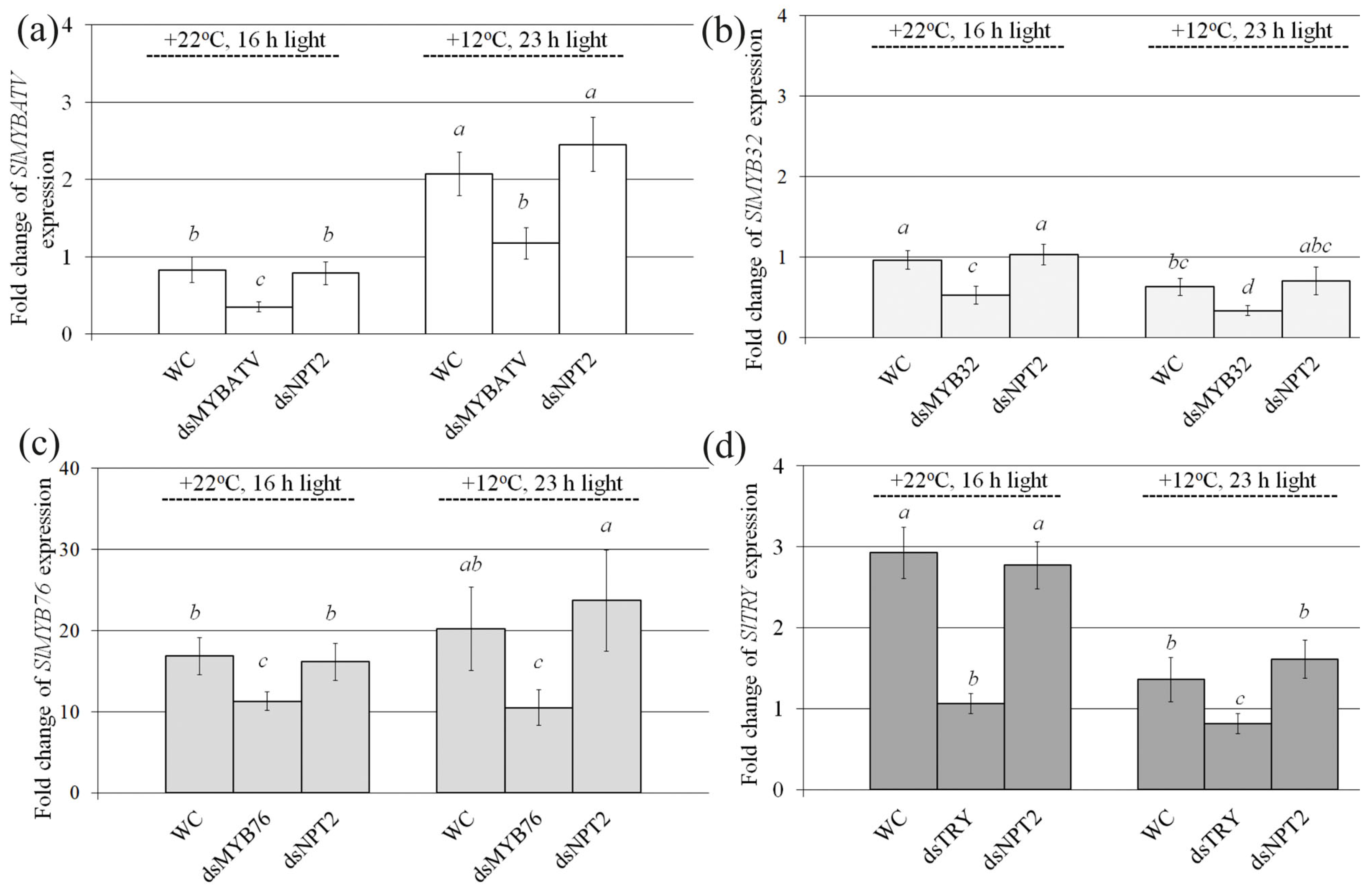
Then, we analyzed whether exogenous application of the pure SlMYBATV-, SlMyb32-, SlMYB76-, SlTRY-, and NPTII-dsRNAs to the foliar surface of four-week-old S. lycopersicum could lead to any changes in the mRNA transcript levels of the SlMYBATV, SlMYB32, SlMYB76, and SlTRY genes in comparison with the water-treated controls seven days after dsRNA application (Figure 2). Since under standard cultivation conditions, anthocyanin production and the expression of anthocyanin biosynthesis genes in S. lycopersicum were low, we divided the treated S. lycopersicum into two groups for post-treatment incubation–plants cultivated under control conditions (+22 °C, 16 h light) or anthocyanin-inducing conditions (+12 °C, 23 h light)—for seven days in order to induce SlCHS expression and anthocyanin biosynthesis and analyze the dsRNA effects.
The qRT–PCR analysis revealed that the mRNA level of the SlMYBATV gene was considerably lower after plant foliar treatment with SlMYBATV-dsRNA than after the application of water or the nonspecific NPTII-dsRNA both under the standard and anthocyanin-inducing conditions (Figure 2a). Similarly, SlMYB32, SlMYB76, and SlTRY mRNA levels were markedly lowered after exogenous application of the SlMYB32-, SlMYB76-, and SlTRY-dsRNAs under the standard and anthocyanin-inducing conditions (Figure 2b–d). Importantly, exogenous plant treatment with the nonspecific NPTII-dsRNA did not considerably affect the expression levels of the SlMYBATV, SlMYB32, SlMYB76, and SlTRY genes compared to the water-treated control, indicating sequence-specificity of the dsRNA-induced gene silencing effects. We also noted that when plants were grown under the anthocyanin-inducing conditions, the expression of the SlMYBATV gene was considerably higher both in water- and dsRNA-treated plants in comparison with plants grown under standard conditions (Figure 2a). mRNA levels of SlMYB32, SlMYB76, and SlTRY genes were reduced or remained at the same level under the anthocyanin-inducing conditions in comparison with control cultivation conditions (Figure 2b–d).

2.2. Exogenous dsRNAs Upregulate mRNA Levels of Anthocyanin Biosynthesis-Related Genes and Anthocyanin Content

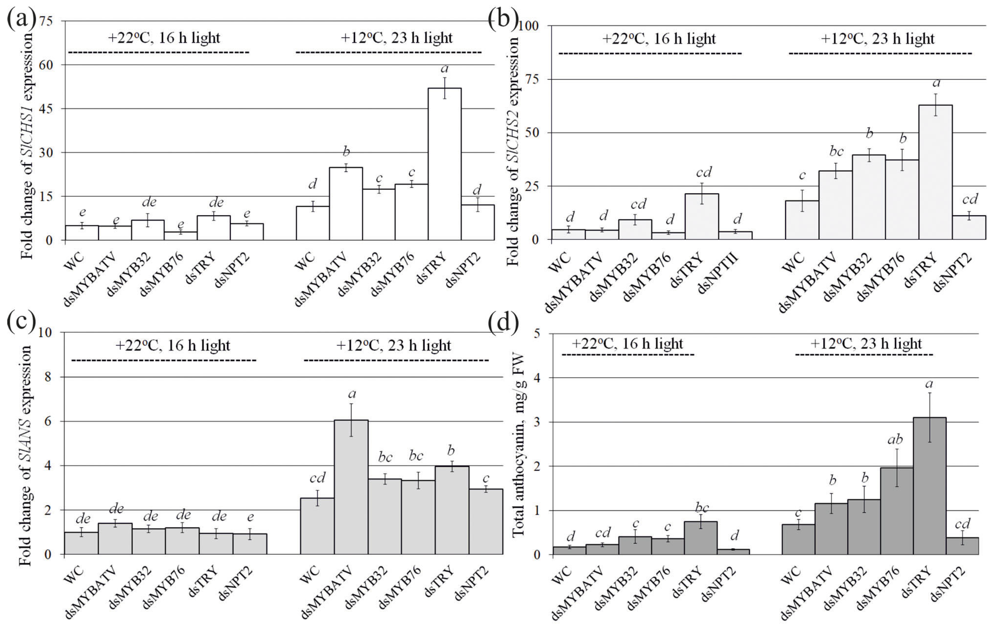
Then, we analyzed the effect of exogenous SlMYBATV-, SlMYB32-, SlMYB76-, SlTRY-, and NPTII-dsRNAs on the expression of three genes, encoding chalcone synthase (CHS) and anthocyanin synthase (ANS), the key enzymes involved in the biosynthesis of anthocyanins, and anthocyanin content itself in comparison with the control water treatment (Figure 3). CHS is the key enzyme in the first committed step of the biosynthesis of flavonoids, including anthocyanins, and catalyzes the stepwise condensation of 4-coumaroyl-CoA and malonyl-CoA to naringenin chalcone, while ANS catalyzes the penultimate step in the biosynthesis of the anthocyanin class of flavonoids [52].
We analyzed the expression of SlCHS1 and SlCHS2 genes encoding chalcone synthase and expression of the SlANS gene encoding anthocyanin synthase (Figure 3a–c). The analysis revealed a pronounced upregulation of SlCHS1, SlCHS2, and SlANS expression in S. lycopersicum grown under the anthocyanin-inducing conditions in comparison with the control conditions (Figure 3a–c). Under the anthocyanin-inducing conditions, expression of the SlCHS1 gene was considerably higher in plants treated with the SlMYBATV-, SlMYB32-, SlMYB76-, and SlTRY-dsRNAs than in the water-treated and NPTII-dsRNA-treated S. lycopersicum (Figure 3a). Expression of the SlCHS2 gene was considerably higher after application of only SlMYB32-, SlMYB76-, and SlTRY-dsRNAs, while expression of the SlANS gene was considerably higher after application of SlMYBATV- and SlTRY-dsRNAs. According to HPLC-MS analysis, this dsRNA-induced decrease in SlMYBATV, SlMYB32, SlMYB76, and SlTRY gene expression correlated with a considerable increase in anthocyanin accumulation, reaching 1.2–3.1 mg/g FW (Figure 3d). The highest anthocyanin level of 3.1 mg/g FW was detected after the application of SlTRY-dsRNA, which was also associated with the most pronounced increase in the expression of the SlCHS1 and SlCHS2 genes (Figure 3a,b,d). The application of SlTRY-dsRNA led to an increase in the total anthocyanin content of 4.3 times under control conditions and 4.6 times under anthocyanin-inducing conditions in comparison with the water control plants (Figure 3d). Notably, the control nonspecific NPTII-dsRNA did not cause any considerable alterations in both the anthocyanin content and expression of the anthocyanin biosynthesis-related genes supporting the gene-specific dsRNA-induced gene silencing effects (Figure 3a–d).
Using HPLC with high-resolution mass spectrometry (HPLC-MS), we detected eight anthocyanin compounds in the water- and dsRNA-treated leaves of S. lycopersicum (Table S1). The main anthocyanins in our samples were petunidin-3-(caffeoyl)-rutinoside-5-glucoside and delphinidin-3-O-(6″-O-p-coumaroyl)-glucoside, whose contents constituted 65.9–77.9% of all individual anthocyanins (Table S1). It is possible that other anthocyanins were present in the analyzed tissues of S. lycopersicum but in trace amounts. Additionally, the content of most individual anthocyanins was higher in the plants grown under the anthocyanin-inducing conditions than at +22 °C (Table S1). We have shown that the treatment of plants with exogenous dsRNAs encoding the analyzed MYB-family transcription factors led to an increase in the total content of anthocyanins, mostly due to an increase in the content of petunidin-3,5-O-diglucoside, petunidin-3-(caffeoyl)-rutinoside-5-glucoside, delphinidin-3-O-(6″-O-p-coumaroyl)-glucoside and malvidin-3-(p-coumaroyl)-rutinoside-5-glucoside in the leaves of S. lycopersicum (Table S1).

3. Discussion

The growing human population and negative impact of environmental stresses promote the development of new molecular tools for crop improvement and plant protection without modification of the plant genome. Public debates about the safety of genetic engineering/genome editing technologies and current legislative limitations on the cultivation of genetically modified plants restrain the development and implementation of these tools. The development of innovative and safe approaches to improve crop traits without genome modifications is an urgent task. SIGS is currently considered as a new promising tool for plant improvement without genetic modifications and includes exogenous treatment of plant surfaces with dsRNAs or siRNAs to induce RNAi-mediated gene silencing and modify plants’ properties [13,14,27,28]. However, there is a limited number of investigations reporting on efficient gene silencing in the plant genome after external dsRNA application.
In this study, we externally applied dsRNAs to tomato foliar surfaces to silence the expression of four blockers of anthocyanin biosynthesis and to affect anthocyanin accumulation in S. lycopersicum, which is an important agricultural crop. While anthocyanins are produced by many plant species, they are produced at low levels or are absent in most cultivated tomatoes due to the incomplete activation of the flavonoid biosynthetic pathway [43,44]. Using the SIGS approach and qRT–PCR, we demonstrated that foliar application of dsRNAs encoding four putative transcription repressors of anthocyanin biosynthesis, SlMYBATV, SlMYB32, SlMYB76, and SlTRY, resulted in highly downregulated mRNA levels of these S. lycopersicum target genes and upregulated mRNA levels of anthocyanin biosynthesis genes. HPLC-MS analysis revealed that these effects were accompanied by considerably increased levels of anthocyanins. Importantly, treatment of S. lycopersicum with the nonspecific NPTII-dsRNAs had no effect on both anthocyanin production or the expression of SlMYBATV, SlMYB32, SlMYB76, and SlTRY genes of S. lycopersicum. This indicates that the observed dsRNA-induced MYB-family gene silencing effect was sequence-specific and was not a result of the dsRNA application itself. Several previous studies have provided evidence for the role of SlMYBATV as a negative regulator of anthocyanin biosynthesis [46,47,48], while much less is known about the role of SlMYB32, SlMYB76, and SlTRY transcription factors in the biosynthesis of anthocyanins. The available studies identified SlMYBATV, SlMYB32, SlMYB76, and SlTRY as putative MYB regulators playing a negative role in anthocyanin biosynthesis based on genome screening [46,48] or SlTRY gene overexpression [49]. These reports are consistent with our finding that SlMYBATV, SlMYB32, SlMYB76, and SlTRY, the four analyzed MYB regulators, act as negative regulators of anthocyanin biosynthesis.
This study revealed the total anthocyanins at the level of 0.17 mg/g FW in tomato leaves under control conditions; these results are comparable with other studies [53] reporting anthocyanin content of 0.14 mg/g FW in different tomato genotypes. Exogenous application of the MYB-specific dsRNAs in our study allowed us to increase anthocyanin levels in tomato leaves to 1.2–3.1 mg/g FW, which is a considerable elevation.
Several tomato cultivars possessing high anthocyanin contents in their fruits and vegetative tissues have been reported and some purple fruited tomato varieties have been generated by conventional breeding [54], overexpressing selected transcription activators in transgenic tomato [55], plant microRNA858 blockage by genetic engineering [56], or CRISPR/Cas and TALEN genome editing technologies [57,58]. However, elevation of anthocyanin levels in vegetative tissues by external plant treatments is also of high interest, since anthocyanins are known to mediate stress tolerance in plants both to abiotic and biotic stresses via plant protection from growth inhibition by reducing oxidative stress, ROS scavenging, and maintaining osmotic balance [42,59]. Anthocyanin-reach tomato cultivars are known to display enhanced stress tolerance to various stress cues [60]. On the other hand, spraying plants with dsRNA solutions with the purpose of changing certain plant traits is a new tool that still needs to be developed. Here, we are still at the very beginning of the path, so changing the properties of plants in any tissues is interesting and relevant.
At present, there is a high number of studies reporting successful external dsRNA application to the plant surfaces for RNAi-mediated silencing of the target genes in infecting plant pathogens, i.e., viruses or fungi, leading to plant pathogen protection (reviewed in [8,9,14]). With regard to SIGS in the plant genome, a limited number of studies demonstrated the possibility of regulating the expression of transgenes in Arabidopsis or tobacco by transgene-specific exogenous dsRNAs [16,50,51,61,62]. There is also a low number of investigations reporting on the silencing of plant endogenous genes after exogenous dsRNA or siRNA application [30,31,32,33,34]. There were also several studies where accessory techniques, such as nanoparticles [35,36] or laser light [37], have been used to achieve efficient exogenously induced gene silencing. We developed our study based on the initial report by Numata et al. [61] who infiltrated A. thaliana leaves with a carrier peptide in a complex with siRNA encoding the AtCHS gene, and on our recent study where we demonstrated that foliar dsRNA treatments of A. thaliana significantly reduced the expression of the AtCHS gene and two genes encoding transcriptional repressors of anthocyanin biosynthesis in Arabidopsis, AtMYBL2 and AtANAC032 [19]. Numata et al. [61] reported a local loss of anthocyanin pigmentation by visual observation, but AtCHS mRNA and anthocyanin levels were not analyzed. The present study demonstrates that the foliar application of naked dsRNA (i.e., pure dsRNA, without nanoclay or other accessory components) to the leaves of S. lycopersicum induced the silencing of SlMYBATV, SlMYB32, SlMYB76, and SlTRY genes which was accompanied by enhanced anthocyanin accumulation, supporting the role of the four analyzed MYB regulators as negative regulators of anthocyanin biosynthesis. The capabilities of this SIGS method make it a promising one to change various characteristics of plants at different intervals of time, which is a unique feature of a biotechnological tool and opens up a great potential of this approach for use in agriculture.
In summary, in this study, we demonstrated for the first time that exogenous foliar treatments of tomato with MYB-specific dsRNAs can be used as a rapid silencing tool in plant gene functional studies to verify plant gene function, and in plant biotechnology to elevate the content of biologically active compounds.

4. Materials and Methods

4.1. Plant Material and Growth Conditions

Seeds of wild-type tomato (S. lycopersicum L.) cultivar Micro-Tom were used in the experiments (seed material from Laboratory of Biotechnology, Federal Scientific Center of the East Asia Terrestrial Biodiversity, Vladivostok, Russia). The tomato seeds were vaporphase sterilized as described [63] (and planted to individual pots (9 cm × 9 cm) containing 200 g of commercially available rich soil (the soil was well-irrigated by filtered water applied at the bottom of the pots). Plants were grown in a chamber (Sanyo MLR-352, Panasonic, Osaka, Japan) at a light intensity of ~120 µmol m−2 s−1 over a 16 h daily light period at 22 °C for four weeks before dsRNA treatments. After the dsRNA treatments, the S. lycopersicum was incubated for seven additional days either under control (+22 °C, 16 h daily light period) or anthocyanin-inducing (+12 °C, 23 h daily light period) conditions in a growth chamber (KS-200, Smolenskoye SKTB SPU, Smolensk, Russia) without further irrigation to induce anthocyanin accumulation.

4.2. Isolation and Sequencing of SlMYBATV, SlMYB32, SlMYB76, and SlTRY Transcripts

Full-length coding cDNA sequences of SlMYBATV-X1 (Solyc07g052490, 519 bp), SlMYB32 (Solyc10g055410.1.1, 822 bp), SlMYB76 (Solyc05g008250, 537 bp), and SlTRY (Solyc01g095640, 285 bp) were amplified by RT–PCR using RNA samples extracted from the adult leaves of S. lycopersicum. Sequence data of SlMYBATV, SlMYB32, SlMYB76, and SlTRY used in the present work for primer design can be also found in the GenBank libraries under the following accession numbers: SlMYBATV-X1 (MF197515 or NM_001365378), SlTRY (XM_010328616), SlMYB32 (NM_001247046.1), and SlMYB76 (MF197513). The primers are listed in Table S2. The RT–PCRs were performed in a Bis-M1105 Thermal Cycler (Bis-N, Novosibirsk, Russia). The RT–PCR products were subcloned into pJET1.2/blunt and sequenced as described previously [64]. The whole genomic sequences of S. lycopersicum are available at the Sol Genomics Network (SGN, https://solgenomics.net/, accessed on 1 March 2023).

4.3. dsRNA Synthesis and Application

All dsRNAs were synthesized using the T7 RiboMAX™Express RNAi System (Promega, Madison, WI, USA). For this purpose, the cloned full-length cDNAs of SlMYBATV, SlTRY, SlMYB32, and SlMYB76 were amplified by PCR for in vitro transcription and dsRNA production. The T7 promoter sequence was introduced into both the 5′ and 3′ ends of the amplified SlMYBATV, SlTRY, SlMYB32, and SlMYB76 in a single PCR for each gene using primers listed in Table S2. The PCRs were performed in the Bis-M1105 Thermal Cycler programmed according to T7 RiboMAX™ Express RNAi System instructions. Then, the obtained PCR products were used as templates for in vitro transcription and dsRNA synthesis following the manufacturer’s protocol. The resultant dsRNAs were analyzed by gel electrophoresis and spectrophotometry to estimate dsRNA purity, integrity, and amount.
The SlMYBATV-, SlTRY-, SlMYB32-, and SlMYB76-dsRNAs were applied to individual four-week-old plants of S. lycopersicum by spraying with 2 mL atomizer polypropylene vials. For each dsRNA treatment, 70 µg of the dsRNA was diluted in 400 µL of nuclease-free water and applied to the foliar surface (all leaves of the tomato for each type of condition were treated on both the adaxial and abaxial sides). In an independent experiment, two plants of S. lycopersicum were used for each type of treatment (Figure 2), i.e., two plants were treated with sterile filtered water (400 µL per plant) and two plants were treated with the dsRNA of each type (400 µL of SlMYBATV-, SlTRY-, SlMYB32-, and SlMYB76-dsRNAs per plant). Then, we divided the treated S. lycopersicum plants into two groups for incubation, under control conditions (+22 °C, 16 h light) and anthocyanin-inducing conditions (+12 °C, and 23 h light), for seven days (Figure 2). At least three independent experiments were performed for each type of analysis. In all experiments, the dsRNAs were applied to four-week-old plants of S. lycopersicum at a late day time (21:00–21:30) under low soil moisture conditions, since the conditions at the time of dsRNA application (appropriate plant age, late day time, and low soil moisture) were important parameters for successful gene silencing in A. thaliana according to our recent analysis [19,50,51]. Soil water content before dsRNA treatment was 50–60%.

4.4. RNA Isolation and Reverse Transcription

For RNA isolation, a typical adult leaf of S. lycopersicum was collected from an individual plant (1) before dsRNA or water application and (2) seven days post-application in an independent experiment. Total RNA was isolated using the cetyltrimethylammonium bromide (CTAB)-based protocol [65]. Complementary DNAs were synthesized using 2.5 µg of total RNA by the MMLV RT Kit (Evrogen, Moscow, Russia). The reactions were performed in 50 µL aliquots of the reaction mixture, which contained the first strand buffer, 5 µL of dNTP mix (10 mM each), 1.85 µL of oligo-(dT)15 primer (100 µM), and 4.3 µL of MMLV reverse transcriptase (100 u/µL), at 37 °C for 1.5 h. The 1 µL samples of reverse transcription products were then amplified by PCR and verified on the absence for DNA contamination using primers listed in Table S2.

4.5. Gene Expression Analysis by qRT–PCR

The qRT–PCRs were performed with SYBR Green I Real-time PCR dye and a real-time PCR kit (Evrogen, Moscow, Russia) as described [66] using two internal controls (SlActin and SlUBI). The expression was calculated by the 2−ΔΔCT method [67]. Then, the obtained data for MYB expression seven days post-treatment were divided to the MYB expression before treatment (fold change in MYB expression relative to respective data before treatment). All gene identification numbers and used primers are listed in Table S2.

4.6. Quantification of Anthocyanins

For HPLC-MS analysis, 200 mg of treated S. lycopersicum leaves were frozen at −20 °C and subsequently homogenized using a mortar and a pestle. Shredded tissue was weighed and extracted for 1 d at 4 °C in 2 mL of 1% (v/v) hydrochloric acid in methanol. Then, the mixture was centrifuged at 13,500 rpm for 15 min, and 1 mL of the supernatant was transferred into another glass tube. The samples were filtered through a 0.25-um nylon membrane for further analysis. The identification of all anthocyanins was performed using a 1260 Infinity analytical HPLC system (Agilent Technologies, Santa Clara, CA, USA) coupled to a Bruker HCT ultra PTM Discovery System (Bruker Daltonik GmbH, Bremen, Germany) equipped with an electrospray ionization (ESI) source. The data for anthocyanins were acquired in a positive ion mode under the operating conditions as described [68]. The MS spectra were recorded across an m/z range of 100–1500, and the individual anthocyanins were identified as described [69]. HPLC with diode array detection (HPLC–DAD) for the quantification of all anthocyanins was performed using a HPLC LC-20AD XR analytical system (Shimadzu, Kyoto, Japan). DAD data were recorded in the 200–800 nm range, and the chromatograms for quantification were acquired at 530 nm. The chromatographic separation was performed on Shim-pack GIST C18 column (150 mm, 2.1 nm i.d., 3-_m part size; Shimadzu, Japan). Anthocyanins were separated using 0.1% formic acid and acetonitrile as mobile phases A and B, respectively, with the following elution profile: 0 to 35 min 0% of B; 35 to 40 min 40% of B; 40 to 50 min 50% of B; 50 to 65 min 100% of B. A volume of 5 µL of the sample extract was injected with a constant column temperature maintained at 40 °C and a flow rate maintained at 0.2 mL/min. All solvents were of HPLC grade. The contents of anthocyanins were determined by external standard methods using the four-point regression calibration curves built with the available standards. The commercial standard cyanidin chloride, petunidin chloride, delphinidin chloride, and malvidin chloride were obtained from Sigma-Aldrich (St. Louis, MO, USA) and used as the control.

4.7. Statistical Analysis

The data are presented as mean ± standard error (SE) and were tested by a paired Student’s t-test. The p < 0.05 level was selected as the point of minimal statistical significance in all analyses. At least three independent experiments were performed for each type of analysis.

Supplementary Materials

The following supporting information can be downloaded at: https://www.mdpi.com/article/10.3390/ijms24119344/s1.

Author Contributions

A.R.S. and K.V.K. performed research design, experiments, data analysis, and participated in paper preparation. A.S.D. was responsible for project administration, funding acquisition, paper preparation, and data analysis. All authors have read and agreed to the published version of the manuscript.

Funding

This work was supported by a grant from the Russian Science Foundation (grant number 23-26-00253, https://rscf.ru/project/23-26-00253/, accessed on 24 May 2023).

Institutional Review Board Statement

Not applicable.

Informed Consent Statement

Not applicable.

Data Availability Statement

The data presented in this study are available within the article and Supplementary Materials.

Conflicts of Interest

The authors declare no conflict of interest.

References

  1. Borges, F.; Martienssen, R.A. The expanding world of small RNAs in plants. Nat. Rev. Mol. Cell Biol. 2015, 507, 727–741. [Google Scholar] [CrossRef] [PubMed]
  2. Zhao, J.H.; Guo, H.S. RNA silencing: From discovery and elucidation to application and perspectives. J. Integr. Plant Biol. 2022, 64, 476–498. [Google Scholar] [CrossRef] [PubMed]
  3. Wilson, R.C.; Doudna, J.A. Molecular mechanisms of RNA interference. Annu. Rev. Biophys. 2013, 42, 217–239. [Google Scholar] [CrossRef] [PubMed]
  4. Guleria, P.; Mahajan, M.; Bhardwaj, J.; Yadav, S.K. Plant small RNAs: Biogenesis, mode of action and their roles in abiotic stresses. Genom. Proteom. Bioinform. 2011, 6, 183–199. [Google Scholar] [CrossRef]
  5. Muhammad, T.; Zhang, F.; Zhang, Y.; Liang, Y. RNA interference: A natural immune system of plants to counteract biotic stressors. Cells 2019, 8, 38. [Google Scholar] [CrossRef]
  6. Tiwari, M.; Sharma, D.; Trivedi, P.K. Artificial microRNA mediated gene silencing in plants: Progress and perspectives. Plant Mol. Biol. 2014, 86, 1–18. [Google Scholar] [CrossRef]
  7. Kamthan, A.; Chaudhuri, A.; Kamthan, M.; Datta, A. Small RNAs in plants: Recent development and application for crop improvement. Front. Plant Sci. 2015, 6, 208. [Google Scholar] [CrossRef]
  8. Morozov, S.Y.; Solovyev, A.G.; Kalinina, N.O.; Taliansky, M.E. Double-stranded RNAs in plant protection against pathogenic organisms and viruses in agriculture. Acta Nat. 2019, 11, 13–21. [Google Scholar] [CrossRef]
  9. Gebremichael, D.E.; Haile, Z.M.; Negrini, F.; Sabbadini, S.; Capriotti, L.; Mezzetti, B.; Baraldi, E. RNA interference strategies for future management of plant pathogenic fungi: Prospects and challenges. Plants 2021, 10, 650. [Google Scholar] [CrossRef]
  10. Koch, A.; Wassenegger, M. Host-induced gene silencing-mechanisms and applications. New Phytol. 2021, 231, 54–59. [Google Scholar] [CrossRef]
  11. Ramegowda, V.; Mysore, K.S.; Senthil-Kumar, M. Virus-induced gene silencing is a versatile tool for unraveling the functional relevance of multiple abiotic-stress-responsive genes in crop plants. Front. Plant Sci. 2014, 5, 323. [Google Scholar] [CrossRef]
  12. Mamta, B.; Rajam, M.V. RNAi technology: A new platform for crop pest control. Physiol. Mol. Biol. Plants 2017, 3, 487–501. [Google Scholar] [CrossRef]
  13. Hoang, B.T.L.; Fletcher, S.J.; Brosnan, C.A.; Ghodke, A.B.; Manzie, N.; Mitter, N. RNAi as a foliar spray: Efficiency and challenges to field applications. Int. J. Mol. Sci. 2022, 23, 6639. [Google Scholar] [CrossRef]
  14. Dubrovina, A.S.; Kiselev, K.V. Exogenous RNAs for gene regulation and plant resistance. Int. J. Mol. Sci. 2019, 20, 2282. [Google Scholar] [CrossRef]
  15. Dalakouras, A.; Wassenegger, M.; McMillan, J.N.; Cardoza, V.; Maegele, I.; Dadami, E.; Runne, M.; Krczal, G.; Wassenegger, M. Induction of silencing in plants by high-pressure spraying of in vitro-synthesized small RNAs. Front. Plant Sci. 2016, 7, 1327. [Google Scholar] [CrossRef]
  16. Mitter, N.; Worrall, E.A.; Robinson, K.E.; Li, P.; Jain, R.G.; Taochy, C.; Fletcher, S.J.; Carroll, B.J.; Lu, G.Q.; Xu, Z.P. Clay nanosheets for topical delivery of RNAi for sustained protection against plant viruses. Nat. Plants 2017, 3, 16207. [Google Scholar] [CrossRef]
  17. Koch, A.; Biedenkopf, D.; Furch, A.; Weber, L.; Rossbach, O.; Abdellatef, E.; Linicus, L.; Johannsmeier, J.; Jelonek, L.; Goesmann, A.; et al. An RNAi-based control of Fusarium graminearum infections through spraying of long dsRNAs involves a plant passage and is controlled by the fungal silencing machinery. PLoS Pathog. 2016, 12, e1005901. [Google Scholar] [CrossRef]
  18. Pampolini, F.; Rodrigues, T.B.; Leelesh, R.S.; Kawashima, T.; Rieske, L.K. Confocal microscopy provides visual evidence and confirms the feasibility of dsRNA delivery to emerald ash borer through plant tissues. J. Pest Sci. 2020, 93, 1143–1153. [Google Scholar] [CrossRef]
  19. Kiselev, K.V.; Suprun, A.R.; Aleynova, O.A.; Ogneva, Z.V.; Kalachev, A.V.; Dubrovina, A.S. External dsRNA downregulates anthocyanin biosynthesis-related genes and affects anthocyanin accumulation in Arabidopsis thaliana. Int. J. Mol. Sci. 2021, 22, 6749. [Google Scholar] [CrossRef] [PubMed]
  20. Konakalla, N.C.; Kaldis, A.; Berbati, M.; Masarapu, H.; Voloudakis, A.E. Exogenous application of double-stranded RNA molecules from TMV p126 and CP genes confers resistance against TMV in tobacco. Planta 2016, 244, 961–969. [Google Scholar] [CrossRef] [PubMed]
  21. Song, X.S.; Gu, K.X.; Duan, X.X.; Xiao, X.M.; Hou, Y.P.; Duan, Y.B.; Wang, J.X.; Zhou, M.G. A myosin5 dsRNA that reduces the fungicide resistance and pathogenicity of Fusarium asiaticum. Pest. Biochem. Physiol. 2018, 150, 1–9. [Google Scholar] [CrossRef]
  22. Song, X.S.; Gu, K.X.; Duan, X.X.; Xiao, X.M.; Hou, Y.P.; Duan, Y.B.; Wang, J.X.; Yu, N.; Zhou, M.G. Secondary amplification of siRNA machinery limits the application of spray-induced gene silencing. Mol. Plant Pathol. 2018, 19, 2543–2560. [Google Scholar] [CrossRef]
  23. Kaldis, A.; Berbati, M.; Melita, O.; Reppa, C.; Holeva, M.; Otten, P.; Voloudakis, A. Exogenously applied dsRNA molecules deriving from the Zucchini yellow mosaic virus (ZYMV) genome move systemically and protect cucurbits against ZYMV. Mol. Plant Pathol. 2008, 19, 883–895. [Google Scholar] [CrossRef]
  24. Gu, K.X.; Song, X.S.; Xiao, X.M.; Duan, X.X.; Wang, J.X.; Duan, Y.B.; Hou, Y.P.; Zhou, M.G. A β2-tubulin dsRNA derived from Fusarium asiaticum confers plant resistance to multiple phytopathogens and reduces fungicide resistance. Pest. Biochem. Physiol. 2019, 153, 36–46. [Google Scholar] [CrossRef]
  25. Werner, B.T.; Gaffar, F.Y.; Schuemann, J.; Biedenkopf, D.; Koch, A.M. RNA-spray-mediated silencing of Fusarium graminearum AGO and DCL genes improve barley disease resistance. Front. Plant Sci. 2020, 11, 476. [Google Scholar] [CrossRef]
  26. Qiao, L.; Lan, C.; Capriotti, L.; Ah-Fong, A.; Nino Sanchez, J.; Hamby, R.; Heller, J.; Zhao, H.; Glass, N.L.; Judelson, H.S.; et al. Spray-induced gene silencing for disease control is dependent on the efficiency of pathogen RNA uptake. Plant Biotechnol. J. 2021, 19, 1756–1768. [Google Scholar] [CrossRef]
  27. Wang, M.; Jin, H. Spray-induced gene silencing: A powerful innovative strategy for crop protection. Trends Microbiol. 2017, 25, 4–6. [Google Scholar] [CrossRef]
  28. Das, P.R.; Sherif, S.M. Application of exogenous dsRNAs-induced RNAi in agriculture: Challenges and triumphs. Front. Plant Sci. 2020, 11, 946. [Google Scholar] [CrossRef]
  29. Akbar, S.; Wei, Y.; Zhang, M.-Q. RNA interference: Promising approach to combat plant viruses. Int. J. Mol. Sci. 2022, 23, 5312. [Google Scholar] [CrossRef]
  30. Lau, S.E.; Schwarzacher, T.; Othman, R.Y.; Harikrishna, J.A. dsRNA silencing of an R2R3-MYB transcription factor affects flower cell shape in a Dendrobium hybrid. BMC Plant Biol. 2015, 15, 194. [Google Scholar] [CrossRef]
  31. Li, H.; Guan, R.; Guo, H.; Miao, X. New insights into an RNAi approach for plant defence against piercing-sucking and stem-borer insect pests. Plant Cell Environ. 2015, 38, 2277–2285. [Google Scholar] [CrossRef] [PubMed]
  32. Marcianò, D.; Ricciardi, V.; Fassolo, E.M.; Passera, A.; BIANCO, P.A.; Failla, O.; Casati, P.; Maddalena, G.; De Lorenzis, G.; Toffolatti, S.L. RNAi of a putative grapevine susceptibility gene as a possible downy mildew control strategy. Front. Plant Sci. 2021, 12, 667319. [Google Scholar] [CrossRef] [PubMed]
  33. Nerva, L.; Guaschino, M.; Pagliarani, C.; De Rosso, M.; Lovisolo, C.; Chitarra, W. Spray-induced gene silencing targeting a glutathione S-transferase gene improves resilience to drought in grapevine. Plant Cell Environ. 2022, 45, 347–361. [Google Scholar] [CrossRef] [PubMed]
  34. Sammons, R.; Ivashuta, S.; Liu, H.; Wang, D.; Feng, P.; Kouranov, A.; Andersen, S. Polynucleotide molecules for gene regulation in plants. U.S. Patent 20110296556, 1 September 2015. [Google Scholar]
  35. Jiang, L.; Ding, L.; He, B.; Shen, J.; Xu, Z.; Yin, M.; Zhang, X. Systemic gene silencing in plants triggered by fluorescent nanoparticle-delivered double-stranded RNA. Nanoscale 2014, 6, 9965–9969. [Google Scholar] [CrossRef] [PubMed]
  36. Molesini, B.; Pennisi, F.; Cressoni, C.; Vitulo, N.; Dusi, V.; Speghini, A.; Pandolfini, T. Nanovector-mediated exogenous delivery of dsRNA induces silencing of target genes in very young tomato flower buds. Nanoscale Adv. 2022, 4, 4542–4553. [Google Scholar] [CrossRef] [PubMed]
  37. Killiny, N.; Gonzalez-Blanco, P.; Gowda, S.; Martini, X.; Etxeberria, E. Plant functional genomics in a few days: Laser-assisted delivery of double-stranded RNA to higher plants. Plants 2021, 10, 93. [Google Scholar] [CrossRef]
  38. Nityagovsky, N.N.; Kiselev, K.V.; Suprun, A.R.; Dubrovina, A.S. Exogenous dsRNA induces RNA interference of a chalcone synthase gene in Arabidopsis thaliana. Int. J. Mol. Sci. 2022, 23, 5325. [Google Scholar] [CrossRef]
  39. Kong, J.M.; Chia, L.S.; Goh, N.K.; Chia, T.F.; Brouillard, R. Analysis and biological activities of anthocyanins. Phytochemistry 2003, 64, 923–933. [Google Scholar] [CrossRef]
  40. Khoo, H.E.; Azlan, A.; Tang, S.T.; Lim, S.M. Anthocyanidins and anthocyanins: Colored pigments as food, pharmaceutical ingredients, and the potential health benefits. Food Nutr. Res. 2017, 61, 1361779. [Google Scholar] [CrossRef]
  41. Konczak, I.; Zhang, W. Anthocyanins—More than nature’s colours. J. Biomed. Biotech. 2004, 5, 239. [Google Scholar] [CrossRef]
  42. Kaur, S.; Tiwari, V.; Kumari, A.; Chaudhary, E.; Sharma, A.; Ali, U.; Garg, M. Protective and defensive role of anthocyanins under plant abiotic and biotic stresses: An emerging application in sustainable agriculture. J. Biotechnol. 2023, 361, 12–29. [Google Scholar] [CrossRef]
  43. Verhoeyen, M.E.; Bovy, A.; Collins, G.; Muir, S.; Robinson, S.; De Vos CH, R.; Colliver, S. Increasing antioxidant levels in tomatoes through modification of the flavonoid biosynthetic pathway. J. Exp. Bot. 2002, 53, 2099–2106. [Google Scholar] [CrossRef]
  44. Gonzali, S.; Mazzucato, A.; Perata, P. Purple as a tomato: Towards high anthocyanin tomatoes. Trends Plant Sci. 2009, 14, 237–241. [Google Scholar] [CrossRef]
  45. Chaves-Silva, S.; Dos Santos, A.L.; Chalfun-Júnior, A.; Zhao, J.; Peres, L.E.P.; Benedito, V.A. Understanding the genetic regulation of anthocyanin biosynthesis in plants—Tools for breeding purple varieties of fruits and vegetables. Phytochemistry 2018, 153, 11–27. [Google Scholar] [CrossRef]
  46. Cao, X.; Qiu, Z.; Wang, X.; Van Giang, T.; Liu, X.; Wang, J.; Wang, X.; Gao, J.; Guo, Y.; Du, Y.; et al. A putative R3 MYB repressor is the candidate gene underlying atroviolacium, a locus for anthocyanin pigmentation in tomato fruit. J. Exp Bot. 2017, 68, 5745–5758. [Google Scholar] [CrossRef]
  47. Colanero, S.; Perata, P.; Gonzali, S. The atroviolacea gene encodes an R3-MYB protein repressing anthocyanin synthesis in tomato plants. Front. Plant Sci. 2018, 9, 830. [Google Scholar] [CrossRef]
  48. Zhao, P.; Li, Q.; Li, J.; Wang, L.; Ren, Z. Genome-wide identification and characterization of R2R3MYB family in Solanum lycopersicum. Mol. Gen. Genom. 2014, 289, 1183–1207. [Google Scholar] [CrossRef]
  49. Nukumizu, Y.; Wada, T.; Tominaga-Wada, R. Tomato (Solanum lycopersicum) homologs of TRIPTYCHON (SlTRY) and GLABRA3 (SlGL3) are involved in anthocyanin accumulation. Plant Signal Behav. 2013, 8, e24575. [Google Scholar]
  50. Kiselev, K.V.; Suprun, A.R.; Aleynova, O.A.; Ogneva, Z.V.; Kostetsky, E.Y.; Dubrovina, A.S. The specificity of transgene suppression in plants by exogenous dsRNA. Plants 2022, 11, 715. [Google Scholar] [CrossRef]
  51. Kiselev, K.V.; Suprun, A.R.; Aleynova, O.A.; Ogneva, Z.V.; Dubrovina, A.S. Physiological conditions and dsRNA application approaches for exogenously induced RNA interference in Arabidopsis thaliana. Plants 2021, 10, 264. [Google Scholar] [CrossRef]
  52. Liu, Y.; Tikunov, Y.; Schouten, R.E.; Marcelis, L.F.M.; Visser, R.G.F.; Bovy, A. Anthocyanin biosynthesis and degradation mechanisms in solanaceous vegetables: A review. Front Chem. 2018, 6, 52. [Google Scholar] [CrossRef] [PubMed]
  53. Tudor-Radu, M.; Vijan, L.E.; Tudor-Radu, C.M.; Tita, I.; Sima, R.; Mitrea, R. Assessment of Ascorbic Acid, Polyphenols, Flavonoids, Anthocyanins and Carotenoids Content in Tomato Fruits. Not. Bot. Horti Agrobot. Cluj-Napoca 2016, 44, 477–483. [Google Scholar] [CrossRef]
  54. Mazzucato, A.; Willems, D.; Bernini, R.; Picarella, M.E.; Santangelo, E.; Ruiu, F.; Tilesi, F.; Soressi, G.P. Novel phenotypes related to the breeding of purple-fruited tomatoes and effect of peel extracts on human cancer cell proliferation. Plant Physiol. Biochem. 2013, 72, 125–133. [Google Scholar] [CrossRef] [PubMed]
  55. Butelli, E.; Titta, L.; Giorgio, M.; Mock, H.P.; Matros, A.; Peterek, S.; Schijlen, E.G.; Hall, R.D.; Bovy, A.G.; Luo, J.; et al. Enrichment of tomato fruit with health-promoting anthocyanins by expression of select transcription factors. Nat. Biotechnol. 2008, 11, 1301–1308. [Google Scholar] [CrossRef] [PubMed]
  56. Jia, X.; Shen, J.; Liu, H.; Li, F.; Ding, N.; Gao, C.; Pattanaik, S.; Patra, B.; Li, R.; Yuan, L. Small tandem target mimic-mediated blockage of microRNA858 induces anthocyanin accumulation in tomato. Planta 2015, 242, 283–293. [Google Scholar] [CrossRef] [PubMed]
  57. Čermák, T.; Baltes, N.J.; Čegan, R.; Zhang, Y.; Voytas, D.F. High-frequency, precise modification of the tomato genome. Genome Biol. 2015, 16, 232. [Google Scholar] [CrossRef]
  58. Tiwari, J.K.; Singh, A.K.; Behera, T.K. CRISPR/Cas genome editing in tomato improvement: Advances and applications. Front Plant Sci. 2023, 14, 1121209. [Google Scholar] [CrossRef]
  59. Naing, A.H.; Kim, C.K. Abiotic stress-induced anthocyanins in plants: Their role in tolerance to abiotic stresses. Physiol. Plant. 2021, 172, 1711–1723. [Google Scholar] [CrossRef]
  60. Faqir Napar, W.P.; Kaleri, A.R.; Ahmed, A.; Nabi, F.; Sajid, S.; Ćosić, T.; Yao, Y.; Liu, J.; Raspor, M.; Gao, Y. The anthocyanin-rich tomato genotype LA-1996 displays superior efficiency of mechanisms of tolerance to salinity and drought. J. Plant Physiol. 2022, 271, 153662. [Google Scholar] [CrossRef]
  61. Numata, K.; Ohtani, M.; Yoshizumi, T.; Demura, T.; Kodama, Y. Local gene silencing in plants via synthetic dsRNA and carrier peptide. Plant Biotechnol. J. 2014, 12, 1027–1034. [Google Scholar] [CrossRef]
  62. Dubrovina, A.S.; Aleynova, O.A.; Kalachev, A.V.; Suprun, A.R.; Ogneva, Z.V.; Kiselev, K.V. Induction of transgene suppression in plants via external application of synthetic dsRNA. Int. J. Mol. Sci. 2019, 20, 1585. [Google Scholar] [CrossRef]
  63. Dubrovina, A.S.; Aleynova, O.A.; Suprun, A.R.; Ogneva, Z.V.; Kiselev, K.V. Transgene suppression in plants by foliar application of in vitro-synthesized small interfering RNAs. Appl. Microbiol. Biotechnol. 2020, 104, 2125–2135. [Google Scholar] [CrossRef]
  64. Dubrovina, A.S.; Aleynova, O.A.; Ogneva, Z.V.; Suprun, A.R.; Ananev, A.A.; Kiselev, K.V. The effect of abiotic stress conditions on expression of calmodulin (CaM) and calmodulin-like (CML) genes in wild-growing grapevine Vitis amurensis. Plants 2019, 8, 602. [Google Scholar] [CrossRef]
  65. Kiselev, K.V.; Dubrovina, A.S.; Shumakova, O.A.; Karetin, Y.A.; Manyakhin, A.Y. Structure and expression profiling of a novel calcium-dependent protein kinase gene, CDPK3a, in leaves, stems, grapes, and cell cultures of wild-growing grapevine Vitis amurensis Rupr. Plant Cell Rep. 2013, 32, 431–442. [Google Scholar] [CrossRef]
  66. Dubrovina, A.S.; Kiselev, K.V. The role of calcium-dependent protein kinase genes VaCPK1 and VaCPK26 in the response of Vitis amurensis (in vitro) and Arabidopsis thaliana (in vivo) to abiotic stresses. Russ. J. Genet. 2019, 55, 319–329. [Google Scholar] [CrossRef]
  67. Livak, K.J.; Schmittgen, T.D. Analysis of relative gene expression data using real-time quantitative PCR and the 2(-Delta Delta C(T)) method. Methods 2001, 25, 402–408. [Google Scholar] [CrossRef]
  68. Abdullin, S.R.; Nikulin, V.Y.; Nikulin, A.Y.; Manyakhin, A.Y.; Bagmet, V.B.; Suprun, A.R.; Gontcharov, A.A. Roholtiella mixta sp. nov. (Nostocales, Cyanobacteria): Morphology, molecular phylogeny, and carotenoid content. Phycologia 2021, 60, 73–82. [Google Scholar] [CrossRef]
  69. Wang, H.; Sun, S.; Zhou, Z.; Qiu, Z.; Cui, X. Rapid analysis of anthocyanin and its structural modifications in fresh tomato fruit. Food Chem. 2020, 333, 127439. [Google Scholar] [CrossRef]
Figure 1. Schematic representation of the dsRNAs and the qRT–PCR primer positions designed to verify the effects of external dsRNA treatments on the levels of endogenous SlMYBATV, SlMYB32, SlMYB76, and SlTRY mRNAs. (a) Representation of SlMYBATV cDNA coding region with positions of the SlMYBATV-dsRNA and primers; (b) representation of SlMYB32 cDNA coding region and position of SlMYB32-specific dsRNA and primers; (c) representation of SlMYB76 cDNA coding region and position of SlMYB76-dsRNA and primers; (d) representation of SlTRY cDNA coding region with position of the SlTRY-dsRNA and primers; (e) representation of NPTII coding region and positions of the NPTII-specific dsRNA and primers. Black arrows indicate positions of the primers (s1, a1, s2, a2, s3, a3, s4, a4, s5, a5) used for SlMYBATV, SlMYB32, SlMYB76, and SlTRY mRNA analysis. UTR—untranslated region, 2 × 35S—the double 35S promoter of the cauliflower mosaic virus (CaMV), Tnos—nopaline synthase terminator.
Ijms 24 09344 g001
Figure 2. Relative fold change in SlMYBATV (a), SlMYB32 (b), SlMYB76 (c), and SlTRY (d) mRNA levels after dsRNA treatments of Solanum lycopersicum compared to untreated plants. WC—S. lycopersicum treated with sterile water; dsMYBATV—S. lycopersicum treated with MYBATV-dsRNA; dsMYB32—S. lycopersicum treated with SlMYB32-dsRNAs; dsMYB76—S. lycopersicum treated with SlMYB76-dsRNAs; dsTRY—S. lycopersicum treated with SlTRY-dsRNAs; dsNPT2—S. lycopersicum treated with NPTII-dsRNA. Total RNA was isolated seven days after dsRNA application and quantitative real-time PCR was used for gene expression analysis. S. lycopersicum was grown under control (+22 °C, 16 h light) and anthocyanin-inducing (+12 °C, 23 h light) conditions. The data are presented as the mean ± SE (three independent experiments). Means on each figure followed by the same letter were not different using one-way analysis of variance (ANOVA), followed by the Tukey HSD multiple comparison test.
Ijms 24 09344 g002
Figure 3. Relative fold change in SlCHS1 (a), SlCHS2 (b), and SlANS (c) mRNA levels after dsRNA treatments of Solanum lycopersicum compared to untreated plants and HPLC results (d) of the total anthocyanin content in the leaves of S. lycopersicum. WC—S. lycopersicum treated with sterile water; dsMYBATV—S. lycopersicum treated with MYBATV-dsRNA; dsMYB32—S. lycopersicum treated with SlMYB32-dsRNAs; dsMYB76—S. lycopersicum treated with SlMYB76-dsRNAs; dsTRY—S. lycopersicum treated with SlTRY-dsRNAs; dsNPT2—S. lycopersicum treated with NPTII-dsRNA. Total RNA was isolated seven days after dsRNA application and quantitative real-time PCR was used for gene expression analysis. S. lycopersicum was grown under control (+22 °C, 16 h light) and anthocyanin-inducing (+12 °C, 23 h light) conditions. Total anthocyanins are expressed as mg per g of fresh weight (mg/g FW). The data are presented as the mean ± SE (three independent experiments). Means on each figure followed by the same letter were not different using one-way analysis of variance (ANOVA), followed by the Tukey HSD multiple comparison test.
Ijms 24 09344 g003
Disclaimer/Publisher’s Note: The statements, opinions and data contained in all publications are solely those of the individual author(s) and contributor(s) and not of MDPI and/or the editor(s). MDPI and/or the editor(s) disclaim responsibility for any injury to people or property resulting from any ideas, methods, instructions or products referred to in the content.