Case Report

Signet-Ring Cell Squamous Cell Carcinoma: A Biphenotypic Neoplasm of the Gastro-Esophageal Junction with Uncertain Biological Potential: Case Report and Literature Review

1
Department of Pathology, George Emil Palade University of Medicine, Pharmacy, Science and Technology, 540012 Targu Mures, Romania
2
Department of Pathology, Clinical County Emergency Hospital, 540136 Targu Mures, Romania
3
Research Center for Oncopathology and Translational Medicine (CCOMT), George Emil Palade University of Medicine, Pharmacy, Science and Technology, 540136 Targu Mures, Romania
4
Department of General Surgery, Clinical County Emergency Hospital, 540136 Targu Mures, Romania
*
Author to whom correspondence should be addressed.
These authors contributed equally to this work.
Int. J. Mol. Sci. 2023, 24(11), 9535; https://doi.org/10.3390/ijms24119535
Submission received: 7 April 2023 / Revised: 27 May 2023 / Accepted: 28 May 2023 / Published: 31 May 2023
(This article belongs to the Special Issue Advanced Research on Biomarkers in Gastrointestinal Cancer)

Abstract

:
The signet-ring cell variant of squamous cell carcinoma (SCC) is an extremely rare histological subtype, with only 24 cases (including the present case) reported in the Medline database: 15 affecting the external surface of the body, 3 in the lung, 2 affecting the uterine cervix, 1 involving the gingiva, another one affecting the esophagus and the present case that is the first reported at the gastro-esophageal junction (GEJ). In one case, the location of the lesion was not mentioned. A 59-year-old male patient underwent segmental eso-gastrectomy for carcinoma of the GEJ. The microscopic examination showed a pT3N1-staged SCC composed of solid nests admixed in over 30% of the tumor, with cells having eccentrically located nuclei and clear vacuolated cytoplasm. The signet-ring cells did not show mucinous secretion and were positive for keratin 5/6 and vimentin, with nuclear expression of β-catenin and Sox2 and focal membrane positivity for E-cadherin. Based on these features, the case was considered a signet-ring SCC with epithelial–mesenchymal transition. Thirty-one months after surgery, the patient was disease-free, with no local recurrence and no known distant metastases. In SCC, a signet-ring cell component might be an indicator of the dedifferentiation of tumor cells towards a mesenchymal molecular subtype.

1. Introduction

According to International Agency for Research on Cancer (IARC) statistics, esophageal/gastro-esophageal junction (GEJ) cancer represents the sixth most frequent cause of cancer-related death globally and the eighth leading cancer diagnosed worldwide [1]. Over 90% of these tumors are squamous cell carcinomas (SCCs) [2].
The histological variants of SCC, which are included in the classification of the World Health Organization (WHO), independently by the anatomical location, are classic SCC, keratoacanthoma, acantholytic variant, spindle cell SCC, verrucous SCC and adenosquamous carcinoma. WHO also defined a group of uncommon variants, including SCC with sarcomatoid differentiation, lymphoepithelioma-like carcinoma and SCC with osteoclast-like giant cells. The aforementioned subtypes vary from well-differentiated, with intracellular or intercellular keratinization, to poorly differentiated and non-keratinized tumors [3]. Besides the histologic variants that are officially recognized by the latest WHO manual, SCC can also have a basaloid, clear cell or adenoid appearance [4].
In this paper, we present an extremely rare histological variant of the SCC of the GEJ. It is about the 24th case of SCC with signet-ring cell morphology described in the Medline database and the second case of the upper gastrointestinal tract. We describe the particularities of the case, from clinical management to molecular level, along with a review of the other 23 cases which were previously reported. A differential diagnosis with a clear cell variant is also presented.

2. Case Presentation

2.1. Personal History

A 59-year-old man was admitted to the General Surgery Department for recurrent pain and postprandial fullness in the epigastrium. The patient reported a 3-month history of the symptoms, which were accompanied by weight loss, lack of appetite, fatigability and constipation. He was a smoker of 20 cigarettes per day and a heavy consumer of alcohol on a daily basis. No family or personal history of oncologic diseases was known.

2.2. Laboratory Examinations

Hematological findings included microcytic hypochromic anemia, with a hematocrit of 34.3% (normal range 39–54%) and hemoglobin of 11.2 g/dL (normal range 13–17 g/dL); leukocytosis (13.77 × 10³ cells/µL; normal range 3.6–10 × 103 cells/µL); and neutrophilia (11.64 × 10³ cells/µL; normal range 1.4–6.5 × 10³ cells/µL). Tumor markers, including carcinoembryonic antigen (CEA), carbohydrate antigen 125 (CA 125), carbohydrate antigen 19-9 (CA 19-9) and carbohydrate antigen 72-4 (CA 72-4), showed normal serum levels. The urine analysis revealed leukocyturia (75 cells/µL; normal value ≤ 20 cells/µL).

2.3. Imaging and Endoscopic Examinations

The preoperative computed tomography (CT scan) showed a heterogeneous, iodophilic, infiltrative thickening of the GEJ wall without the involvement of adjacent anatomical structures and without the suspicion of pulmonary or bone metastases. Upper digestive endoscopy confirmed the presence of a stenotic mass of the GEJ. The biopsy specimen was assessed as high-grade intraepithelial squamous dysplasia. As surgical resection was decided, the informed consent of the patient was obtained prior to surgery for the surgical intervention. The agreement of the Ethical Committee of the Clinical County Emergency Hospital, Targu Mures, Romania, was obtained for the processing and publication of the scientific data.

2.4. Surgery

Dissection of the para-esophageal soft tissue and fascia, followed by removal of the distal esophagus with part of the gastric fundus, was performed. The removed adenopathies were located near the common hepatic artery and left gastric artery and in the peri-esophageal space. The aforementioned adenopathies were excised and sent with the main specimen for histopathological assessment.

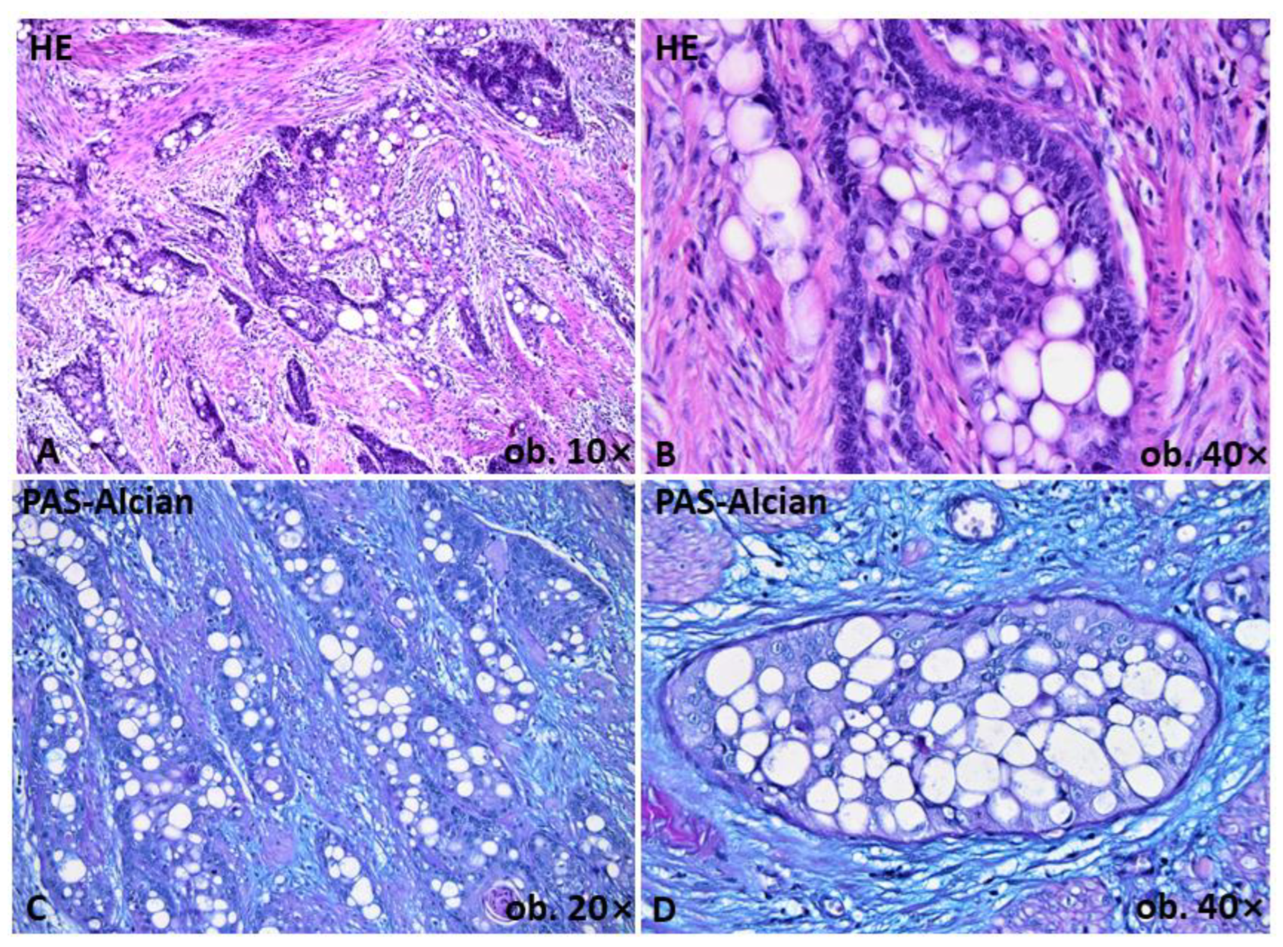
2.5. Gross and Histopathological Assessment of Surgical Specimens

Macroscopically, a 33 × 30 mm ulcerative tumor mass of the GEJ, with a maximum thickness of 23 mm, was confirmed to induce luminal stenosis. On microscopic examination, in situ SCC was observed at the periphery of the tumor. The infiltrative mass crossed the circumferential resection margin and was mainly composed of squamous cells. Besides the squamous component, over 30% of the tumor cells showed a signet-ring cell appearance, with peripherally displaced nuclei, in a crescentic fashion, due to vacuolation of the cytoplasm. Signet-ring cells demonstrated no mucus or glycogen production with the application of periodic acid Schiff and Alcian blue stains (Figure 1). No histological signs of Barrett’s esophagus were seen. Out of the 13 regional lymph nodes, 2 presented metastases. Based on these aspects, the tumor was considered a pT3N1-SCC with a signet-ring cell component.

2.6. Immunohistochemical and Molecular Profile of Tumor Cells

Based on the immunohistochemical profile, which was assessed for the conventional and signet-ring cell components, it was concluded that the signet-ring cells exhibited squamous differentiation. They showed positivity for keratin 5/6, p63 and p40. Mismatch repair (MMR) proficient/microsatellite stable (MSS) status was confirmed by the presence of diffuse nuclear positivity for all markers used to check the MMR profile (MLH1, MSH2, MSH6 and PMS2) in both components. EGFR (epidermal growth factor receptor), VEGF (vascular endothelial growth factor) and maspin showed diffuse positivity (Figure 2). No positivity was proved for CD68, CEA, CA 125, CA 19-9, CA 72-4, p16, PD-1 (programmed death 1), PD-L1 (programmed death-ligand 1) or Oct3/4 (Table 1). The in situ component demonstrated nuclear positivity for Epstein–Barr virus (EBV), but the infiltrative component was negative.
A dual population was revealed, which was considered evidence of epithelial–mesenchymal transition (EMT). EMT is defined as a partial or total loss of E-cadherin positivity, along with positivity for mesenchymal markers such as Slug and vimentin and membrane-to-nuclear translocation of β-catenin [5]. The conventional SCC cells retained diffuse membrane positivity for E-cadherin and β-catenin, demonstrating an epithelial molecular subtype. Meanwhile, the signet-ring cells demonstrated a mesenchymal phenotype, with partial loss of E-cadherin, nuclear translocation of β-catenin and diffuse positivity for Slug and vimentin (Figure 2). Both components were negative for N-cadherin, as well as HER-2 and V-set and Immunoglobulin domain containing 1 (VSIG1), a cell–cell adhesion considered specific for gastric adenocarcinoma [5].
The stemness capacity of tumor cells was checked with Sox2 and CD44. Sox2 showed nuclear positivity only in solid nests, with a loss of positivity in signet-ring cells, while CD44 showed positivity in both tumor populations (Figure 2).
Molecular analysis demonstrated no mutation of BRAF (V600E) and KRAS genes (codons 12 and 13). Gene analysis was performed with qPCR, using in-vitro diagnosis (IVD) BRAF and Kras kit (Qiagen, Venlo, The Netherlands), based on scorpion primers.

2.7. Outcome and Follow-Up

The oncologist decided on a standard regimen of chemotherapy with cisplatin and fluorouracil, combined with radiotherapy at a total dose of 50.4 Gy. Thirty-one months after surgery, at the last periodic follow-up, the patient was disease-free, with no local recurrence and no known distant metastases.

3. Discussion

SCC with a signet-ring cell component is an extremely rare morphologic variant, with only 24 cases reported in the Medline and Web of Science databases, including the present patient. Most of the cases were reported as affecting the external surface of the body, the uterine cervix, the lung or the oral cavity [6,7,8,9,10,11,12,13,14,15,16,17,18,19,20,21,22,23,24] (Table 2). To the best of our knowledge, this is the first case of SCC with a major component of signet-ring cell morphology affecting the GEJ. A similar case was reported in 2016 by Lee, but the signet-ring morphology was only isolated, with osteoclast-like individual and isolated giant cells, on a background of a predominant acantholytic squamous cell esophageal carcinoma [10].
For a long time, the signet-ring cell morphology was thought to be restricted to adenocarcinomas. Later, the signet-ring cell component was reported in SCC, mesothelioma, urothelial carcinoma and lymphoma, and even in mesenchymal tumors [8]. The reports based on histochemical findings postulated that the cellular cytoplasmic changes represent mucin, lipidic, glycogenic or even immunoglobulin storage [8,11]. However, even with electron microscopy, the nature of the vacuoles could not be definitively revealed. In the skin, the enlargement of rough endoplasmic reticulum cisternae was postulated to be present [7]. In our case, mucin and glycogen were excluded as contents of the vacuoles with periodic acid Schiff and Alcian blue staining.
The lower esophagus is predominantly the site of adenocarcinoma and not SCC, which mostly affects the upper two-thirds of the esophagus [4]. Therefore, adenosquamous carcinoma should first be ruled out as a differential diagnosis [4]. A challenging aspect is the differentiation of the signet-ring cell component from the clear cell and acantholytic/adenoid SCC. The signet-ring cell and clear cell variants cannot be differentiated using immunohistochemistry or special stains, both having clear cytoplasm. Therefore, the location of the nucleus—eccentric in the former and central in the latter—helps to differentiate the two variants. Conversely, the acantholytic variant not only shows a different architecture, with tumor nests with central acantholysis, often with giant, bizarre cells, but it also expresses, at least focally, CEA, which was absent in our case (Table 3). Acantholytic/adenoid tumors can also show loss of E-cadherin expression in the discohesive component [10].
Regarding the biphenotypic differentiation, the present case suggests the role of the Wnt/β-catenin-signaling pathway in the EMT of the SCC cells, which transition might induce transformation in cells with a signet-ring appearance. If the EMT phenomenon might be responsible for the behavior of the signet-ring cell component, the stemness of tumor cells might induce the aggressivity of the conventional SCC. In the present case, the nuclear transcription factor Sox2, known as a stem cell marker [21,22], showed nuclear positivity in the solid component only. From these facts, it can be supposed that the stemness features of non-signet-ring tumor cells might precede the development of mesenchymal status, with subsequent transformation into pseudo-signet-ring cells. Once the mesenchymal status is achieved, the stemness features, no longer needed, are lost, according to Sox2 expression. Liu et al., in 2018, also showed that upregulation of Sox2 led to overexpression of EMT markers such as vimentin, Twist, Snail and Slug, with subsequent reduction in E-cadherin membranous expression [23], similar to the present case. Conversely, other studies, such as that published by Bayo et al., demonstrated that tumor aggressivity was enhanced, contrary to Liu et al.’s research, by the loss of Sox2 expression, via upregulation of vimentin and subsequent motility acquirement [23,25]. While Liu et al. demonstrated the overexpression of EMT markers on the same cells that showed upregulation of Sox2 [23], our study emphasizes the idea that Sox2-expressing cells represent the precursors of those that will undergo EMT.
Because of the extremely low number of reports on such cases, the clinical course and prognosis of this particular SCC subtype are not fully known. Although the extreme aggressiveness of signet-ring morphology in glandular tumors is well known, in the present case, the patient had 31 months of disease-free survival at the most recent follow-up. This fact might be an indicator that the molecular changes responsible for signet-ring cell features differ between adenocarcinoma and SCC. The case affecting the lung, reported by Yigit et al. in 2018, was disease-free at 9 months after tumor excision [8]. Further, regarding the tumors affecting the skin, no differences in survival rates were observed between this variant and the classic types [12].

4. Conclusions

This case report highlights the complex implications of both EMT and the acquisition of stemness features of a biphenotypic SCC of the GEJ in enhancing tumor aggressivity and emphasizes the better specificity of Sox2 as a stem cell marker compared with CD44. Understanding the molecular background of the rare histologic subtypes might have therapeutic implications.

Author Contributions

C.B.S. and Z.K. drafted this article, established the study design, and contributed to the histopathological and molecular diagnosis; I.J. contributed to the diagnosis and immunohistochemical assessment; T.B.J. performed surgery and clinical follow-up; S.G. supervised the study design, histopathological diagnosis and immunohistochemical assessment, and conferred the final agreement for publication. All authors have read and agreed to the published version of the manuscript.

Funding

This research was funded by George Emil Palade University of Medicine, Pharmacy, Science and Technology, Targu Mures, Romania—grant number 10127/13/17 December 2020.

Institutional Review Board Statement

This study was conducted in accordance with the Declaration of Helsinki and approved by the Ethics Committee of Clinical County Emergency Hospital, Targu Mures, Romania (No. 914/4 February 2022).

Informed Consent Statement

Informed consent has been obtained from the patient to perform surgical intervention. The agreement of the Ethical Committee of the Clinical County Emergency Hospital, Targu Mures, Romania, was obtained for processing and publication of the scientific data.

Data Availability Statement

The details of the case are available at the corresponding authors upon request.

Acknowledgments

The authors thank Genoveva Rigmanyi for immunohistochemical staining at the Research Center for Oncopathology and Translational Medicine (CCOMT).

Conflicts of Interest

The authors declare no conflict of interest.

References

  1. Ferlay, J.; Ervik, M.; Lam, F.; Colombet, M.; Mery, L.; Pineros, M.; Znaor, A.; Soerjomataram, I.; Bray, F. Global Cancer Observatory: International Agency for Reasearch on Cancer. Available online: https://gco.iarc.fr/ (accessed on 25 July 2020).
  2. Sanchez-Danes, A.; Blanpain, C. Deciphering the cells of origin of squamous cell carcinomas. Nat. Rev. Cancer 2018, 18, 549–561. [Google Scholar] [CrossRef]
  3. Elder, D.E.; Massi, D.; Scolyer, R.A.; Willemze, R. WHO Classification of Skin Tumours, 4th ed.; International Agency for Research on Cancer: Lyon, France, 2018; Volume 4, pp. 35–44. [Google Scholar]
  4. Rinker, M.H.; Fenske, N.A.; Scalf, L.A.; Glass, L.F. Histologic variants of squamous cell carcinoma of the skin. Cancer Control 2001, 8, 354–363. [Google Scholar] [CrossRef] [PubMed]
  5. Gurzu, S.; Sugimura, H.; Szederjesi, J.; Szodorai, R.; Braicu, C.; Kobori, L.; Fodor, D.; Jung, I. Interaction between cadherins, vimentin, and V-set and immunoglobulin domain containing 1 in gastric-type hepatocellular carcinoma. Histochem. Cell Biol. 2021, 156, 377–390. [Google Scholar] [CrossRef] [PubMed]
  6. Cramer, S.F.; Heggeness, L.M. Signet-ring squamous cell carcinoma. Am. J. Clin. Pathol. 1989, 91, 488–491. [Google Scholar] [CrossRef] [PubMed]
  7. McKinley, E.; Valles, R.; Bang, R.; Bocklage, T. Signet-ring squamous cell carcinoma: A case report. J. Cutan. Pathol. 1998, 25, 176–181. [Google Scholar] [CrossRef]
  8. Yigit, N.; Celik, E.; Yavan, I. Prominent signet ring cell morphology in a pulmonary squamous cell carcinoma. Turk. Patol. Derg. 2018, 34, 194–197. [Google Scholar] [CrossRef]
  9. Kupryjanczyk, J.; Kujcwa, M. Signet-ring cells in squamous cell carcinoma of the cervix and in non-neoplastic ectocervical epithelium. Int. J. Gynecol. Cancer 1992, 2, 152–156. [Google Scholar] [CrossRef]
  10. Lee, D. Acantholytic squamous cell carcinoma of the esophagus with prevalent single isolated tumor cells including signet ring cells and many osteoclast-like giant cells. Pathology 2016, 48, 281–283. [Google Scholar] [CrossRef]
  11. Nakajima, K.; Keneko, T.; Aizu, T.; Nakano, H.; Matsuzaki, Y.; Sawamura, D. Signet-ring cutaneous squamous cell carcinoma arising on the back of the finger. Case Rep. Dermatol. 2013, 10, 215–218. [Google Scholar] [CrossRef]
  12. El Demelawy, D.; Onuman, K.; Alowamy, S. Signet ring squamous cell carcinoma-the forgotten variant: Case report and review of the literature. J. Cutan. Pathol. 2011, 38, 306–308. [Google Scholar] [CrossRef]
  13. Park, H.S.; Lee, S. Acantholytic squamous cell carcinoma of the lung showing significant signet ring cell component. Histopathology 2016, 68, 463–465. [Google Scholar] [CrossRef] [PubMed]
  14. Wang, N.R.; Wang, N.M.; Zhou, L.; Liu, Z.L.; Chen, N.P.; Hu, Y.P.; Deng, Y.J.; Qi, X.Q.; Huang, X.F.; Su, Y.; et al. Cutaneous clear cell/signet-ring cell squamous cell carcinoma arising in the right thigh of a patient with type 2 diabetes: Combined morphologic, immunohistochemical and etiologic analysis. Diagn. Pathol. 2016, 11, 36. [Google Scholar] [CrossRef] [PubMed]
  15. Bastian, B.C.; Kutzner, H.; Yen, T.S.B.; LeBoit, P.E. Signet-ring cell formation in cutaneous neoplasms. J. Am. Acad. Dermatol. 1999, 41, 606–613. [Google Scholar]
  16. Proia, A.D.; Selim, M.A.; Reutter, J.C.; Michon, J.J. Basal cell-signet-ring squamous cell carcinoma of the eyelid. Arch. Pathol. Lab. Med. 2006, 130, 393–396. [Google Scholar] [CrossRef] [PubMed]
  17. Lortscher, D.N.; Satter, E.K.; Romero, L.S. Signet ring-like cells: No longer a “signature” of glandular differentiation. Dermatol. Online J. 2012, 18, 3. [Google Scholar] [CrossRef]
  18. Handra-Luca, A. Signet-ring cell morphotype in lung carcinoma. Turk. Patol. Derg. 2020, 36, 275–276. [Google Scholar] [CrossRef]
  19. Findeis, S.K.; Readinger, A.; Mitchell, J.; Agarwal, A. Cutaneous signet-ring cell squamous cell carcinoma. Bayl. Univ. Med. Cent. Proc. 2020, 33, 424–426. [Google Scholar] [CrossRef]
  20. Haghayeghi, K.; Telang, G.; Robinson-Bostom, L. Signet-ring squamous cell carcinoma: A report of a rare variant and review of the literature. Dermatol. Online J. 2020, 26, 6. [Google Scholar] [CrossRef]
  21. Islam, F.; Gopalan, V.; Wahab, R.; Smith, R.A.; Lam, A.K.-Y. Cancer stem cells in esophageal squamous cell carcinoma: Identification, prognostic, and treatment perspectives. Crit. Rev. Oncol. Hematol. 2015, 96, 9–19. [Google Scholar] [CrossRef]
  22. Liu, K.; Lin, B.; Zhao, M.; Yang, X.; Chen, M.; Gao, A.; Liu, F.; Que, J.; Lan, X. The multiple roles for Sox2 in stem cell maintenance and tumorigenesis. Cell. Signal. 2013, 25, 1264–1271. [Google Scholar] [CrossRef]
  23. Liu, X.; Qiao, B.; Zhao, T.; Hu, F.; Lam, A.K.-Y.; Tao, Q. Sox2 promotes tumor aggressiveness and epithelial-mesenchymal transition in tongue squamous cell carcinoma. Int. J. Mol. Med. 2018, 42, 1418–1426. [Google Scholar] [CrossRef] [PubMed]
  24. Mahamad Apandi, N.I.; Ramanathan, A.; Ismail, S.M.; Ranganathan, K. Do Clear Cell Changes in Oral Squamous Cell Carcinoma Warrant It Being Recognised as a Variant? Cureus 2022, 14, e25057. [Google Scholar] [CrossRef] [PubMed]
  25. Bayo, P.; Jou, A.; Stenzinger, A.; Shao, C.; Gross, M.; Jensen, A.; Grabe, N.; Mende, C.H.; Rados, P.V.; Debus, J.; et al. Loss of SOX2 expression induces cell motility via vimentin up-regulation and is an unfavourable risk factor for survival of head and neck squamous cell carcinoma. Mol. Oncol. 2015, 9, 1704–1719. [Google Scholar] [CrossRef] [PubMed]
Figure 1. Histological features of the squamous cell carcinoma of the gastro-esophageal junction emphasized in Hematoxylin-Eosin and Periodic Acid Schiff and Blue Alcian (PAS-Alcian). Squamous nests are admixed with cells with signet-ring appearance in over 30% of tumor (A,B). No mucin or glycogen content is revealed at PAS-Alcian stain (C,D).
Ijms 24 09535 g001
Figure 2. Tumor immunoprofile, confirming the diagnosis of SCC, with positivity of three markers of squamous differentiation—cytokeratin 5/6 (A), p63 (B) and p40 (C). Solid component keeps an epithelial phenotype, while signet-ring cells exhibit epithelial-mesenchymal transition emphasized by focal loss of E-cadherin-red arrows (D), nuclear translocation of β-catenin-red arrows (E) and positivity for vimentin (F) and Slug (G). Stemness capacity of tumor cells is emphasized by Sox2 (H) and CD44 (I). Diffuse positivity for EGFR (J) and VEGF (K) can also be seen, same as mixed nuclear-cytoplasmic maspin expression (L).
Ijms 24 09535 g002
Table 1. Immunohistochemical panel used to confirm the diagnosis of squamous cell carcinoma with signet-ring cell component and to emphasize the particular molecular features and stemness of the two tumor components.
AntibodyCloneManufacturer/
Country
RetrievalDilutionClassic SCC ComponentSignet-Ring Cells
CytokeratinAE1/AE3ImmunoLogic/
Netherland
High pHRTUPositivePositive
Keratin 5/6D5/16B4Dako/DenmarkHigh pHRTUPositivePositive
p634A4ImmunoLogicHigh pH1:100PositivePositive
p40ZR8BioSB/USAHigh pH1:50PositivePositive
MLH1ES05DakoHigh pHRTUPositivePositive
MSH2FE11DakoHigh pHRTUPositivePositive
MSH6EP49DakoHigh pHRTUPositivePositive
PMS2EP51DakoHigh pHRTUPositivePositive
β-cateninβ-catenin1DakoHigh pHRTUMembrane positivityNuclear positivity
E-cadherinNCH-38DakoHigh pHRTUPositiveFocally loss
N-cadherin6G11DakoCitrate1:100NegativeNegative
VimentinV9DakoHigh pH1:800NegativePositive
SlugH-140Santa Cruz/USAHigh pH1:100NegativePositive
HER2PolyclonalDakoHigh pH1:800NegativeNegative
PD-1NAT105A.Menarini/ItalyHigh pH1:50NegativeNegative
PD-L122C3DakoCitrate1:50NegativeNegative
VEGFVG-1Abcam/UKHigh pH1:50NegativeNegative
EGFR/HER1H11DakoHigh pH1:100PositivePositive
Sox2PolyclonalEMD Millipore/USACitrate1:6000NegativePositive
Oct3/4N1NKDakoHigh pH1:50NegativeNegative
CD44DF1485DakoCitrate1:50PositivePositive
CEAII-7DakoHigh pH1:50NegativeNegative
CA125M11DakoCitrate1:20NegativeNegative
CA19-9CA19-9Leica/UKCitrate1:200NegativeNegative
CA72-4B72.3ThermoScientific/
UK
High pH1:200NegativeNegative
EBVCS.1-4DakoCitrate1:100NegativeNegative
p16Anti-p16Biotech/USAHigh pH1:100NegativeNegative
CA—carbohydrate antigen; CD—cluster of differentiation; CEA—carcinoembryonic antigen; EBV—Epstein Barr virus; EGFR—epidermal growth factor receptor; HER—human epidermal growth factor receptor; mb—membrane; MLH—MutL homolog; MSH—MutS homolog; Oct—octamer; PD—programmed death; PD-L—programmed death-ligand; PMS—postmeiotic segregation; RTU—ready to use; Sox—sry-related HMG-box.
Table 2. Cases of SCC with signet-ring cell morphology reported up to date in the Medline database.
Case No.Authors, Year of PublicationPatient’s GenderPatient’s Age (Years)LocalizationDiameter
(mm)
1.Cramer and Heggeness (1988) [6]Male69Skin (forehead)NA
2.Kupryjanczyk et al. (1992) [9]Female74Uterine cervixNA
3.Female84Uterine cervixNA
4.McKinley et al. (1998) [7]Male50Skin (neck)6 mm
5.Bastian et al.
(1999) [15]
Female79Skin (right cheek)NA
6.Bastian et al.
(1999) [15]
Male82Skin (temple)NA
7.Bastian et al.
(1999) [15]
Male83Skin (ear)NA
8.Bastian et al.
(1999) [15]
Male80Skin (forehead)NA
9.Bastian et al.
(1999) [15]
Male87Skin (scalp)NA
10.Bastian et al.
(1999) [15]
----
11.Bastian et al.
(1999) [15]
Male76Skin (forehead)NA
12.Proia et al.
(2006) [16]
Female93Skin (eyelid)17 mm
13.El Demellawy et al. (2011) [12]Female84Upper Lip11 mm
14.Lortscher et al.
(2012) [17]
Male67Skin (canthus/palpebral commissure)15 mm
15.Nakajima et al. (2013) [11]Male83Skin (finger)31 mm
16.Park (2015) [13]Male64Lung29 mm
17.Wang et al. (2016) [14]Female78Skin (thigh)50 mm
18.Lee (2016) [10]Female82Esophagus55 mm
19.Yigit et al. (2018) [8]Male85Lung42 mm
20.Handra-Luca
(2019) [18]
--Lung-
21.Findeis et al.
(2020) [19]
Female Transgender66Skin (parotid region)56 mm
22.Haghayeghi et al.
(2020) [20]
Male71Skin (ear)15 mm
23.Apandi et al.
(2022) [24]
Male65Oral cavity (gingiva)10 mm
24.Present caseMale59Esophagus33 mm
Table 3. The differential diagnosis criteria for three particular histologic subtypes of squamous cell carcinoma with similar features.
Signet-Ring SCCClear Cell SCCAcantholytic/Adenoid/
Pseudo-Glandular SCC
Histology
peripherally displaced nucleus
large cytoplasmic vacuoles
often with a second component of classic SCC
centrally located nucleus
hydropic cytoplasm
type I-keratinizing
type II-nonkeratinizing
type III-pleomorphic
discohesive acantholytic cells
bizarre, often multinucleated cells
Special stains
PAS-negative
Blue Alcian-negative
Mucicarmine-negative
PAS-negative
Blue Alcian-negative
Mucicarmine-negative
PAS-neg
Blue Alcian-negative
Mucicarmine-negative
IHC
keratin AE1/AE3 +
keratin 5/6 +
P63 +
P40 +
CEA –
keratin AE1/AE3 +
keratin 5/6 +
P63 +
P40 +
CEA –
keratin AE1/AE3 +
keratin 5/6 +
P63 +
P40 +
CEA +
EMA +
Clinical behaviorHigh aggressivityModerate/high aggressivityModerate aggressivity
CEA—carcinoembryonic antigen.
Disclaimer/Publisher’s Note: The statements, opinions and data contained in all publications are solely those of the individual author(s) and contributor(s) and not of MDPI and/or the editor(s). MDPI and/or the editor(s) disclaim responsibility for any injury to people or property resulting from any ideas, methods, instructions or products referred to in the content.

