Article

Strontium Attenuates Hippocampal Damage via Suppressing Neuroinflammation in High-Fat Diet-Induced NAFLD Mice

College of Veterinary Medicine, Northwest A&F University, Yangling, Xianyang 712100, China
*
Authors to whom correspondence should be addressed.
These authors contributed equally to this work.
Int. J. Mol. Sci. 2023, 24(12), 10248; https://doi.org/10.3390/ijms241210248
Submission received: 29 May 2023 / Revised: 13 June 2023 / Accepted: 15 June 2023 / Published: 16 June 2023
(This article belongs to the Section Molecular Neurobiology)

Abstract

:
Non-alcoholic fatty liver disease (NAFLD) leads to hippocampal damage and causes a variety of physiopathological responses, including the induction of endoplasmic reticulum stress (ERS), neuroinflammation, and alterations in synaptic plasticity. As an important trace element, strontium (Sr) has been reported to have antioxidant effects, to have anti-inflammatory effects, and to cause the inhibition of adipogenesis. The present study was undertaken to investigate the protective effects of Sr on hippocampal damage in NAFLD mice in order to elucidate the underlying mechanism of Sr in NAFLD. The mouse model of NAFLD was established by feeding mice a high-fat diet (HFD), and the mice were treated with Sr. In the NAFLD mice, we found that treatment with Sr significantly increased the density of c-Fos+ cells in the hippocampus and inhibited the expression of caspase-3 by suppressing ERS. Surprisingly, the induction of neuroinflammation and the increased expression of inflammatory cytokines in the hippocampus following an HFD were attenuated by Sr treatment. Sr significantly attenuated the activation of microglia and astrocytes induced by an HFD. The expression of phospho-p38, ERK, and NF-κB was consistently significantly increased in the HFD group, and treatment with Sr decreased their expression. Moreover, Sr prevented HFD-induced damage to the ultra-structural synaptic architecture. This study implies that Sr has beneficial effects on repairing the damage to the hippocampus induced by an HFD, revealing that Sr could be a potential candidate for protection from neural damage caused by NAFLD.

1. Introduction

Non-alcoholic fatty liver disease (NAFLD) is a potentially progressive liver disease, which can lead to cirrhosis, hepatocellular carcinoma, liver transplantation, and even death [1]. Increasing evidence suggests that NAFLD not only affects liver function but also causes a variety of extrahepatic manifestations, such as depression, cognitive impairment, Alzheimer’s disease, and dementia [2]. Caused by the interactions of genetics, lifestyle, and diet, NAFLD is a multi-factorial disease [3]. A high-fat diet (HFD) is regarded as one of the most vital environmental factors that has resulted in the global NAFLD epidemic, and it also leads to type II diabetes and neurodegenerative diseases [4,5,6]. A long-term HFD, which is closely associated with NAFLD and NAFLD-related diseases, not only causes an abnormally high accumulation of body fat and metabolic disorders but also impairs the function of the central nervous system (CNS) [7].
Strontium (Sr) is a chemical element in the same family as the better-known elements calcium (Ca), barium, and radium of the alkaline earth metals. As an important trace element, Sr is beneficial for the maintenance of life. Previous research on Sr has primarily focused on its effects on bone tissue metabolism and its application in osteoporosis treatment [8,9,10]. Likewise, it has been reported to have antioxidant effects, to have anti-inflammatory effects, and to cause the inhibition of adipogenesis. For instance, the protective effects of Sr were mainly mediated by inhibiting the inflammatory responses via down-regulating the NF-κB pathway [11]. Strontium ranelate attenuated the elevation of β-catenin induced by IL-1β, thus reducing the inflammatory reaction [12]. Morphological damage, a decreased serum testosterone level, and apoptosis in the testes can be partially alleviated by strontium fructose-1,6-diphosphate via suppressing the increased oxidative stress [13]. Additionally, Vidal C and colleagues found that Sr may inhibit adipogenesis by activating the peroxisome proliferator-activated receptors in the adipocytes [14]. These reports indicated that Sr potentially plays an active role under various adverse factors.
It is well known that the hippocampus plays a crucial role in the functions of learning and memory. Hippocampus-dependent learning and memory appear to be particularly vulnerable to HFD-induced damage [15]. Animal studies have suggested that an HFD can result in NAFLD as well as deficits in the learning and memory processes that are dependent on the hippocampus [6,16,17]. An HFD can cause endoplasmic reticulum stress (ERS), inflammation, and lipidic toxicity, playing a major role in the development of NAFLD [7,18,19]. As homologous elements, Sr and Ca have similar chemical properties and biological characteristics. Some studies have shown that Sr can replace Ca in some physiological processes, such as in the process leading to neurohypophysis hormone secretion and in the process of transmitter release at the neuromuscular junction [20,21]. Moreover, a previous report described that Ca could alleviate neuronal dysplasia in the hippocampus [22]. Therefore, we hypothesized that Sr may help prevent and treat the hippocampal damage caused by NAFLD, although there is a lack of available studies on the effects and mechanisms of Sr in hippocampal damage.
This research was undertaken to investigate the protective effects of Sr on HFD-induced hippocampal damage in an NAFLD mouse model. Here, we reported that Sr inhibited apoptosis, ERS, and neuroinflammation and restored hippocampal synaptic plasticity. Our results demonstrated that Sr had beneficial effects on repairing the hippocampal damage induced by an HFD in an NAFLD mouse model, revealing that Sr could perform as a potential candidate to protect from neural lesions caused by NAFLD. Meanwhile, these results also open a way to provide new ideas for preventing and treating HFD-induced hippocampal damage.

2. Results

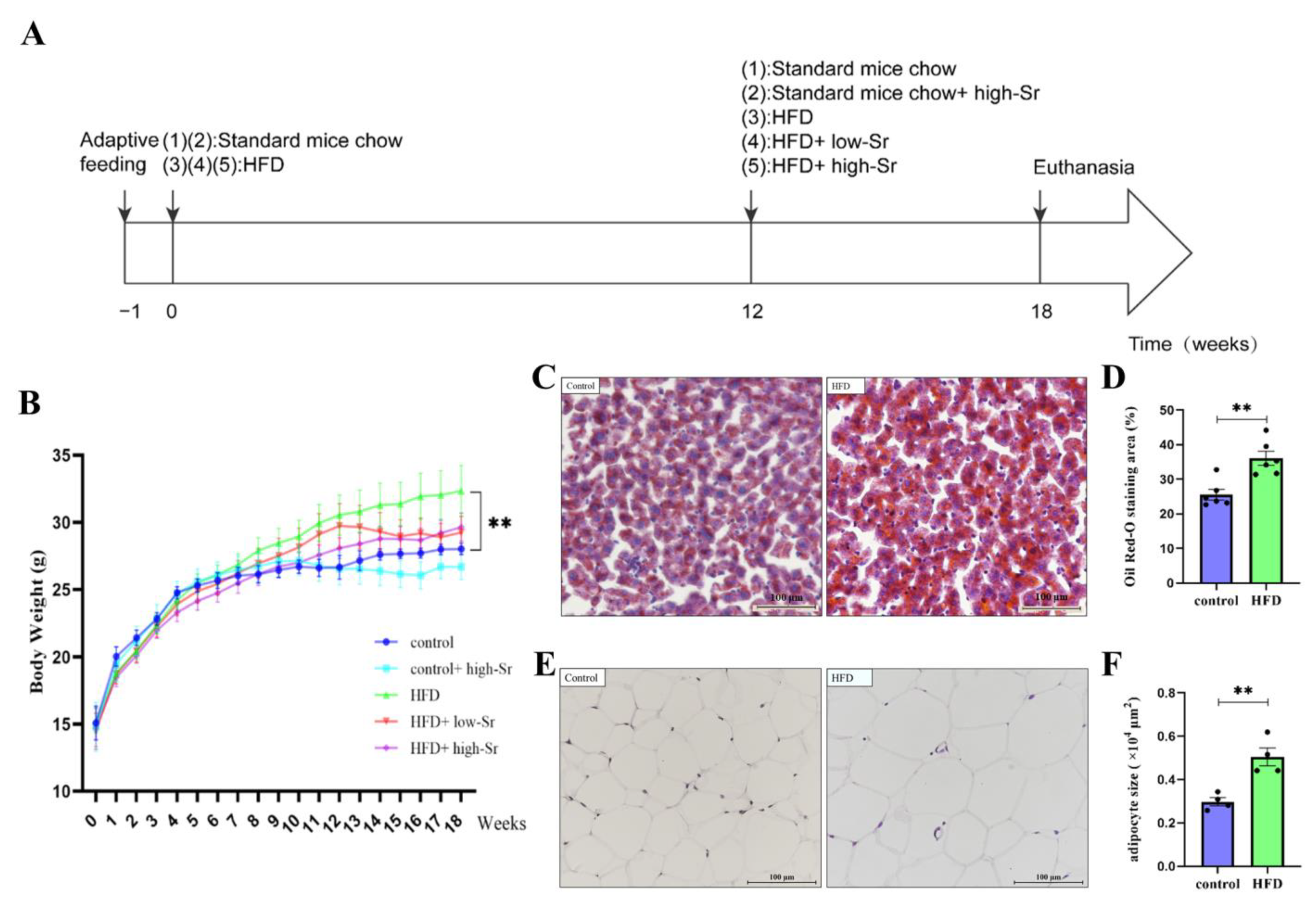
2.1. The NAFLD Mouse Model Induced by an HFD Was Successfully Established

Firstly, the NAFLD model was established according to the method shown in Figure 1A. There was no significant difference among the groups in initial body weight, whereas the body weight of the HFD-fed mice was higher than that in all the standard-mouse-chow-fed groups after 12 weeks of HFD administration (Figure 1B). At the end of the experiment (18 weeks), the HFD group had significant weight gain in comparison with the control group (p = 0.0080; Figure 1B). In the Oil Red O analysis performed to assess hepatic fat accumulation, more lipid droplets with darker staining were observed in the HFD group than in the control group (Figure 1C). There was a significant difference in the percentage of Oil Red-O staining area between these two groups (p = 0.0021; Figure 1D), suggesting that the hepatic fat accumulation in the HFD group was aggravated. Furthermore, the epididymal adipocyte size in the HFD group was markedly enlarged compared to that in the control group (Figure 1E), and the quantitative analysis showed the consistent result (p = 0.0040; Figure 1F). These characteristics indicated that the NAFLD mouse model was successfully constructed.

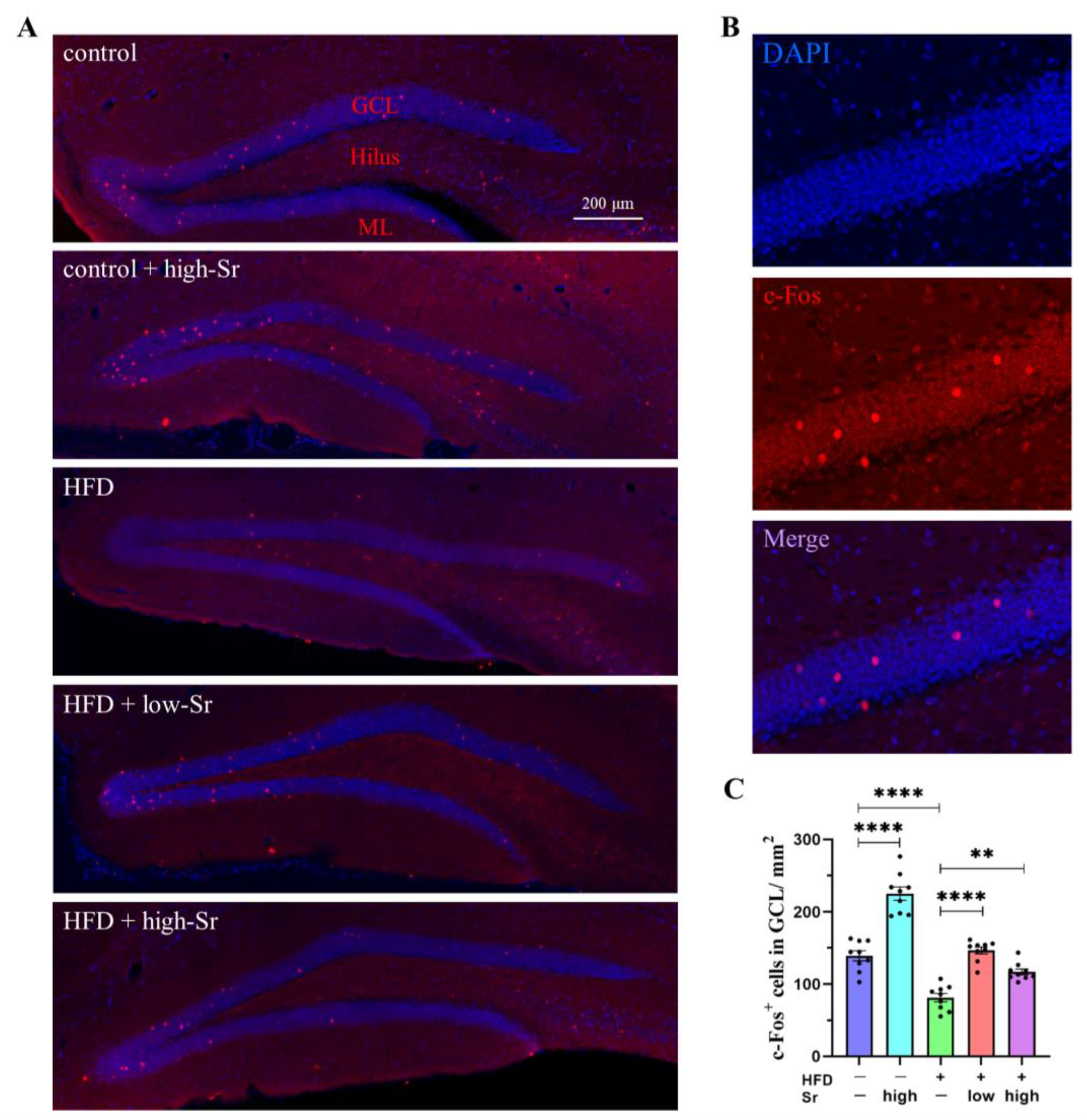
2.2. Sr Increased the Expression of c-Fos in HFD-Fed Mice

As an activator protein 1 transcription factor, it is well known that c-Fos is implicated in the regulation of cell proliferation, survival, and apoptosis [23]. We initially investigated whether Sr affected c-Fos expression in the hippocampi of the mice mediated by an HFD. The expression of c-Fos was evaluated through immunofluorescence staining with a specific antibody, and the c-Fos+ cells in the granule cell layer (GCL) of the hippocampus were counted. The results showed that an HFD considerably reduced the density of c-Fos+ cells in the GCL (p < 0.0001; Figure 2C). However, Sr inhibited the reduction in c-Fos+ cells in the GCL following a long-term HFD (low Sr: p < 0.0001) (high Sr: p = 0.0029) (Figure 2C). Interestingly, in the control group with high Sr, the density of c-Fos+ cells was greatly increased in the GCL (p < 0.0001; Figure 2C). These results demonstrated that Sr could increase the expression of c-Fos in the hippocampal GCL of the HFD-fed mice.

2.3. Sr Suppressed HFD-Induced Apoptosis by Inhibiting ERS

Having demonstrated that an HFD inhibited the expression of c-Fos associated with apoptosis, we investigated whether an HFD could induce apoptosis in the hippocampus. Since caspase-3 is considered to be the primary effector apoptosis molecule [24], its activity was examined to confirm apoptosis. We found that the caspase-3 protein level visibly increased in the HFD group (p = 0.0030; Figure 3B), and treatment with Sr decreased its expression significantly (low Sr: p = 0.0089) (high Sr: p = 0.0008) (Figure 3B). Previous studies indicated that an HFD could result in hippocampal damage through ERS-induced cell apoptosis [25,26,27]. Although the HFD group showed a trend towards a higher GRP78 level in the hippocampus when compared to the control group, the level in the HFD group with high Sr decreased significantly compared to that in the HFD group (p = 0.0010; Figure 3C). To further investigate whether the ERS response is actually induced by an HFD, we analyzed the expression of several ERS-inducible unfolded protein response (UPR) proteins. As expected, the levels of IRE1α, p-IRE1α, XBP1, eIF2α, ATF4, and ATF6 increased significantly in the HFD group compared to those in the control group (IRE1α: p < 0.0001) (p-IRE1α: p < 0.0001) (XBP1: p = 0.0099) (eIF2α: p = 0.0466) (ATF4: p = 0.0499) (ATF6: p = 0.0201) (Figure 3D–G,I,J). Sr significantly decreased the levels of the above proteins in the HFD mice (low Sr: (IRE1α: p < 0.0001) (XBP1: p < 0.0001) (ATF4: p = 0.0014) (ATF6: p = 0.0006)) (high Sr: (IRE1α: p < 0.0001) (p-IRE1α: p = 0.0220) (XBP1: p < 0.0001) (eIF2α: p = 0.0002) (ATF4: p = 0.0002) (ATF6: p < 0.0001)) (Figure 3D–G,I,J). High Sr without HFD provoked an elevation in the IRE1α and p-IRE1α levels (IRE1α: p < 0.0001) (p-IRE1α: p = 0.0123) (Figure 3D,E). Compared with the control group, the expression of p-eIF2α significantly decreased in the HFD group (p-eIF2α: p < 0.0001) (Figure 3H), while the level of p-eIF2α significantly increased (to different extents) with the treatment of low Sr or high Sr (low Sr: p = 0.0095) (high Sr: p < 0.0001) (Figure 3H). The transcription factor CHOP was reported to be a molecule involved in ERS-induced apoptosis that plays an important role in the induction of apoptosis [28,29]. We found that the protein level of CHOP increased significantly in the HFD group (p = 0.0471; Figure 3K), and treatment with Sr markedly decreased its expression (low Sr: p = 0.0005) (high Sr: p < 0.0001) (Figure 3K). Overall, these results showed that Sr could suppress HFD-induced apoptosis by inhibiting ERS.

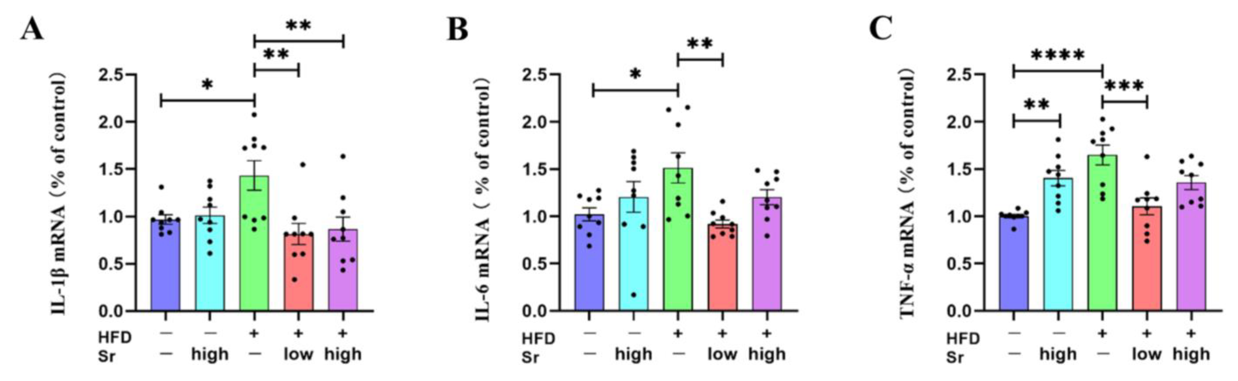
2.4. Sr Inhibited the Production of Inflammatory Cytokines in HFD-Fed Mice

Accumulating studies have demonstrated that prolonged ERS could regulate the inflammatory responses that mediate the production of cytokines [30,31]. An HFD can not only induce neuroinflammation but also increase the expression of inflammatory cytokines [32,33]. To determine whether Sr could suppress the HFD-induced increase in inflammatory cytokines in the hippocampus, an RT-PCR analysis was performed to assess the levels of IL-1β, IL-6, TNF-α, and GAPDH. The results showed that HFD significantly increased the mRNA levels of IL-1β, IL-6, and TNF-α in the hippocampus (IL-1β: p = 0.0410) (IL-6: p = 0.0383) (TNF-α: p < 0.0001) (Figure 4A–C). Low Sr significantly reduced the expression levels of the above inflammatory cytokines compared with the HFD group (IL-1β: p = 0.0031) (IL-6: p = 0.0066) (TNF-α: p = 0.0002) (Figure 4A–C). Although the mRNA level of IL-1β in the HFD group with high Sr was decreased significantly compared to the HFD group, high Sr without an HFD increased the mRNA level of TNF-α compared to the control group (IL-1β: p = 0.0077) (TNF-α: p = 0.0079) (Figure 4A,C). These results suggested that Sr could be capable of inhibiting the HFD-induced production of inflammatory cytokines.

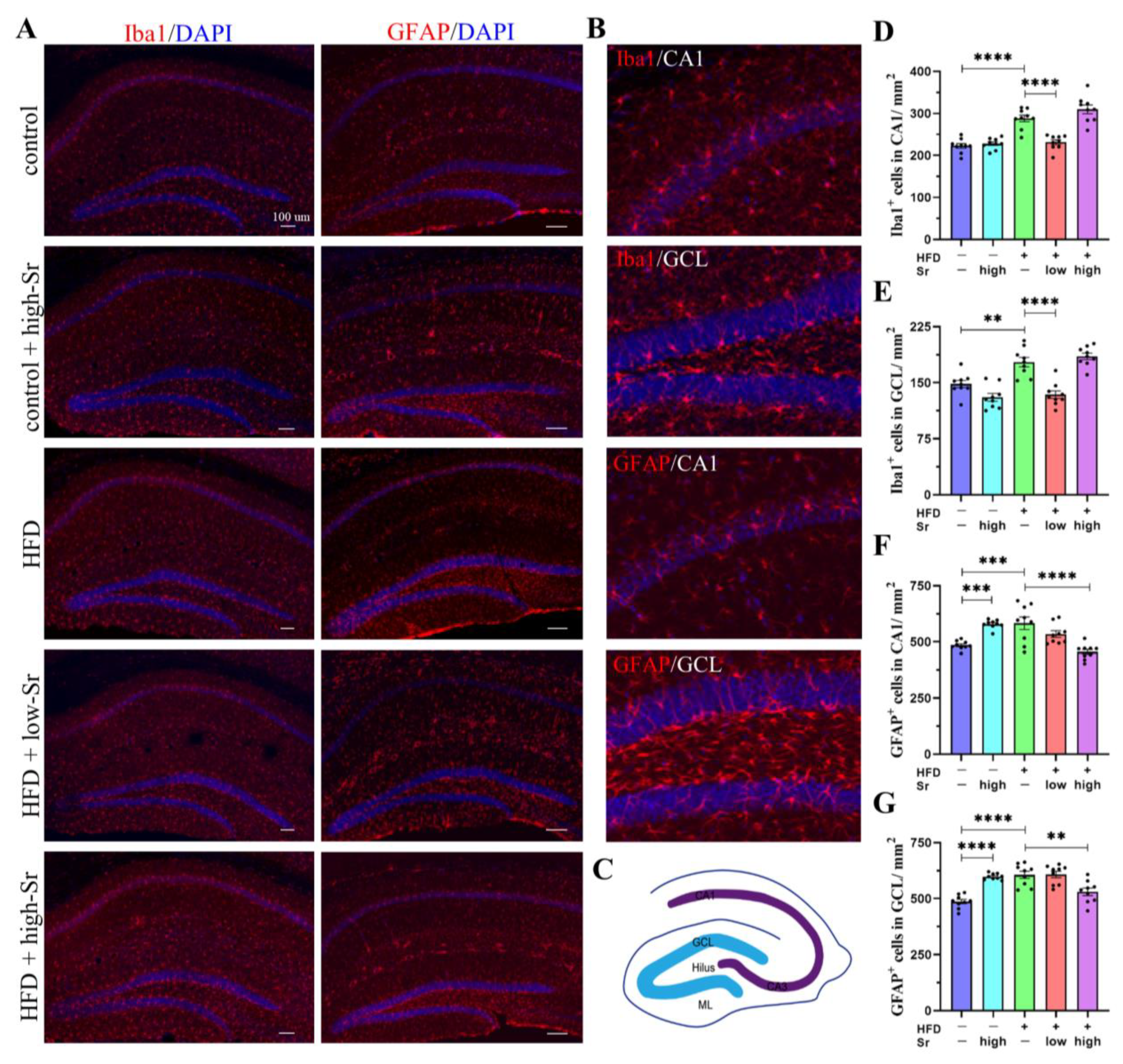
2.5. Sr Suppressed the Activation of Microglia and Astrocytes in the Hippocampus Induced by an HFD

The activation of microglia and astrocytes is thought to be involved in the increase in inflammatory cytokines [34,35,36]. The increase in the number of Iba1+ cells and GFAP+ cells was viewed as the marker of the activation of microglia and astrocytes [37,38]. According to our results, the number of Iba1+ cells and GFAP+ cells in the HFD group increased significantly compared with the control group (Iba1: (CA1: p < 0.0001) (GCL: p = 0.0020)) (GFAP: (CA1: p = 0.0005) (GCL: p < 0.0001)) (Figure 5D–G). Low Sr treatment significantly decreased the number of Iba1+ cells in the CA1 and GCL (CA1: p < 0.0001) (GCL: p < 0.0001) (Figure 5D,E). High Sr treatment not only decreased the number of GFAP+ cells in the HFD group (CA1: p < 0.0001) (GCL: p = 0.0016) (Figure 5F,G) but also substantially increased the number of GFAP+ cells in the control group (CA1: p = 0.0010) (GCL: p < 0.0001) (Figure 5F,G). These findings demonstrated that the activated microglia and astrocytes in the hippocampus induced by an HFD can be inhibited by Sr.

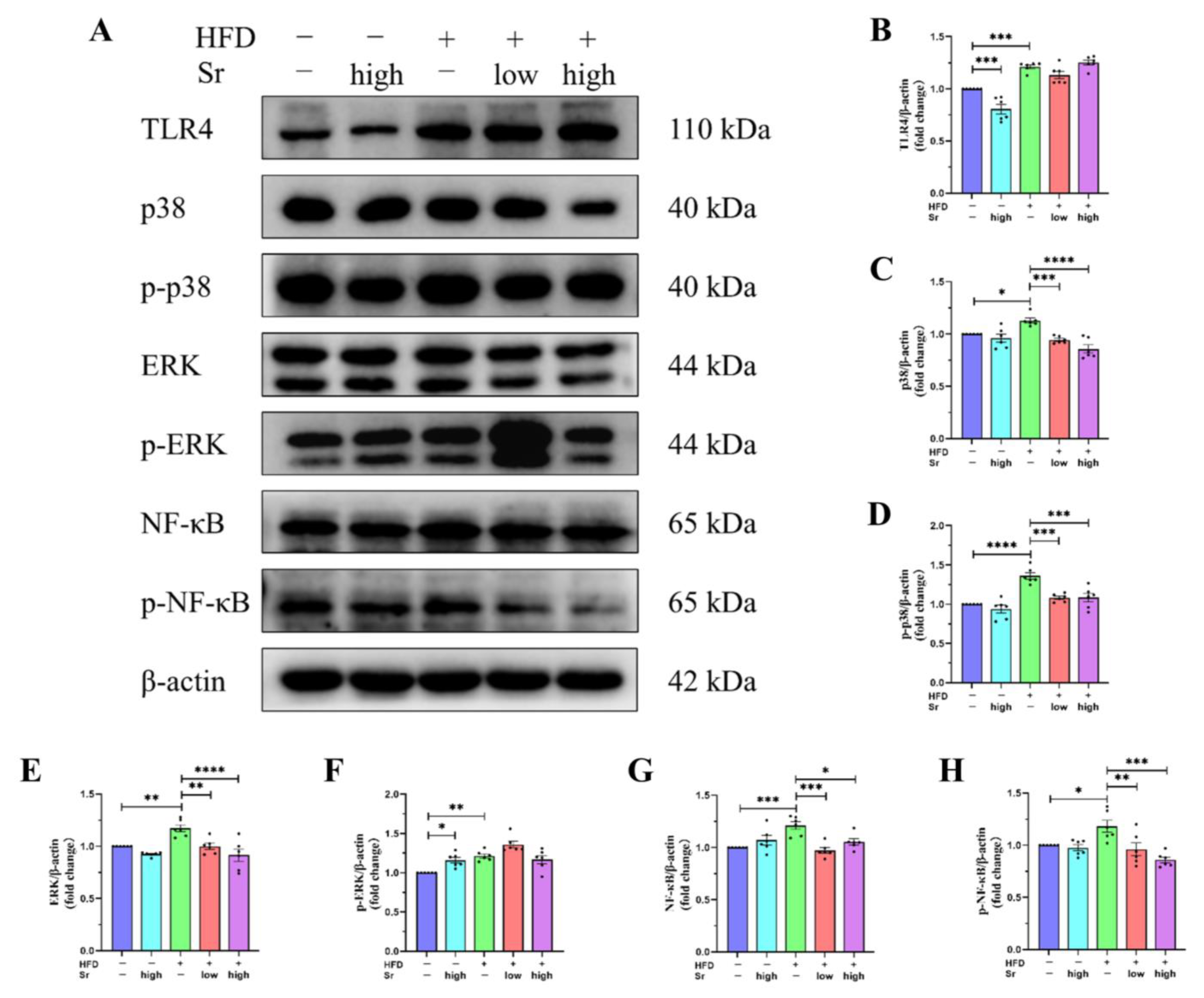
2.6. Sr Inhibited the Activation of the TLR4/p38 MAPK/ERK and NF-κB Pathways Induced by an HFD

Although we demonstrated that Sr can attenuate the HFD-induced secretions of inflammatory cytokines and the massive activation of microglia and astrocytes, the potential mechanisms by which Sr suppresses neuroinflammation remain unclear. To investigate the possible mechanisms, the levels of TLR4, p38, p-p38, ERK, p-ERK, NF-κB, and p-NF-κB in the hippocampus were determined (Figure 6A). The results revealed that the levels of the above inflammation-related proteins were visibly higher in the HFD group than those in the control group (TLR4: p = 0.0002) (p38: p = 0.0205) (p-p38: p < 0.0001) (ERK: p = 0.0088) (p-ERK: p = 0.0025) (NF-κB: p = 0.0008) (p-NF-κB: p = 0.0394) (Figure 6B–H). However, Sr treatment restrained the expression of p38, p-p38, ERK, NF-κB, and p-NF-κB in the HFD group (low Sr: (p38: p = 0.0006) (p-p38: p = 0.0003) (ERK: p = 0.0085) (NF-κB: p = 0.0002) (p-NF-κB: p = 0.0082)) (high Sr: (p38: p < 0.0001) (p-p38: p = 0.0003) (ERK: p < 0.0001) (NF-κB: p = 0.0147) (p-NF-κB: p = 0.0001)) (Figure 6C–E,G,H). As seen in Figure 6B,F, in the control group, the expression of TLR4 was diminished by high Sr treatment, but the expression of p-ERK was increased (TLR4: p = 0.0006) (p-ERK: p = 0.0327). These results indicated that Sr could exert the anti-inflammatory effects induced by an HFD via the TLR4/p38 MAPK/ERK and NF-κB pathways.

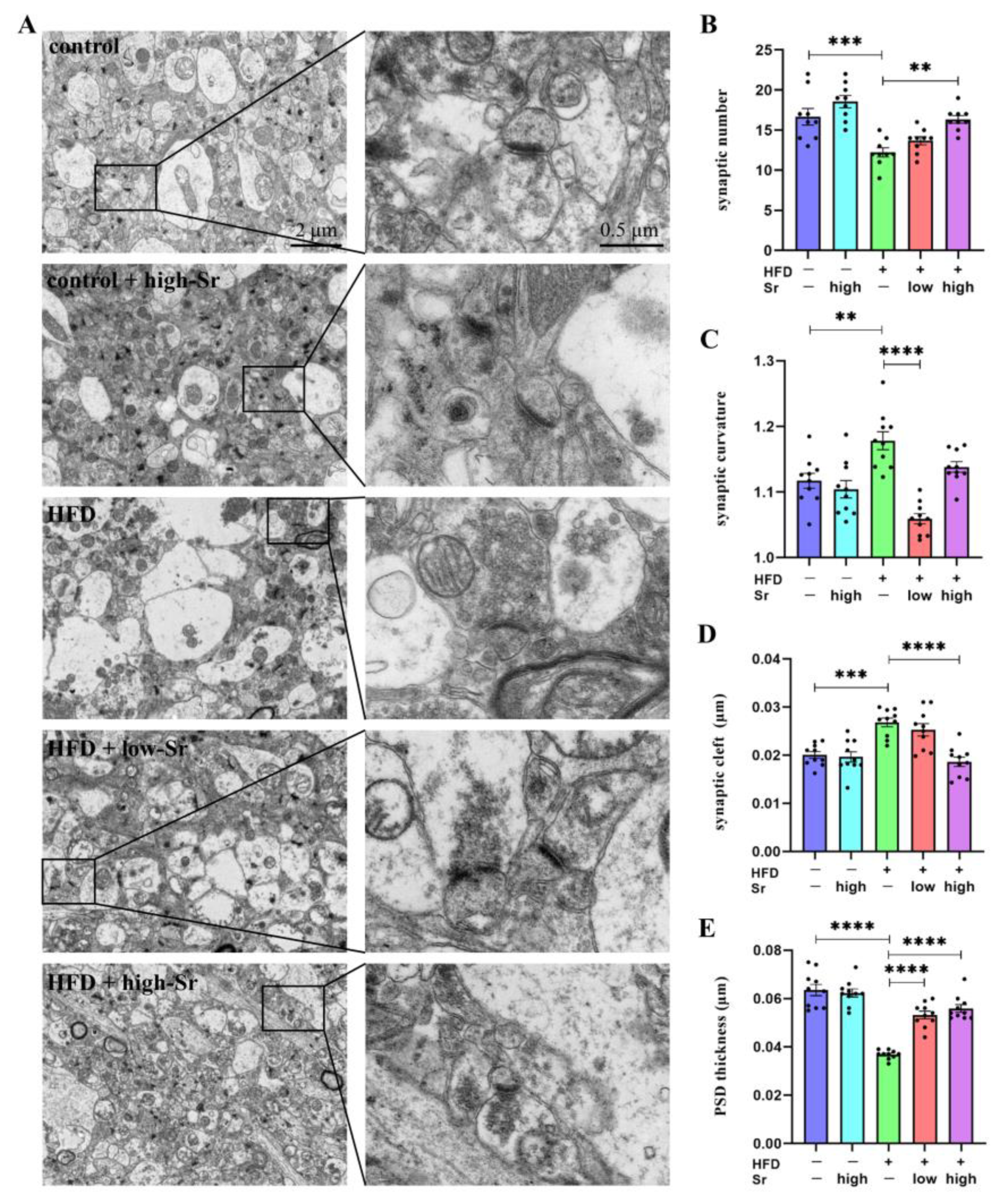
2.7. Sr Suppressed the Alterations in Many Aspects Related to Hippocampal Synaptic Plasticity Induced by an HFD
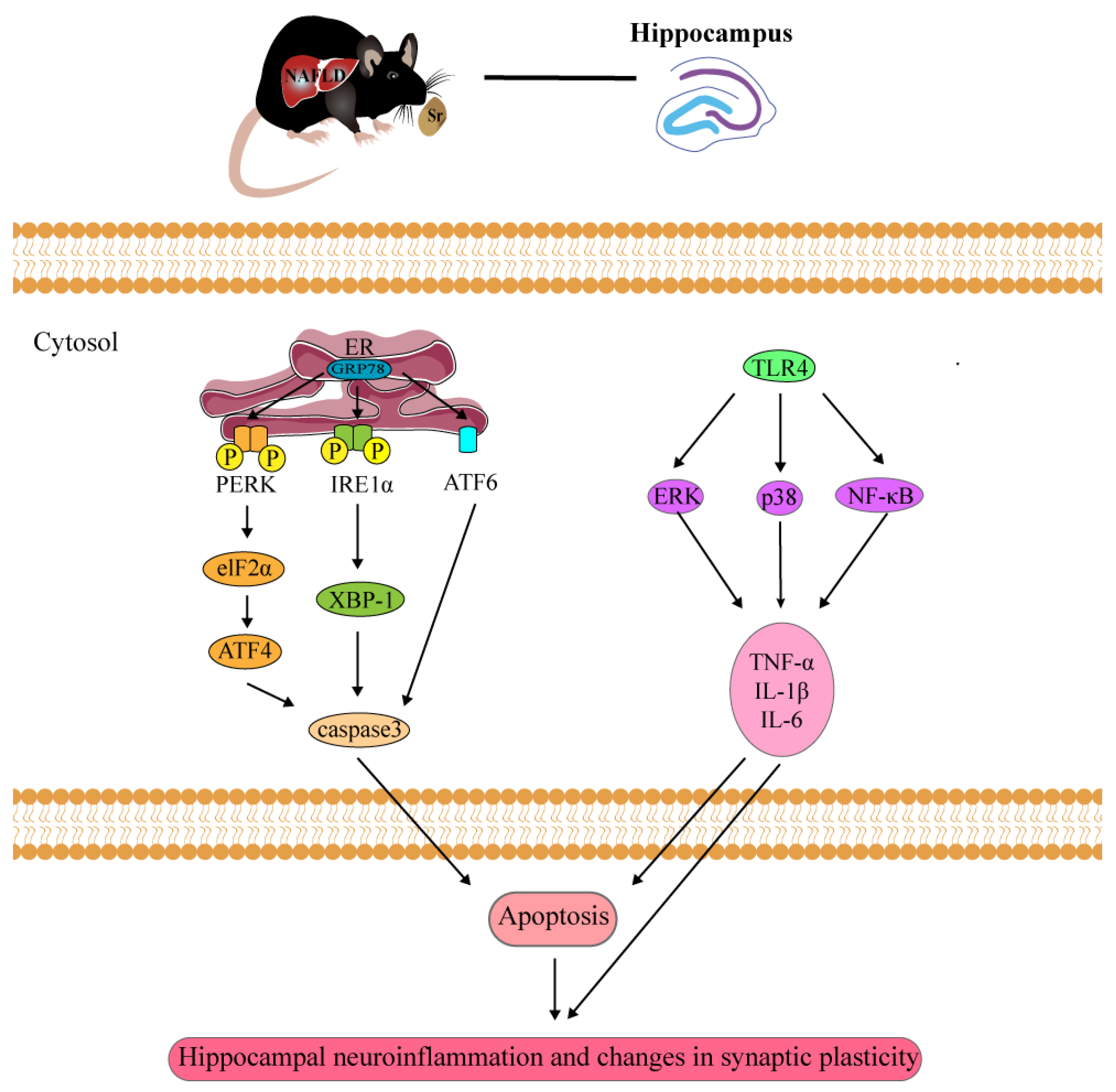
It was previously shown that the hippocampus is particularly vulnerable to damage due to neuroinflammation [39]. Because hippocampal synaptic plasticity is believed to be the neurobiological basis of learning memory and cognitive function, we examined alterations in the synaptic quantity and ultra-structure [40]. As shown in Figure 7A, TEM was performed to measure the ultra-structure of the synapses in the hippocampus, and the number of synapses was counted. The results found that the decreased synaptic number in the HFD-treated mice (p = 0.0005; Figure 7B) was restored by high Sr administration (p = 0.0015; Figure 7B). As displayed in Figure 7C,D, many aspects related to hippocampal synaptic plasticity, including the synaptic curvature and synaptic cleft, increased under the influence of an HFD (synaptic curvature: p = 0.0028) (synaptic cleft: p = 0.0002); the levels of these aspects were decreased by Sr treatment (low Sr: (synaptic curvature: p < 0.0001)) (high Sr: (synaptic cleft: p < 0.0001)). In addition, the postsynaptic density (PSD) thickness was reduced in the HFD group, and that was alleviated by Sr (HFD: p < 0.0001) (low Sr: p < 0.0001) (high Sr: p < 0.0001) (Figure 7E). Taken together, these findings indicated that Sr could suppress the alterations in hippocampal synaptic plasticity in the HFD mice.

3. Discussion

As far as we are aware, this is the first study to investigate the protective effects of Sr on NAFLD-induced hippocampal damage. The pathogenesis and development of NAFLD are quite complex processes involving multiple pathways, including imbalanced diets, lipotoxicity, oxidative stress, and inflammation [2]. It has been illustrated that NAFLD induced by an HFD not only induces inflammation and ERS [7,18,41] but also triggers cognitive deficits and decreased synaptic plasticity [42,43]. Accordingly, the evidence suggested that NAFLD might be a risk factor for CNS diseases [44]. Sr, a promising trace element in the body, triggers new bone formation through the induction of osteoblasts and the prevention of osteoclast activity [45]. In addition, Sr has been proven to be an anti-inflammatory drug with immunomodulatory properties [46,47]. The aim of this research was to investigate whether Sr could exert its neuroprotective effects by inhibiting the ERS, apoptosis, and neuroinflammation induced by an HFD in an NAFLD mouse model. We observed that Sr reduced the increased mRNA expression levels of IL-1β, IL-6, and TNF-α induced by an HFD. Similarly, the activation of microglia and astrocytes was inhibited by Sr administration in the hippocampus. In addition, the observed damage to the ultra-structural synaptic architecture induced by an HFD could be prevented by Sr administration in this study. Mechanism studies suggested that the inhibitory effects of Sr on HFD-induced hippocampal damage in the NAFLD mouse model were mediated by some of the signaling pathways relating to ERS or inflammation (Figure 8). This study implies that Sr has beneficial effects on repairing the damage to the hippocampus induced by an HFD in NAFLD mice.
To examine the function of Sr on the hippocampal damage induced by NAFLD, C57BL/6J mice were fed with an HFD to establish the NAFLD model in vivo. According to the previous study, the same or a similar dietary composition and duration were used to successfully induce liver steatosis, inflammation, and fibrosis [48,49,50]. In the present study, the mice in the model group showed an overall increase in body weight after 12 weeks of HFD feeding. The average weight gain trend appeared to be different in each group. However, it was not possible to regroup the mice because of the fights among them. It is worth noting that the significant weight gain and the large accumulation of fat in the liver and epididymis shown through a histological examination demonstrated the successful establishment of the NAFLD mouse model through HFD feeding.
C-Fos is an immediate-early gene that has been widely used as an indicator of neuronal activation [51]. In the current study, the c-Fos+ cells in the GCL decreased in the HFD mice. Similar data were previously reported that showed that an HFD prevented the enteric activation of c-Fos expression [52]. Because c-Fos is often expressed when neurons fire action potentials, c-Fos in the hippocampus can be used to assess the animal responses to stress [53]. Compared with the control group, the expression of c-fos was significantly decreased in the HFD group, which may be due to the inhibition of neuronal action potentials by a long-term HFD. Treatment with Sr promoted the expression of c-Fos in the HFD-fed mice, indicating that Sr alleviated the impacts of an HFD. However, the expression of c-Fos was also promoted by high Sr in normal mice. The observed increase in c-Fos could be attributed to Sr treatment. It is possible that Sr treatment promoted the neuronal activation of the hippocampus in the brain.
Previous studies have demonstrated that alterations in the expression of c-Fos are associated with ERS [54,55]. The occurrence and development of various long-term HFD-induced metabolic disorders, such as obesity, insulin resistance, and type II diabetes, are closely associated with sustained ERS [56]. Moreover, sustained ERS also has the potential to elicit inflammation and facilitate cell apoptosis [57]. In accordance with recent studies, our results indicated that an HFD could result in the accumulation of unfolded or misfolded proteins in the endoplasmic reticulum (ER) lumen, subsequently activating ERS and leading to apoptosis. When ERS occurs, the key ER regulatory protein GRP78 dissociates from the ER transmembrane proteins, leading to the activation of the transmembrane proteins [58]. Compared with the control group, the expression of GRP78 did not show a significant difference in the HFD group, but there was a certain upward trend. Apart from this, there were significant differences in some of the ERS-associated proteins that we measured. All three ER membrane sensors (IRE1, PERK, and ATF6), which activate the IRE1-spliced XBP1, PERK-eIF2α-ATF4, and ATF6 pathways to the cleaved ATF6 signaling pathway, respectively, induce downstream CHOP, a pro-apoptotic transcription factor [59,60,61]. The activation of CHOP induces caspase-3-mediated apoptotic signaling [62]. Caspase-3 can also be activated in excessive ERS conditions [63]. As a key promoter of apoptosis, caspase-3 activation is the final concentration point of all apoptotic pathways [24]. In this study, the caspase-3 protein level was significantly increased in the hippocampus of the HFD mice, and, importantly, Sr treatment could decrease caspase-3 protein expression. Therefore, we speculate that Sr-mediated anti-apoptosis effects contribute to the increased survival rate of cells in hippocampi under NAFLD.
Neuroinflammation is inextricably linked with apoptosis and ERS. Not unexpectedly, there was an increase in inflammatory cytokines in the HFD mice. In accordance with the present results, previous studies have demonstrated that neuroinflammation is one of the major symptoms of NAFLD and that it is involved in the progression of NAFLD [64]. Microglia and astrocytes are the resident immune cells of the CNS and are the primary mediators of neuroinflammation [65,66]. The proliferation of microglia and astrocytes is characterized as the classic inflammatory phenotype [67,68]. The increased densities of microglia and astrocytes in the HFD mice revealed that they became denser compared to those in the control group. Secreted factors from microglia are known to affect astrocytes and vice versa [69]. Surprisingly, we found that high Sr increased some inflammatory markers in the normal mice. The reason might be that Sr is a trace element, and the body does not normally ingest a high level of Sr. Therefore, high Sr might be a mild irritation in normal mice, leading to certain inflammatory responses, which was especially reflected in TNF-α expression. According to our findings, the release of IL-1β, IL-6, and TNF-α was decreased in the HFD mice with Sr treatment. Hence, it could conceivably be hypothesized that the hippocampal activation of microglia and astrocytes induced by an HFD was suppressed by Sr administration. As expected, treatment with Sr reversed the density changes induced by an HFD, and the density results of the Sr-treated group were almost close to those of the control group, suggesting that Sr played an anti-inflammatory role via modulating the activation of microglia and astrocytes. We further investigated the mechanism by which Sr may inhibit the HFD-induced hippocampal inflammation mediated by the TLR4/p38 MAPK/ERK and NF-κB signaling pathways. It has been demonstrated that the HMGB1/TLR4/NF-κB axis is the critical pro-neuroinflammatory signaling pathway in the CNS [70]. After the activation of TLR4, the recruitment and promotion of NF-κB occurs in neuroinflammation [71]. TLR4 results in the production of inflammatory factors through phosphorylation of p38, ERK, JNK as well as the activation of NF-κB [72,73]. Our results revealed that an HFD increased the expression of phosphorylated p38 and NF-κB in the hippocampus, and Sr inhibited the above increases. According to this, we demonstrated that Sr exerted its protective effects via suppressing the phosphorylation of p38 and NF-κB, which subsequently led to reductions in IL-1β, IL-6, and TNF-α in the mice that were fed an HFD. This may explain why Sr suppressed the activation of microglia and astrocytes induced by an HFD in the NAFLD mice.
CNS neuroinflammatory damage has become a crucial reason for learning/memory impairment [74]. It has been widely recognized that an HFD leads to neuroinflammation in the hippocampus and decreased learning and memory functions [75]. The structural and functional plasticity of the synapses is critical for learning and memory [76]. The plasticity of the synaptic structure is mainly reflected in the ultra-structural changes in the synaptic number, synaptic curvature, synaptic cleft, and PSD thickness [77]. Our results showed that an HFD not only increased the synaptic curvature and synaptic cleft but also decreased the synaptic number and PSD thickness. It was possible that the increases in the synaptic curvature in the HFD group were due to compensatory effects. The most interesting finding was that Sr could inhibit these changes. Based on these findings, we indicated that Sr treatment could prevent HFD-induced damage to the ultra-structural synaptic architecture in the NAFLD model. Additionally, Sr had a protective effect on the hippocampal damage caused by NAFLD.
In summary, our results suggested that an HFD induced both ERS and inflammatory responses in the mice, involving a decrease in the c-Fos+ cell level, the activation of microglia and astrocytes, and increases in the pro-inflammatory cytokine levels. However, Sr did not only inhibit ERS through three UPR signaling pathways (the IRE1α-XBP1, PERK-eIF2α-ATF4, and ATF6 pathways) but also inhibited the inflammation induced by an HFD via blocking the TLR4/p38 MAPK/ERK and NF-κB pathways. In addition, Sr could suppress alterations in hippocampal synaptic plasticity in the NAFLD mice. The protective effect of Sr on hippocampal damage may be mediated by alleviating ERS and the anti-inflammatory effects, which has the potential to become a new therapeutic approach to prevent and treat NAFLD-induced hippocampal damage.

4. Materials and Methods

4.1. Mice, Diets, and Treatments

Male C57BL/6J mice aged six weeks were purchased from Chongqing Byrness Weil biotech Ltd. (Chongqing, China). The mice were housed in a standard rodent room (temperature of 22–26 °C, relative humidity of 40–70%, and 12 h day/night cycle), and body weight was assessed weekly. Before the formal experiment, adaptive feeding was implemented with standard mouse chow and water intake for a week. The mice were randomly divided into 5 groups (n = 12 per group) and housed in cages (n = 4 per cage) after one week of adaptive feeding as follows (Figure 1A):
(1) The control group was fed standard mouse chow for 18 weeks.
(2) The control + high-Sr group was fed standard mouse chow for 12 weeks. Then, the mice were fed standard mouse chow mixed with SrCl2·6H2O (750 mg/kg of Sr) for 6 weeks.
(3) The HFD group was fed a 45% high-fat diet for 18 weeks.
(4) The HFD + low-Sr group was fed a 45% high-fat diet for 12 weeks. Then, the mice were fed a 45% high-fat diet mixed with SrCl2·6H2O (250 mg/kg of Sr) for 6 weeks.
(5) The HFD + high-Sr group was fed a 45% high-fat diet for 12 weeks. Then, the mice were fed a 45% high-fat diet mixed with SrCl2·6H2O (750 mg/kg of Sr) for 6 weeks.
The standard mouse chow (AIN005, 20% protein, 70% carbohydrate, and 10% fat) and the 45% high-fat diet (HD004, 20% protein, 35% carbohydrate, and 45% fat) were purchased from Beijing Botai Hongda Biotechnology Co. The SrCl2·6H2O (V900279) was purchased from Shanghai Sigma–Aldrich. The mouse chow was abraded to 1~5 mm particles using sample mill (SMF2002, Supor, Hangzhou, China). The SrCl2·6H2O was dissolved in distilled water and mixed evenly with diets (dissolve in 10 mL distilled water in per kg control diets and 5 mL in per kg HFD diets). Then, the Sr-feed mixture was pressed into cylindrical chow using feed pellet mill. The chow was then stored at −20 °C separately after drying. Animal investigators performing the experiments were blinded to the group assignment of mice during the experiment. Animal experiments were conducted according to the Guide for Care and Use of Laboratory Animals: Northwest A&F University, and they were approved and performed in accordance with the Animal Care Commission of the College of Veterinary Medicine, Northwest A&F University (Approval No. 2021051).

4.2. Histology

Mouse livers and epididymal adipose were dissected from the control mice and the HFD mice. Livers were placed in 30% sucrose solution in PB at 4 °C overnight before being placed in optimal cutting temperature compound and being frozen in liquid nitrogen. Liver samples underwent frozen sectioning at 8 μm thickness and Oil Red O staining to assess hepatic fat accumulation. Hematoxylin counterstaining was performed to visualize nuclei. Epididymal adipose was fixed in 10% neutral formalin, processed in paraffin blocks, sectioned to a thickness of 20 μm, and stained with hematoxylin and eosin. These frozen sections and paraffin sections were observed and photographed under a microscope (Nikon, Tokyo, Japan). The Oil Red-O staining area and adipocyte size were quantified using ImageJ software (Fiji, https://imagej.net/software/fiji/, accessed on 28 May 2023).

4.3. Western Blot

The brains of mice were rapidly dissected on ice after being sacrificed, and the hippocampus tissues were isolated. The hippocampal samples were homogenized in RIPA lysis buffer containing a protease inhibitor cocktail (P1400) and phosphatase inhibitor cocktail. Protein concentrations were determined with the BCA Protein Assay Kit (Solarbio, Beijing, China). Briefly, the protein samples (20 μg each) were separated through sodium dodecyl sulfate–polyacrylamide gel electrophoresis and were transferred onto polyvinylidene difluoride (PVDF) membranes. We blocked the PVDF membranes for 2 h at room temperature using 5% skim milk (skim milk dissolved in TBST (TBS containing 0.1% Tween-20)). Subsequently, the following primary antibodies were used to incubate the membranes overnight at 4 °C: rabbit anti-NF-κB (#8242), rabbit anti-p38 (#9212), rabbit anti-ERK (#9102), rabbit anti-phospho-ERK (p-ERK, #4370), rabbit anti-phospho-p38 (p-p38, #4511), and anti-caspase-3 (#9662) (purchased from Cell Signaling Technology (Danvers, MA, USA)); mouse anti-ATF6 (EM1701-94) (purchased from Hangzhou Huaan Biotechnology Co., Ltd., Hangzhou, China); rabbit anti-XBP1 (A1731), rabbit anti-phospho-NF-κB (p- NF-κB, AP0475), rabbit anti-GRP78 (A0241), and mouse anti-β-actin (purchased from Wuhan ABclonal Technology Co., Ltd., Wuhan, China); rabbit anti-eIF2α (ab115822), rabbit anti-TLR4 (ab13556), and rabbit anti-phospho-eIF2α (p-eIF2α, ab32157) (purchased from Abcam (Cambridge, MA, USA)); and rabbit anti-CHOP (BM4962), anti-phospho-IRE1α (p-IRE1α, BM4444), rabbit anti-ATF4 (BM5179), and rabbit anti-IRE1α (A00683-1) (purchased from Wuhan BOSTER Biological Technology Co., Ltd., Wuhan, China). The membranes were incubated with horseradish peroxidase (HRP)-conjugated goat anti-rabbit IgG antibody or goat anti-mouse IgG antibody (Cell Signaling Technology) at 4 °C for 5 h and were washed with TBST. The blots were developed with ChemiDoc™ MP Imaging System (Bio-Rad, Hercules, CA, USA) using an enhanced chemiluminescence detection kit (GE Healthcare, Buckinghamshire, UK), and relative band densities were quantified using ImageJ software (Fiji, https://imagej.net/software/fiji/, accessed on 28 May 2023).

4.4. Quantitative RT-PCR

Quantitative RT-PCR analysis was essentially performed as previously described [78]. Total RNA was isolated from hippocampus using TRIzol reagent (Invitrogen, Waltham, MA, USA). cDNA was synthesized from 2 μg of total RNA using the FastKing RT Kit (TIANGEN Biotech, Beijing, China) according to manufacturer’s instructions. The synthesized cDNA was used as the RT-PCR template. This was performed on a Bio-Rad CFX96TM real-time PCR detection system (Bio-Rad, Hercules, CA, USA) using NovoStart® SYBR qPCR SuperMix Plus (Novoprotein Scientific Inc., Nanjing, China). The primers used were as follows. GAPDH: forward primer was 5′-AGGTTGTCTCCTGCGACTGCA-3′, and reverse primer was 5′-GTGGTCCAGGGTTTCTTACTCC-3′. IL-6: forward primer was 5′-CACTTCACAAGTCGGAGGCT-3′, and reverse primer was 5′-CTGCAAGTGCATCATCGTTGT-3′. IL-1β: forward primer was 5′-TGACGGACCCCAAAAGATGA-3′, and reverse primer was 5′-TCTCCACAGCCACAATGAGT-3′. TNF-α: forward primer was 5′-AGTCCGGGCAGGTCTACTTT-3′, and reverse primer was 5′-GTCACTGTCCCAGCATCTTGT-3′. GAPDH was used as an internal control gene. The relative quantification of mRNA expression was calculated with the 2−ΔΔCT method [79].

4.5. Immunofluorescence Staining

We anesthetized the mice with 0.56% sodium pentobarbital and transcardially perfused the mice with 0.9% saline followed by 4% paraformaldehyde (PFA, pH 7.4). All mice brains were obtained and post-fixed in 4% PFA in 0.1 M phosphate buffer (PB, pH 7.4) at 4 °C for over 72 h. The fixed brains were sectioned using a microtome (VT1000S, Leica, Wetzlar, Germany) into coronal slices (thickness of 50 µm). After rinsing three times in PB, sections were incubated with primary antibodies in blocking solution (4% BSA, 1% NGS, and 0.3% Triton in phosphate buffer) for 24 h at 4 °C. The following primary antibodies were used: rabbit anti-c-Fos, rabbit anti-ionized calcium-binding adapter molecule 1 (Iba1), and rabbit anti-glial fibrillary acidic protein (GFAP). The sections were washed three times before incubation with secondary antibodies overnight at 4 °C in the darkness. The secondary antibody used in this study was Alexa Fluor 647 goat anti-rabbit IgG (ab150079, 1:500, Abcam). DAPI was used to stain the nuclei. The slices were washed three times with PB and mounted with Fluorescence Mounting Medium (Dako, Carpentaria, Australia). Stained slides were photographed using a structured illumination microscope (Zeiss Axio Observer Z1, Zeiss, Oberkochen, Germany). The immunofluorescent images were analyzed and processed using ImageJ software.

4.6. Transmission Electron Microscopy (TEM)

Samples of hippocampus tissues (1 mm × 1 mm × 1 mm) were carefully collected to minimize mechanical damage, then primarily fixed in Fixative for TEM (G1102, Servicebio, Wuhan, China) at 4 °C. The sample avoided light after being fixed with 1% OsO4 (Ted Pella Inc., Redding, CA, USA) in 0.1 M PB (pH 7.4) for 2 h at room temperature. After removing OsO4, the tissues were rinsed in 0.1 M PB (pH 7.4) three times. After dehydrating at room temperature, resin penetration and embedding were performed; the embedding models containing the resin and samples were put into an oven at 65 °C for polymerization for over 48 h. After this, the resin blocks were removed from the embedded models and were placed at room temperature for standby application. Then, the resin blocks were cut into thin sections of 60–80 nm using an ultra-microtome (Leica UC7, Leica), and the sections were placed on 150-mesh cuprum grids using formvar film. The sections were stained with 2% uranium acetate saturated alcohol solution and 2.6% lead citrate and observed through TEM (HT7800, Hitachi, Tokyo, Japan).

4.7. Statistical Analysis

GraphPad Prism 8.0 software (San Diego, CA, USA) was used to analyze data, which are expressed as the mean ± standard error of the mean (SEM). The comparisons between groups were performed through one-way analysis of variance (ANOVA) and Tukey’s multiple comparison test. Significant differences were determined when p value was less than 0.05. Analyses were performed by an investigator blinded to the experimental conditions.

Author Contributions

Conceptualization, S.W. (Shuai Wang) and X.Z.; methodology, S.W. (Shuai Wang), F.Z. and J.W.; software, C.X. and S.W. (Shuai Wang); validation, X.F. and Y.M.; formal analysis, Y.M. and S.W. (Shuai Wang); investigation, S.W. (Songlin Wang); resources, S.Z. and X.Z.; data curation, S.W. (Shuai Wang) and X.Z.; writing—original draft preparation, S.W. (Shuai Wang) and J.Y.; writing—review and editing, S.W. (Shuai Wang) and J.Y.; visualization, J.Y.; supervision, X.Z.; project administration, S.Z. and X.Z.; funding acquisition, X.Z. All authors have read and agreed to the published version of the manuscript.

Funding

This work was financially supported by the National Natural Science Foundation of China (No. 32272967).

Institutional Review Board Statement

This study was approved by the Research Ethics Committee of the College of Veterinary Medicine, Northwest A&F University (Approval No. 2021051).

Informed Consent Statement

Not applicable.

Data Availability Statement

The data presented in this study are available on request from the corresponding author.

Acknowledgments

We would like to thank the State Key Laboratory of Crop Stress Biology for Arid Areas in NWAFU for supplying the ChemiDocTM MP Imaging System (Bio-Rad, Hercules, CA, USA).

Conflicts of Interest

The authors declare no conflict of interest.

References

  1. Younossi, Z.M. Non-alcoholic fatty liver disease—A global public health perspective. J. Hepatol. 2019, 70, 531–544. [Google Scholar] [CrossRef] [PubMed] [Green Version]
  2. Colognesi, M.; Gabbia, D.; Martin, S.D. Depression and Cognitive Impairment—Extrahepatic Manifestations of NAFLD and NASH. Biomedicines 2020, 8, 229. [Google Scholar] [CrossRef] [PubMed]
  3. He, J.; Ye, J.; Sun, Y.; Feng, S.; Chen, Y.; Zhong, B. The Additive Values of the Classification of Higher Serum Uric Acid Levels as a Diagnostic Criteria for Metabolic-Associated Fatty Liver Disease. Nutrients 2022, 14, 3587. [Google Scholar] [CrossRef]
  4. Peng, C.; Xu, X.; Li, Y.; Li, X.; Yang, X.; Chen, H.; Zhu, Y.; Lu, N.; He, C. Sex-specific association between the gut microbiome and high-fat diet-induced metabolic disorders in mice. Biol. Sex Differ. 2020, 11, 5. [Google Scholar] [CrossRef] [Green Version]
  5. Nuzzo, D.; Galizzi, G.; Amato, A.; Terzo, S.; Picone, P.; Cristaldi, L.; Mulè, F.; Di Carlo, M. Regular Intake of Pistachio Mitigates the Deleterious Effects of a High Fat-Diet in the Brain of Obese Mice. Antioxidants 2020, 9, 317. [Google Scholar] [CrossRef] [Green Version]
  6. Tsai, C.; Chen, Y.; Yu, H.; Huang, L.; Tain, Y.; Lin, I.; Sheen, J.; Wang, P.; Tiao, M. Long term N-acetylcysteine administration rescues liver steatosis via endoplasmic reticulum stress with unfolded protein response in mice. Lipids Health Dis. 2020, 19, 105. [Google Scholar] [CrossRef]
  7. Tan, B.L.; Norhaizan, M.E. Effect of High-Fat Diets on Oxidative Stress, Cellular Inflammatory Response and Cognitive Function. Nutrients 2019, 11, 2579. [Google Scholar] [CrossRef] [PubMed] [Green Version]
  8. Kołodziejska, B.; Stępień, N.; Kolmas, J. The Influence of Strontium on Bone Tissue Metabolism and Its Application in Osteoporosis Treatment. Int. J. Mol. Sci. 2021, 22, 6564. [Google Scholar] [CrossRef]
  9. Pemmer, B.; Hofstaetter, J.G.; Meirer, F.; Smolek, S.; Wobrauschek, P.; Simon, R.; Fuchs, R.K.; Allen, M.R.; Condon, K.W.; Reinwald, S.; et al. Increased strontium uptake in trabecular bone of ovariectomized calcium-deficient rats treated with strontium ranelate or strontium chloride. J. Synchrot. Radiat. 2011, 18 Pt 6, 835–841. [Google Scholar] [CrossRef] [PubMed] [Green Version]
  10. Marx, D.; Rahimnejad Yazdi, A.; Papini, M.; Towler, M. A review of the latest insights into the mechanism of action of strontium in bone. Bone Rep. 2020, 12, 100273. [Google Scholar] [CrossRef] [PubMed]
  11. Zhu, S.; Hu, X.; Tao, Y.; Ping, Z.; Wang, L.; Shi, J.; Wu, X.; Zhang, W.; Yang, H.; Nie, Z.; et al. Strontium inhibits titanium particle-induced osteoclast activation and chronic inflammation via suppression of NF-κB pathway. Sci. Rep. 2016, 6, 36251. [Google Scholar] [CrossRef]
  12. Yu, H.; Liu, Y.; Yang, X.; He, J.; Zhong, Q.; Guo, X. The anti-inflammation effect of strontium ranelate on rat chondrocytes with or without IL-1β in vitro. Exp. Ther. Med. 2022, 23, 208. [Google Scholar] [CrossRef]
  13. Tang, X.; Zhang, Q.; Dai, D.; Ying, H.; Wang, Q.; Dai, Y. Effects of strontium fructose 1,6-diphosphate on expression of apoptosis-related genes and oxidative stress in testes of diabetic rats. Int. J. Urol. 2008, 15, 251–256. [Google Scholar] [CrossRef] [PubMed]
  14. Vidal, C.; Gunaratnam, K.; Tong, J.; Duque, G. Biochemical changes induced by strontium ranelate in differentiating adipocytes. Biochimie 2013, 95, 793–798. [Google Scholar] [CrossRef]
  15. Gladding, J.M.; Abbott, K.N.; Antoniadis, C.P.; Stuart, A.; Begg, D.P. The Effect of Intrahippocampal Insulin Infusion on Spatial Cognitive Function and Markers of Neuroinflammation in Diet-induced Obesity. Front. Endocrinol. 2018, 9, 752. [Google Scholar] [CrossRef] [Green Version]
  16. Winocur, G.; Greenwood, C.E. Studies of the effects of high fat diets on cognitive function in a rat model. Neurobiol. Aging 2005, 26 (Suppl. S1), 46–49. [Google Scholar] [CrossRef]
  17. Pistell, P.J.; Morrison, C.D.; Gupta, S.; Knight, A.G.; Keller, J.N.; Ingram, D.K.; Bruce-Keller, A.J. Cognitive impairment following high fat diet consumption is associated with brain inflammation. J. Neuroimmunol. 2010, 219, 25–32. [Google Scholar] [CrossRef] [PubMed] [Green Version]
  18. Mullins, C.A.; Gannaban, R.B.; Khan, M.S.; Shah, H.; Siddik, M.A.B.; Hegde, V.K.; Reddy, P.H.; Shin, A.C. Neural Underpinnings of Obesity: The Role of Oxidative Stress and Inflammation in the Brain. Antioxidants 2020, 9, 1018. [Google Scholar] [CrossRef] [PubMed]
  19. Jin, M.; Shen, Y.; Pan, T.; Zhu, T.; Li, X.; Xu, F.; Betancor, M.B.; Jiao, L.; Tocher, D.R.; Zhou, Q. Dietary Betaine Mitigates Hepatic Steatosis and Inflammation Induced by a High-Fat-Diet by Modulating the Sirt1/Srebp-1/Pparα Pathway in Juvenile Black Seabream (Acanthopagrus schlegelii). Front. Immunol. 2021, 12, 694720. [Google Scholar] [CrossRef]
  20. Buchs, M.; Dreifuss, J.J.; Grau, J.D.; Nordmann, J.J. Strontium as a substitute for calcium in the process leading to neurohypophysial hormone secretion. J. Physiol. 1972, 222, 168–169. [Google Scholar]
  21. Miledi, R. Strontium as a Substitute for Calcium in the Process of Transmitter Release at the Neuromuscular Junction. Nature 1966, 212, 1233–1234. [Google Scholar] [CrossRef]
  22. Moser, S.; van der Eerden, B. Osteocalcin-A Versatile Bone-Derived Hormone. Front. Endocrinol. 2019, 9, 794. [Google Scholar] [CrossRef] [Green Version]
  23. Kesarwani, M.; Kincaid, Z.; Gomaa, A.; Huber, E.; Rohrabaugh, S.; Siddiqui, Z.; Bouso, M.F.; Latif, T.; Xu, M.; Komurov, K.; et al. Targeting c-FOS and DUSP1 abrogates intrinsic resistance to tyrosine-kinase inhibitor therapy in BCR-ABL-induced leukemia. Nat. Med. 2017, 23, 472–482. [Google Scholar] [CrossRef] [Green Version]
  24. Ohnishi, T.; Yamada, K.; Iwasaki, K.; Tsujimoto, T.; Higashi, H.; Kimura, T.; Iwasaki, N.; Sudo, H. Caspase-3 knockout inhibits intervertebral disc degeneration related to injury but accelerates degeneration related to aging. Sci. Rep. 2019, 9, 19324. [Google Scholar] [CrossRef] [Green Version]
  25. Marc Francaux, C.P.; Philp, A.; Raymackers, J.M.; Meakin, P.J.; Ashford, M.L.; Delzenne, N.M.; Francaux, M.; Baar, K. The unfolded protein response is activated in skeletal muscle by high-fat feeding: Potential role in the downregulation of protein synthesis. Am. J. Physiol.-Endocrinol. Metab. 2010, 299, E695–E705. [Google Scholar] [CrossRef] [Green Version]
  26. Zhang, K.; Kaufman, R.J. From endoplasmic-reticulum stress to the inflammatory response. Nature 2008, 454, 455–462. [Google Scholar] [CrossRef] [PubMed] [Green Version]
  27. Khan, M.H.; Cai, M.; Deng, J.; Yu, P.; Liang, H.; Yang, F. Anticancer Function and ROS-Mediated Multi-Targeting Anticancer Mechanisms of Copper (II) 2-hydroxy-1-naphthaldehyde Complexes. Molecules 2019, 24, 2544. [Google Scholar] [CrossRef] [PubMed] [Green Version]
  28. Oyadomari, S.; Mori, M. Roles of CHOP/GADD153 in endoplasmic reticulum stress. Cell Death Differ. 2004, 11, 381–389. [Google Scholar] [CrossRef] [PubMed] [Green Version]
  29. Kim, I.; Xu, W.; Reed, J.C. Cell death and endoplasmic reticulum stress: Disease relevance and therapeutic opportunities. Nat. Rev. Drug Discov. 2008, 7, 1013–1030. [Google Scholar] [CrossRef]
  30. Liao, K.; Guo, M.; Niu, F.; Yang, L.; Callen, S.E.; Buch, S. Cocaine-mediated induction of microglial activation involves the ER stress-TLR2 axis. J. Neuroinflamm. 2016, 13, 33. [Google Scholar] [CrossRef] [Green Version]
  31. Henkel, A.S.; Lecuyer, B.; Olivares, S.; Green, R.M. Endoplasmic Reticulum Stress Regulates Hepatic Bile Acid Metabolism in Mice. Cell. Mol. Gastroenterol. Hepatol. 2016, 3, 261–271. [Google Scholar] [CrossRef] [PubMed] [Green Version]
  32. Chang, C.; Lin, C.; Lu, C.; Martel, J.; Ko, Y.; Ojcius, D.M.; Tseng, S.; Wu, T.; Chen, Y.M.; Young, J.D.; et al. Ganoderma lucidum reduces obesity in mice by modulating the composition of the gut microbiota. Nat. Commun. 2015, 6, 7489. [Google Scholar] [CrossRef] [Green Version]
  33. Zheng, Z.; Tu, J.; Li, X.; Hua, Q.; Liu, W.; Liu, Y.; Pan, B.; Hu, P.; Zhang, W. Neuroinflammation induces anxiety- and depressive-like behavior by modulating neuronal plasticity in the basolateral amygdala. Brain Behav. Immun. 2021, 91, 505–518. [Google Scholar] [CrossRef]
  34. Conti, P.; Lauritano, D.; Caraffa, A.; Gallenga, C.E.; Kritas, S.K.; Ronconi, G.; Martinotti, S. Microglia and mast cells generate proinflammatory cytokines in the brain and worsen inflammatory state: Suppressor effect of IL-37. Eur. J. Pharmacol. 2020, 875, 173035. [Google Scholar] [CrossRef]
  35. Gao, Y.; Xie, D.; Wang, Y.; Niu, L.; Jiang, H. Short-Chain Fatty Acids Reduce Oligodendrocyte Precursor Cells Loss by Inhibiting the Activation of Astrocytes via the SGK1/IL-6 Signalling Pathway. Neurochem. Res. 2022, 47, 3476–3489. [Google Scholar] [CrossRef]
  36. Vodret, S.; Bortolussi, G.; Jašprová, J.; Vitek, L.; Muro, A.F. Inflammatory signature of cerebellar neurodegeneration during neonatal hyperbilirubinemia in Ugt1 -/- mouse model. J. Neuroinflamm. 2017, 14, 64. [Google Scholar] [CrossRef] [PubMed] [Green Version]
  37. Sasaki, Y.; Ohsawa, K.; Kanazawa, H.; Kohsaka, S.; Imai, Y. Iba1 Is an Actin-Cross-Linking Protein in Macrophages/Microglia. Biochem. Biophys. Res. Commun. 2001, 286, 292–297. [Google Scholar] [CrossRef] [PubMed]
  38. Sofroniew, M.V. Molecular dissection of reactive astrogliosis and glial scar formation. Trends Neurosci. 2009, 32, 638–647. [Google Scholar] [CrossRef] [PubMed] [Green Version]
  39. Lara-Espinosa, J.V.; Santana-Martínez, R.A.; Maldonado, P.D.; Zetter, M.; Becerril-Villanueva, E.; Pérez-Sánchez, G.; Pavón, L.; Mata-Espinosa, D.; Barrios-Payán, J.; López-Torres, M.O.; et al. Experimental Pulmonary Tuberculosis in the Absence of Detectable Brain Infection Induces Neuroinflammation and Behavioural Abnormalities in Male BALB/c Mice. Int. J. Mol. Sci. 2020, 21, 9483. [Google Scholar] [CrossRef]
  40. Shi, H.; Ge, X.; Ma, X.; Zheng, M.; Cui, X.; Pan, W.; Zheng, P.; Yang, X.; Zhang, P.; Hu, M.; et al. A fiber-deprived diet causes cognitive impairment and hippocampal microglia-mediated synaptic loss through the gut microbiota and metabolites. Microbiome 2021, 9, 223. [Google Scholar] [CrossRef]
  41. Kim, D.; Krenz, A.; Toussaint, L.E.; Maurer, K.J.; Robinson, S.; Yan, A.; Torres, L.; Bynoe, M.S. Non-alcoholic fatty liver disease induces signs of Alzheimer’s disease (AD) in wild-type mice and accelerates pathological signs of AD in an AD model. J. Neuroinflamm. 2016, 13, 1. [Google Scholar] [CrossRef] [PubMed] [Green Version]
  42. Zhigang, L.; Patil, I.Y.; Tianyi, J.; Harsh, S.; Walsh, J.P.; Stiles, B.L.; Fei, Y.; Enrique, C.; Luque, R.M. High-Fat Diet Induces Hepatic Insulin Resistance and Impairment of Synaptic Plasticity. PLoS ONE 2015, 10, e0128274. [Google Scholar] [CrossRef]
  43. Liu, Z.; Sun, Y.; Qiao, Q.; Zhao, T.; Zhang, W.; Ren, B.; Liu, Q.; Liu, X. Sesamol ameliorates high-fat and high-fructose induced cognitive defects via improving insulin signaling disruption in the central nervous system. Food Funct. 2017, 8, 710–719. [Google Scholar] [CrossRef]
  44. Estrada, L.D.; Ahumada, P.; Cabrera, D.; Arab, J.P. Liver Dysfunction as a Novel Player in Alzheimer’s Progression: Looking Outside the Brain. Front. Aging Neurosci. 2019, 11, 174. [Google Scholar] [CrossRef] [Green Version]
  45. Chang, L.C.; Chung, C.Y.; Chiu, C.H.; Lin, H.C.; Yang, J.T. The Effect of Polybutylcyanoacrylate Nanoparticles as a Protos Delivery Vehicle on Dental Bone Formation. Int. J. Mol. Sci. 2021, 22, 4873. [Google Scholar] [CrossRef] [PubMed]
  46. Lourenco, A.H.; Torres, A.L.; Vasconcelos, D.P.; Ribeiro-Machado, C.; Barbosa, J.N.; Barbosa, M.A.; Barrias, C.C.; Ribeiro, C.C. Osteogenic, anti-osteoclastogenic and immunomodulatory properties of a strontium-releasing hybrid scaffold for bone repair. Mater. Sci. Eng. C Mater. Biol. Appl. 2019, 99, 1289–1303. [Google Scholar] [CrossRef]
  47. Pilmane, M.; Salma-Ancane, K.; Loca, D.; Locs, J.; Berzina-Cimdina, L. Strontium and strontium ranelate: Historical review of some of their functions. Mater. Sci. Eng. C 2017, 78, 1222–1230. [Google Scholar] [CrossRef]
  48. Sun, W.; Hua, S.; Li, X.; Shen, L.; Wu, H.; Ji, H. Microbially produced vitamin B12 contributes to the lipid-lowering effect of silymarin. Nat. Commun. 2023, 14, 477. [Google Scholar] [CrossRef]
  49. Xu, Z.; Fan, J.; Ding, X.; Qiao, L.; Wang, G. Characterization of high-fat, diet-induced, non-alcoholic steatohepatitis with fibrosis in rats. Dig. Dis. Sci. 2010, 55, 931–940. [Google Scholar] [CrossRef] [Green Version]
  50. Wang, W.; Zhao, J.; Gui, W.; Sun, D.; Dai, H.; Xiao, L.; Chu, H.; Du, F.; Zhu, Q.; Schnabl, B.; et al. Tauroursodeoxycholic acid inhibits intestinal inflammation and barrier disruption in mice with non-alcoholic fatty liver disease. Br. J. Pharmacol. 2018, 175, 469–484. [Google Scholar] [CrossRef] [Green Version]
  51. Dragunow, M.; Robertson, H.A. Kindling stimulation induces c-fos protein(s) in granule cells of the rat dentate gyrus. Nature 1987, 329, 441–442. [Google Scholar] [CrossRef] [PubMed]
  52. Knauf, C.; Cani, P.D.; Kim, D.H.; Iglesias, M.A.; Chabo, C.; Waget, A.; Colom, A.; Rastrelli, S.; Delzenne, N.M.; Drucker, D.J. Role of Central Nervous System Glucagon-Like Peptide-1 Receptors in Enteric Glucose Sensing. Diabetes 2008, 57, 2603–2612. [Google Scholar] [CrossRef] [Green Version]
  53. Yang, L.; Ton, H.; Zhao, R.; Geron, E.; Li, M.; Dong, Y.; Zhang, Y.; Yu, B.; Yang, G.; Xie, Z. Sevoflurane induces neuronal activation and behavioral hyperactivity in young mice. Sci. Rep. 2020, 10, 11226. [Google Scholar] [CrossRef] [PubMed]
  54. Liu, G.; Liu, N.; Xu, Y.; Ti, Y.; Chen, J.; Chen, J.; Zhang, J.; Zhao, J. Endoplasmic reticulum stress-mediated inflammatory signaling pathways within the osteolytic periosteum and interface membrane in particle-induced osteolysis. Cell Tissue Res. 2015, 363, 427–447. [Google Scholar] [CrossRef] [PubMed] [Green Version]
  55. Shin, S.; Buel, G.R.; Wolgamott, L.; Plas, D.R.; Asara, J.M.; Blenis, J.; Yoon, S. ERK2 Mediates Metabolic Stress Response to Regulate Cell Fate. Mol. Cell. 2015, 59, 382–398. [Google Scholar] [CrossRef] [Green Version]
  56. Salvadó, L.; Xavier, P.; Barroso, E.; Vazquez-Carrera, M. Targeting endoplasmic reticulum stress in insulin resistance. Trends Endocrinol. Metab. 2015, 26, 438–448. [Google Scholar] [CrossRef]
  57. Sprenkle, N.T.; Sims, S.G.; Sánchez, C.L.; Meares, G.P. Endoplasmic reticulum stress and inflammation in the central nervous system. Mol. Neurodegener. 2017, 12, 42. [Google Scholar] [CrossRef] [Green Version]
  58. Rashid, K.; Sil, P.C. Curcumin ameliorates testicular damage in diabetic rats by suppressing cellular stress-mediated mitochondria and endoplasmic reticulum-dependent apoptotic death. Biochim. Biophys. Acta 2015, 1852, 70–82. [Google Scholar] [CrossRef] [Green Version]
  59. Menzie-Suderam, J.M.; Modi, J.; Xu, H.; Bent, A.; Trujillo, P.; Medley, K.; Jimenez, E.; Shen, J.; Marshall, M.; Tao, R.; et al. Granulocyte-colony stimulating factor gene therapy as a novel therapeutics for stroke in a mouse model. J. Biomed. Sci. 2020, 27, 99. [Google Scholar] [CrossRef]
  60. Ron, D.; Walter, P. Signal integration in the endoplasmic reticulum unfolded protein response. Nat. Rev. Mol. Cell Biol. 2007, 8, 519–529. [Google Scholar] [CrossRef]
  61. Minamino, T.; Komuro, I.; Kitakaze, M. Endoplasmic reticulum stress as a therapeutic target in cardiovascular disease. Circ. Res. 2010, 107, 1071–1082. [Google Scholar] [CrossRef] [Green Version]
  62. Hall, G.; Lane, B.M.; Khan, K.; Pediaditakis, I.; Xiao, J.; Wu, G.; Wang, L.; Kovalik, M.E.; Chryst-Stangl, M.; Davis, E.E.; et al. The Human FSGS-Causing ANLN R431C Mutation Induces Dysregulated PI3K/AKT/mTOR/Rac1 Signaling in Podocytes. J. Am. Soc. Nephrol. 2018, 29, 2110–2122. [Google Scholar] [CrossRef] [PubMed] [Green Version]
  63. Xu, F.; Ma, R.; Zhang, G.; Wang, S.; Yin, J.; Wang, E.; Xiong, E.; Zhang, Q.; Li, Y. Estrogen and propofol combination therapy inhibits endoplasmic reticulum stress and remarkably attenuates cerebral ischemia-reperfusion injury and OGD injury in hippocampus. Biomed. Pharmacother. 2018, 108, 1596–1606. [Google Scholar] [CrossRef] [PubMed]
  64. Heo, Y.J.; Choi, S.; Jeon, J.Y.; Han, S.J.; Kim, D.J.; Kang, Y.; Lee, K.W.; Kim, H.J. Visfatin Induces Inflammation and Insulin Resistance via the NF-κB and STAT3 Signaling Pathways in Hepatocytes. J. Diabetes Res. 2019, 2019, 4021623. [Google Scholar] [CrossRef] [Green Version]
  65. Zhang, J.; Liu, Y.; Liu, X.; Li, S.; Cheng, C.; Chen, S.; Le, W. Dynamic changes of CX3CL1/CX3CR1 axis during microglial activation and motor neuron loss in the spinal cord of ALS mouse model. Transl. Neurodegener. 2018, 7, 35. [Google Scholar] [CrossRef]
  66. Ransohoff, R.M.; Brown, M.A. Innate immunity in the central nervous system. J. Clin. Investig. 2012, 122, 1164–1171. [Google Scholar] [CrossRef] [PubMed]
  67. Paul, R.; Borah, A. Global loss of acetylcholinesterase activity with mitochondrial complexes inhibition and inflammation in brain of hypercholesterolemic mice. Sci. Rep. 2017, 7, 17922. [Google Scholar] [CrossRef] [Green Version]
  68. Kettenmann, H.; Hanisch, U.K.; Noda, M.; Verkhratsky, A. Physiology of microglia. Physiol. Rev. 2011, 91, 461–553. [Google Scholar] [CrossRef]
  69. Halleskog, C.; Dijksterhuis, J.P.; Kilander, M.B.C.; Becerril-Ortega, J.; Villaescusa, J.C.; Lindgren, E.; Arenas, E.; Schulte, G. Heterotrimeric G protein-dependent WNT-5A signaling to ERK1/2 mediates distinct aspects of microglia proinflammatory transformation. J. Neuroinflamm. 2012, 9, 111. [Google Scholar] [CrossRef] [Green Version]
  70. Gan, P.; Ding, L.; Hang, G.; Xia, Q.; Huang, Z.; Ye, X.; Qian, X. Oxymatrine Attenuates Dopaminergic Neuronal Damage and Microglia-Mediated Neuroinflammation Through Cathepsin D-Dependent HMGB1/TLR4/NF-κB Pathway in Parkinson’s Disease. Front. Pharmacol. 2020, 11, 776. [Google Scholar] [CrossRef]
  71. Liu, L.; Dong, Y.; Shan, X.; Li, L.; Xia, B.; Wang, H. Anti-Depressive Effectiveness of Baicalin In Vitro and In Vivo. Molecules 2019, 24, 326. [Google Scholar] [CrossRef] [Green Version]
  72. Yao, H.; Hu, C.; Yin, L.; Tao, X.; Xu, L.; Qi, Y.; Han, X.; Xu, Y.; Zhao, Y.; Wang, C. Dioscin reduces lipopolysaccharide-induced inflammatory liver injury via regulating TLR4/MyD88 signal pathway. Int. Immunopharmacol. 2016, 36, 132–141. [Google Scholar] [CrossRef]
  73. Ma, L.; Gong, X.; Kuang, G.; Jiang, R.; Chen, R.; Wan, J. Sesamin ameliorates lipopolysaccharide/d-galactosamine-induced fulminant hepatic failure by suppression of Toll-like receptor 4 signaling in mice. Biochem. Biophys. Res. Commun. 2015, 461, 230–236. [Google Scholar] [CrossRef] [PubMed]
  74. Yu, X.; Li, Z.; Liu, X.; Hu, J.; Liu, R.; Zhu, N.; Li, Y. The Antioxidant Effects of Whey Protein Peptide on Learning and Memory Improvement in Aging Mice Models. Nutrients 2021, 13, 2100. [Google Scholar] [CrossRef] [PubMed]
  75. Ayabe, T.; Ohya, R.; Kondo, K.; Ano, Y. Iso-α-acids, bitter components of beer, prevent obesity-induced cognitive decline. Sci. Rep. 2018, 8, 4760. [Google Scholar] [CrossRef] [PubMed] [Green Version]
  76. Mcleod, F.; Bossio, A.; Marzo, A.; Ciani, L.; Sibilla, S.; Hannan, S.; Wilson, G.A.; Palomer, E.; Smart, T.G.; Gibb, A.; et al. Wnt Signaling Mediates LTP-Dependent Spine Plasticity and AMPAR Localization through Frizzled-7 Receptors. Cell Rep. 2018, 23, 1060–1071. [Google Scholar] [CrossRef] [PubMed] [Green Version]
  77. Kandel, E.R. The Molecular Biology of Memory Storage: A Dialogue Between Genes and Synapses. Science 2001, 294, 1030–1038. [Google Scholar] [CrossRef] [Green Version]
  78. Zhu, X.; Fréchou, M.; Liere, P.; Zhang, S.; Pianos, A.; Fernandez, N.; Denier, C.; Mattern, C.; Schumacher, M.; Guennoun, R. A Role of Endogenous Progesterone in Stroke Cerebroprotection Revealed by the Neural-Specific Deletion of Its Intracellular Receptors. J. Neurosci. 2017, 37, 10998–11020. [Google Scholar] [CrossRef] [Green Version]
  79. Livak, K.J.; Schmittgen, T.D. Analysis of Relative Gene Expression Data Using Real-Time Quantitative PCR and the 2−ΔΔCT Method. Methods 2001, 25, 402–408. [Google Scholar] [CrossRef]
Figure 1. Methods and results of establishing the NAFLD model. (A) The NAFLD model was established according to the method shown. (B) The body weight of control, control + high Sr, HFD, HFD + low Sr and HFD + high Sr groups at the end of every week of treatment is shown (n = 8–12 per group). (C) Representative images of hepatic lipid deposition stained with Oil Red O in 8 μm frozen sections of control and HFD groups. (D) Percentage of Oil Red-O staining area quantitative analysis (n = 6 per group). (E) Representative images of epididymal adipose stained with H&E in 20 μm paraffin sections of control and HFD groups. (F) Quantification of the average adipocyte size of epididymal fat tissue (n = 4 per group). Data are presented as mean ± SEM. ** p < 0.01.
Ijms 24 10248 g001
Figure 2. Sr enhanced the expression of c-Fos in the hippocampal GCL. (A) Representative images of immunostaining of c-Fos (red) in the hippocampus. The sections were counterstained with DAPI (blue) for nuclei to show the architecture of the dentate gyrus (DG). DG can be roughly seen in GCL, ML, and hilus. (B) Merge represents a composite of both channels. (C) Quantitation of c-Fos+ cells staining in the GCL of mice from each group (n = 9 per group). Data are presented as mean ± SEM. ** p < 0.01; **** p < 0.0001.
Ijms 24 10248 g002
Figure 3. Sr restrained the HFD-induced apoptosis by altering expression levels of proteins related to the ERS pathway. (A) Western blot analysis of caspase-3, GRP78, IRE1α, p-IRE1α, XBP1, eIF2α, p-eIF2α, ATF4, ATF6, CHOP, and β-actin. (BK) Relative protein expression of caspase-3 (B), GRP78 (C), IRE1α (D), p-IRE1α (E), XBP1 (F), eIF2α (G), p-eIF2α (H), ATF4 (I), ATF6 (J), and CHOP (K) in the hippocampi of each group of mice was examined through Western blotting (n = 6 per group). Data were normalized with respect to the band of β-actin: the expression of target protein = the intensity of target protein band/the intensity of β-actin band. Results are shown as the ratio of the experimental group to the control group, and the values of the control group were taken as 1. All data are presented as mean ± SEM. * p < 0.05, ** p < 0.01, *** p < 0.001, and **** p < 0.0001.
Ijms 24 10248 g003
Figure 4. Sr inhibited the HFD-induced inflammatory gene expression in the hippocampus. (AC) IL-1β (A), IL-6 (B), and TNF-α (C) mRNA expression levels in the hippocampi of each group of mice were examined through real-time RT-PCR (n = 9 per group). Data are presented as mean ± SEM. * p < 0.05, ** p < 0.01, *** p < 0.001, and **** p < 0.0001.
Ijms 24 10248 g004
Figure 5. Sr inhibited the activation of microglia and astrocytes induced by HFD. (A) Representative images of immunostaining of Iba1 or GFAP (red) in the hippocampus. The sections were counterstained with DAPI (blue) for nuclei to show the architecture. Hippocampus can be roughly seen in GCL, ML, hilus, CA1, and CA3. (B) Merge represents a composite of both channels. (C) Schematic diagram of the hippocampal region; Blue region represents GCL, and purple region represents the central part of hippocampus. (DG) Quantitation of Iba1+ cells and GFAP+ cells staining in the CA1 or GCL of mice from each group (n = 9 per group). Data are presented as mean ± SEM. ** p < 0.01, *** p < 0.001, and **** p < 0.0001.
Ijms 24 10248 g005
Figure 6. Suppressive effects of Sr on HFD-induced activation of the TLR4/p38 MAPK/ERK and NF-κB pathways. (A) Representative bands of TLR4, p38, p-p38, ERK, p-ERK, NF-κB, p-NF-κB, and β-actin expression detected through Western blotting. (BH) Densitometric quantification of TLR4, p38, p-p38, ERK, p-ERK, NF-κB, and p-NF-κB proteins were performed and expressed as ratios of TLR4 (B), p38 (C), p-p38 (D), ERK (E), p-ERK (F), NF-κB (G), and p- NF-κB (H) to β-actin in the hippocampi of each group of mice that were examined (n = 6 per group). Data were normalized with respect to the band of β-actin: the expression of target protein = the intensity of target protein band/the intensity of β-actin band. Results are shown as the ratio of the experimental group to the control group, and the values of the control group were taken as 1. All data are presented as mean ± SEM. * p < 0.05, ** p < 0.01, *** p < 0.001, and **** p < 0.0001.
Ijms 24 10248 g006
Figure 7. Sr suppressed the changes in synaptic plasticity in the hippocampus. (A) Representative images of synaptic ultra-structure. (BE) Quantitative analysis of synaptic number (B), synaptic curvature (C), synaptic cleft (D), and PSD thickness (E) were detected through TEM ((B) n = 9 per group; (CE) n = 10 per group). Data are presented as mean ± SEM. ** p < 0.01, *** p < 0.001, and **** p < 0.0001.
Ijms 24 10248 g007
Figure 8. Schematic diagram of the protective effects and potential therapeutic mechanisms of Sr against HFD-induced hippocampal damage in the NAFLD mouse brain.
Ijms 24 10248 g008
Disclaimer/Publisher’s Note: The statements, opinions and data contained in all publications are solely those of the individual author(s) and contributor(s) and not of MDPI and/or the editor(s). MDPI and/or the editor(s) disclaim responsibility for any injury to people or property resulting from any ideas, methods, instructions or products referred to in the content.

