Article

Influence of Cupric (Cu2+) Ions on the Iron Oxidation Mechanism by DNA-Binding Protein from Starved Cells (Dps) from Marinobacter nauticus

by
João P. L. Guerra
1,2,†,
Daniela Penas
1,2,‡,
Pedro Tavares
1,2,* and
Alice S. Pereira
1,2,*
1
UCIBIO—Applied Molecular Biosciences Unit, Department of Chemistry, NOVA School of Science and Technology, Universidade NOVA de Lisboa, 2829-516 Caparica, Portugal
2
Associate Laboratory i4HB—Institute for Health and Bioeconomy, NOVA School of Science and Technology, Universidade NOVA de Lisboa, 2829-516 Caparica, Portugal
*
Authors to whom correspondence should be addressed.
Current address: Danish Research Institute of Translational Neuroscience—DANDRITE, Nordic EMBL Partnership for Molecular Medicine, Department of Molecular Biology and Genetics, Aarhus University, 8000 Aarhus C, Denmark.
Current address: Fluidic Analytics, Unit A, The Paddocks Business Centre, Cambridge CB1 8DH, UK.
Int. J. Mol. Sci. 2023, 24(12), 10256; https://doi.org/10.3390/ijms241210256
Submission received: 1 June 2023 / Revised: 13 June 2023 / Accepted: 15 June 2023 / Published: 17 June 2023
(This article belongs to the Special Issue Transition Metal Ions in Biology)

Abstract

:
Dps proteins (DNA-binding proteins from starved cells) are multifunctional stress defense proteins from the Ferritin family expressed in Prokarya during starvation and/or acute oxidative stress. Besides shielding bacterial DNA through binding and condensation, Dps proteins protect the cell from reactive oxygen species by oxidizing and storing ferrous ions within their cavity, using either hydrogen peroxide or molecular oxygen as the co-substrate, thus reducing the toxic effects of Fenton reactions. Interestingly, the interaction between Dps and transition metals (other than iron) is a known but relatively uncharacterized phenomenon. The impact of non-iron metals on the structure and function of Dps proteins is a current topic of research. This work focuses on the interaction between the Dps from Marinobacter nauticus (a marine facultative anaerobe bacterium capable of degrading petroleum hydrocarbons) and the cupric ion (Cu2+), one of the transition metals of greater biological relevance. Results obtained using electron paramagnetic resonance (EPR), Mössbauer and UV/Visible spectroscopies revealed that Cu2+ ions bind to specific binding sites in Dps, exerting a rate-enhancing effect on the ferroxidation reaction in the presence of molecular oxygen and directly oxidizing ferrous ions when no other co-substrate is present, in a yet uncharacterized redox reaction. This prompts additional research on the catalytic properties of Dps proteins.

Graphical Abstract

1. Introduction

Transition metals, while kept at controlled low concentrations, are essential to every organism. Both iron and copper are part of the most abundant intracellular metals as these are incorporated as co-factors of key proteins in respiration, oxygen transport or detoxification mechanisms [1]. However, in excess, Fe or Cu ions become toxic due to their propensity to generate reactive oxygen species (ROS), such as the hydroxyl or superoxide anion radicals, through Fenton and Haber–Weiss reactions [2]. Iron also precipitates as ferric hydroxide at physiological pH. Proper iron homeostasis in bacteria is thus both a challenge and a necessity.
The members of the Ferritin family of proteins are the most notable intracellular agents of iron storage and management [3]. In general terms, these proteins perform the oxidation and mineralization of ferrous ions into a non-toxic ferric oxide mineral core lodged within their hollow spherical structure [4,5]. The Ferritin family of proteins can be grouped into maxi-ferritins (assemblies of 24 subunits) and mini-ferritins (also termed DNA-binding protein from starved cells or Dps), which are composed of 12 subunits [6]. Besides the activity of iron oxidation and mineralization common to other ferritins, most Dps proteins also protect the cell through direct binding and condensation of bacterial DNA [7]. The importance and morphology of the Dps-DNA condensates have been the focus of intensive research [8,9,10]. Recently it was reported that Dps proteins can undergo spontaneous complex coacervation with different forms of DNA, being responsible for the compaction of the nucleoid in the stationary phase [11], and may also act as a molecular chaperone under heat stress [12], further expanding the protective functions of these proteins. Contrary to maxi-ferritins, Dps proteins are exclusively present in Prokarya. An example is Marinobacter (M.) nauticus (formerly known as M. hydrocarbonoclasticus [13]), a facultative anaerobe member of the Proteobacteria that produces a Dps protein [14]. This organism has gathered biotechnological significance due to its ability to sustain different levels of oxygen and to degrade non-cyclic hydrocarbons such as those found in oil spills [15,16].
Structurally, Dps proteins present a hollow spherical cube assembly of twelve identical subunits with a highly conserved four-helix bundle motif and N- and C-terminal regions that are mostly unordered [17]. At the interface between each pair of antisymmetric subunits lie the ferroxidase centers (FOCs)—two binding sites per dimer, twelve in total, where the ferroxidation reaction occurs, making the dimer the functional unit of the protein [18]. The amino acid residues responsible for the coordination of the ferrous ions at the FOC are highly conserved throughout the Dps family and usually consist of two histidine and one aspartate/glutamate residues from one subunit and two aspartate/glutamate residues from the opposing subunit [6]. After ferrous iron binding, the subsequent ferroxidation reaction (oxidation of Fe2+ to Fe3+) is coupled to the reduction of a co-substrate, either H2O2 or O2 [19,20]. In contrast to maxi-ferritins, Dps proteins prefer H2O2 as the oxidant, as the ferroxidation rate is generally 100-fold faster (occurring in the millisecond range timescale) when compared to the reaction with molecular oxygen (occurring within the seconds-to-minutes timeframe) [21]. Recently, an alternative mechanism of self-catalytic formation of the mineral in the absence of either co-substrate was also described [14]. Oxidized ferric species are then translocated through intra-protein channels before reaching nucleation sites in the internal surface of the protein, where the ferrihydrite mineral core is formed [22,23]. Up to 500 iron ions can be stored per Dps protein molecule [24].
Besides Fe, Dps proteins are also able to bind to other transition metals [6,25,26]. Mn2+, Zn2+, Co2+ and Cu2+ have been found bound to the FOC with moderate affinity in different Dps homologs [27,28,29,30]. Interestingly, previous studies have shown that these metals can affect the activity of the protein and its iron mineralization mechanisms. For example, the binding of Mn2+ and Zn2+ ions had a rate-enhancing effect in the overall O2-driven iron uptake mechanism in Dps proteins from Kineococcus radiotolerans and Thermosynechococcus elongatus, respectively [27,31]. In other cases, such as the Dps homologs from Listeria innocua, Streptococcus (S.) suis or Nostoc punctiforme, Zn2+ and Tb3+ binding displace the iron from the FOCs, inhibiting the ferroxidation reaction [32,33,34].
So far, the binding of Cu2+ ions to the FOCs has only been reported for the Dps from S. suis [30], with its copper-bound atomic structure revealing the FOCs occupied with Cu2+ in a coordination similar to the Fe2+ ions. Notwithstanding, Cu2+ features as an important co-factor in biological systems, with copper centers being present in several enzymes involved in electron transfer, free radical scavenging or energy production [35].
In this work, we aimed to examine the effect of Cu2+ ions on the structure and function of M. nauticus Dps. This Dps homolog exhibits high structural homology with S. suis Dpr (a member of the Dps family), including a putatively identical FOC coordination site (Figure 1). Further probing if Cu2+ ions bind to Dps, we intended to determine the impact of Cu2+-binding on the iron oxidation mechanism in both anaerobic and aerobic conditions, mimicking the different environments that the bacterium M. nauticus thrives in, using spectroscopic techniques (electron paramagnetic resonance, Mössbauer and UV/Visible spectroscopies).

2. Results

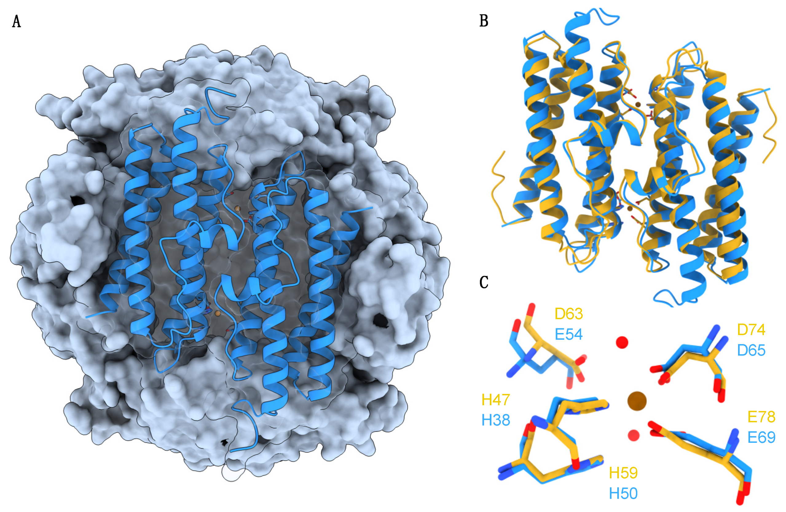
2.1. Binding of Cupric Ions to Dps

Electron paramagnetic resonance (EPR) spectroscopy is often used to probe metal-binding sites in metalloproteins, provided the metal is in a paramagnetic oxidation state, such as Cu2+ ions. The spectra of Dps samples incubated with different amounts of Cu2+ (between 3 and 48 Cu2+/Dps dodecamer), presented in Figure 2A, display axial EPR signals with parallel and perpendicular features characteristic of Type II copper centers (T2Cu), typically resulting from square planar coordination by N or N/O ligands. Samples reacted with 3 to 12 Cu2+/Dps give EPR spectra with a single EPR signal, here termed Site I, with g = 2.361, A = 0.13 mT and g = 2.076 (Figure 2B, in red). With the addition of more than 12 Cu2+/Dps, EPR spectra explained with two sets of EPR parameters were detected. Spectral deconvolution (using Site I as reference spectrum) indicates that those can be described as a sum of Site I and a novel Site II signal, with spectroscopic parameters as follows: g = 2.310, A = 0.15 mT and g = 2.070 (Figure 2B, in blue). Both signals are, therefore, characterized by axial symmetry with copper hyperfine splitting in the parallel region. By global deconvolution of all titration spectra and calculation of the occupancy of each type of Cu2+ signal (Figure 2C), one can conclude that Site I exhibits a higher affinity towards Cu2+ ions, saturating at 12 Cu2+/Dps. This can be interpreted as the occupation of the FOC binding site present in each protein subunit (in a total of 12 FOCs per protein dodecamer), constituted by amino acid side chains compatible with T2Cu EPR signals (histidine, glutamate and aspartate). Furthermore, the occupation of Site II follows a sigmoidal profile, being occupied only after Site I becomes saturated (from 12 up to 48 Cu2+/Dps), which is consistent with the occupation of a second, weaker binding site. The Site II signal is also less defined and broader, which may be explained by Cu2+ ions binding to the electronegative regions in the tri-fold pores and/or intra-protein channels. As such, the EPR data suggest that the Cu2+ ions bind in a sequential manner to at least two distinct sites, the first producing a highly stable species (most likely resulting from the binding of Cu to the FOCs).

2.2. Effect of Cu2+-Binding on the Iron Mineralization Rate by Dps

The iron oxidation and mineralization activity rates of apo-Dps and Cu2+-bound Dps (12 Cu2+ per Dps dodecamer) were compared using UV/visible spectrophotometry. The iron loading assays (from 48 to 384 Fe2+/Dps) were performed in atmospheric oxygen conditions to generate ferric species with a brownish color, characterized by a broad absorption band between 300 and 400 nm. The kinetic profiles obtained monitoring the absorbance at 340 nm after each iron addition (Figure 3) illustrate the effect of cupric ion binding on the iron oxidation mechanism catalyzed by Dps. Whilst the apo-Dps kinetic curves (Figure 3B) present the sigmoidal shape typical of the iron/O2 uptake reaction by Dps proteins, the curves of the 12 Cu2+-Dps samples (Figure 3A) show a pseudo-first-order kinetic behavior, with a much faster iron oxidation reaction rate. In the former, the initial slower phase shortens with increasing Fe2+ substrate concentration added to the protein due to the major contribution of the auto-catalytic iron oxidation activity of the mineral core built up inside the protein cavity.
The initial velocity, v0 as ∆Abs/min, of the iron oxidation reaction was determined by computing the slope of the tangent to the inception (t = 0) of each progress curve (see insets in Figure 3). The results obtained are presented in Table 1 and show up to ~10-fold increase on v0 when Dps was pre-loaded with 12 Cu2+/dodecamer (e.g., from 0.015 ∆Abs/min to 0.161 ∆Abs/min for 192 Fe/Dps). Overall, the results from this experiment demonstrate the functional impact of cupric ion binding to Dps—the initial rate of the iron oxidation reaction (ferroxidation and mineralization) in the presence of molecular oxygen (the least preferred co-substrate) is significantly increased, suggesting that Cu2+ participates in the redox chemistry steps of iron oxidation.

2.3. Characterization of the Anaerobic Iron Uptake Reaction in the Presence of Cu2+ Ions

To better understand the effect observed in the Cu2+-loaded Dps, protein samples of Dps pre-loaded with 12 Cu2+ eqs per dodecamer were incubated for 20 min with 12 Fe2+ eqs (1 Cu2+/1 Fe2+/FOC) in anaerobic conditions, without any additional co-substrate. Parallel samples were analyzed using EPR and Mössbauer spectroscopies (Figure 4 and Table 2).
The EPR spectrum of the initial protein sample, Cu2+-loaded Dps (Figure 4A), shows the previously observed characteristic axial EPR Cu2+ signal (as in Figure 2B). In turn, incubation of apo-Dps with 12 Fe2+/dodecamer (Figure 4E) generates a Mössbauer spectrum explained by the contributions of high-spin ferrous species (isomer shift, δ = 1.26 and 1.31 mm/s and quadrupole splitting, ∆EQ = 3.48 and 3.12 mm/s), as described in previous works [14,37].
Analysis of the spectra obtained after the addition of 12 Fe2+ eqs to 12 Cu2+-Dps samples (Figure 4B,F) suggests that the Cu2+ ions are directly responsible for oxidizing ferrous ions in a 1:1 redox reaction. In this case, the EPR spectrum reflects an identically shaped signal with a 65% loss in spin concentration, whilst the Mössbauer spectrum can be described as the sum of 34% of high-spin ferrous species (red sub-spectrum) and 66% of a new sub-spectrum with parameters characteristic of high-spin ferric species with oxygen/nitrogen ligands (δ = 0.47 mm/s, ∆EQ = 0.57, 1.00 mm/s). Together, these results can be interpreted as evidence for the concurrent oxidation of Fe2+ to Fe3+ coupled with the reduction of Cu2+ to Cu+, with the latter being diamagnetic and thus EPR silent. Please note that in these anoxic experimental conditions, no other oxidant species exist besides the cupric ions.

2.4. The Initial Cu2+ State of Copper Is Regenerated in the Presence of an Oxidant Co-Substrate

Since copper and iron co-occur in biotic environments, and considering the facultative anaerobe nature of M. nauticus (as well as other known Dps-expressing organisms) and its ability to degrade different types of hydrocarbons (including petroleum hydrocarbons), it is important to investigate the potential synergetic effect of both metals. The Mössbauer and EPR data shown above indicate that copper directly oxidizes Fe2+ ions in anaerobic conditions accepting one electron in a stoichiometric redox reaction. Additionally, the kinetic progress curves of 12 Cu2+-Dps samples reacted with Fe2+ ions (48 to 384 Fe/Dps) in atmospheric oxygen conditions demonstrate that the rate-enhancing effect is quantitative, indicating a catalytic role for copper ions. As such, and to clarify the oxidation state of both copper and iron ions during the aerobic iron oxidation reaction, parallel EPR and Mössbauer samples were prepared in different conditions (Figure 4). As discussed in the previous section, the anaerobic incubation of the 12 Cu2+-Dps sample with ferrous ions leads to the loss of the Cu2+-Dps EPR signal with the concomitant stoichiometric appearance of ferric species in the Mössbauer spectrum. However, if such incubation is conducted under aerobic conditions, no loss of the EPR signal is observed (Figure 4D). Furthermore, the addition of an excess of H2O2 after sequential incubation of 12 Cu2+-Dps with 12 Fe2+ in anaerobic conditions for 20 minutes (i.e., addition of an oxidative co-substrate after the 1:1 reaction shown in Figure 4B,F) results in the detection of an EPR signal corresponding to a fully oxidized 12 Cu2+-Dps sample as well as to the presence of 100% ferric species, as judged by Mössbauer spectroscopy (Figure 4C,G). Interestingly, the parameters obtained for the ferric species generated by the reaction with Cu2+ are consistent with the ferric parameters obtained after co-incubating Dps with 12 eqs of Fe2+ with a two-fold molar excess of H2O2 (Figure 4H) and with previous reports of the ferric mineral obtained using these co-substrates of the Dps iron mineralization reaction [14], indicating that the ferric product of the Cu2+:Fe2+ redox reaction in anaerobic conditions most likely shares a similar chemical composition/structure.
Altogether, our results point towards the cupric ion exerting a significant rate-enhancing effect on the catalytic activity of Dps, when compared with the reaction using molecular oxygen as a co-substrate, directly coupling the oxidation of ferrous ions with its reduction to Cu+. The observation that Cu+ can be re-oxidized to Cu2+ by either O2 or H2O2 further supports the catalytic role of copper ions in ferrous iron oxidation and concomitant co-substrate reduction.

3. Discussion

The bimodal cell protective effect accomplished by Dps proteins through ferrous iron scavenging and DNA shielding has been studied for over two decades. In general, the Dps catalytic activity (ferroxidation and mineralization) using H2O2 (preferably) or O2 co-substrate has been characterized since their discovery, with an alternative self-catalytic mineral core growth mechanism in the absence of any co-substrate being only recently described.
In this work, the functional properties of the Dps from M. nauticus were further explored whilst probing the interplay between the protein, iron, and cupric ions. Based on previous data from the literature, we postulated the hypothesis that the occupation of the Dps ferroxidase centers by non-iron biologically relevant transition metals, previously detected using several methods (spectroscopic, calorimetry methods or structure determination), arise from a biological advantage and do not only represent a putative metal binding capacity of the FOCs.
Copper complexes have been extensively studied using multiple spectroscopic techniques, specifically EPR, X-ray absorption and UV/Visible spectroscopies, among others [38,39,40]. In this study, EPR and Mössbauer spectroscopies were applied to detect and quantify the Cu-/Fe-containing species, as both techniques have proven to be important and complementary tools to characterize metalloproteins. While EPR active copper centers present characteristic EPR spectra (for example, type II centers show axial signals with hyperfine splitting in the parallel region), Mössbauer spectroscopy allows the identification of all iron species, regardless of their oxidation state and coordination geometry [41].
The Cu2+ titration experiment performed in this work using EPR spectroscopy (Figure 2) yielded the first direct evidence of the binding of cupric ions to the FOCs of a Dps protein apart from crystallographic information. The EPR spectra of Dps samples incubated with up to 12 eqs of Cu2+ per dodecamer show a single well defined axial signal characteristic of Type II Cu centers. The relatively high sharpness of this signal suggests that the chemical environment around the Cu2+ ions is homogenous, with a coordination sphere of N/O ligands, such as residues His38, His50, Glu54, Asp65 and Glu69 that constitute the FOC in the 12 subunits of the M. nauticus Dps dodecamer (as highlighted in Figure 1), in agreement with previous crystallographic evidence of such Cu2+-FOC centers in S. suis Dpr. After what we believe to be the saturation of the FOC binding sites, the Cu2+ ions most likely start to bind to other sites scattered along the N-terminal pores and/or intra-protein channels, resulting in a second, subsequent Type II Cu EPR broader signal, suggesting different coordination geometries and affinities. Such a type of binding is commonly observed in crystallographic structures of metal–Dps complexes [42,43].
Here, we described, for the first time, the rate-enhancing effect of Cu2+ binding to M. nauticus Dps in the iron uptake reaction, which resulted in up to ~10-fold faster initial velocity of iron oxidation in the presence of O2 (data presented in Figure 3 and Table 1). The impact of non-iron metal binding on the catalytic properties of the protein was described for other metal ions such as Zn2+, Mn2+ and Tb3+ but not Cu2+. In the cases of interactions of Zn2+ ions with the cyanobacterium Thermosynechococcus elongatus Dps A [31] and Mn2+ with the radiation-resistant bacterium Kineococcus radiotolerans Dps [27], the presence of these metal cations resulted in an iron oxidation rate-enhancing effect, especially in the presence of O2, with a ~20-fold faster rate (compared with apo-Dps incubated with iron) in the former. Strikingly, partial or complete inhibition of the iron oxidation reaction using spectrophotometry was detected when Listeria innocua Dps [32] and Nostoc punctiforme Dps4 [34] were previously incubated with Zn2+ and/or Tb3+, suggesting that the catalytic properties of metal–Dps complexes may depend on the type of divalent metal and/or specific protein residues.
Furthermore, this work probed a putative redox reaction between Cu2+ and Fe2+ ions in Dps by spectroscopically characterizing the iron oxidation reaction in anaerobic conditions with no additional co-substrate. Our results show that in these conditions, Cu2+ and Fe2+ participate in a 1:1 electron transfer process that concurrently generates oxidized Fe3+ species and reduced Cu+ ions (Figure 4 and Table 2). The latter may be able to reduce a substrate molecule, regenerating the initial Cu2+ state and thus allowing the next Cu–Fe redox turnover. In general terms, the 1:1 reaction detected in anaerobic conditions is most likely the mechanism behind the rate-enhancing effect detected in atmospheric oxygen conditions using spectrophotometry. The rate-enhancing catalytic effect of Cu2+ ions is independent of the amount of iron added since the reaction should now be controlled by the rate of Cu2+–Cu+ cycling.
Overall, the data presented in this work reveal a yet uncharacterized catalytic synergetic effect between iron and copper in the iron oxidation reaction by Dps, one of the two primary functions of these stress response proteins. The effect of Cu2+ ions on Dps catalysis may represent a response mechanism to multiple stressors in the presence of O2. Under these conditions, Dps shows atypical reactivity sequestering the ferrous iron more efficiently and decreasing cellular exposure to ROS formed by Fenton chemistry (since the rate of iron oxidation is significantly greater than the rate of the apo-form using O2 as co-substrate). Moreover, the ability of Cu2+-loaded Dps to directly oxidize Fe2+ ions in anoxic conditions without any additional co-substrate is of utmost interest and prompts additional research into the mechanistic properties of Dps catalysis in facultative aerobic bacteria such as M. nauticus and the possible significance of non-iron metal ions as regulators of the Dps function.

4. Materials and Methods

4.1. Production of M. nauticus Dps

The production and purification of M. nauticus Dps were carried out following the protocols described by Penas et al. [14].

4.2. Cu2+ Binding Assays by EPR Spectroscopy

A Cu2+ stock solution was prepared using >99% pure CuSO4⋅5H2O (Sigma-Aldrich, now MERCK, Darmstadt, Germany) and Milli-Q water at pH 2.0 and calibrated through the Zincon colorimetric assay [44]. Binding of cupric ions to Dps was monitored by X-band continuous-wave electron paramagnetic resonance (CW-EPR). Triplicate samples of 5–10 µM Dps (dodecamer) in 200 mM MOPS pH 6.0 and 200 mM NaCl buffer were supplemented with increasing concentrations of CuSO4 up to a molar ratio of 48 Cu2+/Dps and incubated for 10 min at room temperature. Samples were then transferred to 2 mm quartz EPR tubes (Wilmad, ATS Life Sciences Scientific Products, Warminster, PA, USA), flash-frozen in liquid nitrogen, and the spectra were acquired in a Miniscope MS400 spectrometer (Magnettech, now Bruker, Billerica, MA, USA) equipped with a cold finger. Spin quantitations (and corresponding Cu2+ concentrations) were calculated by double integration of signals obtained under non-saturating conditions and comparison with a Cu(II)-EDTA standard [45]. Occupancy of each binding site is defined herein as the molar ratio of cupric ions per Dps dodecamer. The data presented in Figure 2 correspond to the average value of the deconvolution of three independent assays, with error bars representing 95% confidence intervals.

4.3. Iron Uptake Kinetic Assays

Dps samples were prepared at a concentration of 0.6 µM (7.2 µM monomer) in the apo-form or pre-loaded with 12 Cu2+ ions per protein (7.2 µM CuSO4) in 200 mM MOPS pH 7.0, 200 mM NaCl buffer. The absorption spectra, between 250 and 800 nm, were registered using an Evo 300 UV/Vis Spectrophotometer (Thermo Fischer Scientific, Waltham, MA, USA) in a 1 cm pathlength quartz cell (Hellma GmbH & Co. KG, Müllheim, Germany) with constant stirring at room temperature. Then, different concentrations of Fe2+ ions were added (between 48 and 384 Fe/Dps corresponding to 28 to 230 µM FeSO4), initiating the reaction. Absorbance spectra were then acquired in 1 min steps, and the absorbance at 340 nm was plotted over time after baseline correction. Ferrous sulfate stock solutions were freshly prepared, and Fe2+ concentration was determined using the 1,10-phenantroline colorimetric assay, as described in [14]. The data presented in Figure 3 are representative of three independent kinetic assays. The initial velocity, v0 as ∆Abs/min, of the iron oxidation reaction was determined by computing the slope of the tangent to the inception (t = 0) of each progress curve, along with estimation of the standard error of the fit with a 95% confidence interval.

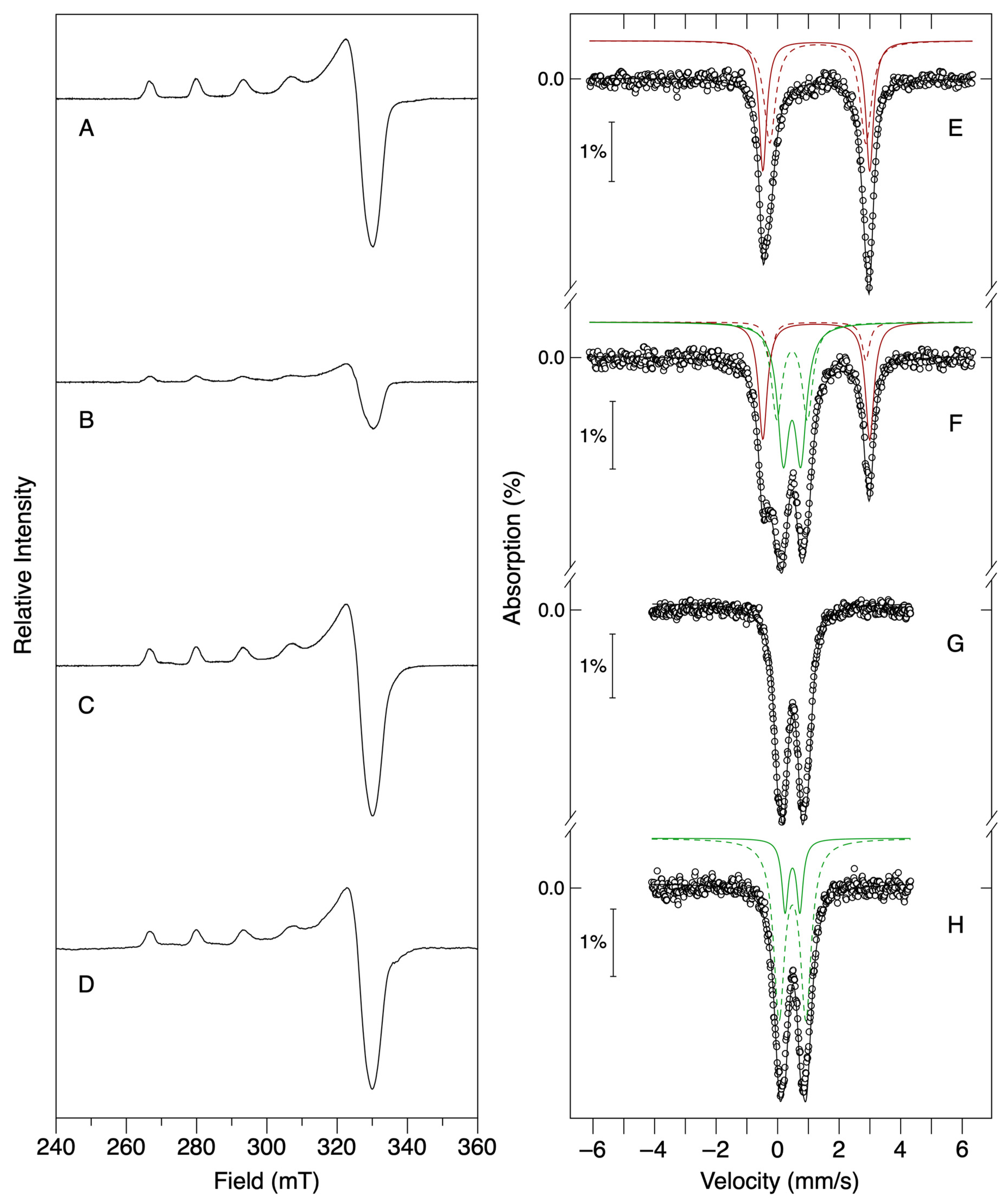
4.4. EPR and Mössbauer Spectroscopies Characterization

Mössbauer and EPR samples in anaerobic conditions were prepared inside an anaerobic chamber (<4 ppm O2; MBLab, MBraun, Garching, Germany). For Mössbauer analysis, a 57FeSO4 stock solution was prepared by dissolution of a 57Fe metal foil with 1 M H2SO4 followed by quantification of the Fe2+ concentration using the 1,10-phenantroline colorimetric assay [14]. Apo-Dps samples at 90 µM in 200 mM MOPS pH 7.0, 200 mM NaCl buffer were incubated anaerobically with 1.08 mM CuSO4 (12 Cu/Dps molar ratio) for 20 min at room temperature followed by the addition of 1.08 mM 57FeSO4 (12 Fe/Dps and 1 Fe/Cu) and left to react for another 20 min at room temperature. Then, 400 μL were transferred to a Mössbauer cell and flash-frozen in liquid nitrogen. At this stage, 2.16 mM of hydrogen peroxide was added (2-fold molar excess over iron) to a parallel sample and further incubated for 20 min before flash-freezing. Mössbauer spectra were recorded on a weak-field spectrometer operating in a constant acceleration mode in transmission geometry, equipped with a Janis Closed Cycle He gas refrigerator cryostat with temperature set at 77 K. The zero velocity of the Mössbauer spectrum is referred to as the centroid of the room-temperature spectrum of a metallic iron foil. Mössbauer analysis was conducted using the WMOSS program (SEE Co., Minneapolis, MN, USA). Parameters (isomer shift, δ, quadrupole splitting, ∆EQ and linewidths), average values, relative percentages and corresponding uncertainties of each iron species in each sample were obtained by spectral deconvolution through least-square fitting using WMOSS. EPR spectra were collected and analyzed as previously described. For the EPR samples subsequently reacted with O2, the anaerobic procedure was reproduced outside the glovebox in atmospheric oxygen conditions.

5. Conclusions

The multifaceted features of Dps proteins have gathered significant interest in bacterial protein biochemistry and bio-nanotechnology for more than two decades. Some works in recent years focused on the interaction between Dps proteins and non-iron metals of biological relevance. The influence of divalent transition metal ions (such as Zn2+, Co2+, Cd2+) regarding protein structural dynamics has been implicated in protein oligomerization and DNA-binding properties, which further the importance of probing possible roles for metal ions other than the ferrous/ferric ions.
This work pays attention to the hitherto lesser-explored interaction between Dps and cupric ions, one of the most relevant metals in biology, of particular importance in redox biochemistry. Our results further advance the recent thought that Dps proteins possibly use metal co-factors to regulate their activity by showing that Cu2+ ions occupy specific sites within the protein and enhance the rate of iron oxidation in aerobic conditions, directly serving as an electron acceptor for the iron oxidation in anoxic conditions with no additional co-substrate. These findings reveal a versatility towards ferrous ion oxidation and storage on par with the stated metabolic versatility of bacteria. In fact, M. nauticus can adapt to different environmental conditions. In the total absence of molecular oxygen, this bacterium obtains energy from nitrate respiration; however, in microaerophilic conditions, the oxidative phosphorylation terminal oxidase reduces molecular oxygen to hydrogen peroxide [46,47]. In these conditions, Dps can rapidly process both ferrous ions and hydrogen peroxide, providing protection and storage functionalities. In aerobic conditions, M. nauticus will shift to the use of a terminal oxidase capable of fully reducing molecular oxygen to water. Under these conditions, it is fair to assume that, with cupric ions available to bind to Dps, molecular oxygen would be more efficiently used as a co-substrate in the ferrous oxidation and storage reaction.

Author Contributions

Conceptualization, P.T. and A.S.P.; methodology, J.P.L.G., D.P., P.T. and A.S.P.; investigation and formal analysis, J.P.L.G., D.P., P.T. and A.S.P.; resources, P.T. and A.S.P.; writing—original draft preparation, J.P.L.G.; writing—review and editing, J.P.L.G., D.P., P.T. and A.S.P.; supervision, A.S.P. and P.T.; funding acquisition, P.T. and A.S.P. All authors have read and agreed to the published version of the manuscript.

Funding

This work was financed by national funds from the Fundação para a Ciência e a Tecnologia (FCT-MCTES), I.P., in the scope of the project UIDB/04378/2020 of the Research Unit on Applied Molecular Biosciences–UCIBIO and LA/P/0140/2020 of the Associate Laboratory Institute for Health and Bioeconomy–i4HB; grant PTDC/BIAPRO/111485/2009 (to P.T.) and PTDC/QUI/64248/2006 (to A.S.P). This work was also supported by the Radiation Biology and Biophysics Doctoral Training Program–RaBBiT, financed by FCT-MCTES, grants PD/00193/2012; UIDB/04378/2020 (UCIBIO) and UIDB/00068/2020 (CEFITEC). The RaBBiT Doctoral Training Program awarded fellowships to J.P.L.G. (PD/BD/135476/2017 and COVID/BD/152497/2022) and to D.P. (SFRH/BD/52535/2014).

Institutional Review Board Statement

Not applicable.

Informed Consent Statement

Not applicable.

Data Availability Statement

Not applicable.

Conflicts of Interest

The authors declare no conflict of interest.

References

  1. Crichton, R.R. An Overview of the Role of Metals in Biology. In Practical Approaches to Biological Inorganic Chemistry; Elsevier: Amsterdam, The Netherlands, 2020; pp. 1–16. ISBN 978-0-444-64225-7. [Google Scholar]
  2. Kruk, J.; Aboul-Enein, H.Y.; Kładna, A.; Bowser, J.E. Oxidative Stress in Biological Systems and Its Relation with Pathophysiological Functions: The Effect of Physical Activity on Cellular Redox Homeostasis. Free Radic. Res. 2019, 53, 497–521. [Google Scholar] [CrossRef]
  3. Andrews, S.C. Iron Storage in Bacteria. Adv. Microb. Physiol. 1998, 40, 281–351. [Google Scholar] [CrossRef]
  4. Andrews, S.C.; Robinson, A.K.; Rodríguez-Quiñones, F. Bacterial Iron Homeostasis. FEMS Microbiol. Rev. 2003, 27, 215–237. [Google Scholar] [CrossRef] [PubMed] [Green Version]
  5. Andrews, S.C. The Ferritin-like Superfamily: Evolution of the Biological Iron Storeman from a Rubrerythrin-like Ancestor. Biochim. Biophys. Acta-Gen. Subj. 2010, 1800, 691–705. [Google Scholar] [CrossRef] [PubMed]
  6. Guerra, J.P.L.; Jacinto, J.P.; Tavares, P. Miniferritins: Small Multifunctional Protein Cages. Coord. Chem. Rev. 2021, 449, 214187. [Google Scholar] [CrossRef]
  7. Chiancone, E.; Ceci, P. Role of Dps (DNA-Binding Proteins from Starved Cells) Aggregation on DNA. Front. Biosci. 2010, 15, 122. [Google Scholar] [CrossRef] [Green Version]
  8. Chesnokov, Y.; Kamyshinsky, R.; Mozhaev, A.; Shtykova, E.; Vasiliev, A.; Orlov, I.; Dadinova, L. Morphological Diversity of Dps Complex with Genomic DNA. Int. J. Mol. Sci. 2023, 24, 8534. [Google Scholar] [CrossRef]
  9. Dubrovin, E.V.; Dadinova, L.A.; Petoukhov, M.V.; Soshinskaya, E.Y.; Mozhaev, A.A.; Klinov, D.V.; Schäffer, T.E.; Shtykova, E.V.; Batishchev, O.V. Spatial Organization of Dps and DNA–Dps Complexes. J. Mol. Biol. 2021, 433, 166930. [Google Scholar] [CrossRef]
  10. Williams, S.M.; Chatterji, D. An Overview of Dps: Dual Acting Nanovehicles in Prokaryotes with DNA Binding and Ferroxidation Properties. In Subcellular Biochemistry; Harris, J.R., Marles-Wright, J., Eds.; Subcellular Biochemistry; Springer International Publishing: Cham, Switzerland, 2021; Volume 96, pp. 177–216. ISBN 978-3-030-28150-2. [Google Scholar]
  11. Gupta, A.; Joshi, A.; Arora, K.; Mukhopadhyay, S.; Guptasarma, P. The Bacterial Nucleoid-Associated Proteins, HU and Dps, Condense DNA into Context-Dependent Biphasic or Multiphasic Complex Coacervates. J. Biol. Chem. 2023, 299, 104637. [Google Scholar] [CrossRef]
  12. Park, J.H.; Lee, E.S.; Jung, Y.J. Functional Characterization of the DNA-Binding Protein from Starved Cells (DPS) as a Molecular Chaperone under Heat Stress. Biochem. Biophys. Res. Commun. 2023, 667, 180–185. [Google Scholar] [CrossRef]
  13. Tindall, B.J. Marinobacter nauticus (Baumann et al. 1972) Comb. Nov. Arising from Instances of Synonymy and the Incorrect Interpretation of the International Code of Nomenclature of Prokaryotes. Arch. Microbiol. 2020, 202, 657–663. [Google Scholar] [CrossRef] [PubMed]
  14. Penas, D.; Pereira, A.S.; Tavares, P. Direct Evidence for Ferrous Ion Oxidation and Incorporation in the Absence of Oxidants by Dps from Marinobacter hydrocarbonoclasticus. Angew. Chem. Int. Ed. 2019, 58, 1013–1018. [Google Scholar] [CrossRef] [PubMed]
  15. Gauthier, M.J.; Lafay, B.; Christen, R.; Fernandez, L.; Acquaviva, M.; Bonin, P.; Bertrand, J.-C. Marinobacter hydrocarbonoclasticus Gen. Nov., Sp. Nov., a New, Extremely Halotolerant, Hydrocarbon-Degrading Marine Bacterium. Int. J. Syst. Bacteriol. 1992, 42, 568–576. [Google Scholar] [CrossRef] [PubMed] [Green Version]
  16. Handley, K.M.; Lloyd, J.R. Biogeochemical Implications of the Ubiquitous Colonization of Marine Habitats and Redox Gradients by Marinobacter Species. Front. Microbiol. 2013, 4, 136. [Google Scholar] [CrossRef] [Green Version]
  17. Grant, R.A.; Filman, D.J.; Finkel, S.E.; Kolter, R.; Hogle, J.M. The Crystal Structure of Dps, a Ferritin Homolog that Binds and Protects DNA. Nat. Struct. Biol. 1998, 5, 294–303. [Google Scholar] [CrossRef]
  18. Ilari, A.; Stefanini, S.; Chiancone, E.; Tsernoglou, D. The Dodecameric Ferritin from Listeria innocua Contains a Novel Intersubunit Iron-Binding Site. Nat. Struct. Biol. 2000, 7, 38–43. [Google Scholar] [CrossRef]
  19. Zhao, G.; Ceci, P.; Ilari, A.; Giangiacomo, L.; Laue, T.M.; Chiancone, E.; Chasteen, N.D. Iron and Hydrogen Peroxide Detoxification Properties of DNA-Binding Protein from Starved Cells. J. Biol. Chem. 2002, 277, 27689–27696. [Google Scholar] [CrossRef] [Green Version]
  20. Yang, X.; Chiancone, E.; Stefanini, S.; Ilari, A.; Chasteen, N.D. Iron Oxidation and Hydrolysis Reactions of a Novel Ferritin from Listeria innocua. Biochem. J. 2000, 349, 783–786. [Google Scholar] [CrossRef] [Green Version]
  21. Su, M.; Cavallo, S.; Stefanini, S.; Chiancone, E.; Chasteen, N.D. The So-Called Listeria innocua Ferritin Is a Dps Protein. Iron Incorporation, Detoxification, and DNA Protection Properties. Biochemistry 2005, 44, 5572–5578. [Google Scholar] [CrossRef]
  22. Pesek, J.; Büchler, R.; Albrecht, R.; Boland, W.; Zeth, K. Structure and Mechanism of Iron Translocation by a Dps Protein from Microbacterium arborescens. J. Biol. Chem. 2011, 286, 34872–34882. [Google Scholar] [CrossRef] [Green Version]
  23. Zeth, K.; Hoiczyk, E.; Okuda, M. Ferroxidase-Mediated Iron Oxide Biomineralization: Novel Pathways to Multifunctional Nanoparticles. Trends Biochem. Sci. 2016, 41, 190–203. [Google Scholar] [CrossRef] [PubMed]
  24. Bozzi, M.; Mignogna, G.; Stefanini, S.; Barra, D.; Longhi, C.; Valenti, P.; Chiancone, E. A Novel Non-Heme Iron-Binding Ferritin Related to the DNA-Binding Proteins of the Dps Family in Listeria innocua. J. Biol. Chem. 1997, 272, 3259–3265. [Google Scholar] [CrossRef] [PubMed] [Green Version]
  25. Guerra, J.P.L.; Blanchet, C.E.; Vieira, B.J.C.; Waerenborgh, J.C.; Jones, N.C.; Hoffmann, S.V.; Pereira, A.S.; Tavares, P. Controlled Modulation of the Dynamics of the Deinococcus grandis Dps N-terminal Tails by Divalent Metals. Protein Sci. 2023, 32, e4567. [Google Scholar] [CrossRef] [PubMed]
  26. Bae, M.K.; Shin, E.; Lee, S.-J. Complementary Roles of Two DNA Protection Proteins from Deinococcus geothermalis. Int. J. Mol. Sci. 2023, 24, 469. [Google Scholar] [CrossRef] [PubMed]
  27. Ardini, M.; Fiorillo, A.; Fittipaldi, M.; Stefanini, S.; Gatteschi, D.; Ilari, A.; Chiancone, E. Kineococcus radiotolerans Dps Forms a Heteronuclear Mn–Fe Ferroxidase Center That May Explain the Mn-Dependent Protection against Oxidative Stress. Biochim. Biophys. Acta BBA Gen. Subj. 2013, 1830, 3745–3755. [Google Scholar] [CrossRef]
  28. Hayden, J.A.; Hendrich, M.P. EPR Spectroscopy and Catalase Activity of Manganese-Bound DNA-Binding Protein from Nutrient Starved Cells. J. Biol. Inorg. Chem. 2010, 15, 729–736. [Google Scholar] [CrossRef] [PubMed]
  29. Haikarainen, T.; Tsou, C.-C.; Wu, J.-J.; Papageorgiou, A.C. Structural Characterization and Biological Implications of Di-Zinc Binding in the Ferroxidase Center of Streptococcus pyogenes Dpr. Biochem. Biophys. Res. Commun. 2010, 398, 361–365. [Google Scholar] [CrossRef] [PubMed]
  30. Haikarainen, T.; Thanassoulas, A.; Stavros, P.; Nounesis, G.; Haataja, S.; Papageorgiou, A.C.; Haikarainen, T.; Stavros, P.; Papageorgiou, A.C.; Thanassoulas, A. Structural and Thermodynamic Characterization of Metal Ion Binding in Streptococcus suis Dpr. J. Mol. Biol. 2011, 405, 448–460. [Google Scholar] [CrossRef]
  31. Alaleona, F.; Franceschini, S.; Ceci, P.; Ilari, A.; Chiancone, E. Thermosynechoccus elongatus DpsA Binds Zn(II) at a Unique Three Histidine-Containing Ferroxidase Center and Utilizes O2 as Iron Oxidant with Very High Efficiency, Unlike the Typical Dps Proteins. FEBS J. 2010, 277, 903–917. [Google Scholar] [CrossRef]
  32. Stefanini, S.; Cavallo, S.; Montagnini, B.; Chiancone, E. Incorporation of Iron by the Unusual Dodecameric Ferritin from Listeria innocua. Biochem. J. 1999, 338, 71–75. [Google Scholar] [CrossRef]
  33. Havukainen, H.; Haataja, S.; Kauko, A.; Pulliainen, A.T.; Salminen, A.; Haikarainen, T.; Finne, J.; Papageorgiou, A.C. Structural Basis of the Zinc- and Terbium-Mediated Inhibition of Ferroxidase Activity in Dps Ferritin-like Proteins. Protein Sci. 2008, 17, 1513–1521. [Google Scholar] [CrossRef] [PubMed] [Green Version]
  34. Howe, C.; Moparthi, V.K.; Ho, F.M.; Persson, K.; Stensjö, K. The Dps4 from Nostoc punctiforme ATCC 29133 Is a Member of His-Type FOC Containing Dps Protein Class That Can Be Broadly Found among Cyanobacteria. PLoS ONE 2019, 14, e0218300. [Google Scholar] [CrossRef] [PubMed] [Green Version]
  35. Festa, R.A.; Thiele, D.J. Copper: An Essential Metal in Biology. Curr. Biol. 2011, 21, R877–R883. [Google Scholar] [CrossRef] [PubMed] [Green Version]
  36. Biasini, M.; Bienert, S.; Waterhouse, A.; Arnold, K.; Studer, G.; Schmidt, T.; Kiefer, F.; Cassarino, T.G.; Bertoni, M.; Bordoli, L.; et al. SWISS-MODEL: Modelling Protein Tertiary and Quaternary Structure Using Evolutionary Information. Nucleic Acids Res. 2014, 42, 252–258. [Google Scholar] [CrossRef]
  37. Guerra, J.P.L.; Blanchet, C.E.; Vieira, B.J.C.; Almeida, A.V.; Waerenborgh, J.C.; Jones, N.C.; Hoffmann, S.V.; Tavares, P.; Pereira, A.S. The Conformation of the N-Terminal Tails of Deinococcus grandis Dps Is Modulated by the Ionic Strength. Int. J. Mol. Sci. 2022, 23, 4871. [Google Scholar] [CrossRef]
  38. Elwell, C.E.; Gagnon, N.L.; Neisen, B.D.; Dhar, D.; Spaeth, A.D.; Yee, G.M.; Tolman, W.B. Copper–Oxygen Complexes Revisited: Structures, Spectroscopy, and Reactivity. Chem. Rev. 2017, 117, 2059–2107. [Google Scholar] [CrossRef] [Green Version]
  39. Solomon, E.I.; Heppner, D.E.; Johnston, E.M.; Ginsbach, J.W.; Cirera, J.; Qayyum, M.; Kieber-Emmons, M.T.; Kjaergaard, C.H.; Hadt, R.G.; Tian, L. Copper Active Sites in Biology. Chem. Rev. 2014, 114, 3659–3853. [Google Scholar] [CrossRef] [Green Version]
  40. Yalovega, G.E.; Myasoedova, T.N.; Shmatko, V.A.; Brzhezinskaya, M.M.; Popov, Y.V. Influence of Cu/Sn Mixture on the Shape and Structure of Crystallites in Copper-Containing Films: Morphological and X-Ray Spectroscopy Studies. Appl. Surf. Sci. 2016, 372, 93–99. [Google Scholar] [CrossRef]
  41. Waerenborgh, J.C.; Tavares, P.; Pereira, A.S. Mössbauer Spectroscopy. In Radiation in Bioanalysis; Pereira, A.S., Tavares, P., Limão-Vieira, P., Eds.; Bioanalysis; Springer International Publishing: Cham, Switzerland, 2019; Volume 8, pp. 213–244. ISBN 978-3-030-28246-2. [Google Scholar]
  42. Zeth, K.; Sancho-Vaello, E.; Okuda, M. Metal Positions and Translocation Pathways of the Dodecameric Ferritin-like Protein Dps. Inorg. Chem. 2019, 58, 11351–11363. [Google Scholar] [CrossRef]
  43. Zeth, K.; Pretre, G.; Okuda, M. Time-Resolved Studies of Ytterbium Distribution at Interfacial Surfaces of Ferritin-like Dps Protein Demonstrate Metal Uptake and Storage Pathways. Biomedicines 2021, 9, 914. [Google Scholar] [CrossRef]
  44. Arnaud, S.; Malatesta, F.; Guigliarelli, B.; Gayda, J.-P.; Bertrand, P.; Miraglio, R.; Denis, M. Purification and Characterization of the Oxidase from the Marine Bacterium Pseudomonas nautica 617. Eur. J. Biochem. 1991, 198, 349–356. [Google Scholar] [CrossRef] [PubMed]
  45. Denis, M.; Arnaud, S.; Malatesta, F. Hydrogen Peroxide Is the End Product of Oxygen Reduction by the Terminal Oxidase in the Marine Bacterium Pseudomonas nautica 617. FEBS Lett. 1989, 247, 475–479. [Google Scholar] [CrossRef] [Green Version]
  46. Säbel, C.E.; Neureuther, J.M.; Siemann, S. A Spectrophotometric Method for the Determination of Zinc, Copper, and Cobalt Ions in Metalloproteins Using Zincon. Anal. Biochem. 2010, 397, 218–226. [Google Scholar] [CrossRef] [PubMed]
  47. Aasa, R.; Vänngård, T. EPR Signal Intensity and Powder Shapes: A Reexamination. J. Magn. Reson. 1969 1975, 19, 308–315. [Google Scholar] [CrossRef]
Figure 1. Structural illustration of the Dps from M. nauticus. (A) Macromolecular structural model of the Dps cube-like dodecamer assembly (light blue surface) with one of its facets (dimer) shown as blue ribbons. (B) The antisymmetric dimer of M. nauticus Dps (in blue) and Streptococcus suis Dpr (in yellow). The residues that compose the two ferroxidase centers (FOCs) formed at the dimer interface are shown as sticks, together with brown spheres representing Cu2+ ions. (C) Magnification of the residues that create the FOCs in M. nauticus Dps (carbon atoms in blue) and Dpr (carbon atoms in yellow). The Cu2+ ion is shown as a brown sphere. Structural waters are presented as red spheres. The M. nauticus Dps model was obtained using SWISS-MODEL [36] as a structure-homology modeling tool and the atomic structure of S. suis Dpr bound to Cu2+ ions as template (PDB ID 2XJN).
Ijms 24 10256 g001
Figure 2. Spectroscopic characterization of the Cu2+ binding sites in Dps by EPR spectroscopy. (A) X-band spectra at 77 K of Dps samples incubated with 3 to 48 eqs of Cu2+ per Dps dodecamer. Other experimental conditions: microwave frequency, 9.45 MHz; microwave power, 20.0 mW; modulation amplitude, 0.1 mT; receiver gain, 5 × 10². (B) Deconvolution of the spectra shown in (A) into two Cu signals (Site I in red and Site II in blue). (C) Contribution of each Cu signal in each spectrum obtained by titrating the protein with Cu2+ (up to 48 Cu2+/Dps) after spectral deconvolution (Site I in red and Site II in blue). Occupancy of each binding site is defined herein as the molar ratio of cupric ions per Dps dodecamer.
Ijms 24 10256 g002
Figure 3. Monitoring the iron oxidation reaction by apo- and 12 Cu2+-loaded Dps. Kinetic profiles obtained upon addition of different amounts of Fe2+/Dps in 200 mM MOPS pH 7.0, 200 mM NaCl in atmospheric oxygen conditions. Addition of 48 to 384 Fe/Dps (from bottom to top traces) to (A) Dps samples pre-loaded with 12 Cu2+ eqs per dodecamer and (B) apo-Dps samples. The insets are magnifications of the first 5 data points used to determine the initial velocities as per Table 1. A representative result of three independent assays is shown. Dotted lines were included as visual aids to data trends.
Ijms 24 10256 g003
Figure 4. Spectroscopic characterization of Dps samples probing the redox reaction between Cu2+ and Fe2+ ions within the protein. Left Panel: EPR spectra recorded at 77 K. Other experimental conditions: microwave frequency, 9.45 MHz; microwave power, 20.0 mW; modulation amplitude, 0.1 mT; receiver gain, 5 × 10². Right panel: Mössbauer spectra recorded at 77 K in the absence of an external applied field. (A) Cu2+-loaded Dps sample (12 eqs. of Cu2+ per Dps dodecamer); (B,F) Cu2+-loaded Dps sample, after reaction with 12 eqs of Fe2+ for 20 min in anaerobic conditions. (C,G) Addition of a 2-fold molar excess of H2O2 to the 12 Cu2+-loaded Dps sample after incubation with 12 Fe2+ per dodecamer. (D) Addition of 12 Fe2+ to the 12 Cu2+-loaded protein sample in atmospheric oxygen conditions. (E) Dps incubated with 12 eqs of Fe2+ per dodecamer; (H) Dps incubated with 12 eqs of Fe2+ and 2-fold molar excess of H2O2. The solid lines overlaying the experimental spectra are the result of least squares fits to the data (relevant parameters listed in Table 2). Contributions of ferrous species shown in red (species I and II in solid and dashed lines, respectively) and ferric species shown in green (species III and IV in solid and dashed lines).
Ijms 24 10256 g004
Table 1. Comparison of the initial velocity of iron oxidation between apo-Dps and 12 Cu2+-Dps samples upon addition of different concentrations of Fe2+ ions.
[Fe2+]/[Dps]Initial Velocity (∆Abs/min)
Apo-Dps12 Cu2+-Dps
480.003 ± 0.0010.031 ± 0.002
960.006 ± 0.0020.074 ± 0.002
1920.015 ± 0.0030.161 ± 0.005
2880.028 ± 0.0030.231 ± 0.009
3840.064 ± 0.0040.290 ± 0.012
Table 2. Mössbauer parameters of the iron species detected during the iron oxidation reaction in the presence of cupric ions by M. nauticus Dps.
Iron SpeciesMössbauer ParametersAbsorption (%)
δ (mm/s)EQ (mm/s)Linewidth (mm/s)12 57Fe2+12 57Fe2+ + 12 Cu2+12 57Fe2+ + 12 Cu2+
+ 24 H2O2
Ferrous species
I1.26 ± 0.02 3.48 ± 0.02 0.30 ± 0.02 46 ± 222 ± 3
II1.30 ± 0.02 3.12 ± 0.030.38 ± 0.03 54 ± 213 ± 3
Ferric species
III0.48 ± 0.020.61 ± 0.020.38 ± 0.0344 ± 362 ± 2
IV0.48 ± 0.021.01 ± 0.020.38 ± 0.0221 ± 338 ± 2
Note: Contribution of each ferrous and ferric species in each spectrum after deconvolution given as percentages.
Disclaimer/Publisher’s Note: The statements, opinions and data contained in all publications are solely those of the individual author(s) and contributor(s) and not of MDPI and/or the editor(s). MDPI and/or the editor(s) disclaim responsibility for any injury to people or property resulting from any ideas, methods, instructions or products referred to in the content.

