Review

The Diagnostic Potential of the Human Blood Microbiome: Are We Dreaming or Awake?

by
Francesca Sciarra
,
Edoardo Franceschini
,
Federica Campolo
and
Mary Anna Venneri
*
Department of Experimental Medicine, Sapienza University of Rome, 00185 Rome, Italy
*
Author to whom correspondence should be addressed.
Int. J. Mol. Sci. 2023, 24(13), 10422; https://doi.org/10.3390/ijms241310422
Submission received: 3 May 2023 / Revised: 1 June 2023 / Accepted: 19 June 2023 / Published: 21 June 2023
(This article belongs to the Section Molecular Pathology, Diagnostics, and Therapeutics)

Abstract

:
Human blood has historically been considered a sterile environment. Recently, a thriving microbiome dominated by Firmicutes, Actinobacteria, Proteobacteria, and Bacteroidetes phyla was detected in healthy blood. The localization of these microbes is restricted to some blood cell populations, particularly the peripheral blood mononuclear cells and erythrocytes. It was hypothesized that the blood microbiome originates from the skin–oral–gut axis. In addition, many studies have evaluated the potential of blood microbiome dysbiosis as a prognostic marker in cardiovascular diseases, cirrhosis, severe liver fibrosis, severe acute pancreatitis, type 2 diabetes, and chronic kidney diseases. The present review aims to summarize current findings and most recent evidence in the field.

1. Introduction

Traditionally, some body compartments such as the upper female gynecological tract [1], brain [2], and blood [3] were considered microbiologically sterile in healthy conditions. However, several works pointed to how these and other body niches cannot be longer considered germ-free in healthy conditions due to the presence of commensal or native microbial flora (collectively named the microbiome) [3]. The existence of metabolically active bacteria in the blood was hypothesized for the first time 50 years ago following the detection of increased absorption of amino acids and nucleosides in the erythrocyte suspension of healthy individuals [4]. Bacterial growth was also confirmed, following osmotic lysis and filtering, in healthy blood samples [5]. Furthermore, the advent of advanced analytical approaches, such as real-time PCR, allowed for the detection of ribosomal DNA in the same samples [6]. Moreover, the use of dark field microscopy techniques, fluorescent in situ hybridization, and flow cytometry led to the identification of pleomorphic bacteria in the blood, capable of altering their characteristics in response to environmental conditions [7]. Moreover, the analysis of blood microbiome morphology in peripheral blood mononuclear cells (PBMCs) using light and electron microscopy highlighted how the blood microbiome displays a complex life cycle, undergoing different morphological transformations, such as vesiculation, tabulation, budding, irregular binary fission, and protrusion–extrusion of progeny cells from large electron-dense bodies [8]. Recently, several studies have managed to increase the “power” of analysis due to a lower abundance of bacterial DNA, investigating healthy blood microbial taxonomy, identifying the most abundant phyla, and sometimes also bacterial genera [9] using 16 s rRNA gene sequencing technology [10,11]. In some blood compartments (red/white blood cells, extracellular vesicles) specific bacteria were detected. Their cellular localization (intracellular, membrane-bound) was linked to specific bacterial behavior (intracellular parasite, commensal), which in turn can depend on environmental conditions [12].
Moreover, blood microbiome sequencing studies highlighted how bacterial abundance differs among European countries. Bacterial DNA abundance distribution was differentially detected according to geographical origin: Germany and Poland show significantly higher abundance values; Italy, Greece, and Finland present intermediate levels, while Belgium and Austria are characterized by lower blood bacterial DNA levels. The main differences among countries were observed in male subjects, together with no age-related differences in bacterial DNA amount detected. This phenomenon could be at least in part attributable to environmental, behavioral, and cultural factors and social habits [13]. Some microbiome-characteristic variations were also correlated with host genetic and innate/adaptive immunity differences, while in other cases, cultural/behavioral features such as diet, hygienic conditions, parasitic load, and environmental exposure appear to have major weight compared to genetic factors [14]. Moreover, an age-based microbiome property difference was not highlighted, suggesting how the environment could have a greater contribution than age [15] even if an association between the blood microbiome and older age cannot be entirely excluded. Indeed, intestinal dysbiosis, malabsorption, and immune response dysregulation are common features of older subjects and lead to chronic systemic inflammatory states [16].
Even if origin and difference in the microbiome have been highlighted, many questions still remain unanswered, the first of which regards the blood microbiome’s source. It is unclear if it comes from other compartments, such as the gut [17], or if it is native to the blood environment [18]. Although members of the microbiota are often referred to as commensals, the symbiosis—persistent interaction—between the microbiota and its mammalian host encompasses various forms of relationship including mutualistic, parasitic, or commensal.
Gut microbiome–host interaction can be highly contextual, with the same microbe developing as a mutualist or parasite according to the nutritional, coinfection, or genetic landscape of its host. Many studies have examined the correlation between the gut microbiota and the immune system by analyzing its role in the development and promotion of autoimmune diseases and several cancer types [19,20].
Moreover, the persistence of blood microbes can lead to the development of many diseases including liver–pancreatic pathologies or cardiovascular and kidney diseases [21,22], although not always necessarily due to an immunomodulatory capacity of circulating commensals.
The bloodstream allows microbes to reach different body sites in healthy individuals; however, the low prevalence of such microbes in healthy blood underscores the transient and infrequent nature of this phenomenon [23].
The aim of this manuscript is to deeply review the relationship among bloodborne bacteria dysbiosis and several human disorders, highlighting the clinical relevance of changes in blood microbiome composition, and to unlock the potential microbiota-derived biomarkers for the diagnosis of a variety of diseases.

2. Blood Microbiome Composition

A healthy gut microbiome is mainly characterized by Firmicutes and Bacteroidetes phyla domination [24]. Knowledge on the healthy blood microbiome is still an under-evaluated topic, even though many research groups focus on this issue. Results from these analyses converge on a microbiome dominated by Firmicutes, Actinobacteria, Proteobacteria, and Bacteroidetes phyla (Table 1) [9,25], with less abundant Fusobacteriota [26,27]. Some genera belonging to the phylum Firmicutes were identified in blood: Bacillus, obliged aerobic bacteria widespread in nature; [9,26,28,29] Lactobacillus, facultative anaerobic able to convert lactose and other sugars in lactic acid by lactic fermentation; Streptococcus, facultative anaerobic able to produce hemolytic exotoxins such as streptolysin; and Romboutsia [30], a gut species isolated in humans [31]. Moreover, the Actinobacteria genus Corynebacterium, commensal human bacteria usually not pathogenic in healthy state; Proteobacteria genus Pseudomonas, bacteria with great metabolic diversity able to colonize a wide range of niches [9]; and Bacteroides, a Bacteroidota genus associated with the gut microbiome, able to process complex polysaccharides into simpler ones were detected in healthy blood [30,32]. The microbial DNA detected in healthy subjects comes from circulating commensals that have asymptomatic coexistence with the host and display immunomodulatory phenotypes. The presence or absence of immunomodulatory phenotypes of the microbiome determines whether an individual with bacteremia is asymptomatic or septic [23].

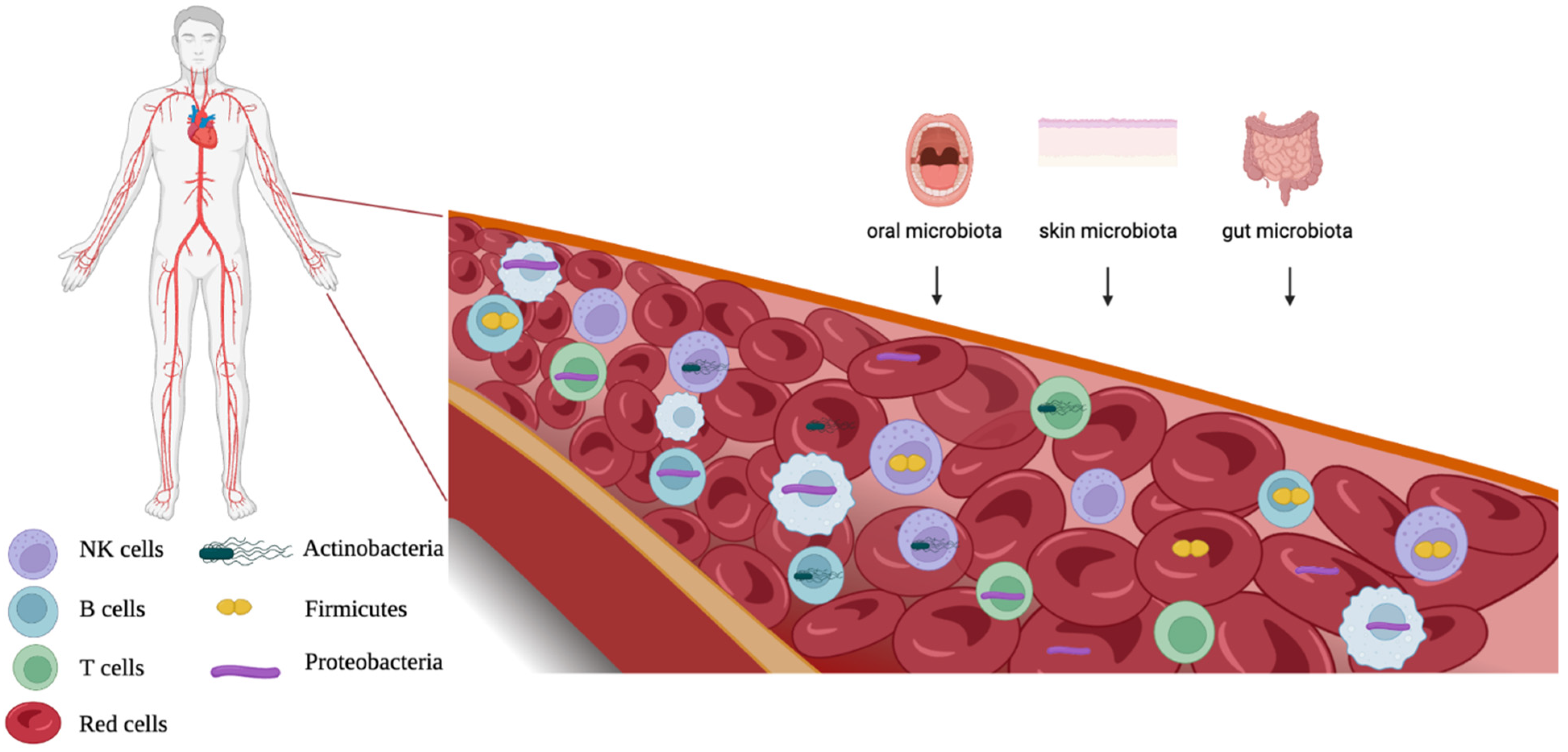
3. Blood Microbiome Origins

Many hypotheses have been postulated regarding the human blood microbiome’s origin, making this issue a question still unsolved. A recent population study rejected the hypothesis of an endogenous blood microbiome, rather supporting the transient and sporadic translocation of commensal microbes from other body sites into the bloodstream [23]. Other works have instead considered such translocations as a frequent event in healthy conditions, frequent enough to support the existence of a stable blood microbial microbiome [37]. In addition, some works have pointed to a bacterial contamination from the skin, laboratory environments, or other environmental sources, discrediting the idea of an endogenous blood microbiome [26,37]. It the origin of the healthy microbiome has also been hypothesized, and it appears that bloodborne microorganisms, especially bacteria, may have a maternal origin [38]. However, based on taxonomy detection, some suitable bacterial origin niches were identified. Several studies detected most of the blood microbiome in the gastrointestinal tract [39] even if it presents marked taxonomic differences compared to the gut microbiome [40]. Other sites of origin are the skin and oral mucosa, where blood bacterial phyla were detected [10,41]. The intestinal immune system is exposed to a number of foreign antigens, coming both from diet and from commensal or pathogenic bacteria presence; thus, the gut mucosa could represent a bacterial route toward the bloodstream. Different bacterial translocation mechanisms across the blood barrier have been hypothesized. One of them involves dendritic cells (DCs). The lamina propria (LP) mucosae contains various populations of DCs, such as CD103(+) and CX(3) CR1(−). DC antigen transport could represent a bacterial translocation way through the intestinal barrier [42]. In addition, other cell types could play a supporting role for intestinal DCs: small intestine mucus-secreting goblet cells (GCs) were found to be able to deliver small molecular antigens from intestinal lumen to CD103(+) DCs [43]. Moreover, intestinal microfold cells (M cells), specialized epithelial cells of the mucosa-associated lymphoid tissues, can sample various antigens and move them, therefore representing a pathway for the intestinal microbiome and antigens [44]. For skin and oral mucosa sources, bacterial translocation may occur when the blood barriers are blocked, as in the case of Porphyromonas gingivalis bacteremia [45].
Maternal origin is an alternative hypothesis to explain the source of the blood microbiome: the vertical bacterial transmission is indeed a phenomenon already known in animals for gut microbiome bacteria [46]. Microbiome presence was detected in various pre-natal tissues, such as umbilical cord blood [47], meconium [48], amnion [49], and in the placenta, which harbors a low-biomass microbiome [50]. All such evidence, although controversial, [1,51] could also point to theoretical maternal bacterial translocation in humans. The mechanisms by which maternal bacterial transfer would work are still unknown, even if some studies suggest a mixed origin, before and after birth. The initial microbial inoculum would be maternal, by oral and gut fetal compartment colonization, and the microbial microbiome would then be enriched during nursing [52]. Another hypothesized bacterial translation way could be the fetal ingestion of amniotic fluid during gestation [53]. Taking into account all these considerations, further investigation is needed to deepen understanding of the blood microbiome’s origin.

4. Blood Microbiome Localization

The healthy blood microbiome has been localized in many human blood sources: in the buffy coat (BC); inside peripheral blood mononuclear cells (PBMCs), such as commensal bacteria associated with red blood cells; and in red blood extracellular vehicles (EVs). The specific blood cell localization reflects bacterial behavior: an obliged intracellular parasite exploits internal cells [12], commensal species use tactics to stick to them [28], while opportunistic bacteria can change their habits depending on the environmental conditions [54]. Most bacterial DNA was detected in the blood buffy coat in healthy individuals [27]. In a significant percentage of healthy donors, PBMC phylum Chlamydiae species Chlamydophila pneumoniae, a human obligate intracellular parasite bacterium and etiologic agent of an atypical pneumonia form [55], was detected (Table 2). Chlamydiae are known for their unique biphasic life cycle, during which they alternate between two morphological forms: infectious extracellular elementary bodies (EBs) transform into metabolically active intracellular reticulate bodies (RBs) after entering the host cell [12].

4.1. Commensal Bacteria

In various blood compounds, phylum Firmicutes species Staphylococcus aureus, a broadly diffused commensal bacterium that can shift to opportunistic pathogen state depending on environmental conditions, was detected, representing one of the most common bacteremia causes, also infecting skin and soft-tissues [54] (Table 2). It represents a versatile bacterial species that can display a relevant number of strategies to exploit host cells [56], such as to being phagocytized by white blood cell neutrophils and macrophages. Some bacterial strains can survive inside white blood cells, deactivating their antibodies, and using them to facilitate bacterial dissemination to other compartments [57]. In addition, Staphylococcus aureus can also exploit red blood cells: it needs iron uptake from the host for its activity and uses complex strategies to gain it. The Staphylococcus aureus heme molecule uptake system uses highly specific cell surface receptors to extract iron ion from hemoglobin and hemoglobin–haptoglobin complexes, to transport it into the cytoplasm, and to degraded it as free iron [58,59] (Table 2). Another commensal phylum Firmicutes species, Streptococcus pneumoniae, manipulates red blood cells and uses them to evade innate immunity. The mechanism by which it works is still under investigation, but recent data point to surface protein PfbA’s (plasmin-fibronectin-binding protein A) role in erythrocytes binding due to its relevant affinity toward red blood cells and moderate affinity toward hemoglobin [60]. Streptococcus pneumoniae is involved in lobar pneumonia disease, giving rise to an inflammatory process in the alveolar spaces, with the exudation of a protein-rich liquid. This fluid works as a bacterial culture medium for dissemination, typically causing pneumonia [61].
Table 2. Localization of bacteria in the blood and identification of their behavior.
PhylaSpeciesLocalizationClinical EffectsBacterial BehaviorReferences
ChlamydiaeChlamydophila pneumoniaePBMCsEtiologic agent of an atypical pneumonia formObligate intracellular parasite [55]
[12]
[62]
FirmicutesStaphylococcus aureusWhite blood cellsCause bacteremiaCommensal/
opportunistic pathogen
[56]
[57]
Red blood cellsExtract iron ion from hemoglobin and hemoglobin–haptoglobin complexes [58]
Streptococcus pneumoniaeInvolved in lobar pneumonia disease onset [60]
[61]
Staphylococcus epidermidisPlasma and Red blood cellsInvolved in nosocomial infections of immunocompromised and transplanted patients [28]
[63]
ProteobacteriaSerratia sp. [29]
PseudomonadotaAcinetobacter baumannii [37]
Stenotrophomonas maltophyla
ActinobacteriaCutibacterium acnesBlood samplesCauses chronic blepharitis and endophthalmitis [28]

4.2. Opportunistic Pathogens

Staphylococcus epidermidis acts as a skin commensal bacterium in healthy conditions, but it can switch to opportunistic pathogenic behavior depending on environmental conditions, causing nosocomial infection to immunocompromised or transplanted patients. It was detected as being associated with the red blood cell fraction of healthy donors [28] (Table 2). Staphylococcus epidermidis has surface and extracellular matrix proteins that can bind blood cells. With the protein polysaccharide intercellular adhesin (PIA), it can create an extracellular material, a biofilm, which can also bind other bacteria and create a multilayer biofilm [63].
Moreover, phylum Actinobacteria species Cutibacterium acnes, a commensal anaerobic bacterium of the human skin microbiome [64], can switch its behavior as an opportunistic pathogen. Some Cutibacterium acnes strains produce several virulence factors involved in polysaccharidic biofilm production, adhesion to target cells, inflammation targeting by enzymatic proteins, and host tissue degradation [65]. Due to their ability to persist on medical devices, they were also involved in a wide range of nosocomial infections, and in chronic disease onset, such as endocarditis [64]. Cutibacterium acnes was detected in blood of healthy donors together with other opportunistic pathogenic bacteria such as Staphylococcus epidermidis [28].
Other environmental opportunistic human pathogens, known to cause nosocomial infections in immunocompromised patients, were also found to be associated with the healthy red blood cell fraction: among them, Proteobacteria genus Serratia [29] and Pseudomonadota species Stenotrophomonas malthophyla and Acinetobacter baumannii [37] (Table 2 and Figure 1). Recently, Proteobacteria, Firmicutes, and Actinobacteria phyla and Staphylococcus, Bacillus, Corynebacterium, Pseudomonas, Acinetobacter, Cutibacterium genera were detected in human femoral arteries [9] (Table 2). Moreover, many indirect clues to bacterial presence in red blood cells of healthy donors’ EVs suggest the presence of lipopolysaccharides (LPS) and outer membrane protein A (OmpA) [66]. In addition, EVs are also able to interact with monocytes, pointing to a microbial crossover among blood cells [66].

5. Clinical Relevance of Blood Microbiome Dysbiosis

Dysbiosis is a term that refers to a change in bacterial composition and metabolite production in microbial communities [67]. The use of microbial dysbiosis as a biomarker for human pathology has mainly been studied for the human gut microbiome [17]. Some recent discoveries, such as a significant positive association between blood microbial DNA copy number and important factors for health, such as glucose, insulin, free fatty acid (FFA) levels, and leukocyte count, led researchers to hypothesize the use of blood microbiome dysbiosis as a potential prognostic biomarker for several human pathologies [13]. However, it should be noticed that specific blood phyla in diseased patients are not indicative of bacterial origin site or of some specific causative role in pathology onset.

5.1. Blood Microbiome and Gastroenteropancreatic and Pulmonary Diseases

Different works have analyzed the blood microbiomes of patients affected by gastro-enteropancreatic diseases highlighting a dysbiosis status. Cirrhotic patients showed an increase in blood total bacterial DNA concentration and higher bacterial heterogeneity compared to healthy subjects [68,69]. Patients affected by severe liver fibrosis showed unique bacterial taxonomy and higher 16 rRNA concentration compared to healthy subjects [70] and showed a specific taxonomic composition for portal vein and central–peripheral venous blood and liver outflow [71]. In severe acute pancreatitis (SAP), patients’ microbial taxonomic diversity appears reduced compared to healthy subjects, with an increase in phyla Bacteroidetes and Firmicutes and a decrease in Actinobacteria (Table 2). Such microbial variations in SAP patients were associated with several immunological dysfunctions characterized by increased levels of serum cytokines, lymphocyte subgroup disruption, and neutrophil proteomic profile alteration [34]. The bacterial and viral microbiomes were identified in chronic obstructive pulmonary disease (COPD) and a relationship between the systemic microbial populations and lung disease was highlighted [72].

5.2. Blood Microbiome and Diabetes

Gut microbiome dysbiosis was associated with diabetes onset [36,73,74] and a link between gut bacterial dysbiosis and intestinal bacteria translocation to bloodstream was also proposed, as diabetic patients’ blood presented a higher rate of gut-derived microbiome than healthy controls [75]. Individuals with type 1 diabetes of long duration and with initial stages of diabetic nephropathy are characterized by aberrant profiles of gut microbiota and plasma metabolites [74]. A case–control study on insulin resistance syndrome highlighted how people predisposed to developing diabetes showed higher 16 S rDNA concentration in their blood compared to people not predisposed to developing the disease [76]. Other works did not detect bacterial dysbiosis in T2D patients, even though study participants who carried the Bacteroides genus in their blood were significantly associated with a lesser risk of developing T2D, while the Sediminibacterium genus bearers showed higher risk of becoming diabetics [36]. Thus, blood Bacteroides and Sediminibacterium could also be considered as T2D biomarkers in the future, although more study is needed.

5.3. Blood Microbiome and Acute Cardiovascular Disease Patients (CVD)

Acute cardiovascular disease (CVD) patients showed a relevant decrease in blood bacterial DNA compared to healthy subjects, and, in addition, patients developing cardiovascular complications showed a nonsignificant increase in Proteobacteria phyla. Proteobacteria augmentation could thus represent an independent biomarker of cardiovascular disease [21]. CVD patients’ blood dysbiotic state has been confirmed by other studies: CVD blood showed a dominance of Actinobacteria and Proteobacteria phyla compared to healthy blood [33]. In addition, an increased proportion of Proteobacteria and Actinobacteria was also detected during cardiac events, such as stroke, coronary heart disease, and myocardial infarction, confirming the proposed role of independent biomarker for Proteobacteria and Actinobacteria in CVD risk [25]. Proteobacteria species Escherichia coli, a common commensal bacterium harbored in the human gut, was detected in the blood of myocardial infarction (MI) coronary thrombosis patients [77]. In particular, the Escherichia coli LPS strain found in MI blood showed an association with several pathological factors involved in MI disease: low-grade endotoxemia, which triggers an inflammatory state characterized by proinflammatory cytokine release; and with soluble P-selectin protein, a marker of platelet activation [78,79]. In addition, Escherichia coli LPS strain also showed significant association with zonulin, a protein marker of gut permeability, also pointing to a factor involved in the bacterial translational pathway from the gut to blood circulation [78]. The role of Proteobacteria phyla in the pathogenesis of cardiovascular diseases was explored, detecting a link between LPS, a relevant Proteobacteria component, and CVD onset [25]. LPS can trigger inflammation directly, by its microbe-associated molecular patterns, or indirectly, inducing the generation of nonmicrobial danger-associated molecular patterns. Translocation of LPS in blood causes endotoxemia, and chronic endotoxemia is in turn involved in the pathogenesis of inflammation-driven conditions such as cardiometabolic disorders (obesity, liver diseases, atherosclerotic cardiovascular diseases) [80]. Moreover, the association between blood bacterial DNA and the risk of cardiovascular mortality was analyzed, detecting three bacterial genera significantly associated with it. Phylum Actinobacteria genus Kokuria and Pseudomonadota genus Enhydrobacter, skin and oral bacteria, were identified as being directly associated with CVD mortality risk, while Pseudomonadota genus Paracoccus was found to be inversely correlated (Table 2) [81]. Such findings highlight a role for blood microbiome dysbiosis in cardiovascular diseases, even though it should be considered that a clinical association between CVD and blood dysbiosis is still inconsistent, and more studies are needed to deepen knowledge on a theoretical causative role of bacteria in cardiovascular diseases.

5.4. Microbiome and Chronic Kidney Disease Patients (CKD)

A pilot study examined circulating microbiome profiles of chronic kidney disease (CKD) patients and compared them with healthy controls. A significant reduction in bacterial diversity in CKD blood and an augmentation of phylum Proteobacteria (Enterobacteridaceae and Pseudomonadaceae families) [22] (Table 2) were detected. The Proteobacteria phylum is a relevant factor in CKD pathogenesis due to its bacterial LPS. High LPS levels in the blood, together with gut-derived uremic toxins and immune deregulation, played a critical role both in CKD and CKD-associated complications [82]. Other studies confirmed CKD blood dysbiosis by the detection of lower bacterial heterogeneity and significant taxonomic variations [83]. In addition, a pilot study explored the link between the gut and blood microbiome in CKD patients with or without vascular calcification (VC), detecting significant changes in specific gut and blood phyla [84].

5.5. Microbiome and Cancer

Unique blood microbiome profiles have recently been identified in several cancer types and whole genome transcriptome studies have shown how unmapped parts of the reads can be frequently assigned to microorganisms, such as bacteria, archaea, and also viruses [85]. Despite intratumor microbial signatures being detected as effective in normal tissue discrimination from tumors, they did not correlate with cancer stages in most of the cancer types [85]. Instead, blood microbial profiles of cancer patients were sensitive enough to detect different types of cancer, even for very early-stage cancers. Such findings point to a blood/intratumor microbiome-based diagnostic method, which could be helpful in the screening and early diagnosis of cancer [18]. In addition, blood bacterial genetics showed potential regarding cancer treatment response prediction: in advanced colorectal cancer, baseline blood microbiota composition was identified as being significantly different among immunochemotherapy responders and non-responders [86].
Relevant findings were also gained studying the potential impact of the blood microbiome on immunotherapy efficacy [86]. Circulating microbiome effect on immunotherapy efficacy was checked on advanced colon/colorectal cancer (CRC) patients subjected to adoptive cellular therapy (ACT) with a mixed dendritic cell/cytokine-induced killer cell product (DC/CIK). The clinical benefits of DC/CIK infusion resulted in being influenced by blood microbiome diversity. DC/CIK responders, which had better immunotherapy impact compared to non-responders, experienced blood microbiome changes, with an enrichment of Actinobacteria genus Bifidobacterium, and Firmicutes genera Lactobacillus and Enterococcus [86]. Some studies on murine models showed how Bifidobacterium, Lactobacillus, and Enterococcus strains showed a promoting impact on immunotherapy effects [87]. Lactobacillus rhamnosus GG oral administration to murine models was connected to an augmentation of immunotherapy anti-programmed cell death 1 (PD-1) antitumor activity, by T-cell and tumor-infiltrating DC increase [88], supporting the potential use of Lactobacillus rhamnosus GG in human CRC patients to raise their antitumor response and immunotherapy efficacy [89]. Several Bifidobacterium longum strains showed antitumor properties in tumor-bearing mice, used alone or in combination with cyclophosphamide (CTX), raising leukocytes levels and significantly inhibiting mouse tumor growth [90]. Some species of the Enterococcus genus, such as Enterococcus faecalis, were found to be connected with an improvement in checkpoint inhibitor immunotherapy in tumor-bearing mice. Enterococcus can express the SagA (Spt–Ada–Gcn5–acetyltransferase) protein, a transcriptional coactivator complex, able to induce the production of immune-active muropeptides, peptydoglican molecules associated with cell walls. The expression of SagA by Enterococcus faecalis was detected, connected to an immunotherapy enhancement in tumor-bearing mice [91]. Even if such evidence is restricted to preclinical models, it clearly suggests how the blood microbiome can exert an effect on immunotherapy clinical response, laying the foundations for considering the blood microbiome as a potential immunotherapy biomarker [87].

5.6. Microbiome and Fertility

Blood microbiome dysbiosis has also been detected in polycystic ovary syndrome (PCOS): the relative abundance of Proteobacteria, Firmicutes, and Bacteroidetes decreased significantly, while Actinobacteria increased significantly (Table 2) [35]. Many studies also point to the potential roles of the gut microbiome in hormonal alteration related to the pathogenesis of PCOS [92,93]. Moreover, blood microbiome composition undergoes a significant alteration in PCOS patients compared to healthy subjects [90]. Variations in the blood microbiome and the co-occurrence of harmful pathogens with other bacteria, fungi, and viruses may play a role in pregnancy-related complications, and their study could be useful to stratify high/low-risk patients [18]. However, up to now, no data on a connection between circulating blood microbiota dysbiosis and male fertility have been produced.

6. Microbial Metabolites Identification in Disease

Several sophisticated techniques, such as next generation sequencing, mass spectrometry, and biosensors/nanosensors, are currently used to analyze circulating microbiomes and their metabolites. Mass spectrometry-based metabolomics constitutes an innovative approach to the detection and characterization of small molecules [94] and microbiome-synthesized metabolites [95]. Furthermore, the knowledge of a specific metabolomic signature represents relevant information that allows for the identification of bacterial sources [96]. Moreover, comparing the blood metabolome after a pharmacologic treatment allows for detecting changes in the bacterial microbiome. Such a bacterial shift could point to an effect of the pharmacological treatment and may also lead to a better understanding of microbial dynamics [97]. The analysis of circulating metabolites in obese patients highlighted a change in the abundance of the Bacteroidota species Bacteroides thetaiotaomicron compared to healthy controls [98]. Such bacterium is an obligate anaerobic species, a common human gut commensal–opportunistic pathogen, whose genome contains a relevant number of polysaccharide digestion-associated genes, making it able to hydrolyze most biological glycosidic bonds [99]. In addition, Bacteroides thetaiotaomicron enacts a decarboxylase action on glutamate, a neurotransmitter, converting it into γ-aminobutyric acid, which has a relevant role in in gut–brain axis homeostasis [100]. Moreover, obese patients display an inverse correlation between blood bacterial load and glutamate concentration. A bariatric surgery weight-loss intervention reversed microbial and metabolic obesity-connected disruptions, such as Bacteroides thetaiotaomicron decrease and a rise in serum glutamate. Such data highlighted a link between gut microbiome metabolic secretion disruption and obesity, also suggesting the use of the serum Bacteroides thetaiotaomicron dysbiosis as an obesity biomarker [98]. A study analyzing major depression-affected patient blood microbiomes revealed that affected patients showed a distinctive blood microbiome compared to controls, and metabolomic profiling showed how diseased patients had alterations in their cyanoaminoacid pathways. After pharmacological treatment, an augmentation of Firmicutes associated with a decrease in Actinobacteria and Pseudomonadota phyla was associated with response to antidepressant treatment. Based on detected metagenomic profiles, bacterial profiles that resulted in association with treatment response were those related with amino acids, lipids, carbohydrates, tryptophan, and drug metabolism [101]. As previously discussed, blood microbiome and metabolomic signature information are relevant to understanding the prediction of risk factor in coronary artery disease (CAD)-affected patients with a distinct serum metabolome compared with control individuals. The Clostridiaceae bacterial family was associated with levels of circulating metabolites, several of which have previously been linked to an increased risk of CAD [102]. These results highlight the powerful utility of the serum metabolome in understanding the basis of risk factor heterogeneity in CAD. All studies have shown that bacterial metabolome detection could represent a useful diagnostic tool; however, further dedicated studies are needed.

7. Final Remarks

Based on the data examined, the microbial presence of the bacterial microbiome in healthy blood is a hot topic in favor of which a still-increasing number of evidence is being collected. All studies focus on circulating microbiome bacterial characterization, on their cellular localization, and on understanding the relationship between the blood microbiome and the development of pathological diseases. Unlike a healthy gut, which is known to be characterized by Firmicutes and Bacteroidetes phyla, many works addressed healthy blood microbiomes dominated by Proteobacteria, Actinobacteria, Firmicutes, and Bacteroidetes. Actually, a few specific blood compounds where healthy a blood microbiome is harbored were localized: in buffy coat; inside peripheral blood mononuclear cells, such as commensal bacteria associated with red blood cells; and in red blood extracellular vesicles.
The immune system is shaped by the human microbiome. It is known that the gut microbiota and immune cells interact with each other through several mechanisms, including the engagement of Toll-like receptors performing immunomodulation, actively impacting multiple host function including circadian rhythmicity, nutritional responses, and metabolism. Perturbation of the microbiome impairing the host–microbiome interface or an altered immune system can result in systemic dissemination of commensal bacteria, susceptibility to infections, and abnormal immune responses.
The identification of potential biomarkers is considered a challenging task for many diseases. Blood microbiome analysis can be considered a noninvasive approach to identifying disease biomarkers and may increase the accuracy of disease classification and the efficacy of therapies. Nevertheless, many unresolved questions remain, such as disentangling microbiome–immune system binomial in homeostasis and disease.
The present review deeply dissected the physiopathology of the blood microbiome from healthy blood microbiome composition, origins, and localization to blood microbiome dysbiosis and its crosstalk with various diseases.
Due to the novelty of the topic, further stringent preclinical and clinical studies are needed to identify the circulating microbiome both in terms of characterization and function, revealing the molecular mechanisms of microbial translocation, and their role in the onset of diseases. Furthermore, this overview could be useful to develop personalized diagnostic and therapeutic microbiome-targeted treatment.

Author Contributions

F.S. and M.A.V.: conception and design; F.S., E.F. and F.C.: data collection and interpretation; F.S., E.F. and F.C.: manuscript writing; M.A.V.: suggestions and manuscript revision for final submission. All authors have read and agreed to the published version of the manuscript.

Funding

This research received no external funding.

Conflicts of Interest

The authors declare no conflict of interest.

References

  1. Venneri, M.A.; Franceschini, E.; Sciarra, F.; Rosato, E.; D’Ettorre, G.; Lenzi, A. Human genital tracts microbiota: Dysbiosis crucial for infertility. J. Endocrinol. Investig. 2022, 45, 1151–1160. [Google Scholar] [CrossRef] [PubMed]
  2. Mayer, E.A.; Nance, K.; Chen, S. The Gut-Brain Axis. Annu. Rev. Med. 2022, 73, 439–453. [Google Scholar] [CrossRef] [PubMed]
  3. Ahlawat, S.; Asha; Sharma, K.K. Gut-organ axis: A microbial outreach and networking. Lett. Appl. Microbiol. 2021, 72, 636–668. [Google Scholar] [CrossRef]
  4. Tedeschi, G.G.; Amici, D.; Paparelli, M. Incorporation of nucleosides and amino-acids in human erythrocyte suspensions: Possible relation with a diffuse infection of mycoplasms or bacteria in the L form. Nature 1969, 222, 1285–1286. [Google Scholar] [CrossRef]
  5. Domingue, G.J.; Schlegel, J.U. Novel bacterial structures in human blood: Cultural isolation. Infect. Immun. 1977, 15, 621–627. [Google Scholar] [CrossRef] [PubMed] [Green Version]
  6. Nikkari, S.; McLaughlin, I.J.; Bi, W.; Dodge, D.E.; Relman, D.A. Does blood of healthy subjects contain bacterial ribosomal DNA? J. Clin. Microbiol. 2001, 39, 1956–1959. [Google Scholar] [CrossRef] [PubMed] [Green Version]
  7. McLaughlin, R.W.; Vali, H.; Lau, P.C.; Palfree, R.G.; De Ciccio, A.; Sirois, M.; Ahmad, D.; Villemur, R.; Desrosiers, M.; Chan, E.C. Are there naturally occurring pleomorphic bacteria in the blood of healthy humans? J. Clin. Microbiol. 2002, 40, 4771–4775. [Google Scholar] [CrossRef] [Green Version]
  8. Tsafarova, B.; Hodzhev, Y.; Yordanov, G.; Tolchkov, V.; Kalfin, R.; Panaiotov, S. Morphology of blood microbiota in healthy individuals assessed by light and electron microscopy. Front. Cell. Infect. Microbiol. 2022, 12, 1091341. [Google Scholar] [CrossRef]
  9. Hidi, L.; Kovács, G.I.; Szabó, D.; Makra, N.; Pénzes, K.; Juhász, J.; Sótonyi, P.; Ostorházi, E. Human blood vessel microbiota in healthy adults based on common femoral arteries of brain-dead multi-organ donors. Front. Cell. Infect. Microbiol. 2022, 12, 1056319. [Google Scholar] [CrossRef]
  10. Whittle, E.; Leonard, M.O.; Harrison, R.; Gant, T.W.; Tonge, D.P. Multi-Method Characterization of the Human Circulating Microbiome. Front. Microbiol. 2018, 9, 3266. [Google Scholar] [CrossRef] [Green Version]
  11. Church, D.L.; Cerutti, L.; Gürtler, A.; Griener, T.; Zelazny, A.; Emler, S. Performance and Application of 16S rRNA Gene Cycle Sequencing for Routine Identification of Bacteria in the Clinical Microbiology Laboratory. Clin. Microbiol. Rev. 2020, 33, e00053-19. [Google Scholar] [CrossRef] [PubMed]
  12. Kumar, S.; Kashyap, B.; Kumar, S.; Kapoor, S. Diagnostic utility of serology and polymerase chain reaction for detection of Mycoplasma pneumoniae and Chlamydophila pneumoniae in paediatric community-acquired lower respiratory tract infections. Indian J. Med. Microbiol. 2020, 38, 152–156. [Google Scholar] [CrossRef] [PubMed]
  13. D’Aquila, P.; Giacconi, R.; Malavolta, M.; Piacenza, F.; Bürkle, A.; Villanueva, M.M.; Dollé, M.E.T.; Jansen, E.; Grune, T.; Gonos, E.S.; et al. Microbiome in Blood Samples From the General Population Recruited in the MARK-AGE Project: A Pilot Study. Front. Microbiol. 2021, 12, 707515. [Google Scholar] [CrossRef]
  14. Gupta, V.K.; Paul, S.; Dutta, C. Geography, Ethnicity or Subsistence-Specific Variations in Human Microbiome Composition and Diversity. Front. Microbiol. 2017, 8, 1162. [Google Scholar] [CrossRef] [PubMed] [Green Version]
  15. Mobeen, F.; Sharma, V.; Tulika, P. Enterotype Variations of the Healthy Human Gut Microbiome in Different Geographical Regions. Bioinformation 2018, 14, 560–573. [Google Scholar] [CrossRef]
  16. DeGruttola, A.K.; Low, D.; Mizoguchi, A.; Mizoguchi, E. Current Understanding of Dysbiosis in Disease in Human and Animal Models. Inflamm. Bowel Dis. 2016, 22, 1137–1150. [Google Scholar] [CrossRef] [Green Version]
  17. Turroni, F.; Milani, C.; Duranti, S.; Lugli, G.A.; Bernasconi, S.; Margolles, A.; Di Pierro, F.; van Sinderen, D.; Ventura, M. The infant gut microbiome as a microbial organ influencing host well-being. Ital. J. Pediatr. 2020, 46, 16. [Google Scholar] [CrossRef]
  18. Cheng, H.S.; Tan, S.P.; Wong, D.M.K.; Koo, W.L.Y.; Wong, S.H.; Tan, N.S. The Blood Microbiome and Health: Current Evidence, Controversies, and Challenges. Int. J. Mol. Sci. 2023, 24, 5633. [Google Scholar] [CrossRef]
  19. Pickard, J.M.; Zeng, M.Y.; Caruso, R.; Núñez, G. Gut microbiota: Role in pathogen colonization, immune responses, and inflammatory disease. Immunol. Rev. 2017, 279, 70–89. [Google Scholar] [CrossRef]
  20. Sepich-Poore, G.D.; Zitvogel, L.; Straussman, R.; Hasty, J.; Wargo, J.A.; Knight, R. The microbiome and human cancer. Science 2021, 371, eabc4552. [Google Scholar] [CrossRef]
  21. Amar, J.; Lange, C.; Payros, G.; Garret, C.; Chabo, C.; Lantieri, O.; Courtney, M.; Marre, M.; Charles, M.A.; Balkau, B.; et al. Blood microbiota dysbiosis is associated with the onset of cardiovascular events in a large general population: The D.E.S.I.R. study. PLoS ONE 2013, 8, e54461. [Google Scholar] [CrossRef] [PubMed]
  22. Shah, N.B.; Allegretti, A.S.; Nigwekar, S.U.; Kalim, S.; Zhao, S.; Lelouvier, B.; Servant, F.; Serena, G.; Thadhani, R.I.; Raj, D.S.; et al. Blood Microbiome Profile in CKD: A Pilot Study. Clin. J. Am. Soc. Nephrol. 2019, 14, 692–701. [Google Scholar] [CrossRef]
  23. Tan, C.C.S.; Ko, K.K.K.; Chen, H.; Liu, J.; Loh, M.; Chia, M.; Nagarajan, N. No evidence for a common blood microbiome based on a population study of 9,770 healthy humans. Nat. Microbiol. 2023, 8, 973–985. [Google Scholar] [CrossRef]
  24. Shanahan, F.; Ghosh, T.S.; O’Toole, P.W. The Healthy Microbiome-What Is the Definition of a Healthy Gut Microbiome? Gastroenterology 2021, 160, 483–494. [Google Scholar] [CrossRef] [PubMed]
  25. Khan, I.; Khan, I.; Jianye, Z.; Xiaohua, Z.; Khan, M.; Hilal, M.G.; Kakakhel, M.A.; Mehmood, A.; Lizhe, A.; Zhiqiang, L. Exploring blood microbial communities and their influence on human cardiovascular disease. J. Clin. Lab. Anal. 2022, 36, e24354. [Google Scholar] [CrossRef]
  26. Eisenhofer, R.; Minich, J.J.; Marotz, C.; Cooper, A.; Knight, R.; Weyrich, L.S. Contamination in Low Microbial Biomass Microbiome Studies: Issues and Recommendations. Trends Microbiol. 2019, 27, 105–117. [Google Scholar] [CrossRef] [PubMed]
  27. Païssé, S.; Valle, C.; Servant, F.; Courtney, M.; Burcelin, R.; Amar, J.; Lelouvier, B. Comprehensive description of blood microbiome from healthy donors assessed by 16S targeted metagenomic sequencing. Transfusion 2016, 56, 1138–1147. [Google Scholar] [CrossRef]
  28. Damgaard, C.; Magnussen, K.; Enevold, C.; Nilsson, M.; Tolker-Nielsen, T.; Holmstrup, P.; Nielsen, C.H. Viable bacteria associated with red blood cells and plasma in freshly drawn blood donations. PLoS ONE 2015, 10, e0120826. [Google Scholar] [CrossRef] [Green Version]
  29. Moriyama, K.; Ando, C.; Tashiro, K.; Kuhara, S.; Okamura, S.; Nakano, S.; Takagi, Y.; Miki, T.; Nakashima, Y.; Hirakawa, H. Polymerase chain reaction detection of bacterial 16S rRNA gene in human blood. Microbiol. Immunol. 2008, 52, 375–382. [Google Scholar] [CrossRef]
  30. Raeisi, J.; Oloomi, M.; Zolfaghari, M.; Siadat, S.D.; Zargar, M.; Pourramezan, Z. Bacterial DNA Detection in the Blood of Healthy Subjects. Iran. Biomed. J. 2022, 26, 230–239. [Google Scholar] [CrossRef]
  31. Ricaboni, D.; Mailhe, M.; Khelaifia, S.; Raoult, D.; Million, M. Romboutsia timonensis, a new species isolated from human gut. New Microbes New Infect. 2016, 12, 6–7. [Google Scholar] [CrossRef] [Green Version]
  32. Bhattacharyya, M.; Ghosh, T.; Shankar, S.; Tomar, N. The conserved phylogeny of blood microbiome. Mol. Phylogenet Evol. 2017, 109, 404–408. [Google Scholar] [CrossRef]
  33. Dinakaran, V.; Rathinavel, A.; Pushpanathan, M.; Sivakumar, R.; Gunasekaran, P.; Rajendhran, J. Elevated levels of circulating DNA in cardiovascular disease patients: Metagenomic profiling of microbiome in the circulation. PLoS ONE 2014, 9, e105221. [Google Scholar] [CrossRef]
  34. Li, Q.; Wang, C.; Tang, C.; Zhao, X.; He, Q.; Li, J. Identification and Characterization of Blood and Neutrophil-Associated Microbiomes in Patients with Severe Acute Pancreatitis Using Next-Generation Sequencing. Front. Cell. Infect. Microbiol. 2018, 8, 5. [Google Scholar] [CrossRef] [Green Version]
  35. Wang, Q.; Wang, Q.; Zhao, L.; Bin, Y.; Wang, L.; Wang, L.; Zhang, K.; Li, Q. Blood Bacterial 16S rRNA Gene Alterations in Women With Polycystic Ovary Syndrome. Front. Endocrinol. 2022, 13, 814520. [Google Scholar] [CrossRef] [PubMed]
  36. Qiu, J.; Zhou, H.; Jing, Y.; Dong, C. Association between blood microbiome and type 2 diabetes mellitus: A nested case-control study. J. Clin. Lab. Anal. 2019, 33, e22842. [Google Scholar] [CrossRef] [PubMed] [Green Version]
  37. Goraya, M.U.; Li, R.; Mannan, A.; Gu, L.; Deng, H.; Wang, G. Human circulating bacteria and dysbiosis in non-infectious diseases. Front. Cell. Infect. Microbiol. 2022, 12, 932702. [Google Scholar] [CrossRef]
  38. Castillo, D.J.; Rifkin, R.F.; Cowan, D.A.; Potgieter, M. The Healthy Human Blood Microbiome: Fact or Fiction? Front. Cell. Infect. Microbiol. 2019, 9, 148. [Google Scholar] [CrossRef] [PubMed] [Green Version]
  39. Wang, C.; Li, Q.; Tang, C.; Zhao, X.; He, Q.; Tang, X.; Ren, J. Characterization of the blood and neutrophil-specific microbiomes and exploration of potential bacterial biomarkers for sepsis in surgical patients. Immun. Inflamm. Dis. 2021, 9, 1343–1357. [Google Scholar] [CrossRef]
  40. Shah, N.B.; Nigwekar, S.U.; Kalim, S.; Lelouvier, B.; Servant, F.; Dalal, M.; Krinsky, S.; Fasano, A.; Tolkoff-Rubin, N.; Allegretti, A.S. The Gut and Blood Microbiome in IgA Nephropathy and Healthy Controls. Kidney360 2021, 2, 1261–1274. [Google Scholar] [CrossRef]
  41. Potgieter, M.; Bester, J.; Kell, D.B.; Pretorius, E. The dormant blood microbiome in chronic, inflammatory diseases. FEMS Microbiol. Rev. 2015, 39, 567–591. [Google Scholar] [CrossRef] [Green Version]
  42. Guendel, F.; Kofoed-Branzk, M.; Gronke, K.; Tizian, C.; Witkowski, M.; Cheng, H.W.; Heinz, G.A.; Heinrich, F.; Durek, P.; Norris, P.S.; et al. Group 3 Innate Lymphoid Cells Program a Distinct Subset of IL-22BP-Producing Dendritic Cells Demarcating Solitary Intestinal Lymphoid Tissues. Immunity 2020, 53, 1015–1032.e1018. [Google Scholar] [CrossRef]
  43. Gustafsson, J.K.; Davis, J.E.; Rappai, T.; McDonald, K.G.; Kulkarni, D.H.; Knoop, K.A.; Hogan, S.P.; Fitzpatrick, J.A.; Lencer, W.I.; Newberry, R.D. Intestinal goblet cells sample and deliver lumenal antigens by regulated endocytic uptake and transcytosis. Elife 2021, 10, e67292. [Google Scholar] [CrossRef]
  44. Beloqui, A.; Brayden, D.J.; Artursson, P.; Préat, V.; des Rieux, A. A human intestinal M-cell-like model for investigating particle, antigen and microorganism translocation. Nat. Protoc. 2017, 12, 1387–1399. [Google Scholar] [CrossRef] [PubMed]
  45. Xie, M.; Tang, Q.; Yu, S.; Sun, J.; Mei, F.; Zhao, J.; Chen, L. Porphyromonas gingivalis disrupts vascular endothelial homeostasis in a TLR-NF-κB axis dependent manner. Int. J. Oral Sci. 2020, 12, 28. [Google Scholar] [CrossRef] [PubMed]
  46. Funkhouser, L.J.; Bordenstein, S.R. Mom knows best: The universality of maternal microbial transmission. PLoS Biol. 2013, 11, e1001631. [Google Scholar] [CrossRef] [PubMed] [Green Version]
  47. Jiménez, E.; Fernández, L.; Marín, M.L.; Martín, R.; Odriozola, J.M.; Nueno-Palop, C.; Narbad, A.; Olivares, M.; Xaus, J.; Rodríguez, J.M. Isolation of commensal bacteria from umbilical cord blood of healthy neonates born by cesarean section. Curr. Microbiol. 2005, 51, 270–274. [Google Scholar] [CrossRef] [PubMed]
  48. Morais, J.; Marques, C.; Teixeira, D.; Durão, C.; Faria, A.; Brito, S.; Cardoso, M.; Macedo, I.; Pereira, E.; Tomé, T.; et al. Extremely preterm neonates have more Lactobacillus in meconium than very preterm neonates-the in utero microbial colonization hypothesis. Gut. Microbes 2020, 12, 1785804. [Google Scholar] [CrossRef]
  49. Rodríguez-Trujillo, A.; Cobo, T.; Vives, I.; Bosch, J.; Kacerovsky, M.; Posadas, D.E.; Ángeles, M.A.; Gratacós, E.; Jacobsson, B.; Palacio, M. Gestational age is more important for short-term neonatal outcome than microbial invasion of the amniotic cavity or intra-amniotic inflammation in preterm prelabor rupture of membranes. Acta Obstet. Gynecol. Scand. 2016, 95, 926–933. [Google Scholar] [CrossRef] [Green Version]
  50. Prince, A.L.; Chu, D.M.; Seferovic, M.D.; Antony, K.M.; Ma, J.; Aagaard, K.M. The perinatal microbiome and pregnancy: Moving beyond the vaginal microbiome. Cold Spring Harb. Perspect. Med. 2015, 5, a023051. [Google Scholar] [CrossRef]
  51. Dos Santos, S.J.; Pakzad, Z.; Elwood, C.N.; Albert, A.Y.K.; Gantt, S.; Manges, A.R.; Dumonceaux, T.J.; Maan, E.J.; Hill, J.E.; Money, D.M. Early Neonatal Meconium Does Not Have a Demonstrable Microbiota Determined through Use of Robust Negative Controls with cpn60-Based Microbiome Profiling. Microbiol. Spectr. 2021, 9, e0006721. [Google Scholar] [CrossRef]
  52. Romano-Keeler, J.; Sun, J. The First 1000 Days: Assembly of the Neonatal Microbiome and Its Impact on Health Outcomes. Newborn 2022, 1, 219–226. [Google Scholar] [CrossRef] [PubMed]
  53. Perez-Muñoz, M.E.; Arrieta, M.C.; Ramer-Tait, A.E.; Walter, J. A critical assessment of the “sterile womb” and “in utero colonization” hypotheses: Implications for research on the pioneer infant microbiome. Microbiome 2017, 5, 48. [Google Scholar] [CrossRef] [PubMed] [Green Version]
  54. Edslev, S.M.; Agner, T.; Andersen, P.S. Skin Microbiome in Atopic Dermatitis. Acta Derm. Venereol. 2020, 100, adv00164. [Google Scholar] [CrossRef] [PubMed]
  55. Karimi, G.; Samiei, S.; Hatami, H.; Gharehbaghian, A.; Vafaiyan, V.; Namini, M.T. Detection of Chlamydia pneumoniae in peripheral blood mononuclear cells of healthy blood donors in Tehran Regional Educational Blood Transfusion Centre. Transfus. Med. 2010, 20, 237–243. [Google Scholar] [CrossRef] [PubMed]
  56. Horn, J.; Stelzner, K.; Rudel, T.; Fraunholz, M. Inside job: Staphylococcus aureus host-pathogen interactions. Int. J. Med. Microbiol. 2018, 308, 607–624. [Google Scholar] [CrossRef]
  57. Groma, M.; Horst, S.A.; Das, S.; Huettel, B.; Klepsch, M.; Rudel, T.; Medina, E.; Fraunholz, M. Identification of a Novel LysR-Type Transcriptional Regulator in Staphylococcus aureus That Is Crucial for Secondary Tissue Colonization during Metastatic Bloodstream Infection. mBio 2020, 11, e01646-20. [Google Scholar] [CrossRef]
  58. Conroy, B.S.; Grigg, J.C.; Kolesnikov, M.; Morales, L.D.; Murphy, M.E.P. Staphylococcus aureus heme and siderophore-iron acquisition pathways. Biometals 2019, 32, 409–424. [Google Scholar] [CrossRef]
  59. Kong, H.H. Skin microbiome: Genomics-based insights into the diversity and role of skin microbes. Trends Mol. Med. 2011, 17, 320–328. [Google Scholar] [CrossRef] [Green Version]
  60. Radhakrishnan, D.; Yamaguchi, M.; Kawabata, S.; Ponnuraj, K. Streptococcus pneumoniae surface adhesin PfbA and its interaction with erythrocytes and hemoglobin. Int. J. Biol. Macromol. 2018, 120, 135–143. [Google Scholar] [CrossRef]
  61. Gómez-Mejia, A.; Gámez, G.; Hammerschmidt, S. Streptococcus pneumoniae two-component regulatory systems: The interplay of the pneumococcus with its environment. Int. J. Med. Microbiol. 2018, 308, 722–737. [Google Scholar] [CrossRef] [PubMed]
  62. Kalkavan, H.; Green, D.R. MOMP, cell suicide as a BCL-2 family business. Cell Death Differ. 2018, 25, 46–55. [Google Scholar] [CrossRef] [PubMed]
  63. Kleinschmidt, S.; Huygens, F.; Faoagali, J.; Rathnayake, I.U.; Hafner, L.M. Staphylococcus epidermidis as a cause of bacteremia. Future Microbiol. 2015, 10, 1859–1879. [Google Scholar] [CrossRef] [Green Version]
  64. Patel, M.S.; Singh, A.M.; Gregori, P.; Horneff, J.G.; Namdari, S.; Lazarus, M.D. Cutibacterium acnes: A threat to shoulder surgery or an orthopedic red herring? J. Shoulder Elbow Surg. 2020, 29, 1920–1927. [Google Scholar] [CrossRef]
  65. Mayslich, C.; Grange, P.A.; Dupin, N. Cutibacterium acnes as an Opportunistic Pathogen: An Update of Its Virulence-Associated Factors. Microorganisms 2021, 9, 303. [Google Scholar] [CrossRef]
  66. Schaack, B.; Hindré, T.; Quansah, N.; Hannani, D.; Mercier, C.; Laurin, D. Microbiota-Derived Extracellular Vesicles Detected in Human Blood from Healthy Donors. Int. J. Mol. Sci. 2022, 23, 13787. [Google Scholar] [CrossRef] [PubMed]
  67. Robles Alonso, V.; Guarner, F. Linking the gut microbiota to human health. Br. J. Nutr. 2013, 109 (Suppl. S2), S21–S26. [Google Scholar] [CrossRef] [Green Version]
  68. Traykova, D.; Schneider, B.; Chojkier, M.; Buck, M. Blood Microbiome Quantity and the Hyperdynamic Circulation in Decompensated Cirrhotic Patients. PLoS ONE 2017, 12, e0169310. [Google Scholar] [CrossRef] [Green Version]
  69. Kajihara, M.; Koido, S.; Kanai, T.; Ito, Z.; Matsumoto, Y.; Takakura, K.; Saruta, M.; Kato, K.; Odamaki, T.; Xiao, J.Z.; et al. Characterisation of blood microbiota in patients with liver cirrhosis. Eur. J. Gastroenterol. Hepatol. 2019, 31, 1577–1583. [Google Scholar] [CrossRef]
  70. Lelouvier, B.; Servant, F.; Païssé, S.; Brunet, A.C.; Benyahya, S.; Serino, M.; Valle, C.; Ortiz, M.R.; Puig, J.; Courtney, M.; et al. Changes in blood microbiota profiles associated with liver fibrosis in obese patients: A pilot analysis. Hepatology 2016, 64, 2015–2027. [Google Scholar] [CrossRef] [Green Version]
  71. Schierwagen, R.; Alvarez-Silva, C.; Madsen, M.S.A.; Kolbe, C.C.; Meyer, C.; Thomas, D.; Uschner, F.E.; Magdaleno, F.; Jansen, C.; Pohlmann, A.; et al. Circulating microbiome in blood of different circulatory compartments. Gut 2019, 68, 578–580. [Google Scholar] [CrossRef] [PubMed] [Green Version]
  72. Morrow, J.D.; Castaldi, P.J.; Chase, R.P.; Yun, J.H.; Lee, S.; Liu, Y.Y.; Hersh, C.P. Peripheral blood microbial signatures in current and former smokers. Sci. Rep. 2021, 11, 19875. [Google Scholar] [CrossRef] [PubMed]
  73. Chen, P.C.; Chien, Y.W.; Yang, S.C. The alteration of gut microbiota in newly diagnosed type 2 diabetic patients. Nutrition 2019, 63–64, 51–56. [Google Scholar] [CrossRef]
  74. Allin, K.H.; Tremaroli, V.; Caesar, R.; Jensen, B.A.H.; Damgaard, M.T.F.; Bahl, M.I.; Licht, T.R.; Hansen, T.H.; Nielsen, T.; Dantoft, T.M.; et al. Aberrant intestinal microbiota in individuals with prediabetes. Diabetologia 2018, 61, 810–820. [Google Scholar] [CrossRef] [Green Version]
  75. Sato, J.; Kanazawa, A.; Ikeda, F.; Yoshihara, T.; Goto, H.; Abe, H.; Komiya, K.; Kawaguchi, M.; Shimizu, T.; Ogihara, T.; et al. Gut dysbiosis and detection of “live gut bacteria” in blood of Japanese patients with type 2 diabetes. Diabetes Care 2014, 37, 2343–2350. [Google Scholar] [CrossRef] [Green Version]
  76. Amar, J.; Serino, M.; Lange, C.; Chabo, C.; Iacovoni, J.; Mondot, S.; Lepage, P.; Klopp, C.; Mariette, J.; Bouchez, O.; et al. Involvement of tissue bacteria in the onset of diabetes in humans: Evidence for a concept. Diabetologia 2011, 54, 3055–3061. [Google Scholar] [CrossRef] [PubMed] [Green Version]
  77. Zullo, A.; Lorenzetti, R.; Hassan, C. Is Escherichia coli involved in the myocardial infarction? Eur. Heart J. 2020, 41, 2220. [Google Scholar] [CrossRef]
  78. Carnevale, R.; Sciarretta, S.; Valenti, V.; di Nonno, F.; Calvieri, C.; Nocella, C.; Frati, G.; Forte, M.; d’Amati, G.; Pignataro, M.G.; et al. Low-grade endotoxaemia enhances artery thrombus growth via Toll-like receptor 4: Implication for myocardial infarction. Eur. Heart J. 2020, 41, 3156–3165. [Google Scholar] [CrossRef]
  79. Liberale, L.; Gorog, D.A. Low-grade endotoxaemia and platelets: A deadly aggregation. Eur. Heart J. 2020, 41, 3166–3168. [Google Scholar] [CrossRef]
  80. Pussinen, P.J.; Kopra, E.; Pietiäinen, M.; Lehto, M.; Zaric, S.; Paju, S.; Salminen, A. Periodontitis and cardiometabolic disorders: The role of lipopolysaccharide and endotoxemia. Periodontology 2000 2022, 89, 19–40. [Google Scholar] [CrossRef]
  81. Lawrence, G.; Midtervoll, I.; Samuelsen, S.O.; Kristoffersen, A.K.; Enersen, M.; Håheim, L.L. The blood microbiome and its association to cardiovascular disease mortality: Case-cohort study. BMC Cardiovasc. Disord. 2022, 22, 344. [Google Scholar] [CrossRef]
  82. Pan, W.; Kang, Y. Gut microbiota and chronic kidney disease: Implications for novel mechanistic insights and therapeutic strategies. Int. Urol. Nephrol. 2018, 50, 289–299. [Google Scholar] [CrossRef] [PubMed]
  83. Mair, R.D.; Sirich, T.L. Blood Microbiome in CKD: Should We Care? Clin. J. Am. Soc. Nephrol. 2019, 14, 648–649. [Google Scholar] [CrossRef] [PubMed]
  84. Merino-Ribas, A.; Araujo, R.; Pereira, L.; Campos, J.; Barreiros, L.; Segundo, M.A.; Silva, N.; Costa, C.; Quelhas-Santos, J.; Trindade, F.; et al. Vascular Calcification and the Gut and Blood Microbiome in Chronic Kidney Disease Patients on Peritoneal Dialysis: A Pilot Study. Biomolecules 2022, 12, 867. [Google Scholar] [CrossRef] [PubMed]
  85. Poore, G.D.; Kopylova, E.; Zhu, Q.; Carpenter, C.; Fraraccio, S.; Wandro, S.; Kosciolek, T.; Janssen, S.; Metcalf, J.; Song, S.J.; et al. Microbiome analyses of blood and tissues suggest cancer diagnostic approach. Nature 2020, 579, 567–574. [Google Scholar] [CrossRef]
  86. Yang, D.; Wang, X.; Zhou, X.; Zhao, J.; Yang, H.; Wang, S.; Morse, M.A.; Wu, J.; Yuan, Y.; Li, S.; et al. Blood microbiota diversity determines response of advanced colorectal cancer to chemotherapy combined with adoptive T cell immunotherapy. Oncoimmunology 2021, 10, 1976953. [Google Scholar] [CrossRef]
  87. Matson, V.; Fessler, J.; Bao, R.; Chongsuwat, T.; Zha, Y.; Alegre, M.L.; Luke, J.J.; Gajewski, T.F. The commensal microbiome is associated with anti-PD-1 efficacy in metastatic melanoma patients. Science 2018, 359, 104–108. [Google Scholar] [CrossRef] [Green Version]
  88. Si, W.; Liang, H.; Bugno, J.; Xu, Q.; Ding, X.; Yang, K.; Fu, Y.; Weichselbaum, R.R.; Zhao, X.; Wang, L. Lactobacillus rhamnosus GG induces cGAS/STING- dependent type I interferon and improves response to immune checkpoint blockade. Gut 2022, 71, 521–533. [Google Scholar] [CrossRef]
  89. Owens, J.A.; Saeedi, B.J.; Naudin, C.R.; Hunter-Chang, S.; Barbian, M.E.; Eboka, R.U.; Askew, L.; Darby, T.M.; Robinson, B.S.; Jones, R.M. Lactobacillus rhamnosus GG Orchestrates an Antitumor Immune Response. Cell. Mol. Gastroenterol. Hepatol. 2021, 12, 1311–1327. [Google Scholar] [CrossRef]
  90. Zhu, L.P.; Yin, Y.; Xing, J.; Li, C.; Kou, L.; Hu, B.; Wu, Z.W.; Wang, J.J.; Xu, G.X. Therapeutic efficacy of Bifidobacterium longum-mediated human granulocyte colony-stimulating factor and/or endostatin combined with cyclophosphamide in mouse-transplanted tumors. Cancer Sci. 2009, 100, 1986–1990. [Google Scholar] [CrossRef]
  91. Griffin, M.E.; Espinosa, J.; Becker, J.L.; Luo, J.D.; Carroll, T.S.; Jha, J.K.; Fanger, G.R.; Hang, H.C. Enterococcus peptidoglycan remodeling promotes checkpoint inhibitor cancer immunotherapy. Science 2021, 373, 1040–1046. [Google Scholar] [CrossRef] [PubMed]
  92. Zeng, B.; Lai, Z.; Sun, L.; Zhang, Z.; Yang, J.; Li, Z.; Lin, J.; Zhang, Z. Structural and functional profiles of the gut microbial community in polycystic ovary syndrome with insulin resistance (IR-PCOS): A pilot study. Res. Microbiol. 2019, 170, 43–52. [Google Scholar] [CrossRef] [PubMed]
  93. Lindheim, L.; Bashir, M.; Münzker, J.; Trummer, C.; Zachhuber, V.; Leber, B.; Horvath, A.; Pieber, T.R.; Gorkiewicz, G.; Stadlbauer, V.; et al. Alterations in Gut Microbiome Composition and Barrier Function Are Associated with Reproductive and Metabolic Defects in Women with Polycystic Ovary Syndrome (PCOS): A Pilot Study. PLoS ONE 2017, 12, e0168390. [Google Scholar] [CrossRef] [PubMed] [Green Version]
  94. Iannone, M.; Dima, A.P.; Sciarra, F.; Botrè, F.; Isidori, A.M. Development and validation of a liquid chromatography-tandem mass spectrometry method for the simultaneous analysis of androgens, estrogens, glucocorticoids and progestagens in human serum. Biomed. Chromatogr. 2022, 36, e5344. [Google Scholar] [CrossRef] [PubMed]
  95. Bauermeister, A.; Mannochio-Russo, H.; Costa-Lotufo, L.V.; Jarmusch, A.K.; Dorrestein, P.C. Mass spectrometry-based metabolomics in microbiome investigations. Nat. Rev. Microbiol. 2022, 20, 143–160. [Google Scholar] [CrossRef]
  96. Galal, A.; Talal, M.; Moustafa, A. Applications of machine learning in metabolomics: Disease modeling and classification. Front. Genet 2022, 13, 1017340. [Google Scholar] [CrossRef]
  97. Lamichhane, S.; Sen, P.; Dickens, A.M.; Orešič, M.; Bertram, H.C. Gut metabolome meets microbiome: A methodological perspective to understand the relationship between host and microbe. Methods 2018, 149, 3–12. [Google Scholar] [CrossRef]
  98. Liu, R.; Hong, J.; Xu, X.; Feng, Q.; Zhang, D.; Gu, Y.; Shi, J.; Zhao, S.; Liu, W.; Wang, X.; et al. Gut microbiome and serum metabolome alterations in obesity and after weight-loss intervention. Nat. Med. 2017, 23, 859–868. [Google Scholar] [CrossRef]
  99. Wexler, H.M. Bacteroides: The good, the bad, and the nitty-gritty. Clin. Microbiol. Rev. 2007, 20, 593–621. [Google Scholar] [CrossRef] [Green Version]
  100. Liu, S.; Wen, B.; Du, G.; Wang, Y.; Ma, X.; Yu, H.; Zhang, J.; Fan, S.; Zhou, H.; Xin, F. Coordinated regulation of Bacteroides thetaiotaomicron glutamate decarboxylase activity by multiple elements under different pH. Food Chem. 2023, 403, 134436. [Google Scholar] [CrossRef]
  101. Ciocan, D.; Cassard, A.M.; Becquemont, L.; Verstuyft, C.; Voican, C.S.; El Asmar, K.; Colle, R.; David, D.; Trabado, S.; Feve, B.; et al. Blood microbiota and metabolomic signature of major depression before and after antidepressant treatment: A prospective case-control study. J. Psychiatry Neurosci. 2021, 46, E358–E368. [Google Scholar] [CrossRef] [PubMed]
  102. Talmor-Barkan, Y.; Bar, N.; Shaul, A.A.; Shahaf, N.; Godneva, A.; Bussi, Y.; Lotan-Pompan, M.; Weinberger, A.; Shechter, A.; Chezar-Azerrad, C.; et al. Metabolomic and microbiome profiling reveals personalized risk factors for coronary artery disease. Nat. Med. 2022, 28, 295–302. [Google Scholar] [CrossRef] [PubMed]
Figure 1. Origins and localization of bacteria in blood cells.
Ijms 24 10422 g001
Table 1. Identification of blood microbiome composition in the healthy and diseased.
ConditionPhylaReferences
HealthyActinobacteria
Firmicutes
Proteobacteria
Bacteroidetes
[9,25]
CVDActinobacteria
Proteobacteria
[33]
SAPBacteroidetes
Firmicutes
[34]
CKDProteobacteria [22]
PCOSProteobacteria
Firmicutes
Bacteroidetes
[35]
T2DBacteroidetes [36]
Nosocomial
Infection
Proteobacteria
Pseudomonadota
[29,37]
Disclaimer/Publisher’s Note: The statements, opinions and data contained in all publications are solely those of the individual author(s) and contributor(s) and not of MDPI and/or the editor(s). MDPI and/or the editor(s) disclaim responsibility for any injury to people or property resulting from any ideas, methods, instructions or products referred to in the content.

