Article

Boosting the Photocatalysis of Plasmonic Au-Cu Nanocatalyst by AuCu-TiO2 Interface Derived from O2 Plasma Treatment

1
College of Environmental Sciences and Engineering, Dalian Maritime University, Dalian 116024, China
2
Laboratory of Plasma Physical Chemistry, Dalian University of Technology, Dalian 116024, China
3
Dalian National Laboratory for Clean Energy, Dalian Institute of Chemical Physics, Chinese Academy of Sciences, Dalian 116023, China
*
Authors to whom correspondence should be addressed.
Int. J. Mol. Sci. 2023, 24(13), 10487; https://doi.org/10.3390/ijms241310487
Submission received: 24 May 2023 / Revised: 15 June 2023 / Accepted: 20 June 2023 / Published: 22 June 2023
(This article belongs to the Special Issue Advanced Catalytic Materials)

Abstract

:
Plasmonic gold (Au) and Au-based nanocatalysts have received significant attention over the past few decades due to their unique visible light (VL) photocatalytic features for a wide variety of chemical reactions in the fields of environmental protection. However, improving their VL photocatalytic activity via a rational design is prevalently regarded as a grand challenge. Herein we boosted the VL photocatalysis of the TiO2-supported Au-Cu nanocatalyst by applying O2 plasma to treat this bimetallic plasmonic nanocatalyst. We found that O2 plasma treatment led to a strong interaction between the Au and Cu species compared with conventional calcination treatment. This interaction controlled the size of plasmonic metallic nanoparticles and also contributed to the construction of AuCu-TiO2 interfacial sites by forming AuCu alloy nanoparticles, which, thus, enabled the plasmonic Au-Cu nanocatalyst to reduce the Schottky barrier height and create numbers of highly active interfacial sites. The catalyst’s characterizations and density functional theory (DFT) calculations demonstrated that boosted VL photocatalytic activity over O2 plasma treated Au-Cu/TiO2 nanocatalyst arose from the favorable transfer of hot electrons and a low barrier for the reaction between CO and O with the construction of large numbers of AuCu-TiO2 interfacial sites. This work provides an efficient approach for the rational design and development of highly active plasmonic Au and Au-based nanocatalysts and deepens our understanding of their role in VL photocatalytic reactions.

Graphical Abstract

1. Introduction

The plasmonic nanostructures of metals (mainly copper, silver, and gold) show significant promise in the conversion of solar energy toward chemical energy [1,2]. Under visible light (VL) illumination, plasmonic metallic nanostructures can strongly interact with resonant photons via localized surface plasmon resonance (LSPR), thus effectively driving a wide variety of chemical reactions. For example, the application of VL photocatalysis in CO oxidation has attracted a great deal of attention because it allows for the efficient oxidation of CO into CO2 at ambient temperatures and solar light irradiation, showing great potential in air purification and the selective oxidation of CO in excess hydrogen. Among the plasmonic metals, gold (Au) is widely used to construct supported plasmonic Au nanocatalysts and draws increasing attention in CO oxidation, mainly due to its high VL absorption cross-sections as well as superior inherent catalytic activity [3,4,5].
Improving VL photocatalytic activity via a rational design is prevalently regarded as a grand challenge for the development of supported plasmonic Au nanocatalysts. Great efforts have been devoted to enhancing the VL photocatalytic activity by modulating the features (such as Au nanoparticle size, support nature, and Au valence state) of Au nanocatalysts [6,7,8]. An alternative avenue is to design plasmonic Au-based nanocatalysts, and previous studies have reported that TiO2-supported Au-Ag and Au-Cu bimetallic nanocatalysts could exhibit a much higher VL photocatalytic activity compared to the monometallic Au/TiO2 nanocatalysts [9,10]. This enhanced VL photocatalytic activity predominately lies in the fact that bimetallic plasmonic catalysts provide highly active metal–support interfaces for photocatalytic reactions [9,11]. Compared with monometallic Au nanocatalysts, the introduction of a secondary metal makes the metal–support interfaces of Au-based bimetallic samples possess a lower Schottky barrier height due to the formation of alloy metals, facilitating the interfacial transfer of hot electrons in photocatalytic reactions [9,12]. Moreover, the electronic and structural features of metal–support interfaces could also be modulated with the presence of a secondary metal in Au-based bimetallic nanocatalysts [13,14], which can improve the adsorption and activation behaviors of reactants at interfaces and, thus, boost photocatalytic reactions.
To construct highly active metal–support interfaces, plasmonic Au-based bimetallic nanocatalysts require efficient treatment/activation [9,13,15]. Calcination (commonly ≥200 °C) has been intensively reported to treat the supported plasmonic Au-based nanocatalysts [16,17] because their thermal treatment could reduce the Au into plasmonic Au nanoparticles and also exhibit great advantages in creating active Au-support interfacial sites by tuning the metal–support interactions [18,19,20]. However, Au nanoparticles tended to coalesce over the support surface during the calcination because of the relatively low Tammann temperature of Au nanoparticles [21,22]. The agglomeration of Au nanoparticles undermined the number of interfacial sites while also causing the separation of Au and the secondary metal, thus suppressing the formation of highly active metal–support interfaces.
Cold plasma is a promising approach when tackling the problems encountered by thermal treatment [23,24], owing to its high reactivity at low temperatures [25]. It has been reported that cold plasma treatment enables Au nanocatalysts to obtain a controlled Au nanoparticle size [15,26] because large numbers of reactive species in cold plasma (energetic electrons, radicals, etc.) effectively reduce cationic Au species at low temperatures (even room temperature). The dispersed metallic nanoparticles on the support, which play the role of expanding the plasma region over the nanocatalysts surface [27,28,29], enable efficient interaction between the reactive species and the surface of Au-based nanocatalysts. This could establish unique chemical environments over the nanocatalyst’s surface, therefore endowing cold plasma with a strong ability to restructure surface species and tailoring the features of metal–support interfaces during the treatment [30].
Herein, we propose a strategy to create highly active interfacial sites for the VL photocatalytic reaction by exploiting O2 plasma to treat the plasmonic Au-Cu/TiO2 nanocatalyst. The VL photocatalytic oxidation (PCO) of CO was used to evaluate the performance of plasmonic nanocatalysts due to its high activity at mild conditions and was widely applied in the fields of environmental protection (for example, indoor air purification) and energy conversion (for example, CO removal from H2-rich gas). It was found that the Au-Cu/TiO2 sample treated by O2 plasma outperformed in VL photocatalytic the monometallic nanocatalysts (Au/TiO2 or Cu/TiO2) or conventional calcined Au-Cu/TiO2 nanocatalyst. We demonstrated how O2 plasma treatment obtained a strong interaction between the Au and Cu species, which played a pivotal role in controlling the size of plasmonic nanoparticles, producing AuCu alloy nanoparticles, and, thus, constructing AuCu-TiO2 interfacial sites. The catalyst characterizations and density functional theory (DFT) calculations suggested that O2 plasma enabled the Au-Cu/TiO2 sample to boost the transfer of hot electrons and a lower barrier for the reaction between CO and dissociated O2 by reducing the Schottky barrier height and creating numbers of highly active AuCu-TiO2 interfacial sites.

2. Results

2.1. Studies of VL PCO of CO

The plasmonic Au-Cu/TiO2 bimetallic nanocatalyst, as well as the monometallic Au and Cu nanocatalysts, were applied to the VL PCO of CO. The Au-Cu/TiO2 nanocatalyst (loadings of Au and Cu are 1 wt.% and 0.5 wt.%, respectively) treated by O2 plasma and calcination (200 °C) were denoted as (Au1Cu0.5/TiO2)OP and (Au1Cu0.5/TiO2)C200, respectively. For comparison, the Au/TiO2 (Au loading of 1 wt.%) and Cu/TiO2 (Cu loading of 1 wt.%) nanocatalysts treated by O2 plasma were prepared and denoted as (Au1/TiO2)OP and (Cu1/TiO2)OP, respectively. Figure 1a shows the VL photocatalytic activity of plasmonic nanocatalysts. The (Au1/TiO2)OP obtained a CO conversion (XCO) of 74%, whereas the (Cu1/TiO2)OP showed no photocatalytic activity for CO oxidation due to its weak LSPR effect and poor intrinsic catalytic activity at room temperature. Similarly, in the absence of Au, TiO2 treated by O2 plasma showed no activity for CO oxidation under VL irradiation. By contrast, (Au1Cu0.5/TiO2)OP achieved the highest XCO (86%) among the plasmonic samples under VL irradiation, indicating a strong synergistic effect between Au and Cu. In addition, the activity of (Au1/TiO2)OP and (Au1Cu0.5/TiO2)OP both exhibited a decline without VL irradiation (see Figure S1a). It could be noted that the Au/Cu ratio showed an obvious influence on the performance of plasmonic Au-Cu/TiO2 nanocatalysts. We investigated the effect of the Au/Cu ratio on photocatalytic activity by keeping Au loading at 1 wt.%. With the decrease in Au/Cu loading, the mass-specific reaction rates of the samples treated by O2 plasma appeared as a peak curve variation (see Figure S1b), and a maximum value of 5.3 mol g−1 h−1 was achieved by the (Au1Cu0.5/TiO2)OP. This indicates that the Au/Cu ratio of two is an optimal ratio for the plasmonic Au-Cu/TiO2 nanocatalysts. The synergism between Au and Cu highly depends on the treatment approaches. As shown in Figure 1a, the (Au1Cu0.5/TiO2)C200, which was treated by conventional calcination, even presented lower photocatalytic activity (XCO of 58%) than the (Au1/TiO2)OP. Further, for the (Au1Cu0.5/TiO2)C200, an obvious induction period can be observed in Figure 1a. The (Au1Cu0.5/TiO2)OP exhibited a much shorter induction period than the (Au1Cu0.5/TiO2)C200, as the strong interaction between Au and Cu facilitated auto-reduction during the VL PCO of CO [9,13]. Apparently, for the plasmonic Au-Cu/TiO2 nanocatalysts, O2 plasma treatment enabled the synergism between Au and Cu and, thus, led to a boost in photocatalytic activity, which suggested the probability of cost reductions in Au-Cu/TiO2 nanocatalysts by partially replacing Au with Cu. To confirm this assumption, we further compared the performance of (Au1/TiO2)OP and (Au0.5Cu0.25/TiO2)OP in the VL PCO of CO (see Figure 1b). During 360 min of continuous test, the (Au0.5Cu0.25/TiO2)OP exhibited comparable activity and stability to the (Au1/TiO2)OP, although it only possessed about half the Au loading of the (Au1/TiO2)OP. The boosted photocatalytic performance of (Au1Cu0.5/TiO2)OP could be attributed to the O2 plasma treatment, which enabled a strong interaction between the Au and Cu species and, thus, the construction of unique metal–support interface structures. Hence, the electronic and structural states of the plasmonic nanocatalysts were investigated and compared.

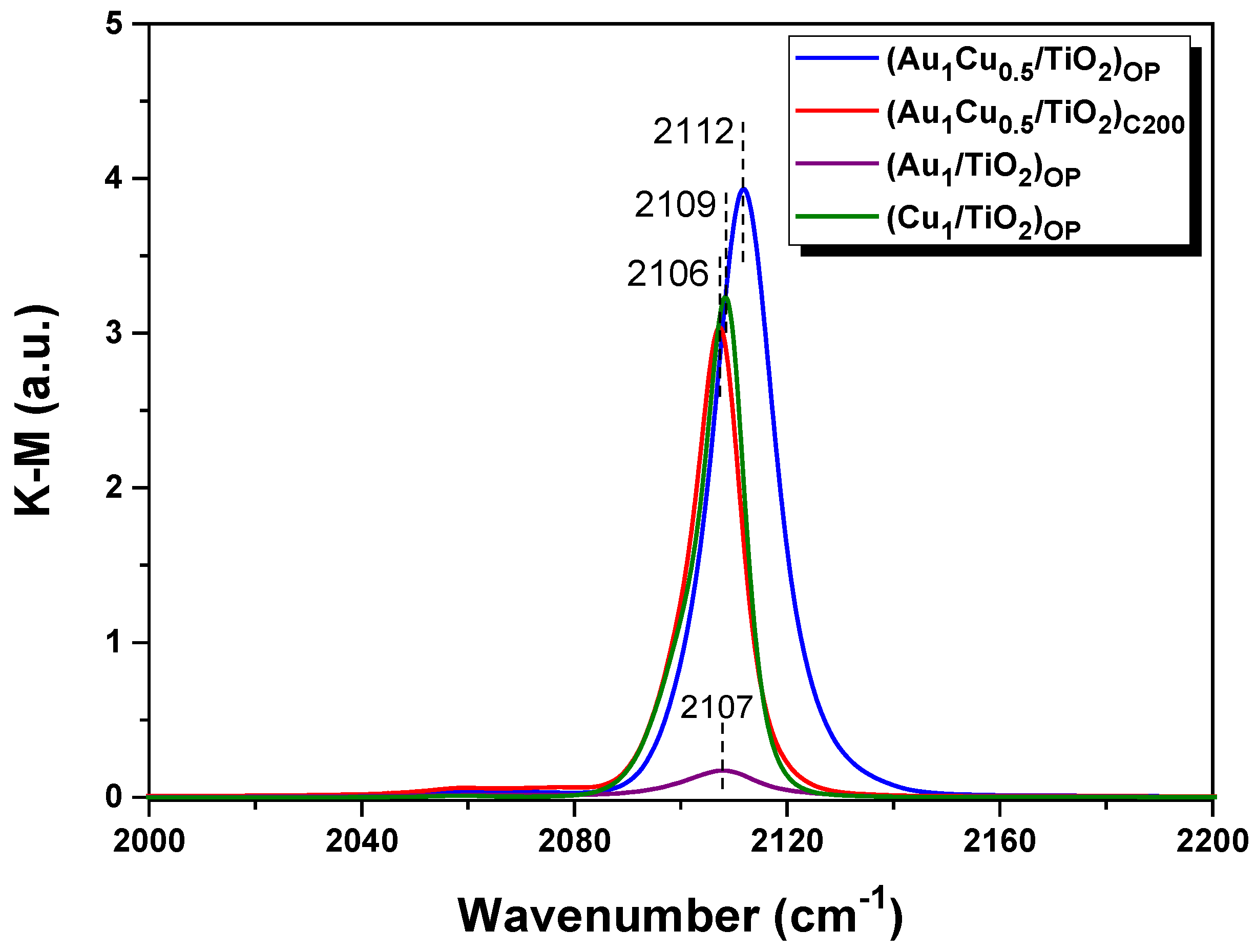
2.2. Electronic State of the Plasmonic Nanocatalysts

The surface electronic state of plasmonic nanocatalysts was measured by XPS. In Figure 2, the binding energies of Au 4f and Cu 3d are deconvoluted, and the results are summarized in Table 1. The Au 4f spectra of Au1Cu0.5/TiO2 nanocatalysts were deconvoluted into Au0 and Auδ+, while the Cu 2p spectra contained Cu2+, Cu+, and Cu0 peaks. Further, the Cu0/Cu+ ratio was evaluated by analyzing the Cu LMM spectra. As listed in Table 1, the contents of Auδ+ and Cu2+ for the fresh (Au1Cu0.5/TiO2)OP were 43% and 35%, respectively. Due to the auto-reduction in cationic Au and Cu species in the induction period, the contents of Auδ+ and Cu2+ for the used (Au1Cu0.5/TiO2)OP decreased to 29% and 32%, respectively. The same phenomenon was also observed for (Au1Cu0.5/TiO2)C200. Interestingly, compared with (Au1Cu0.5/TiO2)C200, the (Au1Cu0.5/TiO2)OP possessed a higher Auδ+ content but lower Cu2+ content. This probably originated from the intimately interacted Au and Cu species in the (Au1Cu0.5/TiO2)OP, as their strong interaction enabled the Cu species to obtain a higher electron density than the Au species [31]. Without the interaction between the Au and Cu species, the (Au1/TiO2)OP and (Cu1/TiO2)OP, respectively, exhibited lower Auδ+ and higher Cu2+ contents than the (Au1Cu0.5/TiO2)OP (see Figure S2 and Table S1). Further, compared with (Au1/TiO2)OP and (Cu1/TiO2)OP, the binding energies of Au 4f and Cu 2p of the (Au1Cu0.5/TiO2)OP exhibited blue- and red-shifts (see Table 1 and Table S1), respectively. These shifts were induced by the interaction between Au and Cu species, which led to a shift in the electron density from the Au to Cu species. Table 1 also shows that O2 plasma treatment endowed the samples with a higher Cu0/Cu+ ratio than the calcined samples, indicating the favorable formation of metallic Cu in the O2 plasma-treated sample. The above results suggest that the Cu species in the (Au1Cu0.5/TiO2)OP tended to achieve low valence, which facilitated the creation of AuCu alloy nanoparticles and, thus, AuCu-TiO2 interfacial sites. Furthermore, the binding energies of Au 4f and Cu 2p for the (Au1Cu0.5/TiO2)OP both presented a blue shift compared with (Au1Cu0.5/TiO2)C200, indicating the electron deficiency of O2 plasma-treated samples. This could be attributed to the fact that the discharge in oxidative O2 enabled strong interactions between the Au, Cu species, and TiO2 support [15,32]. Considering the generation of large numbers of oxygen species (e.g., O2+, O) in O2 discharge, O2 plasma treatment led to a high content of surface oxygen (OS) (see Figures S3 and S4). The content of OS decreased after the VL PCO of CO due to the partial consumption of reactive oxygen species during the reaction. Further, we noted that the presence of Cu resulted in higher OS content (see Figures S3–S6), which could probably be attributed to the construction of AuCu-TiO2 interfacial sites, which benefited the adsorption and activation of oxygen.
To further investigate the interaction between Au and Cu species in the Au-Cu/TiO2 nanocatalysts, the H2-TPR of the fresh plasmonic nanocatalysts was performed. Figure 3 shows that (Au1/TiO2)OP and (Cu1/TiO2)OP both presented two reduction peaks with increasing temperature from −50 °C to 250 °C. The H2 consumption peaks located at low temperatures (35 °C and 32 °C, respectively) could be attributed to a reduction in the surface oxygen species [33], while the peaks located at 65 °C and 123 °C were ascribed to the reduction of Auδ+ and Cuδ+ species, respectively. Figure 3 also shows that (Au1Cu0.5/TiO2)OP exhibited two obvious reduction peaks at 34 °C and 63 °C, respectively, although it possessed the Auδ+ and Cuδ+ species. Apparently, the Auδ+ and Cuδ+ species in (Au1Cu0.5/TiO2)OP only presented one reduction peak at 63 °C, which might be caused by their strong interaction. By contrast, the Auδ+ and Cuδ+ species in (Au1Cu0.5/TiO2)C200 showed a reduction peak at a higher temperature of 87 °C compared to (Au1Cu0.5/TiO2)OP, which might originate from its low content of Auδ+ and the weak interaction between the Auδ+ and Cuδ+ species. Further, (Au1Cu0.5/TiO2)OP possessed the largest peak area of surface oxygen reduction among all the samples (see Figure 3), as the strong interaction between Au and Cu species facilitated the adsorption and activation of oxygen by creating large numbers of AuCu-TiO2 interfacial sites.
CO adsorption behaviors over the plasmonic nanocatalysts are displayed in Figure 4. The (Au1/TiO2)OP showed a band at 2107 cm−1 due to the adsorption of CO onto the metallic Au [34,35], and (Cu1/TiO2)OP appeared with the band at 2109 cm−1, which could be ascribed to CO adsorption on Cu+ species, due to the weak adsorption ability of the metallic Cu and Cu2+ species for CO [36,37]. Furthermore, the (Au1/TiO2)OP exhibited a much weaker band intensity compared with the (Cu1/TiO2)OP, indicating that the Cu+ species possessed a stronger adsorption capacity for CO molecules than metallic Au. The (Au1Cu0.5/TiO2)C200 and (Au1Cu0.5/TiO2)OP presented bands at 2106 cm−1 and 2112 cm−1, respectively, and the adsorption bands for these two samples exhibited a comparable intensity to the monometallic (Cu1/TiO2)OP (see Figure 4). This suggests that the Cu+ species, rather than metallic Au, mainly contributed to CO adsorption in the Au1Cu0.5/TiO2 nanocatalysts. Compared to the (Cu1/TiO2)OP and (Au1Cu0.5/TiO2)C200, the (Au1Cu0.5/TiO2)OP displayed a stronger band intensity, as the strong interaction between the Au and Cu species induced by O2 plasma treatment endowed (Au1Cu0.5/TiO2)OP with large numbers of coordinatively unsaturated sites for CO adsorption [15,38]. Figure 4 also shows that the adsorption band of the (Au1Cu0.5/TiO2)OP had a blue shift of 6 cm−1 compared with that of the (Au1Cu0.5/TiO2)C200. This shift reflects an electron transfer from metallic Au toward Cu+ species, owing to the strong interaction between the Au and Cu species (see H2-TPR analysis). The CO adsorption behaviors of (Au1Cu0.5/TiO2)OP further confirm the pivotal role of O2 plasma treatment in generating a strong interaction between Au and Cu species and constructing highly active AuCu-TiO2 interfacial sites.

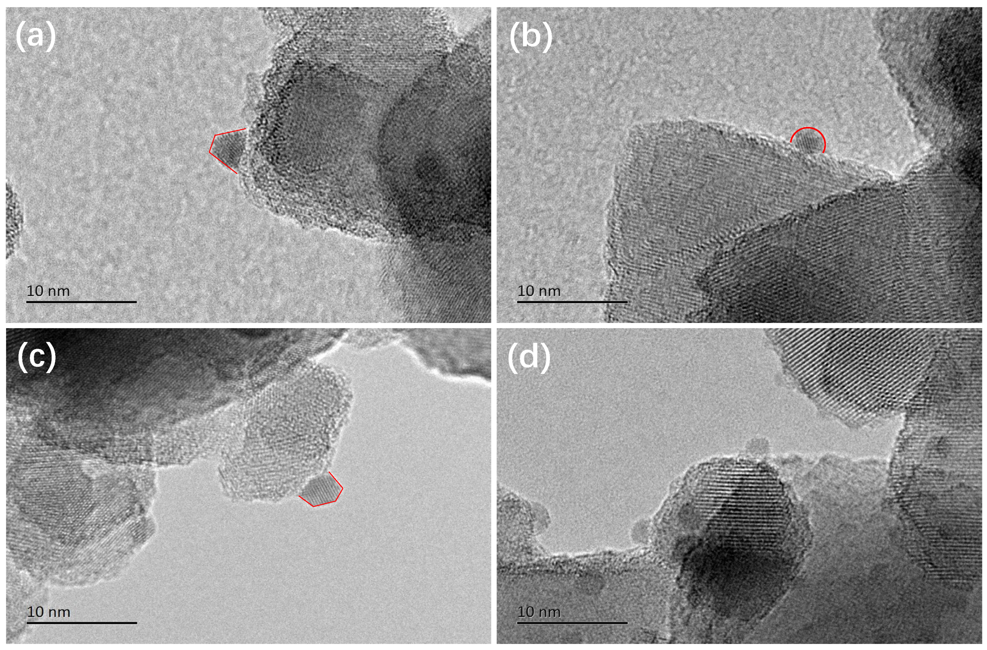
2.3. Structural States of the Plasmonic Nanocatalysts

The morphologies of the plasmonic nanoparticles in different nanocatalysts were observed by HR-TEM. As shown in Figure 5, the metallic nanoparticles on the TiO2 surface for the (Au1Cu0.5/TiO2)OP, (Au1Cu0.5/TiO2)C200, and (Au1/TiO2)OP could be clearly observed. The average size of the metallic nanoparticles for the samples obeyed an order of (Au1Cu0.5/TiO2)OP < (Au1Cu0.5/TiO2)C200 < (Au1/TiO2)OP, and the (Au1Cu0.5/TiO2)OP achieved the smallest average size of 2.4 nm (see Figure S7a–c). It can be noted that (Cu1/TiO2)OP possessed the nanoparticles without clear lattice fringes (see Figure 5d). These nanoparticles were probably ascribed to CuOX, as the Cu2+ species are difficult to be reduced into Cu nanoparticles in the VL PCO of CO. The average size of CuOX nanoparticles in the (Cu1/TiO2)OP was not counted because of the relatively low contrast between CuOX and TiO2 (see Figure S7d). The HR-TEM images confirm that the plasmonic nanocatalysts treated by O2 plasma could obtain metallic plasmonic nanoparticles with small particle sizes. The interaction between the Au and Cu species also contributed to the controlled size of metallic nanoparticles, as demonstrated by the smaller size of plasmonic nanoparticles in (Au1Cu0.5/TiO2)OP compared to that of (Au1/TiO2)OP.
The HAADF-STEM-EDS technique was used to identify the composition of the metallic plasmonic nanoparticles in (Au1Cu0.5/TiO2)OP. The EDS mapping in Figure 6 shows the homogeneous distribution of the Au and Cu elements in a typical nanoparticle, indicating the formation of AuCu alloy nanoparticles in the used (Au1Cu0.5/TiO2)OP. Further, the EDS line profiles of the used (Au1Cu0.5/TiO2)OP illustrate that the peaks of the Au and Cu elements almost exhibited the same variation trend (see Figure S8), demonstrating that the AuCu alloy nanoparticles possessed a uniform structure rather than separated structures (core–shell, Janus, etc.). The formation of AuCu alloy nanoparticles in the used (Au1Cu0.5/TiO2)OP could be attributed to the strong interaction between the Au and Cu species, as the hot electrons produced by metallic Au could reduce adjacent cationic Cu into metallic Cu under VL irradiation. The (Au1Cu0.5/TiO2)C200 with a weak interaction between the Au and Cu species could only generate minor numbers of AuCu alloy nanoparticles, and thus, the AuCu alloy nanoparticles were difficult to identify from the used (Au1Cu0.5/TiO2)C200 in the HAADF-STEM-EDS observation. This suggests that intimately interacting Au and Cu species are essential for obtaining the AuCu alloy nanoparticles.
The above results indicate that the (Au1Cu0.5/TiO2)OP, possessing numerous small AuCu alloy nanoparticles, could create larger numbers of interfacial sites between metallic nanoparticles and the TiO2 support among all the samples. To confirm this point, we estimated and compared the number of interfacial sites that were located at the metal–support interface based on the morphology and average size of plasmonic nanoparticles. A combination of the DFT-based Wulff Construction and physical model reconstruction was used to calculate the proportion of the perimeter metal atoms (see Table S2, Figures S9 and S10) [30]. Table 2 lists that (Au1Cu0.5/TiO2)OP possessed a higher proportion of perimeter metal atoms (6.0%) compared with the (Au1/TiO2)OP (5.3%) and (Au1Cu0.5/TiO2)C200 (4.7%), demonstrating that a larger number of interfacial sites were constructed in the (Au1Cu0.5/TiO2)OP.

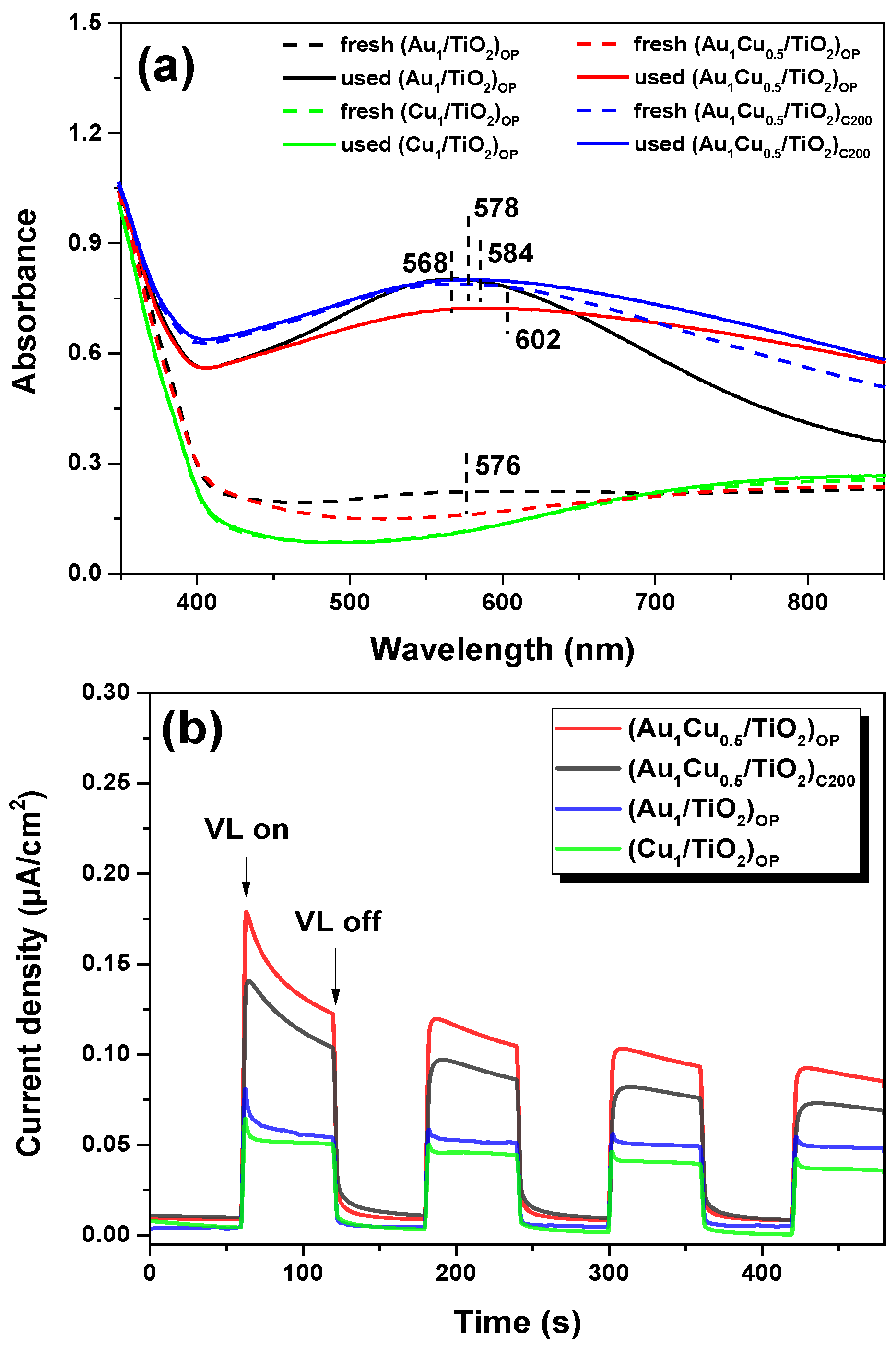
2.4. Photo-Response Properties of the Plasmonic Nanocatalysts

The LSPR effect of plamonic nanocatalysts was measured by UV-vis DRS. Figure 7a shows that a strong light absorption band at ca. 568 nm could be clearly observed for the used (Au1/TiO2)OP due to the LSPR of metallic Au nanoparticles [15]. The fresh (Au1/TiO2)OP only presented a weak light absorption band because of its low Au0 content after O2 plasma treatment, which was consistent with the XPS results. The fresh and used (Cu1/TiO2)OP both appeared as weak absorption bands after 600 nm, indicating the weak light absorption ability of (Cu1/TiO2)OP, owing to their low Cu0 content and relatively low absorption cross-section of Cu nanoparticles [39,40]. The (Au1Cu0.5/TiO2)C200 before and after the VL photocatalytic reaction appeared as strong light absorption bands at 578 nm and 584 nm, respectively. This was because the cationic Au and Cu species were reduced to a metallic Au and Au-Cu alloy, which exhibited strong absorption for the VL. The fresh (Au1Cu0.5/TiO2)OP only exhibited a weak absorption band at 576 nm; however, the used sample exhibited a strong absorption band at around 602 nm after the VL PCO of CO due to an auto-reduction in the sample during the induction period [9]. Considering the strong interaction between Au and Cu species in the fresh (Au1Cu0.5/TiO2)OP, this auto-reduction process could generate a large amount of the Au-Cu alloy and, thus, make the used sample show higher absorption for VL via the strong LSPR effect. Figure 7a also showed that the absorption bands of the used bimetallic nanocatalysts shifted to a higher wavelength compared to the fresh samples, which was due to the formation of AuCu alloy nanoparticles during the VL PCO of CO. The absorption band of the used (Au1Cu0.5/TiO2)OP centered at a higher wavelength than that of (Au1Cu0.5/TiO2)C200, indicating that O2 plasma treatment favored the formation of AuCu alloy nanoparticles. Further, we noted that the used (Au1Cu0.5/TiO2)OP exhibited the broadest absorption band among all the samples, implying its fast rate in LSPR relaxation [41,42]. This facilitated the formation of hot electrons via Landau damping and the following direct injection of hot electrons to the TiO2 support [1,3], which could lead to an enhanced hot electron transfer process during the photocatalytic reaction. The photocurrent densities for different plasmonic nanocatalysts were also confirmed by the enhanced hot electron transfer on the (Au1Cu0.5/TiO2)OP. Figure 7b shows that all plasmonic nanocatalysts exhibited low current densities in the darkness but exhibited distinct photocurrent densities under VL irradiation. Compared to the monometallic samples, the higher photocurrent densities for bimetallic nanocatalysts suggested the favorable hot electron transfer on AuCu-TiO2 interfacial sites. As shown in Figure 7b, the (Au1Cu0.5/TiO2)OP obtained the highest photocurrent density among all the plasmonic samples, which could be attributed to its high dispersion in the AuCu alloy nanoparticles (see the HR-TEM results) when considering the construction of large numbers of AuCu-TiO2 interfacial sites for hot electron transfer. Moreover, compared with the Au-TiO2 interface, the formation of the AuCu-TiO2 interface in (Au1Cu0.5/TiO2)OP lowered the Schottky barrier height [43,44], which enabled more hot electrons to overcome the Schottky barrier under VL irradiation. Additionally, photocurrent densities for the plasmonic nanocatalysts seemed to show a gradual decrease with an increase in the test time (Figure 7b). To confirm this point, we performed a potentiostatic chronoamperometry study of O2 plasma-treated samples after the VL PCO of CO. All samples presented a decrease in their photocurrent density under VL irradiation during 30 min of continuous testing (see Figure S11). Compared with the (Au1/TiO2)OP and (Cu1/TiO2)OP, (Au1Cu0.5/TiO2)OP exhibited a steeper decrease in its photocurrent density, which could probably be attributed to the remarkable changes in its surface properties in the Na2SO4 solution when used for a potentiostatic chronoamperometry test.

3. Discussion

3.1. AuCu-TiO2 Interfacial Sites Construction

In this work, we demonstrate that the plasmonic Au-Cu/TiO2 nanocatalyst treated by O2 plasma showed a remarkable boost of photocatalytic activity in the VL PCO of CO compared with the counterparts. We also confirmed that the construction of AuCu-TiO2 interfacial sites during O2 plasma treatment played an important role in boosting the photocatalytic activity of Au-Cu/TiO2 nanocatalysts. The cationic Au and Cu species were randomly distributed on the surface of the TiO2 support for as-prepared Au-Cu/TiO2 bimetallic nanocatalysts (see Figure 8). During treatment, these cationic Au and Cu species occurred with redispersion and reduction simultaneously, which is essential for the formation of AuCu alloy nanoparticles and, thus, AuCu-TiO2 interfacial sites. It was noted that alteration behaviors of Au and Cu species in the treatment highly depended on the treatment approaches. Calcination could reduce most cationic Au species but hardly formed intimate interactions with the Au and Cu species because of their several migration and aggregations at high temperatures. By contrast, O2 plasma treatment enabled a strong interaction between the cationic Au and Cu species to form particles of mixed oxide AuCuOx, as O2 plasma established a unique redox environment on the nanocatalyst surface through large numbers of charged species [38]. The intimately interacted Au and Cu species benefitted from forming an AuCu alloy and, thus, constructing AuCu-TiO2 interfacial sites and also contributing to and controlling the size of AuCu alloy nanoparticles. These intimately interacted Au and Cu species in (Au1Cu0.5/TiO2)OP could be auto-reduced into the AuCu alloy in the VL PCO of CO since the hot electrons produced by metallic Au nanoparticles and generated from O2 plasma treatment could quickly and efficiently reduce adjacent cationic Cu to form an AuCu alloy under VL irradiation (see Figure 8). This auto-reduction process was completed at the beginning of a photocatalytic reaction (within the induction period) [9]. Apparently, compared with O2 plasma, conventional calcination tended to separate the Au and Cu species, which suppressed the formation of AuCu alloy nanoparticles and, thus, the construction of AuCu-TiO2 interfacial sites.

3.2. The Boost of VL Photocatalysis on AuCu-TiO2 Interfacial Sites

The processes of VL PCO of CO over the Au-Cu/TiO2 bimetallic nanocatalyst could be described by a charge (hot electron) injection mechanism [3]. The AuCu alloy nanoparticles adsorbed CO molecules and also acted as a dye sensitizer to produce hot electrons by absorbing resonant photons under VL illumination. The energetic hot electrons were transferred to the TiO2 support via surmounting the AuCu-TiO2 interfaces. In addition, O2 molecules were adsorbed on the TiO2 support and combined with injected hot electrons to form superoxide (O2). Finally, the O2 dissociates reacted with the adsorbed CO molecules at AuCu-TiO2 interfacial sites to form CO2. It was demonstrated that the (Au1Cu0.5/TiO2)OP possessing large numbers of coordinatively unsaturated sites not only facilitated CO adsorption (see CO chemisorption results) but also enhanced the formation of O2 by generating abundant surface oxygen and hot electrons (see XPS and optical property results) due to the construction of large numbers of AuCu-TiO2 interfacial sites. This provides evidence that (Au1Cu0.5/TiO2)OP exhibits advantages over the counterparts in providing reactants for VL PCO of CO.
For the processes of O2 the dissociation and the following interfacial reactions between CO and O, we further discuss this based on DFT calculation results (see Figure S12). A unique feature of (Au1Cu0.5/TiO2)OP is that it constructs large numbers of AuCu-TiO2 interfacial sites. On this plasmonic Au-Cu/TiO2 nanocatalyst, the dissociation barrier for O2 showed a much higher value (0.64 eV) than the barrier (0.15 eV) for CO to react with dissociated O at the AuCu-TiO2 interfacial sites (see Figure 9). These two elementary reaction processes arrived at similar conclusions on the plasmonic Au/TiO2 and CuOx/TiO2 nanocatalysts (see Figure 9), indicating the rate-limiting step of O2 dissociation. The (Cu1/TiO2)OP presented very poor VL photocatalytic activity, as it had a high dissociation barrier for O2 (0.78 eV) and weak LSPR to generate O2 (see the UV-vis DRS results). Interestingly, the DFT results for (Au1Cu0.5/TiO2)OP and (Au1/TiO2)OP indicate that they possess a comparable barrier for O2 dissociation (see Figure 9a), implying that the construction of AuCu-TiO2 interfacial sites showed a weak influence on the O2 dissociation process. By contrast, the construction of AuCu-TiO2 interfacial sites remarkably reduced the barrier for a reaction between CO and O from 0.42 eV to 0.15 eV in comparison with (Au1/TiO2)OP (see Figure 9b). The above analyses, taken together, suggest that the boost of photocatalysis of the (Au1Cu0.5/TiO2)OP in VL PCO of CO originated from the strong interaction between the Au and Cu species derived from O2 plasma treatment. This strong interaction controlled the size of AuCu alloy nanoparticles, establishing larger numbers of interfacial sites for the hot electron transfer and CO oxidation reaction. Moreover, the intimately interacted Au and Cu species favored the formation of an AuCu alloy, which boosted the hot electron transfer process by lowering the Schottky barrier height and also significantly reduced the barrier for CO to react with O.

4. Materials and Methods

4.1. Catalysts Preparation

The plasmonic Au-Cu/TiO2 bimetallic nanocatalysts were prepared using TiO2 (P25, Degussa) as support and HAuCl4 and Cu(NO3)2 as precursors of plasmonic metals [45]. The solutions of HAuCl4 (2.43 × 10⁻² mol/L; 2.2 mL) and Cu(NO3)2 (4.14 × 10⁻² mol/L; 3.8 mL) were mixed with 1.0 g of TiO2, and the mixture was stirred with a glass rod at room temperature for 10 min. Subsequently, the samples were aged for 14 h at room temperature and then washed with deionized water and an aqueous ammonia solution (pH 8). After washing, the samples were dried at 80 °C for 6 h to obtain the as-prepared Au-Cu/TiO2 nanocatalyst. Unless otherwise specified, the nominal loadings of Au and Cu in as-prepared Au-Cu/TiO2 nanocatalysts were 1 wt.% and 0.5 wt.%, respectively. For comparison, TiO2-supported Au (nominal 1 wt.%) and Cu (nominal 1 wt.%) nanocatalysts were prepared using the same procedures.
The as-prepared plasmonic nanocatalysts (about 30 mg) were coated on a glass substrate of 50 mm (L) × 25 mm (W) × 1 mm (T) [13] and were treated by calcination (200 °C in the air for 2 h) or cold plasma before the photocatalytic activity tests. The plasma treatment was performed in a homemade dielectric barrier discharge (DBD) reactor [9,15]. Our previous work demonstrated that O2 plasma outperformed other plasmas in plasmonic nanocatalyst activation [13], and thus, all plasma treatment (AC discharge; 1.8 kHz; CTP-2000 K, Nanjing Suman Electronics Co., Nanjing, China) experiments were carried out for 10 min at an input power of 10 W with an O2 flow rate of 100 mL/min. The Au1Cu0.5/TiO2 treated by calcination and O2 plasma were denoted as (Au1Cu0.5/TiO2)C200 and (Au1Cu0.5/TiO2)OP, respectively. The Au1/TiO2 and Cu1/TiO2 treated by O2 plasma were denoted as (Au1/TiO2)OP and (Cu1/TiO2)OP, respectively.

4.2. Catalysts Characterization

The actual loadings of Au and Cu in the nanocatalysts were determined by inductively coupled plasma-atomic emission spectroscopy (ICP-AES, Optima 2000DV, PerkinElmer Inc., Waltham, MA, USA). The Au loading for Au1/TiO2 was 0.84 wt.%, and the Cu loading for the Cu1/TiO2 was 0.78 wt.%. The Au and Cu loadings for Au1Cu0.5/TiO2 were 0.82 wt.% and 0.43 wt.%, respectively. Temperature-programmed reduction with H2 (H2-TPR) was performed in an AutoChem 2910 system (Micromeritics, Atlanta, GA, USA). The fresh samples (100 mg) were placed in a quartz reactor and pretreated in helium (50 mL/min) for 30 min at −50 °C. Then, 5% H2 in helium (50 mL/min) was switched to flow through the pretreated samples at −50 °C until there was no change in the signals. After that, the samples were heated to 250 °C at a rate of 5 °C min−1. The X-ray photoelectron spectroscopy (XPS) of the samples was performed at ESCALAB250 (Thermo VG, Waltham, MA, USA) with operating conditions of 15 kV and 300 W. The binding energies were calibrated with reference to the peak of C 1s at 284.6 eV. The in situ diffuse reflectance infrared Fourier transform spectra (DRIFTS) of CO adsorption were recorded by an FT-IR spectrometer (Nicolet 6700, Thermo Fisher, Waltham, MA, USA) at a resolution of 4 cm−1. Before the measurement, the samples placed in the DRIFTS cell were pretreated with N2 (100 mL/min) for 30 min at 80 °C. Subsequently, the samples were cooled down to room temperature and collected with background spectra in N2 flowing (100 mL/min). Finally, CO adsorption tests were performed by switching 1% CO/N2 (50 mL/min) into the DRIFTS cell. The plasmonic nanoparticles of the samples were observed by transmission electron microscopy (TEM; Tecnai G2F30 STWIN, Hillsboro, OR, USA) at 300 kV equipped with a high-angle annular dark-field (HAADF) detector and energy dispersive X-ray spectrometer (EDXS). The UV-vis diffuse reflectance spectroscopy (UV-vis DRS) of the samples was performed at a lambda 750s spectrometer (Perkin-Elmer, Waltham, MA, USA) to measure their photo-response properties. The photocurrent of the samples was measured using a three-electrode cell (0.1 M Na2SO4 solution) equipped with an electrochemical workstation (CHI660E, Chenhua, Shanghai, China). An ITO (indium tin oxide) glass of 20 mm (L) × 20 mm (W) × 1 mm (T) coated with the samples was used as a working electrode, while a platinum sheet and an Ag/AgCl electrode were used as the counter and reference electrodes, respectively. The photocurrent of the samples was measured at a bias voltage of 0.5 V under VL irradiation (58 mW/cm2).

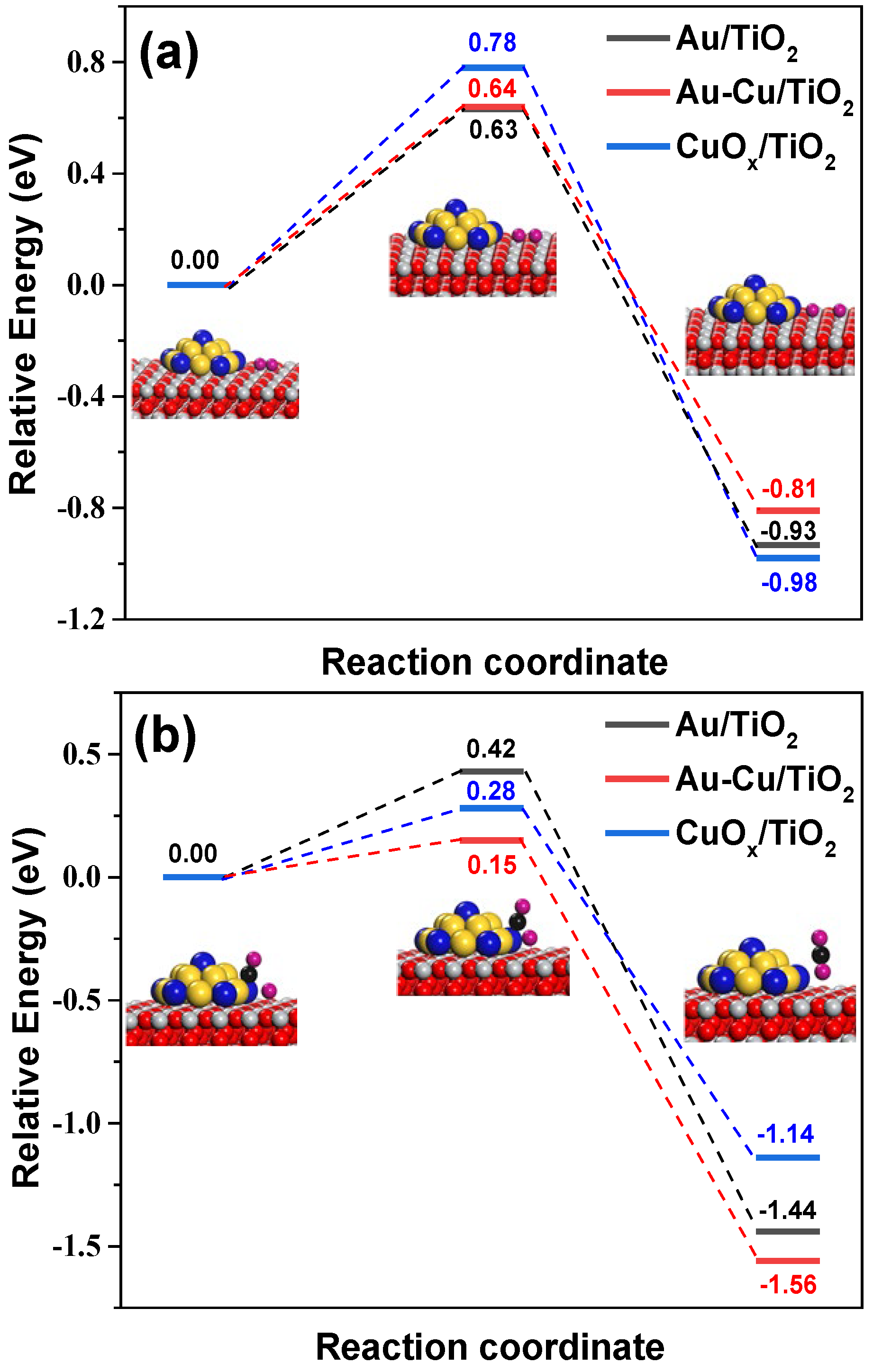
4.3. DFT Calculation

The VL PCO of CO was investigated by Vienna Ab initio Simulation Package (VASP) calculations, in which the Au/TiO2, Au-Cu/TiO2, and CuOx/TiO2 were selected as the calculation models for the comparison. The details of the computational method are described in the Supplementary Material.

4.4. Photocatalytic Reaction Evaluation

To evaluate the performance of the plasmonic nanocatalysts, the VL PCO of CO was performed in a home-made plate reactor [9,13], and VL with a light intensity of 58 mW/cm2 was supplied by a LED lamp [13]. The reactant gas composed of synthetic air (80% N2 + 20% O2) and 920 ppm CO was introduced into the plate reactor at a total flow rate of 300 mL/min. The concentrations of COx (CO and CO2) were monitored online by a COx analyzer (GXH-3011N, Huayun, Beijing, China), and CO conversion (XCO) was defined as Equation (1):
X CO = C C O 2 C CO
where C CO and C C O 2 denote the concentrations of CO in the inlet gas and CO2 in the outlet gas, respectively.

5. Conclusions

In conclusion, this work demonstrates that the plasmonic Au-Cu/TiO2 bimetallic nanocatalyst, prepared with O2 plasma treatment, could exhibit superior photocatalytic activity under VL irradiation. The O2 plasma treatment boosted the VL photocatalytic activity of the Au-Cu/TiO2 nanocatalyst by nearly 50% compared to the conventional calcination treatment. After O2 plasma treatment, the Au-Cu/TiO2 nanocatalyst showed comparable performance to the monometallic Au/TiO2 sample possessing double Au loading. The O2 plasma treatment could initiate the surface alteration of the Au-Cu/TiO2 nanocatalyst to generate a strong interaction between the Au and Cu species. The intimately interacted Au and Cu species decreased their reduction temperature and, thus, were quickly auto-reduced into AuCu alloy nanoparticles at the beginning of the VL PCO of CO. The average size of plasmonic nanoparticles for the O2 plasma-treated Au-Cu/TiO2 nanocatalyst was controlled at around 2.4 nm by the interaction between Au and Cu species, creating larger numbers of interfacial sites for CO adsorption and oxidation in comparison with the conventional calcined sample. Compared with the monometallic plasmonic nanocatalyst, the formation of small AuCu alloy nanoparticles reduced the Schottky barrier height at the metal–support interface and constructed highly active AuCu-TiO2 interfacial sites, which facilitated the interfacial transfer of hot electrons. The DFT calculations confirmed that the construction of AuCu-TiO2 interfacial sites contributed to a low barrier for the reaction between CO and O and, thus, the boosted VL photocatalytic activity, although O2 dissociation was the rate-limiting step for the VL PCO of CO.

Supplementary Materials

The following supporting information can be downloaded at: https://www.mdpi.com/article/10.3390/ijms241310487/s1. References [46,47,48,49,50] are cited in the Supplementary Materials.

Author Contributions

Conceptualization, B.Z. and Y.L.; investigation, X.L.; resources, Y.L. and J.L.; supervision, B.Z.; writing—original draft, X.L.; writing—review and editing, B.Z. and X.Z. All authors have read and agreed to the published version of the manuscript.

Funding

This work is supported by the National Natural Science Foundation of China (22178039, 21808024), Natural Science Foundation of Liaoning Province (2020-MS-126).

Institutional Review Board Statement

Not applicable.

Informed Consent Statement

Not applicable.

Data Availability Statement

The data presented in this study are available upon request from the corresponding author.

Acknowledgments

We thank Yan-Qun Wang for the DFT calculation.

Conflicts of Interest

The authors declare no conflict of interest.

References

  1. Linic, S.; Chavez, S.; Elias, R. Flow and extraction of energy and charge carriers in hybrid plasmonic nanostructures. Nat. Mater. 2021, 20, 916–924. [Google Scholar] [CrossRef] [PubMed]
  2. Yu, S.; Kim, Y.H.; Lee, S.Y.; Song, H.D.; Yi, J. Hot-electron-transfer enhancement for the efficient energy conversion of visible light. Angew. Chem. Int. Ed. 2014, 53, 11203–11207. [Google Scholar] [CrossRef] [PubMed]
  3. Linic, S.; Christopher, P.; Ingram, D.B. Plasmonic-metal nanostructures for efficient conversion of solar to chemical energy. Nat. Mater. 2011, 10, 911–921. [Google Scholar] [CrossRef] [PubMed]
  4. Tsukamoto, D.; Shiraishi, Y.; Sugano, Y.; Ichikawa, S.; Tanaka, S.; Hirai, T. Gold nanoparticles located at the interface of anatase/rutile TiO2 particles as active plasmonic photocatalysts for aerobic oxidation. J. Am. Chem. Soc. 2012, 134, 6309–6315. [Google Scholar] [CrossRef] [PubMed]
  5. Dao, D.V.; Nguyen, T.T.D.; Uthirakumar, P.; Cho, Y.-H.; Kim, G.-C.; Yang, J.-K.; Tran, D.-T.; Le, T.D.; Choi, H.; Kim, H.Y.; et al. Insightful understanding of hot-carrier generation and transfer in plasmonic Au@CeO2 core-shell photocatalysts for light-driven hydrogen evolution improvement. Appl. Catal. B 2021, 286, 119947. [Google Scholar] [CrossRef]
  6. Naya, S.-I.; Tada, H. Dependence of the plasmonic activity of Au/TiO2 for the decomposition of 2-naphthol on the crystal form of TiO2 and Au particle size. J. Catal. 2018, 364, 328–333. [Google Scholar] [CrossRef]
  7. Wang, S.; Zeng, B.; Li, C. Effects of Au nanoparticle size and metal-support interaction on plasmon-induced photocatalytic water oxidation. Chin. J. Catal. 2018, 39, 1219–1227. [Google Scholar] [CrossRef]
  8. Yu, C.; Zhou, W.; Zhu, L.; Li, G.; Yang, K.; Jin, R. Integrating plasmonic Au nanorods with dendritic like α-Bi2O3/Bi2O2CO3 heterostructures for superior visible-light-driven photocatalysis. Appl. Catal. B 2016, 184, 1–11. [Google Scholar] [CrossRef] [Green Version]
  9. Zhu, B.; Zhang, L.-Y.; Liu, J.-L.; Zhang, X.-M.; Li, X.-S.; Zhu, A.-M. TiO2-supported Au-Ag plasmonic nanocatalysts achieved by plasma restructuring and activation. J. Hazar. Mater. 2021, 402, 123508. [Google Scholar] [CrossRef]
  10. Neaţu, S.; Macia-Agullo, J.A.; Concepción, P.; Garcia, H. Gold-copper nanoalloys supported on TiO2 as photocatalysts for CO2 reduction by water. J. Am. Chem. Soc. 2014, 136, 15969–15976. [Google Scholar] [CrossRef]
  11. Tan, T.H.; Scott, J.A.; Ng, Y.H.; Taylor, R.A.; Aguey-Zinsou, K.-F.; Amal, R. Plasmon enhanced selective electronic pathways in TiO2 supported atomically ordered bimetallic Au-Cu alloys. J. Catal. 2017, 352, 638–648. [Google Scholar] [CrossRef]
  12. Dmitry, B.S.; Vishal, J.; Valery, A.N.; Johanna, T.; Maria, E.M.; Jakob, B.W.; Olof, P.; Rainer, T.; Anders, M.; Ivan, M.; et al. Strong Schottky barrier reduction at Au-catalyst/GaAs-nanowire interfaces by electric dipole formation and Fermi-level unpinning. Nat. Commun. 2014, 5, 3221. [Google Scholar]
  13. Zhu, B.; Li, X.; Deng, X.-Q.; Wang, Y.-Q.; Lu, L.-L. Activation of Au-Ag Plasmonic Bimetallic Nanocatalysts with Cold Plasma: The Role of Loading Sequence of Plasmonic Metals and Discharge Atmosphere. Plasma Chem. Plasma Process. 2022, 42, 671–687. [Google Scholar] [CrossRef]
  14. Sugano, Y.; Shiraishi, Y.; Tsukamoto, D.; Ichikawa, S.; Tanaka, S.; Hirai, T. Supported Au-Cu Bimetallic Alloy Nanoparticles: An Aerobic Oxidation Catalyst with Regenerable Activity by Visible-Light Irradiation. Angew. Chem. Int. Ed. 2013, 125, 5403–5407. [Google Scholar] [CrossRef]
  15. Deng, X.-Q.; Zhu, B.; Li, X.-S.; Liu, J.-L.; Zhu, X.; Zhu, A.-M. Visible-light photocatalytic oxidation of CO over plasmonic Au/TiO2: Unusual features of oxygen plasma activation. Appl. Catal. B 2016, 188, 48–55. [Google Scholar] [CrossRef]
  16. Liu, J.-H.; Wang, A.-Q.; Chi, Y.-S.; Lin, H.-P.; Mou, C.-Y. Synergistic Effect in an Au-Ag Alloy Nanocatalyst:  CO Oxidation. J. Phys. Chem. B 2005, 109, 40–43. [Google Scholar] [CrossRef]
  17. Liu, X.; Wang, A.; Li, L.; Zhang, T.; Mou, C.-Y.; Lee, J.-F. Structural changes of Au-Cu bimetallic catalysts in CO oxidation: In situ XRD, EPR, XANES, and FT-IR characterizations. J. Catal. 2011, 278, 288–296. [Google Scholar] [CrossRef]
  18. Du, X.R.; Huang, Y.K.; Pan, X.L.; Han, B.; Su, Y.; Jiang, Q.K.; Li, M.R.; Tang, H.L.; Li, G.; Qiao, B.T. Size-dependent strong metal-support interaction in TiO2 supported Au nanocatalysts. Nat. Commun. 2020, 11, 5811. [Google Scholar] [CrossRef]
  19. Jin, L.; Liu, B.; Louis, M.E.; Li, G.H.; He, J. Highly Crystalline Mesoporous Titania Loaded with Monodispersed Gold Nanoparticles: Controllable Metal-Support Interaction in Porous Materials. ACS Appl. Mater. Interfaces 2020, 12, 9617–9627. [Google Scholar] [CrossRef]
  20. Wang, Y.C.; Widmann, D.; Behm, R.J. Influence of TiO2 Bulk Defects on CO Adsorption and CO Oxidation on Au/TiO2: Electronic Metal-Support Interactions (EMSIs) in Supported Au Catalysts. ACS Catal. 2017, 7, 2339–2345. [Google Scholar] [CrossRef]
  21. Zhu, B.; Li, X.-S.; Liu, J.-L.; Liu, J.-B.; Zhu, X.; Zhu, A.-M. In-situ regeneration of Au nanocatalysts by atmospheric-pressure air plasma: Significant contribution of water vapor. Appl. Catal. B 2015, 179, 69–77. [Google Scholar] [CrossRef]
  22. Hu, S.L.; Li, W.-X. Sabatier principle of metal-support interaction for design of ultrastable metal nanocatalysts. Science 2021, 374, 1360–1365. [Google Scholar] [CrossRef] [PubMed]
  23. Chen, Y.T.; Wang, H.P.; Liu, C.J.; Zeng, Z.Y.; Zhang, H.; Zhou, C.M.; Jia, X.L.; Yang, Y.H. Formation of monometallic Au and Pd and bimetallic Au-Pd nanoparticles confined in mesopores via Ar glow-discharge plasma reduction and their catalytic applications in aerobic oxidation of benzyl alcohol. J. Catal. 2012, 289, 105–117. [Google Scholar] [CrossRef]
  24. Kim, H.H.; Tsubota, S.; Daté, M.; Ogata, A.; Futamura, S. Catalyst regeneration and activity enhancement of Au/TiO2 by atmospheric pressure nonthermal plasma. Appl. Catal. A 2007, 329, 93–98. [Google Scholar] [CrossRef]
  25. Wang, Z.; Zhang, Y.; Neyts, E.C.; Cao, X.X.; Zhang, X.S.; Jang, B.W.-L.; Liu, C.J. Catalyst Preparation with Plasmas: How Does It Work? ACS Catal. 2018, 8, 2093–2110. [Google Scholar] [CrossRef]
  26. Liang, X.; Wang, Z.J.; Liu, C.J. Size-Controlled Synthesis of Colloidal Gold Nanoparticles at Room Temperature Under the Influence of Glow Discharge. Nanoscale Res. Lett. 2010, 5, 124–129. [Google Scholar] [CrossRef] [Green Version]
  27. Kim, H.H.; Kim, J.H.; Ogata, A. Microscopic observation of discharge plasma on the surface of zeolites supported metallic nanoparticles. J. Phys. D Appl. Phys. 2009, 42, 135210. [Google Scholar] [CrossRef]
  28. Mei, D.H.; Zhu, X.B.; He, Y.L.; Yan, J.D.; Tu, X. Plasma-assisted conversion of CO2 in a dielectric barrier discharge reactor: Understanding the effect of packing materials. Plasma Sources Sci. Technol. 2014, 24, 015011. [Google Scholar] [CrossRef] [Green Version]
  29. Zhu, B.; Yan, Y.; Li, M.; Li, X.-S.; Liu, J.-L.; Zhu, Y.-M. Low temperature removal of toluene over Ag/CeO2/Al2O3 nanocatalyst in an atmospheric plasma catalytic system. Plasma Process Polym. 2018, 15, 1700215. [Google Scholar] [CrossRef]
  30. Li, Y.C.; Li, X.S.; Zhu, B.; Zhu, X.; Lian, H.Y.; Zhu, A.M. A facile approach to direct preparation of Pt nanocatalysts from oxidative dechloridation of supported H2PtCl6 by oxygen plasma. J. Catal. 2022, 414, 16–24. [Google Scholar] [CrossRef]
  31. Zhang, Z.Y.; Chen, Z.P.; Qu, C.L.; Chen, L.X. Highly Sensitive Visual Detection of Copper Ions Based on the Shape-Dependent LSPR Spectroscopy of Gold Nanorods. Langmuir 2014, 30, 3625–3630. [Google Scholar] [CrossRef]
  32. Sun, Z.-G.; Li, X.-S.; Liu, J.-L.; Li, Y.-C.; Zhu, B.; Zhu, A.-M. A promising visible-light photocatalyst: H2 plasma-activated amorphous-TiO2-supported Au nanoparticles. J. Catal. 2019, 375, 380–388. [Google Scholar] [CrossRef]
  33. Fu, Q.; Saltsburg, H.; Flytzani-Stephanopoulos, M. Active Nonmetallic Au and Pt Species on Ceria-Based Water-Gas Shift Catalysts. Science 2003, 301, 935–938. [Google Scholar] [CrossRef]
  34. Bollinger, M.A.; Vannice, M.A. A kinetic and DRIFTS study of low-temperature carbon monoxide oxidation over Au-TiO2 catalysts. Appl. Catal. B 1996, 8, 417–443. [Google Scholar] [CrossRef]
  35. Boccuzzi, F.; Chiorino, A. FTIR Study of CO Oxidation on Au/TiO2 at 90 K and Room Temperature. An Insight into the Nature of the Reaction Centers. J. Phys. Chem. B 2000, 104, 5414–5416. [Google Scholar] [CrossRef]
  36. Gao, Y.; Zhang, Z.; Li, Z.; Huang, W. Understanding morphology-dependent CuOx-CeO2 interactions from the very beginning. Chin. J. Catal. 2020, 41, 1006–1016. [Google Scholar] [CrossRef]
  37. Hadjiivanov, K.; Tsoncheva, T.; Dimitrov, M.; Minchev, C.; Knözinger, H. Characterization of Cu/MCM-41 and Cu/MCM-48 mesoporous catalysts by FTIR spectroscopy of adsorbed CO. Appl. Catal. A 2003, 241, 331–340. [Google Scholar] [CrossRef]
  38. Li, Y.-C.; Li, X.-S.; Zhu, B.; Zhu, A.-M. Boosting low-temperature water gas shift reaction over Au/TiO2 nanocatalyst activated by oxygen plasma. Chem. Eng. J. 2022, 430, 133013. [Google Scholar] [CrossRef]
  39. Rycenga, M.; Cobley, C.M.; Zeng, J.; Li, W.; Moran, C.H.; Zhang, Q.; Qin, D.; Xia, Y. Controlling the synthesis and assembly of silver nanostructures for plasmonic applications. Chem. Rev. 2011, 111, 3669–3712. [Google Scholar] [CrossRef] [Green Version]
  40. Ferry, V.E.; Munday, J.N.; Atwater, H.A. Design Considerations for Plasmonic Photovoltaics. Adv. Mater. 2010, 22, 4794–4808. [Google Scholar] [CrossRef]
  41. Patra, K.K.; Gopinath, C.S. Bimetallic and Plasmonic Ag-Au on TiO2 for Solar Water Splitting: An Active Nanocomposite for Entire Visible-Light-Region Absorption. Chemcatchem 2016, 8, 3294–3311. [Google Scholar] [CrossRef]
  42. Kreibig, U. Interface-induced dephasing of Mie plasmon polaritons. Appl. Phys. B 2008, 93, 79–89. [Google Scholar] [CrossRef]
  43. Park, J.Y.; Lee, H.; Renzas, J.R.; Zhang, Y.; Somorjai, G.A. Probing Hot Electron Flow Generated on Pt Nanoparticles with Au/TiO2 Schottky Diodes during Catalytic CO Oxidation. Nano Lett. 2008, 8, 2388–2392. [Google Scholar] [CrossRef] [PubMed] [Green Version]
  44. Yao, G.-Y.; Zhao, Z.-Y.; Liu, Q.-L.; Dong, X.-D.; Zhao, Q.-M. Theoretical calculations for localized surface plasmon resonance effects of Cu/TiO2 nanosphere: Generation, modulation, and application in photocatalysis. Sol. Energ. Mat. Sol. C 2020, 208, 110385. [Google Scholar] [CrossRef]
  45. Zhu, B.; Zhang, L.-Y.; Li, M.; Yan, Y.; Zhang, X.-M.; Zhu, Y.-M. High-performance of plasma-catalysis hybrid system for toluene removal in air using supported Au nanocatalysts. Chem. Eng. J. 2020, 381, 122599. [Google Scholar] [CrossRef]
  46. Avanesian, T.; Dai, S.; Kale, J.M.; Graham, W.G.; Pan, X.; Christopher, P. Quantitative and Atomic-Scale View of CO-Induced Pt Nanoparticle Surface Reconstruction at Saturation Coverage via DFT Calculations Coupled with in Situ TEM and IR. J. Am. Chem. Soc. 2017, 139, 4551–4558. [Google Scholar] [CrossRef]
  47. Barmparis, G.D.; Remediakis, I.N. Dependence on CO adsorption of the shapes of multifaceted gold nanoparticles: A density functional theory. Rev. B Condens. Matter Mater. Phys. 2012, 86, 085457. [Google Scholar] [CrossRef] [Green Version]
  48. Nguyen, T.T.H.; Le, V.K.; Minh, C.L.; Nguyen, N.H. A theoretical study of carbon dioxide adsorption and activation on metal-doped (Fe, Co, Ni) carbon nanotube. Comput. Theor. Chem. 2017, 1100, 46–51. [Google Scholar] [CrossRef]
  49. Liu, C.; Lourenco, M.P.; Hedstrom, S.; Cavalca, F.; Diaz-Morales, O.; Duarte, H.A.; Nilsson, A.; Pettersson, L.G.M. Stability and Effects of Subsurface Oxygen in Oxide-Derived Cu Catalyst for CO2 Reduction. J. Phys. Chem. C 2017, 121, 25010–25017. [Google Scholar] [CrossRef]
  50. Liu, L.M.; McAllister, B.; Ye, H.Q.; Hu, P. Identifying an O2 Supply Pathway in CO Oxidation on Au/TiO2 (110): A Density Functional Theory Study on the Intrinsic Role of Water. J. Am. Chem. Soc. 2006, 128, 4017–4022. [Google Scholar] [CrossRef]
Figure 1. (a) CO conversion as a function of time under VL irradiation over various plasmonic nanocatalysts, and (b) a comparison of VL photocatalytic activity for the (Au1/TiO2)OP and (Au0.5Cu0.25/TiO2)OP. ti represents the induction period.
Ijms 24 10487 g001
Figure 2. XPS Au 4f, and Cu 2p and Cu LMM of the fresh (a) and used (b) (Au1Cu0.5/TiO2)OP, and the fresh (c) and used (d) (Au1Cu0.5/TiO2)C200.
Ijms 24 10487 g002
Figure 3. H2-TPR profiles of fresh plasmonic nanocatalysts.
Ijms 24 10487 g003
Figure 4. In situ DRIFTS of CO adsorption over various plasmonic nanocatalysts at an exposure time of 30 min.
Ijms 24 10487 g004
Figure 5. HR-TEM images of the used (a) (Au1Cu0.5/TiO2)OP, (b) (Au1Cu0.5/TiO2)C200, (c) (Au1/TiO2)OP, and (d) (Cu1/TiO2)OP. The red lines highlight the shape of metallic nanoparticles. The used nanocatalysts were obtained by applying fresh samples to the VL PCO of CO for 90 min.
Ijms 24 10487 g005
Figure 6. (a) General HAADF-STEM image of the used (Au1Cu0.5/TiO2)OP, and (b) enlarged HAADF-STEM image and (ce) the corresponding element mapping patterns of the bimetallic nanoparticle. The dotted frames highlight the location of the enlarged image in a. The used nanocatalyst was obtained by applying the fresh sample to the VL PCO of CO for 90 min.
Ijms 24 10487 g006
Figure 7. (a) UV-vis DRS of fresh and used plasmonic nanocatalysts, and (b) Photocurrent density for used plasmonic nanocatalysts under VL irradiation. The used nanocatalysts were obtained by applying the fresh samples to the VL PCO of CO for 90 min.
Ijms 24 10487 g007
Figure 8. Schematic diagram for the construction processes of AuCu-TiO2 interfacial sites by O2 plasma treatment for the plasmonic Au-Cu/TiO2 bimetallic nanocatalyst.
Ijms 24 10487 g008
Figure 9. Calculated activation barriers for (a) The dissociation of O2 and (b) CO to react with O on different plasmonic nanocatalysts. Inset: O2 dissociation and reaction between CO and O on Au-Cu/TiO2 model interfaces. Color code: Au, golden; Ti, grey; O atom in TiO2, red; O atom in O2, CO and CO2, pink; Cu, blue; C, black.
Ijms 24 10487 g009
Table 1. XPS analysis of the plasmonic bimetallic samples.
SampleAu 4f7/2 (eV)Cu 2p3/2 (eV)Cu LMM (eV)Auδ+/Au (at.%)Cu2+/Cu (at.%)Cu0/Cu+
Auδ+Au0Cu2+Cu0 + Cu+Cu0Cu+
a85.184.2934.0932.5568.7570.143350.88
b84.583.7933.8932.4568.8570.229320.97
c84.283.4932.8931.9568.7570.338400.83
d84.383.4933.0932.0568.5570.024360.91
Note: Au = Au0 + Auδ+; Cu = Cu0 + Cu+ + Cu2+; a and b denote the fresh and used (Au1Cu0.5/TiO2)OP, respectively; c and d represent the fresh and used (Au1Cu0.5/TiO2)C200.
Table 2. The number of perimeter, total atoms and the proportion of perimeter atoms for the (Au1Cu0.5/TiO2)OP, (Au1Cu0.5/TiO2)C200 and (Au1/TiO2)OP.
Samples N p N t Proportion of Perimeter Atoms (%)
(Au1Cu0.5/TiO2)OP162676.0
(Au1Cu0.5/TiO2)C200286014.7
(Au1/TiO2)OP305645.3
Note: Np and Nt are the numbers of the perimeter and total atoms, respectively.
Disclaimer/Publisher’s Note: The statements, opinions and data contained in all publications are solely those of the individual author(s) and contributor(s) and not of MDPI and/or the editor(s). MDPI and/or the editor(s) disclaim responsibility for any injury to people or property resulting from any ideas, methods, instructions or products referred to in the content.

