Review

SWI/SNF Complex Alterations in Tumors with Rhabdoid Features: Novel Therapeutic Approaches and Opportunities for Adoptive Cell Therapy

by
Juan José Soto-Castillo
1,*,
Lucía Llavata-Marti
2,
Roser Fort-Culillas
2,
Pablo Andreu-Cobo
3,
Rafael Moreno
4,
Carles Codony
4,
Xavier García del Muro
1,
Ramon Alemany
4,
Josep M. Piulats
1,4 and
Juan Martin-Liberal
1,*
1
Medical Oncology Department, Catalan Institute of Oncology (ICO), 08908 Hospitalet de Llobregat, Spain
2
Medical Oncology Department, Catalan Institute of Oncology (ICO), 17007 Girona, Spain
3
Medical Oncology Department, Parc Tauli Hospital Universitari, 08208 Sabadell, Spain
4
Cancer Immunotherapy Group, iPROCURE Program, Bellvitge Biomedical Research Institute (IDIBELL), Catalan Institute of Oncology (ICO), 08908 Hospitalet de Llobregat, Spain
*
Authors to whom correspondence should be addressed.
Int. J. Mol. Sci. 2023, 24(13), 11143; https://doi.org/10.3390/ijms241311143
Submission received: 31 May 2023 / Revised: 23 June 2023 / Accepted: 29 June 2023 / Published: 6 July 2023
(This article belongs to the Special Issue Epigenetic Regulation and Cancers)

Abstract

:
The SWItch/Sucrose Non-Fermentable (SWI/SNF) chromatin-remodeling complex is one of the most remarkably altered epigenetic regulators in cancer. Pathogenic mutations in genes encoding SWI/SNF-related proteins have been recently described in many solid tumors, including rare and aggressive malignancies with rhabdoid features with no standard therapies in advanced or metastatic settings. In recent years, clinical trials with targeted drugs aimed at restoring its function have shown discouraging results. However, preclinical data have found an association between these epigenetic alterations and response to immune therapy. Thus, the rationale for immunotherapy strategies in SWI/SNF complex alteration-related tumors is strong. Here, we review the SWI/SNF complex and how its dysfunction drives the oncogenesis of rhabdoid tumors and the proposed strategies to revert this alteration and promising novel therapeutic approaches, including immune checkpoint inhibition and adoptive cell therapy.

1. Introduction

Epigenetic reprogramming mechanisms have recently been identified as a new hallmark of cancer [1]. These mechanisms go beyond pathogenic mutations in oncogenes and tumor suppressor genes mutations since the packaging and assembly of the DNA molecule and its interactions with histones are important steps in the regulation of gene expression.
The SWItch/Sucrose Non-Fermentable (SWI/SNF) complex (SWI/SNFc) is a family of ATP-dependent chromatin remodeling complexes found in eukaryotes. Its main function is to regulate histone–DNA interactions in the reassembly of nucleosomes using the energy released by ATP hydrolysis. The dynamics among nucleosomes act as ejection and/or sliding motions that translate into easier or harder access to chromatin, allowing genes to be activated or repressed [2].
Data from The Cancer Genome Atlas (TCGA) project have shown that mutations in genes encoding subunits of SWI/SNFc are present in nearly 25% of all cancers [3,4]. In vitro and in vivo studies support that SWI/SNF mutations are tumor-promoting, as the majority of these alterations produce a loss-of-function phenotype. Consequently, most of the genes involved in this complex are considered to be tumor suppressors [5,6].
SWI/SNFc alterations were first implicated in oncogenesis after the discovery of SMARCB1. Biallelic-inactivating mutations in SMARCB1 were characterized in 1998 in malignant rhabdoid tumors, an aggressive type of pediatric soft-tissue sarcoma [7]. It has also been shown that genetically engineered mice with inactivation of this gene rapidly develop cancer with 100% penetrance [8]. With the advent of data from TCGA, other genes were added to the list of solid tumors harboring mutations in genes encoding the SWI/SNFc [9,10,11,12]. Some of these tumors are considered extremely rare and have a poor prognosis. Moreover, the few therapeutic options—mainly chemotherapy—for the treatment of advanced/metastatic disease have shown poor results, so enrollment of these patients in clinical trials is encouraged. Currently, drugs targeting epigenetic and DNA repair pathways are being tested, both in monotherapy and in combination with tyrosine kinase inhibitors (TKI) or chemotherapy. However, clinical results have not been as good as expected.
One of the most exciting vulnerabilities that have recently emerged is the link between SWI/SNF aberrations and immunogenicity. For example, SMARCB1-mutant rhabdoid tumors are infiltrated by clonally expanded populations of T lymphocytes, suggesting a tumor-specific immune response [13,14]. These and several other studies broaden the horizons for further research into how SWI/SNFc-mutant monogenic tumors might be sensitive to novel therapeutic strategies, such as immune-checkpoint inhibitors (ICI) or adoptive cell therapy (ACT). In this review, we discuss the role of SWI/SNc alterations in rare rhabdoid tumors and provide the latest evidence on new therapeutic approaches.

2. The SWI/SNF Complex

2.1. Description

DNA as a naked chain of nucleotides is extremely unusual in cells. For most of the cell cycle, this molecule is bound to histones and other proteins so that a few meters of nucleic acid are stored in a compact shape inside the nucleus as chromatin.
There are many protein complexes that play a key role in chromatin remodeling. The SWI/SNFc is one of them. It consists of an evolutionarily conserved family of enzymes broadly found in eukaryotes, which essentially work in an ATP-dependent manner, using the energy from its hydrolysis to modify the interactions between histone and nucleosomes, as well as the distance between them, thereby changing the way DNA is packaged [15].
The SWI/SNFc was first identified in yeast. This complex has been conserved across lineages and species as an essential factor in controlling chromatin accessibility. However, it has diverged into different subclasses with species-specific subunits, resulting in a heterogeneous array of regulatory proteins [16]. SWI/SNFc integrates paralogous subunits (homologous genes with different origins within the same genome) according to specific cell types or during certain developmental processes, such as those mediating cell differentiation (e.g., in hematopoietic cells, osteoblasts, skeletal muscle, among others), or lineage specification in embryonic stem cells [16,17,18]. Thus, the enormous diversity of SWI/SNF subunits composition leads to many functional implications in the transcriptional regulation of genes. The location of regulators provides diversity in how chromatin can be transcribed. Therefore, the interaction of SWI/SNF with enhancers or promoters of genes can have multiple transcriptional consequences [19,20].

2.2. Proteins Involved and Mechanism of Action

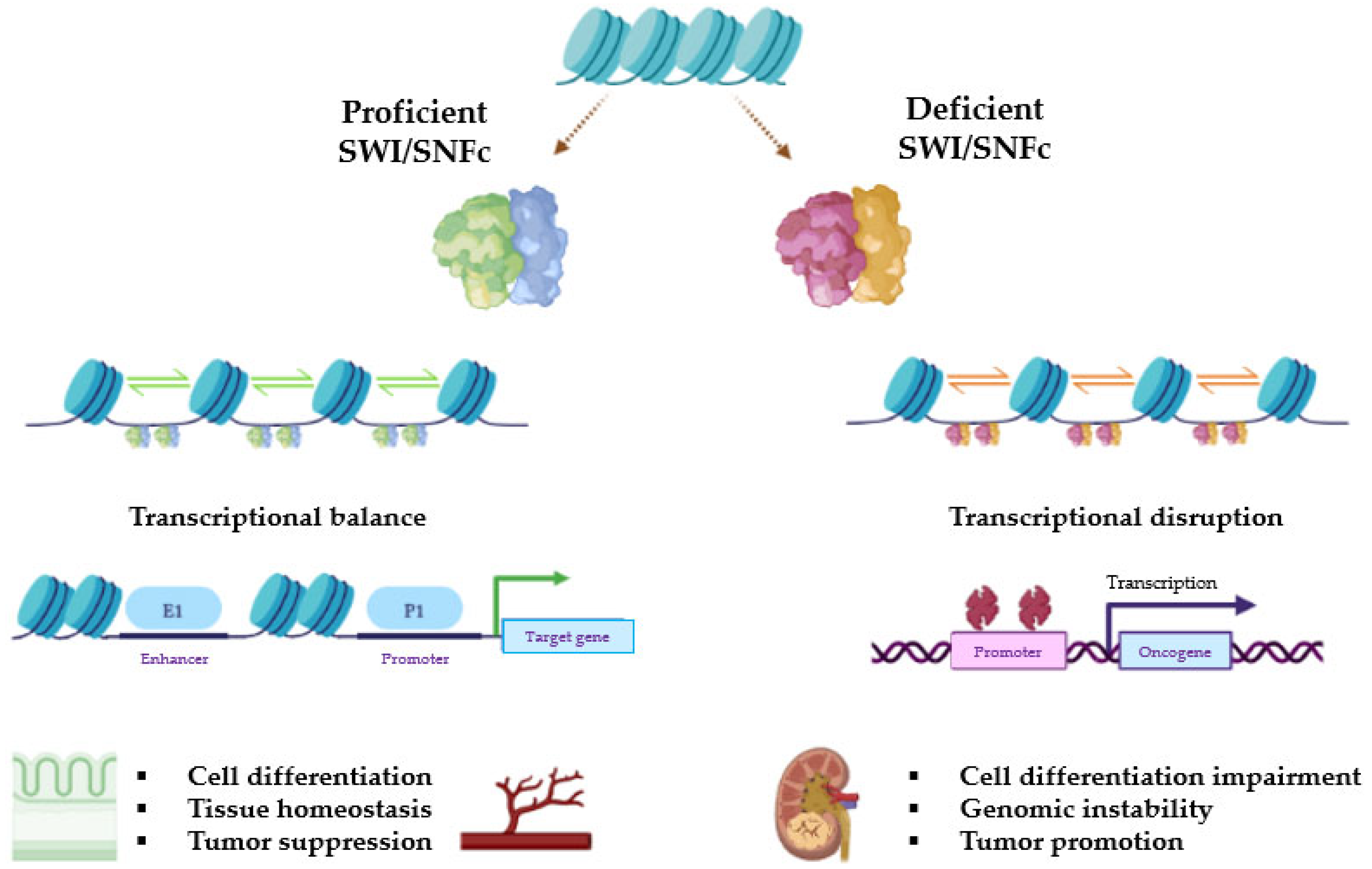
Based on their composition, SWI/SNF complexes in mammals are divided into three major subfamilies: BR-associated factor (BAF, or SWI/SNF-α), polybromo BRG1-associated factor (PBAF, or SWI/SNF-β), and non-canonical BAF (ncBAF/GBAF) [21]. All complexes contain three types of subunits: the ATPase subunits (SMARCA4/BRG1 or SMARCA2/BRM), which are present in a mutually exclusive manner and harbor the catalytic function, the core subunits (SMARCC1/BAF155, SMARCC2/BAF170, and SMARCB1/BAF47/INI1), which are important for assembly and stabilization of the ATPase, and the complex-specific subunits (e.g., ARID1A/BAF250A and ARID1B/BAF250B for BAF, ARID2/BAF200 and PBRM1/BAF180 for PBAF, or BICRA/GLTSCR1 for ncBAF, among others). Many other proteins are shared among BAF, PBAF, and ncBAF, so the SWI/SNFc may be variable in composition and be encoded by multiple genes (Figure 1). The characteristic combination of these proteins is essential for the identity of these three complexes and their diverse biological functions [16,19,22]. Figure 2 represents general actions and consequences of proficient and deficient SWI/SNF.
Despite this variable composition, the structure of the complexes is largely conserved. Structural models in animals have shown that the SWI/SNFc envelops the nucleosome by forming a clamp shape, with the ATPase and core subunits in contact with the nucleosome. However, SWI/SNFc has no intrinsic ability to bind DNA, but it tends to be recruited by transcription factors close to gene promoters. The core subunits act as a hinge to stabilize the connection between the nucleosome and the complex, while the ATPase can recognize the superhelicoidal location 2.5 (SHL2.5) domain within the DNA, where the energy released from ATP hydrolysis breaks the contacts between histones and DNA [23,24]. This causes localized disruption to the chromatin structure, making it more accessible to transcriptional regulators and RNA. Chromatin remodeling is carried out by dynamic forces that slide, displace, or destabilize nucleosome components or even eject histone dimers, leading to the regulation of transcription in large sets of genes [25,26]. cBAF is mainly active at enhancers, whereas PBAF and ncBAF are reported to be enriched at promoters, although they can also bind to some enhancers [27].
Bromodomains in SMARCA4 and SMARCA2 play an important role. They typically bind to acetylated lysines on the N-terminal tails of histones H3 and H4, as well as other proteins. This binding is critical for stable interaction with promoters, which are essential for differentiation-specific gene programs [28]. Bromodomain-containing proteins have been classified into different groups based on their structure. SMARCA2, SMARCA4, and PBRM1 belong to family VIII of bromodomains, which are located in the C-terminal region of the protein [29].

2.3. Role in Carcinogenesis

SWI/SNF plays a critical role in various cellular processes, such as cell cycle control, cell differentiation, apoptosis, or metabolism.
In 1997, Trouche et al. showed that retinoblastoma (RB) protein must cooperate with BRG1 to induce complete G1 arrest through inhibition of the E2F1 transcription factor [30]. A few decades later, Ruijtenberg et al. demonstrated that SWI/SNFc could induce cell cycle arrest in proliferating muscle cell precursors. Loss of SWI/SNF function also impaired the specific gene expression in differentiated cell types, also promoting failure to exist in the cell cycle [31]. Further research has evidenced the key role of some SWI/SNF subunits in cell cycle regulation by promoting arrest [32,33] or programmed cell death [34].
It has been proposed that SWI/SNFc is also involved in many key differentiation and developmental processes in mammalian tissues, such as adipocytes, hematopoietic cells, neurons, osteoblasts, or muscle cells [24]. Klochendler–Yeivin et al. showed that loss-of-function mutations in the SNF5 gene were detrimental to the early development of cell embryos, with lethality in nullizygous blastocysts [6].
DNA damage repair (DDR) is another well-established non-transcriptional function involving SWI/SNF. Between 2009 and 2010, a number of authors reported that BAF and PBAF could be gathered around sites of DNA damage, including the phosphorylation of BAF170 dependent on ATM and ATR, or the existence of cooperative structures between SWI/SNF and γ-H2A.X for the repair of DNA double-strand breaks (DSBs) [35,36]. DSBs are one of the most deleterious forms of DNA damage, leading to genome instability if not repaired.
There is a robust signaling cascade initiated in response to DNA DSBs that can lead to transcriptional upregulation of repair genes, cell cycle arrest, and, in some cases, programmed cell death, in which the SWI/SNFc can participate [37]. Once DNA DSBs are detected and signaled, two major repair pathways are activated: non-homologous end joining (NHEJ) and homologous recombination (HR). SWI/SNFc has been implicated in both, as well as in other cellular pathways, such as alternative end joining, although its role in these is limited. Current evidence suggests that BAF plays a critical role in the process of NHEJ. Specifically, BAF is required to restructure chromatin adjacent to DNA DSBs in order to facilitate the binding of repair factors [38].
The role of chromatin remodeling complexes in the process of HR—particularly the SWI/SNFc—is important, given the requirement for manipulation of the chromatin flanking the DSB and the sister chromatid during strand invasion [35]. In addition, the HR process does not function effectively in cells lacking SWI/SNFc subunits [39,40]. Therefore, the loss of SWI/SNFc function can lead to defective DNA repair and increased sensitivity to DNA damage.
SWI/SNF also has a role in maintaining chromosomal stability. First, many specific regions critical for chromosome organization, such as the binding sites of CTCF and cohesins, are enriched with BAF and PBAF, suggesting that these complexes are critical for regulating the overall chromatin structure [41,42]. Moreover, Brownlee et al. found that a deleterious function of PBRM1 facilitated aneuploidy due to its role in sister chromatid cohesion [43].
Crosstalk with key proliferation, survival, and cell cycle control pathways has also been described. Recent evidence has shown that SWI/SNFc may interact with canonical proto-oncogenes such as MYC and tumor suppressor genes such as TP53, CDKN2A, RB1, or BRCA1 [44,45,46,47]. Although the nature and impact of these interactions on carcinogenesis remain poorly understood, it is well established that the SWI/SNFc plays a crucial role in facilitating p53’s ability to mediate gene expression and exert its tumor suppressor functions. In addition, this interaction is relevant for the regulation of VEGFR2 through chromatin remodeling [48].
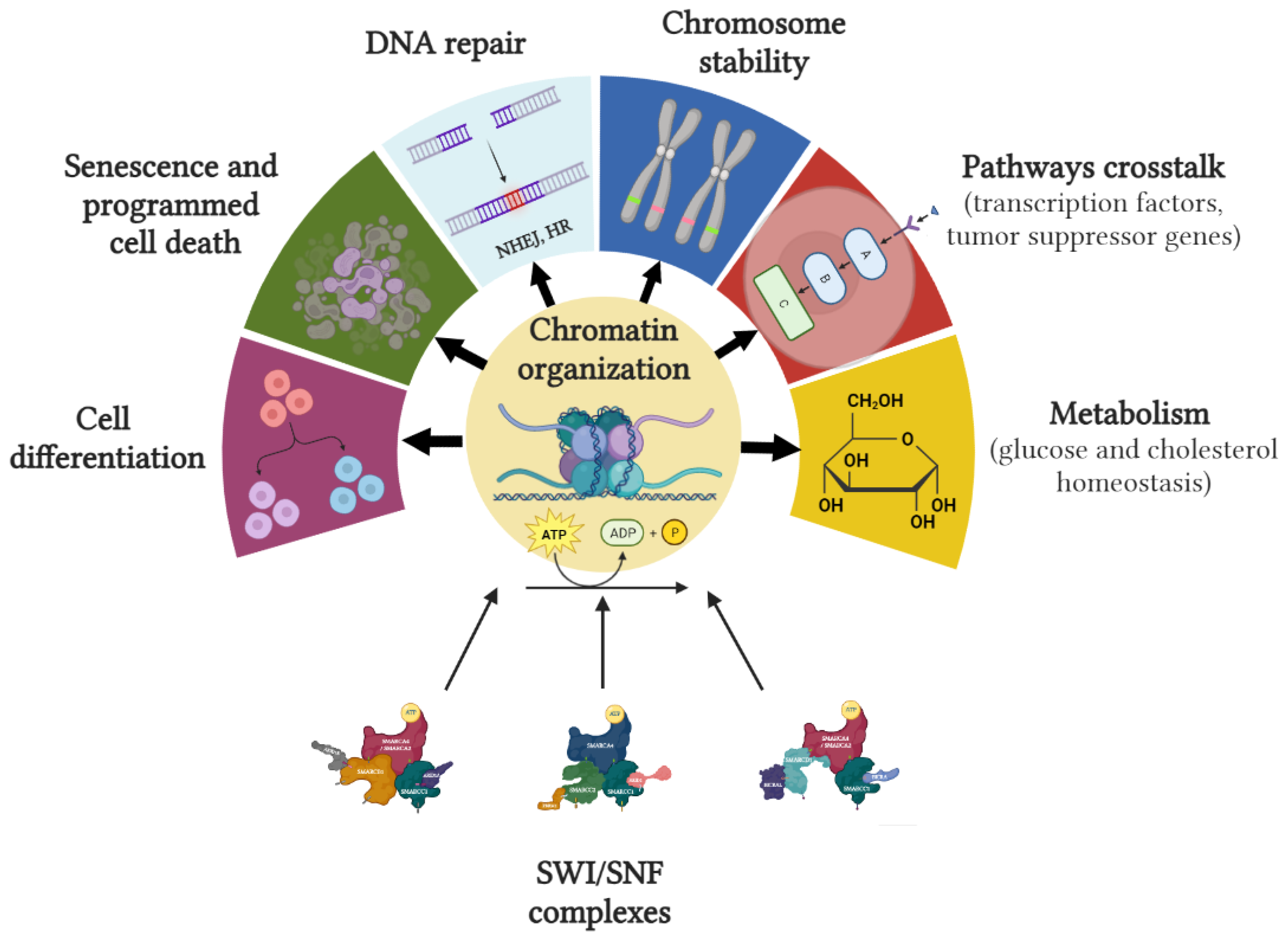
Figure 3 summarizes cellular processes in which SWI/SNF is involved.
Given that large-scale cancer analyses, such as the TCGA, have demonstrated a high rate of mutations in genes involved in SWI/SNFc (nearly 25% in more than 100,000 tumors), there is a strong rationale for accelerating research and finding therapeutic approaches that target SWI/SNF aberrations. Loss-of-function mutations are the most common alterations leading to SWI/SNF inactivation, but the TCGA also revealed other genomic aberrations, like amplifications or overexpression. Consequently, mutations in SWI/SNF genes can lead to tumor suppressor or oncogene functions, the former being the most commonly described in the literature [49]. Biallelic inactivation of SMARCB1 due to nonsense mutations or gene deletions, resulting in complete loss of protein, was the first SWI/SNF alteration found [50]. In contrast to this tumor suppressor role, gain-of-function of ARID1A and SMARCA4 was found in hepatocellular carcinoma and in breast cancer, respectively, suggesting that SWI/SNFc alterations could also act as oncogenes [51]. Deregulation of SMARCA4 has also been described in lung adenocarcinoma, medulloblastoma, pancreatic adenocarcinoma, and Burkitt’s lymphoma, with a variable mutation rate [52,53,54,55]. ARID1A mutations have also been reported in gastric cancer, colorectal cancer, and cholangiocarcinoma [56,57,58].
In summary, cellular function and cell cycle phase appear to be important in defining the potential consequences of SWI/SNFc alterations in human tumors.

3. Rhabdoid Tumors Associated with SWI/SNF Complex Alterations

Mutations in SWI/SNF can arise either during tumor development or as an initiating oncogenic driver event. Solid tumors with rhabdoid features are one of the best examples of SWI/SNF disruption leading to carcinogenesis, sometimes by well-characterized complete loss of SMARCA4 or SMARCB1. These SWI/SNF-mutated monogenic diseases correspond to a large number of solid malignancies with an aggressive clinical course and an ominous prognosis but also with a clear target to be reverted.
Etymologically, “rhabdoid” comes from the Greek term rhabdoeidēs, meaning “rod-shaped”. In Pathology, this feature is characterized by the presence of sheets and clusters of variably cohesive, large cells (sometimes with prominent nucleoli) and large, paranuclear intracytoplasmic hyaline globules [59]. Rhabdoid tumors include a few rare and aggressive tumors, most of which are sarcomas, affecting mainly the pediatric population.
Epithelioid sarcoma (ES): ES is a rare and aggressive soft tissue sarcoma (<1% of all sarcomas) of young adulthood. Characteristically, it grows as a painless mass in the distal limbs. Up to 30–50% of cases may present with lymph node or visceral metastases. This tumor is molecularly characterized by a complete lack of SMARCB1 expression, identified as loss of INI1 immunohistochemical stain in 90% of cases [60,61]. Biallelic-inactivating SMARCB1 mutations are the most frequent aberration. Nonsense frameshift and splice site mutations complete the causes of SMARCB1 loss, but these are quite rare [62,63,64]. Preclinical data have suggested that some miRNAs (miR-193a-5p, miR-206, miR-381, miR-671-5p) are involved in SMARCB1 inactivation through epigenetic mechanisms [65,66,67]. However, this hypothesis remains to be confirmed. SMARCB1 deficiency disrupts essential molecular pathways of cell cycle control, gene transcription, and cell survival, leading to upregulation of MYC, Wnt/β-catenin, and Sonic Hedgehog signaling [68,69,70] and enhancing the oncogenic process. In particular, the antagonistic activity between SMARCB1 and EZH2, the catalytic subunit of Polycomb Repressive Complex 2 (PRC2), has provided the rationale for the approval of tazemetostat in these patients, as discussed later.
Malignant rhabdoid tumor (MRT): This is a rare and highly malignant neoplasm of childhood (usually occurring within the first 2 years of life). Regardless of location, all rhabdoid tumors are highly aggressive and have a poor prognosis. Anatomically, MRT can arise from any site, but the central nervous system (CNS) is the most common site, followed by the kidney and soft tissues. Nearly 95% of MRTs have mutations in SMARCB1, and in very few cases, SMARCA4 is altered (<5%) [71]. Complete inactivation of SMARCB1 has been associated with large deletions of chromosome 22, whole exon duplications or deletions, and point mutations leading to stop codons [72]. A predisposition syndrome has also been described in families with germline mutations in these genes [73], requiring a second somatic hit given the tumor suppressor nature of SMARCB1. Loss of SMARC leads to destabilization of SWI/SNF function and results in a more hypomethylated state of the chromatin in cell lines [74]. Despite these homogeneous molecular features, MRT phenotypes are highly variable, and many subclassifications have been proposed [14]. Multimodal therapy, including radical surgery followed by chemotherapy, intrathecal methotrexate (in CNS MRT), and radiotherapy, is the recommended approach. Recently, tazemetostat has shown good results on MRT [63].
Small-cell carcinoma of the ovary, hypercalcemic variant (SCCOHT): SCCOHT is a very rare tumor of the ovary (<0.5% of malignant ovarian tumors), mostly affecting women under the age of 30. A painful pelvic mass associated with hypercalcemia should prompt consideration of this diagnosis, as this endocrine disorder is present in 60% of patients [75,76]. The prognosis is poor, with a long-term survival rate of approximately 30%. More than 95% of SCCOHTs harbor deleterious mutations in SMARCA4 (biallelic in 25%), and these have been described in both germline and somatic lines [77]. Loss of SMARCA2 by epigenetic inactivation [78] and SMARCB1/ARID1A inactivating mutations may also occur but are extremely rare [75,79]. Cytotoxic chemotherapy is the cornerstone of treatment in the advanced setting, but further research is needed due to poor outcomes. In an early clinical trial, tazemetostat was shown to control disease in some anecdotal cases [63]. Different approaches targeting histone deacetylases, tyrosine kinase receptors (TKR), and ICI are under investigation.
Renal medullary carcinoma (RMC): RMC is a rare tumor that originates in the kidney and tends to be aggressive and resistant to standard therapy. It usually occurs in young patients who are characteristically affected by sickle cell traits or other hemoglobinopathies [80]. Molecularly, RMC shows a complete loss of SMARCB1 expression, which may occur due to inactivating translocations or deletions [81]. The hypertonic environment of the interstitial space within the renal medulla is thought to be a major trigger for DNA double-strand breaks, which, in turn, would be the underlying mechanism of SMARCB1 inactivation [67].
Malignant peripheral nerve sheath tumor (MPNST): This is an invasive soft tissue sarcoma arising from both malignant schwannoma cells and malignant rhabdomyoblasts. Approximately 50% of cases are associated with neurofibromatosis type 1 [82]. The combined loss of NF1 and SMARCB1 is explained by large deletions or biallelic mutations following a sequential “4-hit” mechanism. In addition, some studies have reported that germline mutations in SMARCB1 can cause familial schwannomatosis and meningiomas [83]. MPNST also frequently shows chromosomal abnormalities, such as copy number variants (CNVs) of chromosome 17 or 9p21 losses and aberrations in TP53, CDKN2A, SUZ12, and RASSF1 (the latter two are core components of the PRC2). Several single nucleotide variants (SNVs) have been found in TKR, but their oncogenic contribution remains unclear [84].
Myoepithelial carcinoma: This is a rare tumor that mainly affects the salivary glands, breast, soft tissues, and, less commonly, other organs such as the lungs. It may mimic a pleomorphic adenoma but has rhabdoid features and SMARCB1 mutations [85].
Extra-skeletal myxoid chondrosarcoma (ESMC): This is a sarcoma with a propensity for local recurrence and development of metastases despite an indolent clinical course. It is resistant to chemotherapy. This tumor is associated with tumor-specific translocations involving the Ewing’s Sarcoma (EWS) gene and not infrequently with SMARCB1 mutations [86].
Poorly differentiated chordoma: This tumor is rare, usually affects adults, and arises at the base of the skull and spine. Cases of chordoma diagnosed at a young age are often poorly differentiated, with cytologic atypia, increased cellularity, and mitosis, and their aggressive behavior is associated with a high incidence of metastatic and short patient survival. Recent studies have described the loss of SMARCB1 in poorly differentiated chordomas [87].
Rhabdomyosarcoma: This is the most common sarcoma in childhood, accounting for up to 5–10% of all pediatric malignancies. Four subtypes have been described, all with different clinical features. Molecularly, rhabdomyosarcoma can be characterized by oncogenic drivers, such as FOXO1 fusions, MYOD1 mutations, VGLL2 fusions, and TFCP2 fusions. The embryonal subtype harbors mutations affecting the RAS family proteins and TP53 [88]. No specific SWI/SNFc aberrations characterize this tumor, but recent data support the importance of SMARCA4 expression in the maintenance of alveolar and embryonal rhabdomyosarcoma cells. In these cases, SMARCA4 expression favors cell growth, as knockdown of the gene experimentally affected the viability of cell lines. On the other hand, high expression of SMARCA2 has been associated with reduced survival in a cohort of patients [89].

4. Therapeutic Approaches

4.1. Strategies Focusing on SWI/SNFc and Related Targets

Table 1 lists early clinical trials testing drugs in SWI/SNF-altered solid tumors, with a focus on rhabdoid tumors. Different therapeutic strategies have been grouped into five categories according to their mechanism of action or targeted pathway, as described below.

4.1.1. Targeting SWI/SNF Subunits

Mutations in genes encoding specific SWI/SNF subunits create shared dependencies with other subunits and partners of the complex, conferring a vulnerability that can promote synthetic lethal mechanisms. For example, a mutated subunit would not completely disable SWI/SNF function because it could be partially compensated by its paralogue gene. However, if both deficiencies occur together, this would lead to cell death, causing lethality [90]. In rhabdomyosarcoma, dual depletion of SMARCA4 and SMARCA2 by protein degradation enzymes has been shown to inhibit tumor growth. Also, SMARCA4 inhibition with histone deacetylase 3 is another approach to target SWI/SNF in wild-type tumors.
Several intra- and inter-complex vulnerabilities have been found to be associated with synthetic lethal phenotype in SWI/SNF-deficient tumors. Examples of intra-complex dependencies are mutations of ARID1A with its paralogue ARID1B or mutations of SMARCA4 with SMARCA2. There are also extra-complex dependencies, such as mutations of SMARCB1 with its no-paralogue gene BRD9 [27].
Drugs targeting SWI/SNF have mainly focused on the ATPase and SMARCA2/4 bromodomain subunits, seeking synthetical lethality by inactivating specific proteins of the complex or neutralizing their overexpression. Furthermore, there are some ongoing trials testing BRD9 inhibitors in tumors with SMARCB1 loss or SMARCA2 degradation in SMARCA4-mutant tumors (Table 1).

4.1.2. Targeting PRC via EZH2

SWI/SNF and PRC have opposing gene-regulatory functions. While SWI/SNF normally locates at sites marked by histone H3K27 and cooperates with transcription factors to open chromatin for transcription, PRC (mainly PCR2) acts through its enzymatic subunit EZH2, blocking H3K27 by methylation and repressing transcription.
There is an antagonistic relationship between EZH2 and SMARCB1 (as well as other subunits of the SWI/SNFc), resulting in genetic dependence on EZH2 in some SWI/SNF-mutant cancers. SMARCB1 inactivation results in increased levels of methylated H3K27 (promoted by PCR2) [27]. High levels of EZH2 often correlate with advanced tumor stage and poor prognosis, so inhibition of EZH2 may block proliferation and survival. Currently, EZH2 is a therapeutic target with an approved drug, tazemetostat. It was approved by the Food and Drug Administration (FDA) in January 2020 following the results of a phase 2 clinical trial in advanced ES with loss of INI1/SMARCB1. Tazemetostat activity was evaluated in 62 patients with metastatic or locally advanced ES with INI1 loss. The overall response rate (ORR) was 15%. Of these responses, 67% lasted ≥ 6 months with acceptable tolerability [63].
Given this benefit of tazemetostat, many other ongoing phase 1 and 2 trials are assessing its activity in rhabdoid tumors (Table 1).

4.1.3. Targeting DDR Process

Non-transcriptional roles of SWI/SNF in DNA repair may be exploited therapeutically, as loss-of-function mutations in SWI/SNF genes could be potential biomarkers for inhibitors of the DNA damage response, such as PARP inhibitors (Table 1).

4.1.4. Targeting TKR

The presence of links to other known oncogenic pathways (MYC, RAS) may serve as therapeutic targets that could potentially be inhibited by small molecules. However, most SWI/SNF proteins act as tumor suppressors, making restoration of their function with current drugs extremely challenging.
Interestingly, a dependency between rhabdoid tumors and ARID1A-mutated ovarian cancer and some TKRs has been described, especially EGFR and HER2, but also FGFR, IGF, and c-MET [27,91]. As a result, studies are underway to evaluate the blockade of aberrant TKR in these tumors (Table 1).

4.1.5. Targeting Kinases Involved in Cell Cycle

CDK4/6 inhibitors and other agents that may induce synthetic lethality by impairing DDR (ATR inhibitors and platinum chemotherapy) are also under investigation. A synthetic lethal interaction between SMARCA4 loss and CDK4/6 inhibition, mediated by cyclin D1 deficiency, has been demonstrated. Loss of SMARCA4 causes downregulation of cyclin D1, and there are data supporting that this vulnerability mediates drug sensitivity to CDK4/6 inhibition in SMARCA4-deficient cancer cells [92]. Geoerger et al. evaluated the safety and showed preliminary activity of ribociclib in pretreated neuroblastomas, MRT, and other cancers with documented cyclin D–CDK4/6–INK4–Rb pathways aberrations, including those with SMARCB1 loss [93].
Table 1. Clinical trials targeting SWI/SNF subunits and other molecular pathways in solid tumors harboring SWI/SNFc alterations.
Author/YearNCTStudy DesignNTumorDrugEndpoints/Results and Grade 3–5 AEs
Targeting SWI/SNF complexes
Ongoing05639751Phase I86Advanced SMARCA4-mutant solid tumorsPRT3789 (SMARCA2 degrader)Safety (DLT, MTD, AEs). PK, PD.
Efficacy (ORR, PFS, DOR, BOR).
Ongoing04965753Phase I104Advanced synovial sarcoma and advanced SMARCB1-loss tumorsFHD-609 (BRD9 inhibitor)Safety (TRAEs, AEs, DLTs). PK, PD.
Efficacy (ORR, DOR, PFS, OS).
Ongoing05355753Phase I/II110Adolescents and adults with advanced SMARCB1-altered tumorsCFT8634 (BRD9 inhibitor)Safety (AEs, DLTs). PK, PD.
Efficacy (ORR, DOR, PFS, OS).
Ongoing03297424Phase I/II60–136Advanced malignancies with a known ARID1A mutationPLX2853 (BET inhibitor)Safety (DLT, AEs). PK, PD.
Efficacy (ORR, DOR, OS, PFS).
Targeting PCR via EZH2
Gounder et al. (2020) [63]02601950Phase II62Advanced epithelioid sarcoma with loss of INI1/SMARCB1TazemetostatEfficacy: ORR 15%; DOR not reached; median PFS 5.5 months, median OS 19 months.
AEs: Anemia G3 (6%), weight loss G3 (3%). No G4-5 AEs.
Ongoing01897571Phase II420Advanced-stage solid tumors or B cell lymphomasTazemetostatEfficacy.
Safety (MTD, bioavailability).
Ongoing02601937Phase I82Children with MRT, ATR, RTK, and other tumors with rhabdoid featuresTazemetostatSafety (AEs)
Ongoing03213665Phase II49Children R/R solid tumors, NHL or histiocytic disorders EZH2, SMARCB1, or SMARCA4-mutatedTazemetostatEfficacy (ORR, PFS).
Safety (AEs).
Ongoing02601950Phase II250Adults MRT, ATRT, RTK with loss of SMARCB1 or SMARCA4 or EZH2-mutated tumorsTazemetostatEfficacy (ORR, DOR, PFS). Effect of tazemetostat on immune priming.
Ongoing02875548Phase II300Adults MRT, ATRT, RTK, synovial, or epithelioid sarcoma, mesothelioma, DLBLCTazemetostatEfficacy (PFS, OS). Safety (AEs).
Ongoing05151588Phase II30Locally Advanced SMARCB1-deficient sinonasal carcinomaChemotherapy + TazemetostatEfficacy (BOR, PFS, OS, orbit preservation rate).
Safety (AEs).
Ongoing02601937Phase I109Children and adolescents with R/R INI1-negative tumors or synovial sarcomaTazemetostatSafety (MTD, AEs). PK.
Efficacy (ORR, PFS, OS).
Inhibitors of DNA damage repair
Ongoing0405269Phase II40–116ARID1A-deficient gynecological tumorsCelasertib ± OlaparibEfficacy (BOR)
Ongoing03682289Phase II89BAF250-negative solid tumorsCelasertib ± Olaparib or DurvalumabEfficacy (ORR, DOR, PFS, OS)
Safety (AEs)
Ongoing03207347Phase II57Adults with BAP1 and ARID1-mutant tumorsNiraparibEfficacy (ORR, PFS, OS)
Ongoing02576444Phase II64Adults with cancer containing mutations in homologous DNA repair or other DDR genes, including ARID1AOlaparib + CapivasertibEfficacy (ORR)
Ongoing04065269Phase II40Adults with relapsed gynecological cancers, with or without loss of ARID1AOlaparib + Ceralasertib (AZD6738)Efficacy (ORR, DCR, PFS, TTP, OS)
Ongoing05523440Phase II92Recurrent ovarian or endometrial cancer with ARID1A mutationBevacizumab ± NiraparibEfficacy (ORR, DOR, PFS). Safety (AEs)
Targeting tyrosine kinase receptors
Ongoing03718091Phase II223Adults with advanced-stage solid tumors (including an ARID1-mutant cohort)Berzosertib (VX-970 and M6620; ATR)Efficacy (changes in Phospo-CHK1, ɣH2AX levels and DCR
Ongoing02059265Phase II35Adults with recurrent or persistent gynecological cancer, with or without BAF250 lossDasatinibEfficacy (ORR, PFS, OS) Safety
Ongoing04284202Phase II30Adults with NSCLC ARID1-mutantDasatinib + ToripalimabEfficacy (PFS, OS)
Targeting kinases involved in the cell cycle
Ongoing02114229Phase II180Children and young adults with ATR and or extra-CNS MRT (with loss of SMARCB1 and/or extra-CNS MRT (with loss of SMARCB1 or SMARCA4)Alisertib (Aurora A inhibitor)Efficacy (ORR, PFS); PK; PD
Geoerger et al. (2017) [93]01747876Phase I32Children and young adults with SMARCB1-loss tumorsRibociclibEfficacy: ORR 0%
AEs: Neutropenia G3-4 (63%), leukopenia G3-4 (38%), thrombopenia G3-4 (28%), fatigue G3-4 (3%), AST increased G3-4 (3%), anemia G3-4 (3%), decreased appetite G3-4 (3%)
Abbreviations: AEs: adverse events; ATRT: atypical teratoid rhabdoid tumor; BOR: best overall response; DCR: disease control rate; DLBLC: diffuse large B cell lymphoma; DLT: dose-limiting toxicity; DOR: duration of response; MTD: maximum tolerated dose; NHL: non-Hodgkin lymphoma; ORR: overall response rate; OS: overall survival; PD: pharmacodynamics; PFS: progression-free survival; PK: pharmacokinetics; R/R: recurrent or relapsed; RTK: rhabdoid tumor of the kidney; TRAEs: treatment-related adverse events.

4.2. Immunotherapy Strategies

The first immunotherapy was approved for the treatment of an advanced solid tumor more than three decades ago [94]. Since then, cancer immunotherapy has become one of the cornerstones of cancer treatment, particularly with the advent of ICI. Today, the number of approvals in both advanced and adjuvant settings is increasing as a number of antibodies targeting the programmed cell death protein 1 (anti-PD-1) or its ligand (anti-PD-L1) are being tested in several phase III trials [95,96].
SWI/SNFc aberrations were first proposed as potential predictors of response in a retrospective analysis of patients with advanced renal cell carcinoma treated with ICI. PBRM1 truncating or loss-of-function mutations were demonstrated to be involved in the activation of several pathways (IL6/JAK-STAT3, TNFα signaling via NF-κB and hypoxia-responsive signature), especially in the context of immunostimulatory responses, and thus associated with responsiveness to nivolumab (anti-PD1). Clonality and a high proportion of cells harboring this PBRM1-deficient alteration were associated with tumor responses [97].
In addition, Pan et al. showed how the inactivation of ARID2 or BRD7 in melanoma cell lines attracted effector T cells via interferon (IFN)-γ signaling [97]. Other preclinical studies have associated ARID1A-mutant cancers with an increased number of TIL, higher PD-L1 expression, and the benefit from ICI treatment [98,99,100].
Rhabdoid tumors with specific SWI/SNFc subunit mutations (mostly SMARCB1 and SMARCA4) have been shown to be associated with immune infiltration in the tumor microenvironment, monocytes/macrophages and CD8+ T lymphocytes being the two most predominant subtypes. SMARCB1 deficiency seems to impact critically on epigenetic regulation and immunogenicity, as SMARCB1 mutation is the predominant genomic aberration able to trigger the IFN pathway in these tumors. These preliminary results require prospective clinical validation [13].
Those findings may be applicable to other rare malignancies. For example, Leruste et al. reported complete responses in mice with rhabdoid tumors receiving anti-PD1, and Jelinic et al. published durable responses in four patients with SCCOHT treated with anti-PD1 monotherapy, most of whom had deleterious mutations of SMARCA4 [13,101]. An RNA sequencing study in mouse cell lines found a correlation between lower expression of ARID2 and PBRM1 and higher CD8+ T cell-mediated cytotoxicity. Furthermore, data extracted from TCGA melanoma patients showed that ARID2 mRNA levels correlated with survival depending on CD8+ expression [98]. The same investigators demonstrated the enrichment of dendritic cells and a favorable ratio of M1-like macrophages to tumor-promoting M2-like macrophages in PBRM1-deficient tumors, providing additional evidence for the use of immunotherapy targeting the tumor microenvironment.
Therefore, previously described alterations in individual SWI/SNF proteins may confer susceptibility to immunotherapy in rhabdoid tumors with scarce therapeutic options and poor prognosis.

4.2.1. Immune-Checkpoint Inhibition

Preclinical evidence and case reports of susceptibility to ICI are increasingly being reported. Recently, Wang et al. published the first study of alterations in the 31 members of the SWI/SNFc and their association with ICI outcomes, demonstrating favorable overall survival (OS) and progression-free survival (PFS) in some cancer types, supporting the results of previous publications [102].
Another study, the AcSé trial, showed 50% ORR in the subgroup of SMARCA4-deficient rhabdoid tumors, higher than in other rare sarcomas included. One-year PFS and OS were also remarkable (83% and 62.5%, respectively) [103].
Table 2 provides a summary of current phase I and II trials with immunotherapy in patients with rhabdoid tumors harboring SWI/SNF alterations.

4.2.2. Adoptive Cell Therapy

Adoptive cell therapy (ACT) encompasses a range of strategies based on isolation, modification, and infusion of T lymphocytes to overcome tumor evasion and redirect the immune response. The most common modalities of ACT are engineered T cell receptor (TCR) therapy, chimeric antigen receptor (CAR) T cell therapy, and tumor-infiltrating lymphocyte (TIL) therapy [104].
As epigenetic disruption leads to the accumulation of errors in replication, with subsequent mutations, the production of neoantigens increases. These aberrant antigens are derived from tumor cells and can act as potent activators of CD8+ T cell responses [105]. Immunogenicity in rhabdoid tumors with defective SWI/SNFc may derive from the antigens themselves, although the tumor mutational burden (TMB) is another factor to justify T cell infiltration. Despite the hypothetical increased antigen presentation, not all solid tumors with a higher production of self-antigens express high TMB, as can be observed in certain sarcomas, such as synovial and desmoplastic round cell tumors [106].
ACT could be proposed as a new therapeutic option in SWI/SNFc-altered rhabdoid tumors due to its ability to recognize specific antigens. The main feature that makes these solid tumors candidates for ACT is the predominance of monogenic mutations in a high percentage of cases (e.g., SMARCB1 in ES, MRT or RMC, SMARCA4 in SCCOHT). In addition, natural T lymphocyte infiltration can be exploited to potentially use ICI with ACT to enhance the cytotoxic response.
TCR therapy is dependent on major histocompatibility complex (MHC) presentation, whereas CAR T and TIL strategies are not. The former requires the isolation of T cells that recognize specific tumor antigens and subsequent transduction of the receptor α and β chains by lentiviral or retroviral vectors. Although this treatment is highly specific, it is not generalizable due to its dependence on the MHC.
CAR T therapy involves the generation of a chimeric molecule combining two fragments: one from an antibody (with recognition capability) and one from the TCR. This avoids the MHC-mediated recognition step, making it applicable to any patient. Currently, CAR T therapies are not approved for the treatment of solid tumors. A major concern is the high rate of toxicity (on-target/off-tumor and cytokine release syndrome) due to the ubiquitous nature of the targeted antigens, which limits their use.
Clinical data on TCR and CAR T strategies in tumors with rhabdoid features are limited to a few phase I and II trials including soft tissue sarcomas, with overall poor results [107,108,109,110,111]. New York esophageal squamous cell carcinoma (NY-ESO)-1 and melanoma-associated antigen (MAGE)-A4 have been selected for engineered TCR (restricted by HLA). To date, no clinical trials have been conducted targeting SWI/SNF antigens.
TIL therapy consists of isolating tumor-specific T lymphocytes within the tumor that recognize tumor-specific antigens. TILs are artificially expanded and later administered following a protocol of lymphodepleting chemotherapy. This strategy has the advantage of using more TCR subpopulations than CAR T and TCR therapies, which target different specific antigens. A number of studies using TILs have been conducted in solid tumors, mainly melanoma [112,113,114,115]. TILs therapy may represent a novel approach for rhabdoid tumors, as TILs could recognize specific aberrations in SWI/SNFc, such as complete loss of SMARCA4 or SMARCB1 or other neoantigens created by epigenetic changes occurring within the tumor. Moreover, high lymphocytic infiltration allows for the possibility of expansion if the tumor is amenable to surgery or fresh biopsy.
A multicenter phase II study of TILs treatment in advanced tumors with monogenic SWI/SNFc alterations (the TILTS study) will evaluate the clinical activity of autologous TILs as a single therapy in patients affected by unresectable or advanced rhabdoid tumors (EU CT number 2023-504632-17-00).

5. Conclusions

SWI/SNF alterations are present in up to 25% of solid tumors. Their role in oncoge-nesis has attracted preclinical and clinical research, as several cellular processes depend on the correct function of SWI/SNF genes. However, the huge diversity of proteins involved in SWI/SNF makes this novel therapeutic approach challenging.
Rhabdoid tumors are closely associated with SWI/SNF alterations. The lack of effective therapies in the metastatic setting is an unmet clinical need for these patients. As a result, many phase I and II clinical trials have tested drugs targeting a variety of pathways, including SWI/SNF itself, PCR through the EZH2 subunit, or cell cycle kinases. Despite these efforts, only tazemetostat has been approved for the treatment of metastatic or locally advanced epithelioid sarcoma that is ineligible for complete resection. The other therapeutic strategies have not been successful, but a large number of ongoing trials with novel monotherapy or combinatorial approaches will expand our knowledge in this setting.
T cell infiltration and the presence of an immunogenic microenvironment have been described in SWI/SNF-altered rhabdoid tumors. For this reason, immunotherapy has emerged as a promising treatment for these tumors. Indeed, some responses with prolonged survival have been reported. Currently, some early clinical trials are recruiting using anti-PD(L)-1-based strategies. On the other hand, ACT represents a new treatment option for patients with solid tumors harboring defective SWI/SNF subunits. Therefore, further research on ACT (especially TILs therapy) in patients with rhabdoid tumors is warranted.

Author Contributions

Conceptualization, J.J.S.-C. and J.M.-L.; methodology, J.J.S.-C., L.L.-M., R.F.-C., P.A.-C. and J.M.-L.; software, J.J.S.-C.; validation, J.J.S.-C., R.M., C.C., X.G.d.M., R.A., J.M.P. and J.M.-L.; formal analysis, J.J.S.-C. and J.M.-L.; writing—original draft preparation, J.J.S.-C. and J.M.-L.; writing—review and editing J.J.S.-C., L.L.-M., R.F.-C., P.A.-C. and J.M.-L.; supervision, J.M.-L.; project administration, C.C.; funding acquisition, J.M.-L. All authors have read and agreed to the published version of the manuscript.

Funding

This research was funded by Instituto de Salud Carlos III through the project ICI21/00110. Funded by European Union—NextGenerationEU. We thank CERCA Programme/Generalitat de Catalunya for institutional support.

Institutional Review Board Statement

Not applicable.

Informed Consent Statement

Not applicable.

Data Availability Statement

Data sharing not applicable. No new data were created or analyzed in this study. Data sharing is not applicable to this article.

Conflicts of Interest

J.J.S.-C. has received travel and accommodations grants from Eisai, MSD, Pfizer; speaker’s bureau from Novartis. L.L.-M. and R.F.-C. declare no conflict of interest. P.A.-C. has received an advisory fee from Ferrer. R.M., C.C. and X.G.M. declare no conflict of interest. R.A. has received honoraria fees from Theriva Biologics for research unrelated to the content described in this paper. J.M.-L. has received lecture fees from Astellas, Bristol-Myers Squibb, MSD, Novartis, Pierre Fabre, Pfizer, Roche, Sanofi; advisory fees from Bristol-Myers Squibb, Highlight Therapeutics, Novartis, Pierre Fabre, Roche, Sanofi; and travel grants from Bristol-Myers Squibb, MSD, Novartis, Pierre Fabre, Pfizer, Roche, Ipsen.

References

  1. Hanahan, D. Hallmarks of Cancer: New Dimensions. Cancer Discov. 2022, 12, 31–46. [Google Scholar] [CrossRef]
  2. Kwon, H.; Imbalzano, A.N.; Khavari, P.A.; Kingston, R.E.; Green, M.R. Nucleosome Disruption and Enhancement of Activator Binding by a Human SW1/SNF Complex. Nature 1994, 370, 477–481. [Google Scholar] [CrossRef] [PubMed]
  3. Shain, A.H.; Pollack, J.R. The Spectrum of SWI/SNF Mutations, Ubiquitous in Human Cancers. PLoS ONE 2013, 8, e55119. [Google Scholar] [CrossRef] [PubMed] [Green Version]
  4. Kadoch, C.; Hargreaves, D.C.; Hodges, C.; Elias, L.; Ho, L.; Ranish, J.; Crabtree, G.R. Proteomic and Bioinformatic Analysis of Mammalian SWI/SNF Complexes Identifies Extensive Roles in Human Malignancy. Nat. Genet. 2013, 45, 592–601. [Google Scholar] [CrossRef] [Green Version]
  5. Roberts, C.W.M.; Galusha, S.A.; McMenamin, M.E.; Fletcher, C.D.M.; Orkin, S.H. Haploinsufficiency of Snf5 (Integrase Interactor 1) Predisposes to Malignant Rhabdoid Tumors in Mice. Proc. Natl. Acad. Sci. USA 2000, 97, 13796–13800. [Google Scholar] [CrossRef] [PubMed]
  6. Klochendler-Yeivin, A.; Fiette, L.; Barra, J.; Muchardt, C.; Babinet, C.; Yaniv, M. The Murine SNF5/INI1 Chromatin Remodeling Factor Is Essential for Embryonic Development and Tumor Suppression. EMBO Rep. 2000, 1, 500–506. [Google Scholar] [CrossRef] [Green Version]
  7. Sévenet, N.; Sheridan, E.; Amram, D.; Schneider, P.; Handgretinger, R.; Delattre, O. Constitutional Mutations of the HSNF5/INI1 Gene Predispose to a Variety of Cancers. Am. J. Hum. Genet. 1999, 65, 1342–1348. [Google Scholar] [CrossRef] [Green Version]
  8. Tolstorukov, M.Y.; Sansam, C.G.; Lu, P.; Koellhoffer, E.C.; Helming, K.C.; Alver, B.H.; Tillman, E.J.; Evans, J.A.; Wilson, B.G.; Park, P.J.; et al. Swi/Snf Chromatin Remodeling/Tumor Suppressor Complex Establishes Nucleosome Occupancy at Target Promoters. Proc. Natl. Acad. Sci. USA 2013, 110, 10165–10170. [Google Scholar] [CrossRef]
  9. Wiegand, K.C.; Shah, S.P.; Al-Agha, O.M.; Zhao, Y.; Tse, K.; Zeng, T.; Senz, J.; McConechy, M.K.; Anglesio, M.S.; Kalloger, S.E.; et al. ARID1A Mutations in Endometriosis-Associated Ovarian Carcinomas. N. Engl. J. Med. 2010, 363, 1532–1543. [Google Scholar] [CrossRef] [Green Version]
  10. Varela, I.; Tarpey, P.; Raine, K.; Huang, D.; Ong, C.K.; Stephens, P.; Davies, H.; Jones, D.; Lin, M.-L.; Teague, J.; et al. Exome Sequencing Identifies Frequent Mutation of the SWI/SNF Complex Gene PBRM1 in Renal Carcinoma. Nature 2011, 469, 539–542. [Google Scholar] [CrossRef] [Green Version]
  11. Brennan, B.; Stiller, C.; Bourdeaut, F. Extracranial Rhabdoid Tumours: What We Have Learned so Far and Future Directions. Lancet Oncol. 2013, 14, e329–e336. [Google Scholar] [CrossRef]
  12. Lu, B.; Shi, H. An In-Depth Look at Small Cell Carcinoma of the Ovary, Hypercalcemic Type (SCCOHT): Clinical Implications from Recent Molecular Findings. J. Cancer 2019, 10, 223–237. [Google Scholar] [CrossRef] [PubMed]
  13. Leruste, A.; Tosello, J.; Ramos, R.N.; Tauziède-Espariat, A.; Brohard, S.; Han, Z.-Y.; Beccaria, K.; Andrianteranagna, M.; Caudana, P.; Nikolic, J.; et al. Clonally Expanded T Cells Reveal Immunogenicity of Rhabdoid Tumors. Cancer Cell 2019, 36, 597–612.e8. [Google Scholar] [CrossRef]
  14. Chun, H.-J.E.; Johann, P.D.; Milne, K.; Zapatka, M.; Buellesbach, A.; Ishaque, N.; Iskar, M.; Erkek, S.; Wei, L.; Tessier-Cloutier, B.; et al. Identification and Analyses of Extra-Cranial and Cranial Rhabdoid Tumor Molecular Subgroups Reveal Tumors with Cytotoxic T Cell Infiltration. Cell Rep. 2019, 29, 2338–2354.e7. [Google Scholar] [CrossRef]
  15. Kassabov, S.R.; Zhang, B.; Persinger, J.; Bartholomew, B. SWI/SNF Unwraps, Slides, and Rewraps the Nucleosome. Mol. Cell 2003, 11, 391–403. [Google Scholar] [CrossRef]
  16. Hernández-García, J.; Diego-Martin, B.; Kuo, P.H.; Jami-Alahmadi, Y.; Vashisht, A.A.; Wohlschlegel, J.; Jacobsen, S.E.; Blázquez, M.A.; Gallego-Bartolomé, J. Comprehensive Identification of SWI/SNF Complex Subunits Underpins Deep Eukaryotic Ancestry and Reveals New Plant Components. Commun. Biol. 2022, 5, 549. [Google Scholar] [CrossRef]
  17. Lessard, J.; Wu, J.I.; Ranish, J.A.; Wan, M.; Winslow, M.M.; Staahl, B.T.; Wu, H.; Aebersold, R.; Graef, I.A.; Crabtree, G.R. An Essential Switch in Subunit Composition of a Chromatin Remodeling Complex during Neural Development. Neuron 2007, 55, 201–215. [Google Scholar] [CrossRef] [PubMed] [Green Version]
  18. Ho, L.; Ronan, J.L.; Wu, J.; Staahl, B.T.; Chen, L.; Kuo, A.; Lessard, J.; Nesvizhskii, A.I.; Ranish, J.; Crabtree, G.R. An Embryonic Stem Cell Chromatin Remodeling Complex, EsBAF, Is Essential for Embryonic Stem Cell Self-Renewal and Pluripotency. Proc. Natl. Acad. Sci. USA 2009, 106, 5181–5186. [Google Scholar] [CrossRef]
  19. Mashtalir, N.; D’Avino, A.R.; Michel, B.C.; Luo, J.; Pan, J.; Otto, J.E.; Zullow, H.J.; McKenzie, Z.M.; Kubiak, R.L.; St. Pierre, R.; et al. Modular Organization and Assembly of SWI/SNF Family Chromatin Remodeling Complexes. Cell 2018, 175, 1272–1288.e20. [Google Scholar] [CrossRef] [Green Version]
  20. Michel, B.C.; D’Avino, A.R.; Cassel, S.H.; Mashtalir, N.; McKenzie, Z.M.; McBride, M.J.; Valencia, A.M.; Zhou, Q.; Bocker, M.; Soares, L.M.M.; et al. A Non-Canonical SWI/SNF Complex Is a Synthetic Lethal Target in Cancers Driven by BAF Complex Perturbation. Nat. Cell Biol. 2018, 20, 1410–1420. [Google Scholar] [CrossRef]
  21. Zhou, M.; Yuan, J.; Deng, Y.; Fan, X.; Shen, J. Emerging Role of SWI/SNF Complex Deficiency as a Target of Immune Checkpoint Blockade in Human Cancers. Oncogenesis 2021, 10, 3. [Google Scholar] [CrossRef] [PubMed]
  22. Pulice, J.L.; Kadoch, C. Composition and Function of Mammalian SWI/SNF Chromatin Remodeling Complexes in Human Disease. Cold Spring Harb. Symp. Quant. Biol. 2016, 81, 53–60. [Google Scholar] [CrossRef] [Green Version]
  23. Sahu, R.K.; Singh, S.; Tomar, R.S. The Mechanisms of Action of Chromatin Remodelers and Implications in Development and Disease. Biochem. Pharmacol. 2020, 180, 114200. [Google Scholar] [CrossRef]
  24. Romero, O.A.; Sanchez-Cespedes, M. The SWI/SNF Genetic Blockade: Effects in Cell Differentiation, Cancer and Developmental Diseases. Oncogene 2014, 33, 2681–2689. [Google Scholar] [CrossRef] [PubMed] [Green Version]
  25. Clapier, C.R.; Iwasa, J.; Cairns, B.R.; Peterson, C.L. Mechanisms of Action and Regulation of ATP-Dependent Chromatin-Remodelling Complexes. Nat. Rev. Mol. Cell Biol. 2017, 18, 407–422. [Google Scholar] [CrossRef]
  26. Harrod, A.; Lane, K.A.; Downs, J.A. The Role of the SWI/SNF Chromatin Remodelling Complex in the Response to DNA Double Strand Breaks. DNA Repair 2020, 93, 102919. [Google Scholar] [CrossRef]
  27. Mittal, P.; Roberts, C.W.M. The SWI/SNF Complex in Cancer—Biology, Biomarkers and Therapy. Nat. Rev. Clin. Oncol. 2020, 17, 435–448. [Google Scholar] [CrossRef]
  28. Awad, S.; Hassan, A.H. The Swi2/Snf2 Bromodomain Is Important for the Full Binding and Remodeling Activity of the SWI/SNF Complex on H3- and H4-Acetylated Nucleosomes. Ann. N. Y. Acad. Sci. 2008, 1138, 366–375. [Google Scholar] [CrossRef]
  29. Khavari, P.A.; Peterson, C.L.; Tamkun, J.W.; Mendel, D.B.; Crabtree, G.R. BRG1 Contains a Conserved Domain of the SWI2/SNF2 Family Necessary for Normal Mitotic Growth and Transcription. Nature 1993, 366, 170–174. [Google Scholar] [CrossRef]
  30. Trouche, D.; Le Chalony, C.; Muchardt, C.; Yaniv, M.; Kouzarides, T. RB and Hbrm Cooperate to Repress the Activation Functions of E2F1. Proc. Natl. Acad. Sci. USA 1997, 94, 11268–11273. [Google Scholar] [CrossRef]
  31. Ruijtenberg, S.; van den Heuvel, S. G1/S Inhibitors and the SWI/SNF Complex Control Cell-Cycle Exit during Muscle Differentiation. Cell 2015, 162, 300–313. [Google Scholar] [CrossRef] [PubMed] [Green Version]
  32. Isakoff, M.S.; Sansam, C.G.; Tamayo, P.; Subramanian, A.; Evans, J.A.; Fillmore, C.M.; Wang, X.; Biegel, J.A.; Pomeroy, S.L.; Mesirov, J.P.; et al. Inactivation of the Snf5 Tumor Suppressor Stimulates Cell Cycle Progression and Cooperates with P53 Loss in Oncogenic Transformation. Proc. Natl. Acad. Sci. USA. 2005, 102, 17745–17750. [Google Scholar] [CrossRef]
  33. Flores-Alcantar, A.; Gonzalez-Sandoval, A.; Escalante-Alcalde, D.; Lomelí, H. Dynamics of Expression of ARID1A and ARID1B Subunits in Mouse Embryos and in Cells during the Cell Cycle. Cell Tissue Res. 2011, 345, 137–148. [Google Scholar] [CrossRef]
  34. Napolitano, M.A.; Cipollaro, M.; Cascino, A.; Melone, M.A.B.; Giordano, A.; Galderisi, U. Brg1 Chromatin Remodeling Factor Is Involved in Cell Growth Arrest, Apoptosis and Senescence of Rat Mesenchymal Stem Cells. J. Cell Sci. 2007, 120, 2904–2911. [Google Scholar] [CrossRef]
  35. Peng, G.; Yim, E.-K.; Dai, H.; Jackson, A.P.; van der Burgt, I.; Pan, M.-R.; Hu, R.; Li, K.; Lin, S.-Y. BRIT1/MCPH1 Links Chromatin Remodelling to DNA Damage Response. Nat. Cell Biol. 2009, 11, 865–872. [Google Scholar] [CrossRef] [Green Version]
  36. Lee, H.-S.; Park, J.-H.; Kim, S.-J.; Kwon, S.-J.; Kwon, J. A Cooperative Activation Loop among SWI/SNF, γ-H2AX and H3 Acetylation for DNA Double-Strand Break Repair. EMBO J. 2010, 29, 1434–1445. [Google Scholar] [CrossRef]
  37. Shibata, A.; Jeggo, P.A. DNA Double-Strand Break Repair in a Cellular Context. Clin. Oncol. 2014, 26, 243–249. [Google Scholar] [CrossRef]
  38. Park, Y.; Chui, M.H.; Suryo Rahmanto, Y.; Yu, Z.-C.; Shamanna, R.A.; Bellani, M.A.; Gaillard, S.; Ayhan, A.; Viswanathan, A.; Seidman, M.M.; et al. Loss of ARID1A in Tumor Cells Renders Selective Vulnerability to Combined Ionizing Radiation and PARP Inhibitor Therapy. Clin. Cancer Res. 2019, 25, 5584–5594. [Google Scholar] [CrossRef]
  39. Vélez-Cruz, R.; Manickavinayaham, S.; Biswas, A.K.; Clary, R.W.; Premkumar, T.; Cole, F.; Johnson, D.G. RB Localizes to DNA Double-Strand Breaks and Promotes DNA End Resection and Homologous Recombination through the Recruitment of BRG1. Genes Dev. 2016, 30, 2500–2512. [Google Scholar] [CrossRef] [Green Version]
  40. de Castro, R.O.; Previato, L.; Goitea, V.; Felberg, A.; Guiraldelli, M.F.; Filiberti, A.; Pezza, R.J. The Chromatin-Remodeling Subunit Baf200 Promotes Homology-Directed DNA Repair and Regulates Distinct Chromatin-Remodeling Complexes. J. Biol. Chem. 2017, 292, 8459–8471. [Google Scholar] [CrossRef] [Green Version]
  41. Bell, A.C.; Felsenfeld, G. Methylation of a CTCF-Dependent Boundary Controls Imprinted Expression of the Igf2 Gene. Nature 2000, 405, 482–485. [Google Scholar] [CrossRef] [PubMed]
  42. Euskirchen, G.M.; Auerbach, R.K.; Davidov, E.; Gianoulis, T.A.; Zhong, G.; Rozowsky, J.; Bhardwaj, N.; Gerstein, M.B.; Snyder, M. Diverse Roles and Interactions of the SWI/SNF Chromatin Remodeling Complex Revealed Using Global Approaches. PLoS Genet 2011, 7, e1002008. [Google Scholar] [CrossRef] [PubMed] [Green Version]
  43. Brownlee, P.M.; Chambers, A.L.; Cloney, R.; Bianchi, A.; Downs, J.A. BAF180 Promotes Cohesion and Prevents Genome Instability and Aneuploidy. Cell Rep. 2014, 6, 973–981. [Google Scholar] [CrossRef] [Green Version]
  44. Oh, J.; Sohn, D.H.; Ko, M.; Chung, H.; Jeon, S.H.; Seong, R.H. BAF60a Interacts with P53 to Recruit the SWI/SNF Complex. J. Biol. Chem. 2008, 283, 11924–11934. [Google Scholar] [CrossRef] [PubMed] [Green Version]
  45. Zhang, J.; Ohta, T.; Maruyama, A.; Hosoya, T.; Nishikawa, K.; Maher, J.M.; Shibahara, S.; Itoh, K.; Yamamoto, M. BRG1 Interacts with Nrf2 to Selectively Mediate HO-1 Induction in Response to Oxidative Stress. Mol. Cell. Biol. 2006, 26, 7942–7952. [Google Scholar] [CrossRef] [Green Version]
  46. Marignani, P.A.; Kanai, F.; Carpenter, C.L. LKB1 Associates with Brg1 and Is Necessary for Brg1-Induced Growth Arrest. J. Biol. Chem. 2001, 276, 32415–32418. [Google Scholar] [CrossRef] [Green Version]
  47. Shanahan, F.; Seghezzi, W.; Parry, D.; Mahony, D.; Lees, E. Cyclin E Associates with BAF155 and BRG1, Components of the Mammalian SWI-SNF Complex, and Alters the Ability of BRG1 To Induce Growth Arrest. Mol. Cell. Biol. 1999, 19, 1460–1469. [Google Scholar] [CrossRef] [Green Version]
  48. Pfister, N.T.; Fomin, V.; Regunath, K.; Zhou, J.Y.; Zhou, W.; Silwal-Pandit, L.; Freed-Pastor, W.A.; Laptenko, O.; Neo, S.P.; Bargonetti, J.; et al. Mutant P53 Cooperates with the SWI/SNF Chromatin Remodeling Complex to Regulate VEGFR2 in Breast Cancer Cells. Genes Dev. 2015, 29, 1298–1315. [Google Scholar] [CrossRef] [Green Version]
  49. Orlando, K.A.; Nguyen, V.; Raab, J.R.; Walhart, T.; Weissman, B.E. Remodeling the Cancer Epigenome: Mutations in the SWI/SNF Complex Offer New Therapeutic Opportunities. Expert Rev. Anticancer Ther. 2019, 19, 375–391. [Google Scholar] [CrossRef]
  50. Biegel, J.A. Molecular Genetics of Atypical Teratoid/Rhabdoid Tumors. Neurosurg. Focus 2006, 20, E11. [Google Scholar] [CrossRef] [Green Version]
  51. Sun, X.; Wang, S.C.; Wei, Y.; Luo, X.; Jia, Y.; Li, L.; Gopal, P.; Zhu, M.; Nassour, I.; Chuang, J.-C.; et al. Arid1a Has Context-Dependent Oncogenic and Tumor Suppressor Functions in Liver Cancer. Cancer Cell 2017, 32, 574–589.e6. [Google Scholar] [CrossRef] [Green Version]
  52. Imielinski, M.; Berger, A.H.; Hammerman, P.S.; Hernandez, B.; Pugh, T.J.; Hodis, E.; Cho, J.; Suh, J.; Capelletti, M.; Sivachenko, A.; et al. Mapping the Hallmarks of Lung Adenocarcinoma with Massively Parallel Sequencing. Cell 2012, 150, 1107–1120. [Google Scholar] [CrossRef] [Green Version]
  53. Shain, A.H.; Giacomini, C.P.; Matsukuma, K.; Karikari, C.A.; Bashyam, M.D.; Hidalgo, M.; Maitra, A.; Pollack, J.R. Convergent Structural Alterations Define SWItch/Sucrose NonFermentable (SWI/SNF) Chromatin Remodeler as a Central Tumor Suppressive Complex in Pancreatic Cancer. Proc. Natl. Acad. Sci. USA 2012, 109, E252–E259. [Google Scholar] [CrossRef]
  54. Robinson, G.; Parker, M.; Kranenburg, T.A.; Lu, C.; Chen, X.; Ding, L.; Phoenix, T.N.; Hedlund, E.; Wei, L.; Zhu, X.; et al. Novel Mutations Target Distinct Subgroups of Medulloblastoma. Nature 2012, 488, 43–48. [Google Scholar] [CrossRef] [Green Version]
  55. Love, C.; Sun, Z.; Jima, D.; Li, G.; Zhang, J.; Miles, R.; Richards, K.L.; Dunphy, C.H.; Choi, W.W.L.; Srivastava, G.; et al. The Genetic Landscape of Mutations in Burkitt Lymphoma. Nat. Genet. 2012, 44, 1321–1325. [Google Scholar] [CrossRef] [Green Version]
  56. Wang, K.; Kan, J.; Yuen, S.T.; Shi, S.T.; Chu, K.M.; Law, S.; Chan, T.L.; Kan, Z.; Chan, A.S.Y.; Tsui, W.Y.; et al. Exome Sequencing Identifies Frequent Mutation of ARID1A in Molecular Subtypes of Gastric Cancer. Nat. Genet. 2011, 43, 1219–1223. [Google Scholar] [CrossRef]
  57. The Cancer Genome Atlas Network. Comprehensive Molecular Characterization of Human Colon and Rectal Cancer. Nature 2012, 487, 330–337. [Google Scholar] [CrossRef] [Green Version]
  58. Chan-on, W.; Nairismägi, M.-L.; Ong, C.K.; Lim, W.K.; Dima, S.; Pairojkul, C.; Lim, K.H.; McPherson, J.R.; Cutcutache, I.; Heng, H.L.; et al. Exome Sequencing Identifies Distinct Mutational Patterns in Liver Fluke-Related and Non-Infection-Related Bile Duct Cancers. Nat. Genet. 2013, 45, 1474–1478. [Google Scholar] [CrossRef]
  59. Gokden, N.; Pfeifer, J.D.; Humphrey, P.A. Renal Cell Carcinoma with Rhabdoid Features. Am. J. Surg. Pathol. 2000, 24, 1329–1338. [Google Scholar] [CrossRef]
  60. Del Savio, E.; Maestro, R. Beyond SMARCB1 Loss: Recent Insights into the Pathobiology of Epithelioid Sarcoma. Cells 2022, 11, 2626. [Google Scholar] [CrossRef]
  61. Needs, T.; Fillman, E.P. Epithelioid Sarcoma. In StatPearls; StatPearls Publishing: Treasure Island, FL, USA, 2023. Available online: https://www.ncbi.nlm.nih.gov/books/nbk532911/ (accessed on 23 April 2023).
  62. Le Loarer, F.; Zhang, L.; Fletcher, C.D.; Ribeiro, A.; Singer, S.; Italiano, A.; Neuville, A.; Houlier, A.; Chibon, F.; Coindre, J.-M.; et al. Consistent SMARCB1 Homozygous Deletions in Epithelioid Sarcoma and in a Subset of Myoepithelial Carcinomas Can Be Reliably Detected by FISH in Archival Material: SMARCB1 Homozygous Deletions in Epithelioid Sarcoma. Genes Chromosomes Cancer 2014, 53, 475–486. [Google Scholar] [CrossRef] [PubMed] [Green Version]
  63. Gounder, M.; Schöffski, P.; Jones, R.L.; Agulnik, M.; Cote, G.M.; Villalobos, V.M.; Attia, S.; Chugh, R.; Chen, T.W.-W.; Jahan, T.; et al. Tazemetostat in Advanced Epithelioid Sarcoma with Loss of INI1/SMARCB1: An International, Open-Label, Phase 2 Basket Study. Lancet Oncol. 2020, 21, 1423–1432. [Google Scholar] [CrossRef] [PubMed]
  64. Baker, T.G.; Lyons, M.J.; Leddy, L.; Parham, D.M.; Welsh, C.T. Epithelioid Sarcoma Arising in a Long-Term Survivor of an Atypical Teratoid/Rhabdoid Tumor in a Patient with Rhabdoid Tumor Predisposition Syndrome. Pediatr. Dev. Pathol. 2021, 24, 164–168. [Google Scholar] [CrossRef] [PubMed]
  65. Kohashi, K.; Yamamoto, H.; Kumagai, R.; Yamada, Y.; Hotokebuchi, Y.; Taguchi, T.; Iwamoto, Y.; Oda, Y. Differential MicroRNA Expression Profiles between Malignant Rhabdoid Tumor and Epithelioid Sarcoma: MiR193a-5p Is Suggested to Downregulate SMARCB1 MRNA Expression. Mod. Pathol. 2014, 27, 832–839. [Google Scholar] [CrossRef] [PubMed] [Green Version]
  66. Papp, G.; Krausz, T.; Stricker, T.P.; Szendrői, M.; Sápi, Z. SMARCB1 Expression in Epithelioid Sarcoma Is Regulated by MiR-206, MiR-381, and MiR-671-5p on Both MRNA and Protein Levels: Smarcb1 Regulation by MiRNAs In Epithelioid Sarcoma. Genes Chromosomes Cancer 2014, 53, 168–176. [Google Scholar] [CrossRef]
  67. Sápi, Z.; Papp, G.; Szendrői, M.; Pápai, Z.; Plótár, V.; Krausz, T.; Fletcher, C.D.M. Epigenetic Regulation of SMARCB1 By MiR-206, -381 and -671-5p Is Evident in a Variety of SMARCB1 Immunonegative Soft Tissue Sarcomas, While MiR-765 Appears Specific for Epithelioid Sarcoma. A MiRNA Study of 223 Soft Tissue Sarcomas: Epigenetic Regulation of SMARCB1 by Mirnas in Soft Tissue Sarcomas. Genes Chromosomes Cancer 2016, 55, 786–802. [Google Scholar] [CrossRef]
  68. Msaouel, P.; Malouf, G.G.; Su, X.; Yao, H.; Tripathi, D.N.; Soeung, M.; Gao, J.; Rao, P.; Coarfa, C.; Creighton, C.J.; et al. Comprehensive Molecular Characterization Identifies Distinct Genomic and Immune Hallmarks of Renal Medullary Carcinoma. Cancer Cell 2020, 37, 720–734.e13. [Google Scholar] [CrossRef]
  69. Mora-Blanco, E.; Mishina, Y.; Tillman, E.; Cho, Y.J.; Thom, C.S.; Pomeroy, S.L.; Shao, W.; Roberts, C.W.M. Activation of β-catenin/TCF targets following loss of the tumor suppressor SNF5. Oncogene 2014, 33, 933–938. [Google Scholar] [CrossRef] [Green Version]
  70. Jagani, Z.; Mora-Blanco, E.L.; Sansam, C.G.; McKenna, E.S.; Wilson, B.; Chen, D.; Klekota, J.; Tamayo, P.; Nguyen, P.T.L.; Tolstorukov, M.; et al. Loss of the Tumor Suppressor Snf5 Leads to Aberrant Activation of the Hedgehog-Gli Pathway. Nat. Med. 2010, 16, 1429–1433. [Google Scholar] [CrossRef] [Green Version]
  71. Finetti, M.A.; Grabovska, Y.; Bailey, S.; Williamson, D. Translational Genomics of Malignant Rhabdoid Tumours: Current Impact and Future Possibilities. Semin. Cancer Biol. 2020, 61, 30–41. [Google Scholar] [CrossRef]
  72. Jackson, E.M.; Sievert, A.J.; Gai, X.; Hakonarson, H.; Judkins, A.R.; Tooke, L.; Perin, J.C.; Xie, H.; Shaikh, T.H.; Biegel, J.A. Genomic Analysis Using High-Density Single Nucleotide Polymorphism-Based Oligonucleotide Arrays and Multiplex Ligation-Dependent Probe Amplification Provides a Comprehensive Analysis of INI1/SMARCB1 in Malignant Rhabdoid Tumors. Clin. Cancer Res. 2009, 15, 1923–1930. [Google Scholar] [CrossRef] [Green Version]
  73. Schneppenheim, R.; Frühwald, M.C.; Gesk, S.; Hasselblatt, M.; Jeibmann, A.; Kordes, U.; Kreuz, M.; Leuschner, I.; Subero, J.I.M.; Obser, T.; et al. Germline Nonsense Mutation and Somatic Inactivation of SMARCA4/BRG1 in a Family with Rhabdoid Tumor Predisposition Syndrome. Am. J. Hum. Genet. 2010, 86, 279–284. [Google Scholar] [CrossRef] [Green Version]
  74. Erkek, S.; Johann, P.D.; Finetti, M.A.; Drosos, Y.; Chou, H.-C.; Zapatka, M.; Sturm, D.; Jones, D.T.W.; Korshunov, A.; Rhyzova, M.; et al. Comprehensive Analysis of Chromatin States in Atypical Teratoid/Rhabdoid Tumor Identifies Diverging Roles for SWI/SNF and Polycomb in Gene Regulation. Cancer Cell 2019, 35, 95–110.e8. [Google Scholar] [CrossRef] [Green Version]
  75. Tischkowitz, M.; Huang, S.; Banerjee, S.; Hague, J.; Hendricks, W.P.D.; Huntsman, D.G.; Lang, J.D.; Orlando, K.A.; Oza, A.M.; Pautier, P.; et al. Small-Cell Carcinoma of the Ovary, Hypercalcemic Type–Genetics, New Treatment Targets, and Current Management Guidelines. Clin. Cancer Res. 2020, 26, 3908–3917. [Google Scholar] [CrossRef] [Green Version]
  76. Matias-Guiu, X.; Prat, J.; Young, R.H.; Capen, C.C.; Rosol, T.J.; Delellis, R.A.; Scully, R.E. Human Parathyroid Hormone-Related Protein in Ovarian Small Cell Carcinoma. An Immunohistochemical Study. Cancer 1994, 73, 1878–1881. [Google Scholar] [CrossRef]
  77. Witkowski, L.; Carrot-Zhang, J.; Albrecht, S.; Fahiminiya, S.; Hamel, N.; Tomiak, E.; Grynspan, D.; Saloustros, E.; Nadaf, J.; Rivera, B.; et al. Germline and Somatic SMARCA4 Mutations Characterize Small Cell Carcinoma of the Ovary, Hypercalcemic Type. Nat. Genet. 2014, 46, 438–443. [Google Scholar] [CrossRef]
  78. Karnezis, A.N.; Wang, Y.; Ramos, P.; Hendricks, W.P.; Oliva, E.; D’Angelo, E.; Prat, J.; Nucci, M.R.; Nielsen, T.O.; Chow, C.; et al. Dual Loss of the SWI/SNF Complex ATPases SMARCA4/BRG1 and SMARCA2/BRM Is Highly Sensitive and Specific for Small Cell Carcinoma of the Ovary, Hypercalcaemic Type. J. Pathol. 2016, 238, 389–400. [Google Scholar] [CrossRef] [Green Version]
  79. Lin, D.I.; Chudnovsky, Y.; Duggan, B.; Zajchowski, D.; Greenbowe, J.; Ross, J.S.; Gay, L.M.; Ali, S.M.; Elvin, J.A. Comprehensive Genomic Profiling Reveals Inactivating SMARCA4 Mutations and Low Tumor Mutational Burden in Small Cell Carcinoma of the Ovary, Hypercalcemic-Type. Gynecol. Oncol. 2017, 147, 626–633. [Google Scholar] [CrossRef]
  80. Msaouel, P.; Tannir, N.M.; Walker, C.L. A Model Linking Sickle Cell Hemoglobinopathies and SMARCB1 Loss in Renal Medullary Carcinoma. Clin. Cancer Res. 2018, 24, 2044–2049. [Google Scholar] [CrossRef] [Green Version]
  81. Calderaro, J.; Masliah-Planchon, J.; Richer, W.; Maillot, L.; Maille, P.; Mansuy, L.; Bastien, C.; de la Taille, A.; Boussion, H.; Charpy, C.; et al. Balanced Translocations Disrupting SMARCB1 Are Hallmark Recurrent Genetic Alterations in Renal Medullary Carcinomas. Eur. Urol. 2016, 69, 1055–1061. [Google Scholar] [CrossRef]
  82. Rizzo, D.; Fréneaux, P.; Brisse, H.; Louvrier, C.; Lequin, D.; Nicolas, A.; Ranchère, D.; Verkarre, V.; Jouvet, A.; Dufour, C.; et al. SMARCB1 Deficiency in Tumors from the Peripheral Nervous System: A Link between Schwannomas and Rhabdoid Tumors? Am. J. Surg. Pathol. 2012, 36, 964–972. [Google Scholar] [CrossRef] [PubMed]
  83. Eaton, K.W.; Tooke, L.S.; Wainwright, L.M.; Judkins, A.R.; Biegel, J.A. Spectrum of SMARCB1/INI1 Mutations in Familial and Sporadic Rhabdoid Tumors: SMARCB1 Mutations in Rhabdoid Tumor. Pediatr. Blood Cancer 2011, 56, 7–15. [Google Scholar] [CrossRef] [PubMed] [Green Version]
  84. Pemov, A.; Li, H.; Presley, W.; Wallace, M.R.; Miller, D.T. Genetics of Human Malignant Peripheral Nerve Sheath Tumors. Neuro-Oncol. Adv. 2020, 2 (Suppl. S1), i50–i61. [Google Scholar] [CrossRef]
  85. Xu, B.; Katabi, N. Myoepithelial Carcinoma. Surg. Pathol. Clin. 2021, 14, 67–73. [Google Scholar] [CrossRef] [PubMed]
  86. Stacchiotti, S.; Baldi, G.G.; Morosi, C.; Gronchi, A.; Maestro, R. Extraskeletal Myxoid Chondrosarcoma: State of the Art and Current Research on Biology and Clinical Management. Cancers 2020, 12, 2703. [Google Scholar] [CrossRef]
  87. Curcio, C.; Cimera, R.; Aryeequaye, R.; Rao, M.; Fabbri, N.; Zhang, Y.; Hameed, M. Poorly Differentiated Chordoma with Whole-genome Doubling Evolving from a SMARCB1-deficient Conventional Chordoma: A Case Report. Genes Chromosomes Cancer 2021, 60, 43–48. [Google Scholar] [CrossRef]
  88. Leiner, J.; Le Loarer, F. The Current Landscape of Rhabdomyosarcomas: An Update. Virchows Arch. 2020, 476, 97–108. [Google Scholar] [CrossRef]
  89. Bharathy, N.; Cleary, M.M.; Kim, J.-A.; Nagamori, K.; Crawford, K.A.; Wang, E.; Saha, D.; Settelmeyer, T.P.; Purohit, R.; Skopelitis, D.; et al. SMARCA4 Biology in Alveolar Rhabdomyosarcoma. Oncogene 2022, 41, 1647–1656. [Google Scholar] [CrossRef]
  90. Helming, K.C.; Wang, X.; Roberts, C.W.M. Vulnerabilities of Mutant SWI/SNF Complexes in Cancer. Cancer Cell 2014, 26, 309–317. [Google Scholar] [CrossRef] [Green Version]
  91. Huang, P.H. Targeting SWI/SNF Mutant Cancers with Tyrosine Kinase Inhibitor Therapy. Expert Rev. Anticancer. Ther. 2017, 17, 1–3. [Google Scholar] [CrossRef] [Green Version]
  92. Xue, Y.; Meehan, B.; Macdonald, E.; Venneti, S.; Wang, X.Q.D.; Witkowski, L.; Jelinic, P.; Kong, T.; Martinez, D.; Morin, G.; et al. CDK4/6 Inhibitors Target SMARCA4-Determined Cyclin D1 Deficiency in Hypercalcemic Small Cell Carcinoma of the Ovary. Nat. Commun. 2019, 10, 558. [Google Scholar] [CrossRef]
  93. Geoerger, B.; Bourdeaut, F.; DuBois, S.G.; Fischer, M.; Geller, J.I.; Gottardo, N.G.; Marabelle, A.; Pearson, A.D.J.; Modak, S.; Cash, T.; et al. A Phase I Study of the CDK4/6 Inhibitor Ribociclib (LEE011) in Pediatric Patients with Malignant Rhabdoid Tumors, Neuroblastoma, and Other Solid Tumors. Clin. Cancer Res. 2017, 23, 2433–2441. [Google Scholar] [CrossRef] [Green Version]
  94. Eno, J. Immunotherapy Through the Years. J. Adv. Pract. Oncol. 2017, 8, 747–753. [Google Scholar] [CrossRef] [Green Version]
  95. Haanen, J.; Obeid, M.; Spain, L.; Carbonnel, F.; Wang, Y.; Robert, C.; Lyon, A.R.; Wick, W.; Kostine, M.; Peters, S.; et al. Management of Toxicities from Immunotherapy: ESMO Clinical Practice Guideline for Diagnosis, Treatment and Follow-Up. Ann. Oncol. 2022, 33, 1217–1238. [Google Scholar] [CrossRef]
  96. Miao, D.; Margolis, C.A.; Gao, W.; Voss, M.H.; Li, W.; Martini, D.J.; Norton, C.; Bossé, D.; Wankowicz, S.M.; Cullen, D.; et al. Genomic Correlates of Response to Immune Checkpoint Therapies in Clear Cell Renal Cell Carcinoma. Science 2018, 359, 801–806. [Google Scholar] [CrossRef] [Green Version]
  97. Pan, D.; Kobayashi, A.; Jiang, P.; Ferrari de Andrade, L.; Tay, R.E.; Luoma, A.M.; Tsoucas, D.; Qiu, X.; Lim, K.; Rao, P.; et al. A Major Chromatin Regulator Determines Resistance of Tumor Cells to T Cell–Mediated Killing. Science 2018, 359, 770–775. [Google Scholar] [CrossRef] [Green Version]
  98. Shen, J.; Ju, Z.; Zhao, W.; Wang, L.; Peng, Y.; Ge, Z.; Nagel, Z.D.; Zou, J.; Wang, C.; Kapoor, P.; et al. ARID1A Deficiency Promotes Mutability and Potentiates Therapeutic Antitumor Immunity Unleashed by Immune Checkpoint Blockade. Nat. Med. 2018, 24, 556–562. [Google Scholar] [CrossRef] [PubMed]
  99. Buglioni, S.; Melucci, E.; Sperati, F.; Pallocca, M.; Terrenato, I.; De Nicola, F.; Goeman, F.; Casini, B.; Amoreo, C.A.; Gallo, E.; et al. The Clinical Significance of PD-L1 in Advanced Gastric Cancer Is Dependent on ARID1A Mutations and ATM Expression. OncoImmunology 2018, 7, e1457602. [Google Scholar] [CrossRef] [Green Version]
  100. Kim, Y.; Ahn, J.M.; Bae, W.J.; Sung, C.O.; Lee, D. Functional Loss of ARID1A Is Tightly Associated with High PD-L1 Expression in Gastric Cancer. Int. J. Cancer 2019, 145, 916–926. [Google Scholar] [CrossRef] [PubMed]
  101. Jelinic, P.; Ricca, J.; Van Oudenhove, E.; Olvera, N.; Merghoub, T.; Levine, D.A.; Zamarin, D. Immune-Active Microenvironment in Small Cell Carcinoma of the Ovary, Hypercalcemic Type: Rationale for Immune Checkpoint Blockade. JNCI J. Natl. Cancer Inst. 2018, 110, 787–790. [Google Scholar] [CrossRef] [PubMed] [Green Version]
  102. Wang, D.; Wang, J.; Zhou, D.; Wu, Z.; Liu, W.; Chen, Y.; Chen, G.; Zhang, J. SWI/SNF Complex Genomic Alterations as a Predictive Biomarker for Response to Immune Checkpoint Inhibitors in Multiple Cancers. Cancer Immunol. Res. 2023, 11, 646–656. [Google Scholar] [CrossRef] [PubMed]
  103. Blay, J.-Y.; Penel, N.; Ray-Coquard, I.L.; Schott, R.; Saada-Bouzid, E.; Bertucci, F.; Chevreau, C.M.; Bompas, E.; Coquan, E.; Cousin, S.; et al. High Clinical Benefit Rates of Pembrolizumab in Very Rare Sarcoma Histotypes: First Results of the AcSé Pembrolizumab Study. Ann. Oncol. 2019, 30, v517. [Google Scholar] [CrossRef]
  104. Kershaw, M.H.; Westwood, J.A.; Darcy, P.K. Gene-Engineered T Cells for Cancer Therapy. Nat. Rev. Cancer 2013, 13, 525–541. [Google Scholar] [CrossRef]
  105. Strickler, J.H.; Hanks, B.A.; Khasraw, M. Tumor Mutational Burden as a Predictor of Immunotherapy Response: Is More Always Better? Clin. Cancer Res. 2021, 27, 1236–1241. [Google Scholar] [CrossRef]
  106. Worley, B.S.; van den Broeke, L.T.; Goletz, T.J.; Pendleton, C.D.; Daschbach, E.M.; Thomas, E.K.; Marincola, F.M.; Helman, L.J.; Berzofsky, J.A. Antigenicity of Fusion Proteins from Sarcoma-Associated Chromosomal Translocations. Cancer Res. 2001, 61, 6868–6875. [Google Scholar]
  107. Gyurdieva, A.; Zajic, S.; Chang, Y.-F.; Houseman, E.A.; Zhong, S.; Kim, J.; Nathenson, M.; Faitg, T.; Woessner, M.; Turner, D.C.; et al. Biomarker Correlates with Response to NY-ESO-1 TCR T Cells in Patients with Synovial Sarcoma. Nat. Commun. 2022, 13, 5296. [Google Scholar] [CrossRef] [PubMed]
  108. D’Angelo, S.P.; Druta, M.; Liebner, D.A.; Schuetze, S.; Somaiah, N.; Van Tine, B.A.; Tap, W.D.; Pulham, T.; Chagin, K.; Norry, E.; et al. Pilot Study of NY-ESO-1c259 T Cells in Advanced Myxoid/Round Cell Liposarcoma. J. Clin. Oncol. 2018, 36, 3005. [Google Scholar] [CrossRef]
  109. Huang, X.; Park, H.; Greene, J.; Pao, J.; Mulvey, E.; Zhou, S.X.; Albert, C.M.; Moy, F.; Sachdev, D.; Yee, D.; et al. IGF1R- and ROR1-Specific CAR T Cells as a Potential Therapy for High Risk Sarcomas. PLoS ONE 2015, 10, e0133152. [Google Scholar] [CrossRef]
  110. Lehner, M.; Götz, G.; Proff, J.; Schaft, N.; Dörrie, J.; Full, F.; Ensser, A.; Muller, Y.A.; Cerwenka, A.; Abken, H.; et al. Redirecting T Cells to Ewing’s Sarcoma Family of Tumors by a Chimeric NKG2D Receptor Expressed by Lentiviral Transduction or MRNA Transfection. PLoS ONE 2012, 7, e31210. [Google Scholar] [CrossRef] [Green Version]
  111. Leuci, V.; Casucci, M.; Grignani, G.; Rotolo, R.; Rossotti, U.; Vigna, E.; Gammaitoni, L.; Mesiano, G.; Fiorino, E.; Donini, C.; et al. CD44v6 as Innovative Sarcoma Target for CAR-Redirected CIK Cells. OncoImmunology 2018, 7, e1423167. [Google Scholar] [CrossRef] [Green Version]
  112. Rohaan, M.W.; Borch, T.H.; van den Berg, J.H.; Met, Ö.; Kessels, R.; Geukes Foppen, M.H.; Stoltenborg Granhøj, J.; Nuijen, B.; Nijenhuis, C.; Jedema, I.; et al. Tumor-Infiltrating Lymphocyte Therapy or Ipilimumab in Advanced Melanoma. N. Engl. J. Med. 2022, 387, 2113–2125. [Google Scholar] [CrossRef]
  113. Huang, H.; Nie, C.; Liu, X.; Song, B.; Yue, J.; Xu, J.; He, J.; Li, K.; Feng, Y.; Wan, T.; et al. Phase I Study of Adjuvant Immunotherapy with Autologous Tumor-Infiltrating Lymphocytes in Locally Advanced Cervical Cancer. J. Clin. Investig. 2022, 132, e157726. [Google Scholar] [CrossRef] [PubMed]
  114. Stevanović, S.; Helman, S.R.; Wunderlich, J.R.; Langhan, M.M.; Doran, S.L.; Kwong, M.L.M.; Somerville, R.P.T.; Klebanoff, C.A.; Kammula, U.S.; Sherry, R.M.; et al. A Phase II Study of Tumor-Infiltrating Lymphocyte Therapy for Human Papillomavirus–Associated Epithelial Cancers. Clin. Cancer Res. 2019, 25, 1486–1493. [Google Scholar] [CrossRef] [PubMed]
  115. Creelan, B.C.; Wang, C.; Teer, J.K.; Toloza, E.M.; Yao, J.; Kim, S.; Landin, A.M.; Mullinax, J.E.; Saller, J.J.; Saltos, A.N.; et al. Tumor-Infiltrating Lymphocyte Treatment for Anti-PD-1-Resistant Metastatic Lung Cancer: A Phase 1 Trial. Nat. Med. 2021, 27, 1410–1418. [Google Scholar] [CrossRef] [PubMed]
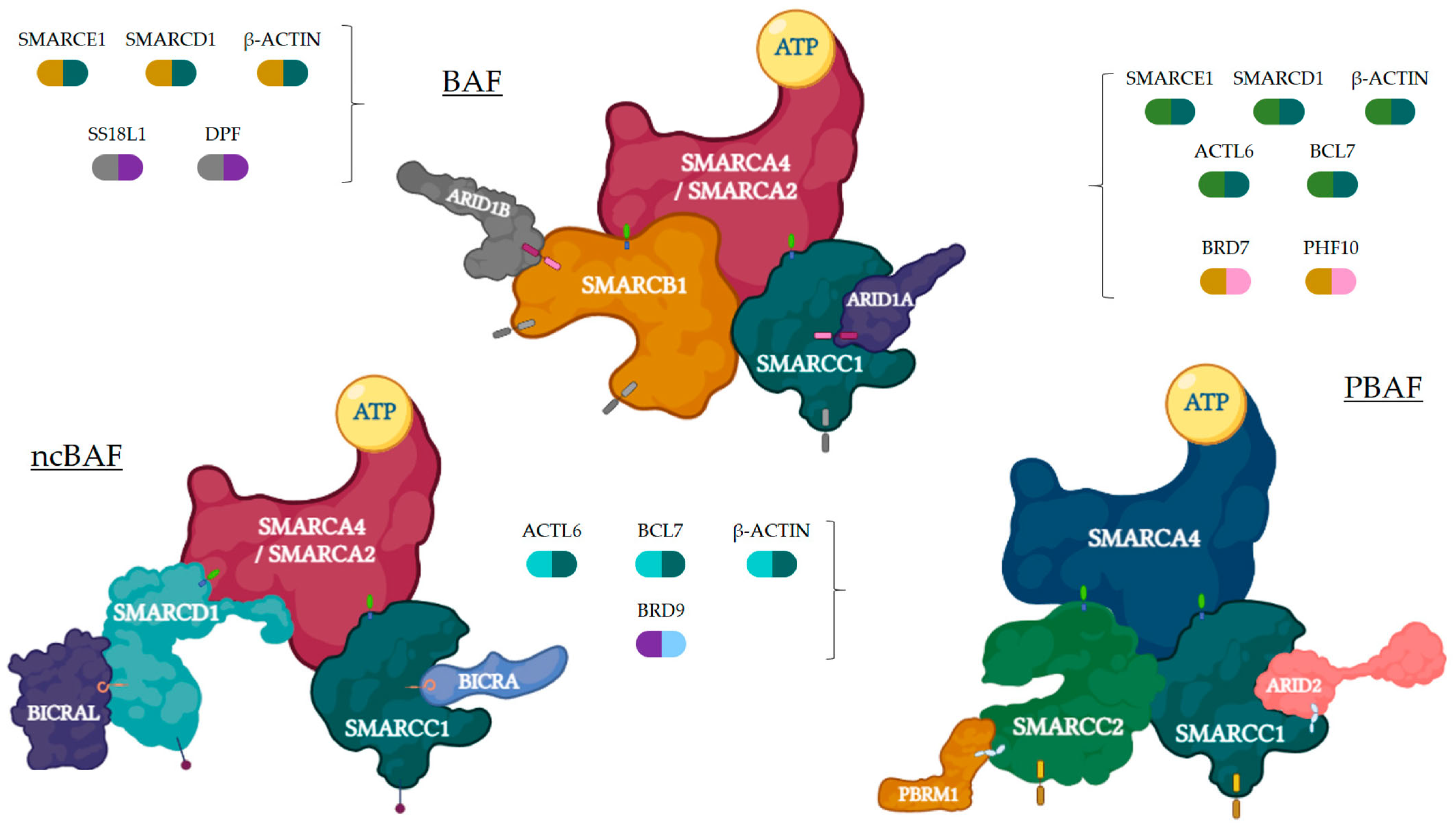
Figure 1. Three SWI/SNFc complexes are represented with their main subunits. Each complex may contain a different core (mustard and dark teal for BAF, turquoise and dark teal for ncBAF, green and dark teal for PBAF) and specific (grey and purple for BAF, sky blue and purple for ncBAF, mustard and pink for PBAF) subunits, apart from the ones presented in the figure, which are represented by bicolor capsules. Figure created with BioRender.com.
Ijms 24 11143 g001
Figure 2. Representative diagram of the transcriptional consequences of whether the SWI/SNFc works properly or not due to deleterious mutations in key subunits. Relative positions among nucleosomes may be pathogenically altered when ATP hydrolysis or histone binding is disrupted. As a result, the accessibility of enhancers provokes different effects on gene transcription. Figure created with BioRender.com.
Ijms 24 11143 g002
Figure 3. In mammals, SWI/SNFc regulates critical cellular processes, including cell cycle progression, programmed cell death, cell differentiation and development, genomic stability, DNA repair, and metabolism. Figure created with BioRender.com.
Ijms 24 11143 g003
Table 2. Clinical trials testing ICI, alone or combined with other agents, in rhabdoid and other tumors characterized by specific SWI/SNF alterations.
Author/YearNCTStudy DesignNTumorDrugEndpoints/Results and Grade 3–5 AEs
Blay et al. (2019) [103]03012620Phase II21Rare sarcomas, including rhabdoid and SMARCA4-deficient sarcomasPembrolizumabORR 15%; 1-year PFS 50% (SMARCA4-MRT)
None reported G3-4 AEs
Ongoing05286801Phase I/II86Children (1–18 years) with R/R SMARCB1 or SMARCA4-deficient tumorsTiragolumab and AtezolizumabSafety (AEs); PK;
Efficacy (ORR, PFS, OS, DOR)
Ongoing05407441Phase I/II49Children and young adults (<24 years) with INI1-neg/SMARCA4-deficient tumorsTazemetostat + Nivolumab/IpilimumabSafety (AEs, MTD, RP2D)
Efficacy (ORR, OS, PFS)
Ongoing04416568Phase II45Children, adolescents, and adults with R/R INI1-negative cancersNivolumab + IpilimumabEfficacy (ORR, PFS, OS, DCR)
Safety (AEs)
Ongoing04284202Phase II30Adults with NSCLC ARID1-mutantToripalimab + DasatinibEfficacy (PFS, OS)
Ongoing04957615Phase II30Metastatic or unresectable solid tumors with ARID1A mutationNivolumabEfficacy (ORR, OS, PFS)
Ongoing04953104Phase II30Metastatic urothelial cancer with ARID1A mutationNivolumabEfficacy (ORR, OS, PFS)
Abbreviations: AEs: Adverse events; DCR: disease control rate; DOR: duration of response; MTD: maximum tolerated dose; ORR: overall response rate; OS: overall survival; PFS: progression-free survival; PK: pharmacokinetics; RP2D: recommended phase 2 dose; R/R: recurrent or relapsed.
Disclaimer/Publisher’s Note: The statements, opinions and data contained in all publications are solely those of the individual author(s) and contributor(s) and not of MDPI and/or the editor(s). MDPI and/or the editor(s) disclaim responsibility for any injury to people or property resulting from any ideas, methods, instructions or products referred to in the content.